

Pompe's disease - RePub - Erasmus Universiteit Rotterdam
Pompe's disease - RePub - Erasmus Universiteit Rotterdam
Pompe's disease - RePub - Erasmus Universiteit Rotterdam

Create successful ePaper yourself
Turn your PDF publications into a flip-book with our unique Google optimized e-Paper software.
Chapter 1<br />
In lysosomal storage disorders, the affected cell type usually plays a major role in the<br />
degradation of the (accumulated) substrate. For example, cerebral white matter is affected<br />
in patients with defects in the degradation of sphingolipids. Connective tissue is involved<br />
in mucopolysaccharide storage disorders. The symptoms are obviously related to the site<br />
of accumulated undegradable materials, but the exact cause of cell death or dysfunction<br />
is often unclear. All the disorders are progressive and can be fatal. Defi nitive diagnosis is<br />
accomplished by measuring specifi c enzyme activities or function in leukocytes or cultured<br />
skin fi broblasts or other tissue specimens, selected on the basis of clinical symptoms. Light<br />
or electron microscopy, and DNA analysis can confi rm the diagnosis. There is extensive<br />
heterogeneity within each of the disorders, exemplifi ed by infantile, juvenile or adult onset<br />
of symptoms, mostly related to residual (enzyme) function. The different types of mutations<br />
within the same gene totally eliminating or partially reducing the enzyme activity or lysosomal<br />
protein function explain this. In addition, expression of modifying genes also infl uences the<br />
phenotype (7, 8).<br />
1.2 Cell biology of lysosomes<br />
Processing of lysosomal enzymes<br />
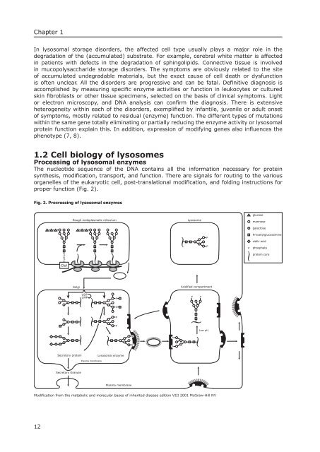
The nucleotide sequence of the DNA contains all the information necessary for protein<br />
synthesis, modifi cation, transport, and function. There are signals for routing to the various<br />
organelles of the eukaryotic cell, post-translational modifi cation, and folding instructions for<br />
proper function (Fig. 2).<br />
Fig. 2. Procressing of lysosomal enzymes<br />
12<br />
P<br />
P<br />
Dol<br />
Secretory protein<br />
Secretory Granule<br />
Rough endoplasmatic reticulum Lysosome<br />
Golgi<br />
UDP<br />
UMP<br />
Plasma membrane<br />
P<br />
P<br />
P<br />
P<br />
Lysosomal enzyme<br />
Plasma membrane<br />
P<br />
P<br />
P<br />
P<br />
Acidified compartment<br />
P P<br />
Modification from the metabolic and molecular bases of inherited <strong>disease</strong> edition VIII 2001 McGraw-Hill NY.<br />
P<br />
P<br />
Low pH<br />
P<br />
glucose<br />
mannose<br />
galactose<br />
N-acetylglucosamine<br />
sialic acid<br />
phosphate<br />
protein core


