Academy of Laser Dentistry
Academy of Laser Dentistry
Academy of Laser Dentistry

You also want an ePaper? Increase the reach of your titles
YUMPU automatically turns print PDFs into web optimized ePapers that Google loves.
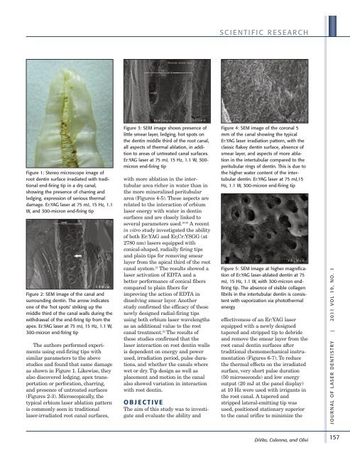
Figure 1: Stereo microscope image <strong>of</strong><br />
root dentin surface irradiated with traditional<br />
end-firing tip in a dry canal,<br />
showing the presence <strong>of</strong> charring and<br />
ledging, expression <strong>of</strong> serious thermal<br />
damage. Er:YAG laser at 75 mJ, 15 Hz, 1.1<br />
W, and 300-micron end-firing tip<br />
Figure 2: SEM image <strong>of</strong> the canal and<br />
surrounding dentin. The arrow indicates<br />
one <strong>of</strong> the ‘hot spots’ striking up the<br />
middle third <strong>of</strong> the canal walls during the<br />
withdrawal <strong>of</strong> the end-firing tip from the<br />
apex. Er:YAG laser at 75 mJ, 15 Hz, 1.1 W,<br />
300-micron end-firing tip<br />
The authors performed experiments<br />
using end-firing tips with<br />
similar parameters to the above<br />
studies and found that same damage<br />
as shown in Figure 1. Likewise, they<br />
also discovered ledging, apex transportation<br />
or perforation, charring,<br />
and presence <strong>of</strong> untreated surfaces<br />
(Figures 2-3). Microscopically, the<br />
typical erbium laser ablation pattern<br />
is commonly seen in traditional<br />
laser-irradiated root canal surfaces,<br />
Figure 3: SEM image shows presence <strong>of</strong><br />
little smear layer, ledging, hot spots on<br />
the dentin middle third <strong>of</strong> the root canal,<br />
all aspects <strong>of</strong> thermal ablation, in addition<br />
to areas <strong>of</strong> untreated canal surfaces.<br />
Er:YAG laser at 75 mJ, 15 Hz, 1.1 W, 300micron<br />
end-firing tip<br />
with more ablation in the intertubular<br />
area richer in water than in<br />
the more mineralized peritubular<br />
area (Figures 4-5). These aspects are<br />
related to the interaction <strong>of</strong> erbium<br />
laser energy with water in dentin<br />
surfaces and are closely linked to<br />
several parameters used. 19-20 A recent<br />
in vitro study investigated the ability<br />
<strong>of</strong> both Er:YAG and Er,Cr:YSGG (at<br />
2780 nm) lasers equipped with<br />
conical-shaped, radially firing tips<br />
and plain tips for removing smear<br />
layer from the apical third <strong>of</strong> the root<br />
canal system. 21 The results showed a<br />
laser activation <strong>of</strong> EDTA and a<br />
better performance <strong>of</strong> conical fibers<br />
compared to plain fibers for<br />
improving the action <strong>of</strong> EDTA in<br />
dissolving smear layer. Another<br />
study confirmed the efficacy <strong>of</strong> these<br />
newly designed radial-firing tips<br />
using both erbium laser wavelengths<br />
as an additional value to the root<br />
canal treatment. 22 The results <strong>of</strong><br />
these studies confirmed that the<br />
laser interaction on root dentin walls<br />
is dependent on energy and power<br />
used, irradiation period, pulse durations,<br />
and whether the canals where<br />
wet or dry. Tip design as well as<br />
placement and motion in the canal<br />
also showed variation in interaction<br />
with root dentin.<br />
O B J E C T I V E<br />
The aim <strong>of</strong> this study was to investigate<br />
and evaluate the ability and<br />
S C I E N T I F I C R E S E A R C H<br />
Figure 4: SEM image <strong>of</strong> the coronal 5<br />
mm <strong>of</strong> the canal showing the typical<br />
Er:YAG laser irradiation pattern, with the<br />
classic flakey dentin surface, absence <strong>of</strong><br />
smear layer, and aspects <strong>of</strong> more ablation<br />
in the intertubular compared to the<br />
peritubular rings <strong>of</strong> dentin. This is due to<br />
the higher water content <strong>of</strong> the intertubular<br />
dentin. Er:YAG laser at 75 mJ,15<br />
Hz, 1.1 W, 300-micron end-firing tip<br />
Figure 5: SEM image at higher magnification<br />
<strong>of</strong> Er:YAG laser-ablated dentin at 75<br />
mJ, 15 Hz, 1.1 W, with 300-micron endfiring<br />
tip. The absence <strong>of</strong> visible collagen<br />
fibrils in the intertubular dentin is consistent<br />
with vaporization via photothermal<br />
energy<br />
effectiveness <strong>of</strong> an Er:YAG laser<br />
equipped with a newly designed<br />
tapered and stripped tip to debride<br />
and remove the smear layer from the<br />
root canal dentin surfaces after<br />
traditional chemomechanical instrumentation<br />
(Figures 6-7). To reduce<br />
the thermal effects on the irradiated<br />
surface, very short pulse duration<br />
(50 microseconds) and low energy<br />
output (20 mJ at the panel display)<br />
at 10 Hz were used with irrigants in<br />
the root canal. A tapered and<br />
stripped lateral-emitting tip was<br />
used, positioned stationary superior<br />
to the canal orifice to minimize the<br />
DiVito, Colonna, and Olivi<br />
J O U R N A L O F L A S E R D E N T I S T R Y | 2 011 V O L 19 , N O . 1<br />
157