Academy of Laser Dentistry
Academy of Laser Dentistry
Academy of Laser Dentistry

You also want an ePaper? Increase the reach of your titles
YUMPU automatically turns print PDFs into web optimized ePapers that Google loves.
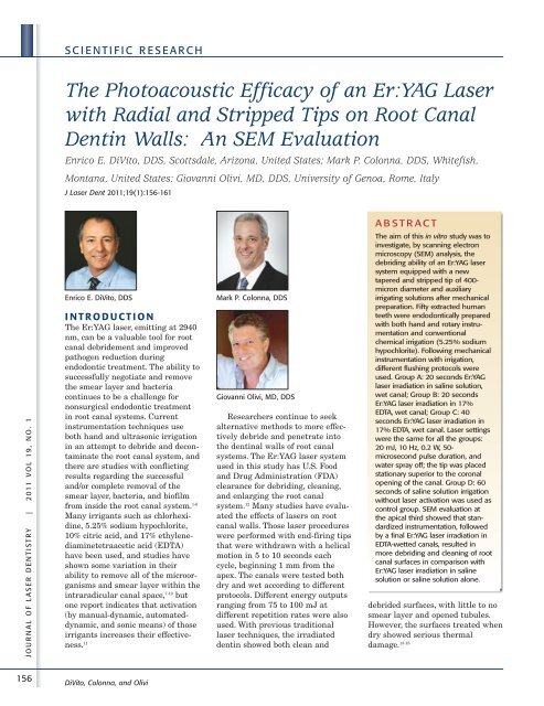
J O U R N A L O F L A S E R D E N T I S T R Y | 2 011 V O L 19 , N O . 1<br />
156<br />
S C I E N T I F I C R E S E A R C H<br />
The Photoacoustic Efficacy <strong>of</strong> an Er:YAG <strong>Laser</strong><br />
with Radial and Stripped Tips on Root Canal<br />
Dentin Walls: An SEM Evaluation<br />
Enrico E. DiVito, DDS, Scottsdale, Arizona, United States; Mark P. Colonna, DDS, Whitefish,<br />
Montana, United States; Giovanni Olivi, MD, DDS, University <strong>of</strong> Genoa, Rome, Italy<br />
J <strong>Laser</strong> Dent 2011;19(1):156-161<br />
Enrico E. DiVito, DDS Mark P. Colonna, DDS<br />
I N T R O D U C T I O N<br />
The Er:YAG laser, emitting at 2940<br />
nm, can be a valuable tool for root<br />
canal debridement and improved<br />
pathogen reduction during<br />
endodontic treatment. The ability to<br />
successfully negotiate and remove<br />
the smear layer and bacteria<br />
continues to be a challenge for<br />
nonsurgical endodontic treatment<br />
in root canal systems. Current<br />
instrumentation techniques use<br />
both hand and ultrasonic irrigation<br />
in an attempt to debride and decontaminate<br />
the root canal system, and<br />
there are studies with conflicting<br />
results regarding the successful<br />
and/or complete removal <strong>of</strong> the<br />
smear layer, bacteria, and bi<strong>of</strong>ilm<br />
from inside the root canal system. 1-6<br />
Many irrigants such as chlorhexidine,<br />
5.25% sodium hypochlorite,<br />
10% citric acid, and 17% ethylene -<br />
diaminetetraacetic acid (EDTA)<br />
have been used, and studies have<br />
shown some variation in their<br />
ability to remove all <strong>of</strong> the microorganisms<br />
and smear layer within the<br />
intraradicular canal space, 7-10 but<br />
one report indicates that activation<br />
(by manual-dynamic, automateddynamic,<br />
and sonic means) <strong>of</strong> those<br />
irrigants increases their effectiveness.<br />
11<br />
DiVito, Colonna, and Olivi<br />
Giovanni Olivi, MD, DDS<br />
Researchers continue to seek<br />
alternative methods to more effectively<br />
debride and penetrate into<br />
the dentinal walls <strong>of</strong> root canal<br />
systems. The Er:YAG laser system<br />
used in this study has U.S. Food<br />
and Drug Administration (FDA)<br />
clearance for debriding, cleaning,<br />
and enlarging the root canal<br />
system. 12 Many studies have evaluated<br />
the effects <strong>of</strong> lasers on root<br />
canal walls. Those laser procedures<br />
were performed with end-firing tips<br />
that were withdrawn with a helical<br />
motion in 5 to 10 seconds each<br />
cycle, beginning 1 mm from the<br />
apex. The canals were tested both<br />
dry and wet according to different<br />
protocols. Different energy outputs<br />
ranging from 75 to 100 mJ at<br />
different repetition rates were also<br />
used. With previous traditional<br />
laser techniques, the irradiated<br />
dentin showed both clean and<br />
A B S T R A C T<br />
The aim <strong>of</strong> this in vitro study was to<br />
investigate, by scanning electron<br />
microscopy (SEM) analysis, the<br />
debriding ability <strong>of</strong> an Er:YAG laser<br />
system equipped with a new<br />
tapered and stripped tip <strong>of</strong> 400micron<br />
diameter and auxiliary<br />
irrigating solutions after mechanical<br />
preparation. Fifty extracted human<br />
teeth were endodontically prepared<br />
with both hand and rotary instrumentation<br />
and conventional<br />
chemical irrigation (5.25% sodium<br />
hypochlorite). Following mechanical<br />
instrumentation with irrigation,<br />
different flushing protocols were<br />
used. Group A: 20 seconds Er:YAG<br />
laser irradiation in saline solution,<br />
wet canal; Group B: 20 seconds<br />
Er:YAG laser irradiation in 17%<br />
EDTA, wet canal; Group C: 40<br />
seconds Er:YAG laser irradiation in<br />
17% EDTA, wet canal. <strong>Laser</strong> settings<br />
were the same for all the groups:<br />
20 mJ, 10 Hz, 0.2 W, 50microsecond<br />
pulse duration, and<br />
water spray <strong>of</strong>f; the tip was placed<br />
stationary superior to the coronal<br />
opening <strong>of</strong> the canal. Group D: 60<br />
seconds <strong>of</strong> saline solution irrigation<br />
without laser activation was used as<br />
control group. SEM evaluation at<br />
the apical third showed that standardized<br />
instrumentation, followed<br />
by a final Er:YAG laser irradiation in<br />
EDTA-wetted canals, resulted in<br />
more debriding and cleaning <strong>of</strong> root<br />
canal surfaces in comparison with<br />
Er:YAG laser irradiation in saline<br />
solution or saline solution alone.<br />
debrided surfaces, with little to no<br />
smear layer and opened tubules.<br />
However, the surfaces treated when<br />
dry showed serious thermal<br />
damage. 13-18
Figure 1: Stereo microscope image <strong>of</strong><br />
root dentin surface irradiated with traditional<br />
end-firing tip in a dry canal,<br />
showing the presence <strong>of</strong> charring and<br />
ledging, expression <strong>of</strong> serious thermal<br />
damage. Er:YAG laser at 75 mJ, 15 Hz, 1.1<br />
W, and 300-micron end-firing tip<br />
Figure 2: SEM image <strong>of</strong> the canal and<br />
surrounding dentin. The arrow indicates<br />
one <strong>of</strong> the ‘hot spots’ striking up the<br />
middle third <strong>of</strong> the canal walls during the<br />
withdrawal <strong>of</strong> the end-firing tip from the<br />
apex. Er:YAG laser at 75 mJ, 15 Hz, 1.1 W,<br />
300-micron end-firing tip<br />
The authors performed experiments<br />
using end-firing tips with<br />
similar parameters to the above<br />
studies and found that same damage<br />
as shown in Figure 1. Likewise, they<br />
also discovered ledging, apex transportation<br />
or perforation, charring,<br />
and presence <strong>of</strong> untreated surfaces<br />
(Figures 2-3). Microscopically, the<br />
typical erbium laser ablation pattern<br />
is commonly seen in traditional<br />
laser-irradiated root canal surfaces,<br />
Figure 3: SEM image shows presence <strong>of</strong><br />
little smear layer, ledging, hot spots on<br />
the dentin middle third <strong>of</strong> the root canal,<br />
all aspects <strong>of</strong> thermal ablation, in addition<br />
to areas <strong>of</strong> untreated canal surfaces.<br />
Er:YAG laser at 75 mJ, 15 Hz, 1.1 W, 300micron<br />
end-firing tip<br />
with more ablation in the intertubular<br />
area richer in water than in<br />
the more mineralized peritubular<br />
area (Figures 4-5). These aspects are<br />
related to the interaction <strong>of</strong> erbium<br />
laser energy with water in dentin<br />
surfaces and are closely linked to<br />
several parameters used. 19-20 A recent<br />
in vitro study investigated the ability<br />
<strong>of</strong> both Er:YAG and Er,Cr:YSGG (at<br />
2780 nm) lasers equipped with<br />
conical-shaped, radially firing tips<br />
and plain tips for removing smear<br />
layer from the apical third <strong>of</strong> the root<br />
canal system. 21 The results showed a<br />
laser activation <strong>of</strong> EDTA and a<br />
better performance <strong>of</strong> conical fibers<br />
compared to plain fibers for<br />
improving the action <strong>of</strong> EDTA in<br />
dissolving smear layer. Another<br />
study confirmed the efficacy <strong>of</strong> these<br />
newly designed radial-firing tips<br />
using both erbium laser wavelengths<br />
as an additional value to the root<br />
canal treatment. 22 The results <strong>of</strong><br />
these studies confirmed that the<br />
laser interaction on root dentin walls<br />
is dependent on energy and power<br />
used, irradiation period, pulse durations,<br />
and whether the canals where<br />
wet or dry. Tip design as well as<br />
placement and motion in the canal<br />
also showed variation in interaction<br />
with root dentin.<br />
O B J E C T I V E<br />
The aim <strong>of</strong> this study was to investigate<br />
and evaluate the ability and<br />
S C I E N T I F I C R E S E A R C H<br />
Figure 4: SEM image <strong>of</strong> the coronal 5<br />
mm <strong>of</strong> the canal showing the typical<br />
Er:YAG laser irradiation pattern, with the<br />
classic flakey dentin surface, absence <strong>of</strong><br />
smear layer, and aspects <strong>of</strong> more ablation<br />
in the intertubular compared to the<br />
peritubular rings <strong>of</strong> dentin. This is due to<br />
the higher water content <strong>of</strong> the intertubular<br />
dentin. Er:YAG laser at 75 mJ,15<br />
Hz, 1.1 W, 300-micron end-firing tip<br />
Figure 5: SEM image at higher magnification<br />
<strong>of</strong> Er:YAG laser-ablated dentin at 75<br />
mJ, 15 Hz, 1.1 W, with 300-micron endfiring<br />
tip. The absence <strong>of</strong> visible collagen<br />
fibrils in the intertubular dentin is consistent<br />
with vaporization via photothermal<br />
energy<br />
effectiveness <strong>of</strong> an Er:YAG laser<br />
equipped with a newly designed<br />
tapered and stripped tip to debride<br />
and remove the smear layer from the<br />
root canal dentin surfaces after<br />
traditional chemomechanical instrumentation<br />
(Figures 6-7). To reduce<br />
the thermal effects on the irradiated<br />
surface, very short pulse duration<br />
(50 microseconds) and low energy<br />
output (20 mJ at the panel display)<br />
at 10 Hz were used with irrigants in<br />
the root canal. A tapered and<br />
stripped lateral-emitting tip was<br />
used, positioned stationary superior<br />
to the canal orifice to minimize the<br />
DiVito, Colonna, and Olivi<br />
J O U R N A L O F L A S E R D E N T I S T R Y | 2 011 V O L 19 , N O . 1<br />
157
J O U R N A L O F L A S E R D E N T I S T R Y | 2 011 V O L 19 , N O . 1<br />
158<br />
S C I E N T I F I C R E S E A R C H<br />
above-cited collateral damages seen<br />
in the canals with the end-firing tips.<br />
M AT E R I A LS A N D<br />
M E T H O D S<br />
Sample Preparation<br />
Fifty (50) freshly extracted premolar<br />
teeth, stored in saline solution at<br />
21°C, were used for this study.<br />
Root Canal Treatment<br />
Traditional mechanical techniques<br />
were used to open the access cavities<br />
and to expose the pulp chambers and<br />
canals. The teeth were then instrumented<br />
with two different methods:<br />
• Twenty-five teeth were prepared<br />
to size #30 at the apex by using a<br />
K-File #30 hand file (Lexicon,<br />
DENTSPLY Tulsa Dental, Tulsa,<br />
Okla., USA).<br />
• The remaining 25 teeth were<br />
prepared to size #30 at the apex<br />
by using NiTi rotary instrumentation<br />
(Pr<strong>of</strong>ile ® GT , Dentsply<br />
Tulsa Dental), size 30/06.<br />
Irrigation <strong>of</strong> all the samples<br />
during preparation was accomplished<br />
using a combination <strong>of</strong> 2 ml<br />
root canal conditioner containing<br />
EDTA and carbamide peroxide<br />
(ProLube ® , Dentsply Tulsa Dental),<br />
2 ml <strong>of</strong> 5.5% sodium hypochlorite<br />
solution, and 2 ml <strong>of</strong> 17% EDTA.<br />
Each was introduced into the canal<br />
in the aforementioned order using<br />
a dual-ported 27-gauge needle<br />
(Appli-Vac TM and Vista-Probe TM irrigating<br />
tips, Vista Dental Products,<br />
Racine, Wis., USA) in a sterile<br />
syringe.<br />
After instrumentation was<br />
completed, additional final irrigation<br />
was performed using two<br />
cycles, 30 seconds each, <strong>of</strong> irrigation<br />
with saline solution. Handand<br />
rotary-prepared teeth were<br />
randomly divided into four groups<br />
for the final laser irradiation with<br />
different protocols.<br />
<strong>Laser</strong> Irradiation Method<br />
A 2940-nm Er:YAG laser (Fidelis,<br />
Fotona d.d., Ljubljana, Slovenia) was<br />
used with a newly designed, 14-mmlong,<br />
400-micron diameter tapered<br />
DiVito, Colonna, and Olivi<br />
Figure 6: Diagrammatic representation <strong>of</strong><br />
the 400-micron tapered and stripped tip<br />
used for this laser study<br />
Figure 7: Operative image shows the tip,<br />
positioned stationary and immersed in<br />
irrigant-filled chamber, at the orifice<br />
opening <strong>of</strong> the palatal (palatine) canal<br />
tip; 4 mm <strong>of</strong> the polyamide sheath<br />
were stripped back from the end.<br />
<strong>Laser</strong> settings were 10 Hz, 20<br />
mJ, with 50 microseconds pulse<br />
duration, with air/water spray <strong>of</strong>f.<br />
Four groups were established,<br />
and each group had the same<br />
number <strong>of</strong> canals prepared by K<br />
and NiTi files:<br />
• Group A (n = 12) was laser-irradiated<br />
for 20 sec in saline<br />
solution-wetted canal.<br />
• Group B (n = 12) was laser-irradiated<br />
for 20 sec in 17%<br />
EDTA-wetted canal.<br />
• Group C (n = 12) was laser-irradiated<br />
for 40 sec in 17%<br />
EDTA-wetted canal.<br />
• Group D (n = 14) was not laserirradiated,<br />
and served as the<br />
control group.<br />
During laser irradiation, the root<br />
canals were continuously irrigated<br />
with 2 mL <strong>of</strong> the same fluid (saline<br />
solution or 17% EDTA) to maintain<br />
hydration and fluid levels using a<br />
25-gauge needle (same manufacturer<br />
as above) in a sterile syringe,<br />
positioned above the laser tip in the<br />
coronal aspect <strong>of</strong> the access opening.<br />
The prepared samples were then<br />
sectioned longitudinally and examined<br />
with a field emission electron<br />
microscope (F4000, Hitachi, Tokyo,<br />
Japan).<br />
Temperature Measurements<br />
Temperature variations were measured<br />
on the root surface <strong>of</strong> five<br />
samples for each laser group (for a<br />
total <strong>of</strong> 15 samples). A modified<br />
thermocouple measurement sensor<br />
<strong>of</strong> 1.5-mm diameter (TEL-11854 K-<br />
Type NiCr-Ni Immersion Sensor,<br />
TEL-Atomic Inc., Jackson, Mich.,<br />
USA) was used. It was placed on<br />
the external root surface at 5 mm<br />
from the apex and attached with a<br />
silicon-based heat-conductive<br />
compound (Dow Corning ® 340 Heat<br />
Sink Compound, Dow Corning<br />
Corp., Midland, Mich., USA). The<br />
temperature variations were monitored<br />
continuously during all the<br />
irradiation procedure periods (20<br />
sec for Groups 2 and 3, and 40 sec<br />
for Group 4) using a digital thermometer<br />
(TEL-11853 Digital Quick<br />
Response Pocket Thermometer,<br />
TEL-Atomic Inc.).<br />
R E S U LT S<br />
• The samples in Group A, treated<br />
with the Er:YAG laser and saline<br />
solution, showed partially clean<br />
surfaces and open tubules, with<br />
residual debris and thin smear<br />
layer remaining. (Note: There is<br />
no accompanying illustration<br />
included in this article.)<br />
• The samples from Group D, with<br />
saline solution applied conventionally,<br />
demonstrated a<br />
noticeable smear layer and closed<br />
tubules (Figures 8-9).<br />
• However, the Group B samples<br />
that incorporated the Er:YAG<br />
laser irradiation for 20 seconds,<br />
the tapered and stripped tip, and
Figures 8-9: Group D sample: SEM<br />
images <strong>of</strong> the coronal third show<br />
adherent mechanical debris present in<br />
conventional mechanically instrumented<br />
canals, followed by rinsing with saline<br />
solution for 1 minute<br />
EDTA irrigation showed clean,<br />
open tubules with essentially no<br />
smear layer or debris remaining<br />
(Figures 10-11).<br />
• In addition, the Group C samples<br />
with Er:YAG irradiation for 40<br />
seconds, the tapered and stripped<br />
tip, and EDTA irrigation show<br />
the collagen fibers and internal<br />
hydroxyapatite matrices intact at<br />
increased SEM magnification at<br />
20,000X (Figures 12-13).<br />
• Temperature increases were<br />
observed at the root surface<br />
during laser irradiation. Groups<br />
A and B (20 seconds <strong>of</strong> laser<br />
exposure) exhibited a rise <strong>of</strong><br />
1.2°C, and Group C (40 seconds)<br />
showed a 1.5°C increase.<br />
D I S C U S S I O N<br />
Currently used chemomechanical<br />
instrumentation still falls short <strong>of</strong><br />
successfully removing the smear<br />
layer from inside the root canal<br />
system. 5 This was also confirmed<br />
Figures 10-11: Group B sample: SEM<br />
images <strong>of</strong> the coronal third show clean<br />
surface after laser irradiation in 17%<br />
EDTA for 20 seconds and smear layer and<br />
debris removal from dentin tubules. The<br />
surface does not exhibit evidence <strong>of</strong> the<br />
application <strong>of</strong> thermal energy. Er:YAG<br />
laser at 20 mJ, 10 Hz, 0.2 W, 400-micron<br />
tapered and stripped tip<br />
from the results seen in the control<br />
group <strong>of</strong> this study, when the laser<br />
technique was not employed.<br />
This study introduced several<br />
modifications to the commonly used<br />
laser-assisted techniques and protocols,<br />
to reduce the thermal effect <strong>of</strong><br />
laser radiation on dentinal walls.<br />
The thermal and ablative effect <strong>of</strong><br />
laser radiation on the root canal<br />
surface may be reduced using less<br />
ablative parameters. The dentin ablation<br />
threshold is currently known to<br />
be about 1 to 4 J/cm 2 for the Er:YAG<br />
laser. 23-24 Based on these values and<br />
using a 400-micron tip, this study<br />
utilized the higher but still valid<br />
threshold ablative energy value <strong>of</strong> 20<br />
mJ (measured at the panel display),<br />
which is 4 J/cm 2 . As mentioned<br />
above, this pulse energy is significantly<br />
below those reported in<br />
previous studies, but did not produce<br />
S C I E N T I F I C R E S E A R C H<br />
Figure 12: Group C: SEM image <strong>of</strong> apical<br />
third presents completely removed smear<br />
layer from the dentin tubules following<br />
laser radiation in 17% EDTA wetted canal<br />
for 40 seconds. There is no evidence <strong>of</strong><br />
morphological thermal damage at 20 mJ,<br />
10 Hz, 0.2 W, 400-micron tapered and<br />
stripped tip<br />
Figure 13: Group C: SEM image <strong>of</strong> middle<br />
third shows the collagen fibers and<br />
internal hydroxyapatite matrices left<br />
intact, indicating the absence <strong>of</strong> laser<br />
thermal energy, at 20 mJ, 10 Hz, 0.2 W,<br />
400-micron tapered and stripped tip<br />
the thermal damage. Furthermore,<br />
with the pulse duration <strong>of</strong> 50<br />
microseconds, a peak power <strong>of</strong> 400 W<br />
per pulse was achieved.<br />
According to a study by George<br />
and Walsh, the use <strong>of</strong> tapered and<br />
stripped laser tips showed greater<br />
lateral emissions and corresponding<br />
lower forward emissions than<br />
conventional blunt tips, thus<br />
reducing the thermal side effects <strong>of</strong><br />
the end-firing tips. They also<br />
reported that two different erbium<br />
laser systems equipped with a<br />
radial fiber design elicited temperature<br />
increases less than 2.5°C<br />
during lasing, confirming the role<br />
<strong>of</strong> water irrigation in attenuating<br />
the thermal effects <strong>of</strong> each lasing<br />
cycle. 22<br />
DiVito, Colonna, and Olivi<br />
J O U R N A L O F L A S E R D E N T I S T R Y | 2 011 V O L 19 , N O . 1<br />
159
J O U R N A L O F L A S E R D E N T I S T R Y | 2 011 V O L 19 , N O . 1<br />
160<br />
S C I E N T I F I C R E S E A R C H<br />
Because <strong>of</strong> the high affinity <strong>of</strong><br />
the Er:YAG laser for water and the<br />
very short pulse duration utilized<br />
in this laser protocol, the resultant<br />
high peak power <strong>of</strong> 400 W generated<br />
a cavitation and shock wave<br />
effect in the liquid. Since the<br />
volume <strong>of</strong> the liquid in the root<br />
canal is very small, this effect is<br />
amplified and improves the<br />
removal <strong>of</strong> the smear layer and<br />
residual tissue tags, which was<br />
confirmed by another study. 21 The<br />
authors speculate that this<br />
phenomenon is responsible for the<br />
removal <strong>of</strong> smear layer observed in<br />
group A, in which laser irradiation<br />
was combined with saline solution.<br />
Limited information exists at this<br />
time regarding the induction <strong>of</strong><br />
mechanical effects due either to<br />
ultrasonic or laser stimulation, and<br />
other authors have described<br />
similar expanding and imploding<br />
vapor bubbles, shock waves, with<br />
secondary cavitation effects with<br />
fluid movements in endodontics as<br />
a result <strong>of</strong> the erbium laser<br />
(Er:YAG and Er,Cr:YSGG) thermal<br />
activation <strong>of</strong> irrigants, within the<br />
canal, termed laser-activated irrigation.<br />
25-27 However, those studies<br />
utilized lasers equipped with<br />
different tips, and different parameters<br />
and techniques were<br />
employed. In this study, the tip was<br />
placed only within the coronal<br />
portion <strong>of</strong> the root canal system;<br />
and, with 20 mJ per pulse, the<br />
previously described harmful<br />
thermal effects 13-18 did not occur.<br />
The authors postulate that the<br />
smear layer and debris were not<br />
removed via thermal vaporization,<br />
but from the laser-activated coronal<br />
irrigation fluid. They use the<br />
acronym PIPS to describe this<br />
Photon-Induced Photoacoustic<br />
Streaming .<br />
The use <strong>of</strong> EDTA together with<br />
the described Er:YAG laser protocol<br />
produces a synergistic effect. This<br />
contributes to improved treatment<br />
efficacy when compared to the<br />
erbium laser-saline solution group<br />
(Group A) or the nonirradiated<br />
DiVito, Colonna, and Olivi<br />
control group (Group D), leading to<br />
an efficient debriding <strong>of</strong> the root<br />
canal as seen in Groups B and C.<br />
The SEM images verified also<br />
the minimally disruptive effects on<br />
dentin walls, and even on the<br />
hydroxyapatite and collagenous<br />
fiber matrices.<br />
C O N C LU S I O N<br />
This SEM study demonstrated the<br />
effective ability <strong>of</strong> an Er:YAG laser<br />
equipped with a tapered and<br />
stripped tip, set at low energy and<br />
short pulse duration, to clean and<br />
debride the root canal system. This<br />
laser system and the technique<br />
used appeared to have no thermal<br />
effects on the dentinal walls by<br />
virtue <strong>of</strong> decreased energy settings<br />
and short pulse duration and by<br />
remote placement <strong>of</strong> the radial and<br />
stripped tip from the target site (at<br />
the orifice <strong>of</strong> the root canal). The<br />
dentin surface and its collagen and<br />
hydroxyapatite matrices were<br />
essentially undisturbed and clean.<br />
The use <strong>of</strong> the laser utilized in this<br />
study appeared to enhance the<br />
removal <strong>of</strong> the smear layer from<br />
dentin tubules when used with<br />
EDTA for both 20 and 40 seconds.<br />
A U T H O R B I O G R A P H I E S<br />
Dr. Enrico DiVito has been in practice<br />
since 1980. He established and<br />
runs the Arizona Center for <strong>Laser</strong><br />
<strong>Dentistry</strong> in Scottsdale, Arizona.<br />
He is always exploring potential<br />
uses for innovations in the field<br />
and holds a number <strong>of</strong> patents for<br />
dental products. He lectures<br />
nationally and internationally. Dr.<br />
DiVito is the founder and director<br />
<strong>of</strong> the state-accredited Arizona<br />
School <strong>of</strong> Dental Assisting. He also<br />
teaches as a clinical instructor at<br />
the Arizona School <strong>of</strong> <strong>Dentistry</strong> and<br />
Oral Health and is responsible for<br />
helping to create and implement<br />
their Department <strong>of</strong> <strong>Laser</strong><br />
<strong>Dentistry</strong>. In 2007 he was awarded<br />
the Biolase Clinical Case <strong>of</strong> the<br />
Year. Dr. DiVito may be contacted<br />
by e-mail at edivito@azcld.com.<br />
Disclosure: Dr. DiVito receives honoraria<br />
from Lares Research for lectures<br />
on laser dentistry. He is also a shareholder<br />
in Medical Dental Advanced<br />
Technology Group, which has<br />
performed the research for PIPS.<br />
Dr. Mark Colonna founded and<br />
operates the Montana Center for<br />
<strong>Laser</strong> <strong>Dentistry</strong>, his private practice<br />
and advanced laser training<br />
center. He is a pioneer in the development<br />
<strong>of</strong> techniques and<br />
instruments for dentistry without<br />
the use <strong>of</strong> drills. He lectures,<br />
teaches, and writes nationally and<br />
internationally about laser<br />
dentistry. Dr. Colonna holds many<br />
awards, including the Clinician <strong>of</strong><br />
the Year Award from the World<br />
Congress <strong>of</strong> Minimally Invasive<br />
<strong>Dentistry</strong>. He has achieved<br />
Mastership status from the World<br />
Clinical <strong>Laser</strong> Institute. Dr.<br />
Colonna may be contacted by email<br />
at doc@mtlaserdentistry.com.<br />
Disclosure: Dr. Colonna receives<br />
honoraria from Lares Research for<br />
lectures on laser dentistry. He is also a<br />
shareholder in Medical Dental<br />
Advanced Technology Group, which<br />
has performed the research for PIPS.<br />
Dr. Giovanni Olivi is a Pr<strong>of</strong>essor in<br />
Restorative and Endodontics at the<br />
University <strong>of</strong> Genoa Faculty <strong>of</strong><br />
<strong>Dentistry</strong>, and a Board member <strong>of</strong><br />
Scientific Committee <strong>of</strong> the<br />
University <strong>of</strong> Genoa “<strong>Laser</strong> in<br />
<strong>Dentistry</strong>” Master Course. He is a<br />
member <strong>of</strong> the <strong>Academy</strong> <strong>of</strong> <strong>Laser</strong><br />
<strong>Dentistry</strong> and received Mastership<br />
from the ALD. Dr. Giovanni is the<br />
2007 recipient <strong>of</strong> ALD’s Leon<br />
Goldman Award for Clinical<br />
Excellence. He is a co-author <strong>of</strong> the<br />
books Pediatric <strong>Laser</strong> <strong>Dentistry</strong> and<br />
<strong>Laser</strong>s in Dental Traumatology.<br />
Pr<strong>of</strong>. Giovanni Olivi may be<br />
contacted by e-mail at<br />
olivi.g@tiscali.it.<br />
Disclosure: Dr. Olivi has no commercial<br />
or financial interest relative to<br />
this manuscript.
R E F E R E N C E S<br />
1. Hülsmann M, Peters OA, Dummer<br />
PMH. Mechanical preparation <strong>of</strong><br />
root canals: Shaping goals, techniques<br />
and means. Endod Topics<br />
2005;10(1):30-76.<br />
2. Baumgartner JC, Cuenin PR.<br />
Efficacy <strong>of</strong> several concentrations <strong>of</strong><br />
sodium hypochlorite for root canal<br />
irrigation. J Endod 1992;18(12):605-<br />
612.<br />
3. Hülsmann M, Rümmelin C, Schäfers<br />
F. Root canal cleanliness after<br />
preparation with different<br />
endodontic handpieces and hand<br />
instruments: A comparative SEM<br />
investigation. J Endod<br />
1997;23(5):301-306.<br />
4. Gu L, Kim JR, Ling J, Choi KK,<br />
Pashley DH, Tay FR. Review <strong>of</strong><br />
contemporary irrigant agitation<br />
techniques and devices. J Endod<br />
2009;35(6):791-804.<br />
5. Ricucci D, Siqueira JF Jr. Fate <strong>of</strong><br />
the tissue in lateral canals and<br />
apical ramifications in response to<br />
pathologic conditions and treatment<br />
procedures. J Endod 2010;36(1):1-<br />
15.<br />
6. Metzger Z, Teperovich E, Cohen R,<br />
Zary R, Paqué F, Hülsmann M. The<br />
self-adjusting file (SAF). Part 3:<br />
Removal <strong>of</strong> debris and smear layer –<br />
A scanning electron microscope<br />
study. J Endod 2010;36(4):697-702.<br />
7. Byström A, Sundqvist G.<br />
Bacteriologic evaluation <strong>of</strong> the effect<br />
<strong>of</strong> 0.5 percent sodium hypochlorite<br />
in endodontic therapy. Oral Surg<br />
Oral Med Oral Pathol<br />
1983;55(3):307-312.<br />
8. White RR, Hays GL, Janer LR.<br />
Residual antimicrobial activity after<br />
canal irrigation with chlorhexidine.<br />
J Endod 1997;23(4):229-231.<br />
9. Berutti E, Marini R, Angeretti A.<br />
Penetration ability <strong>of</strong> different irrigants<br />
into dentinal tubules. J Endod<br />
1997;23(12):725-727.<br />
10. Khedmat S, Shokouhinejad N.<br />
Comparison <strong>of</strong> the efficacy <strong>of</strong> three<br />
chelating agents in smear layer<br />
removal. J Endod 2008;34(5):599-<br />
602.<br />
11. Caron G, Nham K, Bronnec F,<br />
Machtou P. Effectiveness <strong>of</strong> different<br />
final irrigant activation protocols on<br />
smear layer removal in curved<br />
canals. J Endod 2010;36(8):1361-<br />
1366.<br />
12. Sulewski JG. Making the most <strong>of</strong><br />
the 17th annual conference and<br />
exhibition: A practical orientation<br />
for attendees. J <strong>Laser</strong> Dent<br />
2010;18(Suppl).<br />
13. Takeda FH, Harashima T, Kimura Y,<br />
Matsumoto K. Comparative study<br />
about the removal <strong>of</strong> smear layer by<br />
three types <strong>of</strong> laser devices. J Clin<br />
<strong>Laser</strong> Med Surg 1998;16(2):117-122.<br />
14. Matsuoka E, Kimura Y, Matsumoto<br />
K. Studies on the removal <strong>of</strong> debris<br />
near the apical seats by Er: YAG<br />
laser and assessment with a fiberscope.<br />
J Clin <strong>Laser</strong> Med Surg<br />
1998;16(5):255-261.<br />
15. Takeda FH, Harashima T, Eto JN,<br />
Kimura Y, Matsumoto K. Effect <strong>of</strong><br />
Er:YAG laser treatment on the root<br />
canal walls <strong>of</strong> human teeth: An SEM<br />
study. Endod Dent Traumatol<br />
1998;14(6):270-273.<br />
16. Takeda FH, Harashima T, Kimura Y,<br />
Matsumoto K. Efficacy <strong>of</strong> Er: YAG<br />
laser irradiation in removing debris<br />
and smear layer on root canal walls.<br />
J Endod 1998;24(8):548-551.<br />
17. Takeda FH, Harashima T, Kimura Y,<br />
Matsumoto K. A comparative study<br />
<strong>of</strong> the removal <strong>of</strong> smear layer by<br />
three endodontic irrigants and two<br />
types <strong>of</strong> laser. Int Endod J<br />
1999;32(1):32-39.<br />
18. Pecora JD, Brugnera-Júnior A,<br />
Cussioli AL, Zanin F, Silva R.<br />
Evaluation <strong>of</strong> dentin root canal<br />
permeability after instrumentation<br />
and Er:YAG laser application.<br />
<strong>Laser</strong>s Surg Med 2000; 26(3):277-<br />
281.<br />
19. Kimura Y, Yonaga K, Yokoyama K,<br />
Kinoshita J-i, Ogata Y, Matsumoto<br />
K Root surface temperature<br />
increase during Er:YAG laser irradiation<br />
<strong>of</strong> root canals. J Endod<br />
2002;28(2):76-78.<br />
20. Ebihara A, Majaron B, Liaw L-HL,<br />
Krasieva TB, Wilder-Smith P.<br />
Er:YAG laser modification <strong>of</strong> root<br />
canal dentine: Influence <strong>of</strong> pulse<br />
duration, repetitive irradiation and<br />
water spray. <strong>Laser</strong>s Med Sci 2002;<br />
17(3):198-207.<br />
S C I E N T I F I C R E S E A R C H<br />
21. George R, Meyers IA, Walsh LJ.<br />
<strong>Laser</strong> activation <strong>of</strong> endodontic irrigants<br />
with improved conical laser<br />
fiber tips for removing smear layer<br />
in the apical third <strong>of</strong> the root canal.<br />
J Endod 2008;34(12):1524-1527.<br />
22. George R, Walsh LJ. Thermal effects<br />
from modified endodontic laser tips<br />
used in the apical third <strong>of</strong> root<br />
canals with erbium-doped yttrium<br />
aluminium garnet and erbium,<br />
chromium-doped yttrium scandium<br />
gallium garnet lasers. Photomed<br />
<strong>Laser</strong> Surg 2010;28(2):161-165.<br />
23. Li Z-z, Code JE, Van De Merwe WP.<br />
Er:YAG <strong>Laser</strong> ablation <strong>of</strong> enamel<br />
and dentin <strong>of</strong> human teeth:<br />
Determination <strong>of</strong> ablation rates at<br />
various fluences and pulse repetition<br />
rates. <strong>Laser</strong>s Surgery Med<br />
1992;12(6):625-630.<br />
24. Majaron B, Lukač M, Šušterčič D,<br />
Funduk N, Skalerič U. Threshold<br />
and efficiency analysis in Er:YAG<br />
laser ablation <strong>of</strong> hard dental tissue.<br />
In: Altshuler GB, Chiesa F,<br />
Geschwind HJ, Hibst R, Krasner N,<br />
Laffitté F, Maira G, Neumann R,<br />
Pini R, Reidenbach H-D, Roggan A,<br />
Serra I Mila A, editors. <strong>Laser</strong> applications<br />
in medicine and dentistry,<br />
September 7-10, 1996, Vienna,<br />
Austria. Proc. SPIE 2922.<br />
Bellingham, Wash.: SPIE – The<br />
International Society for Optical<br />
Engineering, 1996:233-242.<br />
25. Blanken J, De Moor RJG, Meire M,<br />
Verdaasdonk R. <strong>Laser</strong> induced<br />
explosive vapor and cavitation<br />
resulting in effective irrigation <strong>of</strong><br />
the root canal. Part 1: A visualization<br />
study. <strong>Laser</strong>s Surg Med<br />
2009;41(7):514-519.<br />
26. De Moor RJG, Blanken J, Meire M,<br />
Verdaasdonk R. <strong>Laser</strong> induced<br />
explosive vapor and cavitation<br />
resulting in effective irrigation <strong>of</strong><br />
the root canal. Part 2: Evaluation <strong>of</strong><br />
the efficacy. <strong>Laser</strong>s Surg Med<br />
2009;41(7):520-523.<br />
27. De Moor RJG, Meire M, Goharkhay<br />
K, Moritz A, Vanobbergen J. Efficacy<br />
<strong>of</strong> ultrasonic versus laser-activated<br />
irrigation to remove artificially<br />
placed dentin debris plugs. J Endod<br />
2010;36(9):1580-1583. nn<br />
DiVito, Colonna, and Olivi<br />
J O U R N A L O F L A S E R D E N T I S T R Y | 2 011 V O L 19 , N O . 1<br />
161