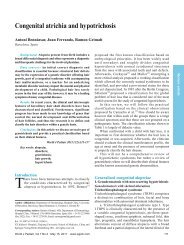
Chest-deformities: a proposal for a classification - Wjpch.com
Chest-deformities: a proposal for a classification - Wjpch.com
Chest-deformities: a proposal for a classification - Wjpch.com

Create successful ePaper yourself
Turn your PDF publications into a flip-book with our unique Google optimized e-Paper software.
Review article<br />
Background: In this article we assess the significance<br />
of classifying chest-<strong>de<strong>for</strong>mities</strong> based on morphological<br />
fi ndings in type-related treatment and its results.<br />
Data sources: Recent publications on chest-<strong>de<strong>for</strong>mities</strong><br />
in children and youth were retrieved from PubMed and<br />
Medline and from our clinical and intraoperative fi ndings.<br />
Results: <strong>Chest</strong>-<strong>de<strong>for</strong>mities</strong> are diagnosed by thoraxmeasurements<br />
using a flexible meter projected on a<br />
graph-paper by MR/CT investigations and color coded<br />
videorasterstereography. In addition an ultrasound guided<br />
mediastinal analysis is per<strong>for</strong>med on the heart, the great<br />
vessels and mediastinal organs. These investigations could<br />
determine meticulously the morphology of the sternum,<br />
the sterno-costal segments and the costal arch, enabling<br />
to find different chest wall <strong>de<strong>for</strong>mities</strong>, i.e., 11 different<br />
types. The clinical and surgical significance of such a<br />
<strong>classification</strong> can be shown by <strong>com</strong>paring postoperative<br />
results of non-classified chest-<strong>de<strong>for</strong>mities</strong> with those of<br />
classified. Preoperatively non-classified chest-<strong>de<strong>for</strong>mities</strong><br />
often have postoperative asymmetric shapes, partial local<br />
recurrences, costal arch eversions and a platythorax. Such<br />
a <strong>classification</strong> can be used to analyze and predict socalled<br />
"secondary associated alterations" of the vertebral<br />
column or mediastinal organs.<br />
Conclusions: Determining the specific type of a<br />
thorax de<strong>for</strong>mity could be considered a type-related<br />
physiotherapy as conservative treatment or vacuum<br />
treatment and if surgery is indicated a type-related<br />
surgical correction can be per<strong>for</strong>med. A type-related<br />
and adapted surgical correction can prevent subsequent<br />
mitral valve prolapse, recurrent infections, vertebral<br />
disturbances caused by kypho-scoliosis and increasing<br />
psychological irritation. Typing chest-<strong>de<strong>for</strong>mities</strong> are an<br />
Author Affiliations: Pediatric Surgical Clinic, University Clinic Graz,<br />
Austria (Saxena A); Pediatric Surgical Research Institute Münster,<br />
Germany (Schütze U); Surgical Clinic Hospital Mühldorf/Inn, Munich,<br />
Germany (Richter W)<br />
Corresponding Author: Günter H. Willital, Professor, MD, Pediatric<br />
Surgical Research Institute Münster, Am Getterbach 49e, D-48163<br />
Münster, Germany (Tel: +49-251-717555; Fax: +49-251-712661; Email:<br />
g.willital@web.de)<br />
doi: 10.1007/s12519-011-0263-y<br />
©Children's Hospital, Zhejiang University School of Medicine, China and<br />
Springer-Verlag Berlin Heidelberg 2011. All rights reserved.<br />
118<br />
World Journal of Pediatrics<br />
<strong>Chest</strong>-<strong>de<strong>for</strong>mities</strong>: a <strong>proposal</strong> <strong>for</strong> a classifi cation<br />
G.H. Willital, A.K. Saxena, U. Schütze, W. Richter<br />
Münster, Germany<br />
additional and essential help <strong>for</strong> the surgeon to per<strong>for</strong>m<br />
specific surgical procedures: detorsion of the sternum,<br />
correction of the sterno-costal region, the costal arch<br />
bow and the kind of chest wall immobilization by metal<br />
struts. It can also <strong>com</strong>pare the postoperative results more<br />
accurately in similar types of chest-<strong>de<strong>for</strong>mities</strong>.<br />
World J Pediatr 2011;7(2):118-123<br />
Key words: chest-<strong>de<strong>for</strong>mities</strong>;<br />
classifi cation;<br />
funnel chest<br />
Introduction<br />
<strong>Chest</strong>-<strong>de<strong>for</strong>mities</strong> have an incidence of<br />
approximately 1:1000. [1] Genetic studies have<br />
proved a congenital origin. [2] <strong>Chest</strong>-<strong>de<strong>for</strong>mities</strong><br />
must not always be present at birth. They can develop<br />
gradually in the first 5 years of life. [3] Histological<br />
and electron microscopic investigations have shown<br />
an increased growth of cartilage at the sternocostal<br />
junction. [4] Chondrocyts in this region have a<br />
hypoplastic structure causing a diminished stability of<br />
the sterno-costal region. <strong>Chest</strong>-<strong>de<strong>for</strong>mities</strong> are seen in<br />
4.3% of patients associated with 29 different syndromes<br />
such as Marfan syndrome or Jeune syndrome.<br />
Since August 2010, 2926 patients have been<br />
operated on at the Pediatric Surgical University<br />
Hospitals in Münster and Erlangen in a period of 35<br />
years (1975-2010). These patients accounted <strong>for</strong> 28%<br />
of all admitted patients with chest-<strong>de<strong>for</strong>mities</strong>.<br />
This review aims to find out whether chest<strong>de<strong>for</strong>mities</strong><br />
differ in morphology and whether a derived<br />
classifi cation is of value <strong>for</strong> surgery, physiotherapy and<br />
<strong>com</strong>parison of the results.<br />
Diagnostic methods<br />
Be<strong>for</strong>e the start of specific diagnostic procedures, [5,6]<br />
we speak to the parents about the history, the onset of<br />
symptoms, psychological alterations of the children<br />
and check the heart, lung and vertebral column. [7,8]<br />
In addition an ultrasound examination of the heart<br />
provides some in<strong>for</strong>mation about the shape and<br />
location of the heart, the mitral and aortic valve and the<br />
configuration of the aortic arch (dilatation of the aorta).<br />
World J Pediatr, Vol 7 No 2 . May 15, 2011 . www.wjpch.<strong>com</strong>
With a flexible meter, modelled to the anterior and<br />
posterior part of the thorax, the shape of chest de<strong>for</strong>mity<br />
can be reproduced on a graph-paper. This non-radiation<br />
method can determine the depth and protusion of the<br />
de<strong>for</strong>mity, the external sterno-vertebral distance as well<br />
as torsion of the sternum or eversion of the costal arch<br />
(Figs. 1, 2). [9]<br />
1. MR/CT investigations are per<strong>for</strong>med in a transverse<br />
and vertical direction. They can diagnose mal<strong>for</strong>mations of<br />
the sternum including the mediastinal shape of the sternum,<br />
the sterno-costal junction, <strong>com</strong>pression and dislocation<br />
of the heart and the mediastinal organs. In addition,<br />
curvatures of the spine related to the chest de<strong>for</strong>mity can be<br />
determined (Fig. 3). [10,11]<br />
2. We have used color-coded videorasterstereography<br />
<strong>for</strong> 8 years to illustrate the shape of the anterior wall<br />
of the thorax color coded. With this method pre- and<br />
postoperative results are <strong>com</strong>pared accurately (Fig. 4).<br />
Types of chest-<strong>de<strong>for</strong>mities</strong><br />
The above mentioned diagnostic methods are used to<br />
investigate chest-<strong>de<strong>for</strong>mities</strong> accurately. [12]<br />
In a 35-year period, we operated on 2926 children<br />
and adolescent patients between the age of 4 and 18<br />
years with chest-<strong>de<strong>for</strong>mities</strong>. Preoperative findings<br />
A B<br />
Fig. 1. With a fl exible meter which is placed on the anterior side of the<br />
thorax (A), the shape of the de<strong>for</strong>mity can be reproduced on a graphpaper<br />
(B). The external sterno-vertebral distance can be determined<br />
if the flexible meter is also placed on the posterior side of the thorax.<br />
The distance is <strong>com</strong>pared with normal values. The size of protusion or<br />
the depth of the thorax can be expressed as a percentage of the normal<br />
fi ndings age related.<br />
Deepest<br />
location<br />
Depth of funnel chest Guideline <strong>for</strong> surgery<br />
Fig. 2. Determination of chest <strong>de<strong>for</strong>mities</strong> using a fl exible meter.<br />
<strong>Chest</strong>-<strong>de<strong>for</strong>mities</strong>: a <strong>proposal</strong> <strong>for</strong> a classifi cation<br />
Cross sections of the thorax<br />
were found to be correlated with intraoperative<br />
findings concerning the different shapes of chest<strong>de<strong>for</strong>mities</strong>.<br />
The most reliable diagnostic methods were<br />
measurements of the chest using a flexible meter and<br />
MRI. Based on these measurements, we found 11 types<br />
of chest-<strong>de<strong>for</strong>mities</strong> (Fig. 5).<br />
Results<br />
Typing chest-<strong>de<strong>for</strong>mities</strong> enables to get further<br />
in<strong>for</strong>mation about necessary operative steps, accurate<br />
findings of secondary alterations of the vertebral<br />
B<br />
Fig. 3. Significance of thorax MR/CT-measurements: this is an MRinvestigation<br />
in a 12 years old boy with a funnel chest (A). The depth<br />
was 45% of the normal sterno-vertebral distance. The funnel chest is<br />
symmetrically shaped. The sternum is not twisted. The heart is shifted to<br />
the left side and <strong>com</strong>pressed by the sternum (B).<br />
Preoperative Postoperative<br />
Fig. 4. The color-coded videorasterstereography enables an easy<br />
documentation of depths or protusions of the sternum, the ribs and<br />
shows hypoplasia of muscle layers. Pre- and postoperative fi ndings can<br />
be <strong>com</strong>pared with each other, and the out<strong>com</strong>e of physiotherapy can<br />
be ascertained easily by improvements of the muscle layers. It is also<br />
possible to documentate symmetric and asymmetric contures of the<br />
thorax. Depressions are in blue, protusion are in red color.<br />
World J Pediatr, Vol 7 No 2 . May 15, 2011 . www.wjpch.<strong>com</strong> 119<br />
A<br />
Review article
Review article<br />
120<br />
type 1: symmetrical<br />
funnel chest<br />
type 5: symmetrical<br />
pigeon chest<br />
type 9: <strong>com</strong>bination of pigeon<br />
chest and funnel chest<br />
Fig. 5. Survey of different types of chest-<strong>de<strong>for</strong>mities</strong>.<br />
column and intrathoracic organs, and precise<br />
re<strong>com</strong>mendations <strong>for</strong> postoperative physiotherapy <strong>for</strong><br />
preventing recurrences (Table 1). [13-18] To evaluate the<br />
significance of such <strong>classification</strong> in chest-<strong>de<strong>for</strong>mities</strong>,<br />
we <strong>com</strong>pared postoperative results in patients of<br />
the same age, gender and surgical techniques used<br />
with those without use of this <strong>classification</strong>. Thus<br />
<strong>com</strong>plications and extraordinary courses related to the<br />
surgical technique could be excluded (Table 2). [19]<br />
Preoperative chest wall typing in patients with<br />
chest-<strong>de<strong>for</strong>mities</strong> is helpful to differentiate surgical<br />
planning: detorsion, splitting and elevation of the<br />
sternum, immobilization of the anterior chest wall by<br />
one or additional metal struts, placed above, transverse<br />
or behind the sternum, single or multiple chondrotomies,<br />
correction of costal arch protrusions, and reimplantations<br />
of excized cartilage/bone covered by bio-absorbable<br />
patches.<br />
Symmetrical funnel chests types 1-3 are found<br />
in 58% of costal arch protrusions. There<strong>for</strong>e it is<br />
re<strong>com</strong>mended to correct this costal arch protrusion by<br />
additional chondrotomies after the sternum is elevated in<br />
a normal anatomical position.<br />
World Journal of Pediatrics<br />
type 2: asymmetrical<br />
funnel chest<br />
type 6: asymmetrical<br />
pigeon chest<br />
Thorax <strong>de<strong>for</strong>mities</strong><br />
type 3: symmetrical funnel<br />
chest and platythorax<br />
type 7: <strong>com</strong>bination of pigeon<br />
chest and platythorax symmetrical<br />
type 4: asymmetrical funnel<br />
chest and platythorax<br />
type 8: <strong>com</strong>bination of pigeon<br />
chest and platythorax asymmetrical<br />
Type<br />
n = 2926<br />
Percentage<br />
1 76.0<br />
2-4 14.0<br />
5-8 6.0<br />
9 3.5<br />
10 0.2<br />
11 0.3<br />
type 10: aplasia of ribs type 11: cleft sternum Frequency<br />
Asymmetrical funnel chests types 2-4 are found in<br />
56% of costal arch protrusions of different dimensions<br />
or sternal torsion. They need a wedge osteotomy at the<br />
level of the third intercostal space and detorsion of the<br />
sternum, depending on the degree of the torsion. This<br />
operation can be done by either open surgery or minimal<br />
invasive surgery via a thoracoscopic procedure. In<br />
addition, the different levels of the costal arch protrusions<br />
should be corrected in a straight way, otherwise a pseudo<br />
funnel chest may occur.<br />
In case of funnel chests with a platythorax (types 3<br />
and 4) it is re<strong>com</strong>mended not only to lift up the sternum<br />
in a normal position but also to elevate the sterno-costal<br />
junction and small costal segments.<br />
In case of <strong>com</strong>bination of pigeon chest and funnel<br />
chest (type 9), several surgical steps must be planned<br />
to avoid recurrences: elevation or subsiding of parts of<br />
the chest wall by multiple chondrotomies and multiple<br />
sternotomies.<br />
For the surgeon, a preoperative <strong>classification</strong><br />
of chest-<strong>de<strong>for</strong>mities</strong> can help to plan the procedure<br />
more accurately in order to get better results. In other<br />
words, classifying chest-<strong>de<strong>for</strong>mities</strong> enables to identify<br />
World J Pediatr, Vol 7 No 2 . May 15, 2011 . www.wjpch.<strong>com</strong>
Type Description Mal<strong>for</strong>mation<br />
sternum<br />
morphological details of the chest wall accurately. This<br />
may be a useful indicator <strong>for</strong> surgical procedures to avoid<br />
local recurrences. [20]<br />
In addition, such a classifi cation proves to be useful<br />
<strong>for</strong> a differentiated physiotherapy. Patients with a funnel<br />
chest (types 1-4) in 87% have a poor posture with round-<br />
shaped shoulders and a curved abdominal wall due<br />
to costal arch protrusion in contrast to patients with a<br />
pigeon chest (types 5-8). Reasons <strong>for</strong> this are hypoplastic<br />
and weak muscles of the back, the shoulders and the<br />
<strong>Chest</strong>-<strong>de<strong>for</strong>mities</strong>: a <strong>proposal</strong> <strong>for</strong> a classifi cation<br />
Table 1. Signifi cance of preoperative typing chest-<strong>de<strong>for</strong>mities</strong> in planning the surgical procedure and in regard to postoperative results<br />
Type 1 Symmetrical funnel<br />
(45%) chest otherwise<br />
normal thorax<br />
Type 2 Asymmetrical funnel<br />
(15%) chest otherwise normal<br />
thorax<br />
Type 3 Symmetrical funnel<br />
(22%) chest and platythorax<br />
Type 4<br />
(8%)<br />
Type 5<br />
(2%)<br />
Type 6<br />
(1%)<br />
Type 7<br />
(2%)<br />
Type 8<br />
(1%)<br />
Type 9<br />
(2%)<br />
Asymmetrical funnel<br />
chest and platythorax<br />
Symmetrical pigeon<br />
chest, otherwise<br />
normal thorax<br />
Asymmetrical pigeon<br />
chest otherwise<br />
normal thorax<br />
Symmetrical pigeon<br />
chest and platythorax<br />
Asymmetrical pigeon<br />
chest and platythorax<br />
Combination of funnel<br />
chest and pigeon chest<br />
Mal<strong>for</strong>mation<br />
sterno-costal junction<br />
Mal<strong>for</strong>mation<br />
costal arch<br />
Sternal depression Sterno-costal depression Bilateral costal<br />
arch protrusions<br />
Sternal depression and<br />
sternal rotation<br />
Sternal depression and<br />
fl at thorax<br />
Sternal depression and<br />
sternal rotation and<br />
fl at thorax<br />
Sterno-costal depression<br />
and different steep walls<br />
of funnel<br />
Sterno-costal depression<br />
and fl at thorax<br />
Sterno-costal depression<br />
and different steep walls<br />
of funnel, fl at thorax<br />
Sternal protrusion Sterno-costal<br />
protrusion<br />
Sternal protrusion and<br />
sternal rotation<br />
Sternal protrusion and<br />
fl at thorax<br />
Sternal protrusion and<br />
sternal rotation and<br />
fl at thorax<br />
Sternal depression and<br />
sternal protrusion<br />
Sterno-costal protrusion<br />
and different steep<br />
walls of protrusion<br />
Sterno-costal protrusion<br />
and fl at thorax<br />
Sterno-costal protrusion,<br />
different steep walls of<br />
protrusion, fl at thorax<br />
Sterno-costal protrusion<br />
and sterno-costal<br />
depression<br />
Bilateral costal arch<br />
prortusions at<br />
different levels<br />
Minimal costal arch<br />
protrusions bilateral<br />
Minimal costal arch<br />
protrusion unilateral<br />
Platythorax<br />
Special points<br />
No Location of deepest sternal<br />
depression either in upper<br />
or middle or lower part<br />
No Location of deepest sternal<br />
depression either in upper<br />
or middle or lower part<br />
Yes No<br />
Yes No<br />
No No Mal<strong>for</strong>mation starts at<br />
manubrium – sternal<br />
junction<br />
No No Mal<strong>for</strong>mation starts at<br />
manubrium – sternal<br />
junction<br />
No Yes No<br />
No Yes No<br />
No No Sternal mal<strong>for</strong>mation at<br />
different parts of the<br />
sternum<br />
Type 10 Aplasia of the ribs<br />
(1%)<br />
No No No No Special surgical technique<br />
Type 11<br />
(1%)<br />
Manubrium -<br />
sternal cleft<br />
Sternal cleft No No No Special surgical technique<br />
Table 2. Different postoperative results if preoperative chest<strong>de<strong>for</strong>mities</strong><br />
have been classifi ed<br />
Results<br />
(1 y after operation)<br />
Classifying chest-<strong>de<strong>for</strong>mities</strong> regarding<br />
postoperative results<br />
Not classified (n=43) Classified (n=43)<br />
Partial recurrence 7 2<br />
Total recurrence 3 0<br />
Costal arch protrusion 8 1<br />
Asymmetrical surface 7 1<br />
Parasternal protrusion 7 1<br />
Sternal depression 6 0<br />
Protrusion of ribs 5 0<br />
abdominal wall. After reconstruction of the chest an<br />
intensive training of these muscles is necessary in order<br />
to avoid postoperative recurrences. It is important to<br />
speak with the parents and the patients be<strong>for</strong>e operation<br />
to plan postoperative physiotherapeutic treatments as part<br />
of the whole treatment. [21]<br />
Furthermore, such a <strong>classification</strong> can be of<br />
diagnostic help regarding alterations of the vertebral<br />
column, the heart and the lung. Symmetrical chest<strong>de<strong>for</strong>mities</strong><br />
(types 1, 3, 5, 7 and 9) can lead to kyphosis,<br />
whereas asymmetrical chest <strong>de<strong>for</strong>mities</strong> (types 2, 4,<br />
6 and 8) can lead to scoliosis of the vertebral column<br />
causing backache in youth and early adult life and later<br />
slip discs if no additional physiotherapeutic treatment<br />
is prescribed. These alterations of the vertebral<br />
column are described as secondary pathology of chest<strong>de<strong>for</strong>mities</strong>.<br />
If these secondary alterations are present it<br />
is re<strong>com</strong>mended to train, after chest wall reconstruction,<br />
muscle layers of the back by a special physiotherapeutic<br />
therapy. Symmetrical funnel chests (type 1) can<br />
cause a <strong>com</strong>pression of the heart, reduced heart beat<br />
volume, and functional disorders of the heart or mitral<br />
valve prolapse. They can also lead to a <strong>com</strong>pression<br />
World J Pediatr, Vol 7 No 2 . May 15, 2011 . www.wjpch.<strong>com</strong> 121<br />
Review article
Review article<br />
of the trachea or of lung segments, documented by<br />
perfusionscintigraphy. [22]<br />
Discussion<br />
There are diseases in pediatric surgery, which have been<br />
investigated meticulously and in which a <strong>classification</strong><br />
has been proposed <strong>for</strong> diagnostic or therapeutic<br />
purposes. Such diseases include esophageal atresia,<br />
anorectal anomalies, biliary atresia, diaphragmatic<br />
hernia and others. <strong>Chest</strong> wall <strong>de<strong>for</strong>mities</strong> may be a<br />
disease in which different morphological types can<br />
be distinguished following morphological alterations<br />
of the sternum, the sterno-costal segments and the<br />
ribs. This <strong>classification</strong> describes accurately the<br />
morphological alterations of chest-<strong>de<strong>for</strong>mities</strong> and<br />
provides in<strong>for</strong>mation on possible secondary alterations<br />
of the heart, lung and vertebral column as well as<br />
re<strong>com</strong>mendations <strong>for</strong> physiotherapy improving<br />
unilateral or bilateral deficiencies of muscle layers of<br />
the abdominal wall or back. [23]<br />
<strong>Chest</strong>-<strong>de<strong>for</strong>mities</strong> necessitate a preoperative plan <strong>for</strong><br />
surgical reconstruction: location of chondrotomies, costal<br />
arch corrections, partial sternotomy or determination<br />
of the number, location and shape of metal struts. In<br />
our cases we <strong>com</strong>pared preoperative morphological<br />
fi ndings with intraoperative fi ndings. Preoperative typing<br />
of chest-<strong>de<strong>for</strong>mities</strong> helps to predict the prognosis of<br />
postoperative results: symmetrical funnel chests (type<br />
1) have the best postoperative results and prognosis: a<br />
partial recurrence rate of 2.4% and a total recurrence<br />
rate of 0.8%, whereas asymmetric funnel chests with a<br />
plathythorax (type 4) have a partial recurrence rate of<br />
4.2% and a total recurrence rate of 1.7%.<br />
Further experience should be accumulated by a<br />
type-related conservative treatment of chest-<strong>de<strong>for</strong>mities</strong><br />
using physiotherapy. [24] Complications and extraordinary<br />
courses are internet-based and collected by the IDBEC<br />
(the International Database <strong>for</strong> Complications and<br />
Extraordinary Courses). [25-29]<br />
Conclusions<br />
Typing chest-<strong>de<strong>for</strong>mities</strong> is advantageous <strong>for</strong> both<br />
patients and surgeons. Distinguishing between different<br />
types of chest-<strong>de<strong>for</strong>mities</strong> enables the surgeons to plan<br />
the surgical procedure more accurately <strong>for</strong> detorsion of<br />
the sternum in asymmetric chest-<strong>de<strong>for</strong>mities</strong> (types 2<br />
and 4), correction of associated costal arch protusions<br />
<strong>for</strong> types 1 and 2, and physiotherapeutic treatment of<br />
muscle hypoplasia (type 4). Such a classifi cation might<br />
be in<strong>for</strong>mative <strong>for</strong> surgeons to <strong>com</strong>pare the results of<br />
different types of funnel chests with each other.<br />
122<br />
World Journal of Pediatrics<br />
Funding: None.<br />
Ethical approval: Not needed.<br />
Competing interest: No benefits in any regard have been or<br />
will be received from any <strong>com</strong>mercial party related directly or<br />
indirectly to the topic of this article.<br />
Contributors: All authors contributed to the content and approved<br />
the fi nal version.<br />
References<br />
1 Willital GH, Kiely E, Gohary AM, Gupta DK, Li M, Tsuchida<br />
Y. Atlas of Children's Surgery, 18-26. Pabst Science Publishers<br />
(Lengerich, Berlin, Bremen, Miami, Riga, Viernheim, Wien,<br />
Zagreb), 2005.<br />
2 Willital GH, Lehmann RR. Surgery in Childhood, 33-67. Spitta<br />
Verlag, Balingen / Rothacker Verlag, München 2000/2005.<br />
3 Saxena AK, Willital GH. Valuable lessons from two decades of<br />
pectus repair with the Willital-Hegemann procedure. J Thorac<br />
Cardiovasc Surg 2007;134:871-876.<br />
4 Fokin AA, Robicsek F. Acquired <strong>de<strong>for</strong>mities</strong> of the anterior<br />
chest wall. Thorac Cardiovasc Surg 2006;54:57-61.<br />
5 Glinkowski W, Sitnkik R, Witkowski M, Kocon H, Bolewicki<br />
P, Goreci A. Method of pectus excavatum measurement based<br />
on structured light technique. J Biomed Opt 2009;14:044041.<br />
6 Guntheroth WG, Spiers PS. Cardiac function be<strong>for</strong>e and after<br />
surgery <strong>for</strong> pectus excavatum. Am J Cardiol 2007;99:1762-<br />
1764.<br />
7 Leutschaft R, Geyer E. The preoperative funnel chest ECG<br />
and postoperative changes after long term observation. Arch<br />
Kreislauff 1968;57:257-272.<br />
8 Malek MH, Berger DE, Housh TJ, Marelich WD, Coburn JW,<br />
Beck TW. Cardiovascular function following surgical repair of<br />
pectus excavatum: a metaanalysis. <strong>Chest</strong> 2006;130:506-516.<br />
9 Lawson ML, Barnes-Eley M, Burke BL, Mitchell K, Katz<br />
ME, Dory CL, et al. Reliability of a standardized protocol to<br />
calculate cross-sectional chest area and severity indices to<br />
evaluate pectus excavatum. J Pediatr Surg 2006;41:1219-1225.<br />
10 Haller JA Jr, Kramer SS, Lietmann SA. Use of CT scans<br />
in selection of patients <strong>for</strong> pectus excavatum surgery: A<br />
preliminary report. J Pediatr Surg 1987;22:904-906.<br />
11 Poncet P, Kravarusic D, Richart T, Evison R, Ronsky JL,<br />
Alassiri A, et al. Clinical impact of optical imaging with<br />
3-D-reconstruction of torso topography in <strong>com</strong>mon anterior<br />
chest wall anomalies. J Pediatr Surg 2007;42:898-903.<br />
12 Colombani PM. Preoperative assessment of chest wall<br />
<strong>de<strong>for</strong>mities</strong>. Semin Thorax Cardiovasc Surg 2009;21:58-63.<br />
13 Cartoski MJ, Nuss D, Goretsky MJ, Proud VK, Croitoru DP,<br />
Gustin T, et al. Classifi cation of the dysmorphology of pectus<br />
excavatum. J Pediatr Surg 2006;41:1573-1581.<br />
14 Jaroszewski DE, Fonkalsrud EW. Repair of pectus chest<strong>de<strong>for</strong>mities</strong><br />
in 320 adult patients: 21 year experience. Ann<br />
Thorac Surg 2007;84:429-433.<br />
15 Metzelder ML, Kuebler JF, Leonhardt J, Ure BM, Petersen C.<br />
Self and parental assessment after minimally invasive repair of<br />
pectus excavatum: lasting satisfaction after bar removal. Ann<br />
Thorac Surg 2007;83:1844-1849.<br />
16 Saxena AK. Pectus less invasive extrapleural repair (PLIER). J<br />
Plast Reconstr Aesthet Surg 2009;62:663-668.<br />
17 Schaarschmidt K, Kolberg-Schwerdt A, Lempe M, Schlesinger<br />
F. New endoscopic minimal access pectus carinatum<br />
repair using subpectoral carbon dioxide. Ann Thorac Surg<br />
World J Pediatr, Vol 7 No 2 . May 15, 2011 . www.wjpch.<strong>com</strong>
2006;81:1099-1103.<br />
18 Zeng Q, Zhang N, Chen CH, He YR. Classification of the<br />
pectus excavatum and minimally invasive Nuss procecure.<br />
Zhonghuar Wai Ke Za Zhi 2008;46:1160-1162.<br />
19 Weber PG, Huemmer HP, Reingruber B. Forces to be over<strong>com</strong>e<br />
in correction of pectus excavatum. J Thorac Cardiovasc Surg<br />
2006;132:1369-1373.<br />
20 Genc O, Gurkok S, Gözübüyük A, Dakak M, Caylak H, Yücel<br />
O. Repair of pectus <strong>de<strong>for</strong>mities</strong>: experience and out<strong>com</strong>e in<br />
317 cases. Ann Saudi Med 2006;26:370-374.<br />
21 Shin S, Goretsky MJ, Kelly RE Jr, Gustin T, Nuss D. Infectious<br />
<strong>com</strong>plications after the Nuss repair in a series of 863 patients. J<br />
Pediatr Surg 2007;42:87-92.<br />
22 Crossland DS, Auldist AW, Davis AM. Malignant pectus<br />
excavatum. Heart 2006;92:1511.<br />
23 Shamberger RC, Welch KJ. Cardiopulmonary function in<br />
pectus excavatum. Surg Gynecol Obstet 1988;166:383-391.<br />
24 Schier F. Vacuum treatment of pectus excavatum. Eur J<br />
Cardiothorac Surg 2006;30:687.<br />
25 Kelly RE Jr, Shamberger RC, Mellins RB, Mitchell<br />
KK, Lawson ML, Oldham K, et al. Excavatum: design,<br />
<strong>Chest</strong>-<strong>de<strong>for</strong>mities</strong>: a <strong>proposal</strong> <strong>for</strong> a classifi cation<br />
perioperative <strong>com</strong>plications, pain, and baseline pulmonary<br />
function facilitated by internet-based data collection. J Am Coll<br />
Surg 2007;205:205-216.<br />
26 Moretto G, Pollini GP, Pellini F, Nardo A, Stimamiglio P,<br />
Sandrini R, et al. Surgical repair of pectus excavatum by<br />
internal metal strut fixation. Clinical experience in 51 cases.<br />
Minerva Chir 2000;55:835-840.<br />
27 Nagasao T, Miyamoto J, Tamaki T, Ichihara K, Jiang H,<br />
Taguchi T, et al. Stress distribution on the thorax after the Nuss<br />
procedure <strong>for</strong> pectus excavatum results in different patterns<br />
between adult and child patients. J Thorac Cardiovasc Surg<br />
2007;134:1502-1507.<br />
28 Nuss D. Minimal invasive repair of pectus excavatum. Chir<br />
Pediatr 2002;15:1-2.<br />
29 Park HJ, Lee SY, Lee CS, Youm W, Lee KR. The Nuss<br />
procedure <strong>for</strong> pectus excavatum: evolution of techniques and<br />
early results on 322 patients. Ann Thorac Surg 2004;77:289-<br />
295.<br />
Received June 29, 2009<br />
Accepted after revision September 27, 2010<br />
World J Pediatr, Vol 7 No 2 . May 15, 2011 . www.wjpch.<strong>com</strong> 123<br />
Review article