Time Table of Normal Foetal Brain Development - mattes verlag ...
Time Table of Normal Foetal Brain Development - mattes verlag ...
Time Table of Normal Foetal Brain Development - mattes verlag ...

You also want an ePaper? Increase the reach of your titles
YUMPU automatically turns print PDFs into web optimized ePapers that Google loves.
<strong>Time</strong> <strong>Table</strong> <strong>of</strong> <strong>Normal</strong> <strong>Foetal</strong> <strong>Brain</strong> <strong>Development</strong> 13<br />
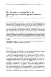
<strong>Table</strong> 4. Glial cells.<br />
Cell type<br />
Structure<br />
Radial glia<br />
Long radial processes spanning the<br />
thickness <strong>of</strong> the cortical wall<br />
Astrocytes<br />
Support cells with short, thick processes<br />
for neurons (“protoplasmic” astrocytes)<br />
or long, thin processes for nerve fibers<br />
(“fibrous” astrocytes)<br />
Oligodendrocytes<br />
Schwann cells<br />
Small cells with few processes<br />
Microglia<br />
Resemble blood monocytes<br />
Functions<br />
Progenitors <strong>of</strong> neurons and astrocytes<br />
Guidance <strong>of</strong> neurons and nerve fibers<br />
Regulation <strong>of</strong> synaptic plasticity<br />
Structural support <strong>of</strong> nerve fibers and cell bodies<br />
Secretion and elimination <strong>of</strong> neurotransmitters<br />
Chemical homeostasis<br />
Oxygen and nutrient supply for neurons<br />
Blood-brain barrier<br />
Regulation <strong>of</strong> local blood flow<br />
Myelin production; functions <strong>of</strong> myelin:<br />
• increases the speed <strong>of</strong> nerve conduction by ten to<br />
one hundred times;<br />
• prevents loss <strong>of</strong> activation by ion diffusion and erratic<br />
activation <strong>of</strong> adjacent axons<br />
• Inhibition <strong>of</strong> the formation <strong>of</strong> new fibres for new<br />
connection (reduction <strong>of</strong> plasticity);<br />
• involved in learning and cognition.<br />
Immune cells (phagocytosis <strong>of</strong> pathogens, cell debris)<br />
the formation <strong>of</strong> new connections to match the number <strong>of</strong> outgrowing fibres to<br />
the capacity <strong>of</strong> target cells (Lossi and Merighi 2003; Saxena and Caroni 2007).<br />
The fittest neurons survive in competition for limited resources in the brain<br />
as electrical impulses, neurotransmitters (e.g. nerve growth factor) and nutrients<br />
within the neural network. Active cells with many connections to target cells receive<br />
more <strong>of</strong> these life-savers than less active neurons. Thus, overproduction<br />
and subsequent elimination <strong>of</strong> excess neurons and connections are not a waste<br />
<strong>of</strong> resources, but necessary to allow optimal locations and interconnections <strong>of</strong><br />
neurons.<br />
Synapses are newly formed and eliminated throughout life. This allows continuous<br />
reorganization <strong>of</strong> the neural network in accordance with the requirements<br />
<strong>of</strong> the environment and is thus the basis <strong>of</strong> life-long neural development and<br />
plasticity (Goda and Davis 2003). Between the onset <strong>of</strong> puberty and adult age approximately<br />
40% <strong>of</strong> synapses and nerve fibres (Bourgeois 2002) and a substantial<br />
portion <strong>of</strong> neurons are eliminated, particularly in the prefrontal cortex, the brain<br />
region involved in major cognitive abilities. In accordance with the “use it or lose<br />
it” principle, cells with apparently redundant connections for unused (not useless!)<br />
skills are discarded to enhance abilities that have been extensively utilized<br />
(Lopez et al. 2008).<br />
Adjustment <strong>of</strong> neurons and connections to the demands <strong>of</strong> the individual environment<br />
usually makes sense, but can result in severe impairments <strong>of</strong> sensory,<br />
behavioural and cognitive functions, if the foetus or young infant is deprived<br />
from normal sensory input or exposed to severe stress (Fabricius 2008). The hippocampus<br />
(stores memory!) is particularly sensitive to the apoptotic actions <strong>of</strong>