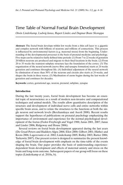
Time Table of Normal Foetal Brain Development - mattes verlag ...
Time Table of Normal Foetal Brain Development - mattes verlag ...
Time Table of Normal Foetal Brain Development - mattes verlag ...

Create successful ePaper yourself
Turn your PDF publications into a flip-book with our unique Google optimized e-Paper software.
Int. J. Prenatal and Perinatal Psychology and Medicine Vol. 21 (2009) No. 1/2, pp. 4–16<br />
<strong>Time</strong> <strong>Table</strong> <strong>of</strong> <strong>Normal</strong> <strong>Foetal</strong> <strong>Brain</strong> <strong>Development</strong><br />
Otwin Linderkamp, Ludwig Janus, Rupert Linder, and Dagmar Beate Skoruppa<br />
Abstract: The foetal brain develops within few weeks from a thin cell layer to a gigantic<br />
and complex network with billions <strong>of</strong> neurons and trillions <strong>of</strong> connections. This process<br />
is influenced by environmental factors (e.g. maternal stress) from the beginning. Understanding<br />
<strong>of</strong> the developmental processes is the basis <strong>of</strong> prenatal medicine and psychology.<br />
Five steps can be related to fairly defined time periods: (1) From 7 to 22 weeks <strong>of</strong> gestation<br />
20 billion neurons are produced and migrate to their final locations in the brain. (2) From<br />
20 to 35 weeks the transient subplate structure lays the foundation <strong>of</strong> the cortex. (3) The<br />
organization <strong>of</strong> the neural network (nerve fibre and synapse formation) starts at 24 weeks<br />
gestation and continues throughout life. (4) Individual adjustment <strong>of</strong> the neural network<br />
by elimination <strong>of</strong> more than 50% <strong>of</strong> the neurons and circuits also starts at 24 weeks, and<br />
shapes the brain in three waves. (5) Myelination <strong>of</strong> axons begins during the last weeks <strong>of</strong><br />
gestation and continues for decades.<br />
Keywords: cortex, gestational age, neuron, prenatal, subplate, synapse<br />
Introduction<br />
During the last twenty years, foetal brain development has become an essential<br />
topic <strong>of</strong> neuroscience as a result <strong>of</strong> modern non-invasive and computational<br />
techniques and animal models. The results allow quantitative description <strong>of</strong> the<br />
structure and development <strong>of</strong> individual nerve cells and entire networks within<br />
specific brain areas, and to relate the structures to the functions at both the single<br />
neuron and network levels (Berzhanskaya and Ascoli 2008). Recent results<br />
support the hypotheses <strong>of</strong> publications on prenatal psychology emphasizing the<br />
importance <strong>of</strong> environment and experience for the normal psychological development<br />
<strong>of</strong> the foetus (Fedor-Freybergh and Vogel 1988; Janus 2001, 2007; Janus<br />
and Linder 2006; Ridgeway and House 2006).<br />
Several reviews on foetal brain development appeared during the last years<br />
(De Graaf-Peters and Hadders-Algra 2006; Eliot 2000; Gilbert 2001; Hüther and<br />
Krens 2006; Lagercrantz et al. 2002; Linderkamp 2005; Ridley 2003; Rutter 2006;<br />
Turkewitz 2007). Our present review is designed to summarize the present knowledge<br />
<strong>of</strong> foetal brain development with emphasis on the time-table <strong>of</strong> the events<br />
shaping the brain. Our paper provides the basis <strong>of</strong> understanding experiencedependent<br />
brain development and effects <strong>of</strong> maternal anxiety and stress on the<br />
brain and long-term outcome. Subsequent papers <strong>of</strong> our group will focus on these<br />
topics (Linderkamp et al. 2010a, b).
<strong>Time</strong> <strong>Table</strong> <strong>of</strong> <strong>Normal</strong> <strong>Foetal</strong> <strong>Brain</strong> <strong>Development</strong> 5<br />
Early Human <strong>Brain</strong> <strong>Development</strong><br />
The brain development begins at approximately three weeks after conception<br />
(5 weeks <strong>of</strong> gestation) with the formation <strong>of</strong> the neural plate at the back <strong>of</strong> the<br />
embryo. A few days later the plate folds to form the neural tube around a canal.<br />
In the brain the canal later widens to the ventricles, in the spinal cord it forms the<br />
central canal. At the time <strong>of</strong> neural tube closure the neural wall consists <strong>of</strong> one<br />
or two layers <strong>of</strong> epithelial cells (neuroepithelium) which are the precursors <strong>of</strong> an<br />
enormous variety <strong>of</strong> neurons and the macroglia.<br />
The development <strong>of</strong> the cerebral cortex occurs in precisely-timed stages<br />
(<strong>Table</strong> 1, Fig. 1). Each developmental process is also a vulnerable period which<br />
is sensitive to environmental insults rendering the brain susceptible to structural<br />
malformations and functional impairments.<br />
Neurulation<br />
Neural proliferation<br />
Neuronal migration<br />
Subplate neurons<br />
Axon growth<br />
Synapse formation<br />
Glia proliferation<br />
Myelination<br />
Neuronal death<br />
Fibre retraction<br />
Synapse elimination<br />
10 15 20 25 30 35 40 6 12 24 4 8 16 32<br />
10 15 20 25 30 35 40 6 12 24 4 8 16 32<br />
weeks months years<br />
Gestational Age<br />
Postnatal Age<br />
Fig. 1. <strong>Time</strong> table <strong>of</strong> developmental events <strong>of</strong> the human brain during foetal and postnatal<br />
life. Black shaded areas indicate peak activities, open lined areas indicate low or medium<br />
activity.<br />
Neurogenesis: “Raw Material” for the <strong>Brain</strong><br />
Billions <strong>of</strong> nerve cells (neurons) are produced during the development <strong>of</strong> the central<br />
nervous system. Neurogenesis mainly occurs at the inner edge <strong>of</strong> the neural<br />
tube wall, the later ventricles (brain) and central canal (spinal cord), respectively<br />
(Fig. 2). In preterm infants the reproduction zone is still visible on ultrasound<br />
scans (“subependymal germinal matrix”). Cell division begins once the neural
6 Otwin Linderkamp et al.<br />
<strong>Table</strong> 1. Major events in foetal cortical development.<br />
Peak occurrence* Major developmental events Abnormal development<br />
5-9 wk Primary neurulation (neural tube<br />
formation)<br />
Prosencephalon formation<br />
(precursor <strong>of</strong> haemispheres)<br />
12-18 wk<br />
(6 wk to life long)<br />
12-20 wk<br />
(8-30 wk)<br />
22-34 wk<br />
(15-38 wk)<br />
24 wk to 15 mo<br />
(10 wk to life-long)<br />
24-38 wk<br />
(20-44 wk)<br />
24 wk to life-long<br />
15 wk to 18 mo<br />
(6 wk to life-long)<br />
Anencephaly<br />
Encephalocele**<br />
Meningomyelocele<br />
Spina bifida**<br />
Neuronal proliferation (neurogenesis) Encephalocele, microbrain**<br />
Schizophrenia**<br />
Neuronal migration<br />
Formation <strong>of</strong> cortical cell layers<br />
Subplate neurons (guidance <strong>of</strong> axons<br />
between thalamus, cortex and<br />
subcortical structures;<br />
final migration <strong>of</strong> neurons)<br />
Outgrowth <strong>of</strong> axons<br />
Outgrowth <strong>of</strong> dendrites<br />
Synaptogenesis<br />
Selective death <strong>of</strong> neurons<br />
Elimination <strong>of</strong> synapses<br />
Heterotopias (wrong place);<br />
reduced or no gyration:<br />
reduced attention and cognition,<br />
depressive signs**<br />
Impaired development <strong>of</strong> thalamus<br />
and cortex and connecting circuits:<br />
disorders <strong>of</strong> frontal, temporal and parietal<br />
centers**<br />
White matter reduction<br />
Cortical dysplasias:<br />
Down, fragile-X syndrome sensory,<br />
behavioural, cognitive disorders**<br />
Excessive loss <strong>of</strong> neurons and connecting<br />
circuits: cognitive, sensory, behavioural,<br />
psychiatric disorders**<br />
Glial cells proliferate und differentiate Impaired neuronal migration<br />
(structural support, neuronal migration, Loss <strong>of</strong> dendrites and synapses in frontal<br />
myelin, “clean up”)<br />
cortex, hippocampus, amygdale<br />
35 wk to 24 mo Myelination Dysfunction <strong>of</strong> axons: psychiatric,<br />
(15 wk to adulthood)<br />
cognitive disorders**<br />
Abbreviatons: IQ, intelligence quotient; p.n., mo, months postnatal; wk, weeks gestation<br />
*Gestational (postmenstrual) age; in parentheses, occurrence prolonged at slower pace.<br />
**Increased risk due to maternal stress has been shown in human foetuses or animal models (from Linderkamp et al. 2010b).<br />
tube has closed at 4 to 5 weeks after conception (6 to 7 weeks <strong>of</strong> gestation). The<br />
majority <strong>of</strong> neurons are formed at 12 to 18 weeks <strong>of</strong> gestation. Approximately<br />
100 000 neurons are produced during each second to provide a number <strong>of</strong> at least<br />
200 billion (2 × 10 11 ) neurons in the human brain and 40 billion in the neocortex<br />
alone. Approximately 50% <strong>of</strong> the neurons are eliminated during the later maturation<br />
process, resulting in a final number <strong>of</strong> 100 billion neurons at 40 weeks<br />
(full-term).<br />
Proliferation <strong>of</strong> neurons during the first 22 weeks <strong>of</strong> gestation is mainly determined<br />
by genetic factors (Bourgois 2002). However, severe maternal stress during<br />
the first trimester (i.e. neurulation and early neurogenesis) has been linked to an<br />
increased risk <strong>of</strong> encephalocele (Hansen et al. 2000) and schizophrenia (Khashan<br />
et al. 2008), suggesting that the expression <strong>of</strong> genes in early foetal life is influenced<br />
by external factors. Stress-induced reduction <strong>of</strong> neurons in late foetal life<br />
is probably the result <strong>of</strong> increased damage <strong>of</strong> neurons (Fabricius et al. 2008).
<strong>Time</strong> <strong>Table</strong> <strong>of</strong> <strong>Normal</strong> <strong>Foetal</strong> <strong>Brain</strong> <strong>Development</strong> 7<br />
Fig. 2. Section through the cortex at approximately 24 weeks <strong>of</strong> gestation. Note that the<br />
germinal zone adjoins to the ventricle at the inner edge <strong>of</strong> the cortex. Newly formed neurons<br />
migrate along the radial glia through the subplate and previously formed neuronal<br />
layers to the upper layer <strong>of</strong> the cortical plate.<br />
If the brain is sufficiently used and trained, new neurons are generated throughout<br />
life. Neural stem cells and pluripotent radial glia cells are able to differentiate<br />
into neurons in the adult brain (Mo et al. 2007). In mice, neurogenesis increased<br />
the efficiency <strong>of</strong> learning, but did not affect long-term memory (Zhang et al.<br />
2008). The formation <strong>of</strong> new synapses and the prevention <strong>of</strong> neuronal damage<br />
are far more important mechanisms for life-long learning than the formation <strong>of</strong><br />
new neurons (Uylings et al. 2005).<br />
Migration <strong>of</strong> Neurons: Finding the Right Place<br />
After several divisions neuroblasts lose their ability to divide and they begin to<br />
move away from the inner multiplication zone to the outer edges <strong>of</strong> the growing<br />
neural tube wall. Once a neuron has reached its final destination within the correct<br />
cortical layer, it will stay there for life. The first neurons start migration with the<br />
beginning <strong>of</strong> multiplication, the majority <strong>of</strong> cells move to their layer between 12<br />
and 20 weeks <strong>of</strong> gestation (Gressens 2005).<br />
Both passive pushing by subsequently migrating neurons and active movement<br />
<strong>of</strong> neurons are mechanisms <strong>of</strong> migration. In the cortex, neurons move radially<br />
outwards to the surface along specialized radial glial fibres (Fig. 2), which span<br />
the entire thickness <strong>of</strong> the hemisphere from the ventricular surface to the external<br />
pial surface (Rakic 2003). This “ladder” facilitates the journey through the earlier<br />
arriving cell layers. At the brain surface, the neurons leave the ladder and move
8 Otwin Linderkamp et al.<br />
<strong>Table</strong> 2. Maturation <strong>of</strong> nerve cells in the cortex.<br />
Step Events<br />
Neurogenesis Subventricular stem cells divide symmetrically. The last division results in<br />
larger neurons before they migrate.<br />
Radial glia Generated from same stem cells a neurons.<br />
Form long processes through the entire cortex (fig. 2).<br />
Migration Neurons climb on radial glia to cortical surface.<br />
Contact to subplate<br />
neurons<br />
Formation <strong>of</strong> six<br />
cortical layers<br />
Migration through subplate neurons (fig. 2) and contact to thalamo-cortical<br />
and cortico-cortical fibres may accelerate their maturation.<br />
Neurons migrate through previously formed layers to the surface. Thus, the<br />
early-migrating cells form the superficial layer, the latest the deepest until six<br />
cortical layers have been established. Neurons assemble in columns above<br />
the stem cells and are therefore clonally related.<br />
Astrocyte formation Astrocytes are generated from radial glia.<br />
Life-long neurogenesis New neurons are generated from remaining subventricular stem cells and<br />
locally from radial glia.<br />
laterally to give way to the subsequently arriving neurons and to form a layer at<br />
the surface <strong>of</strong> the cortex. Then the next group <strong>of</strong> migrating cells passes through<br />
this layer and forms a new layer at the surface. This process continues until six<br />
layers have been formed. Thus, the earlier generated neurons form the deepest<br />
cortical layer, and the latest cells settle in the most superficial layer (inside-out<br />
order). The radial migration <strong>of</strong> neurons originating from the same reproduction<br />
site results in columns <strong>of</strong> clonally related cells. This may be important for their<br />
specialized functions in their final cortical destination.<br />
Insufficient movement or migrations to wrong places result in heterotopias<br />
which may be associated with serious malformations as lissencephaly (reduced<br />
gyration, “flat brain”), epilepsy and mental retardation (Gressens 2005; Nicolic<br />
and Reynolds 2008). Although normal migration <strong>of</strong> neurons to the right location<br />
is probably determined by genes (Rutter 2006), abnormal migration is mostly the<br />
result <strong>of</strong> environmental factors. Maternal stress during the gestational age <strong>of</strong> maximal<br />
neuronal migration has been shown to predispose the <strong>of</strong>fspring to a variety <strong>of</strong><br />
impairments including reduced attention span, cognitive problems and depressive<br />
symptoms (van den Bergh et al. 2008).<br />
Organisation <strong>of</strong> the Neural Network<br />
The first two steps, multiplication and migration <strong>of</strong> primitive nerve cells, are mostly<br />
completed at 22 weeks <strong>of</strong> gestation. At the beginning <strong>of</strong> migration neurons are<br />
not yet specialized, but they lose their pluripotency once they have reached their<br />
final position in a specialized region <strong>of</strong> the central nervous system.<br />
Organization <strong>of</strong> an individual neuron refers to the establishment <strong>of</strong> connections<br />
with other cells and the specialization to distinct functions within the neural<br />
network. Organization <strong>of</strong> the total central nervous system refers to the formation<br />
<strong>of</strong> the entire neuronal network and its capacity to operate as an integrated whole.
<strong>Time</strong> <strong>Table</strong> <strong>of</strong> <strong>Normal</strong> <strong>Foetal</strong> <strong>Brain</strong> <strong>Development</strong> 9<br />
<strong>Table</strong> 3. Major steps <strong>of</strong> neural organisation <strong>of</strong> the cortex (modified from Volpe 2008).<br />
Goal:<br />
Establishment <strong>of</strong> a functioning neural network<br />
Major period:<br />
20 weeks <strong>of</strong> gestation to years after birth<br />
Steps:<br />
• Formation <strong>of</strong> subplate neurons with initial fibre and synapse formation.<br />
• Formation <strong>of</strong> the cortical plate with six layers <strong>of</strong> aligned neurons.<br />
• Outgrowth <strong>of</strong> nerve fibres (axons, dendrites) and their ramifications.<br />
• Synptogenesis.<br />
• Selective elimination <strong>of</strong> neurons (apoptosis), nerve fibres and synapses.<br />
• Proliferation and differentiation <strong>of</strong> neuroglia.<br />
The process <strong>of</strong> organization starts at approximately 22 weeks <strong>of</strong> gestation and<br />
includes actions <strong>of</strong> subplate neurons, outgrowth <strong>of</strong> neural fibres, synaptogenesis<br />
and myelination.<br />
Subplate Neurons: Pioneers Paving the Wire Tracks<br />
Subplate neurons play a major role in the development <strong>of</strong> the gigantic network<br />
connecting billions <strong>of</strong> neurons and are probably responsible for the evolution <strong>of</strong><br />
the neocortex. The subplate zone is situated between the intermediate zone (precursor<br />
<strong>of</strong> white matter) and the cortical plate with the six layers <strong>of</strong> neurons (Fig. 2).<br />
In magnetic resonance images, the subplate is visible as a continuous band in the<br />
entire cortex at 20–27 weeks <strong>of</strong> gestation, starts to disappear in the parietal lobe<br />
at 28 weeks, but remains prominent in the frontal lobe up to 35 weeks (Perkins et<br />
al. 2008). At 38 weeks, 90% <strong>of</strong> the subplate neurons have disappeared.<br />
The subplate neurons excrete neurotransmitters that attract axons ascending<br />
from the thalamus and dendrites descending from cortical neurons for transient<br />
connections with the subplate neurons. When the subplate neurons die, the thalamic<br />
and cortical neurons become directly connected (thalamo-cortical tracts).<br />
Moreover, subplate neurons help cortical neurons to establish connections with<br />
other cortical neurons in both hemispheres and to guide the final migration <strong>of</strong><br />
cortical neurons within the six layers. They help to balance excitation and inhibition<br />
in cortical layers, which is important for the “plasticity” <strong>of</strong> brain functions<br />
(Kanold and Shatz 2006). The transient connections among various brain centres<br />
via subplate neurons are the basis for early foetal (and preterm’s) behaviour<br />
(Kostovics and Jovanov-Milosevic 2006).<br />
Maternal stress during the peak actions <strong>of</strong> subplate neurons from 22 to 34<br />
weeks gestation has been linked to developmental delays, lower IQ, behavioural<br />
problems and schizophrenia in <strong>of</strong>fsprings (Bergman et al. 2007). It is likely that<br />
the stress exposure <strong>of</strong> preterm infants during intensive care can alter subplate<br />
neurons, thereby contributing to the high risk <strong>of</strong> preterm infants to long-term<br />
cognitive and behavioural problems.
10 Otwin Linderkamp et al.<br />
Wiring the Neural Network: Axons, Dendrites and Synapses<br />
The set-up <strong>of</strong> a functioning neural network connecting all parts <strong>of</strong> the central<br />
nervous system and other target organs requires trillions <strong>of</strong> connections among<br />
neurons via axons, dendrites and synapses. The migrating cells have no functioning<br />
axons and dendrites. Having migrated to the appropriate position, axons and<br />
dendrites begin to grow out <strong>of</strong> the young neurons.<br />
Usually one axon only arises from each cell (Fig. 3). Axons are the long nerve<br />
fibres connecting distant parts within the central nervous system and with peripheral<br />
organs (e.g. muscles and glands). Their final length can be more than<br />
a meter in adults, but also just a few µm, if they connect adjacent neurons. Axons<br />
develop many branches at the tip and each final branch can form a synapse<br />
with a final branch <strong>of</strong> a dendrite or sometimes another axon or a nerve cell body.<br />
Dendrites emerge from many points along the cell body and appear very much<br />
like branches on a tree. Axons and dendrites find their target cells principally<br />
by growing in the direction <strong>of</strong> the targets. This growth is guided by molecules<br />
bound to cells (for short-range chemoattraction) or diffused in the environment<br />
(long-range chemoattraction, e.g. nerve growth factor). Target cells also present<br />
and secrete chemorepellents that inhibit the growth <strong>of</strong> connecting nerve fibres<br />
to these cells. The search <strong>of</strong> outgrowing fibres for target neurons can be highly<br />
specific or more or less arbitrary. Specific connections are formed between neurons<br />
that express specific marker molecules, thereby giving the connecting cells<br />
no choice (cell specificity). Other neurons are attracted to send fibres to neurons<br />
in a defined region (topographic specificity).<br />
Synapses are formed by proteins acting as molecular switches between two<br />
nerve fibres. Chemoattractants determine when and where synapses are formed<br />
and their specificity and stability. Moreover, formation, specificity and stability <strong>of</strong> a<br />
synapse depend on the quality and quantity <strong>of</strong> impulses travelling through the connecting<br />
fibres. Synaptic activity provides critical information about the usefulness<br />
<strong>of</strong> synaptic connections, thereby influencing synapse stability and maintenance<br />
(Waites et al. 2005). Synaptic activity promotes the formation <strong>of</strong> new synapses<br />
and strengthens existing synapses in the neighbourhood. Thus, synapse formation<br />
and stabilization are dynamic processes, requiring bi-directional communication<br />
between connected partners. Subtle alterations in synaptic connections are the<br />
means by which learning wires the pathways to memory (Ge et al. 2007).<br />
Although the first synapses are produced already at 8 weeks <strong>of</strong> gestation,<br />
synapse formation is slow until 24 weeks <strong>of</strong> gestation resulting in a total number<br />
<strong>of</strong> synapses that is not much higher than the total number <strong>of</strong> neurons. From<br />
24 weeks gestation to 12 months <strong>of</strong> postnatal age, a myriad <strong>of</strong> connections is<br />
formed among billions <strong>of</strong> neurons. At full-term each cortical neuron is linked<br />
with approximately 2500 other neurons, at 12 months <strong>of</strong> postnatal age with 15 000<br />
(Petanjek et al. 2008). Synaptogenesis begins in a relatively short time period in all<br />
cortical regions, but the maximum synaptic density is reached at different times<br />
after full-term, ranging from 3 months in the auditory and visual cortex to 15<br />
months in the prefrontal cortex (Bourgeois 2002).<br />
After the first year <strong>of</strong> postnatal life the total synapse number slowly increases<br />
and reaches the maximum at five years when the child’s brain weighs almost as<br />
much as in adults. Then the number <strong>of</strong> synapses plateaus until about 10 years and
<strong>Time</strong> <strong>Table</strong> <strong>of</strong> <strong>Normal</strong> <strong>Foetal</strong> <strong>Brain</strong> <strong>Development</strong> 11<br />
Fig. 3. Neurons with one axon and several dendrites arising from the neuronal cell body.<br />
The left neuron represents the development in the sensory cortex at approximately 24–<br />
28 weeks, the right neuron at 32–40 weeks. Note the marked differences in ramifications<br />
between the two neurons.<br />
begins to decrease by approximately 40% with the onset <strong>of</strong> puberty. Thus, during<br />
the first 5–10 years <strong>of</strong> life, the child achieves the highest number <strong>of</strong> synapses,<br />
thereby enabling the child to acquire enormous behavioural, social, environmental,<br />
linguistic and cultural information. After the age <strong>of</strong> five years, synaptogenesis<br />
continues as a local event (Bourgeois 2002) in dependence on the activity <strong>of</strong><br />
neighbouring synapses. Formation <strong>of</strong> new synapses and changes <strong>of</strong> specificity and<br />
stability <strong>of</strong> synapses are fundamental to life-long learning, memory and cognition<br />
in the mature brain (Waites et al. 2005).<br />
Outgrowth <strong>of</strong> fibres and formation <strong>of</strong> synapses are largely influenced by environmental<br />
factors, including sensory experience. Both decreased sensory input <strong>of</strong><br />
the foetus and maternal stress may cause a marked reduction <strong>of</strong> axons, dendrites<br />
and synapses in the prefrontal cortex, the hippocampus and other brain centres<br />
(Linderkamp et al. 2010b).<br />
Glial Cells and Myelination<br />
Glial cells (also called neuroglia) are non-neuronal cells that outnumber neurons<br />
by about 10 to 1, but constitute only half <strong>of</strong> the brain volume, since they are<br />
smaller than neurons. Glial cells surround neurons and hold them in place, play<br />
an important role in neuronal and axonal guidance, supply nutrients and oxygen<br />
to neurons, produce and remove chemical transmitters, insulate axons by myelin,
12 Otwin Linderkamp et al.<br />
destroy pathogens, dead neurons and other debris, and contribute to formation<br />
<strong>of</strong> new neurons. Glial cells are crucial in the development <strong>of</strong> the nervous system<br />
and in processes such as synaptic plasticity and synaptogenesis. Various types <strong>of</strong><br />
glial cells are defined by origin, appearance and functions (<strong>Table</strong> 4).<br />
Macroglial cells comprise radial glia, astrocytes and oligodendrocytes and develop<br />
from the same stem cells in the ventricular zone <strong>of</strong> the neural tube as the<br />
neurons. Radial glia cells are the progenitors <strong>of</strong> astrocytes, some oligodendrocytes<br />
and neurons. In the developing brain, radial glia functions as a “ladder”<br />
upon which neurons migrate to the surface <strong>of</strong> the cortex. Microglia are specialized<br />
immune cells capable <strong>of</strong> phagocytosis. They are derived from haemopoietic<br />
precursors as other immune cells.<br />
Oligodendrocytes produce myelin that forms insulating sheaths around axons.<br />
Schwann cells provide myelination to axons in the peripheral nervous system.<br />
Myelin is a white fatty material wrapped around most neural axons. It prevents<br />
the leakage <strong>of</strong> ions and thus <strong>of</strong> electrical current from the axon, thereby increasing<br />
the speed <strong>of</strong> nerve conduction by ten to one hundred times. Moreover, myelin prevents<br />
erratic activation <strong>of</strong> adjacent axons. Without myelin, electric activity would<br />
be aimlessly distributed throughout the brain, and information would become<br />
chaotic. Myelination also inhibits plasticity, since a myelinated axon has less ability<br />
to branch out and connect with other neurons. Myelin is involved in cognitive<br />
functions and learning (Fields 2008).<br />
Myelination starts in the spinal cord (at about 12 weeks gestation), then in<br />
brain stem (14 weeks) and thalamic axons (20 weeks), and finally in the cortex<br />
(35 weeks) and continues for decades in the human brain (Miller et al. 2003).<br />
Axons connecting the frontal-limbic system (responsible for complex cognitive<br />
functions) start to myelinate after birth. Late myelination explains that the brains<br />
<strong>of</strong> infants and young children are slow compared with adult brains. Myelination<br />
is modifiable by experience and severe maternal or postnatal stress may inhibit<br />
myelination, thereby contributing to psychiatric disorders, including schizophrenia<br />
and depression, and cognitive impairment (Fields 2008).<br />
Shaping the <strong>Brain</strong> by Elimination <strong>of</strong> Excess Neurons and Circuits<br />
At least twice as many neurons as necessary are produced during the time period<br />
<strong>of</strong> active neuronal multiplication, and most <strong>of</strong> the excess neurons are eliminated<br />
during maturation <strong>of</strong> the neuronal network (“programmed cell death” or apoptosis).<br />
Three peak periods <strong>of</strong> neuronal death can be distinguished (Fig. 1): 1) at<br />
the beginning <strong>of</strong> neurogenesis; 2) from 24 to 38 weeks gestation; and 3) between<br />
the onset <strong>of</strong> puberty and adulthood (Lossi and Merighi 2003).<br />
The second and third periods are linked to selective elimination <strong>of</strong> axons, dendrites<br />
and synapses. Production <strong>of</strong> neurons and growth <strong>of</strong> axons and dendrites in<br />
the direction <strong>of</strong> target cells are not very selective and result in overproduction <strong>of</strong><br />
connections. The initial wiring is diffuse, with a lot <strong>of</strong> overlap making communication<br />
inaccurate and disorganized. Elimination <strong>of</strong> fibres, synapses and entire<br />
neurons allows quantitative adjustments <strong>of</strong> connections between neurons and to<br />
compensate for errors <strong>of</strong> cell migration (mislocation) and projection <strong>of</strong> axons and<br />
dendrites (misprojection). Elimination <strong>of</strong> neurons, fibres and synapses parallels
<strong>Time</strong> <strong>Table</strong> <strong>of</strong> <strong>Normal</strong> <strong>Foetal</strong> <strong>Brain</strong> <strong>Development</strong> 13<br />
<strong>Table</strong> 4. Glial cells.<br />
Cell type<br />
Structure<br />
Radial glia<br />
Long radial processes spanning the<br />
thickness <strong>of</strong> the cortical wall<br />
Astrocytes<br />
Support cells with short, thick processes<br />
for neurons (“protoplasmic” astrocytes)<br />
or long, thin processes for nerve fibers<br />
(“fibrous” astrocytes)<br />
Oligodendrocytes<br />
Schwann cells<br />
Small cells with few processes<br />
Microglia<br />
Resemble blood monocytes<br />
Functions<br />
Progenitors <strong>of</strong> neurons and astrocytes<br />
Guidance <strong>of</strong> neurons and nerve fibers<br />
Regulation <strong>of</strong> synaptic plasticity<br />
Structural support <strong>of</strong> nerve fibers and cell bodies<br />
Secretion and elimination <strong>of</strong> neurotransmitters<br />
Chemical homeostasis<br />
Oxygen and nutrient supply for neurons<br />
Blood-brain barrier<br />
Regulation <strong>of</strong> local blood flow<br />
Myelin production; functions <strong>of</strong> myelin:<br />
• increases the speed <strong>of</strong> nerve conduction by ten to<br />
one hundred times;<br />
• prevents loss <strong>of</strong> activation by ion diffusion and erratic<br />
activation <strong>of</strong> adjacent axons<br />
• Inhibition <strong>of</strong> the formation <strong>of</strong> new fibres for new<br />
connection (reduction <strong>of</strong> plasticity);<br />
• involved in learning and cognition.<br />
Immune cells (phagocytosis <strong>of</strong> pathogens, cell debris)<br />
the formation <strong>of</strong> new connections to match the number <strong>of</strong> outgrowing fibres to<br />
the capacity <strong>of</strong> target cells (Lossi and Merighi 2003; Saxena and Caroni 2007).<br />
The fittest neurons survive in competition for limited resources in the brain<br />
as electrical impulses, neurotransmitters (e.g. nerve growth factor) and nutrients<br />
within the neural network. Active cells with many connections to target cells receive<br />
more <strong>of</strong> these life-savers than less active neurons. Thus, overproduction<br />
and subsequent elimination <strong>of</strong> excess neurons and connections are not a waste<br />
<strong>of</strong> resources, but necessary to allow optimal locations and interconnections <strong>of</strong><br />
neurons.<br />
Synapses are newly formed and eliminated throughout life. This allows continuous<br />
reorganization <strong>of</strong> the neural network in accordance with the requirements<br />
<strong>of</strong> the environment and is thus the basis <strong>of</strong> life-long neural development and<br />
plasticity (Goda and Davis 2003). Between the onset <strong>of</strong> puberty and adult age approximately<br />
40% <strong>of</strong> synapses and nerve fibres (Bourgeois 2002) and a substantial<br />
portion <strong>of</strong> neurons are eliminated, particularly in the prefrontal cortex, the brain<br />
region involved in major cognitive abilities. In accordance with the “use it or lose<br />
it” principle, cells with apparently redundant connections for unused (not useless!)<br />
skills are discarded to enhance abilities that have been extensively utilized<br />
(Lopez et al. 2008).<br />
Adjustment <strong>of</strong> neurons and connections to the demands <strong>of</strong> the individual environment<br />
usually makes sense, but can result in severe impairments <strong>of</strong> sensory,<br />
behavioural and cognitive functions, if the foetus or young infant is deprived<br />
from normal sensory input or exposed to severe stress (Fabricius 2008). The hippocampus<br />
(stores memory!) is particularly sensitive to the apoptotic actions <strong>of</strong>
14 Otwin Linderkamp et al.<br />
corticosteroids transmitted to the foetus as a result <strong>of</strong> maternal stress (Fenoglio<br />
et al. 2006).<br />
References<br />
Bergman K, Sarkar P, O’Connor TG, Modi N, Glover V (2007) Maternal stress during<br />
pregnancy predicts cognitive ability and fearfulness in infancy. J Am Acad Child Adolesc<br />
Psychiatry 46: 1454–1463<br />
Berzhanskaya J, Ascoli G (2008) Computational neuroanatomy. Scholarpedia 3:1313<br />
(www.scholarpedia.org)<br />
Bourgeois JP (2002) Synaptogenesis in the neocortex <strong>of</strong> the newborn. in: Lagercrantz H,<br />
Hanson M, Evrard P, Rodeck C (eds) The Newborn <strong>Brain</strong>. Cambridge University Press,<br />
Cambridge, UK, pp 91–113<br />
Colvert E, Rutter M, Kreppner J, Beckett C, Castle J, Groothues C, Hawkins A, Stevens<br />
S, Sonuga-Barke EJ (2008) Do theory <strong>of</strong> mind and executive function deficits underlie<br />
the adverse outcomes associated with pr<strong>of</strong>ound early deprivation?: Findings from the<br />
English and Romanian adoptees study. J Abnorm Child Psychol [Epub ahead <strong>of</strong> print]<br />
De Graaf-Peters VB, Hadders-Algra M (2006) Ontogeny <strong>of</strong> the human central nervous<br />
system: what is happening when? Early Hum Dev 82: 257–266<br />
Eliot L (2000) What’s Going on in There? How the <strong>Brain</strong> and Mind Develop in the First<br />
Five Years <strong>of</strong> Life. Bantam, New York, U.S.A.<br />
Fabricius K, Wörtwein G, Pakkenberg B (2008) The impact <strong>of</strong> maternal separation on adult<br />
mouse behaviour and on the total neuron number in the mouse hippocampus. <strong>Brain</strong><br />
Struct Funct 212: 403–416<br />
Fedor-Freybergh PG, Vogel LV (1988) Prenatal and Perinatal Psychology and Medicine:<br />
Encounter With the Unborn. Parthenon, Carnforth, UK<br />
Fenoglio KA, Brunson KL, Baram TZ (2006) Hippocampal neuroplasticity induced by<br />
early-life stress: functional and molecular aspects. Front Neuroendocrinol 27: 180–192<br />
Fields RD (2008) White matter in learning, cognition and psychiatric disorders. Trends<br />
Neurosci [Epub ahead <strong>of</strong> print]<br />
Ge S, Jang CH, Hsu KS, Ming GL, Song H (2007) A critical period for enhanced synaptic<br />
plasticity in newly generated neurons <strong>of</strong> the adult brain. Neuron 54: 559–566<br />
Gilbert G (2001) Individual <strong>Development</strong> and Evolution: The Genesis <strong>of</strong> Novel Behavior.<br />
Lawrence Erlbaum Ass., Mahwah, NJ, U.S.A.<br />
Goda Y, Davis GW (2003) Mechanisms <strong>of</strong> synapse assembly and disassembly. Neuron 40:<br />
243–264<br />
Gressens P (2005) Neuronal migration disorders. J Child Neurol 20: 969–971<br />
Hansen D, Lou HC, Olsen J (2000) Serious life event and congenital malformations: a<br />
national study with complete follow-up. Lancet 356: 975–980<br />
Hüther G, Krens I (2006) Das Geheimnis der ersten neun Monate. Unsere frühesten<br />
Prägungen. Walter, Düsseldorf, Germany.<br />
Janus L (2001) Enduring Effects <strong>of</strong> Prenatal Experiences. Mattes, Heidelberg, Germany.<br />
Janus L (2007) Seelenraum des Ungeborenen: Pränatale Psychologie und Therapie. Patmos,<br />
Düsseldorf, Germany.<br />
Janus L, Linder R (2006) Psychologische und psychosomatische Aspekte von Schwangerschaft<br />
und Geburt. Prenat Perinat Psychol Med 18: 57–70<br />
Kanold PO, Shatz CJ (2006) Subplate neurons regulate maturation <strong>of</strong> cortical inhibition<br />
and outcome <strong>of</strong> ocular dominance plasticity. Neuron 51: 627–638<br />
Khashan AS, Abel KM, McNamee R, Pedersen MG, Webb RT, Baker PN, Kenny LC,<br />
Mortensen PB (2008) Higher risk <strong>of</strong> <strong>of</strong>fspring schizophrenia following antenatal maternal<br />
exposure to severe adverse life events. Arch Gen Psychiatry 65: 146–152
<strong>Time</strong> <strong>Table</strong> <strong>of</strong> <strong>Normal</strong> <strong>Foetal</strong> <strong>Brain</strong> <strong>Development</strong> 15<br />
Kostovic I, Jovanov-Milosevic N (2006) The development <strong>of</strong> cerebral connections during<br />
the first 20–45 weeks’ gestation. Semin Fetal Neonatal Med 11: 415–422<br />
Lagercrantz H, Hanson M, Evrard P, Rodeck C (2002) The Newborn <strong>Brain</strong>. Neuroscience<br />
and clinical applications. Cambridge University Press, Cambridge, UK<br />
Linderkamp O (2005) Gehirnentwicklung bei Feten und Frühgeborenen, in: Frühgeborene<br />
optimal ernähren und pflegen, Frank C, Linderkamp O, Pohlandt F, pp. 126–131. Kirchheim,<br />
Mainz<br />
Linderkamp O, Janus L, Linder R, Skoruppa D (2009a) <strong>Development</strong> <strong>of</strong> the foetal brain.<br />
Genetics and experience-driven plasticity. Int J Prenat Perinat Psychol Med (in press)<br />
Linderkamp O, Janus L, Linder R, Skoruppa D (2009b) Effects <strong>of</strong> prenatal stress on brain<br />
development. Int J Prenat Perinat Psychol Med (in press)<br />
Lopez B, Schwartz SJ, Prado G, Campo AE, Pantin H (2008) Adolescent neurological<br />
development and its implications for adolescent substance use prevention. J Prim Prev<br />
29: 5–35<br />
Lossi L, Merighi A (2003) In vivo cellular and molecular mechanisms <strong>of</strong> neuronal apoptosis<br />
in the mammalian CNS. Prog Neurobiol 69: 287–312<br />
Maffei A, Nataraj K, Nelson SB, Turrigiano GG (2006) Potentiation <strong>of</strong> cortical inhibition<br />
by visual deprivation. Nature 443: 81–84<br />
Miller JH, McKinstry RC, Philip JV, Mukherjee P, Neil JJ (2003) Diffusion-tensor MR<br />
imaging <strong>of</strong> normal brain maturation: a guide to structural development and myelination.<br />
Am J Roentgenol 180: 851–859<br />
Mo Z, Moore AR, Filipovic R, Ogawa Y, Kazuhiro I, Antic SD, Zecevic N (2007) Human<br />
cortical neurons originate from radial glia and neuron-restricted progenitors. J Neurosci<br />
27: 4132–4145<br />
Naves G, Cooke SF, Bliss TV (2008) Synaptic plasticity, memory and the hippocampus: a<br />
neural network approach to causality. Nat Rev Neurosci 9: 65–75<br />
Nicolic M, Reynolds R (2008) Mechanisms governing neuronal migration and morphology.<br />
Dev Neurosci 30: 1–222<br />
Perkins L, Hughes E, Srinivasan L, Allsop J, Glover A, Kumar S, Fisk N, Rutherford M<br />
(2008) Exploring cortical subplate evolution using magnetic resonance imaging <strong>of</strong> the<br />
fetal brain. Dev Neurosci 30: 211–220<br />
Petanjek Z, Judas M, Kostovic I, Uylings HB (2008) Lifespan alterations <strong>of</strong> basal dendritic<br />
trees <strong>of</strong> pyramidal neurons in the human prefrontal cortex: a layer-specific pattern.<br />
Cereb Cortex 18: 915–929<br />
Plomin R, Spinath FM (2004) Intelligence: Genetics, genes, and genomics. J Personal Soc<br />
Psychol 86: 112–129<br />
Rakic P (2003) <strong>Development</strong>al and evolutionary adaptations <strong>of</strong> cortical radial glia. Cereb<br />
Cortex 13: 541–549<br />
Ridgway R, House SH (2006) The Unborn Child. Beginning a Whole Life and Overcoming<br />
Problems <strong>of</strong> Early Origin. Karnac, London, UK<br />
Ridley M (2003) Nature via Nurture. Genes, Experience and What Makes us Human.<br />
Harper Collins, London<br />
Rutter M (2006) Genes and Behavior: Nature-Nurture Interplay Explained. Blackwell,<br />
Cambridge, MA<br />
Saxena S, Caroni P (2007) Mechanisms <strong>of</strong> axon degeneration: from development to disease.<br />
Prog Neurobiol 83: 174–191<br />
Turkewitz G (2007) The relevance <strong>of</strong> the fetal and neonatal period for the development<br />
<strong>of</strong> novelty preferences, learning, habituation and hemispheric specialization. Dev Psychobiol<br />
49: 780–787
16 Otwin Linderkamp et al.<br />
Uylings HB, Mal<strong>of</strong>eeva LI, Bogolepova IN, Jacobsen AM, Amunts K, Zilles K (2005) No<br />
postnatal doubling <strong>of</strong> number <strong>of</strong> neurons in human Broca’s areas (Brodmann areas 44<br />
and 45)? A stereological study. Neuroscience 136: 715–728<br />
Van den Bergh BR, Van Calster B, Smits T, Van Huffel S, Lagae L (2008) Antenatal maternal<br />
anxiety is related to HPA-axis dysregulation and self-reported depressive symptoms<br />
in adolescence: A prospective study on the fetal origins <strong>of</strong> depressed mood. Neuropsychopharmacology<br />
33: 536–545<br />
Volpe JJ (2008) Neurology <strong>of</strong> the Newborn, 5th ed. Saunders Co., Philadelphia<br />
Waites CL, Craig AM, Garner CC (2005) Mechanisms <strong>of</strong> vertebrate synaptogenesis. Annu<br />
Rev Neurosci 28: 251–274<br />
Zhang CL, Zou Y, He W, Gage FH, Evans RM (2008) sA role for adult TLX positive<br />
neural stem cells in learning and behaviour. Nature 451: 1004–1007<br />
Correspondence to: Otwin Linderkamp, Pr<strong>of</strong>. Dr., Kornmarkt 3, 69117 Heidelberg, Germany,<br />
E-Mail olinderkamp@yahoo.de