Time Table of Normal Foetal Brain Development - mattes verlag ...
Time Table of Normal Foetal Brain Development - mattes verlag ...
Time Table of Normal Foetal Brain Development - mattes verlag ...

You also want an ePaper? Increase the reach of your titles
YUMPU automatically turns print PDFs into web optimized ePapers that Google loves.
<strong>Time</strong> <strong>Table</strong> <strong>of</strong> <strong>Normal</strong> <strong>Foetal</strong> <strong>Brain</strong> <strong>Development</strong> 11<br />
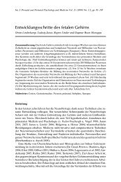
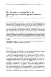
Fig. 3. Neurons with one axon and several dendrites arising from the neuronal cell body.<br />
The left neuron represents the development in the sensory cortex at approximately 24–<br />
28 weeks, the right neuron at 32–40 weeks. Note the marked differences in ramifications<br />
between the two neurons.<br />
begins to decrease by approximately 40% with the onset <strong>of</strong> puberty. Thus, during<br />
the first 5–10 years <strong>of</strong> life, the child achieves the highest number <strong>of</strong> synapses,<br />
thereby enabling the child to acquire enormous behavioural, social, environmental,<br />
linguistic and cultural information. After the age <strong>of</strong> five years, synaptogenesis<br />
continues as a local event (Bourgeois 2002) in dependence on the activity <strong>of</strong><br />
neighbouring synapses. Formation <strong>of</strong> new synapses and changes <strong>of</strong> specificity and<br />
stability <strong>of</strong> synapses are fundamental to life-long learning, memory and cognition<br />
in the mature brain (Waites et al. 2005).<br />
Outgrowth <strong>of</strong> fibres and formation <strong>of</strong> synapses are largely influenced by environmental<br />
factors, including sensory experience. Both decreased sensory input <strong>of</strong><br />
the foetus and maternal stress may cause a marked reduction <strong>of</strong> axons, dendrites<br />
and synapses in the prefrontal cortex, the hippocampus and other brain centres<br />
(Linderkamp et al. 2010b).<br />
Glial Cells and Myelination<br />
Glial cells (also called neuroglia) are non-neuronal cells that outnumber neurons<br />
by about 10 to 1, but constitute only half <strong>of</strong> the brain volume, since they are<br />
smaller than neurons. Glial cells surround neurons and hold them in place, play<br />
an important role in neuronal and axonal guidance, supply nutrients and oxygen<br />
to neurons, produce and remove chemical transmitters, insulate axons by myelin,