Time Table of Normal Foetal Brain Development - mattes verlag ...
Time Table of Normal Foetal Brain Development - mattes verlag ...
Time Table of Normal Foetal Brain Development - mattes verlag ...

You also want an ePaper? Increase the reach of your titles
YUMPU automatically turns print PDFs into web optimized ePapers that Google loves.
<strong>Time</strong> <strong>Table</strong> <strong>of</strong> <strong>Normal</strong> <strong>Foetal</strong> <strong>Brain</strong> <strong>Development</strong> 5<br />
Early Human <strong>Brain</strong> <strong>Development</strong><br />
The brain development begins at approximately three weeks after conception<br />
(5 weeks <strong>of</strong> gestation) with the formation <strong>of</strong> the neural plate at the back <strong>of</strong> the<br />
embryo. A few days later the plate folds to form the neural tube around a canal.<br />
In the brain the canal later widens to the ventricles, in the spinal cord it forms the<br />
central canal. At the time <strong>of</strong> neural tube closure the neural wall consists <strong>of</strong> one<br />
or two layers <strong>of</strong> epithelial cells (neuroepithelium) which are the precursors <strong>of</strong> an<br />
enormous variety <strong>of</strong> neurons and the macroglia.<br />
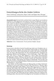
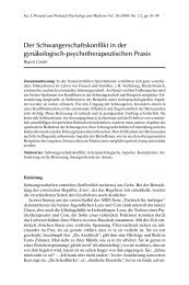
The development <strong>of</strong> the cerebral cortex occurs in precisely-timed stages<br />
(<strong>Table</strong> 1, Fig. 1). Each developmental process is also a vulnerable period which<br />
is sensitive to environmental insults rendering the brain susceptible to structural<br />
malformations and functional impairments.<br />
Neurulation<br />
Neural proliferation<br />
Neuronal migration<br />
Subplate neurons<br />
Axon growth<br />
Synapse formation<br />
Glia proliferation<br />
Myelination<br />
Neuronal death<br />
Fibre retraction<br />
Synapse elimination<br />
10 15 20 25 30 35 40 6 12 24 4 8 16 32<br />
10 15 20 25 30 35 40 6 12 24 4 8 16 32<br />
weeks months years<br />
Gestational Age<br />
Postnatal Age<br />
Fig. 1. <strong>Time</strong> table <strong>of</strong> developmental events <strong>of</strong> the human brain during foetal and postnatal<br />
life. Black shaded areas indicate peak activities, open lined areas indicate low or medium<br />
activity.<br />
Neurogenesis: “Raw Material” for the <strong>Brain</strong><br />
Billions <strong>of</strong> nerve cells (neurons) are produced during the development <strong>of</strong> the central<br />
nervous system. Neurogenesis mainly occurs at the inner edge <strong>of</strong> the neural<br />
tube wall, the later ventricles (brain) and central canal (spinal cord), respectively<br />
(Fig. 2). In preterm infants the reproduction zone is still visible on ultrasound<br />
scans (“subependymal germinal matrix”). Cell division begins once the neural