Dental Lasers - Academy of Laser Dentistry
Dental Lasers - Academy of Laser Dentistry
Dental Lasers - Academy of Laser Dentistry

Create successful ePaper yourself
Turn your PDF publications into a flip-book with our unique Google optimized e-Paper software.
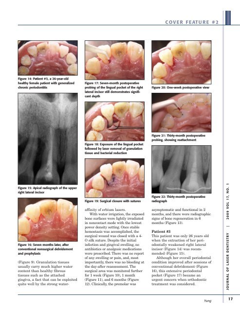
Figure 14: Patient #3, a 26-year-old<br />
healthy female patient with generalized<br />
chronic periodontitis<br />
Figure 15: Apical radiograph <strong>of</strong> the upper<br />
right lateral incisor<br />
Figure 16: Seven months later, after<br />
conventional nonsurgical debridement<br />
and prophylaxis<br />
(Figure 9). Granulation tissues<br />
usually carry much higher water<br />
content than healthy fibrous<br />
tissues such as the attached<br />
gingiva, a fact that can be exploited<br />
quite well by the strong water-<br />
Figure 17: Seven-month postoperative<br />
probing <strong>of</strong> the lingual pocket <strong>of</strong> the right<br />
lateral incisor still demonstrates significant<br />
depth<br />
Figure 18: Exposure <strong>of</strong> the lingual pocket<br />
followed by laser removal <strong>of</strong> granulation<br />
tissue and bacterial reduction<br />
Figure 19: Surgical closure with sutures<br />
affinity <strong>of</strong> erbium lasers.<br />
With water irrigation, the exposed<br />
bone surfaces were lightly irradiated<br />
in noncontact mode with the lowest<br />
power density setting. Once stable<br />
hemostasis was accomplished, the<br />
surgical wound was closed with a 4-<br />
O silk suture. Despite the initial<br />
infection and gingival swelling, no<br />
antibiotics or analgesic medications<br />
were prescribed. There was no report<br />
<strong>of</strong> any swelling or pain, and, most<br />
importantly, there was no bleeding at<br />
the day-after reassessment. The<br />
surgical area was monitored further<br />
for 1 week (Figure 10), 1 month<br />
(Figure 11), and 6 months (Figure<br />
12). Clinically, the premolar was<br />
COVER FEATURE #2<br />
Figure 20: One-week postoperative view<br />
Figure 21: Thirty-month postoperative<br />
probing, showing reattachment<br />
Figure 22: Thirty-month postoperative<br />
radiograph<br />
asymptomatic and functional in 2<br />
months, and there were radiographic<br />
signs <strong>of</strong> bone regeneration in 6<br />
months (Figure 13).<br />
Patient #3<br />
This patient was only 26 years old<br />
when the extraction <strong>of</strong> her periodontally<br />
weakened right lateral<br />
incisor (Figure 14) was recommended<br />
(Figure 15).<br />
Although her overall periodontal<br />
condition improved after sessions <strong>of</strong><br />
conventional debridement (Figure<br />
16), this extensive periodontal<br />
pocket (Figure 17) became an<br />
urgent concern when orthodontic<br />
treatment was considered.<br />
Yung<br />
JOURNAL OF LASER DENTISTRY | 2009 VOL 17, NO. 1<br />
17