
You also want an ePaper? Increase the reach of your titles
YUMPU automatically turns print PDFs into web optimized ePapers that Google loves.
with<br />
Prof Charles McGhee<br />
& A/Prof Dipika Patel<br />
Series Editors<br />
Atypical infectious keratitis<br />
– a rising scourge<br />
ASSOCIATE PROFESSOR DIPIKA PATEL<br />
& PROFESSOR CHARLES McGHEE*<br />
Corneas may be infected by a myriad of<br />
pathogens, and in temperate countries such<br />
as New Zealand, bacterial infections account<br />
for the majority of cases of infectious keratitis.<br />
All forms of infectious keratitis have one thing in<br />
common – their potential to cause devastating<br />
visual loss.<br />
Although relatively uncommon, corneal<br />
infections with atypical microbial pathogens<br />
(eg. Acanthamoeba, fungal, microsporidia, nontuberculous<br />
mycobacteria) are notoriously difficult<br />
to diagnose and treat, often resulting in poor visual<br />
outcomes.<br />
The past decade has seen a rise in the incidence of<br />
atypical infectious keratitis. An outbreak of fusarium<br />
keratitis was described in Singapore in 2005 and<br />
was linked to the use of ReNu with MoistureLoc. In<br />
2006, an increase in the incidence of Acanthamoeba<br />
keratitis was linked to the use of AMO Complete<br />
MoisturePlus. Although product recalls led to a<br />
dramatic drop in cases of fusarium keratitis, the<br />
incidence of acanthamoeba keratitis continues to<br />
rise, the cause of which remains uncertain. Indeed,<br />
recent data from Greenlane Clinical Centre indicates<br />
that the number of cases of Acanthamoeba keratitis<br />
presenting annually has doubled in the period 2009-<br />
2016 compared to 2001-2009.<br />
Risk factors<br />
Taking a detailed history is crucial when it comes<br />
to raising suspicion of an atypical corneal infection.<br />
The vast majority of cases of Acanthamoeba<br />
keratitis occur in contact lens wearers and is typically<br />
associated with swimming, using hot-pools/hottubs<br />
or showering with contact lenses in situ. Risk<br />
factors also include washing contact lenses in tap<br />
water, particularly if sourced from a water tank.<br />
A major red flag for fungal infection is trauma<br />
involving vegetable matter. Other risk factors<br />
include recent travel to a tropical country, chronic<br />
ocular surface disease or systemic immune<br />
deficiency, and poor contact lens hygiene.<br />
Non-tuberculous mycobacterial corneal<br />
infections are rare and are usually preceded by a<br />
surgical intervention (most commonly LASIK), or<br />
corneal trauma.<br />
silmoparis.com<br />
24 NEW ZEALAND OPTICS <strong>March</strong> <strong>2017</strong><br />
SHOW<br />
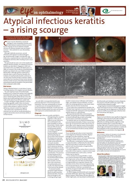
Fig 1. Slit lamp biomicroscopy images of atypical infectious keratitis showing (a) corneal epitheliopathy (arrows) in a patient with Acanthamoeba keratitis, (b) a dense<br />
stromal infiltrate in severe fungal keratitis, and (c) a focal stromal infiltrate (arrow) with intact overlying epithelium in a case of microsporidium keratitis.<br />
Fig 2. In vivo confocal microscopy images of the cornea showing (a) double walled cysts (arrows) in Acanthamoeba keratitis,<br />
(b) branching lines in fusarium keratitis and (c) diffuse fine hyper-reflective spots in microsporidium keratitis.<br />
Up until 2003, microsporidial keratitis was<br />
primarily observed in immunocompromised<br />
individuals. There was subsequently an increase<br />
in reported cases of microsporidia keratitis in<br />
immunocompetent individuals in South East Asia.<br />
Risk factors include contact lens wear and trauma<br />
with exposure to contaminated water or soil.<br />
Diagnosis<br />
Clinical signs alone are usually unreliable in<br />
distinguishing the causative<br />
organism. In the case of<br />
atypical corneal infections,<br />
this is attributed to the<br />
great variability in clinical<br />
presentation. Studies report<br />
that over 90% of cases of<br />
Acanthamoeba keratitis<br />
are initially misdiagnosed<br />
as viral, fungal or<br />
bacterial keratitis. Corneal<br />
epitheliopathy (fig 1a)<br />
occurs early in the course of<br />
the disease and may have<br />
a dendritiform appearance<br />
similar to that observed<br />
in herpetic keratitis.<br />
Subepithelial infiltrates may<br />
mimic adenoviral keratitis.<br />
Other presentations include<br />
ring-shaped or focal stromal<br />
infiltrates and corneal melt<br />
or perforation. The presence<br />
of radial keratoneuritis is<br />
virtually pathognomonic<br />
for Acanthamoeba keratitis,<br />
as it occurs rarely in<br />
other keratitis, but this<br />
sign is also uncommon<br />
in acanthamoeba and is<br />
usually only observed early<br />
in the course of the disease.<br />
The stromal keratitis<br />
caused by fungal infection<br />
(fig 1b) usually resembles<br />
bacterial keratitis. Features<br />
that are thought to aid<br />
in distinguishing fungal<br />
keratitis include stromal infiltrates with feathery<br />
edges, and satellite stromal infiltrates. In some<br />
cases, the overlying epithelium may remain intact<br />
despite extensive stromal involvement.<br />
In non-tuberculous mycobacterium keratitis, the<br />
infiltrates have may have an unusual, focal, waxy or<br />
“cracked windshield” appearance and may develop<br />
satellite lesions or a ring infiltrate.<br />
Microsporidial keratitis often mimics herpetic<br />
keratitis, presenting with multifocal epitheliopathy,<br />
or stromal infiltrates (fig 1c) with surrounding<br />
corneal oedema and keratic precipitates.<br />
For patients who are on empirical treatment for<br />
presumed bacterial keratitis, if there is not at least<br />
some sign of improvement within the first four to<br />
seven days, viral or atypical causes of the keratitis<br />
should be actively considered and the temptation<br />
to use corticosteroids should be actively avoided.<br />
Investigation<br />
Tissue sampling and culture remain imperative<br />
in the diagnosis of infectious keratitis. Atypical<br />
pathogens are often fastidious, requiring<br />
specialised culture systems and some cultures may<br />
take days to weeks to become positive.<br />
The difficulty in isolating the causative organism<br />
in atypical keratitis is reflected by the observation<br />
that only 30 to 40% of cultured cases among<br />
patients with Acanthamoeba or fungal keratitis<br />
have a positive culture.<br />
In culture negative cases where there is a lack of a<br />
favourable clinical response, a repeat corneal scrape<br />
is recommended and, in some cases, a corneal<br />
biopsy may be required.<br />
In vivo confocal microscopy (IVCM) is a rapid,<br />
non-invasive technique that enables imaging of<br />
the living human cornea at the cellular level. IVCM<br />
is a useful adjunctive tool when Acanthamoeba<br />
or fungal keratitis are suspected. However, the<br />
resolution limits of this instrument (approaching<br />
one micron) preclude its use in detecting bacterial<br />
or viral infections. IVCM has a sensitivity and<br />
specificity of approximately 90% for the detection<br />
of fungi or Acanthamoeba.<br />
On IVCM imaging, Acanthamoeba cysts may appear<br />
as double-walled cysts, signet rings, and bright spots<br />
(fig 2a). However, inflammatory cells also appear as<br />
similar bright spots, and may easily be confused with<br />
Acanthamoeba cysts leading to erroneous diagnosis.<br />
The presence of double-walled cysts, signet rings<br />
should therefore always be sought.<br />
Fungal hyphae characteristically appear as bright<br />
linear branching structures on IVCM images (fig 2b).<br />
Microsporidia may be diagnosed on IVCM by the<br />
presence of diffuse punctate hyper-reflective inter<br />
and intracellular dots (fig 2c).<br />
Conclusion<br />
Atypical corneal infections pose significant diagnostic<br />
challenges, particularly due to the wide variability<br />
in presentation, overlapping clinical signs, and<br />
difficulties in isolating causative organisms. Early<br />
detection of these cases is crucial and relies on<br />
having a high level of suspicion based on the history,<br />
clinical signs and response to treatment. In particular,<br />
the temptation to start corticosteroids should be<br />
avoided if there is uncertainty in the diagnosis. ▀<br />
References<br />
Patel DV, Rayner S, McGhee CN. Resurgence of Acanthamoeba<br />
keratitis in Auckland, New Zealand: a 7-year review<br />
of presentation and outcomes. Clin Exp Ophthalmol.<br />
2010;38(1):15-20<br />
Patel DV, McGhee CN. Acanthamoeba keratitis: a<br />
comprehensive photographic reference of common and<br />
uncommon signs. Clin Exp Ophthalmol. 2009;37(2):232-8<br />
Kheir WJ, Sheheitli H, Abdul Fattah M, Hamam RN.<br />
Nontuberculous Mycobacterial Ocular Infections: A Systematic<br />
Review of the Literature. Biomed Res Int. 2015;2015:164989.<br />
Garg P. Microsporidia infection of the cornea--a unique and<br />
challenging disease. Cornea. 2013 Nov;32 Suppl 1:S33-8.<br />
Garg P.Fungal, Mycobacterial, and Nocardia infections and the<br />
eye: an update. Eye (Lond). 2012 Feb;26(2):245-51.<br />
About the authors:<br />
* Associate professor Dipika Patel is based in the Department<br />
of Ophthalmology at the University of Auckland. Her research<br />
interests include anterior segment imaging and investigating<br />
potential therapeutic applications for corneal stem cells.<br />
* Professor Charles McGhee is head of the Department of<br />
Ophthalmology at the University of Auckland, and senior<br />
ophthalmic surgeon at Auckland City Hospital. Prof McGhee’s<br />
clinical interests<br />
include corneal<br />
diseases such as<br />
keratoconus, corneal<br />
dystrophies, corneal<br />
transplantation,<br />
cataract surgery and<br />
complex anterior<br />
A/Prof Patel<br />
Prof McGhee<br />
segment surgery<br />
following trauma.