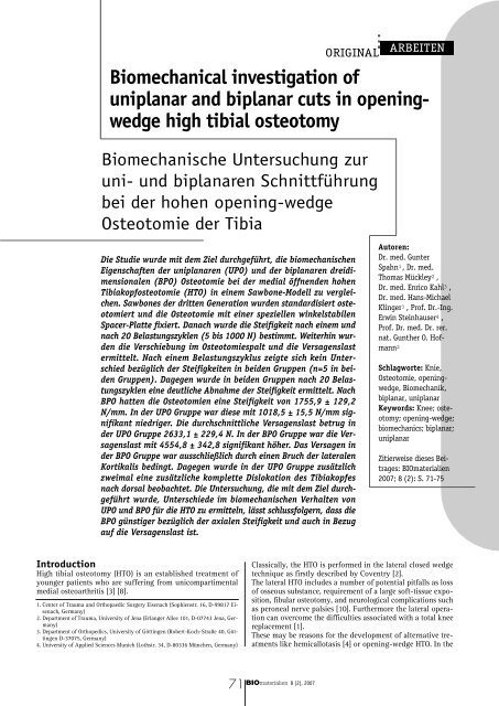
Biomechanical investigation of uniplanar and biplanar cuts in ...
Biomechanical investigation of uniplanar and biplanar cuts in ...
Biomechanical investigation of uniplanar and biplanar cuts in ...

You also want an ePaper? Increase the reach of your titles
YUMPU automatically turns print PDFs into web optimized ePapers that Google loves.
<strong>Biomechanical</strong> <strong><strong>in</strong>vestigation</strong> <strong>of</strong><br />
<strong>uniplanar</strong> <strong>and</strong> <strong>biplanar</strong> <strong>cuts</strong> <strong>in</strong> open<strong>in</strong>gwedge<br />
high tibial osteotomy<br />
Biomechanische Untersuchung zur<br />
uni- und <strong>biplanar</strong>en Schnittführung<br />
bei der hohen open<strong>in</strong>g-wedge<br />
Osteotomie der Tibia<br />
Die Studie wurde mit dem Ziel durchgeführt, die biomechanischen<br />
Eigenschaften der <strong>uniplanar</strong>en (UPO) und der <strong>biplanar</strong>en dreidimensionalen<br />
(BPO) Osteotomie bei der medial öffnenden hohen<br />
Tibiakopfosteotomie (HTO) <strong>in</strong> e<strong>in</strong>em Sawbone-Modell zu vergleichen.<br />
Sawbones der dritten Generation wurden st<strong>and</strong>ardisiert osteotomiert<br />
und die Osteotomie mit e<strong>in</strong>er speziellen w<strong>in</strong>kelstabilen<br />
Spacer-Platte fixiert. Danach wurde die Steifigkeit nach e<strong>in</strong>em und<br />
nach 20 Belastungszyklen (5 bis 1000 N) bestimmt. Weiterh<strong>in</strong> wurden<br />
die Verschiebung im Osteotomiespalt und die Versagenslast<br />
ermittelt. Nach e<strong>in</strong>em Belastungszyklus zeigte sich ke<strong>in</strong> Unterschied<br />
bezüglich der Steifigkeiten <strong>in</strong> beiden Gruppen (n=5 <strong>in</strong> beiden<br />
Gruppen). Dagegen wurde <strong>in</strong> beiden Gruppen nach 20 Belastungszyklen<br />
e<strong>in</strong>e deutliche Abnahme der Steifigkeit ermittelt. Nach<br />
BPO hatten die Osteotomien e<strong>in</strong>e Steifigkeit von 1755,9 ± 129,2<br />
N/mm. In der UPO Gruppe war diese mit 1018,5 ± 15,5 N/mm signifikant<br />
niedriger. Die durchschnittliche Versagenslast betrug <strong>in</strong><br />
der UPO Gruppe 2633,1 ± 229,4 N. In der BPO Gruppe war die Versagenslast<br />
mit 4554,8 ± 342,8 signifikant höher. Das Versagen <strong>in</strong><br />
der BPO Gruppe war ausschließlich durch e<strong>in</strong>en Bruch der lateralen<br />
Kortikalis bed<strong>in</strong>gt. Dagegen wurde <strong>in</strong> der UPO Gruppe zusätzlich<br />
zweimal e<strong>in</strong>e zusätzliche komplette Dislokation des Tibiakopfes<br />
nach dorsal beobachtet. Die Untersuchung, die mit dem Ziel durchgeführt<br />
wurde, Unterschiede im biomechanischen Verhalten von<br />
UPO und BPO für die HTO zu ermitteln, lässt schlussfolgern, dass die<br />
BPO günstiger bezüglich der axialen Steifigkeit und auch <strong>in</strong> Bezug<br />
auf die Versagenslast ist.<br />
Introduction<br />
High tibial osteotomy (HTO) is an established treatment <strong>of</strong><br />
younger patients who are suffer<strong>in</strong>g from unicompartimental<br />
medial osteoarthritis [3] [8].<br />
1. Center <strong>of</strong> Trauma <strong>and</strong> Orthopaedic Surgery Eisenach (Sophienstr. 16, D-99817 Eisenach,<br />
Germany)<br />
2. Department <strong>of</strong> Trauma, University <strong>of</strong> Jena (Erlanger Allee 101, D-07743 Jena, Germany)<br />
3. Department <strong>of</strong> Orthopedics, University <strong>of</strong> Gött<strong>in</strong>gen (Robert-Koch-Straße 40, Gött<strong>in</strong>gen<br />
D-37075, Germany)<br />
4. University <strong>of</strong> Applied Sciences Munich (Lothstr. 34, D-80336 München, Germany)<br />
ARBEITEN<br />
Autoren:<br />
Dr. med. Gunter<br />
Spahn 1, Dr. med.<br />
Thomas Mückley 2,<br />
Dr. med. Enrico Kahl 3,<br />
Dr. med. Hans-Michael<br />
Kl<strong>in</strong>ger 3,Pr<strong>of</strong>. Dr.-Ing.<br />
Erw<strong>in</strong> Ste<strong>in</strong>hauser 4,<br />
Pr<strong>of</strong>. Dr. med. Dr. rer.<br />
nat. Gunther O. H<strong>of</strong>mann<br />
2<br />
Schlagworte: Knie,<br />
Osteotomie, open<strong>in</strong>gwedge,<br />
Biomechanik,<br />
<strong>biplanar</strong>, <strong>uniplanar</strong><br />
Keywords: Knee; osteotomy;<br />
open<strong>in</strong>g-wedge;<br />
biomechanics; <strong>biplanar</strong>;<br />
<strong>uniplanar</strong><br />
Zitierweise dieses Beitrages:<br />
BIOmaterialien<br />
2007; 8 (2): S. 71-75<br />
Classically, the HTO is performed <strong>in</strong> the lateral closed wedge<br />
technique as firstly described by Coventry [2].<br />
The lateral HTO <strong>in</strong>cludes a number <strong>of</strong> potential pitfalls as loss<br />
<strong>of</strong> osseous substance, requirement <strong>of</strong> a large s<strong>of</strong>t-tissue exposition,<br />
fibular osteotomy, <strong>and</strong> neurological complications such<br />
as peroneal nerve palsies [10]. Furthermore the lateral operation<br />
can overcome the difficulties associated with a total knee<br />
replacement [1].<br />
These may be reasons for the development <strong>of</strong> alternative treatments<br />
like hemicallotasis [4] or open<strong>in</strong>g-wedge HTO. In the<br />
BIOmaterialien 8 (2), 2007<br />
71<br />
ORIGINAL
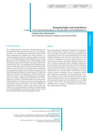
ORIGINAL ARBEITEN Gunter Spahn: <strong>Biomechanical</strong> <strong><strong>in</strong>vestigation</strong> <strong>of</strong> <strong>uniplanar</strong> <strong>cuts</strong> <strong>in</strong> open<strong>in</strong>g-wedge high tibial osteotomy<br />
Figure 1 Technique <strong>of</strong> transversal <strong>uniplanar</strong> (UPO) <strong>and</strong> <strong>biplanar</strong> (BPO) osteotomy.<br />
The osteotomy by us<strong>in</strong>g an oscillat<strong>in</strong>g saw started on the medial<br />
cortex 2.5 cm below the medial jo<strong>in</strong>t space.<br />
For transversal, <strong>uniplanar</strong> osteotomy (UPO) the cut started 4 cm below the<br />
medial jo<strong>in</strong>t l<strong>in</strong>e <strong>and</strong> was aimed at a po<strong>in</strong>t 3 cm below the lateral jo<strong>in</strong>t l<strong>in</strong>e<br />
(figure 1a).<br />
The dorsal cut <strong>of</strong> the <strong>biplanar</strong> osteotomy (BPO) also started 4 cm below the<br />
medial jo<strong>in</strong>t l<strong>in</strong>e <strong>and</strong> was directed to a po<strong>in</strong>t 3 cm below the lateral jo<strong>in</strong>t l<strong>in</strong>e.<br />
Only the posterior two thirds <strong>of</strong> the tibia were cut. The frontal third <strong>of</strong><br />
the tibia was leaved <strong>in</strong>tact. The anterior cut was aimed to a po<strong>in</strong>t 2 cm below<br />
the ventral jo<strong>in</strong>t l<strong>in</strong>e (figure 1b).<br />
last years the medial open<strong>in</strong>g-wedge HTO became very popular.<br />
This operation is able to avoid typical complications <strong>of</strong> lateral<br />
procedure <strong>and</strong> is relatively easy to perform. But also, the<br />
medial technique <strong>in</strong>cludes some potential pitfalls <strong>and</strong> specific<br />
complications [11].<br />
On the one h<strong>and</strong>, the success <strong>of</strong> the medial HTO depends on the<br />
quality <strong>of</strong> the <strong>in</strong>ternal fixation. The fixation is generally possible<br />
by conventional plates [16] with or without autologous bone<br />
grafts or bone substitutes [5]. Puddu created a spacer plate<br />
which is able to support the osteotomy gap dur<strong>in</strong>g osseous consolidation<br />
[15]. In the last year’s angle stable plates for <strong>in</strong>ternal<br />
fixation after HTO became popular [6]. In recent studies the biomechanical<br />
properties <strong>of</strong> different implants were evaluated [6].<br />
St<strong>of</strong>fel et al. [14] compared the biomechnical properties <strong>of</strong> a<br />
very massive implant (Tom<strong>of</strong>ix, Synthes) with a small implant<br />
(Puddu plate, Arthrex). The differences <strong>in</strong> dimension produced<br />
different mechanical behaviour [13]. Aga<strong>in</strong>st Spahn et al. found<br />
a superior biomechanical behavoir <strong>in</strong> <strong>in</strong>ternal fixation by plates<br />
which comb<strong>in</strong>e angle stability <strong>and</strong> spacers [12].<br />
On the other h<strong>and</strong>, the stability after HTO depends on the oste-<br />
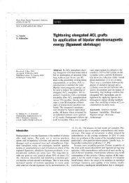
Figure 2 Angle stable <strong>and</strong> a mobile spacer conta<strong>in</strong><strong>in</strong>g plate for open<strong>in</strong>g wedge<br />
HTO (Varibale-w<strong>in</strong>kelstabile HTO Platte, Königsee-Implantate, Germany)<br />
otomy technique dur<strong>in</strong>g the operation. Classically, the osteotomy<br />
is performed as a transverse, s<strong>in</strong>gle cut [2]. The cut was<br />
aimed at the caudal third <strong>of</strong> the tibi<strong>of</strong>ibular jo<strong>in</strong>t [9]. Alternatively<br />
Lobenh<strong>of</strong>fer et al. published the results <strong>of</strong> 112 patients<br />
who were treated by a <strong>biplanar</strong> osteotomy technique [6] Biplanar<br />
osteotomy: <strong>in</strong> addition to the transverse osteotomy <strong>of</strong><br />
the posterior tibia a second ascend<strong>in</strong>g osteotomy <strong>in</strong> the coronary<br />
plane underneath the tibial tuberosity is performed. This<br />
shall improve rotational stability <strong>of</strong> the osteotomy <strong>and</strong> create<br />
an anterior buttress aga<strong>in</strong>st sagittal tilt<strong>in</strong>g <strong>of</strong> the osteotomy<br />
planes.<br />
This study was aimed to compare the biomechanical properties<br />
<strong>of</strong> <strong>uniplanar</strong> transversal with the <strong>biplanar</strong> cutt<strong>in</strong>g technique<br />
<strong>in</strong> HTO <strong>in</strong> a sawbone model.<br />
Materials <strong>and</strong> Methods<br />
Specimen preparation, HTO, <strong>and</strong> fixation<br />
The bone model consisted <strong>of</strong> Sawbones third-generation composite<br />
tibiae (No. 3302) (Sawbones Europe, Malmö, Sweden).<br />
The specimen were shortened to a length <strong>of</strong> 20 cm.<br />
The transversal, <strong>uniplanar</strong> osteotomy (UPO) the cut started 4<br />
cm below the medial jo<strong>in</strong>t l<strong>in</strong>e <strong>and</strong> was aimed to a po<strong>in</strong>t 3 cm<br />
below the lateral jo<strong>in</strong>t l<strong>in</strong>e (figure 1a).<br />
The dorsal cut <strong>of</strong> the <strong>biplanar</strong> osteotomy (BPO) also started 4<br />
cm below the medial jo<strong>in</strong>t l<strong>in</strong>e <strong>and</strong> was aimed to a po<strong>in</strong>t 3 cm<br />
below the lateral jo<strong>in</strong>t l<strong>in</strong>e. Only the posterior two thirds <strong>of</strong><br />
the tibia were cut. The frontal third <strong>of</strong> the tibia was leaved <strong>in</strong>tact.<br />
The anterior cut was aimed to a po<strong>in</strong>t 2 cm below the<br />
ventral jo<strong>in</strong>t l<strong>in</strong>e (figure 1b). A total <strong>of</strong> five specimens was<br />
prepared <strong>in</strong> each group.<br />
A HTO angle <strong>of</strong> 10° was used <strong>in</strong> all experiments.<br />
The osteotomies were fixed by an osteotomy plate which conta<strong>in</strong>ed<br />
a mobile spacer (Variabel-w<strong>in</strong>kelstabile HTO-Platte®,<br />
Königsee-Implantate, Germany). The implant is designed as<br />
angle stable <strong>and</strong> a mobile spacer conta<strong>in</strong><strong>in</strong>g plate (figure 2<br />
<strong>and</strong> 3).<br />
<strong>Biomechanical</strong> test<br />
The distal parts <strong>of</strong> the specimens were embedded with dental<br />
plaster (Sockeldent, Albaum, Lehrte, Germany) <strong>in</strong> a steel cyl<strong>in</strong>der<br />
(5 x 10 cm). The tests were performed <strong>in</strong> a mechanical<br />
test<strong>in</strong>g mach<strong>in</strong>e (Z010, Zwick Germany). The proximal stamp<br />
was a femoral part <strong>of</strong> an endoprosthesis as shown <strong>in</strong> figure 4.<br />
An even axial load <strong>of</strong> the specimens was possible by us<strong>in</strong>g<br />
this experimental design.<br />
Prelim<strong>in</strong>ary measurements with five native saw-bones had demonstrated<br />
a high reproducibility <strong>of</strong> this test<strong>in</strong>g design.<br />
In the first step, the specimens had undergone a preload <strong>of</strong> 50<br />
N for 3 m<strong>in</strong>utes. The distance <strong>of</strong> the dorsal part <strong>of</strong> the osteotomy<br />
gap was measured by a precision measurement <strong>in</strong>strument<br />
(Kraftfix, Schalkaldia GmbH, Schmalkalden, Germany).The<br />
device has a precision <strong>of</strong> 0.1 mm. The po<strong>in</strong>t for me-<br />
BIOmaterialien 8 (2), 2007<br />
72<br />
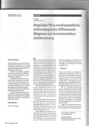
Figure 3<br />
Osteotomy specimen<br />
after <strong>in</strong>ternal fixation
Gunter Spahn: <strong>Biomechanical</strong> <strong><strong>in</strong>vestigation</strong> <strong>of</strong> <strong>uniplanar</strong> <strong>cuts</strong> <strong>in</strong> open<strong>in</strong>g-wedge high tibial osteotomy<br />
Figure 4<br />
<strong>Biomechanical</strong> test setup<br />
asurement was marked with a water pro<strong>of</strong> f<strong>in</strong>e l<strong>in</strong>er on the<br />
dorsal tibial edge.<br />
The load was <strong>in</strong>creased from 50 to 1000 N at a velocity <strong>of</strong> 5<br />
mm/s. After a precondition<strong>in</strong>g load cycle the test was stopped<br />
by a 1000 N load for 1 m<strong>in</strong>ute.<br />
Than a total <strong>of</strong> 20 cycles were performed. The load-displacement<br />
curves were registered for the 1st <strong>and</strong> <strong>in</strong> the 20th cycle.<br />
The mach<strong>in</strong>e was stopped aga<strong>in</strong> at a load <strong>of</strong> 1000 N for 1 m<strong>in</strong>ute<br />
to determ<strong>in</strong>e the distance <strong>of</strong> the osteotomy gap.<br />
F<strong>in</strong>ally the specimens were stressed by an <strong>in</strong>creas<strong>in</strong>g load to<br />
failure (5 mm/m<strong>in</strong>). After failure the load decreased rapidly.<br />
The tests were stopped by achiev<strong>in</strong>g 30% <strong>of</strong> the maximum load.<br />
The failure tests were recorded by a video tape. This had made<br />
it possible to register the cause <strong>of</strong> failure for every specimen.<br />
Statistics<br />
The s<strong>of</strong>tware program testXpert 10.0 (Zwick, Ulm, Germany)<br />
was used to evaluate the results <strong>of</strong> the mechanical tests.<br />
Statistical analyses were performed on a personal computer<br />
us<strong>in</strong>g SPSS version 13.0, SPSS Inc Chicago IL, USA. The Kolmogorov-Smirnov<br />
test <strong>and</strong> analysis <strong>of</strong> variance were used <strong>in</strong><br />
addition to the Mann-Whitney U-test <strong>and</strong> chi-square test for<br />
statistical analysis, with the level <strong>of</strong> significance set at p <<br />
0.05.<br />
Results<br />
Displacement <strong>and</strong> Stiffness<br />
The mean load-dependent displacement is shown <strong>in</strong> figure 5.<br />
All specimens showed a load-dependent displacement. After<br />
20 load<strong>in</strong>g cycles the displacement <strong>in</strong>creased <strong>in</strong> both groups.<br />
Stiffness was calculated for 1000 N axial load. The stiffness<br />
<strong>in</strong> BPO specimens after 1 cycle was 2102.8 ± 167.8 N/mm. After<br />
1 cycle UPO specimen had a significant m<strong>in</strong>or stiffness<br />
(1500.6 ± 121.0 N/mm) than BPO specimens.<br />
The undergo<strong>in</strong>g <strong>of</strong> 20 load<strong>in</strong>g cycles caused a significant loss<br />
<strong>of</strong> stiffness. After these 20 cycles BPO specimen had a stiffness<br />
<strong>of</strong> 1755.9 ± 129.2 N/mm. The stiffness <strong>in</strong> UPO specimen<br />
was 1018.5 ± 15.5 N/mm. The difference between the groups<br />
was significant.<br />
Displacement with<strong>in</strong> the Osteotomy Gap<br />
The osteotomy gap underwent a deformation dur<strong>in</strong>g axial load<strong>in</strong>g.<br />
This deformation depended on the number <strong>of</strong> load<strong>in</strong>g<br />
cycles, on the one h<strong>and</strong>. On the other h<strong>and</strong>, the osteotomy<br />
techniques also <strong>in</strong>fluence the displacement with<strong>in</strong> the osteotomy<br />
gap. After one load<strong>in</strong>g cycle no differences between<br />
BPO <strong>and</strong> UPO specimens were evaluated. After 20 cycles BPO<br />
specimen had a significant m<strong>in</strong>or deformation under an axial<br />
load <strong>of</strong> 1000 N (table 1).<br />
Failure Tests<br />
The mean maximum load at failure <strong>in</strong> UPO specimen was<br />
2633.1 ± 229.4 N. Always the failure was caused by an <strong>in</strong>fraction<br />
<strong>of</strong> the lateral cortex. Two times a dorsal dislocation <strong>of</strong><br />
the tibial head was registered.<br />
Specimens after BPO had a significant higher maximum load<br />
at failure <strong>of</strong> 4554.8 ± 342.8 N. Every time, the failure also was<br />
caused by an <strong>in</strong>fraction <strong>of</strong> the lateral cortex but no dislocation<br />
regard<strong>in</strong>g to the longitud<strong>in</strong>al axis <strong>of</strong> the tibia was observed.<br />
Discussion<br />
The success after medial open<strong>in</strong>g HTO <strong>in</strong> treatment <strong>of</strong> unicompartimental<br />
osteoarthritis ma<strong>in</strong>ly depends on a sufficient<br />
operation technique. This requires sufficient <strong>in</strong>ternal fixation<br />
<strong>in</strong> the first l<strong>in</strong>e. In the last years the results <strong>of</strong> numerous <strong><strong>in</strong>vestigation</strong>s<br />
for evaluat<strong>in</strong>g the biomechanical properties <strong>of</strong><br />
different <strong>in</strong>ternal fixations after open<strong>in</strong>g wedge HTO were published<br />
(Spahn et al., 2006c, Spahn <strong>and</strong> Wittig, 2002, St<strong>of</strong>fel et<br />
al., 2004c, Stuart et al., 1999). In these studies a relative high<br />
primary stability <strong>of</strong> various <strong>in</strong>ternal fixations was suggested.<br />
Another important biomechanical aspect <strong>in</strong> HTO is the direction<br />
<strong>and</strong> the design <strong>of</strong> the osteotomy. The osteotomy classically<br />
is performed as a transversal <strong>uniplanar</strong> cut which spares<br />
the lateral 1/4 <strong>of</strong> the tibia.<br />
Lobenh<strong>of</strong>fer et al. [6] [7] had hypothesized that a three-dimensional<br />
<strong>biplanar</strong> osteotomy produces a higher primary stable<br />
osteotomy than a <strong>uniplanar</strong> transversal (“classical”) osteotomy.<br />
This biomechanical study was undertaken to compare<br />
the UPO with the tri-dimensional BPO <strong>in</strong> a Sawbone osteotomy<br />
model.<br />
In the last years synthetic bone models e.g saw bones became<br />
important <strong>in</strong> biomechanical <strong><strong>in</strong>vestigation</strong>s. The synthetic specimens<br />
have identical dimensions <strong>and</strong> material properties. They<br />
are available <strong>in</strong> high numbers <strong>and</strong> there are no <strong>in</strong>dividual limitations<br />
given by age, sex, or osteoporosis.<br />
The ma<strong>in</strong> effect <strong>of</strong> the medial open<strong>in</strong>g wedge is the carefully<br />
widen<strong>in</strong>g <strong>of</strong> the elastic bone with<strong>in</strong> the osteotomy gap by chisels,<br />
aimed to spare an <strong>in</strong>tact lateral 1/4 <strong>of</strong> the tibial bone.<br />
Because the osteotomy specimens had identical dimensions<br />
<strong>and</strong> disposition <strong>of</strong> the saw<strong>in</strong>g <strong>cuts</strong>, the biomechanical tests<br />
BIOmaterialien 8 (2), 2007<br />
73<br />
ORIGINAL<br />
ARBEITEN<br />
Figure 5<br />
Mean load-displacement curves for the specimens had undergone a total <strong>of</strong><br />
20 load-relief-cycles. The power-dependent displacement was evaluated<br />
from the protocol <strong>of</strong> the test<strong>in</strong>g mach<strong>in</strong>e. The stiffness was calculated after<br />
one <strong>and</strong> after twenty cycles.
ORIGINAL ARBEITEN Gunter Spahn: <strong>Biomechanical</strong> <strong><strong>in</strong>vestigation</strong> <strong>of</strong> <strong>uniplanar</strong> <strong>cuts</strong> <strong>in</strong> open<strong>in</strong>g-wedge high tibial osteotomy<br />
Width the dorsal osteotomy gap [mm]<br />
under a load <strong>of</strong> 1000 N<br />
After 1 cycle After 20 cycles<br />
BPO 9.6±0.3 8.6±0.5<br />
UPO 9.6±0.1 7.8±0.4*<br />
Table 1: Load dependent displacement with<strong>in</strong> the osteotomy gap<br />
with an identically fixed osteotomy are sufficient for evaluation.<br />
The tibia undergoes a significant loss <strong>of</strong> stiffness after medial<br />
open<strong>in</strong>g HTO. This loss <strong>of</strong> stiffness correlates with the number<br />
<strong>of</strong> load<strong>in</strong>g cycles. In our tests the specimens were loaded with<strong>in</strong><br />
a range <strong>of</strong> 50 to 1000 N. This is accord<strong>in</strong>g to the load <strong>of</strong> 20<br />
gait cycles. Our results are conform<strong>in</strong>g to the results <strong>of</strong> other<br />
<strong>in</strong>vestigators. St<strong>of</strong>fel et al. [14] also found a significant reduction<br />
<strong>of</strong> axial stiffness. In Sawbone models fixed by a spacer<br />
conta<strong>in</strong><strong>in</strong>g a Puddu plate this reduction was 76% [15]. By<br />
usage <strong>of</strong> an angle-stable plate (Tom<strong>of</strong>ix) the loss <strong>of</strong> reduction<br />
only amounted 41% [6]. These experiments always were performed<br />
by an UPO. In our experiments, the loss <strong>of</strong> axial stiffness<br />
<strong>in</strong> UPO was 32.1%. This m<strong>in</strong>or loss <strong>of</strong> stiffness may be<br />
caused by a better biomechanical property <strong>of</strong> an angle-stable<br />
<strong>and</strong> spacer conta<strong>in</strong><strong>in</strong>g <strong>in</strong>ternal fixation. The BPO produced a<br />
significant m<strong>in</strong>or loss <strong>of</strong> axial stiffness (16.5 %). This correlated<br />
with a significant lesser displacement with<strong>in</strong> the osteotomy<br />
gap.<br />
F<strong>in</strong>ally, the ma<strong>in</strong> load <strong>of</strong> failure was significantly higher <strong>in</strong><br />
BPO specimen compared with UPO specimen. In BPO specimens<br />
the failure always was caused by an <strong>in</strong>fraction <strong>of</strong> the lateral<br />
cortex. This is the weak po<strong>in</strong>t <strong>of</strong> the system anyway. These<br />
dislocations have cl<strong>in</strong>ical relevance <strong>in</strong> vivo (Spahn, 2004b).<br />
A displacement <strong>of</strong> the tibial head after HTO can namely produce<br />
a catastrophically cl<strong>in</strong>ical outcome. Our results suggest<br />
that BPO widely prevents this dislocation dur<strong>in</strong>g axial load<strong>in</strong>g.<br />
Conclusions<br />
This study was undertaken to evaluate the biomechanical properties<br />
<strong>of</strong> a UPO <strong>and</strong> BPO <strong>in</strong> medial wedge osteotomy. The results<br />
<strong>of</strong>fer a significant superior axial stiffness under cyclic<br />
load<strong>in</strong>g as well as resistance aga<strong>in</strong>st load to failure <strong>in</strong> BPO<br />
specimens.<br />
Abstract<br />
This study was aimed to determ<strong>in</strong>e the biomechanical properties<br />
<strong>of</strong> <strong>uniplanar</strong> tranversial (UPO) <strong>and</strong> <strong>biplanar</strong>, three-dimensional<br />
(BPO) osteotomy <strong>in</strong> medial open<strong>in</strong>g wedge high tibial<br />
osteotomy (HTO) <strong>in</strong> a comparable Sawbone model.<br />
Third generation tibial Sawbones were osteotomized <strong>in</strong> a st<strong>and</strong>ardized<br />
manner <strong>and</strong> fixed with an angle-stable, spacer conta<strong>in</strong><strong>in</strong>g<br />
plate. Axial stiffness after one <strong>and</strong> twenty load<strong>in</strong>g cycles<br />
(range 50 to 1000 N) was registered as well as displacement<br />
with<strong>in</strong> the osteotomy gap <strong>and</strong> load at failure.<br />
After one load<strong>in</strong>g cycle no differences between BPO (n=5) <strong>and</strong><br />
UPO (n=5) specimens were evaluated. The undergo<strong>in</strong>g <strong>of</strong> 20<br />
load<strong>in</strong>g cycles caused a significant loss <strong>of</strong> stiffness. After the<br />
cycles BPO specimen had a stiffness <strong>of</strong> 1755.9 ± 129.2 N/mm.<br />
The stiffness <strong>in</strong> UPO specimen was 1018.5 ± 15.5 N/mm. The<br />
difference between the groups was significant.<br />
The mean maximum load at failure <strong>in</strong> UPO specimens was<br />
2633.1 ± 229.4 N. Specimens after BPO had a significant higher<br />
maximum load at failure <strong>of</strong> 4554.8 ± 342.8 N. In BPO specimens<br />
the failure always was caused by an <strong>in</strong>fraction <strong>in</strong> the lateral<br />
cortex, whereas <strong>in</strong> UPO the failure was caused <strong>in</strong> two-times<br />
by an additional dorsal dislocation <strong>of</strong> the tibial head.<br />
This study was undertaken to evaluate the biomechanical properties<br />
<strong>of</strong> a UPO <strong>and</strong> BPO <strong>in</strong> medial wedge osteotomy. The results<br />
<strong>of</strong>fer a significant advantage for the BPO regard<strong>in</strong>g to<br />
axial stiffness under cyclic load<strong>in</strong>g as well as resistance aga<strong>in</strong>st<br />
load to failure.<br />
References<br />
[1] Bergenudd H,Sahlstrom A,Sanzen L. Total knee arthroplasty<br />
after failed proximal tibial valgus osteotomy. J Arthroplasty<br />
1997; 12: 635-638.<br />
[2] Coventry MB. Osteotomy <strong>of</strong> the upper portion <strong>of</strong> the tibia<br />
for degenerative arthritis <strong>of</strong> the knee. J Bone Jo<strong>in</strong>t Surg Am<br />
1965; 47: 984-990.<br />
[3] Insall JN,Joseph DM,Msika C. High tibial osteotomy for<br />
varus gonarthrosis. A long-term follow-up study. J Bone Jo<strong>in</strong>t<br />
Surg Am 1984; 66: 1040-1048.<br />
[4] Kl<strong>in</strong>ger HM,Lorenz F,Harer T. Open wedge tibial osteotomy<br />
by hemicallotasis for medial compartment osteoarthritis.<br />
Arch Orthop Trauma Surg 2001; 121: 245-247.<br />
[5] Kosh<strong>in</strong>o T,Murase T,Takagi T,Saito T. New bone formation<br />
around porous hydroxyapatite wedge implanted <strong>in</strong> open<strong>in</strong>g<br />
wedge high tibial osteotomy <strong>in</strong> patients with osteoarthritis.<br />
BIOmaterialien 8 (2), 2007<br />
74<br />
Dr. med.<br />
Gunter Spahn<br />
Korrespondenzadresse:<br />
Center <strong>of</strong> Trauma <strong>and</strong><br />
Orthopaedic Surgery Eisenach<br />
Sophienstr. 16<br />
D-99817 Eisenach<br />
Phone +49-3691-73500<br />
Fax +49-3691-735011<br />
Mail spahn@pk-eisenach.de <strong>and</strong> spahn.esa@t-onl<strong>in</strong>e.de<br />
Akademischer Lebenslauf<br />
1980 – 1985 Mediz<strong>in</strong>studium an der<br />
Friedrich-Schiller-Universität Jena<br />
1985 Promotion<br />
1985 – 1986 Pflichtassistenz am Kl<strong>in</strong>ikum Gera<br />
1986 – 1988 Militärarzt <strong>in</strong> Spremberg<br />
1988 – 1994 Kl<strong>in</strong>ikum Gera: Kl<strong>in</strong>ik für Allgeme<strong>in</strong>-<br />
Visceral- und K<strong>in</strong>derchirurgie, Kl<strong>in</strong>ik<br />
für Unfallchirurgie<br />
1993 Facharzt für Chirurgie<br />
1994-1997 Chefarzt der Medical Center Praxiskl<strong>in</strong>ik<br />
Eisnach und ärztlicher Leiter des<br />
Reha Zentrums für Orthopädie und<br />
Traumatologie Eisenach<br />
seit 1997 Praxiskl<strong>in</strong>ik für Unfallchirurgie und<br />
Orthopädie <strong>in</strong> Eisenach<br />
Schwerpunkte: Knorpelschaden und Arthrose,<br />
Biomechanik, Implantatentwicklung
Gunter Spahn: <strong>Biomechanical</strong> <strong><strong>in</strong>vestigation</strong> <strong>of</strong> <strong>uniplanar</strong> <strong>cuts</strong> <strong>in</strong> open<strong>in</strong>g-wedge high tibial osteotomy<br />
Biomaterials 2001; 22: 1579-1582.<br />
[6] Lobenh<strong>of</strong>fer P,Agneskirchner J,Zoch W. [Open valgus<br />
alignment osteotomy <strong>of</strong> the proximal tibia with fixation by<br />
medial plate fixator]. Orthopade 2004; 33: 153-160.<br />
[7] Lobenh<strong>of</strong>fer P,Agneskirchner JD. Improvements <strong>in</strong> surgical<br />
technique <strong>of</strong> valgus high tibial osteotomy. Knee Surg<br />
Sports Traumatol Arthrosc 2003; 11: 132-138.<br />
[8] Naudie D,Bourne RB,Rorabeck CH,Bourne TJ. The Install<br />
Award. Survivorship <strong>of</strong> the high tibial valgus osteotomy. A<br />
10- to -22-year followup study. Cl<strong>in</strong> Orthop Relat Res 1999;<br />
18-27.<br />
[9] Silverton CG,Kentsch ARW,Müller W. Osteotomies about<br />
the knee. In: Callaghan JJ,Rosenberg AG,Rubash HE,Simonian<br />
PT,Wickiewicz TL, editors. The adult knee. Philadelphia, Baltimore,<br />
New York, London, Buenos Aires, Hong Kong, Sydney,<br />
Tokyo: Lip<strong>in</strong>cott Williams <strong>and</strong> Wilk<strong>in</strong>s 2003: 991-1016.<br />
[10] Slawski DP,Schoenecker PL,Rich MM. Peroneal nerve <strong>in</strong>jury<br />
as a complication <strong>of</strong> pediatric tibial osteotomies: a review<br />
<strong>of</strong> 255 osteotomies. J Pediatr Orthop 1994; 14: 166-172.<br />
[11] Spahn G. Complications <strong>in</strong> high tibial (medial open<strong>in</strong>g<br />
wedge) osteotomy. Arch Orthop Trauma Surg 2004; 124: 649-<br />
653.<br />
[12] Spahn G,Muckley T,Kahl E,H<strong>of</strong>mann GO. <strong>Biomechanical</strong><br />
<strong><strong>in</strong>vestigation</strong> <strong>of</strong> different <strong>in</strong>ternal fixations <strong>in</strong> medial open<strong>in</strong>gwedge<br />
high tibial osteotomy. Cl<strong>in</strong> Biomech (Bristol , Avon )<br />
2006; 21: 272-278.<br />
[13] Spahn G,Wittig R. Primary stability <strong>of</strong> various implants <strong>in</strong><br />
tibial open<strong>in</strong>g wedge osteotomy: a biomechanical study. J Orthop<br />
Sci 2002; 7: 683-687.<br />
[14] St<strong>of</strong>fel K,Stachowiak G,Kuster M. Open wedge high tibial<br />
osteotomy: biomechanical <strong><strong>in</strong>vestigation</strong> <strong>of</strong> the modified Arthrex<br />
Osteotomy Plate (Puddu Plate) <strong>and</strong> the TomoFix Plate.<br />
Cl<strong>in</strong> Biomech (Bristol , Avon ) 2004; 19: 944-950.<br />
[15] Stuart MJ,Beachy AM,Grabowski JJ,An KN,Kaufman KR.<br />
<strong>Biomechanical</strong> evaluation <strong>of</strong> a proximal tibial open<strong>in</strong>g-wedge<br />
osteotomy plate. Am J Knee Surg 1999; 12: 148-153.<br />
[16] W<strong>in</strong>ker KH,Weller S. [Extra-ligamentous valgisation additive<br />
tibial head osteotomy. Indications-technic-complications-errors.<br />
Critical analysis]. Z Orthop Ihre Grenzgeb 1990;<br />
128: 58-62.<br />
BIOmaterialien 8 (2), 2007<br />
75<br />
ORIGINAL<br />
ARBEITEN