news & views MAY 2013

Create successful ePaper yourself
Turn your PDF publications into a flip-book with our unique Google optimized e-Paper software.
ARTICLE<br />
African radiographers: How to bridge the gap?<br />
Boniface Yao, ISRRT Regional Coordinator Africa/Professional Practice<br />
Radiologic Technologist (diagnostic radiology and medical imaging), Institut National de Formation des Agents de Santé (INFAS) Cote d’Ivoire<br />
The discovery of ionizing radiations revealed radiography, the<br />
pathway to clinical settings. Diagnostic imaging is crucial in<br />
diagnosis of pathologies and several imaging technologies exist<br />
today: Conventional x-ray imaging, Ultrasound imaging, Computed<br />
tomography, Magnetic Resonance Imaging and Nuclear medicine.<br />
Moreover, the growth of modern imaging depends on advances in<br />
digital image technologies that contain more information, easier<br />
to handle and archive. However, in Africa, hundreds of hospitals<br />
and institutions do not have the possibilities to perform the most<br />
fundamental imaging procedures, for lack of equipment, malfunction,<br />
or even insufficient skills.<br />
Despite the recurrent shortage in their area, African Technologists<br />
keep on struggling to bridge the gap with the objective of seeking<br />
for alternatively affordable and easy to use means of practicing<br />
radiology.<br />
The general methodology of approach consists, upon analysis of<br />
examination procedure, to select equipments which offer a higher<br />
flexibility in transposition from manufactured design to homemade<br />
conception with local material. Then comprehensive prototypes are<br />
designed in purpose of local construction after quality and suitability<br />
evaluation.<br />
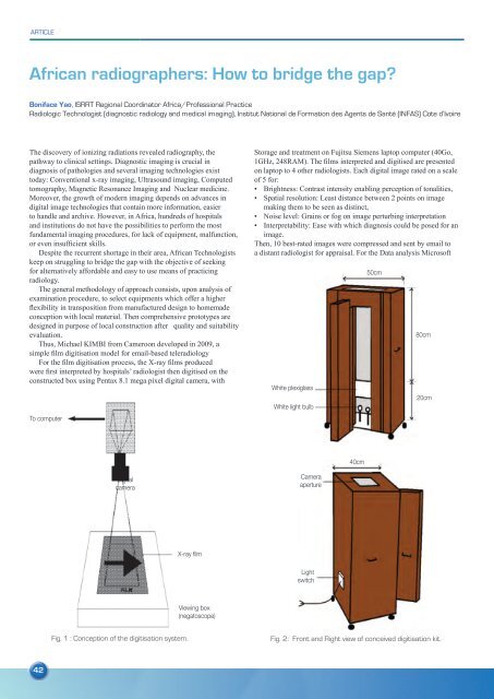
Thus, Michael KIMBI from Cameroon developed in 2009, a<br />
simple film digitisation model for email-based teleradiology<br />
For the film digitisation process, the X-ray films produced<br />
were first interpreted by hospitals’ radiologist then digitised on the<br />
constructed box using Pentax 8.1 mega pixel digital camera, with<br />
To computer<br />
Storage and treatment on Fujitsu Siemens laptop computer (40Go,<br />
1GHz, 248RAM). The films interpreted and digitised are presented<br />
on laptop to 4 other radiologists. Each digital image rated on a scale<br />
of 5 for:<br />
• Brightness: Contrast intensity enabling perception of tonalities,<br />
• Spatial resolution: Least distance between 2 points on image<br />
making them to be seen as distinct,<br />
• Noise level: Grains or fog on image perturbing interpretation<br />
• Interpretability: Ease with which diagnosis could be posed for an<br />
image.<br />
Then, 10 best-rated images were compressed and sent by email to<br />
a distant radiologist for appraisal. For the Data analysis Microsoft<br />
White plexiglass<br />
White light bulb<br />
50cm<br />
80cm<br />
20cm<br />
40cm<br />
Digital<br />
camera<br />
Camera<br />
aperture<br />
X-ray film<br />
Light<br />
switch<br />
Viewing box<br />
(negatoscope)<br />
Fig. 1 : Conception of the digitisation system.<br />
Fig. 2: Front and Right view of conceived digitisation kit.<br />
42