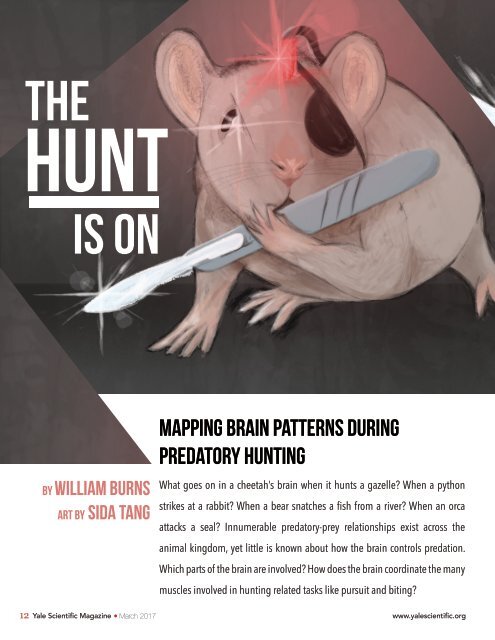
the hunt is on by William Burns art by Sida Tang MAPPING BRAIN PATTERNS DURING PREDATORY HUNTING What goes on in a cheetah’s brain when it hunts a gazelle? When a python strikes at a rabbit? When a bear snatches a fish from a river? When an orca attacks a seal? Innumerable predatory-prey relationships exist across the animal kingdom, yet little is known about how the brain controls predation. Which parts of the brain are involved? How does the brain coordinate the many muscles involved in hunting related tasks like pursuit and biting? 12 Yale Scientific Magazine March 2017 www.yalescientific.org
neuroscience FOCUS Researchers at the Yale School of Medicine, led by Ivan de Araujo, associate professor of Psychiatry, are looking for answers. The team of scientists identified a sub-region of the amygdala, an olive-shaped region of the brain, as the epicenter for predatory hunting. The amygdala has long been recognized as a contributor to emotions like fear and aggression, but these researchers dug deeper to understand the molecular pathways which drive predatory instincts. Shining flashes of laser light into the mice’s brains, the researchers could turn on or off the neurons that fire when mice hunt. Accordingly, the researchers could transform the mice at will from passive creatures to gluttonous predators. Through their findings, the team of scientists uncovered clues relating to the evolution of the brain. Firing the Neurons Previous studies showed that when rats hunt insects, the central amygdala (CeA), an almond-shaped sub-region of the amygdala, surges in activity. This led the researchers at Yale to question how the CeA of predators’ brains coordinate the plethora of muscles involved with hunting. “Predatory behavior is a particularly complex task because an animal needs a number of different muscles to run, jump on things, and use its head to kill,” De Araujo said. First, the researchers injected the central amygdala of the mice with a virus that contained a gene encoding a light-sensitive ion channel. Ion channels allow positively charged sodium ions to flow into the negatively charged axon of the neuron. The change in charge distribution along the length of the axon, called depolarization, causes the neuron to fire. Thus, the virus made neurons in the CeA of the mice more prone to firing. The signal traveled through the body until it reached certain target muscles, such as the jaw and neck muscles. To control when the neurons fired, the researchers inserted an optrode, an optic fiber cable connected to an electrode. Through this cable, the researchers could send a specific wavelength of laser light into the mice’s brains to turn on the neurons involved when a mouse hunts. This technique, called optogenetics, allowed the researchers to use the energy from the laser to manually stimulate the parts of brains in mice which control hunting and biting. “Optogenetics has two great advantages. First, you can target and study a specific group of neurons. Second, the control over when the neurons fire is specific. The technique is very transient and very fast,” said Wenfei Han, first author of the paper. Optogenetic activation triggered only the neurons injected with the virus to fire more intensely according to electromyogram (EMG) recordings, which measure electrical activity in muscles. Initiating the Hunt When the CeA was stimulated using optogenetics, the normally indifferent, docile mice transformed into bestial predators. For example, when a cricket was placed in the cage, the mice captured and killed their prey in a much shorter time than they did when the CeA was not stimulated. The mice attacked even inanimate objects; when a bottle cap was placed in front of them, the mice grasped and bit the cap as if it were prey. Such attacks were not observed when the laser was off. Upon laser stimulation, the mice attacked the closest object they could find. When a food pellet was placed in the opposite corner of the cage and the laser was turned on, some mice started to eat the cotton bed on which they were resting before stimulation. In some cases, the mice would grab onto the laser cable itself and chew straight through it. Unsurprisingly, when the cable split, the mice ceased hunting and returned to their previous docility. Even when no food was placed in the cage, the CeA-stimulated mice positioned themselves in an eccentric feeding position, tensing their hind legs and holding their front legs to their mouths as if they were eating. The researchers called this phenomenon “fictive feeding,” as it reflected the feeding www.yalescientific.org