YSM Issue 90.2

You also want an ePaper? Increase the reach of your titles
YUMPU automatically turns print PDFs into web optimized ePapers that Google loves.
neuroscience<br />
FOCUS<br />
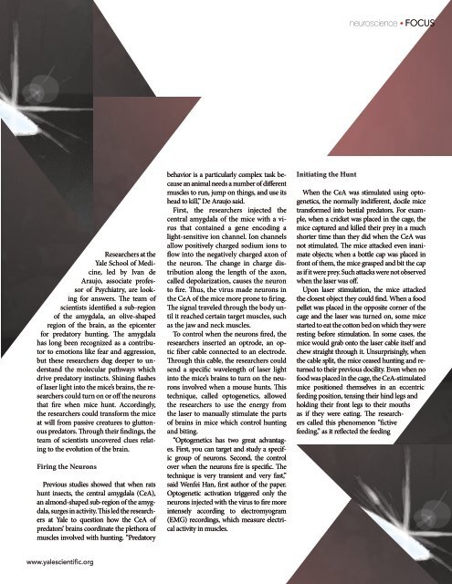
Researchers at the<br />
Yale School of Medicine,<br />
led by Ivan de<br />
Araujo, associate professor<br />
of Psychiatry, are looking<br />
for answers. The team of<br />
scientists identified a sub-region<br />
of the amygdala, an olive-shaped<br />
region of the brain, as the epicenter<br />
for predatory hunting. The amygdala<br />
has long been recognized as a contributor<br />
to emotions like fear and aggression,<br />
but these researchers dug deeper to understand<br />
the molecular pathways which<br />
drive predatory instincts. Shining flashes<br />
of laser light into the mice’s brains, the researchers<br />
could turn on or off the neurons<br />
that fire when mice hunt. Accordingly,<br />
the researchers could transform the mice<br />
at will from passive creatures to gluttonous<br />
predators. Through their findings, the<br />
team of scientists uncovered clues relating<br />
to the evolution of the brain.<br />
Firing the Neurons<br />
Previous studies showed that when rats<br />
hunt insects, the central amygdala (CeA),<br />
an almond-shaped sub-region of the amygdala,<br />
surges in activity. This led the researchers<br />
at Yale to question how the CeA of<br />
predators’ brains coordinate the plethora of<br />
muscles involved with hunting. “Predatory<br />
behavior is a particularly complex task because<br />
an animal needs a number of different<br />
muscles to run, jump on things, and use its<br />
head to kill,” De Araujo said.<br />
First, the researchers injected the<br />
central amygdala of the mice with a virus<br />
that contained a gene encoding a<br />
light-sensitive ion channel. Ion channels<br />
allow positively charged sodium ions to<br />
flow into the negatively charged axon of<br />
the neuron. The change in charge distribution<br />
along the length of the axon,<br />
called depolarization, causes the neuron<br />
to fire. Thus, the virus made neurons in<br />
the CeA of the mice more prone to firing.<br />
The signal traveled through the body until<br />
it reached certain target muscles, such<br />
as the jaw and neck muscles.<br />
To control when the neurons fired, the<br />
researchers inserted an optrode, an optic<br />
fiber cable connected to an electrode.<br />
Through this cable, the researchers could<br />
send a specific wavelength of laser light<br />
into the mice’s brains to turn on the neurons<br />
involved when a mouse hunts. This<br />
technique, called optogenetics, allowed<br />
the researchers to use the energy from<br />
the laser to manually stimulate the parts<br />
of brains in mice which control hunting<br />
and biting.<br />
“Optogenetics has two great advantages.<br />
First, you can target and study a specific<br />
group of neurons. Second, the control<br />
over when the neurons fire is specific. The<br />
technique is very transient and very fast,”<br />
said Wenfei Han, first author of the paper.<br />
Optogenetic activation triggered only the<br />
neurons injected with the virus to fire more<br />
intensely according to electromyogram<br />
(EMG) recordings, which measure electrical<br />
activity in muscles.<br />
Initiating the Hunt<br />
When the CeA was stimulated using optogenetics,<br />
the normally indifferent, docile mice<br />
transformed into bestial predators. For example,<br />
when a cricket was placed in the cage, the<br />
mice captured and killed their prey in a much<br />
shorter time than they did when the CeA was<br />
not stimulated. The mice attacked even inanimate<br />
objects; when a bottle cap was placed in<br />
front of them, the mice grasped and bit the cap<br />
as if it were prey. Such attacks were not observed<br />
when the laser was off.<br />
Upon laser stimulation, the mice attacked<br />
the closest object they could find. When a food<br />
pellet was placed in the opposite corner of the<br />
cage and the laser was turned on, some mice<br />
started to eat the cotton bed on which they were<br />
resting before stimulation. In some cases, the<br />
mice would grab onto the laser cable itself and<br />
chew straight through it. Unsurprisingly, when<br />
the cable split, the mice ceased hunting and returned<br />
to their previous docility. Even when no<br />
food was placed in the cage, the CeA-stimulated<br />
mice positioned themselves in an eccentric<br />
feeding position, tensing their hind legs and<br />
holding their front legs to their mouths<br />
as if they were eating. The researchers<br />
called this phenomenon “fictive<br />
feeding,” as it reflected the feeding<br />
www.yalescientific.org