YSM Issue 91.1

You also want an ePaper? Increase the reach of your titles
YUMPU automatically turns print PDFs into web optimized ePapers that Google loves.
Yale Scientific<br />
Established in 1894<br />
THE NATION’S OLDEST COLLEGE SCIENCE PUBLICATION<br />
MARCH 2018 VOL. 91 NO. 1 | $6.99<br />
SNEAKING<br />
ORGANS<br />
PAST THE<br />
IMMUNE SYSTEM<br />
15<br />
DELIVERING<br />
18<br />
A TALE OF ICE<br />
20<br />
BRAVING<br />
THE INHIBITOR AND SNOW<br />
THE COLD
Yale Scientific Magazine<br />
VOL. 90 ISSUE NO. 5<br />
CONTENTS<br />
MARCH 2018<br />
NEWS 6<br />
FEATURES 25<br />
ON THE COVER<br />
12 SNEAKING<br />
ORGANS PAST THE<br />
IMMUNE SYSTEM<br />
Current research done by Mark Saltzman<br />
and Jordan Pober combines<br />
techniques from multiple fields—<br />
nanoparticle delivery and transplant<br />
techniques—to improve long-term<br />
results after transplant surgery.<br />
15<br />
DELIVERING THE<br />
INHIBITOR<br />
Cancer, diabetes, and many other<br />
diseases involve dysregulation of<br />
the same signaling pathway critical<br />
for regulating protein function.<br />
Researchers have developed a<br />
drug delivery system facilitating the<br />
inhibition of proteins implicated in<br />
the disruption of this pathway.<br />
18<br />
A TALE OF ICE AND<br />
SNOW<br />
Why do clouds behave the way they<br />
do, sometimes producing snow and<br />
ice and rain? Yale professor Amir Haji-Akbari<br />
investigates these processes<br />
using computational techniques.<br />
20<br />
BRAVING THE<br />
COLD<br />
Yale researchers identify a genetic<br />
adaptation in certain species of<br />
rodents that reduces their sensitivity<br />
to the cold, allowing them to hibernate<br />
in temperatures just above<br />
freezing.<br />
22 DIVERGENCE<br />
What makes the human brain so<br />
unique? Although it is around three<br />
times larger than the brains of our<br />
closest living relatives, the complexity<br />
in the connections between cells and<br />
the differences between cells themselves<br />
hint at a deeper explanation.<br />
www.yalescientific.org<br />
More articles available online at www.yalescientific.org<br />
March 2018<br />
Yale Scientific Magazine<br />
3
Q&A<br />
HOW COLD CAN WATER GET?<br />
By Alice Tao<br />
Way colder than you’d think! In fact, water<br />
doesn’t always freeze when the temperature<br />
reaches zero degrees Celsius, the<br />
value regularly cited as the freezing point<br />
of water. Under certain conditions, water<br />
can undergo “supercooling” and exist in<br />
a liquid state far below its usual freezing<br />
point—at temperatures as low as -42.6 degrees<br />
Celsius.<br />
Previously, researchers encountered difficulties<br />
determining the lowest possible<br />
temperature of liquid water due to the rapid<br />
rate of ice crystal formation. In Germany,<br />
Robert Grisenti at the GSI Helmholtz Centre<br />
for Heavy Ion Research is developing<br />
new techniques for accurate measurement<br />
of the temperature of supercooled water<br />
with greater precision than ever before.<br />
Grisenti’s team began by spraying microscopic<br />
droplets of water into a vacuum. The<br />
exceptionally low pressure in the vacuum<br />
DO PENGUINS SNACK?<br />
By Hannah Verma<br />
Male emperor penguins have long<br />
been thought to make the ultimate sacrifice:<br />
fasting for almost three months<br />
as they guard their eggs. As it turns out,<br />
however, the penguins are not quite as<br />
selfless as they’ve previously been portrayed.<br />
Researchers from the Scripps<br />
Institution of Oceanography discovered<br />
that male emperor penguins sometimes<br />
break their fast during the incubation<br />
period.<br />
Typically, females leave the egg with<br />
the males for approximately for three or<br />
four months; the male’s feet keep the egg<br />
warm by cradling it with a fleshy layer<br />
of skin. Scientists have long thought<br />
that the male penguins stay by the<br />
egg’s side at all times during this period.<br />
They often spend these bone-chilling<br />
Arctic months sleeping to preserve<br />
their energy, but they still lose up to<br />
IMAGE COURTESY OF PEXELS<br />
Grisenti’s team supercooled water to -42.6 degrees<br />
Celsius.<br />
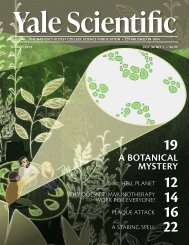
IMAGE COURTESY OF FLICKR<br />
Two emperor penguins stand protectively over a young<br />
chick.<br />
causes fast evaporative cooling, where<br />
evaporation cools the tiny water droplets<br />
much faster than ice can form. With<br />
the understanding that droplet size is<br />
proportionally related to temperature,<br />
the researchers determined the droplets’<br />
temperature by measuring their size<br />
with a laser that had 10-nanometer precision.<br />
They calculated a record low for<br />
liquid water, -42.6 degrees Celsius.<br />
Supercooled water and its transformation<br />
into atmospheric ice occur naturally<br />
in the Earth’s upper atmosphere.<br />
“Atmospheric climate models need an<br />
accurate representation of such processes<br />
for a realistic description of cloud formation<br />
and precipitation,” Grisenti said.<br />
The hope is that this research can ultimately<br />
provide leading climate scientists<br />
with better insight for developing more<br />
reliable climate-predicting models.<br />
half of their body weight. It is such a<br />
challenging task that some emperor<br />
penguins do not survive the winter.<br />
Other males, however, have other<br />
ideas. After tracking several emperor<br />
penguins, the researchers observed<br />
that these males snuck off<br />
in the middle of the night to hunt<br />
for fish in the open water. This phenomenon<br />
is thought to be more<br />
common among penguin colonies<br />
based near the southern coast,<br />
where the journey to the ocean is<br />
much shorter. These “early feedings”<br />
are significant because they<br />
increase the male’s chance of survival<br />
in the winter. By snacking, the<br />
male makes it far more likely that<br />
he will successfully incubate the egg<br />
and ensures the chick’s chance at<br />
life in the coming months.
Science belongs to everyone.<br />
From doctors and biomedical engineers to astronomers and ecologists, science forms the<br />
roots from which we grow. With science, we explore our planet and beyond, from modeling<br />
the mysteries of the clouds in our sky (pg. 18) to imaging the galaxies that exist beyond the<br />
scope of our sight (pg. 30). We attempt to describe the life that surrounds us, including bird<br />
feather patterns (pg. 9), dog behavior (pg. 11), and butterfly evolution (pg. 34). We push the<br />
limits of science to create new technologies and innovations such as quantum computers<br />
(pg. 6), infrared imaging satellites (pg. 7), and dandelion lab tools (pg. 35).<br />
But perhaps most importantly, science contributes to people. From exploring depression<br />
(pg. 8), to inspiring young researchers (pg. 36), to challenging diabetes (pg. 32) and<br />
attacking HIV (pg. 10), science drives our emotional, mental, and physical progress. Our<br />
lives are supported and shaped by the scope of our scientific understanding and thus we<br />
are defined by science and science is defined by us. This now brings us to you, the readers,<br />
and the people that we hope to inspire.<br />
Our cover article this issue gives hope towards a future of both improved organ transplant<br />
outcomes and of increased transplant organ viability and thus availability (pg. 12).<br />
This future is an encapsulation of the discoveries that mark years of progress in many<br />
fields of research, where each breakthrough is built on the previous. But what are we<br />
building towards?<br />
The interconnected nature of science creates a pattern of progress that cannot begin to<br />
be described as uniform. A step in one direction may lead to many in another, and thus the<br />
nonlinearity of scientific discovery becomes increasingly evident. It is our responsibility<br />
as scientific journalists to explore the direction of each scientific path and ultimately map<br />
these fields of discovery. We attempt to guide you through what seems to be a labyrinth of<br />
research and progress. We aim to understand science and share this understanding.<br />
With our new 2018 masthead comes a new era of writing, editing, and design. We maintain<br />
the same scientific excitement and curiosity that our predecessors celebrated and<br />
shared with us. We cherish your continuing support and your high expectations, both of<br />
which serve as inspirations for future improvement. I asked earlier what we are building<br />
towards; the reality may be that we approach nothing in particular. Rather, we build towards<br />
everything. Just as the universe expands, science reaches in all directions. It is driven<br />
by the passions of people and thus represents the ideas of all. Science is shared and we are<br />
lucky to be the ones to share it with you.<br />
Yale Scientific<br />
Established in 1894<br />
THE NATION’S OLDEST COLLEGE SCIENCE PUBLICATION<br />
MARCH 2018 VOL. 91 NO. 1 | $6.99<br />
SNEAKING<br />
ORGANS<br />
PAST THE<br />
IMMUNE SYSTEM<br />
DELIVERING<br />
A TALE OF ICE BRAVING<br />
15THE INHIBITOR 18 20<br />
AND SNOW<br />
THE COLD<br />
F R O M T H E E D I T O R<br />
Sharing Science<br />
A B O U T T H E A R T<br />
Eileen Norris<br />
Editor-in-Chief<br />
I’m so excited to serve as arts editor this upcoming<br />
year with <strong>YSM</strong>’s amazing board! For my<br />
first cover piece, I wanted to capture the magnitude<br />
of a new breakthrough in organ transplant<br />
technology. I illustrated a surgeon’s hands<br />
cradling a precious kidney, an organ that tens<br />
of thousands of Americans are in desperate<br />
need of, that has been treated by a nanoparticle<br />
drug delivery system represented in glowing<br />
green. By decreasing the frequency of organ<br />
rejections, more patients will receive their vital<br />
organs more quickly-making this a wonderful,<br />
revolutionary development in medicine.<br />
Editor-in-Chief<br />
Managing Editors<br />
News Editor<br />
Features Editor<br />
Articles Editor<br />
Online Editor<br />
Copy Editors<br />
Yale Scientific<br />
M A G A Z I N E<br />
Established in 1894<br />
MARCH 2018 VOL. 91 NO. 1<br />
Production Manager<br />
Layout Editor<br />
Art Editor<br />
Photography Editor<br />
Outreach Designer<br />
Webmaster<br />
Publisher<br />
Operations Manager<br />
Advertising Manager<br />
Synapse President<br />
Synapse Vice President<br />
Outreach Coordinators<br />
Social Media Coordinator<br />
Staff<br />
Victoria Dombrowik<br />
Alice Li<br />
Tiger Zhang<br />
Erica Lin<br />
Sunnie Liu<br />
Miriam Ross<br />
Grace Niewijk<br />
Sonia Wang<br />
Mindy Le<br />
Anna Sun<br />
Liz Ruddy<br />
Christine Xu<br />
Alice Tao<br />
Hannah Verma<br />
Megha Chawla<br />
Vikram Shaw<br />
Lisa Wu<br />
Jason Yang<br />
Advisory Board<br />
Priyamvada Natarajan<br />
Sandy Chang<br />
Kurt Zilm, Chair<br />
Fred Volkmar<br />
Stanley Eisenstat<br />
James Duncan<br />
Stephen Stearns<br />
Jakub Szefer<br />
Werner Wolf<br />
John Wettlaufer<br />
William Summers<br />
Scott Strobel<br />
Robert Bazell<br />
Craig Crews<br />
Ayaska Fernando<br />
Robert Cordova<br />
Annie Yang<br />
Jau Tung Chan<br />
Antonio Medina<br />
Isaac Wendler<br />
Leslie Sim<br />
Marcus Sak<br />
Urmila Chadayammuri<br />
Emma Healy<br />
Grace Chen<br />
Diyu Pearce-Fisher<br />
Matthew Kegley<br />
Hannah Geller<br />
Lauren Telesz<br />
Allie Olson<br />
Sandra Li<br />
Yulan Zhang<br />
Theo Kuhn<br />
Joshua Perez-Cruet<br />
Eileen Norris<br />
Diane Rafizadeh<br />
Stephanie Smelyansky<br />
Will Burns<br />
Charlie Musoff<br />
Allie Forman<br />
Conor Johnson<br />
Marcus Sak<br />
Joshua Matthew<br />
Sunnie Liu<br />
Kelly Zhou<br />
Ivory Fu<br />
Eric Wang<br />
Laurie Wang<br />
Alice Wu<br />
Kevin Chang<br />
Jiyoung Kang<br />
Allie Olson<br />
Jessica Trinh<br />
Nasser Odetallah<br />
Seth Anderson<br />
Lisa Wu<br />
Leslie Sim<br />
Lukas Corey<br />
Lauren J Kim<br />
Sonia Wang<br />
Fangchen Zhu<br />
Vikram Shaw<br />
Jason Yang<br />
Emma Healy<br />
Emma Wilson<br />
Anusha Bishop<br />
Lauren Gatta<br />
Sunnie Liu<br />
Elissa Johnson<br />
Astronomy<br />
Biological and Biomedical Sciences<br />
Chemistry<br />
Child Study Center<br />
Computer Science<br />
Diagnostic Radiology<br />
Ecology & Evolutionary Biology<br />
Electrical Engineering<br />
Emeritus<br />
Geology & Geophysics<br />
History of Science, Medicine, & Public Health<br />
Molecular Biophysics & Biochemistry<br />
Molecular, Cellular, & Developmental Biology<br />
Molecular, Cellular, & Developmental Biology<br />
Undergraduate Admissions<br />
Yale Science & Engineering Association<br />
The Yale Scientific Magazine (<strong>YSM</strong>) is published four times a year by<br />
Yale Scientific Publications, Inc. Third class postage paid in New Haven,<br />
CT 06520. Non-profit postage permit number 01106 paid for May 19,<br />
1927 under the act of August 1912. ISN:0091-287. We reserve the right<br />
to edit any submissions, solicited or unsolicited, for publication. This<br />
magazine is published by Yale College students, and Yale University<br />
is not responsible for its contents. Perspectives expressed by authors<br />
do not necessarily reflect the opinions of <strong>YSM</strong>. We retain the right to<br />
reprint contributions, both text and graphics, in future issues as well as<br />
a non-exclusive right to reproduce these in electronic form. The <strong>YSM</strong><br />
welcomes comments and feedback. Letters to the editor should be under<br />
200 words and should include the author’s name and contact information.<br />
We reserve the right to edit letters before publication. Please send<br />
questions and comments to ysm@yale.edu. Special thanks to Yale STC.
NEWS<br />
in brief<br />
THE ROLE OF ERROR IN QUANTUM<br />
COMPUTING<br />
By Victoria Dombrowik<br />
COURTESY OF QUATRONICS LABORATORY<br />
An image of a mother board, the most<br />
important piece of a binary computer.<br />
The future of information technology may<br />
be in the qubit. The term, a combination of<br />
quantum and bit, is used to refer to an electronic<br />
circuit that functions as the basis of<br />
quantum computing. It relies on the principle<br />
of superposition, which holds that a physical<br />
system may exist simultaneously in two different<br />
states. While the computer bits of today<br />
can store information in either a 0 or 1<br />
state, qubits are capable of storing a 0 and 1 at<br />
the same time, expanding the ability to cache<br />
data exponentially. Such a device could function<br />
with unimaginable speed and precision,<br />
making considerable advancements in fields<br />
like medicine and cyber security.<br />
The next step is to build a quantum computer<br />
that is capable of using qubits. The<br />
foremost problem facing developers today is<br />
the extreme sensitivity of quantum systems.<br />
Even a slight interaction of the qubit with<br />
the surroundings could lead to decoherence,<br />
or a slow collapse of the quantum mechanical<br />
properties of the system, which would<br />
in turn lead to errors in calculations. Michel<br />
Devoret, the F. W. Beinecke Professor of Applied<br />
Physics at Yale University, and his team<br />
at the Quantronics Laboratory are investigating<br />
a method to improve coherence through<br />
the use of quantum error correction, which<br />
protects the integrity of the qubit. Quantum<br />
error correction does this correcting for both<br />
decoherence and quantum noise, which is the<br />
uncertainty in the original quantum system.<br />
They believe that mastering this technique<br />
will lead to the design of systems that remain<br />
coherent indefinitely. Devoret was also optimistic<br />
about the future of the quantum computer.<br />
“There is no roadblock, no physics that<br />
would prevent it. If it is possible, then humans<br />
will find a way to create it.”<br />
TRIGGERING THE RESPONSE<br />
By Alice Li<br />
PHOTOGRAPHY BY WIKIMEDIA COMMONS<br />
An image of a healthy human T-cell.<br />
You probably had a sore deltoid muscle<br />
after your flu vaccine this year. This is because<br />
this standard shot is delivered straight into<br />
muscle. But there’s another problem besides<br />
you getting a sore shoulder: by injecting the<br />
vaccine into the muscle, the vaccine bypasses<br />
most of the potent cells needed to initiate<br />
an immune response, meaning high vaccine<br />
doses must be given to stimulate immunity.<br />
In an epidemic, however, there might not be<br />
enough vaccine to immunize everyone with<br />
the high doses needed for muscle injection.<br />
To find a better injection site, a team<br />
of scientists led by professor Stephanie<br />
Eisenbarth from the Yale School of Medicine<br />
investigated immune cells responsible for<br />
generating an effective vaccine response.<br />
The team knew that antigens in the vaccine<br />
activate immune cells called T-follicular<br />
helper cells (Tfh), which in turn enable a<br />
second type of immune cell, the B-cells, to<br />
make antibodies. However, the scientists<br />
found that another type of immune cell,<br />
called Type 2 dendritic cell, recognizes the<br />
antigens from vaccines and presents them to<br />
Tfh cells, which then activate B-cells in an<br />
immune cell relay race.<br />
Vaccinating into muscle misses most of the<br />
dendritic cells needed to initiate the response<br />
chain. Instead, the findings, published in<br />
Science Immunology, suggest a new injection<br />
site for your next flu shot: the skin. “If we<br />
deliver the vaccine through the skin, then<br />
we are presenting the vaccine directly to the<br />
dendritic cells,” said Eisenbarth. This means<br />
that existing vaccine stores less likely to run<br />
out in the case of an epidemic, and that the<br />
vaccine can be more widely available.<br />
This new delivery method creates a more<br />
effective immune response and requires<br />
only one-tenth of the original dose. While<br />
injecting into the skin is more painful, the<br />
10-fold increase in efficiency outweighs the<br />
additional discomfort.<br />
6 Yale Scientific Magazine March 2018 www.yalescientific.org
in brief<br />
NEWS<br />
UNDER THE SEA: UNUSUAL MANTLE<br />
BEHAVIOR<br />
By Tiger Zhang<br />
Though hidden, Earth’s interior is full of<br />
activity. Heat from Earth’s interior drives<br />
plate tectonics, the movement of giant slabs<br />
of the Earth’s crust. At certain areas, such<br />
as along the coast of Chile, one plate slides<br />
under another, sinking into the mantle in<br />
a process called subduction. New research<br />
by Yale geoscientists Kanani Lee, associate<br />
professor of geology and geophysics, and<br />
graduate student Jie Deng helps explains why<br />
subducting plates do not sink directly down,<br />
but stagnate at around 1000 km in the lower<br />
mantle.<br />
The researchers studied ferropericlase, the<br />
second most abundant mineral in Earth’s<br />
mantle, under high temperature and pressure<br />
conditions simulating Earth’s interior. By<br />
looking at how ferropericlase’s melting<br />
behavior affects its viscosity, or its resistance to<br />
flow, the researchers hypothesized that around<br />
1000 km below Earth’s crust, ferropericlase<br />
helps create a region in the mantle a hundred<br />
times more viscous than the surrounding<br />
rock.<br />
This layer would hinder the sinking of<br />
tectonic slabs, causing the slabs to buckle<br />
and stagnate. In the other direction, the layer<br />
would affect mantle plumes, such as those<br />
beneath Hawaii and Iceland, that contribute<br />
to volcanic activity. The layer’s high viscosity<br />
would slow down and deflect plumes of hot<br />
rock rising from Earth’s core, causing the<br />
plumes to bend sideways.<br />
“There have been other ways to explain<br />
the stagnation of subducting slabs, however<br />
this mechanism also explains the deflection<br />
of rising plumes,” Lee said. This new theory<br />
is the first to account for both stagnation of<br />
subducting plates and deflection of mantle<br />
plumes.<br />
COURTESY OF WIKIMEDIA COMMONS<br />
Volcanic activity in hot spots such as the<br />
Hawaiian Islands is fed by rising plumes<br />
of hot rock. The upward movement<br />
of the plume is altered by the high<br />
viscosity rock present around 1000 km<br />
below the Earth’s surface.<br />
NIGHTTIME LIGHTS, DATA, ACTION<br />
By Erica Lin<br />
Close your eyes and imagine yourself on<br />
the moon. It is night, and before you, the<br />
glow of cities, sprawl, and human activity<br />
illuminate Earth.<br />
Yale Professor of Geography and<br />
Urbanization Karen Seto and Ph.D.<br />
Candidate Eleanor Stokes spend their time<br />
marveling at these lights—but not with their<br />
bare eyes. Instead, they see through the eyes<br />
of Visible Infrared Imaging Radiometer<br />
Suite (VIIRS), a real-time sensor on the<br />
NASA/NOAA Suomi-NPP satellite that<br />
collects high-resolution nighttime imagery<br />
of Earth.<br />
The Seto Lab processed raw VIIRS<br />
imagery and analyzed nighttime light<br />
intensity patterns for cities across the globe.<br />
They discovered that after accounting for<br />
confounding factors like light pollution or<br />
moon reflectance, VIIRS data acts as a proxy<br />
for electricity usage within individual cities.<br />
The connection between nighttime lights<br />
and electricity usage may seem intuitive,<br />
but previous metrics have typically relied on<br />
national rather than localized data, leading<br />
to misgeneralizations about individual cities’<br />
energy usages. Thus, VIIRS provides more<br />
precise energy usage analyses. “I’m very<br />
excited about the scope, that it’s global, yet<br />
we also look at the within-urban scale to<br />
assess the world,” Stokes said.<br />
Cities can use the new data to tackle<br />
local sustainability issues. Potential VIIRS<br />
applications include facilitating disaster relief<br />
planning, foreign urbanization development,<br />
and transportation sustainability. For<br />
example, tracking road connectivity via<br />
lights could pinpoint traffic-congested<br />
zones in need of redesign, and tracing light<br />
usage after earthquakes may reveal areas<br />
with limited resource access. Indeed, VIIRS<br />
can drive sustainability efforts by using<br />
data relevant to each glowing city in our<br />
illuminated world.<br />
PHOTOGRAPHY BY NASA EARTH OBSERVATORY<br />
Nighttime lights map of the United<br />
States<br />
www.yalescientific.org<br />
March 2018<br />
Yale Scientific Magazine<br />
7
NEWS<br />
psychology<br />
KNOW TOO MUCH?<br />
The downside of genetic testing for depression<br />
BY SUNNIE LIU<br />
Picture yourself as a participant in a study: you receive a kit<br />
containing mouthwash and a color-changing strip of paper.<br />
Sleek text on the professional label reads “Saliva Self-Testing Kit<br />
for 5-Hydroxylindoleacetic Acid.” Following the instructions,<br />
you rinse your mouth and hold the test strip to your tongue,<br />
immediately transforming the test strip from blue to brownish<br />
green. While this kit may appear to be a saliva-based genetic<br />
test, it is actually a placebo test. However, you were not the only<br />
person fooled; 786 participants in the study also believed that<br />
the kit was a real genetic test.<br />
In this study, Yale psychologist Woo-Kyong Ahn and Columbia<br />
University postdoctoral research fellow Matthew S. Lebowitz explored<br />
the negative effects of genetic testing for depression, which<br />
had been mostly overlooked during the growing popularization<br />
of personalized genetic testing for explaining and predicting<br />
health issues in the twenty-first century. Public opinion is shifting<br />
to believe that depression and other mental disorders, like physical<br />
diseases, stem from biological causes, leading to higher interest<br />
in genetic testing for mental disorders.<br />
“There was hope that genetic testing would further decrease<br />
the stigma around mental health issues,” Lebowitz explained.<br />
However, only recognizing the benefits of genetic testing leaves<br />
out details about potentially adverse consequences, resulting in a<br />
dangerously overoptimistic view of genetic testing. This recently<br />
published Yale Thinking Lab study helped show the emotional<br />
downside of genetic testing that so often gets ignored.<br />
The researchers divided the participants into three groups: a<br />
depression gene-absent group and two depression gene-present<br />
groups. The test strip turned brown for all three groups, so all<br />
the participants received the same placebo test results, but the researchers<br />
randomly assigned different meaning to the results. The<br />
gene-absent group was told they did not carry the gene that would<br />
make them more susceptible to depression, while both gene-present<br />
groups were told that they carried the gene.<br />
One of the two gene-present groups watched a short intervention<br />
video explaining that genes alone cannot make a person depressed<br />
because complicated factors—such as epigenetics, which<br />
turn genes on or off, the interactions between many different<br />
genes, and environmental and experiential factors—also play important<br />
roles in the development of depression. The other group<br />
did not. Finally, all three groups completed a Negative Mood Regulation<br />
(NMR) scale, which measures how well one expects to be<br />
able to control one’s negative emotions in the future.<br />
The gene-absent group reported higher NMR scores than the<br />
two gene-present groups, suggesting that the gene-present groups<br />
felt less confident in their ability to cope with depressive symptoms<br />
than the gene-absent group. In other words, the people who<br />
thought they were genetically predisposed to depression felt more<br />
helpless and hopeless in regulating their mood, and thus, viewed<br />
themselves as more susceptible to depression. “We basically created<br />
depression in three minutes,” said Ahn.<br />
Adding further complexity, the gene-present group shown<br />
the educational video scored significantly higher on the NMR<br />
scale than the gene-present group who did not watch the video.<br />
This difference in NMR scores demonstrates that the educational<br />
video effectively mitigated the negative effects of the genetic<br />
testing on the people who believed that they were genetically<br />
disposed to depression.<br />
Personalized genetic testing for susceptibility to depression and<br />
other mental disorders is already common among the general<br />
public and will most likely become even more prevalent in the<br />
future. Ahn thinks the popularity of genetic testing keeps growing<br />
because it promises easy answers, even if they may be misleading.<br />
“Genetic testing is a way for people to simplify this complicated<br />
world, but problems arise with oversimplifying,” said Ahn.<br />
Celebrating only the beneficial aspects of genetic testing overlooks<br />
its negative implications, potentially leading to tragic clinical<br />
effects. For example, this study showed that genetic testing for<br />
depression could actually increase one’s risk for depression, because<br />
test results showing genetic predisposition to depression exacerbate<br />
people’s pessimism in their ability to cope with and overcome<br />
depressive symptoms. This negative consequence of genetic<br />
testing is especially concerning because faith in one’s own possibility<br />
of overcoming depression can be a self-fulfilling prophecy,<br />
so abandoning self-confidence can prevent improvement.<br />
The researchers hope their results will spark thoughtful consideration<br />
of genetic testing. “I would like for people who are otherwise<br />
gung-ho about rolling out genetic testing in all fields to be<br />
more cautious,” said Lebowitz.<br />
COURTESY OF DR. AHN<br />
Photograph shows closed container of “Saliva Self-Testing Kit for<br />
5-Hydroxylindoleacetic Acid,” the fake genetic testing kit used in<br />
the study.<br />
8 Yale Scientific Magazine March 2018 www.yalescientific.org
ecology<br />
NEWS<br />
THE SINGLE BIRDS’ BAR<br />
The evolution of super-black feathers in birds-of-paradise<br />
BY MIRIAM ROSS<br />
IMAGE COURTESY OF WIKIPEDIA<br />
The feathers of the superblack bird of paradise trap all of the light.<br />
Just like a teenager before a first date, male birds-of-paradise<br />
spend hours checking their looks and practicing their dance<br />
moves. But unlike a teenager, who might change clothes five<br />
times before leaving the house, male birds-of-paradise wear the<br />
same outfit their whole life, so they make it as flashy as possible.<br />
Their plumage is astonishingly black, much more so than any tuxedo.<br />
A new study by Yale researchers Todd Harvey and Richard<br />
Prum examined what causes the velvety, super-black quality in<br />
the feathers of these birds.<br />
Understanding the processes behind bird coloration is important<br />
to understand evolution, sexual selection, and speciation.<br />
Birds-of-paradise have some of the most intricate courtship<br />
dances and feather coloration in the world, making them<br />
ideal to study. The researchers hypothesized that the birds’ super-black<br />
feathers evolved to intensify the perceived brightness<br />
of neighboring colorful feather patches, appealing more to females<br />
during the courtship dance.<br />
Two processes contribute to bird feather coloration. Molecular<br />
pigments absorb light only at specific wavelengths, reflecting<br />
the rest to produce color. The second process, known as structural<br />
absorption, generates color by scattering light at multiple<br />
angles. In both cases, we see the reflected light as color. A material<br />
that reflects all light will appear white, and a material that<br />
absorbs all light will look black.<br />
The researchers discovered that the birds’ super-black color<br />
comes from this second process of structural absorption. When<br />
light is scattered, the feather absorbs some of that scattered light.<br />
By causing more scattering, structurally absorbent objects like<br />
bird feathers can take in more light and appear much darker.<br />
Bird-of-paradise feathers look so black because they are essentially<br />
trapping all the light.<br />
The researchers studied the function of this structural absorption<br />
in feathers from seven different bird-of-paradise species.<br />
They discovered that super-black feathers structurally absorbed<br />
99.95% of incoming light, the most efficient rate of any natural<br />
material. The shape, position, and texture of feather barbs and<br />
barbules, hook-like structures holding the feather together, all<br />
affect the coloration. The experimenters found that the barbule<br />
arrays of super-black feathers tilted vertically, forming deep and<br />
curved structural cavities.<br />
The scientists hypothesized that the tilted feather microstructure,<br />
combined with the courtship dance, evolved to elevate vibrant<br />
feathers to their maximal brilliance. In both butterflies and<br />
birds-of-paradise, super-black areas are located next to structurally<br />
colorful feathers. The super-black feathers make other colors<br />
appear brighter by eliminating the environmental signals females<br />
typically use to judge color intensity. Specifically, they override<br />
specular highlights, the spots of light that show up on illuminated<br />
shiny objects, and define physical boundaries. The feathers absorb<br />
all the incident light, making the neighboring colors seem so radiant<br />
they lose their boundaries and hover in space.<br />
The researchers also found that super-black feathers have a<br />
strong directional reflectance bias, making them look darkest<br />
when facing a viewer head on. Male birds-of-paradise strut their<br />
stuff at a particular angle relative to watchful females, consistent<br />
with the optimal angle for a super-black appearance.<br />
However, only feathers used in the courtship dance were super-black.<br />
Other feathers, such as those on the birds’ backs, reflected<br />
more light. The difference in structure and color between<br />
feathers involved in the courtship dance and those that are not<br />
was striking, suggesting a highly specialized purpose. “Other organisms<br />
that evolved super-black, a West African viper, or a butterfly...in<br />
both cases they evolve this super-black material for a<br />
different purpose than our birds-of-paradise,” said Harvey. “Our<br />
birds-of-paradise are not developing it for camouflage...they’re developing<br />
it because it makes them shockingly beautiful to a mate.”<br />
But much research remains to understand in what ways structural<br />
absorption and multiple scattering of light affect the female<br />
birds’ color correction. Moving forward, Prum’s lab will likely focus<br />
on how female birds-of-paradise perceive a male’s plumage<br />
during a courtship dance. Furthermore, the researchers hope that<br />
super-black feathers will inspire new biomimetic materials. Structural<br />
absorption has major potential for a variety of mechanical,<br />
thermal, and solar technologies, like the lining inside space telescopes.<br />
Perhaps, like a male bird-of-paradise, the next batch of<br />
space photography will blind us with its beauty.<br />
www.yalescientific.org<br />
March 2018<br />
Yale Scientific Magazine<br />
9
NEWS<br />
medicine<br />
ERADICATING HIV<br />
Targeting latently-infected T-cells<br />
BY GRACE CHEN<br />
IMAGE COURTESY OF NIH<br />
Scanning electron micrograph of a T cell infected infected with HIV.<br />
One of the most pressing problems today in the effort to<br />
eradicate HIV is latency. Infected T cells may harbor the virus<br />
but lie dormant for years, making up a latent reservoir<br />
that evades the drugs, which effectively kill only active cells<br />
replicating the virus. Researchers have recently discovered<br />
a new way to uncover cells harboring latent forms of the virus,<br />
opening the door to a new approach to extinguishing<br />
reservoirs of infected T cells inside a patient’s body.<br />
Recently published in Nature Scientific Reports, the<br />
project was headed by Linda Fong, a graduate student in<br />
the laboratory of Kathryn Miller-Jensen, Associate Professor<br />
of Biomedical Engineering at Yale University. Her<br />
team studied five cell-signaling pathways in an attempt<br />
to distinguish infected cells from healthy ones. Signaling<br />
pathways are essential in multicellular organisms, since<br />
they facilitate how external and internal stimuli trigger<br />
changes in cells. These changes include events like releasing<br />
a hormone or expressing a gene. Before Fong’s team<br />
could study these pathways, however, they needed to reactivate<br />
the latently infected T cells.<br />
To do this, several classes of latency reversing agents<br />
(LRAs) were used to stimulate the cells. One type of LRA<br />
functions by opening up the cell’s chromatin, where the<br />
virus’s genetic information is found, to allow for the transcription<br />
and expression of the HIV genome; others are<br />
able to activate specific proteins leading to expression of<br />
the HIV genome. These agents are currently of great interest<br />
to HIV researchers looking to “activate-and-kill” the latent<br />
reservoir.<br />
Once the T cells were treated with LRAs, researchers<br />
compared levels of kinase phosphorylation in infected cells<br />
and healthy ones. Kinases are enzymes that play an essential<br />
role in cell signaling pathways by transferring a phosphate<br />
from ATP, a molecule in which energy is stored, to another<br />
molecular substrate—usually a key protein in a cell signaling<br />
pathway—effectively activating or deactivating the<br />
protein. This transfer releases energy so that the signaling<br />
pathway can continue. The researchers realized that infected<br />
cells exhibit a significantly higher level of kinase phosphorylation<br />
than do healthy cells. Mathematical analyses<br />
further confirmed that the extent of variation in phosphorylation<br />
between infected and healthy cells was sufficient to<br />
differentiate them. This increased level of phosphorylation<br />
in infected cells indicates that the virus has managed to deregulate<br />
crucial cell processes, shedding light into one of<br />
the many ways the virus impacts the cells harboring it.<br />
“You can imagine that if scores of scientists were working<br />
on this, you could find so many other pathways that are<br />
dysregulated,” Fong said. “That comes back to pathway engineering<br />
and the idea that you can get a cell to do what you<br />
want if you understand its circuitry.”<br />
Previous research has compared latently infected and<br />
healthy T cells in a state prior to reactivation by LRAs.<br />
These studies were unable to find an accurate, specific way<br />
to distinguish latently infected T cells from healthy cells.<br />
These findings could have meaningful clinical implications,<br />
and Fong is working towards eventually conducting<br />
trials in HIV patients. For now, she has transitioned from<br />
testing healthy cells that are manually infected with HIV to<br />
working with cells extracted from the blood of HIV-positive<br />
patients. Ultimately, Fong and her team hope that their<br />
work will enable more selective and specific eradication<br />
strategies that target only infected T cells while leaving the<br />
healthy ones intact in patients with HIV.<br />
IMAGE COURTESY OF LINDA FONG<br />
Linda Fong pipettes her samples in the Miller-Jensen Lab.<br />
10 Yale Scientific Magazine March 2018 www.yalescientific.org
evolutionary biology<br />
NEWS<br />
THINKING ON FOUR FEET<br />
Peering into the mystery of canine eye contact<br />
BY GRACE NIEWIJK<br />
PHOTO BY LINDA CHANG<br />
Eye contact may have facillitated the healthy relationship between<br />
humans and dogs.<br />
Humans use eye contact all the time, from bonding with<br />
our babies to sharing an awkward glance with someone<br />
during an embarrassing situation. Eye contact isn’t just<br />
for humans though—dogs use it too. Man’s best friend has<br />
learned to use eye contact to connect and communicate<br />
with humans extraordinarily well. Researchers at Yale’s Canine<br />
Cognition Center (CCC) set out to learn more about<br />
how this behavior developed over the course of the domestication<br />
process by comparing dogs, wolves, and dingoes.<br />
We can learn a lot about ourselves by observing the<br />
behavior of animals that spend a lot of time around us.<br />
“Across domestication, dogs have come to learn from humans<br />
in much the same way as human children learn from<br />
adults, so dogs and dingoes offer us the unique opportunity<br />
to examine how these human-like abilities may have<br />
evolved,” said Yale graduate student Angie Johnston. Johnston<br />
works in the CCC alongside Laurie Santos, Ph.D.,<br />
who directs the center, observing canine behavior to answer<br />
these types of questions.<br />
Back in 2015, a Japanese group found that both dogs and<br />
humans experience a rush of oxytocin—a hormone associated<br />
with bonding and warm fuzzy feelings—when they<br />
make eye contact with each other. In contrast, wolves that<br />
underwent the same experiments rarely made eye contact<br />
with their handlers and didn’t show similar oxytocin<br />
spikes even when their eyes did meet.<br />
For dogs, eye contact has practical uses that extend beyond<br />
warm fuzzy feelings. When dogs were given a difficult<br />
puzzle to solve, they looked at their owners more<br />
frequently, seeking help or looking for solutions based on<br />
where the human’s gaze is directed. On the other hand,<br />
labs that compared dogs’ problem-solving behavior to<br />
wolves’ found that the wolves tackled the puzzle independently<br />
and mostly ignored the humans.<br />
The CCC added nuance to these previous studies by collecting<br />
observations from Australian dingoes that underwent<br />
the same experiments. Wolves are considered the standard<br />
undomesticated ancestor; in contrast, dingoes associate frequently<br />
with humans but have never been selectively bred<br />
like dogs. The last shared ancestor between dingoes and<br />
modern dogs existed roughly 5000 years ago. As a result, dingoes<br />
represent an intermediate step in canine domestication.<br />
By studying dingoes, researchers can notice subtle effects<br />
of complete domestication that may be overlooked<br />
when comparing dogs to wolves. “If we see differences in<br />
dogs and dingoes, it’s coming from a really tiny window<br />
of domestication,” said Santos. The dingoes in this recent<br />
study made eye contact with humans less often than dogs,<br />
but more often than wolves, indicating that some motivation<br />
to make eye contact developed even before the tiny<br />
window separating dingoes and dogs.<br />
“Our study in particular suggests that eye contact between<br />
humans and canids may have evolved relatively early<br />
in the domestication process, before humans began actively<br />
breeding dogs,” said Johnston. “This is significant<br />
because it suggests that one of the most foundational aspects<br />
of canine social cognition was already being shaped<br />
very early in domestication.”<br />
The results led researchers to hypothesize that the<br />
bond between humans and dogs may have developed in<br />
two stages. Early efforts at domestication might have favored<br />
dogs that showed some tendency to make eye contact,<br />
since that would have elicited some of the same warm<br />
fuzzy feelings as parent-child eye contact. Once some<br />
bond was established, humans probably started treating<br />
dogs as social partners, which would have prompted dogs<br />
to start learning eye contact as a form of communication.<br />
Looking ahead, Johnston expresses enthusiasm about<br />
how she expects this research to move forward. She’s especially<br />
interested in diving deeper into social cognition<br />
and the communicative aspects of eye contact. She<br />
points out that understanding domestication and canine<br />
cognition not only helps unravel history but can also<br />
have practical implications for our day-to-day interactions<br />
with the canines in our lives. “Understanding more<br />
about how the bond between our two species develops<br />
may help promote healthy relationships between people<br />
and their pet dogs, therapy dogs, service dogs, and emotional<br />
support dogs,” she said.<br />
www.yalescientific.org<br />
March 2018<br />
Yale Scientific Magazine<br />
11
FOCUS<br />
biotechnology<br />
Sneaking Organs<br />
Past the Immune<br />
System<br />
By SONIA WANG<br />
Art by SUNNIE LIU<br />
12 Yale Scientific Magazine April 2015 www.yalescientific.org
The diagnosis comes in: patient X has end-stage<br />
renal failure. His kidneys no longer work, and he has<br />
the choice of either staying hooked up to a machine<br />
for dialysis treatment a few times each week, or<br />
obtaining a transplant organ. Luckily, he is able to<br />
receive a donated kidney—hard to come by.<br />
biomedical engineering<br />
FOCUS<br />
But a new diagnosis comes in three<br />
months after the surgery: his new kidney<br />
is failing. His body has rejected the new organ,<br />
and his immune system is slowly eating<br />
away at it. Once again, he is forced to begin<br />
dialysis treatment, depending on a blinking<br />
machine to carry out the same function that<br />
his kidneys used to do.<br />
This story is not uncommon: around fifteen<br />
to twenty percent of kidney transplants<br />
fail within five years of transplantation. Currently,<br />
the main way physicians attempt to<br />
decrease the rejection rate is by giving patients<br />
drugs that suppress the immune system,<br />
thereby reducing the body’s ability to<br />
attack and reject the transplant.<br />
But Yale researchers are seeking to develop<br />
a new way to reduce immune rejection. A<br />
long-standing study done by Yale professors<br />
Mark Saltzman and Jordan Pober in collaboration<br />
with researchers at Cambridge University<br />
seeks to use carefully designed, tiny<br />
nanoparticles to deliver drugs to transplant<br />
organs before they are placed in the body.<br />
They hope their process will improve longterm<br />
outcomes for transplant patients.<br />
America’s transplant problem<br />
The organ transplant waiting list is a national<br />
list compiled by the United Network<br />
for Organ Sharing (UNOS), a non-profit established<br />
to manage the federal organ transplant<br />
system and to objectify the complex<br />
matching process between donors and recipients.<br />
Factors such as a patient’s medical<br />
urgency, the compatibility between donor<br />
and recipient, and the time on the waiting<br />
list guide how organs are distributed.<br />
Despite this, there are around 116,000<br />
people waiting for a vital organ transplant,<br />
while in 2017 there were only about 35,000<br />
organ transplants. The average wait time for<br />
a liver transplant is around 11 months; for a<br />
kidney transplant that number increases to<br />
5 years.<br />
Even after a patient receives a transplant,<br />
there’s still no guarantee that their new organ<br />
will prove an effective treatment. In the case<br />
of a bad match between the recipient and<br />
donor, the recipient’s immune system will<br />
recognize the new organ as a foreign object<br />
and will attack it, sometimes damaging it irreversibly.<br />
Though transplant rejection can<br />
be minimized using drugs that suppress the<br />
immune system, these immunosuppressant<br />
drugs can make the patient more susceptible<br />
to other diseases.<br />
Patients need a more targeted approach to<br />
prevent the immune system from destroying<br />
the transplant, but also allow normal immune<br />
function to occur—perhaps through<br />
a delivery system of some sorts. Enter Professor<br />
of Biomedical Engineering Mark<br />
Saltzman, who has long worked on using<br />
nanoparticles to create better drug delivery<br />
systems.<br />
Small but mighty<br />
What is the smallest object visible to the<br />
human eye? Those with imperfect vision<br />
might squint to see the words on a page in<br />
front of them. Others might say a human<br />
hair, just 0.1 millimeters wide, or 10 percent<br />
of the width of your typical credit card. The<br />
typical human cell is far smaller than what<br />
the eye can see—around ten cells can fit in<br />
the thickness of a single average hair. But<br />
on the nano-scale, thousands to hundreds<br />
of thousands of tiny nanoparticles can fit<br />
across a hair.<br />
Science has turned its focus to nanoparticles<br />
as a potential drug delivery mechanism<br />
because of their size. Because they are so tiny,<br />
they not only have a comparatively large surface<br />
area available for reactions, but also are<br />
able to cross cell and tissue barriers that current<br />
delivery systems cannot, making them a<br />
more efficient system for drug delivery.<br />
The key is finding ways to engineer<br />
nanoparticles to target specific cells, such as<br />
delivering growth-suppressing drugs to tumors<br />
in cancer patients. Nanoparticles can<br />
IMAGE COURTESY OF FLICKR<br />
Nanoparticles can be used to target drug<br />
delivery to specific types of cells.<br />
be designed with specific properties to increase<br />
their effectiveness—for instance, by<br />
giving them a positive charge to interact better<br />
with the drug they are carrying. Antibodies<br />
that recognize and bind to characteristic<br />
targets on the cell types of interest can also<br />
be attached to the surface of the nanoparticles<br />
to make the delivery more specific.<br />
In a typical transplant organ, the circulating<br />
blood from the host primarily interacts with<br />
cells that line the blood vessels of the transplant<br />
organ called endothelial cells. White<br />
blood cells, the fighters of the immune system,<br />
are found in the blood and interact with<br />
major histocompatibility complex (MHC)<br />
proteins found on the surface of the endothelial<br />
cells. If an MHC protein not typically<br />
produced by the body is found, then the white<br />
blood cells hone in on those cells and initiate<br />
inflammatory responses, which can then kill<br />
the cells of the transplant organ.<br />
IMAGE COURTESY OF WIKIMEDIA COMMONS<br />
A kidney facing end stage renal disease. After<br />
kidney failure, patients can only be put on<br />
dialysis or undergo transplant surgery.<br />
www.yalescientific.org<br />
March 2018<br />
Yale Scientific Magazine<br />
13
FOCUS<br />
biomedical engineering<br />
perfused for one to two hours, we could treat<br />
them in other ways to make them less prone<br />
to rejection,” Saltzman said.<br />
The researchers decided to target CD31,<br />
a protein found on all endothelial cells.<br />
Nanoparticles coated with antibodies able to<br />
recognize CD31 were injected into the perfusion<br />
device while blood passed through<br />
a donor kidney, along with a non-targeted<br />
set of nanoparticles without the antibodies.<br />
The results, a colorful set of images showing<br />
where each set of nanoparticles accumulated,<br />
indicated that targeted particles could<br />
accumulate to levels two to five times higher<br />
than in the control group, whereas some<br />
areas showed profound targeting with levels<br />
up to ten times higher than the control.<br />
“That was one surprise. No one ever<br />
looked at where particles go in a human organ<br />
before at this level,” Saltzman said. “It<br />
allowed us to make some hypotheses about<br />
what would give you the best distribution<br />
through the kidney.”<br />
But by effectively showing that nanoparticles<br />
can be targeted to the endothelial cells of<br />
an organ through machine perfusion, the researchers<br />
are one step closer to engineering<br />
a drug delivery system that can use machine<br />
perfusion to improve transplant outcomes.<br />
Combining past and present<br />
Yale professor Mark Saltzman’s lab works on nanoparticle drug delivery systems.<br />
Some approaches now aim to mask the<br />
transplant organ from the immune system<br />
by decreasing the amount of MHC protein<br />
recognizable as foreign. Jordan Pober, Professor<br />
of Immunobiology at Yale, has long<br />
been interested in the role of endothelial<br />
cells in the immune response. Together with<br />
Saltzman, he worked on a project in which<br />
MHC protein was deleted in a mouse with<br />
a transplanted human artery by delivering<br />
molecules called small interfering RNAs<br />
(siRNAs) through a nanoparticle delivery<br />
system. This effectively prevented the immune<br />
system from attacking the transplant<br />
and allowed the new organ to heal.<br />
But another problem with current drug<br />
delivery systems is that injecting drugs into<br />
the bloodstream may not get to the target at<br />
sufficient levels to be effective. Thus, Pober<br />
and Saltzman began collaborating with researchers<br />
from the University of Cambridge<br />
to create a system able to treat organs to improve<br />
long-term outcomes, before they are<br />
even transplanted into the body.<br />
IMAGE COURTESY OF WIKIMEDIA COMMONS<br />
There is still far more to go, and the researchers<br />
have received another grant to<br />
work on the project. “It’s a new area. We’re<br />
treating human organs outside the body, and<br />
[there are] studies we need to do to show this<br />
is safe before we can use them in humans,”<br />
Saltzman said. “But I love the mystery.”<br />
Right now, the research on machine perfusion<br />
has shown that scientists can target<br />
endothelial cells, but getting to the right targets—the<br />
cells on the transplant organ—is<br />
another question. “Now the question is can<br />
we improve the delivery, but also can we<br />
choose the right targets,” Pober said. Saltzman<br />
and Pober hope to combine their research<br />
on knocking down MHC proteins in<br />
the transplant organ with their research on<br />
machine perfusion, in hopes of creating a<br />
new transplant treatment system to improve<br />
outcomes.<br />
“There just aren’t enough organs. Now<br />
there are two things you can do about that:<br />
one, to have people who have [transplants]<br />
to keep from losing them, and second, using<br />
tissue engineering to keep [transplants]<br />
from the invading immune system,” Pober<br />
said. They hope to decrease the frequency of<br />
the first.<br />
In the future, hopefully cases like Patient X<br />
will be far less common through the help of<br />
treatments being developed by Saltzman and<br />
Pober’s labs. And with an increase in viability<br />
of organs, perhaps the organ transplant<br />
waiting list will decrease and more people<br />
will receive life-saving treatments.<br />
Targeting from the start<br />
Ex vivo normothermic machine perfusion<br />
(NMP) is a mouthful to pronounce, but it may<br />
be the key to improving transplant outcomes<br />
and increasing the number of transplant organs<br />
available. The process involves pumping<br />
warm blood at body temperature through a<br />
transplant organ outside of the body, keeping<br />
the organ alive for longer and helping to<br />
repair damage to the organ. This allows even<br />
organs that previously did not seem viable to<br />
become suitable for transplantation.<br />
“We thought, if these organs were being<br />
ABOUT THE AUTHOR<br />
SONIA WANG<br />
SONIA WANG is a current senior in Jonathan Edwards College majoring in Biochemistry and<br />
Economics. She used to be managing editor and news editor for the Yale Scientific, and looks<br />
forward to writing more for them this semester. She currently works in the Joan Steitz lab on<br />
microRNA degradation.<br />
THE AUTHOR WOULD LIKE TO THANK Mark Saltzman and Jordan Pober for giving their time to<br />
this article.<br />
FURTHER READING<br />
Tietjen, Gregory T., et al. “Nanoparticle targeting to the endothelium during normothermic machine<br />
perfusion of human kidneys.” Science translational medicine 9.418 (2017): eaam6764.<br />
14 Yale Scientific Magazine March 2018 www.yalescientific.org
DELIVERING<br />
THE INHIBITOR<br />
Delivering therapeutic treatments<br />
to cancer, diabetes, and<br />
neurodegenerative targets<br />
by Mindy Le<br />
art by Elissa Martin<br />
www.yalescientific.org<br />
December 2017<br />
Yale Scientific Magazine<br />
15
FOCUS<br />
organic chemistry<br />
Within our bodies’ cells,<br />
a myriad of chemical<br />
reactions orchestrate life.<br />
These reactions ensure our health and are<br />
essential to all of our bodily functions, including<br />
metabolism and homeostasis within<br />
the bodily environment. But when these reactions<br />
are unable to function properly, disease<br />
can result.<br />
One of the important chemical reactions<br />
that occur in our bodies is protein tyrosine<br />
phosphorylation, which is a modification<br />
of newly synthesized proteins. When regulation<br />
of this reaction is disturbed, diseases<br />
such as diabetes, cancer, and neurodegeneration<br />
arise. Jonathan Ellman, Professor of<br />
Chemistry and Pharmacology at Yale, and<br />
his team of researchers have developed a<br />
method for delivering therapeutic drugs to<br />
target certain proteins involved in the dysregulation<br />
of the protein tyrosine phosphorylation<br />
pathway.<br />
Striking a balance<br />
During protein tyrosine phosphorylation,<br />
a phosphate molecule is added to an amino<br />
acid called tyrosine, which is a common<br />
building block of proteins. To help the reaction<br />
happen more efficiently, this addition<br />
is catalyzed by an enzyme called protein tyrosine<br />
kinase. Kinases are a class of proteins<br />
that add phosphates to other molecules.<br />
Proteins tyrosine kinases act concurrently<br />
with protein tyrosine phosphatases (PTPs),<br />
which remove phosphate groups from tyrosine.<br />
The body requires a proper balance of<br />
these kinases and phosphatases to ensure a<br />
proper balance of tyrosine phosphorylation<br />
levels on proteins within our cells.<br />
The wide and seemingly unrelated range of<br />
diseases related to dysregulation of protein<br />
tyrosine phosphorylation pathways suggest<br />
a universal importance for these molecules<br />
within our bodies. Specifically, it highlights<br />
the significance of the enzymes that catalyze<br />
such reactions. Of particular interest are the<br />
aforementioned PTPs, a family of enzymes<br />
with the general function of removing phosphate<br />
groups from tyrosine. For example,<br />
bacterial PTPs have interestingly been found<br />
to exacerbate infections such as tuberculosis.<br />
Medical interest in PTPs arose due to their<br />
implications in human disease. However,<br />
challenges involving PTP-based drugs have<br />
made such research and drug development<br />
difficult. “PTPs continue to be challenging<br />
targets for progressing inhibitors to the clinic<br />
because their active sites are highly conserved<br />
and charged,” Ellman said. Active<br />
sites are areas within enzymes such as PTP<br />
that specifically bind to protein targets. For<br />
PTPs, the target is the phosphate group on a<br />
tyrosine found within different kinds of proteins.<br />
Because PTP active sites are charged<br />
and conserved, meaning that they possess an<br />
electrical charge and are universal to many<br />
PTP types, designing drugs to specifically<br />
target and successfully react at the active site<br />
is tricky. “Thus, it is difficult to develop inhibitors<br />
that are potent and selective against<br />
a specific PTP while also having appropriate<br />
physicochemical properties to be effective<br />
drugs, such as level of polarity to efficiently<br />
cross cell membranes,” Ellman added.<br />
Motivated to study PTPs, Ellman tackled<br />
the problem of PTP drug development<br />
by designing a platform to inhibit PTPs for<br />
disease treatment. The platform consists of<br />
glutathione-responsive selenosulfide prodrugs<br />
that have a specific function of inhibiting<br />
PTPs. Prodrugs are inactive precursors<br />
to drugs that, once processed by the body,<br />
can exert their biological function in a controlled<br />
manner. This selectivity in the prodrug’s<br />
mechanism is crucial for designing and<br />
understanding how the prodrug acts within<br />
the human body.<br />
The mechanism<br />
IMAGE COURTESY OF WIKIMEDIA COMMONS<br />
As part of their drug delivery system, Ellman<br />
made use of the natural concentration<br />
differences of this molecule, glutathione, in<br />
order to deliver their PTP inhibitor. Glutathione<br />
is an antioxidant important for preventing<br />
damage to our cells.<br />
Glutathione (GSH) is an antioxidant important<br />
for preventing free radical damage<br />
to our cells, which is spontaneous damage<br />
that occurs all over our body due to things<br />
like ultraviolet (UV) radiation from sunlight<br />
and even from the body’s own metabolism.<br />
GSH is synthesized in our body from food<br />
sources obtained from our diet. Because<br />
there is a large difference between GSH levels<br />
inside and outside of our cells, the research<br />
group used this natural difference in<br />
concentration to activate a specific PTP inhibitor,<br />
which comes from a novel group of<br />
chemicals called selenosulfide phosphatase<br />
inhibitors. This class is named after the key<br />
part of the inhibitor structure responsible<br />
for labeling the enzyme: the inhibitor targets<br />
a sulfur-containing group found within<br />
the phosphatase enzyme, hence the “sulfide”<br />
in the name. The researchers chose the<br />
GSH-responsive motif as a method for prodrug<br />
delivery due to these cellular properties.<br />
The drug’s mechanism of action relies on<br />
its selenosulfide pharmacophore, the part of<br />
the drug that is responsible for its pharmacological<br />
interaction, which reacts with cysteine,<br />
an amino acid in the active site of PTP,<br />
to form a product that inhibits PTP. The inhibitor<br />
is useful because its structure contains<br />
sites available for certain molecules to<br />
be added in order to change the potency and<br />
selectivity of the inhibitor for a specific PTP.<br />
The researchers then took their platform<br />
further by developing specific PTP inhibitors<br />
that could act against two PTP targets:<br />
the virulence factor mPTPA secreted by Mycobacterium<br />
tuberculosis and the striatal-enriched<br />
protein tyrosine phosphatase (STEP),<br />
a tyrosine phosphatase that is specific to the<br />
central nervous system. They chose to do<br />
this as a proof-of-concept experiment to<br />
demonstrate the efficacy of their prodrug<br />
platform. Both molecules were found to inhibit<br />
their respective targets potently and selectively.<br />
Drug efficacy in the test tube<br />
Tuberculosis, the lung disease that infects<br />
one-third of the world’s population<br />
and causes over one million annual deaths,<br />
is caused by the Mycobacterium tuberculosis<br />
bacterium. On top of that, over 50 million<br />
people develop multidrug resistant tuberculosis,<br />
and current treatments for this disease<br />
are limited. As such, when two PTPs secreted<br />
by the bacterium, mPTPA and mPTPB,<br />
were identified as potential drug targets, this<br />
discovery spurned new interest in developing<br />
tuberculosis treatments. “Tuberculosis<br />
drug resistance is a serious, ongoing prob-<br />
16 Yale Scientific Magazine March 2018 www.yalescientific.org
organic chemistry<br />
FOCUS<br />
lem and often occurs through mechanisms<br />
that limit a drug’s accessibility to its biomolecular<br />
target. Tuberculosis PTPs are intriguing<br />
because the bacteria secrete these<br />
enzymes, rendering them much more accessible<br />
than the targets of most tuberculosis<br />
drugs, which reside within bacterial cells.<br />
However, additional research is needed to<br />
validate mPTPA and mPTPB as drug targets,”<br />
Ellman remarked.<br />
This work also addressed a key problem<br />
in PTP inhibitor development. Namely,<br />
there is a high amount of structural similarity<br />
among PTPs that makes it difficult<br />
to achieve high selectivity of their developed<br />
inhibitors. The researchers evaluated<br />
the selectivity of their mPTPA inhibitor<br />
against a collection of known human PTPs,<br />
and also a generic cysteine protease, which<br />
is an enzyme that breaks down proteins using<br />
a key cysteine amino acid found within<br />
the protein of interest. Here they found that<br />
their mPTPA inhibitor had great selectivity<br />
against each enzyme in this panel, indicating<br />
that their inhibitor could act in a controlled<br />
and predictable manner.<br />
Drug efficacy in a biological setting<br />
After testing their PTP inhibitors in a testtube<br />
setting, the next step was to evaluate their<br />
prodrug in a cellular context. However, in animal<br />
models, it was found that both mPTPA<br />
and mPTPB inhibitors were needed for significant<br />
antibacterial activity. Because they<br />
chose only to develop an inhibitor against<br />
mPTPA at this stage of their research, they instead<br />
decided to develop selenosulfide prodrug<br />
inhibitors to another PTP target in order<br />
to do a more simple and straightforward analysis<br />
of the prodrug activity in the cell.<br />
The second target, STEP, is a central nervous<br />
system (CNS)-specific tyrosine phosphatase<br />
that may be a therapeutic target for<br />
neurological disorders like Alzheimer’s disease.<br />
After testing a variety of potential prodrugs,<br />
they identified one that could inhibit<br />
STEP in rat cortical neurons.<br />
After demonstrating the activity and specificity<br />
of their PTP inhibitors, they reported<br />
their success in developing a prodrug strategy<br />
to facilitate the delivery of a novel class of<br />
PTP inhibitors into cells in an efficient manner.<br />
Their development of inhibitors for two<br />
PTPs that can selectively inhibit mPTPA and<br />
STEP very potently also acted as a robust<br />
ABOUT THE AUTHOR<br />
IMAGE COURTESY OF WIKIPEDIA<br />
Protein tyrosine phosphatase (PTP), shown here, is a target for treatment for several diseases<br />
including diabetes, cancer, and neurodegenerative disorders. Researchers at Yale have designed a<br />
method for delivering PTP inhibitors in order to restore balance of tyrosine phosphorylation levels<br />
within our cells.<br />
proof-of-concept demonstration, showing<br />
that their strategy for targeting PTPs is feasible<br />
and has great potential.<br />
Future promises of PTP-inhibitor drugs<br />
In the future, Ellman hopes to expand<br />
upon this research. “We intend to investigate<br />
a number of questions to advance the<br />
approach. For example, we will evaluate proteome-wide<br />
specificity of identified inhibitors,”<br />
he said. Of the inhibitors developed<br />
in his lab so far, their group will need to see<br />
how these inhibitors act across the entire<br />
proteome, which is the collection of all proteins<br />
present in our cells. In doing so, they<br />
can determine if the inhibitor acts on a different<br />
protein or group of proteins that was<br />
not anticipated, which could have severe<br />
consequences if the inhibitor targeted a protein<br />
essential for our survival.<br />
Furthermore, Ellman hopes to expand<br />
upon the collection of PTP inhibitors already<br />
developed in his lab. “We additionally<br />
intend to test the generality of the approach<br />
by developing potent and selective inhibitors<br />
of other PTPs as well as other enzymes,”<br />
Ellman said. If successful, this could result<br />
in a greater number of potential drugs for<br />
disease treatment involving PTP inhibition.<br />
For example, some PTPs have been implicated<br />
in cancer, and inhibitors of these enzymes<br />
have been suggested as potential<br />
drug candidates to be used in combination<br />
with immunotherapy treatments. Although<br />
such treatments would require more study<br />
and clinical tests, the future of cancer treatment<br />
using PTP inhibitors remains promising.<br />
The use of PTP inhibitors extends<br />
beyond cancer treatment, having vast implications<br />
in both neurodegenerative disorders<br />
and diabetes, two diseases with wide prevalence<br />
in society that warrant crucial further<br />
research and drug development.<br />
MINDY LE<br />
MINDY LE is a junior in Ezra Stiles College studying Molecular, Cellular, and Developmental<br />
Biology. She is an avid squirrel enthusiast who works in Professor Patrick Sung’s lab, researching<br />
DNA repair in the context of breast and ovarian cancer.<br />
THE AUTHOR WOULD LIKE TO THANK both Professor Jonathan Ellman and Caroline Chandra Tjin<br />
for their time and dedication to their research.<br />
FURTHER READING<br />
Tonks, N. K. 2013. “Protein tyrosine phosphatases--from housekeeping enzymes to master regulators of<br />
signal transduction.” FEBS J. 280: 346-378.<br />
www.yalescientific.org<br />
March 2018<br />
Yale Scientific Magazine<br />
17
Surprises<br />
in the<br />
CLOUDS<br />
Understanding cloud<br />
behavior through<br />
computational modeling<br />
by CHRISTINE XU | art by LAUREN GATTA<br />
On some days during the coldest months<br />
of winter, we are greeted by fluffy snow<br />
falling from the sky when we venture outside.<br />
On other days, it’s rain or an unpleasant<br />
combination of freezing sleet and snow.<br />
What comes from the clouds on a given day<br />
might seem random, but scientists are coming<br />
up with new ways to predict these seemingly<br />
mysterious weather patterns.<br />
Amir Haji-Akbari, assistant professor of<br />
Chemical and Environmental Engineering<br />
at Yale, uses computational simulations to<br />
study how ice and snow form from microdroplets<br />
of water in clouds. Clouds are large,<br />
visible masses of condensed water vapor<br />
floating high up in the atmosphere; studying<br />
the behavior of the water molecules<br />
that make up these clouds can therefore<br />
help scientists understand different weather<br />
patterns. Haji-Akbari employs computer<br />
models to predict how the water droplets in<br />
clouds form into frozen particles in a process<br />
called nucleation, providing some insight<br />
into weather patterns.<br />
“Ice formation is a very important component<br />
of what happens in clouds. It’s a very<br />
important part of cloud microphysics, and<br />
the amount of ice you have in a cloud determines<br />
how likely it is to produce rain and<br />
snow,” Haji-Akbari said.<br />
Clouds contain water droplets that are light<br />
enough to float in the air without falling.<br />
These water droplets come from evaporating<br />
water from the Earth’s surface that condenses<br />
or freezes as it gets higher up in the atmosphere,<br />
often times around a nucleus such as<br />
a dust particle or an aerosol. Eventually, that<br />
water comes back to the Earth’s surface in the<br />
form of rain or snow. However, it is unclear<br />
18 Yale Scientific Magazine March 2018 www.yalescientific.org
environmental science<br />
FOCUS<br />
IMAGE COURTESY OF PIXABAY<br />
Computational techniques can be used to examine the processes that occur within clouds containing microdroplets of water that can condense to<br />
form ice and snow.<br />
how and when water droplets and ice crystals<br />
form, and scientists like Haji-Akbari are trying<br />
to develop new ways of understanding the<br />
physics behind these processes.<br />
In a recent study published in PNAS, Haji-Akbari<br />
investigated how the vapor-liquid<br />
interface affects the freezing of water molecules.<br />
He simulated the formation of two<br />
different types of ice crystal structures called<br />
the hexagonal cage structure and the double<br />
diamond cage structure. The hexagonal<br />
cage structures tend to be found in the kind<br />
of ice we see in everyday life, because their<br />
chemical structures make them more stable.<br />
Double-diamond cage structures, on the other<br />
hand, are typically found in cubic ice and<br />
only form at extremely low temperatures. By<br />
studying the behavior of these ice structures,<br />
Haji-Akbari analyzed how likely they were to<br />
form under various conditions. Specifically,<br />
he observed that the vapor-liquid interface<br />
in thin films of water promotes the formation<br />
of double-diamond cages. Haji-Akbari’s findings<br />
answer longstanding questions about ice<br />
formation near surfaces.<br />
In order to carry out his study, Haji-Akbari<br />
used computational techniques to simulate<br />
molecular behavior, which involves<br />
generating models to predict events on a microscopic<br />
scale in a way that wouldn’t be possible<br />
through traditional physical experimentation.<br />
One technique used was forward flux<br />
sampling, which maps out the pathway that a<br />
system takes during the occurrence of a rare<br />
event, such as ice and snow formation.<br />
“These are phenomena that occur very<br />
quickly, but you have to wait a very long time<br />
for them to occur,” Haji-Akbari said. “You can<br />
think of, for example, a power outage. You have<br />
to wait a long time for it to happen, but when<br />
it happens it happens in a matter of seconds.”<br />
Haji-Akbari studies how a system transitions<br />
from the pre-rare event to the post-rare event<br />
state, such as when ice forms in clouds.<br />
Haji-Akbari is now pursuing several other<br />
research questions that build on his past findings.<br />
He is interested in a phenomenon called<br />
contact freezing, in which a collision between<br />
a liquid droplet and a dry nucleating particle<br />
causes freezing. The question is whether<br />
the mechanical impact of the collision causes<br />
freezing, or whether this is due to an interaction<br />
between the solid-liquid and liquid-vapor<br />
interface. Additionally, Haji-Akbari is studying<br />
how new computational methods could better<br />
predict the waiting time required for rare<br />
events such as ice formation to occur.<br />
This research has many applications not<br />
only in understanding and predicting climate<br />
patterns but also in potentially developing<br />
strategies to address climate-related<br />
issues. For instance, cloud seeding is a<br />
technique already used in many parts of the<br />
ABOUT THE AUTHOR<br />
world to induce rain. In cloud seeding, crystals<br />
such as salts are dispersed into clouds in<br />
order to provide additional nuclei for droplet<br />
condensation. Haji-Akbari’s research focuses<br />
on a related process, liquid droplets<br />
nucleating to form ice crystals. He hopes<br />
that research such as his will improve on the<br />
efficiency and safety of techniques to modify<br />
the weather, for instance to induce or prevent<br />
snow and ice formation.<br />
Haji-Akbari pointed out climate patterns<br />
around the world that have recently been<br />
disrupted by climate change, noting examples<br />
of drought in several countries. To him,<br />
asking and trying to address scientific questions<br />
about atmospheric dynamics can lead<br />
to a better understanding of our environment<br />
and even the ability to change the climate.<br />
“These are problems that are not only<br />
theoretically interesting, but also useful for<br />
the challenges that our society and our species<br />
face in the world,” Haji-Akbari said.<br />
CHRISTINE XU<br />
CHRISTINE XU is a senior in Saybrook College. She has been writing for Yale Scientific Magazine<br />
since her freshman year and was the previous News Editor. She enjoys both nonfiction and<br />
creative writing, and also does neurobiology research at the Yale School of Medicine. In the future,<br />
she hopes to pursue a career in medicine, research, and writing, and importantly would like to<br />
own a cat.<br />
THE AUTHOR WOULD LIKE TO THANK Dr. Haji Akbari for both his time and his enthusiasm in<br />
sharing his work.<br />
FURTHER READING<br />
Haji-Akbari, Amir, and Pablo G. Debenedetti. “Direct calculation of ice homogeneous nucleation rate<br />
for a molecular model of water.” Proceedings of the National Academy of Sciences 112, no. 34 (2015):<br />
10582-0588. doi:10.1073/pnas.1509267112.<br />
Haji-Akbari, Amir, and Pablo G. Debenedetti. “Computational investigation of surface freezing in a<br />
molecular model of water.” Proceedings of the National Academy of Sciences 114, no. 13 (2017): 3316-<br />
321. doi:10.1073/pnas.1620999114.<br />
www.yalescientific.org<br />
March 2018<br />
Yale Scientific Magazine<br />
19
FOCUS<br />
neuroscience<br />
BRAVING<br />
THE COLD<br />
A genetic adaptation makes certain<br />
squirrels and hamsters immune to the cold<br />
by Elizabeth Ruddy<br />
art by Emma Wilson<br />
You step outside of your building,<br />
bundled up in four different layers and<br />
a marshmallow coat. Buried beneath<br />
scarves, hats, and gloves, it’s still not<br />
enough to keep the cold out. Your face<br />
stings from the biting wind, and you<br />
can feel icicles forming on your nose.<br />
There’s snow melting in your boots and<br />
you’ve long since lost the ability to feel<br />
your hands. You feel like you’ll never be<br />
warm again. While we may cower in the<br />
face of cold weather, for some animals,<br />
the cold is no big deal. In fact, they don’t<br />
even feel it.<br />
A team of researchers at Yale led by Elena<br />
Gracheva and Sviatoslav Bagriantsev<br />
are currently investigating a molecular adaptation<br />
in the Syrian hamster and a species<br />
of squirrel native to North America,<br />
called the thirteen-lined ground squirrel,<br />
that enables them to endure harsh winters.<br />
The body temperatures of these rodents<br />
adjust to match the air around them,<br />
enabling them to hibernate for months in<br />
temperatures just above freezing without<br />
noticing the gruelling winter.<br />
Bring on the cold<br />
The root cause of this ability comes down<br />
to genetics. “Being sensitive to the cold<br />
would prevent the rodents from hibernating,<br />
much like how being cold would<br />
prevent us from sleeping,” Gracheva said.<br />
Therefore, there must be some quirk in<br />
their DNA that allows them to withstand<br />
the extreme cold for such extended periods<br />
of time.<br />
In behavioral studies performed on the<br />
rodents, the researchers found that when<br />
While we may cower in<br />
the face of cold weather,<br />
for some animals, the<br />
cold is no big deal. In<br />
fact, they don’t even feel<br />
it.<br />
given the choice between two plates of<br />
different temperatures, the squirrels and<br />
the hamsters did not avoid the cold as<br />
strongly as mice did. This suggests that<br />
there are genetic differences among rodents<br />
that allow some species to endure<br />
the cold but not others. This diminished<br />
sensitivity to the cold could be<br />
caused by a number of different factors<br />
such as a reduced ability to perceive cold<br />
in the nerves responsible for registering<br />
temperature, known as somatosensory<br />
nerves, or a suppression of the instinct to<br />
avoid cold in the central nervous system<br />
of certain rodents.<br />
A cold-sensing protein<br />
In a study published in Cell Reports,<br />
Gracheva and her colleagues found that by<br />
imaging the somatosensory neurons of the<br />
rodents, they were able to isolate the adaptation<br />
to a specific protein called TRPM8.<br />
TRPM8 is an ion channel, which is a type<br />
of protein found in a cell’s membrane that<br />
allows specific charged atoms or molecules<br />
to pass through. Many ion channels open<br />
and close in response to certain stimuli in<br />
order change the electric potential of the<br />
cellular membrane, and thus are particularly<br />
important in allowing nerve cells to<br />
relay electrical signals to the brain. Specifically,<br />
TRPM8 is a cold-activated ion channel—when<br />
the temperature decreases, this<br />
channel opens and excites neurons, leading<br />
to generation of electrical signals that<br />
are transmitted throughout the rodents’<br />
nervous system. “The neuron then sends<br />
electric impulses to signal to the brain that<br />
20 Yale Scientific Magazine March 2018 www.yalescientific.org
neuroscience<br />
FOCUS<br />
cold temperature has been encountered,”<br />
Bagriantsev said.<br />
Something different in the TRPM8 of<br />
the thirteen-lined ground squirrels and<br />
Syrian hamsters makes the protein insensitive<br />
to the cold. Upon isolating the protein,<br />
the researchers were able to identify<br />
the adaptation in a specific group of six<br />
amino acids, the building blocks of proteins,<br />
that are the difference between cold<br />
sensitive and cold insensitive organisms.<br />
Although the scientists do not know exactly<br />
when the hibernators developed this<br />
adaptation evolutionarily, they do know<br />
that the squirrels and the hamsters developed<br />
it independently. “They use different<br />
structural elements at the molecular<br />
level but arrive at the same end product,<br />
which is cold-insensitive ion channels,”<br />
Gracheva said.<br />
Because the adaptations were developed<br />
independently, there are slight differences<br />
in temperature reactions between the<br />
two species. Specifically, hamsters are<br />
slightly more sensitive to the cold than<br />
the squirrels. However, when compared<br />
to species without the adaptation, such as<br />
the mice, these differences are negligible.<br />
In a previous report published by the lab,<br />
researchers found that the thirteen-lined<br />
ground squirrel is also extremely resistant<br />
to heat, enabling it to survive in harsh climates<br />
such as deserts, though this ability<br />
stems from an adaptation in a protein<br />
other than TRPM8.<br />
Next steps<br />
IMAGE COURTESY OF WIKIPEDIA<br />
The Syrian hamster has also demonstrated a<br />
marked tolerance for the cold.<br />
Now that the exact adaptation has been<br />
located, the researchers are working<br />
to reintroduce cold sensitivity into the<br />
TRPM8 of the squirrels and hamsters by<br />
substituting their key amino acids for the<br />
ones found in the mice. The researchers<br />
are also attempting the opposite—inserting<br />
genetic substitutions into the mice to<br />
see if they can become cold-insensitive.<br />
Through this research, the team has taken<br />
significant steps towards a better understanding<br />
of the hibernation puzzle. Scientists<br />
still do not fully understand what<br />
induces hibernation among certain species<br />
and how it is possible biologically. For<br />
example, little is understood about what<br />
causes heart rates to slow down, how these<br />
species go months without consuming nutrients<br />
or water, or how the animals do not<br />
lose bone mass during this time of inactivity.<br />
TRPM8 is one piece to the puzzle. The<br />
genetic adaptation explains how certain<br />
hibernating species can sleep through<br />
extremely cold temperatures. However,<br />
it is not that TRPM8 becomes cold-insensitive<br />
specifically for the hibernation<br />
period. Rather, the adaptation is encoded<br />
in their DNA such that they have this<br />
cold-insensitivity from birth. “We still<br />
don’t know everything about what causes<br />
hibernation,” Gracheva said. “It’s possible<br />
that certain genes do turn on when<br />
the rodents enter this state but TRPM8 is<br />
not specifically related to hibernation. It<br />
is cold-insensitive all the time.”<br />
In addition to studying hibernation,<br />
the group also plans to investigate whether<br />
cold-insensitive rodents can adapt to<br />
temperatures below ten degrees Celsius.<br />
The group’s goal is to the test the limit of<br />
the animals’ tolerance, as well as to try to<br />
understand how these animals ward off<br />
hypothermia.<br />
Potential human applications<br />
Don’t put away your jackets yet though,<br />
because even though the researchers are<br />
ABOUT THE AUTHOR<br />
IMAGE COURTESY OF ELENA GRACHEVA<br />
Graphical abstract of the difference between<br />
the cold-insensitive rodents and the control<br />
group of mice.<br />
working on replicating the cold-insensitivity<br />
in mice, there’s a long way to go before<br />
we could even think about substituting<br />
the adaptation into humans.<br />
But there are other ways this research<br />
applies to humans—specifically in the<br />
field of medicine. For example, one major<br />
detriment to chemotherapy is that patients<br />
often develop an extreme aversion<br />
to even mild cold. It is possible that this<br />
research could be used to minimize these<br />
effects and help patients be able to better<br />
tolerate the cold.<br />
“This study furthers our understanding<br />
of how vertebrates, including humans,<br />
feel cold. It will help develop<br />
approaches toward better organ preservation<br />
and contribute to the development<br />
of novel techniques for lowering<br />
human body temperature, which is required<br />
during some medical procedures<br />
and may be useful for long-term space<br />
travel,” Bagriantsev said.<br />
So good luck tomorrow on your walk<br />
through the bitter February cold. Just<br />
remember to channel your inner thirteen-lined<br />
ground squirrel.<br />
ELIZABETH RUDDY<br />
ELIZABETH RUDDY is a Sophomore Physics major in Berkeley. She enjoys dancing, writing for the<br />
Yale Scientific, and hibernating.<br />
THE AUTHOR WOULD LIKE TO THANK Professor Gracheva and Professor Bagriantsev for sharing<br />
their time for this article.<br />
FURTHER READING<br />
Kimzey, S. L. “Temperature Adaptation of Active Sodium-Potassium Transport and of Passive Permeability<br />
in Erythrocytes of Ground Squirrels.” The Journal of General Physiology, vol. 58, no. 6, 1971, pp. 634–<br />
649., doi:10.1085/jgp.58.6.634.<br />
www.yalescientific.org<br />
March 2018<br />
Yale Scientific Magazine<br />
21
FOCUS<br />
evolutionary biology<br />
DIVERGENCE<br />
The Molecular and Cellular Basis of the Human Brain Evolution<br />
by Anna Sun || art by Emma Healy<br />
Millions of years have passed since humans parted ways with our closest nonhuman<br />
primates on the evolutionary pathway. During this time, humans have developed<br />
languages and writing skills, harnessed fire and begun cooking, created<br />
innovative technologies that now govern our daily lives, and even studied how life itself<br />
works. So why have no other nonhuman primates ever rivaled our level of cognitive ability?<br />
In hopes of answering that very question,<br />
much debate among scientists today<br />
centers around the differences between<br />
the brain structures of humans and nonhuman<br />
primates. While some argue that<br />
the larger size of the human brain alone<br />
is responsible for higher-order thinking,<br />
others insist that there is more to the story.<br />
Perhaps in addition to increased size,<br />
the connections between cells and the<br />
different cells themselves provide a better<br />
explanation. André Sousa and Ying<br />
Zhu, researchers at the Yale School of<br />
Medicine, analyzed tissue samples from<br />
sixteen regions of the brain to further investigate<br />
the cellular and molecular differences<br />
between human and nonhuman<br />
primate brains. Examining individual<br />
gene expression differences in the brains<br />
of chimpanzees, macaques, and humans,<br />
these researchers discovered human-specific<br />
differences in the expression of the<br />
TH gene responsible for dopamine production<br />
and the MET gene that is related<br />
to Autism Spectrum Disorder, gaining<br />
insight into the basis of certain neurological<br />
and psychiatric disorders.<br />
Our closest relatives<br />
In order to determine human-specific<br />
differences in the brain structures, the<br />
researchers chose to study chimpanzees,<br />
our closest living relative, as well<br />
as the rhesus macaque, one of the most<br />
commonly studied nonhuman primates.<br />
“Ideally, the easiest way to study human<br />
brain evolution would be to analyze<br />
the brains of all extinct human species,”<br />
Sousa remarked. However, since<br />
the brain does not fossilize, he and his<br />
colleagues instead had to compare the<br />
human brain with the brains of our closest<br />
living relatives to determine which<br />
features are most likely human-specific.<br />
For ethical reasons, they could only use<br />
postmortem tissue for direct molecular<br />
experimentation.<br />
The researchers particularly analyzed<br />
the upregulation or downregulation of<br />
22 Yale Scientific Magazine March 2018 www.yalescientific.org
evolutionary biology<br />
FOCUS<br />
genes, to correspondingly compare increased<br />
or decreased gene expressions<br />
in different parts of the brain in the<br />
different species studied. “What drives<br />
these differences in gene expression are<br />
often changes in regulatory non-coding<br />
regions,” explains Sousa. “Additionally,<br />
mutations in non-coding regions of<br />
DNA don’t change the protein product,<br />
but rather change when, where, and how<br />
much is produced.”<br />
Because humans and chimpanzees diverged<br />
from a common ancestor more<br />
recently than they did from macaques,<br />
the researchers were able to use these<br />
three branches of a much more extensive<br />
evolutionary tree in order to narrow<br />
down the origins of a modified<br />
gene. For example, if a specific gene<br />
appeared to be upregulated in humans<br />
but downregulated in both chimpanzees<br />
and macaques, then this would indicate<br />
a human-specific change. Similarly, if<br />
a gene was upregulated in humans and<br />
macaques, but downregulated in chimpanzees,<br />
then this would demonstrate a<br />
chimpanzee-specific change. This gene<br />
regulation would then correspond to an<br />
increase or decrease in the production of<br />
proteins in cells and thus a change in the<br />
trait, or phenotype, displayed by each<br />
species.<br />
A glance at brain structure<br />
The human brain is about three times<br />
larger than the chimpanzee brain. The<br />
primary assumption is that a bigger<br />
brain should be able to hold more information<br />
and form more complex circuits<br />
between nerve cells. “We believe<br />
that instead of one big change in brain<br />
structure that accounts for a lot of differences,<br />
there are actually many small but<br />
distinct differences in human brains that<br />
“<br />
This image shows the molecular characterization of TH+ cells.<br />
Instead of one big change in brain<br />
structure that accounts for a lot<br />
of differences, there are actually<br />
many small but distinct differences<br />
in human brains that add<br />
together to make big differences.<br />
www.yalescientific.org<br />
”<br />
- André Sousa<br />
add together to make big differences, for<br />
example, in cognition,” Sousa said.<br />
Uncertain of where they would find<br />
the most differences in the brains of the<br />
three species, the researchers approached<br />
this study without a main brain region of<br />
interest. Because the neocortex is known<br />
predominantly for its role in higher cognitive<br />
function, they hypothesized that<br />
the greatest number of differences across<br />
the species would be located in this region.<br />
Their data demonstrated that, as<br />
expected, most genes were similar and<br />
therefore conserved among humans,<br />
chimpanzees, and macaques. However,<br />
the most interspecies differences in<br />
changes in gene expression were actually<br />
discovered not in the neocortex but in<br />
the striatum, a region of the brain primarily<br />
involved in voluntary movement,<br />
planning, and reward. “This is likely because<br />
it is a transition station between<br />
the neocortex and other regions of the<br />
brain, so changes in this region may also<br />
lead to changes in the neocortex,” Zhu<br />
reasoned.<br />
The key is in dopamine<br />
IMAGE COURTESY OF ANDRE MIGUEL SOUSA<br />
All primates have dopamine, a crucial<br />
neurotransmitter responsible for motor<br />
control and emotional responses. The<br />
researchers discovered that the TH gene,<br />
responsible for the production of dopamine,<br />
was more expressed in the human<br />
striatum than in the striata of chimpanzees<br />
and macaques, which indicates that<br />
humans most likely have more dopamine<br />
in the striatum than in the other<br />
species studied. “There are two possible<br />
explanations for this observation,” Sousa<br />
said. “First—there are exactly the same<br />
number of TH cells among the three species,<br />
but each human cell is producing<br />
considerably more dopamine; or second—humans<br />
have more TH cells than<br />
chimpanzees and macaques.” Because<br />
these comparisons were made at the<br />
tissue level, the researchers performed<br />
cellular-level analysis and were able to<br />
conclude that the latter case was true:<br />
humans have more TH cells in the striatum<br />
than the other two species. In fact,<br />
chimpanzees and the other non-human<br />
African great apes (bonobos and gorilas)<br />
March 2018<br />
Yale Scientific Magazine<br />
23
FOCUS<br />
evolutionary biology<br />
have no TH cells at all in the neocortex.<br />
All primate species have dopamine production<br />
centralized in specialized structures<br />
of the midbrain. However, it is a<br />
novel discovery that humans likely have<br />
another localized production of dopamine<br />
in the neocortex. “This is important<br />
because it is the first time we showed<br />
that cells in the neocortex are also able to<br />
produce dopamine,” Sousa said.<br />
Additionally, since TH was expressed<br />
less in the neocortex of both chimpanzees<br />
and gorillas, this suggests that the<br />
expression of the gene was absent in the<br />
most recent common ancestor of the African<br />
great apes and reappeared in humans<br />
somewhere recent along the evolutionary<br />
timeline.<br />
Dopamine’s involvement in motor<br />
control, learning and memory, and the<br />
reward system therefore holds great relevance<br />
to human evolution and even<br />
modern neurological diseases. For example,<br />
Parkinson’s disease, a neurodegenerative<br />
disorder that particularly affects<br />
movement, is caused by a lack of dopamine<br />
from the death of dopamine producing<br />
cells. “This loss of cells may also<br />
be related to some form of intellectual<br />
impairment, but this is purely hypothesized<br />
right now,” Zhu commented.<br />
Still unraveling the mystery<br />
In addition to the TH gene, this study<br />
found small but distinct differences in<br />
other genes, including MET and ZP2.<br />
Specifically, the MET gene, which is associated<br />
with autism spectrum disorder,<br />
was enriched only in the human prefrontal<br />
cortex, an area of the brain related to<br />
very high cognitive functions. A potential<br />
area of research would involve studying<br />
whether this increase in MET levels<br />
in the prefrontal cortex makes humans<br />
more susceptible to autism.<br />
As for the ZP2, this gene was found only<br />
in the human brain and not in chimpanzees<br />
or macaques. “What’s most surprising<br />
about this gene is its location,” Sousa said.<br />
This gene, which has been studied extensively<br />
in the context of the reproductive<br />
system, is crucial for the recognition and<br />
mediation of the sperm in the egg. Future<br />
research could also be directed at studying<br />
this particular gene in order to figure out<br />
what it is also doing in the brain and how<br />
Human TH+ interneurons synthesize and transport dopamine.<br />
it got there. With the discoveries of this<br />
study in mind, researchers could examine<br />
these specific genes to gain clearer insight<br />
into the genetic and molecular changes in<br />
evolution of the human brain.<br />
Future direction<br />
“We believe that there is a cumulative<br />
effect of all of these small changes we<br />
found that helps explain the evolution<br />
of the human brain and differentiate it<br />
between the brains of nonhuman primates,”<br />
Sousa said. Human-specific differences<br />
have huge implications for the<br />
onset of neurological diseases, such as<br />
Parkinson’s disease, in which a depletion<br />
of TH + cells in the neocortex could be<br />
detrimental to cognitive function. Additionally,<br />
this study focused solely on<br />
adult brains among the three species, but<br />
ABOUT THE AUTHOR<br />
IMAGE COURTESY OF ANDRE MIGUEL SOUSA<br />
the researchers are interested in expanding<br />
their research to cover other human<br />
developmental stages in hopes of learning<br />
about the differences in how brains<br />
change over time.<br />
A possible addition to future research<br />
would be to include a more extensive<br />
comparison including many more species.<br />
The interspecies comparisons between<br />
humans, chimpanzees, and macaques<br />
in this study already demonstrate<br />
substantial differences between humans<br />
and our closest nonhuman primates,<br />
which could then possibly affect the way<br />
we employ animal models to study human<br />
disorders or develop pharmaceutical<br />
drugs. “Although we have made a<br />
big step towards understanding cellular<br />
and molecular distinctions in the human<br />
brain, there is still much work to do,”<br />
both researchers conclude.<br />
ANNA SUN<br />
ANNA SUN is a prospective Molecular, Cellular and Developmental Biology major in Pierson<br />
College ‘21. She is very interested in bioinformatics and plans to spend this summer in a<br />
laboratory conducting genetics research. In addition to writing for the Yale Scientific Magazine,<br />
she is involved in the MCDB Student Advisory Committee at Yale and loves to spend time with her<br />
friends discovering the food scene in New Haven.<br />
THE AUTHOR WOULD LIKE TO THANK Dr. Sousa and Dr. Zhu for sharing their time and enthusiasm<br />
about their research.<br />
FURTHER READING<br />
Enard, Wolfgang. “The Molecular Basis of Human Brain Evolution.” Current Biology, 24 Oct. 2016,<br />
doi:10.1016/j.cub.2016.09.030.<br />
24 Yale Scientific Magazine March 2018 www.yalescientific.org
FEATURE biomedical engineering<br />
BY ANTONIO MEDINA<br />
PRINTING OUT THE MIND<br />
Bioprinting 3-D organs with soft, tissue-like materials<br />
IMAGE COURTESY OF ZHENGCHU TAN<br />
The second harmonic microscope onstructed by the researchers<br />
used here to image a glass capillary.<br />
Cars, toys, knick-knacks and prototypes—the array of objects<br />
that can be brought to life with a 3D printer is vast and<br />
growing. 3D printers are getting more impressive, capable of<br />
making anything from usable engine parts to full-scale houses.<br />
Now, an even greater goal is within reach: printing the brain.<br />
3D printing technology has recently made significant breakthroughs<br />
in diverse fields, including medicine, engineering,<br />
and design. We live in a world where a technician can print a<br />
prototype of an invention in just a few hours. The ability to create<br />
a 3-D object, one that models the intricacies of a physical<br />
system that was once only imaginable on a computer screen,<br />
has opened the doors to hundreds of applications, particularly<br />
in the field of biomedical engineering. These ambitions are<br />
coming to fruition thanks to the work done by researchers at<br />
Imperial College London, whose novel 3D printing process<br />
creates material that can closely model the human brain.<br />
Models of the human body made by 3D printing have been<br />
around for the past 20 years. For example, Yale researchers at<br />
the Center for Engineering Innovation and Design successfully<br />
printed a large-scale model of a neuron in 2015. Academically,<br />
this provides a new “dimension” to the way students and neuroscientists<br />
can observe and study the human neural system. Bioprinting<br />
takes this technology one step further. 3D bioprinting<br />
uses the same techniques as a standard printer, which ejects material<br />
to construct precisely mapped layers, but uses a combination<br />
of cells and hydrogels as its material instead.<br />
Bioprinting technology has already made waves in drug<br />
testing, regenerative medicine, and even surgical tissue transplants.<br />
The challenge of using these printers with real people,<br />
however, is that whatever is produced often cannot accurately<br />
represent an actual human organ; the product might be too<br />
stiff or too dense, or it might misbehave when placed in the<br />
same conditions as the organ it is modeled after.<br />
Fortunately, cryogenic freezing is a promising solution: a<br />
team of researchers at Imperial College London has developed<br />
an innovative modification to standard bioprinting in which<br />
a solution of a composite hydrogel (CH) is frozen rapidly to<br />
produce material that is as soft as the tissue in the brain or<br />
lung. The procedure uses precision technology to accomplish<br />
a process known as rapid cooling. When a liquid material is<br />
cooled below its freezing point, it transitions into a solid. The<br />
final product is much harder than the liquid, in the way water<br />
is much “softer” than ice. Controlled cooling slows down<br />
the moving molecules of a liquid until they can’t move around.<br />
These molecules try to arrange themselves in the most tightly<br />
packed way possible so that when the material freezes, it is<br />
stiff. This rearrangement of molecules takes some time, so if<br />
one can rapidly freeze a material, the final product will be a<br />
much softer, more flexible solid because the molecules didn’t<br />
have time to pack optimally.<br />
Using this principle, the new cryogenic bioprinting procedure<br />
uses dry ice to rapidly freeze the CH ink solution as it<br />
extrudes layer by layer. The result, according to Zhengchu Tan,<br />
the postgraduate researcher spearheading the procedure, is<br />
something that now enables the creation of arrangements that<br />
match complicated biological structures, such as the tissue in<br />
the brain. The innovative procedure provides an alternative to<br />
cast molding organ replicas, where the hydrogel is poured into<br />
a mold. “That’s how they’re normally made, but you can’t get<br />
complex geometries like that—hollow geometries,” Tan said.<br />
By using a 3D printer, one can now create the complex interiors<br />
of distinct shapes and textures, not just the exteriors.<br />
Another significant consequence of the cryogenic freezing<br />
method is that the printed tissue behaves similarly to the brain<br />
in the appropriate environment. “The resulting material structure<br />
is as soft as the brain, so while it does hold its shape, the<br />
brain is also deformed under gravity, and that’s a massive issue<br />
during neurosurgeries,” Tan said. Their bioprinted tissue successfully<br />
models this behavior. When hydrated and kept in the<br />
same conditions as the brain, the material is also able to keep<br />
its shape or deform as necessary.<br />
Tan’s research paves the way for enormous opportunities in<br />
medical research. It may soon be possible that 3D printed organs<br />
can replace live subjects for drug testing or risky surgical<br />
practices. Day by day, layer by layer, bioprinting technology<br />
overcomes challenges in innovative ways. With each advancement,<br />
more medical dreams become reality as we print our<br />
way to a better future.<br />
25 Yale Scientific Magazine March 2018 www.yalescientific.org
FEATURE environmental science<br />
IT’S GETTING HOT IN HERE<br />
Global warming takes its toll on sea turtles<br />
BY ISAAC WENDLER<br />
Sea turtles are the latest species affected by the rising<br />
temperatures characteristic of global warming. Researchers<br />
at the National Oceanic and Atmospheric<br />
Administration (NOAA) fisheries have shown that the<br />
rising ocean temperatures caused by global warming are<br />
resulting in a greater number of newborn female sea turtles<br />
than males.<br />
Sea turtles, like many reptiles and fish, undergo temperature-dependent<br />
sex determination (TSD). In organisms<br />
affected by TSD, the sex of a member of the species<br />
is determined by the temperature of the embryo<br />
during development. Sea turtles nest on the beach, so<br />
in this case, the increasing heat of the sand is directly<br />
responsible for an individual’s sex. In general, a higher<br />
temperature is correlated with a higher percentage of female<br />
individuals in a sea turtle population. This finding<br />
is well-established in the field of ecology, but this study<br />
marks the first time that the trend has been found in major<br />
populations of wild sea turtles.<br />
As both ocean temperatures and terrestrial sand temperatures<br />
continue to rise on beaches across the Great<br />
Barrier Reef, a greater number of sea turtle populations<br />
are giving birth to generations of mostly-female offspring.<br />
In fact, the percentage of female adult offspring<br />
in some populations has been observed to be as high as<br />
86.8 percent.<br />
This imbalance can be bad news for the species, as<br />
such high frequencies of females can lead to an overall<br />
decrease in male fertility. In general, a population must<br />
have a stable ratio of sexes in order to achieve an ecological<br />
balance and, if this ratio is not achieved, the species<br />
may eventually be driven toward extinction.<br />
Although this news is troubling, especially for the ecosystems<br />
in and around the Great Barrier Reef, this study<br />
also marks the development of a novel method for studying<br />
sea turtles, one that can be generalized to the study<br />
of other species.<br />
The conventional method of reviewing the sex frequency<br />
of sea turtle populations was by anatomical examination<br />
of a nest, which is far more impractical and<br />
less revealing than the genetics-based method used in<br />
this study.<br />
The research team, led by Dr. Michael Jenson, makes<br />
use of a combination of both endocrinology and genetic<br />
markers on sea turtles they find in the water. These<br />
markers can then be traced back to the specific nesting<br />
location of the turtle, which can then show the conditions<br />
of the nest, including temperature. With this approach,<br />
the researchers are not forced to travel across<br />
many different nest locations in order to examine the<br />
turtles inside.<br />
Although the status of the sea turtle population of the<br />
Great Barrier Reef is likely not on most people’s minds, it<br />
serves as an omen as to what may come if global warming<br />
is not curbed. After all, these turtles are only one of<br />
many species affected by the consistent warming of the<br />
planet. This relentless force influences other groups as<br />
well: polar bears, whose habitat is melting by the day;<br />
coral, whose shells are weakening from the acidification<br />
of the oceans; maybe even humans, whose sources of<br />
food are dwindling as ecosystems around the world begin<br />
to decay.<br />
Dr. Mary Beth Decker, professor in the Yale Department<br />
of Ecology and Evolutionary Biology, has some<br />
sage advice for those who want to help the fight against<br />
global warming. “Communicate with your state, federal,<br />
and local representatives and encourage them to make<br />
good policy decisions with respect to energy and climate.<br />
You can also always talk with your friends and family<br />
and encourage them to do the same,” Decker said.<br />
IMAGE COURTESY OF WIKIMEDIA<br />
The heat of the sand that sea turtles nest on is directly responsible<br />
for their sex.<br />
26 Yale Scientific Magazine March 2018 www.yalescientific.org
genetics<br />
FEATURE<br />
IMMUNE TO OUR FOOD<br />
Fast food, slow recovery<br />
BY LESLIE SIM<br />
IMAGE COURTESY OF THE LATZ LAB<br />
Dr. Latz and Dr. Christ of the Latz Lab at University of Bonn.<br />
Our world is plastered with weight-loss advertisements.<br />
For every McDonald’s commercial, there is another one<br />
for Lean Cuisine or Weight Watchers. In America, obesity<br />
and health have become national concerns, as over a<br />
third of the adult population is obese—a record high. It<br />
seems that unhealthy eating habits don’t just contribute<br />
to obesity; they also have long-term consequences that<br />
originate from our immune systems and DNA.<br />
Our immune systems have a memory similar to that of<br />
our muscles and brains. For example, our immune systems<br />
are able to recognize pathogens such as the influenza<br />
virus so that in the case of a second infection, we<br />
know how to fight the infection. This evolutionary advantage<br />
in humans allows us to ward off infections and<br />
illnesses on the daily. Without our innate immune system’s<br />
memory, we would die from the common cold.<br />
The Latz lab at University of Bonn, in which postdoctoral<br />
fellow Anette Christ has researched the immune<br />
system, has delved into these health concerns with a scientific<br />
mindset. By pairing a problem we see in society<br />
with curiosities about the innate immune system and its<br />
response to certain types of diets, the researchers discovered<br />
that the immune system responds similarly to the<br />
typical Western fast food diet—high in fat, high in sugar,<br />
and low in fiber—as it does to pathogens and infections.<br />
The experiment was performed on three groups of<br />
mice: one was fed a standard healthy chow diet, a second<br />
was fed a Western diet, and a third group was given<br />
a Western fast food diet and then switched over to the<br />
chow diet after a period of time. After performing genetic<br />
analyses on the different groups’ bone marrow cells, the<br />
Latz lab discovered the presence of signatures linked to<br />
inflammation and immune cell differentiation called inflammasomes<br />
that release inflammatory messengers in<br />
the group of mice on a Western diet. Inflammasomes are<br />
usually only triggered by bacterial infections in order to<br />
keep the immune system ready for a subsequent infection.<br />
These signatures were originally observed in the group<br />
that had switched diets, but they later disappeared after<br />
the mice were put onto chow diet. Although still curious<br />
about how exactly these signatures recognize characteristics<br />
of the Western fast food diet, the lab was surprised<br />
to discover that the immune system may treat high-sugar,<br />
high-fat foods the way it treats bacterial infections.<br />
Furthermore, they found that the Western fast food<br />
diet affects histone-packaging in the DNA, which means<br />
certain portions of the DNA unwind to cause a change in<br />
the expression of genetic material of the cell. These epigenetic<br />
changes coupled with inflammation have been<br />
shown to play a major role in the development of atherosclerosis,<br />
diabetes, and heart disease in the mice. This<br />
finding suggests that nutrition and diet choices can have<br />
major consequences on our health.<br />
The next step in this research is determining whether<br />
it applies to humans. In the near future, Christ hopes to<br />
conduct a clinical study in which healthy volunteers will<br />
be exposed to different diets for several different time<br />
periods. While this study will have more variables, she<br />
believes that it will produce results similar to those of the<br />
study she has already performed with the Latz lab.<br />
As health gurus and health movements are on the rise,<br />
we often find ourselves wondering which diets are the<br />
best for us: vegan? vegetarian? A raw diet? The answer<br />
is probably none of the above. “There is no ‘correct’ diet<br />
out there for us,” Christ said. Everyone is different—due<br />
to different food resources and traditional cuisines, people<br />
from different races or geographical locations may<br />
have varied intestinal environments and genetic makeups<br />
that complicate the answer. It’s nonetheless important<br />
for people to be informed about what types of foods<br />
they should choose for themselves in an attempt to live<br />
a healthy life. We are what we eat, and our immune systems<br />
agree.<br />
www.yalescientific.org<br />
March 2018<br />
Yale Scientific Magazine<br />
27
DISEASE DOUBLE WHAMMY<br />
Crohn’s disease and Parkinson’s disease are now linked by the LRRK2 gene<br />
BY MARCUS SAK<br />
ART BY ELISSA MARTIN<br />
At the 2016 Summer<br />
Olympics in Rio, seven years after<br />
being diagnosed with Crohn’s disease (CD), Kathleen<br />
Baker set a swimming world record and bagged two medals. It<br />
was after she had just set two national swimming records for her<br />
age group back in 2010. Diagnosed, but with no effective treatment,<br />
she would suffer from stomach cramps, nausea, and whooping cough,<br />
dividing her time between doctors’ offices and pool practice. Baker’s<br />
story is even more remarkable considering that she will never be cured<br />
of CD. She gives herself biweekly injections. Like Baker, the other<br />
million or so CD patients worldwide, usually diagnosed in their<br />
teenage years, fight similar battles—they try to keep alive their<br />
dreams of going to college, getting married, and pursuing careers.<br />
Judy Cho, MD, Professor of Medicine and Gastroenterology<br />
at Mount Sinai, heads a research group that works to identify<br />
the genetic bases of CD and related inflammatory diseases. The<br />
researchers hope that understanding the complex network of<br />
interdependent molecular processes in cells will lead to a cure.<br />
CD is extremely challenging to treat and manage, but Cho finds<br />
treating CD to be personally rewarding. “CD is my favorite disease<br />
to treat, because I wound up treating a lot of young adults<br />
with whom I could make a big difference,” Cho said.<br />
One big step towards a better understanding of CD was<br />
recently taken in a study led by Cho and Inga Peter, Professor<br />
of Genetics and Genomic Sciences at Mount Sinai.<br />
This four-year-long study, involving 51 collaborators from<br />
26 institutions, identified mutations in the LRRK2 gene<br />
(pronounced “lurk-two”) strongly associated with CD. Since<br />
LRRK2 has long been known to be the major genetic cause for
the neurodegenerative disorder Parkinson’s disease (PD), this study<br />
provides a direct link between seemingly unrelated CD and PD and<br />
hints at a common molecular basis for both diseases.<br />
From its inception, this genetic study was unusual—the researchers<br />
did not start with a hypothesis. Like any other disease, they knew<br />
that certain variants in the genetic code were responsible for CD.<br />
As such, they began by screening hundreds of thousands of possible<br />
gene variants in order to compare the genes of CD patients with those<br />
of healthy subjects. This initial comparison was done on 5,699 Ashkenazi<br />
Jewish patients, since CD is more common in this population.<br />
The researchers succeeded in identifying two categories of mutations:<br />
risk mutations, which were more likely to be found in CD<br />
patients, and protective mutations, which were more common in<br />
healthy subjects. The mutations were determined to lie within the<br />
LRRK2 gene, which was implicated in cellular processes central to<br />
CD. The strong association between LRRK2 and PD raised many<br />
questions, and it gave the project a direction.<br />
To establish a proper link between CD and PD, the researchers<br />
focused on individual mutations and expanded their screening to<br />
include 24,570 CD and PD cases plus healthy controls. Ultimately, the<br />
individual variants associated with CD were also strongly linked to<br />
PD; furthermore, they correlated in the same direction—risk variants<br />
for CD were also risk variants for PD, and vice versa. This suggested a<br />
similar genetic architecture underlying the two diseases.<br />
At this point, a problem arose. Mutations in LRRK2 are inherited<br />
together with those in its gene neighbors, so association signals were<br />
also being detected from its neighboring regions. To prove that it was<br />
LRRK2 responsible for CD and not a neighboring gene, the researchers<br />
turned to computational biology. They constructed a model of all<br />
gene-gene interactions in the intestine, and then stripped it down to<br />
the essential CD genes, intentionally excluding the genes known to<br />
be involved in PD, including LRRK2. They then ran a simulation of<br />
CD, knowing that only the genes required for CD would be pulled<br />
back into their system. LRRK2 was the only gene in its neighborhood<br />
that came up.<br />
Up to this point, the researchers had been working based on statistical<br />
evidence alone. Now came the most challenging part: to determine<br />
whether the identified LRRK2 variants indeed led to biological<br />
effects. Peter and her coworkers recalled CD carriers registered with<br />
Mount Sinai Medical Center, from whom they obtained blood for<br />
biological testing. “For a genetic epidemiologist, functional studies<br />
are the most frustrating because you have no control over the data,”<br />
Peter said. The risk variant worked—in functional studies, cells that<br />
carried this variant exhibited traits characteristic of experimental<br />
models of CD and PD. Unexpectedly, there was no indication that<br />
the most strongly correlated protective variant had any functional<br />
significance. The researchers had seemingly wasted a whole year of<br />
collecting and testing blood samples.<br />
Peter and her coworkers quickly moved on to the second most<br />
promising neighboring protective variant; testing revealed that it<br />
was the variant actually responsible for CD. Interestingly, all people<br />
who were recalled for additional blood draws had both protective<br />
variants, which explained why the statistical evidence alone was misleading.<br />
This costly detour lays out an important lesson in genetic<br />
studies. “In this type of analysis, statistical significance is not everything,”<br />
Peter said. Often, statistics cannot account for the complexity<br />
of biological processes.<br />
Having confirmed the effects of the mutations on human cells,<br />
medicine<br />
FEATURE<br />
the researchers moved on to the clinical scale, where they examined<br />
the effects of LRRK2 mutations on the disease course. They<br />
found that the risk mutation led to CD onset at a younger age, so<br />
Peter wants to include LRRK2 mutations as markers during genetic<br />
screening, which would allow clinicians to determine whether a<br />
patient is susceptible to CD early on.<br />
For Peter, incorporating LRRK2 in genetic screening is just a first<br />
step. The discovery that the risk mutation leads to an overactive<br />
LRRK2 protein implies that drugs could be developed to inhibit<br />
LRRK2 and, thus, treat CD. Many PD studies have shown encouraging<br />
results. In one study, LRRK2 inhibitors were found to rescue brain<br />
cell degeneration in mice. In trying to reverse PD, however, the inhibitors<br />
also had unforeseen effects on other cells that use LRRK2. More<br />
research is needed on this cell type-specific targeting. The protective<br />
variant is a promising candidate, as it reflects the natural biochemical<br />
pathways that cells evolved to protect themselves against CD.<br />
IMAGE COURTESY OF WIKIMEDIA COMMONS<br />
Crohn’s disease intestinal cells, which are responsible for the inflammation,<br />
observed under microscope.<br />
Regardless of whether a drug can be developed, this study has<br />
far-reaching implications for PD treatment. PD is notoriously hard<br />
to treat because it does not show symptoms until years after its onset,<br />
at which point treatment is no longer effective. As CD has an earlier<br />
age of onset, clinicians can use the identified LRRK2 mutations to<br />
determine which CD patients are at high risk of PD and administer<br />
preventative measures.<br />
Ultimately, the discoveries made use of a multipronged treatment<br />
of mountains of data, including computational biology, statistical<br />
analysis, as well as clinical and functional studies. “No one sophisticated<br />
bioinformatics approach will allow you to get to the bottom of<br />
the problem,” Peter said. She believes that this study demonstrates an<br />
important research strategy: attack the problem from as many angles<br />
as possible, and then confirm, confirm, and reconfirm the findings.<br />
CD is still far from curable. Based on the findings in this study,<br />
Peter and her coworkers are examining the effect of LRRK2 inhibitors<br />
on reversing colon inflammation in mice. This push is driven by<br />
the promise of genetics in answering many biological problems. “I<br />
think we’re going to enter a golden age of medicine, where instead<br />
of being a descriptive science, medicine is going to be a molecular<br />
science,” Cho said.<br />
www.yalescientific.org<br />
March 2018<br />
Yale Scientific Magazine<br />
29
BIG BANG GIVE<br />
ME A TWIRL<br />
by Urmila Chadayammuri<br />
art by Jason Yang<br />
Radio telescopes reveal surprisingly neat rotation<br />
in extremely young galaxies<br />
“We are made of star stuff.” These timeless words from astrophysicist<br />
and space evangelist Carl Sagan are more than poetic rhetoric. The carbon,<br />
nitrogen, oxygen and silicon—so central to life on earth—and the<br />
iron in our blood are all produced when hydrogen fuses into heavier<br />
elements in the cores of massive stars. The nuclear fusion process is<br />
what powers these stars to light up our sky. As the stars age and die, they<br />
release these elements into the gas around them, which will eventually<br />
form planets, sometimes populated by sentient organisms that build<br />
telescopes to peer back out into where they came from.<br />
A team led by Renske Smit, a postdoctoral fellow at Cambridge University,<br />
recently measured the star-forming gas in two galaxies that sent<br />
us light when the Universe was just one seventh of its current size. This<br />
means that any photon—a packet of light—that left a source at that time<br />
would, therefore, have its wavelength stretched out, or made redder on<br />
the electromagnetic spectrum, by this same factor. Astronomers say the<br />
galaxies are at “redshifts” of 6.8. In effect, we are seeing these galaxies as<br />
they were when the Universe was just 800 million years old.<br />
Using the Atacama Large Millimeter Array (ALMA), a collection of<br />
radio telescopes in the high, dry Chilean desert, Smit looked for the light<br />
emitted by carbon atoms. When a carbon atom collides with another<br />
particle, the energy of that collision can kick one of the outer electrons of<br />
the carbon to a higher energy level, which is called collisional excitation.<br />
But electrons like to stay in the lowest energy states available, so it will<br />
soon jump back down. In the process, it releases a photon of wavelength<br />
157.7 micrometers, which matches the energy difference between those<br />
two electron levels.<br />
Quantum mechanics dictates that an atom will always emit a photon<br />
of exactly the same wavelength for a given electron transition. However,<br />
30 Yale Scientific Magazine March 2018 www.yalescientific.org
astronomy<br />
FEATURE<br />
IMAGE COURTESY OF WIKIMEDIA COMMONS<br />
The James Webb Space Telescope is a space telescope developed in<br />
coordination with NASA.<br />
if the atom is moving away from us, this wavelength will get longer and<br />
redder; if it is moving towards us, the light gets shorter and bluer. This<br />
is known as the Doppler effect; most of us experience the aural version<br />
of it every time an ambulance drives past, its siren getting shriller as it<br />
approaches and then dropping as it drives away.<br />
Since the Universe is expanding away from us, light coming from<br />
its most distant sources is red-shifted. Smit explains that the brightest<br />
emission in the most distant galaxies, which has optical wavelengths in<br />
a lab, becomes redshifted into the mid-infrared. In her PhD thesis at<br />
the Leiden observatory in the Netherlands, Smit used the infrared space<br />
telescope, Spitzer, to measure redshifts precisely for a very large sample<br />
of galaxies. The CII line, with a wavelength of 157.7 micrometers, is<br />
already in the infrared, but it gets further redshifted into longer radio<br />
waves. This is exactly what ALMA can detect.<br />
“Getting time on ALMA is hard,” said Pascal Oesch, former postdoctoral<br />
fellow at Yale and now assistant professor at the University of<br />
Geneva, a co-author on the paper. The entire array of telescopes must be<br />
reconfigured every time a researcher wants to look at a different range<br />
of wavelengths. “You really have to know the redshifts and locations of<br />
the targets, and Renske constrained those tightly with her Spitzer observations,”<br />
Oesch said.<br />
Now picture a disk of swirling gas, moving clockwise. Rotate the disk<br />
so you’re viewing it from the side. Gas to the right half of the disk will<br />
appear to be moving towards you, and on the left it’ll be moving away<br />
from you. If there were carbon atoms emitting photons at exactly 158<br />
micrometers everywhere in the disk, you’d think the light from the right<br />
side of the disk actually had a wavelength shorter and bluer than that,<br />
and that from the left larger and redder. Now think of two coins sitting<br />
on this disk, at different distances from the center. As the disk rotates, the<br />
coin farther from the center moves a longer total distance than the one<br />
closer in. In other words, the velocity of the disk is greater at larger radii.<br />
So the 157.7 micrometer line is redshifted increasingly more the further<br />
left you look from the center, and blueshifted more the further you look<br />
right. The line gets broadened into a bell shaped curve, the width of<br />
which tells you how fast the gas in the disk is rotating.<br />
Current simulations of galaxy formation in the early Universe show<br />
these galaxies colliding with their neighbors often in what are called mergers.<br />
These mergers disrupt the formation of any disks, and tend to leave<br />
IMAGE COURTESY OF WIKIMEDIA COMMONS<br />
The Atacama Large Millimeter Array (ALMA) is a collection of radio<br />
telescopes in the Chile, five kilometers (sixteen thousand feet) above sea<br />
level. Since light from a single source in the sky lands at slightly different<br />
points on each telescope, the images can be combined using a technique<br />
called interferometry to get extremely high resolution. Together, the<br />
telescopes can see fainter objects than any one of them could on its own.<br />
Source: The European Space Organization.<br />
behind gas clumps with lots of star formation in the center. “We expected<br />
the carbon emission to be pretty concentrated,” Smit said. Therefore, she<br />
only expected to see lines from the center of the rotating disk. Instead, the<br />
line-emission was extended enough that she could measure the velocity<br />
variations across the galaxy. “The fact that we could actually see the rotation<br />
meant that CII emission was more spread out,” Smit said.<br />
That was not the only surprise. The CII line emission is only generated<br />
if the carbon undergoes a lot of collisions, usually with electrons coming<br />
from dust. “We see the carbon line but we don’t see any dust, and that is<br />
a puzzle we haven’t solved yet,” Smit said.<br />
Oesch doesn’t think it’s that surprising to find so little dust in these galaxies:<br />
dust is released during a relatively late stage in a galaxies’ evolution,<br />
and since they are still so young, the stars simply may not have reached<br />
this phase of their lives. Further, he says, they may not have found<br />
dust because they were looking for a very specific kind. “You have to<br />
make assumptions of the temperature of the dust to predict how much<br />
emission you would see,” said Oesch. It is just another example of how<br />
carefully astronomers have to design their experiments to encompass all<br />
of the components of a galaxy that they’re interested in.<br />
Astronomers are also very careful about making inferences from<br />
small samples. Smit is excited about the upcoming James Webb Space<br />
Telescope, which will detect hundreds or even thousands of galaxies<br />
at these high redshifts. James Webb will have an IFU, or Integral Field<br />
Unit, a device that takes a spectrum on every pixel of the camera.<br />
“We’ll at least be able to get low resolution but large samples,” she said.<br />
Smit already has time on ALMA to look at six more galaxies, which<br />
will help us build a picture of how common galaxy disks really are in the<br />
early Universe. She is also preparing to observe one of these galaxies at<br />
a much higher resolution. “We’ll be able to see how organized the disk<br />
is, or if its messier than we thought. It’ll tell us about the physics of at<br />
least one disk in much more detail,” Oesch said. She emphasizes that<br />
this really a new frontier of observation. “Maybe it’s not a single disk—<br />
maybe it’s multiple clumps merging! We really haven’t been able to do<br />
any of this before.”<br />
www.yalescientific.org<br />
March 2018<br />
Yale Scientific Magazine<br />
31
FEATURE biomedical engineering<br />
Defeating<br />
Diabetes<br />
Advances in Cell Encapsulation<br />
Technology<br />
By EMMA HEALY<br />
Art by ANUSHA BISHOP<br />
A young boy is rushed into the Emergency Department after<br />
being discovered unconscious. He’s with his mother, who reports<br />
that earlier that evening, her son had been thirsty, nauseous, and<br />
urinating frequently. He’s now gasping for air, and his breath smells<br />
fruity and sweet, like a sugary pear candy. It’s the smell of ketone<br />
bodies, molecules produced by the liver that cells use as fuel, and<br />
their presence is indicative of ketoacidosis—a dangerous complication<br />
of diabetes. Ketone bodies are acidic, so as they accumulate,<br />
the blood’s pH drops, leading to hyperventilation, nausea, and, in<br />
extreme cases, severe neurological and cardiac complications.<br />
Given his symptoms, the boy likely suffers from type 1 diabetes,<br />
an autoimmune disease in which the patient’s immune system<br />
destroys islet cells in his pancreas. These cells are responsible for<br />
producing insulin, a hormone that helps your body absorb glucose<br />
from the bloodstream. Without sufficient insulin, blood sugar levels<br />
increase and contribute to disease. Diabetic ketoacidosis is a<br />
rapid-onset complication of type 1 diabetes that occurs because<br />
glucose is trapped in the bloodstream, so cells need an alternative<br />
source of energy—the ketone bodies—to keep functioning.<br />
Diabetes treatments focus on maintaining normal insulin levels<br />
with daily injections, which sound easier than they are. These insulin<br />
injections can be uncomfortable, and remembering to keep<br />
to a schedule can be stressful and tiring. Furthermore, figuring out<br />
correct doses can be challenging, as these injections serve multiple<br />
purposes: patients must inject to maintain background levels of insulin,<br />
prepare for meals, and correct high blood sugar. Complicating<br />
the issue, different insulin products act on different time scales,<br />
and each person’s insulin sensitivity is unique. There is always the<br />
risk of overdose, especially after a missed meal, which could lower<br />
blood sugar beyond safe levels. For these reasons, researchers at<br />
Cornell University are improving designs on an alternative treatment<br />
for type 1 diabetes: cell transplantation. “Instead of delivering<br />
insulin through injection, we are trying to develop a technology to<br />
deliver cells, which can sense the glucose concentration and secrete<br />
insulin autonomously,” said Duo An, the PhD candidate at Cornell<br />
who led the research.<br />
As with any transplant procedure, islet cell transplantation has<br />
risks. Since the cells are foreign to the host, the body recognizes<br />
them as invaders and launches an immune response. To prevent<br />
transplant rejection, patients must take immunosuppressive medications<br />
for the remainder of their lives, decreasing their ability to<br />
fight infectious diseases. Despite its dangers, immunosuppression<br />
is often a necessity unless the transplanted cells can be protected<br />
against the host’s immune system, as Cornell’s team is trying to do<br />
with cell encapsulation, a technique where they deliver cells within<br />
special membranes.<br />
Cell encapsulation is not a new procedure. Attempts to coat<br />
transplanted materials with protective membranes occurred in as<br />
early as the 1960s; however, the technology is far from perfect. Even<br />
a current and promising cell encapsulation system, called hydrogel<br />
microcapsules, has a critical flaw: the microcapsules are difficult<br />
to retrieve completely after implantation. “To cure type 1 diabetes<br />
patients, we estimate that 500,000 pancreatic cell aggregates are<br />
required, which means you need to put tens of thousands of these<br />
microcapsules into the patients,” said An. “Because they are individual<br />
microcapsules, it’s almost impossible to retrieve all of the<br />
materials.” Without a better way to remove the microcapsules from<br />
a patient, clinical application of these devices has been restricted. If<br />
the membrane failed or the cells died and the microcapsules could<br />
not be safely removed, the situation could be dangerous to the recipient.<br />
Recognizing this obstacle to cell encapsulation technology,<br />
the Cornell research team sought to design an encapsulation<br />
32 Yale Scientific Magazine March 2018 www.yalescientific.org
iomedical engineering<br />
FEATURE<br />
IMAGE COURTESY OF HEALTH.MIL<br />
For patients with diabetes, blood tests to monitor glucose levels are<br />
important for the management of the disease.<br />
IMAGE COURTESY OF WIKIMEDIA COMMONS<br />
Brown algae is an important component of alginate hydrogels, the<br />
material that made the cell encapsulation device biocompatible.<br />
device that is therapeutically successful, scalable, and retrievable.<br />
Their original concept was simple. “At the beginning, we were<br />
thinking, ‘What if we used a thread to connect all of these microcapsules,<br />
like a necklace? Then they can be easily implanted and<br />
retrieved through a simple procedure,’” An said. Interestingly,<br />
this preliminary design was inspired by a spider’s web—the necklace-like<br />
structure would look like a strand of spider silk collecting<br />
droplets of dew, and the thread itself would mimic the properties of<br />
adhesive spider silk. In the final design, however, the hydrogel was<br />
layered uniformly around the string and islet cells, more closely<br />
resembling a tube than a strand with beads.<br />
While the concept was simple, the design proved to be more difficult.<br />
The hydrogel, the islet cells, and the modified sutures used to<br />
make the underlying thread all had to be compatible. Coordinating<br />
these components required a diverse array of knowledge, which<br />
challenged the researchers. “I needed to learn from basic chemistry,<br />
materials science, cellular biology, and biomedical engineering.<br />
I even needed to have some medical knowledge for the surgical<br />
procedure,” An said. In the end, the team’s efforts paid off, and they<br />
built a successful device.<br />
The base layer for the device is a nylon suture, a biocompatible,<br />
medical-grade material that is often used for stiches. The suture<br />
was a good starting point, since it is commercially available and<br />
has been proven safe, yet it lacked certain properties that the researchers<br />
desired. Using a chemical treatment, they modified the<br />
sutures to contain small pores and to release calcium chloride.<br />
Both of these modifications improved the thread’s ability to bind<br />
uniformly to the hydrogel: the porous surface allowed the hydrogel<br />
to penetrate the thread and strengthen the adhesion, and calcium<br />
promoted chemical bonds between the materials.<br />
Once the modified thread had been made, the researchers crosslinked<br />
it with a hydrogel made from alginate, a biomaterial obtained<br />
from brown seaweed. The uniform layer of alginate hydrogel<br />
made the device biocompatible and prevented fibrosis, the thickening<br />
and scarring of tissue that sometimes occurs when foreign<br />
materials are implanted into animals. Blocking fibrosis around the<br />
device was critically important, since fibrosis would have blocked<br />
substances from diffusing to and from the device. While the cell<br />
encapsulation system is designed to protect transplanted cells from<br />
the immune system, it must still be semi-permeable so oxygen and<br />
nutrients can reach the cells.<br />
After fabricating the device, the researchers tested the system’s biocompatibility<br />
and its ability to correct diabetes in mice. They also<br />
performed experiments in dogs to test whether they could increase<br />
the scale of transplantation in a larger mammal. The thread-reinforced<br />
hydrogel microcapsule system was successful in each trial,<br />
causing little to no fibrosis around sites of implantation, demonstrating<br />
therapeutic potential in diabetic mice, and remaining intact for<br />
complete retrieval from dogs. These results are a major step forward<br />
for cell encapsulation technology, as the new device will likely minimize<br />
the risks associated with this type of transplantation, making it<br />
a more viable option for treating type 1 diabetes.<br />
At the moment, the Cornell team hopes to improve their cell encapsulation<br />
system, modifying it to become even more biocompatible<br />
and mechanically stable. They also want to scale up further, so<br />
they can deliver enough cells to cure a human patient. Research is<br />
ongoing in all of these areas, and the team eventually hopes to get<br />
the device into clinical trials.<br />
Type 1 diabetes currently affects over one million Americans and<br />
is most often diagnosed in children and young adults. For these<br />
children, the leading cause of diabetes-related death is diabetic ketoacidosis,<br />
and it occurs most frequently when someone fails to<br />
administer a proper dose of insulin. “The final goal of this research<br />
is to find a cure to type 1 diabetes, so that patients no longer need<br />
to get painful and tedious insulin injections every day,” An said.<br />
While we are still far from curing type 1 diabetes, a cell encapsulation<br />
device could simplify management of this disease, reducing its<br />
burden on millions of lives.<br />
www.yalescientific.org<br />
March 2018<br />
Yale Scientific Magazine<br />
33
FEATURE environmental science<br />
COUNTERPOINT<br />
WHICH CAME FIRST, THE BUTTERFLY OR THE FLOWER?<br />
by Annie Yang<br />
A butterfly lands on top of a flower and imbibes nectar with<br />
its coiled mouthpiece; the butterfly and the flower seem to<br />
be an inseparable pair. For a long time, our understanding<br />
of the symbiotic relationship between the two organisms led<br />
scientists to believe that Lepidoptera, the order of insects<br />
that includes butterflies and moths, evolved alongside the<br />
first angiosperms or flowering plants. However, a group of<br />
researchers recently discovered new evidence that suggests<br />
that the evolutionary history of Lepidoptera and angiosperms<br />
may not be as simple as scientists had previously delineated.<br />
Paul K. Strother, a researcher and professor at Boston<br />
College, visited microfossil paleontologist Bas van de<br />
Schootbrugge in Germany in 2012 because they were<br />
interested in looking for prehistoric remnants of freshwater<br />
algae. When the research team drilled into the Schandelah-1<br />
well, however, they inadvertently uncovered enigmatic scales<br />
in sediments from the late Triassic and early Jurassic periods.<br />
While they deduced that these were insect scales, they were<br />
unable to immediately conclude what insects they belonged<br />
to because many different insects have wings lined with<br />
thousands of these kinds of scales. Thus, they had to develop<br />
a method to extract the infinitesimal and delicate scales from<br />
the sample in order to determine their origin.<br />
Timo van Eldijk, an undergraduate at Utrecht University<br />
at the time, took charge of the laboratorial work. To isolate<br />
the scales, he exposed the sediments to harsh acids and then<br />
used a needle tipped with a piece of human nostril hair to<br />
transfer each one individually to a separate slide. He then<br />
IMAGE COURTESY OF TIMO VAN ELDIJK<br />
This image depicts a living descendant of a moth that did not<br />
possess a proboscis. Solid scales extracted from this group of<br />
primitive Lepidoptera support that the earliest butterflies and<br />
moths had jawlike mouthparts.<br />
examined the slides under a microscope and discovered that<br />
there were two types of scales in the sample: solid, round<br />
ones and hollow, jagged ones. By comparing the scales to<br />
previously classified specimens, van Eldijk determined that<br />
the solid scales belonged to the earliest Lepidoptera, who<br />
relied on mandibles or jawlike mouthparts to chew their<br />
food, corroborating the results of earlier insect evolutionary<br />
studies.<br />
The discovery of the newfound hollow scales, however,<br />
revealed something truly remarkable. Because hollow scales<br />
are characteristic of extant, or currently living, Lepidoptera,<br />
specifically the Glossata, a suborder containing moths<br />
and butterflies that have an elongated feeding and sucking<br />
mouthpart called a proboscis, the discovery of these hollow<br />
wings in the sample suggests that moths and butterflies had<br />
already evolved proboscises during the late Triassic period,<br />
nearly 70 million years before plants evolved flowers.<br />
If this is true and there were no flowers during the late Triassic<br />
period, what did moths and butterflies use their proboscises<br />
for? The researchers have considered the possibility that<br />
flowering plants existed earlier than fossil records show, but<br />
Strother finds this explanation unconvincing. “There are<br />
individual plant specimens [from the Triassic Period] that<br />
might have been what was to become of a flower,” Strother<br />
said. “But the thing is that there is always a glitch. There is no<br />
robust record to support early flowers claims.” He explains<br />
that the more plausible explanation is that they evolved<br />
the proboscis in response to the dry and arid climate. The<br />
appendage would allow them to feed on pollination droplets<br />
and saps from gymnosperms, or seed-bearing plants like pine<br />
trees, in order to replenish their lost moisture.<br />
The results of this research alter our understanding of<br />
Lepidoptera and angiosperm coevolution. The research<br />
suggests that the Lepidoptera fed primarily on gymnosperms<br />
but changed their feeding preferences when angiosperms<br />
evolved, consequently coevolving with them and forming the<br />
symbiotic relationship as we know it today.<br />
Since the scales that van Eldijk studied are only a small<br />
part of what was uncovered from the well, there are still<br />
vestiges of other organisms that have yet to be explored.<br />
Strother explains that the next step is to analyze the rest of<br />
the sample. Furthermore, because prehistoric fossil records<br />
are still largely elusive, the research team is also beginning<br />
to look at sediments from other time periods. “Now that we<br />
can recognize these things, what we want to do is to have<br />
a stepwise, continuous record of understanding. There is a<br />
punctuated record, so we are working to fill in for earlier<br />
times,” Strother said.<br />
34 Yale Scientific Magazine March 2018 www.yalescientific.org
FOCUS<br />
technology<br />
repurposed<br />
by<br />
Jau Tung<br />
Chan<br />
From Garden Destroyer to Lab Assistant<br />
Taraxacum officinale, or more commonly<br />
known as dandelions, are a well-known nuisance<br />
in gardens throughout the world because they<br />
infest crops. In a recent development, however,<br />
dandelions may have turned over a new leaf,<br />
having found a purpose in an unlikely setting: the<br />
scientific laboratory.<br />
Last year, a team of high school students, under<br />
the guidance of professors from Xi’an Jiaotong<br />
University in Xi’an, China, completed an in-depth<br />
study about how dandelion seeds can be used as<br />
pipettes to create and manipulate microlitersized<br />
droplets, a fifth the size of droplets from<br />
eyedroppers. In their research, they were not only<br />
able to capture and release consistent droplet sizes<br />
repeatedly, but were also able to model the droplet<br />
sizes quantitatively based on characteristics of the<br />
seeds, such as the length of their hairs.<br />
To use the dandelion seed as a pipette, the<br />
researchers first pushed the seed downward against<br />
a liquid surface to form a dip, much like the dip<br />
when someone stands in the middle of a trampoline.<br />
When they pulled the seed upwards out of the<br />
liquid, the seed remarkably did not completely<br />
separate from it. Instead, the hairs on the seed—<br />
which traditionally allowed wind to disperse<br />
the seed—formed the shape of a paintbrush tip,<br />
enclosing a droplet of the liquid within them.<br />
The key to the consistent droplet sizes—according<br />
to Feng Xu, one of the professors involved in<br />
the research—lies in a fine balance between two<br />
opposing factors: the surface tension of the liquid,<br />
and the stiffness of hairs on the seed. Surface<br />
tension is an elastic force on the surface of liquids,<br />
arising from the attraction of liquid molecules to<br />
each other, akin to how the elastic membrane of<br />
an inflated balloon keeps the balloon in a spherical<br />
shape instead of popping. Surface tension tends to<br />
reduce the droplet size. On the other hand, the stiff<br />
hairs on the seed tend to increase the droplet size<br />
by tending to straighten themselves out, similar to<br />
how a plastic ruler snaps back straight after being<br />
bent. The combination of these two effects creates<br />
a unique balancing point for droplet size, like the<br />
stalemate arising when both sides of a tug-of-war<br />
are equally strong.<br />
By quantitatively modelling these forces and<br />
experimentally verifying their predictions, the<br />
researchers were able to optimize the system<br />
to hold the largest droplet size. They varied the<br />
number, distribution, and length of hairs on<br />
the dandelion seed. Xu recounts an unexpected<br />
result. “Nature optimized the dandelion seed<br />
to give it the capability to capture the maximum<br />
amount of liquid,” he said. In hindsight, this makes<br />
evolutionary sense, since dandelion seeds need<br />
maximal water for optimal growth.<br />
There currently aren’t many tools for creating<br />
and manipulating microliter-sized droplets. A<br />
significant advantage dandelion seeds have over<br />
regular micropipettes is that they are omniphilic,<br />
which means that they can be used for both water<br />
and oil. Most common materials can only do one<br />
or the other, but not both.<br />
That being said, there are certainly restrictions<br />
to this new laboratory tool. In order to release a<br />
droplet from the dandelion seed, the researchers<br />
must place the seed into a liquid with lower surface<br />
tension than that of the droplet liquid so that the<br />
droplet will be released. This condition limits the<br />
contexts in which the method may be used. Still,<br />
Xu is optimistic about using the dandelion seed to<br />
inspire new synthetic fiber structures—similarly to<br />
how Velcro was inspired by the clinging of burrs<br />
of the Burdock plant to clothes. For example,<br />
some synthetic materials can change their stiffness<br />
in response to environmental factors, such as<br />
temperature or light. If the fiber structure is<br />
manufactured from the dandelion seeds, then just<br />
by adjusting those environmental factors—without<br />
a need to even contact the structure—we can allow<br />
stiffness to win that tug-of-war with surface tension,<br />
thereby releasing the droplet anywhere we want. An<br />
added advantage of the consistent manufacturing<br />
of such fiber structures is that droplet sizes will be<br />
more precise than regular micropipettes.<br />
Looking ahead, future directions for this research<br />
include the manufacturing of such micro-scale<br />
synthetic fiber structures, and the adaptation of<br />
this method for other scientific tools. Currently,<br />
Xu is pursuing the latter, developing similar fiber<br />
structures to manipulate cells instead of liquids,<br />
which could potentially change the way human<br />
tissues are created. Ultimately, the researchers have<br />
uniquely repurposed a common garden weed into<br />
a scientific tool.<br />
35 Yale Scientific Magazine March 2018
COLIN HEMEZ (ES ‘18)<br />
THINKING DEEPLY ABOUT ENGINEERING<br />
BY JASON YANG<br />
UNDERGRADUATE PROFILE<br />
IMAGE COURTESY OF COLIN HEMEZ<br />
Hemez, a Goldwater Scholar, has been working in Professor Isaacs’ Lab at<br />
the Systems Biology Institute since sophomore year.<br />
Yale College senior Colin Hemez (ES ’18) cherishes long runs,<br />
rock-climbing, and sleep. These routines ground Hemez, allowing<br />
him to reflect on himself and the world around him. For Hemez, life<br />
is not about success or achievement; instead, it’s about thoughtfully<br />
exploring problems that are interesting and important. “I really think<br />
deeply about what kinds of problems I want to work on, maybe more<br />
deeply than I should,” said Hemez. His deep reflection has led him to<br />
genome-editing research, a double major in Biomedical Engineering<br />
and Art History, and the combined Bachelor’s and Master’s program<br />
through the Yale School of Public Health.<br />
Born in France but raised in New Mexico near Los Alamos National<br />
Labs, Hemez was exposed to the world of scientists from an early age.<br />
Naturally, during his first year at Yale, he joined the iGEM (International<br />
Genetically Engineered Machine) competition team at Yale. As<br />
a team, they conducted synthetic biology research and presented their<br />
findings at the annual iGEM International Jamboree. More importantly,<br />
Hemez got to know one of the team’s sponsors: his future research<br />
mentor, Dr. Farren Isaacs, Associate Professor of Molecular, Cellular,<br />
and Development Biology.<br />
Since sophomore year, Hemez has been working in Professor Isaacs’<br />
lab at the Systems Biology Institute on Yale’s West Campus. Conventional<br />
biology is very bottom-up: it asks how specific molecules, genes,<br />
or pieces fit into the bigger picture. However, systems biology is more<br />
top-down, using many of the hottest recent developments in biology,<br />
including deep sequencing, DNA synthesis, and novel imaging tools<br />
to a get a broader view of biology. Very interdisciplinary, systems biology<br />
brings together molecular biologists, biomedical engineers, ecologists,<br />
and evolutionary biologists.<br />
“What really gets me excited about the work I do in the Isaacs lab<br />
is that it’s really at the intersection of basic science and engineering,”<br />
Hemez said. Hemez loves the study of science, but at the end of the<br />
day, he is an engineer by training. One of his most recent projects is<br />
understanding how bacteria whose genetic information isn’t stored<br />
in DNA can still interact with normal bacteria that use DNA. He explains<br />
that potential applications include engineering gut bacteria that<br />
have specific genetic capabilities and do not interact unintentionally<br />
with normal bacteria, as bacteria with alternative genetic codes cannot<br />
exchange genetic information with other bacterial community members.<br />
Not only is Hemez studying biological principles, but he also<br />
hopes to use them to better the world.<br />
So how does art history fit into all of this? Hemez’s interest in art<br />
history budded when he took an online course in high school. Though<br />
Hemez is not necessarily looking to find the intersection of biomedical<br />
engineering and art history, the technical analysis he performs in art<br />
history has, in turn, informed his scientific pursuits. “The visual stimuli<br />
we see around us are hugely influential on the way we make sense<br />
of the world. Science is communicated visually,” Hemez said. He hopes<br />
that future scientists will strengthen the way science is presented. In<br />
art history, he gets a chance to think deeply about specific works, and<br />
he believes that, in science, there is something to be gained in slowing<br />
down, closely analyzing, and “digesting,” as engineers must understand<br />
the motivations and ethics behind the problems they’re facing.<br />
Hemez feels very honored to be a Goldwater Scholar, one of only<br />
three Yale students in his class to be given this distinction. He additionally<br />
attributes his incredible experience in research to the Beckman<br />
Scholars program, which provides research support for 18 months, an<br />
exceptionally long time for a scholarship. “The Beckman Foundation<br />
really encourages the scientists they’re funding to dig deep into the<br />
problems they’re studying, to take their time with it, to see what comes<br />
out of it,” Hemez said. He values the process of deeply exploring science<br />
and letting his work evolve organically along various tangents,<br />
but he worries that many scientists fear the pressure to publish.<br />
Hemez hopes to apply his engineering to solving public health problems,<br />
particularly engineering microbial communities that work together<br />
to produce medicines, biofuels, and other important natural<br />
products. He knows that graduate school is essential to his goals, and<br />
could ultimately lead to a faculty position, which he believes would<br />
provide unparalleled freedom to work on useful projects that interest<br />
him. Hemez feels very grateful to Isaacs and his graduate student<br />
mentors, and is often humbled by their brilliance. Whatever Hemez’s<br />
future holds for him, he hopes to continue exploring his interests, all<br />
while thinking deeply about engineering problems.<br />
36 Yale Scientific Magazine March 2018 www.yalescientific.org
BY LISA WU<br />
ALUMNI PROFILE<br />
DR. HUGH TAYLOR (SM ‘83)<br />
LEADING RESEARCH IN REPRODUCTIVE HEALTH<br />
IMAGE COURTESY OF DR. HUGH TAYLOR<br />
Dr. Taylor’s laboratory has identified transdermal estrogen treatments<br />
(commonly referred to as the “patch”) as more effective at enhancing<br />
sexual function when compared to placebo and oral estrogen treatments<br />
Dr. Hugh Taylor (SM ’83) sort of wound up becoming Chief of Obstetrics<br />
at a prominent hospital. Taylor remarks that he was always<br />
interested in science, but he still didn’t necessarily expect to end up<br />
going to medical school, spend an additional four years in a laboratory,<br />
and rise through the ranks to become the Chief of Obstetrics and<br />
Gynecology at Yale-New Haven Hospital, as well as the Anita O’Keeffe<br />
Young Professor of Obstetrics, Gynecology, and Reproductive Sciences.<br />
Even once he entered medical school, he certainly didn’t expect<br />
to choose gynecology as his specialty. Taylor’s path was by no means<br />
completely pre-determined, but he ended up exactly where he needed<br />
to be—a reassuring reminder to millions of concerned pre-medical<br />
candidates that arbitrary chance and fate, as volatile and uncertain as<br />
they seem, can be a good thing.<br />
Taylor, like many other doctors and surgeons, grew up interested in<br />
science and in people. His path towards becoming a physician-scientist<br />
began at Yale, where a campus culture strongly centered on community<br />
involvement empowered him to commit to a life of public<br />
service. “I think the responsibility that comes with a Yale education<br />
is important,” Taylor said. For him, that responsibility compelled him<br />
to join a laboratory during his residency to learn more about using<br />
laboratory research to help patients. In addition to seeing patients,<br />
performing some of his field’s most difficult surgeries, and leading<br />
his department, Taylor now runs a laboratory that examines the underlying<br />
mechanisms of endometriosis, a disease in which the mucous<br />
membrane lining the uterus that thickens during the menstrual<br />
cycle becomes displaced. The experience has been a long haul and a<br />
long-term investment—overwhelming workload with little financial<br />
incentive—but he maintains that it is well worth the outcome. “As a<br />
doctor, I get immediate, personal feedback that my work is helping an<br />
individual. And as a scientist, I get to impact a much wider audience,”<br />
Taylor said.<br />
Taylor’s desire to benefit his community through his work also influenced<br />
his decision to specialize in gynecology. During medical<br />
school, Taylor was interested in several different medical specialties,<br />
but gynecology stood out as an area in which clear progress could be<br />
made. “I considered internal medicine, but OB-GYN was extremely<br />
exciting,” he said. “There were so many unsolved issues, and I saw<br />
some clear opportunities for research.”<br />
When it comes to reproductive health, the truth is that the United<br />
States is lagging behind the rest of the world. “It’s amazing that despite<br />
the United States’ status as one of the most developed nations,<br />
our country still has one of the highest rates of complications associated<br />
with pre-term delivery of babies in the world,” Taylor said. Taylor’s<br />
laboratory has been prolific in its efforts to close the gap; his laboratory<br />
has identified a group of stem cells in the uterus that have been<br />
linked to endometrial growth. They can be extracted with a simple office<br />
biopsy—a procedure that removes a small amount of tissue—and<br />
induced to develop into insulin-producing cells, cartilage, and neuronal<br />
cells. Taylor recently completed a four-year long study that found<br />
that when compared to a placebo and oral estrogen treatments, the<br />
transdermal estrogen replacement patch resulted in increased sexual<br />
function. However, transdermal hormone replacement therapy can<br />
still cause adverse side-effects. Consequently, Taylor emphasizes that<br />
the treatment of sexual dysfunction, like the treatment of any condition,<br />
should be modified specifically to each patient’s needs. “It’s all<br />
a part of a general effort in medicine, whether it be cancer or gynecology,<br />
to personalize therapy to deliver optimal results,” Taylor said.<br />
Taylor may have not have entered Phelps Gate with a definitive<br />
plan, but he was still able to embark on a medical career spanning the<br />
lab bench, patient care centers, and hospital conference rooms. “You<br />
should think big and dream,” he said. “It’s a big world, and there are<br />
a lot of opportunities in medicine to make the world a better place.”<br />
www.yalescientific.org<br />
March 2018<br />
Yale Scientific Magazine<br />
37
Science in the Spotlight<br />
The Evolution of Beauty<br />
By Megha Chawla<br />
PHOTOGRAPHY BY KELLY ZHOU<br />
4.5/5<br />
The Evolution of Beauty is a compelling<br />
and insightful look at beauty in nature<br />
through the eyes of Richard O. Prum, the<br />
William Robertson Coe Professor of Ornithology<br />
at Yale. The book is Prum’s response<br />
to decades of research in the evolutionary sciences that have<br />
embraced Darwinian thought and theory of natural selection, while<br />
woefully ignoring Darwin’s theory of sexual selection. Through a<br />
lifetime of observing birds in nature and researching the evolution<br />
of ornamental beauty, Prum contends to revive the arbitrary sexual<br />
selection hypothesis, or as he calls it, “Beauty Happens.”<br />
According to Prum, animals have subjective tastes and preferences<br />
and the agency to act upon them via mate choice. Prum<br />
compares adaptationists, who believe that all traits have evolved<br />
to provide a better chance of survival and communicate information<br />
about mate quality, to economic theorists, who expect actors<br />
in a free market to behave completely rationally and purposefully.<br />
“Of course,” Prum says, “Evolution, like markets, is spurred by<br />
the irrational and subjective choices of its actors.” He provides a<br />
motley of colorful examples from the bird world to support this<br />
hypothesis, like the evolution of the male peacock’s seemingly<br />
useless tail which may even be harmful to survival, but has persisted<br />
because the ladies like it.<br />
In the animal kingdom, it is often the female who chooses a mate<br />
among available males. Because of this, Prum’s argument also has<br />
a feminist flair. He describes how male and female ducks’ genitalia<br />
and the male bowerbird’s elaborate bower-building ritual might<br />
have evolved for the same purpose—to protect females from forced<br />
copulation. Prum even extends his hypothesis to the evolution of<br />
the female orgasm in great apes and the size and shape of the human<br />
penis, claiming that female sexual autonomy has led to their<br />
evolution—or, in his words, “Pleasure Happens.”<br />
The Evolution of Beauty is interdisciplinary at its core, and Prum<br />
passionately comments on evolution from multiple perspectives.<br />
While the book has been well-received—the New York Times named<br />
it as one of the ten best books of 2017—Prum says the response<br />
from the scientific community has been mostly mute. “I’m perfectly<br />
happy to lose the battle and win the war,” he said, hoping his work<br />
will inspire further research recognizing aesthetic preference as a<br />
strong force in evolution.<br />
On the whole, The Evolution of Beauty endures as a brilliant<br />
anthology of beauty and desire in the natural world, written in<br />
engaging prose that is vivid, graphic, and at times unexpectedly<br />
funny. Prum humorously and appropriately quotes Sean Hannity’s<br />
remarks on his research: “Don’t we really need to know about duck<br />
sex?” If you thought you, like Sean Hannity, didn’t care about the<br />
mating habits of ducks, or never gave them any thought at all, this<br />
book will make you strongly reevaluate your indifference.<br />
38 Yale Scientific Magazine March 2018 www.yalescientific.org
Science in the Spotlight<br />
The Great Quake<br />
By Vikram Shaw<br />
IMAGE COURTESY OF USGS<br />
5/5<br />
At 5:36 p.m. on Good Friday, March 27, 1964,<br />
a violent shaking disrupted a peaceful Friday<br />
night in the village of Chenega, Alaska. The<br />
villagers, primarily Alutiiq natives, noticed<br />
the water level of their cove rapidly recede, curiously<br />
exposing the bottom of the ocean floor. The villagers<br />
knew what it meant, but many of them did not have enough<br />
time to get out. Moments later, a thirty-five-foot-high wall of<br />
water came barreling towards them.<br />
In a compelling tale of disaster, resilience, and scientific discovery,<br />
New York Times journalist and Yale graduate Henry<br />
Fountain tells the story of the biggest recorded earthquake<br />
in the history of North America in his new book, The Great<br />
Quake. After Fountain wrote an article in the New York Times<br />
about the quake’s 50th anniversary, an editor at a New York<br />
book publisher told him that the story sounded like a good<br />
book. Fountain quickly mobilized, and by the end of 2015, he<br />
had visited Alaska for a couple of months and completed most<br />
of his research. “I wanted to tell the narrative of the people affected<br />
and tell the tale of science through the eyes of George,”<br />
Fountain said.<br />
George Plafker, a geologist who started with little knowledge<br />
of earthquakes but a lot of knowledge of the Alaskan<br />
backcountry, was one of the main players behind the scientific<br />
breakthroughs that the quake catalyzed. At the turn of the<br />
century, earthquake science was young and misguided. Eduard<br />
Suess, for example, put forth the leading earthquake theory of<br />
the time by comparing the earth to a drying apple that would<br />
wrinkle as it shrank. A small minority of scientists instead favored<br />
the emerging ideas—rejected by most—that would later<br />
become known as plate tectonics. It was not until after the<br />
Alaskan quake, however, that Plafker helped settle the debate.<br />
The book tells the stories of the two communities that were<br />
hit the hardest by the quake: Valdez, a small costal town that<br />
arose from the Gold Rush, and Chenega, a village of around<br />
eighty native Alaskans and an American schoolteacher. Following<br />
these two villages through the disaster, the book is both<br />
exciting to read and informative. Fountain makes sure that no<br />
details are spared when it comes to the moments leading up<br />
to and just after the quake. He also details the months on end<br />
spent by many geologists, including Plafker, who dedicated<br />
their lives to understanding what happened.<br />
Fountain closes his book with a sobering reminder: by several<br />
estimates, including Plafker’s, the Pacific Northwest is due<br />
for a megathrust earthquake—the same kind that occurred in<br />
Alaska. “The Northwest will probably have a quake like it in<br />
the next 50 years,” Fountain said. “They’re all expecting it to<br />
happen.”<br />
www.yalescientific.org<br />
March 2018<br />
Yale Scientific Magazine<br />
39
C A R T O O N S<br />
TEARS<br />
FELLOWSHIPS WISHING WELL: OUT OF ORDER<br />
MIDTERM SEASONAL SPECIALS<br />
THE ALL-NIGHTER<br />
ZOMBIE<br />
DROWN<br />
SORROWS<br />
IN SUGAR<br />
ENERGY<br />
DRINK<br />
EMOTIONAL<br />
SUPPORT<br />
THE LAST-MINUTE<br />
PANICKER<br />
DESPERATION<br />
QUADRUPLE<br />
SHOT<br />
ESPRESSO<br />
TEA BECAUSE<br />
YOU ARE ON<br />
TOP OF IT<br />
THE OBNOXIOUS<br />
OVERACHEIVER