2005 Scientific Report

You also want an ePaper? Increase the reach of your titles
YUMPU automatically turns print PDFs into web optimized ePapers that Google loves.
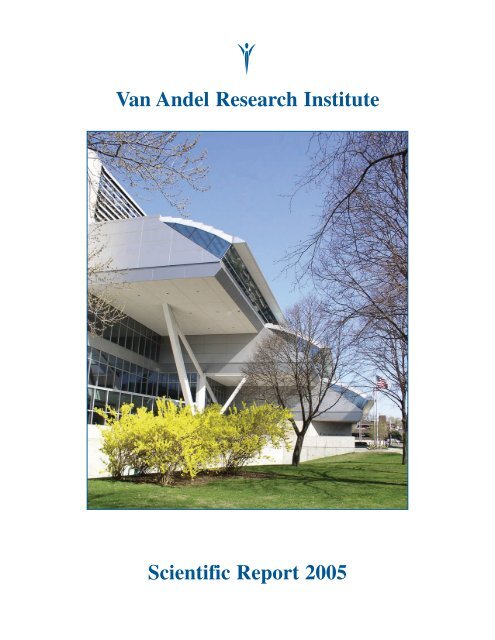
Van Andel Research Institute<br />
<strong>Scientific</strong> <strong>Report</strong> <strong>2005</strong>
®<br />
333 Bostwick Avenue, N.E., Grand Rapids, MI 49503<br />
Phone (616) 234-5000; Fax (616) 234-5001; Web site: www.vai.org<br />
Cover photograph of the Van Andel Institute building, Grand Rapids, Michigan
Van Andel Research Institute<br />
<strong>Scientific</strong> <strong>Report</strong><br />
<strong>2005</strong>
Title page photo: Podosomes in transformed cells<br />
This image shows podosomes in NIH3T3 mouse fibroblasts transformed with activated Src tyrosine<br />
kinase. Podosomes are the rounded structures at the tips of many of the cell extensions. They are rich in<br />
filamentous actin, which has been stained green with a conjugated phalloidin dye. The cells are co-stained<br />
with an antibody that recognizes the adhesion protein CrkL (red; or where co-localized with actin and its<br />
phalloidin stain, yellow). The protein CrkL is involved in integrin-induced cell adhesion and migration.<br />
The study of these proteins (Src, F-actin, CrkL, integrins) in these and other tumorigenic cell types may<br />
shed light on the mechanisms of podosome-mediated cancer cell metastasis and invasion.<br />
(Eduardo F. Azucena, Jr., Darren Seals, James Resau, and Sara Courtneidge)<br />
Published June <strong>2005</strong><br />
©<strong>2005</strong> by the Van Andel Institute<br />
All rights reserved<br />
Van Andel Institute<br />
333 Bostwick Avenue, N.E.<br />
Grand Rapids, Michigan 49503, U.S.A.
Contents<br />
Director’s Introduction . . . . . . . . . . . . . . . . . . . . . . . . . . . . . . . . . . . . . . . . . . . . . . . . . . . . . . . . . . 3<br />
Laboratory <strong>Report</strong>s<br />
Cell Structure and Signal Integration . . . . . . . . . . . . . . . . . . . . . . . . . . . . . . . . . . . . . . . . . . . . . . . . . 9<br />
Arthur S. Alberts, Ph.D.<br />
Antibody Technology . . . . . . . . . . . . . . . . . . . . . . . . . . . . . . . . . . . . . . . . . . . . . . . . . . . . . . . . . . . . . 13<br />
Brian Cao, M.D.<br />
Mass Spectrometry and Proteomics . . . . . . . . . . . . . . . . . . . . . . . . . . . . . . . . . . . . . . . . . . . . . . . . . . 16<br />
Gregory S. Cavey, B.S.<br />
Signal Regulation and Cancer . . . . . . . . . . . . . . . . . . . . . . . . . . . . . . . . . . . . . . . . . . . . . . . . . . . . . . 18<br />
Sara A. Courtneidge, Ph.D.<br />
Cancer and Developmental Cell Biology . . . . . . . . . . . . . . . . . . . . . . . . . . . . . . . . . . . . . . . . . . . . . 20<br />
Nicholas S. Duesbery, Ph.D.<br />
Vivarium and Transgenics . . . . . . . . . . . . . . . . . . . . . . . . . . . . . . . . . . . . . . . . . . . . . . . . . . . . . . . . . 24<br />
Bryn Eagleson, A.A., RLATG<br />
Bioinformatics . . . . . . . . . . . . . . . . . . . . . . . . . . . . . . . . . . . . . . . . . . . . . . . . . . . . . . . . . . . . . . . . . . 26<br />
Kyle Furge, Ph.D.<br />
Cancer Immunodiagnostics . . . . . . . . . . . . . . . . . . . . . . . . . . . . . . . . . . . . . . . . . . . . . . . . . . . . . . . . 28<br />
Brian B. Haab, Ph.D.<br />
Molecular Medicine and Virology . . . . . . . . . . . . . . . . . . . . . . . . . . . . . . . . . . . . . . . . . . . . . . . . . . . 31<br />
Sheri L. Holmen, Ph.D.<br />
Integrin Signaling and Tumorigenesis . . . . . . . . . . . . . . . . . . . . . . . . . . . . . . . . . . . . . . . . . . . . . . . . 34<br />
Cindy K. Miranti, Ph.D.<br />
Analytical, Cellular, and Molecular Microscopy . . . . . . . . . . . . . . . . . . . . . . . . . . . . . . . . . . . . . . . . 38<br />
and<br />
Microarray Technology and Molecular Diagnostics<br />
James H. Resau, Ph.D.<br />
Germline Modification . . . . . . . . . . . . . . . . . . . . . . . . . . . . . . . . . . . . . . . . . . . . . . . . . . . . . . . . . . . . 42<br />
Pamela J. Swiatek, Ph.D., M.B.A.<br />
Cancer Genetics . . . . . . . . . . . . . . . . . . . . . . . . . . . . . . . . . . . . . . . . . . . . . . . . . . . . . . . . . . . . . . . . . 45<br />
Bin T. Teh, M.D., Ph.D.<br />
iii
Molecular Oncology . . . . . . . . . . . . . . . . . . . . . . . . . . . . . . . . . . . . . . . . . . . . . . . . . . . . . . . . . . . . . 49<br />
George F. Vande Woude, Ph.D.<br />
Tumor Metastasis and Angiogenesis . . . . . . . . . . . . . . . . . . . . . . . . . . . . . . . . . . . . . . . . . . . . . . . . . 53<br />
Craig P. Webb, Ph.D.<br />
Chromosome Replication . . . . . . . . . . . . . . . . . . . . . . . . . . . . . . . . . . . . . . . . . . . . . . . . . . . . . . . . . 58<br />
Michael Weinreich, Ph.D.<br />
Cell Signaling and Carcinogenesis . . . . . . . . . . . . . . . . . . . . . . . . . . . . . . . . . . . . . . . . . . . . . . . . . . 60<br />
Bart O. Williams, Ph.D.<br />
Structural Sciences . . . . . . . . . . . . . . . . . . . . . . . . . . . . . . . . . . . . . . . . . . . . . . . . . . . . . . . . . . . . . . . 63<br />
H. Eric Xu, Ph.D.<br />
Mammalian Developmental Genetics . . . . . . . . . . . . . . . . . . . . . . . . . . . . . . . . . . . . . . . . . . . . . . . . 66<br />
Nian Zhang, Ph.D.<br />
Daniel Nathans Memorial Award . . . . . . . . . . . . . . . . . . . . . . . . . . . . . . . . . . . . . . . . . . . . . . . . . .71<br />
Postdoctoral Fellowship Program . . . . . . . . . . . . . . . . . . . . . . . . . . . . . . . . . . . . . . . . . . . . . . . . .75<br />
Student Programs . . . . . . . . . . . . . . . . . . . . . . . . . . . . . . . . . . . . . . . . . . . . . . . . . . . . . . . . . . . . . .79<br />
Han-Mo Koo Memorial Seminar Series . . . . . . . . . . . . . . . . . . . . . . . . . . . . . . . . . . . . . . . . . . . .85<br />
Organization . . . . . . . . . . . . . . . . . . . . . . . . . . . . . . . . . . . . . . . . . . . . . . . . . . . . . . . . . . . . . . . . . . .91<br />
Recent VARI Photos . . . . . . . . . . . . . . . . . . . . . . . . . . . . . . . . . . . . . . . . . . . . . . . . . . . . . . . . . . . . .99<br />
iv
Director’s Introduction
Director’s Introduction<br />
We are now in our<br />
fifth year since the<br />
opening of the Van<br />
Andel Institute. The<br />
Institute was created<br />
because of the<br />
generosity of Jay Van<br />
Andel and his wife<br />
Betty, both of whom<br />
George F. Vande Woude<br />
passed away in 2004.<br />
While deeply saddened by their loss,<br />
we continue our endeavor to fulfill their vision of<br />
a center for research and education excellence in<br />
the heart of Grand Rapids.<br />
I know Jay was able to see the beginning of<br />
what a great contribution he made to society and<br />
the betterment of human health. Sharing his and<br />
Betty’s vision, we have begun to expand our<br />
research beyond cancer biology into the field of<br />
neurological disorders. David and Carol Van<br />
Andel, in Jay’s honor, have created an endowed<br />
chair dedicated to Parkinson disease (PD)<br />
research. The search for a leading scientist to<br />
take this chair is now underway.<br />
Already, Jim Resau and Bin Teh have<br />
initiated a collaboration with Australian<br />
scientists Alan Mackay-Sim and Peter Silburn at<br />
Queensland University in Australia to better<br />
understand Parkinson disease and how it<br />
develops. Alan is a developmental biologist who<br />
has initiated some exciting discoveries using<br />
adult olfactory stem cells. Peter is a neurologist<br />
who studies and treats PD patients. Together<br />
with these new collaborators, we will increase<br />
our understanding of the function, growth, and<br />
death of human nerve cells as models for PD. In<br />
addition, these approaches will be used in the<br />
study of Alzheimer disease pathophysiology.<br />
Our scientists have also partnered with the St.<br />
Mary’s Hauenstein Parkinson Center—named in<br />
honor of lead donor (and VAI trustee) Ralph<br />
Hauenstein—to determine the interactions of the<br />
environment and genes associated with PD. We<br />
will learn how these genes correlate with disease<br />
progression and drug response.<br />
We are pleased to announce that, in<br />
collaboration with the Van Andel Education<br />
Institute, the Research Institute is developing a<br />
graduate school with a program leading to the<br />
Ph.D. degree in cellular and molecular genetics,<br />
with an emphasis on translation. We expect the<br />
graduate program to contribute to the vitality and<br />
creativity of our successful research programs<br />
and to also address the nation’s need for<br />
expertise in the life sciences and biotechnology.<br />
We have received a charter from the state of<br />
Michigan and have appointed a Board of<br />
Directors for the school. Our goal is to have our<br />
first students on board in September 2006.<br />
Personnel<br />
A special occasion occurred this past year<br />
with our first promotion review. We are very<br />
proud to announce the promotion of Bin Tean Teh<br />
to Distinguished <strong>Scientific</strong> Investigator, our<br />
highest appointment level. Bin has made major<br />
contributions to the understanding of kidney,<br />
nasopharyngeal, and endocrine cancer. In recognition<br />
of his efforts, Bin was recently appointed<br />
to the Medical Advisory Board of the Kidney<br />
Cancer Association. We are also proud to<br />
announce the appointment of Rick Hay as a<br />
Senior <strong>Scientific</strong> Investigator. Rick will establish<br />
the Laboratory of Animal Imaging. Our congratulations<br />
to both scientists!<br />
Sara Courtneidge has taken a position at the<br />
Burnham Institute and is relocating her<br />
laboratory in early summer <strong>2005</strong>. Sara and I<br />
have been friends and colleagues for over 20<br />
years. I was delighted when she joined VARI<br />
and I am grateful for all her contributions to the<br />
Institute, especially her efforts in helping to<br />
establish the graduate program with MSU and<br />
the VARI postdoctoral advisory committee. The<br />
Burnham Institute is very fortunate to have<br />
recruited her, where she will be reunited with her<br />
many West Coast friends. I am sorry to see her<br />
leave, but I wish her great success and look<br />
forward to our continued scientific interactions.<br />
Publications and Competitive Funding<br />
As of April 1, <strong>2005</strong>, there have been 190<br />
peer-reviewed articles published by VARI<br />
investigators. In December 2004, a VARI article<br />
was featured on the cover of the Journal of Bone<br />
3
and Mineral Research; in January <strong>2005</strong>, we had a<br />
featured article in Molecular Cell; and in<br />
February, a VARI article in Cancer Cell was the<br />
source of the issue’s cover photo. Our<br />
investigators have also demonstrated their<br />
competitive research abilities in terms of receiving<br />
grants for funding of their work. In fiscal year<br />
2004, extramural funding for VARI dramatically<br />
increased over that in 2003. Seventeen of our<br />
scientists and six of our postdoctoral fellows<br />
received funding from 42 grants.<br />
Our new major awards have come from a<br />
variety of sources. The National Institutes of<br />
Health (NIH) National Cancer Institute (NCI)<br />
made two awards to Nick Duesbery. The first<br />
was an R01 grant for studying MEK signaling in<br />
sarcoma growth and vascularization. The second<br />
grant, an R21, was for investigating the antitumor<br />
effects of an anthrax toxin moiety (which<br />
we have termed tumor lethal factor, or “TLF”) on<br />
Kaposi sarcoma. Nick’s lab has been developing<br />
TLF as a potential cancer therapeutic.<br />
Brian Haab was awarded an R21 NCI grant for a<br />
two-year project entitled “Longitudinal Cancer-<br />
Specific Serum Protein Signatures.” This project<br />
seeks to develop protein microarray methods for<br />
detecting and diagnosing prostate cancer by<br />
examining changes over time in several cancerrelated<br />
proteins in serum. And, Art Alberts<br />
received an R21 award from the NIH to exploit a<br />
discovery in his lab with a long-term goal of<br />
developing novel anti-cancer therapies.<br />
The American Cancer Society awarded two<br />
Research Scholar Grants to our researchers.<br />
One grant, to Art Alberts, is for a four-year<br />
project to study Diaphanous-related formins in<br />
myelodysplasia. Art has been studying the role<br />
of formins in cancer. A second American Cancer<br />
Society grant was awarded to Michael Weinreich.<br />
Michael is identifying small-molecule inhibitors<br />
of Cdc7 kinase for study of its regulation in DNA<br />
replication, with a long-term goal of identifying<br />
novel targets for cancer diagnosis and therapy.<br />
Two major grants have also been received<br />
from the Michigan Technology Tri-Corridor<br />
(MTTC). One award went to Rick Hay for the<br />
development of novel agents for nuclear imaging<br />
and therapy of Met-expressing human tumors.<br />
The project is a collaboration among scientists at<br />
VARI, Michigan State University, the Department<br />
of Veterans Affairs Healthcare System in Ann<br />
Arbor, ApoLife, Inc., and the National Cancer<br />
Institute. A second grant was awarded to Bin Teh<br />
for the development of the “RenoChip,”<br />
a diagnostic and prognostic tool for use against<br />
kidney cancer. In addition, the Department of<br />
Defense awarded Eric Xu a grant for a three-year<br />
study of the structure and function of the<br />
androgen receptor in prostate cancer. Eric’s lab<br />
aims to make progress in understanding the<br />
androgen dependence (or independence) of<br />
prostate cancer.<br />
Funding from other sources in the past year<br />
has included Brian Haab’s grant from DHHS/<br />
NCI via the University of Michigan for a project<br />
entitled “Accelerated Cancer Biomarker<br />
Discovery.” This project is being undertaken by<br />
a consortium of laboratories and focuses on the<br />
development and application of new proteomics<br />
technologies for cancer biomarker discovery.<br />
Bin Teh has received a grant from the<br />
Schregardus Family Foundation for a project on<br />
renal cell carcinoma (RCC) in which his lab will<br />
be studying the prognostic value of genes for<br />
improving the clinical management of RCC<br />
patients. Bin is also the recipient of a grant from<br />
the Gerber Foundation for gene expression<br />
profiling in newborns with congenital chromosomal<br />
abnormalities.<br />
We are also proud that three of our<br />
postdoctoral fellows have received awards.<br />
Carrie Graveel (Vande Woude lab) and Kate<br />
Eisenmann (Alberts lab) have received National<br />
Research Service Awards from the NIH, while<br />
Jennifer Bromberg-White (Webb lab) received a<br />
fellowship award from the Multiple Myeloma<br />
Research Foundation.<br />
Looking to the Future<br />
We now look to expanding not only our<br />
research goals but the Institute itself. On May<br />
17th we celebrated our fifth anniversary, and our<br />
CEO, David Van Andel, announced that in 2006<br />
we will begin the second construction phase of<br />
our Institute. The new building, a model of<br />
which is displayed in the Cook-Hauenstein Hall,<br />
will join and mirror our current exceptional<br />
facility, but it will provide two-and-a-half times<br />
the existing laboratory space, or an additional<br />
150,000 square feet.<br />
4
As we plan and begin our expansion, we are<br />
part of the unprecedented growth in the health<br />
industry that Grand Rapids is experiencing.<br />
The area of Grand Rapids in which the Institute is<br />
situated has been appropriately renamed “Medical<br />
Hill.” Our neighbor across Bostwick Avenue,<br />
Spectrum Health, has celebrated the opening of<br />
the new Fred and Lena Meijer Heart Center.<br />
Furthermore, the hospital will soon break ground<br />
for the construction of a new cancer center as well<br />
as a new pediatric hospital. In another project, our<br />
own Rick Hay serves as the chairman of a<br />
combined VARI and Grand Valley State<br />
University group that is planning a good<br />
manufacturing practices (GMP) facility. This<br />
facility is being established with state and federal<br />
funding, and it will produce small quantities of<br />
clinical-grade biological products that can be<br />
tested in patients. Finally, there is the strong<br />
possibility of Michigan State University’s College<br />
of Human Medicine relocating to Grand Rapids,<br />
which would certainly be a major event in the<br />
development of the biomedical community here.<br />
Overall, with the new hospital facilities at<br />
Spectrum Health and St. Mary’s, Grand Valley<br />
State University’s strength in health sciences, and<br />
our own research program and future expansion,<br />
we will witness in the next decade exciting and<br />
dramatic developments in biomedical research,<br />
scientific discovery, and health care delivery<br />
taking place in Grand Rapids. We look forward<br />
to these many exciting changes and to the<br />
formation of a center of excellence in biomedical<br />
disciplines in western Michigan.<br />
5
Van Andel Research Institute<br />
Laboratory <strong>Report</strong>s
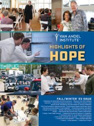
<strong>2005</strong> VARI<br />
<strong>Scientific</strong> Retreat<br />
8
Laboratory of Cell Structure and Signal Integration<br />
Arthur S. Alberts, Ph.D.<br />
Dr. Alberts received his Ph.D. in physiology and pharmacology at the University of<br />
California, San Diego, in 1993, where he studied with James Feramisco. From 1994<br />
to 1997, he served as a postdoctoral fellow in Richard Treisman’s laboratory at the<br />
Imperial Cancer Research Fund in London, England. From 1997 through 1999, he<br />
was an Assistant Research Biochemist in the laboratory of Frank McCormick at the<br />
Cancer Research Institute, University of California, San Francisco. Dr. Alberts joined<br />
VARI as a <strong>Scientific</strong> Investigator in January 2000.<br />
Staff<br />
Art Alberts, Ph.D.<br />
Jun Peng, M.D.<br />
Yunju Chen, Ph.D.<br />
Kathryn Eisenmann, Ph.D.<br />
Holly Holman, Ph.D.<br />
Susan Kitchen, B.S.<br />
Laboratory Members<br />
Students<br />
Aaron DeWard, B.S.<br />
Yaojian Liu, B.S.<br />
Katharine Collins<br />
Visiting Scientists<br />
Stephen Matheson, Ph.D.<br />
Brad Wallar, Ph.D.<br />
Research Interests<br />
T<br />
he actin cytoskeleton is a dynamic,<br />
tightly regulated protein network that<br />
plays a crucial role in mediating<br />
diverse cellular processes including cell division,<br />
migration, endocytosis, vesicle trafficking, and<br />
cell shape. The research focus of the lab is the<br />
genetics and molecular biology of the Rho<br />
family of small GTPases and their effectors,<br />
which together control multiple aspects of<br />
cytoskeletal dynamics. The guiding hypothesis<br />
of the laboratory is that cytoskeletal dynamics<br />
defines the what, where, and how of signal<br />
transduction pathways, control responses to<br />
growth factors, and other extracellular cues, and<br />
that defects in these tightly controlled dynamics<br />
can contribute to cancer pathophysiology.<br />
Support for this hypothesis is observed in human<br />
cancers that carry mutations in genes encoding<br />
regulators of Rho GTPase activity. Ultimately,<br />
our goal is to exploit our understanding of the<br />
mechanics of GTPase-effector relationships in<br />
order to develop anti-cancer therapeutics.<br />
PAK1 negatively regulates the activity of the<br />
Rho exchange factor NET1<br />
Rho GTPases act as molecular switches in<br />
cells, alternating between on and off states while<br />
bound to GTP and GDP nucleotides, respectively.<br />
The activated, GTP-bound proteins preferentially<br />
interact with numerous autoregulated downstream<br />
effector proteins. Rho GTPases are activated by<br />
Rho guanine nucleotide exchange factors (Rho<br />
GEFs), one of which is the neuroepithelioma<br />
transforming gene 1 (NET1). Recently it was<br />
demonstrated that wild-type NET1 is localized in<br />
the nucleus and that truncation of the amino<br />
terminus results in relocalization of a fraction of<br />
the NET1 to the cytoplasm. This is at least<br />
partially due to the elimination of two putative<br />
nuclear localization signals within the amino<br />
terminus. Thus, NET1 activity is regulated at least<br />
in part through subcellular localization.<br />
Rho family members can modulate the<br />
activity of other Rho proteins. The protein kinase<br />
PAK1 down-regulates the activity of the RhoAspecific<br />
GEF NET1. Specifically, PAK1<br />
phosphorylates NET1 on three sites in vitro:<br />
serines 152, 153, and 538. Replacement of<br />
serines 152 and 153 with glutamate residues<br />
reduces the activity of NET1 as an exchange<br />
factor in vitro, as well as its ability to stimulate<br />
actin stress fiber formation in cells. Using a<br />
phospho-specific antibody, PAK1 can be shown<br />
to phosphorylate NET1 on serine 152 in cells, and<br />
Rac1, which activates PAK1, stimulates serine<br />
152 phosphorylation in a PAK1-dependent<br />
manner. Furthermore, coexpression of<br />
constitutively active PAK1 inhibits NET1<br />
stimulation of actin polymerization only when<br />
serines 152 and 153 are present. These<br />
observations provide a novel mechanism for the<br />
control of RhoA activity.<br />
9
Signal transduction and spatially controlled<br />
assembly of F-actin networks<br />
One well-characterized actin nucleator, the<br />
Arp2/3 complex, induces the formation of<br />
branched actin filaments. Arp2/3 works by<br />
complexing with G-actin or by binding to the side<br />
of preexisting filaments. Its nucleation and<br />
filament-binding activity is tightly regulated by<br />
interactions with nucleation-promoting factors<br />
(NPFs), the most prominent being the WASp/Scar<br />
family. WASp is an autoregulated molecular<br />
switch controlled by yet other switch-like proteins<br />
such as Cdc42, a Rho family small GTPase. Thus,<br />
NPFs integrate signals controlling growth<br />
factor–stimulated actin nucleation and branching.<br />
Cdc42-activated WASp induction of Arp2/3<br />
activity is shown schematically in Fig. 1A.<br />
The mammalian Diaphanous-related formins<br />
Formins are a highly conserved family of<br />
proteins implicated in a diverse array of cellular<br />
functions including the cytoskeletal remodeling<br />
events necessary for cytokinesis, bud formation<br />
in yeast, establishment of cell/organelle polarity,<br />
and endocytosis. Formins have the ability to<br />
stabilize microtubules, which (like F-actin) are<br />
assembled by tightly controlled cycles of<br />
polymerization and depolymerization.<br />
The mammalian Diaphanous-related formin<br />
(mDia) proteins are a subfamily of formins that<br />
share a loosely conserved Rho GTPase-binding<br />
domain (GBD) in the amino terminus and a<br />
highly conserved Diaphanous-autoregulatory<br />
domain (DAD) in the carboxy terminus.<br />
The GBDs can interact with their internal DAD<br />
partners in vitro, leading to the autoregulation<br />
model depicted in Fig. 1B. The model shows that<br />
while the formin proteins dimerize through their<br />
FH2 domains, it is the GBD-DAD interaction that<br />
is the linchpin of autoregulation. GTP-bound<br />
Rho can interact with the GBD and interrupt the<br />
autoinhibited conformation, leading to nucleation<br />
and elongation of nonbranched actin filaments.<br />
It has been unclear whether GTPase binding<br />
simply activates the mDia proteins’ ability to<br />
nucleate actin, or provides other signals that<br />
direct subcellular targeting and recruitment of<br />
mDia-associated proteins. Another question is,<br />
do mDia proteins activated in specific cellular<br />
contexts (i.e., on vesicles or at sites of adhesion)<br />
associate with or work in parallel with other<br />
modifiers of actin polymerization to generate<br />
site-specific F-actin networks? We are studying<br />
such questions using FRET technology.<br />
Site-specific interactions between Rho<br />
GTPases and mDia proteins<br />
Fluorescence resonance energy transfer<br />
(FRET) is a powerful technique that allows us to<br />
assay protein-protein interactions in cells by<br />
using two fluorophores, in this case, cyan<br />
fluorescent protein (CFP) and yellow fluorescent<br />
protein (YFP). When fused to the GTPase and<br />
excited at the appropriate wavelength, CFP acts<br />
as a fluorescent donor which then excites YFP,<br />
which is fused to a Drf protein (Fig. 1B). FRET<br />
Figure 1. Drfs are actin nucleators whose activity is regulated through interactions with small<br />
GTPases. A) A model for formin (mDia1-3) and WASp collaborating in cells with activated Cdc42, in which<br />
Cdc42 interacts with mDia2 in cells at specific sites associated with membrane protrusions. In this model,<br />
activated WASp nucleates branched filaments from the side of mDia2 nucleated “mother” filaments;<br />
alternatively, mDia2 binds to and processively elongates filaments after nucleation by Arp2/3. B) GTP-bound<br />
Rho GTPase binding disrupts intramolecular interactions between the GBD and DAD of a DRF. If the GTPase<br />
and GBD are linked to fluorophores (ECFP and EYFP), the proximity of the two can be determined by FRET.<br />
10
occurs only when the donor/acceptor pair is in<br />
close proximity (less than 30 Å) .<br />
Fusion proteins are expressed following<br />
microinjection of their expression plasmids into<br />
cells, which are then fixed 4 h later. We have<br />
shown that YFP-mDia2 is expressed with CFP-<br />
Cdc42, primarily at the leading cell edge (or<br />
cortex) and at the microtubule-organizing center.<br />
In other experiments, we have shown that this<br />
interaction depends upon the integrity of the<br />
CRIB motif within the mDia2 GBD. CRIB<br />
motifs are necessary for binding to the GTPase.<br />
Our observations indicate that one particular<br />
GTPase-formin pair, Cdc42 and mDia2, may<br />
have a role in remodeling actin at the cell edge.<br />
What this pair contributes to actin or microtubule<br />
dynamics at the MTOC, however, remains an<br />
open question. We speculate that the pair may<br />
participate in microtubule regulation at the minus<br />
(–) end of the tubules, which (like actin) are<br />
assembled and disassembled in a polarized<br />
fashion. Other GTPase-formin pairs may be<br />
working at the plus end to direct them to focal<br />
adhesions or other sites. From collaborative<br />
efforts with the Gundersen lab, it has been shown<br />
that mDia1 and mDia2 can complex with the<br />
MT(+)-end binding proteins APC and EB1.<br />
In contrast to the speculative role of Cdc42<br />
and mDia2, RhoB is known to have a role in<br />
endocytic or vesicular trafficking, and it interacts<br />
with mDia2 on endosomes (Fig. 2). This result<br />
is consistent with our discovery of both mDia1<br />
and mDia2 on endosomes. The expression of<br />
either activated RhoB or deregulated versions of<br />
mDia1, mDia2, and mDia3 blocks the movement<br />
of vesicles and increases their number. One<br />
interpretation is that expression of RhoB or<br />
deregulated mDia1–3 triggers an inappropriate<br />
transition from fast microtubule-dependent<br />
transport to actin-dependent transport.<br />
Formins as anti-cancer drug targets<br />
A dynamic cytoskeleton is required for tumor<br />
cell growth. Drugs that stabilize the cytoskeleton<br />
are emerging as effective anti-cancer<br />
therapeutics. For example, Taxol binds directly<br />
to the components that comprise the microtubule<br />
cytoskeleton and blocks their dynamics. A<br />
cyclopeptide derived from a sea sponge,<br />
jasplakinolide, has similar effects on actin.<br />
Figure 2. RhoB<br />
interacts with mDia2<br />
on vesicles. CFP-fused<br />
activated RhoB-G14V<br />
and YFP-fused mDia2<br />
were co-injected into<br />
cells and FRET was<br />
assessed 4 h later. A<br />
FRET signal is observed<br />
between activated RhoB<br />
and mDia2 upon<br />
endosomes. These data<br />
indicate that individual<br />
GTPase-formin pairs<br />
would appear to be<br />
functionally distinct,<br />
promoting the formation<br />
of different actin<br />
structures at different<br />
sites. Since both RhoB<br />
and Cdc42 have been<br />
implicated in endocytosis<br />
and vesicle trafficking, it<br />
is possible that both<br />
GTPases use mDia2<br />
sequentially or in parallel<br />
as effectors to transport<br />
cargo within cells.<br />
Previously, we found that a peptide derived<br />
from the DAD region of mDia proteins, when<br />
expressed in cells, stabilized both the actin and<br />
microtubule cytoskeletons. Because the<br />
mechanism of DAD action is unique—it binds to<br />
cellular formins and disrupts their normal<br />
autoregulatory mechanism—DAD represents a<br />
novel class of anti-tumor drugs.<br />
Because DAD is unable to enter cells as a<br />
drug, we have begun searching for functional<br />
analogs of DAD that could have similar properties<br />
under a drug-development program funded by the<br />
National Cancer Institute. We hypothesize that by<br />
deregulating specific mDia molecules in tumor<br />
cells, we can arrest dynamic remodeling of the<br />
cytoskeleton, a process required for cell motility<br />
and cytokinesis. We will characterize the<br />
structural and functional requirements for DADinduced<br />
cell death and develop a high-throughput<br />
screen for the identification of novel molecules<br />
that can deregulate mDia proteins and kill tumor<br />
cells. Our objectives are to initiate a drug<br />
discovery program by validating mDia proteins as<br />
molecular targets in cancer and to determine the<br />
physical requirements for DAD interactions with<br />
the mDia GBD.<br />
11
External Collaborators<br />
Philippe Chavrier, Institut Curie, Paris, France<br />
Jeff Frost, University of Houston, Texas<br />
Gregg Gundersen, Columbia University, New York<br />
George Prendergast, Lankenau Institute, Wynnewood, Pennsylvania<br />
Kathy Siminovitch, University of Toronto, Canada<br />
Recent Publications<br />
Alberts, A.S., H. Qin, H.S. Carr, and J.A. Frost. In press. PAK1 negatively regulates the activity of<br />
the Rho exchange factor NET1. Journal of Biological Chemistry.<br />
Eisenmann, K.M., J. Peng, B.J. Wallar, and A.S. Alberts. In press. Rho GTPase-formin pairs in<br />
cytoskeletal remodeling. In Signaling Networks in Cell Shape and Motility, London, U.K.:<br />
Novartis Foundation.<br />
Wen, Ying, Christina H. Eng, Jan Schmoranzer, Noemi Cabrera-Poch, Edward J.S. Morris, Michael<br />
Chen, Bradley J. Wallar, Arthur S. Alberts, and Gregg G. Gundersen. 2004. EB1 and APC bind<br />
to mDia to stabilize microtubules downstream of Rho and promote cell migration. Nature Cell<br />
Biology 6(9): 820–830.<br />
Left to right: Liu, Holman, DeWard, Chen, Peng, Collins, Alberts, Kitchen, Eisenmann<br />
12
Laboratory of Antibody Technology<br />
Brian Cao, M.D.<br />
Dr. Cao obtained his M.D. from Beijing Medical University, People’s Republic of<br />
China, in 1986. On receiving a CDC fellowship award, he was a visiting scientist at<br />
the National Center for Infectious Diseases, Centers for Disease Control and<br />
Prevention (1991–1994). He next served as a postdoctoral fellow at Harvard<br />
(1994–1995) and at Yale (1995–1996). From 1996 to 1999, Dr. Cao was a Scientist<br />
Associate in charge of the Monoclonal Antibody Production Laboratory at the<br />
Advanced BioScience Laboratories–Basic Research Program at the National<br />
Cancer Institute–Frederick Cancer Research and Development Center, Maryland.<br />
Dr. Cao joined VARI as a Special Program Investigator in June 1999.<br />
Staff<br />
Ping Zhao, M.S.<br />
Tessa Grabinski, B.S.<br />
Laboratory Members<br />
Visiting Scientist<br />
Mei Guo, M.S.<br />
Students<br />
Yong-jun Jiao<br />
Xin Wang<br />
Jin Zhu<br />
Research Interests<br />
H<br />
epatocyte<br />
growth factor/scatter factor<br />
(HGF/SF) is a multifunctional<br />
heterodimeric protein produced by<br />
mesenchymal cells and is an effector of cells<br />
expressing the tyrosine kinase receptor Met. Met,<br />
the protein product of the c-met protooncogene, is<br />
from the same family as epidermal growth factor<br />
(EGF) receptors. The activation of Met by<br />
HGF/SF affects downstream signaling pathways<br />
(including other protein kinases) responsible for<br />
cellular differentiation, motility, proliferation,<br />
organogenesis, angiogenesis, and apoptosis.<br />
Aberrant expression of the Met-HGF/SF<br />
receptor-ligand complex—resulting either from<br />
mutations in the complex or in conjunction with<br />
mutations in other oncogenes—is associated with<br />
an invasive/metastatic phenotype in most solid<br />
human tumors. Met-HGF/SF and downstream<br />
kinases are therefore attractive targets for new<br />
anti-cancer agents for clinical diagnosis,<br />
prognosis, and treatment.<br />
The aberrant expression of the Met receptor<br />
kinase by two-thirds of localized prostate cancers,<br />
and apparently by all osseous metastases,<br />
suggests that Met provides a strong mechanism of<br />
selection for metastatic development. We have<br />
generated and characterized several anti-Met<br />
murine monoclonal antibodies (mAbs) that have<br />
high affinity for and specifically recognize Met<br />
extracellular domains in their native<br />
conformation. In collaborative studies, we are<br />
using two of these radiolabeled anti-Met mAbs,<br />
designated Met3 and Met5, to study mouse<br />
xenograft and orthotopic models of localized and<br />
metastatic prostate cancer via clinical nuclear<br />
imaging. Moreover, we will soon be testing these<br />
two radiolabeled mAbs on dog spontaneous<br />
prostate cancer and bone metastasis models.<br />
In collaboration with the Nanjing Medical<br />
University of China, we have initiated a project to<br />
construct a phage-display antibody fragment<br />
library. This technique involves the construction<br />
and use of animal/human, immunized/naïve Fab<br />
and scFv antibody gene repertoires by phage<br />
display. The ability to co-select antibodies and<br />
their genes allows the isolation of high-affinity,<br />
antigen-specific mAbs derived from either<br />
immunized animals or non-immunized humans. A<br />
number of procedures for selecting such antibodies<br />
from recombinant libraries have been described,<br />
and some useful antibodies have been produced<br />
with this approach. Over the past two years, we<br />
have closely followed the development of this<br />
technology for producing novel recombinant<br />
antibody-like molecules. We have constructed a<br />
human naïve Fab library with the diversity of 2 ×<br />
10 9 and have screened out some mAb fragments<br />
against tumor marker proteins. In particular, we<br />
have selected from this library and characterized<br />
one specific anti-Met Fab fragment, designated as<br />
Fab-Met-1, using a subtractive whole-cell panning<br />
approach (Figs. 1 and 2).<br />
We have also established the technology of a<br />
phage-display peptide library for mAb epitope<br />
13
A<br />
B<br />
mapping. A random peptide library<br />
is constructed by genetically fusing<br />
oligonucleotides to the coding<br />
sequence of a coat protein of<br />
bacteriophage, resulting in display<br />
of the fused polypeptide on the<br />
surface of the virion. Phage display<br />
has been used to create a physical<br />
linkage between a vast library of<br />
random peptide sequences and the<br />
DNA encoding each sequence,<br />
allowing rapid identification of<br />
peptide ligands for a variety of<br />
target molecules such as antibodies.<br />
A library of phage is exposed to a<br />
plate coated with mAb. Unbound<br />
phages are washed away, and<br />
specifically bound phages are eluted<br />
by lowering the pH. The eluted pool of phage is<br />
amplified, and the process is repeated for two more<br />
rounds. Individual clones are isolated, screened by<br />
ELISA, and sequenced. We have successfully<br />
epitope-mapped a variety of important mAbs<br />
including anti-HGF/SF, anti-Met, and anti-anthrax<br />
lethal factor. We are now exploring the use of this<br />
technology on protein-protein interactions; one<br />
example is the mapping of the HGF/SF-Met<br />
binding site in an in vitro system, and several<br />
interesting peptides have been selected from the<br />
library as being potential Met antagonists.<br />
Functioning as an antibody production core<br />
facility at the Van Andel Research Institute, this<br />
lab has extensive capabilities in the generation,<br />
characterization, scaled-up production, and<br />
purification of mAbs using comprehensive<br />
cutting-edge technologies. The technologies and<br />
services available in the core include animal<br />
immunization and antigen preparation; peptide<br />
design; DNA immunization (Gene-gun<br />
technology); immunization of a wide range of<br />
antibody-producing models (including mice,<br />
rats, rabbits, human cells, and transgenic or<br />
knock-out mice); and in vitro immunization.<br />
Other services we provide include the generation<br />
of hybridomas from spleen cells of immunized<br />
mice, rats, and rabbits; hybridoma expansion and<br />
subcloning; cryopreservation of hybridomas<br />
secreting mAbs; monoclonal antibody isotyping;<br />
ELSIA screening of hybridoma supernatants;<br />
monoclonal antibody characterization by<br />
immunoprecipitation, Western blot, immunohistochemistry,<br />
immunofluorescence staining,<br />
FACS, or in vitro bioassays; production of bulk<br />
quantities of mAbs using high-density cell culture;<br />
purification of mAbs using FPLC affinity columns;<br />
generation of bi-specific mAbs by secondary<br />
fusion; conjugation of mAbs to detection enzymes<br />
(biotin/streptavidin, fluorescence reporters, etc.);<br />
and the development of detection methods/kits<br />
such as sandwich ELISA. Over the past few years,<br />
this facility has generated more than 200 different<br />
mAbs, 10 of which have been licensed to<br />
commercial companies. We have also contracted<br />
services to local biotechnology companies to<br />
generate, characterize, produce, and purify mAbs<br />
for their research/diagnostic kit development.<br />
S114<br />
Figure 1. A) SDS–PAGE of Fab-<br />
Met-1 fragment purified by affinity<br />
chromatography. Lane 1, standard<br />
molecular weight markers; lane 2,<br />
purified Fab fragment under reducing<br />
conditions. The concentration of the<br />
running gel was 12%.<br />
B) Immunoprecipitation analysis of<br />
Fab-Met-1. Met from cell extracts<br />
was immunoprecipitated with purified<br />
Fab-Met-1 and detected by western<br />
blot analysis. Lane 1 is S114 cell<br />
lysate immunoprecipitated with C-28<br />
(rabbit anti-human Met polyclonal<br />
antibody). Lanes 2–5 are cell lysates<br />
immunoprecipitated with Fab-Met-1:<br />
lane 2, S-114 (Met+); lane 3, MKN45<br />
(Met+); lane 4, M14 cell (Met–); and<br />
lane 5, NIH3T3 (Met–).<br />
M14<br />
Figure 2. The binding affinity of a selected Fab<br />
fragment (Fab-Met-1) was tested by FACS analysis.<br />
Two cell lines, S114 (Met+) and M14 (Met–), were<br />
incubated with purified Fab-Met-1. Bound Fab was<br />
detected by staining the cells with secondary goat<br />
anti-human Fab–FITC conjugate and analyzing by<br />
FACS (black lines). Green lines indicate staining<br />
with the secondary antibody only.<br />
14
External Collaborators<br />
Zhen-qing Feng, Nanjing Medical University, China<br />
Milton Gross, Department of Veterans Affairs Medical Center/University of Michigan, Ann Arbor<br />
Xiao-hong Guan, Nanjing Medical University, China<br />
Beatrice Knudsen, Fred Hutchinson Cancer Research Center, Seattle, Washington<br />
Yi Ren, Cancer Center, Cleveland Clinic Foundation, Cleveland, Ohio<br />
Kang-lin Wan, Chinese Centers for Disease Control and Prevention, Beijing, China<br />
David Waters, Gerald P. Murphy Cancer Foundation, Seattle, Washington<br />
David Wenkert, Michigan State University, East Lansing<br />
Wei-cheng You, Beijing Institute for Cancer Research, China<br />
Recent Publications<br />
Jiao, Y., P. Zhao, J. Zhu, T. Grabinski, Z. Feng, X. Guan, R.S. Skinner, M.D. Gross, Y. Su,<br />
G.F. Vande Woude, R.V. Hay, and B. Cao. In press. Construction of human naïve Fab<br />
library and characterization of anti-Met Fab fragment generated from the library.<br />
Molecular Biotechnology.<br />
Zhang, Yu-Wen, Yanli Su, Nathan Lanning, Margaret Gustafson, Nariyoshi Shinomiya, Ping Zhao,<br />
Brian Cao, Galia Tsarfaty, Ling-Mei Wang, Rick Hay, and George F. Vande Woude. <strong>2005</strong>.<br />
Enhanced growth of human Met-expressing xenografts in a new strain of immunocompromised<br />
mice transgenic for human hepatocyte growth factor/scatter factor. Oncogene 24(1): 101–106.<br />
Tan, Min-Han, Carl Morrison, Pengfei Wang, Ximing Yang, Carola J. Haven, Chun Zhang, Ping<br />
Zhao, Maria S. Tretiakova, Eeva Korpi-Hyovalti, John R. Burgess, Khee Chee Soo, Wei-Keat<br />
Cheah, Brian Cao, James Resau, Hans Morreau, and Bin Tean Teh. 2004. Loss of parafibromin<br />
immunoreactivity is a distinguishing feature of parathyroid carcinoma. Clinical Cancer<br />
Research 10(19): 6629–6637.<br />
Zhao, Ping, Xudong Liang, Jessica Kalbfleisch, Han-Mo Koo, and Brian Cao. 2003. Neutralizing<br />
monoclonal antibody against anthrax lethal factor inhibits intoxication in a mouse model.<br />
Human Antibodies 12(4): 129–135.<br />
From left to right: Zhu, Wang, Cao, Ferrell, Grabinski, Zhao<br />
15
Laboratory of Mass Spectrometry and Proteomics<br />
Gregory S. Cavey, B.S.<br />
Mr. Cavey received his B.S. degree from Michigan State University in 1990. Prior to<br />
joining VARI he was employed at Pharmacia in Kalamazoo, Michigan, for nearly 15<br />
years. As a member of a biotechnology development unit, he was group leader for<br />
a protein characterization core laboratory. More recently as a research scientist in<br />
discovery research, he was principal in the establishment and application of a stateof-the-art<br />
proteomics laboratory for drug discovery. Mr. Cavey joined VARI as a<br />
Special Program Investigator in July 2002.<br />
Staff<br />
Paula Davidson, M.S.<br />
Laboratory Members<br />
Student<br />
Wendy Johnson<br />
Research Interests<br />
The Mass Spectrometry and Proteomics<br />
program works with many of the<br />
research labs at the Institute, using stateof-the-art<br />
mass spectrometers in combination with<br />
analytical protein separation and purification<br />
methods to help answer a wide range of biological<br />
questions. Using mass spectrometry data and<br />
database search software, proteins can be<br />
identified and characterized with unprecedented<br />
sensitivity and throughput. Since proteomics is a<br />
relatively new scientific discipline, many of the<br />
analytical techniques are rapidly changing;<br />
therefore our mission involves using established<br />
protocols, improving them, and developing new<br />
approaches to expand the scope of biological<br />
challenges being addressed.<br />
Protein-protein interactions<br />
Analyzing samples representing different<br />
cellular conditions or disease states is a step toward<br />
understanding the role of a protein with an<br />
unknown function or understanding the regulatory<br />
mechanism of several proteins in a given pathway.<br />
In this approach, a known protein is affinitypurified<br />
from a nondenatured sample. The purified<br />
protein and its binding partners are separated using<br />
two-dimensional (2D) electrophoresis gels or<br />
SDS-PAGE. After staining, the proteins are cut<br />
from the gel, enzymatically digested into peptides,<br />
and then analyzed by nanoscale high-pressure<br />
liquid chromatography on line with a mass<br />
spectrometer (LC-MS). The mass spectrometer<br />
fragments the peptides and the resulting spectra are<br />
used to search protein or translated DNA<br />
databases. Identifications are made using the<br />
amino acid sequences derived from the mass<br />
spectrometry data. We have optimized all aspects<br />
of this analysis for sample recovery yields and<br />
high-sensitivity protein identification.<br />
Recently, we have been evaluating newly<br />
developed software that allows us to eliminate the<br />
electrophoresis separation step from these analyses,<br />
giving the potential to identify more proteins from<br />
complex mixtures. With this software, affinitypurified<br />
protein complexes are compared to a<br />
control sample via peptide differential display. The<br />
proteins are digested into peptides in solution rather<br />
than from gels and are analyzed by LC-MS.<br />
Peptides unique to the experimental sample relative<br />
to the control are used to identify proteins that are<br />
part of a protein complex.<br />
Protein characterization<br />
Our laboratory also characterizes proteins and<br />
their post-translational modifications. Purified<br />
proteins are analyzed by protein electrospray to<br />
confirm the average protein molecular weight<br />
before proceeding to labor-intensive studies such<br />
as protein crystallization.<br />
Mapping the post-translational modifications<br />
of proteins such as phosphorylation is an important<br />
undertaking in cancer research. Phosphorylation<br />
regulates many protein pathways, several of which<br />
could serve as potential drug targets for cancer<br />
therapy. In recent years, mass spectrometry has<br />
emerged as a primary tool that helps investigators<br />
determine exactly which amino acids of a protein<br />
are modified. This undertaking is complicated by<br />
many factors, but principally by the fact that<br />
pathway regulation can occur when only 0.01% of<br />
the molecules of a given protein are<br />
16
phosphorylated. Thus, we are dealing with an<br />
extremely small number of molecules, in addition<br />
to the fact that the purification of phosphopeptides<br />
is always difficult. Our lab collaborates with<br />
investigators to map protein phosphorylation using<br />
techniques including immobilized metal affinity<br />
purification following esterification; immunoaffinity<br />
purification of phosphoproteins and<br />
peptides; and phosphorylation-specific mass<br />
spectrometry detection.<br />
Protein expression<br />
As mass spectrometry instruments and protein<br />
separation methods develop, we hope to identify<br />
and quantitate all the proteins expressed in a given<br />
cell or tissue, as a means of evaluating all of the<br />
physiological processes occurring within. This<br />
approach, termed systems biology, aims at<br />
understanding how all proteins interact to affect a<br />
biological outcome. Traditionally this approach<br />
has used 2D gel electrophoresis, image analysis of<br />
stained proteins, and identification of proteins<br />
from gels using mass spectrometry. Due to the<br />
labor-intensive nature of 2D gels and the<br />
underrepresentation of some classes of proteins<br />
(such as membrane proteins), proteomics has been<br />
moving toward solution-based separations and<br />
direct mass spectrometry. Our first approach is to<br />
digest all proteins into peptides and label their<br />
C-terminus with 18 O water to effect a mass shift.<br />
Experimental and control samples are then mixed<br />
and separated by multidimensional high-pressure<br />
liquid chromatography using strong-cation ion<br />
exchange and reverse-phase separation modes.<br />
Peptides that are differentially expressed in<br />
experimental and control samples according to<br />
their 16 O/ 18 O ratio are identified using mass<br />
spectrometry and database searching.<br />
We intend to apply this or other mass<br />
spectrometry–based approaches in the discovery<br />
of biomarkers for early cancer detection, for morespecific<br />
diagnosis, and for more-accurate<br />
prognosis following drug treatment.<br />
External Collaborators<br />
Greg Fraley, Hope College, Holland, Michigan<br />
Gary Gibson, Henry Ford Hospital, Detroit, Michigan<br />
Brett Phinney, Michigan State University, East Lansing<br />
Recent Publications<br />
Li, Yong, Mihwa Choi, Greg Cavey, Jennifer Daugherty, Kelly Suino, Amanda Kovach, Nathan C.<br />
Bingham, Steven A. Kliewer, and H. Eric Xu. <strong>2005</strong>. Crystallographic identification and functional<br />
characterization of phospholipids as ligands for the orphan nuclear receptor steroidogenic factor-1.<br />
Molecular Cell 17(4): 491–502.<br />
From left to right: Davidson, Johnson, Cavey<br />
17
Laboratory of Signal Regulation and Cancer<br />
Sara A. Courtneidge, Ph.D.<br />
Dr. Courtneidge completed her Ph.D. at the National Institute for Medical Research<br />
in London. She began her career in the basic sciences in 1978 as a postdoctoral<br />
fellow in the laboratory of J. Michael Bishop at the University of California School of<br />
Medicine. She later joined her alma mater as a member of the scientific staff. In<br />
1985 Dr. Courtneidge joined the European Molecular Biology Laboratory as group<br />
leader and in 1991 was appointed a senior scientist with tenure. She joined Sugen<br />
in 1994 as Vice President of Research, later becoming Senior Vice President of<br />
Research and then Chief Scientist. Dr. Courtneidge joined VARI in January 2001<br />
as a Distinguished <strong>Scientific</strong> Investigator.<br />
Staff<br />
Eduardo Azucena, Ph.D.<br />
Paul Bromann, Ph.D.<br />
Hasan Korkaya, Ph.D.<br />
Laboratory Members<br />
Ian Pass, Ph.D.<br />
Darren Seals, Ph.D.<br />
Laila Al-Duwaisan<br />
Research Interests<br />
Our laboratory wants to understand at the<br />
molecular level how proliferation is<br />
controlled in normal cells and by what<br />
mechanisms these controls are subverted in tumor<br />
cells. We largely focus on a family of oncogenic<br />
tyrosine kinases, the Src family. The prototype of<br />
the family, vSrc, originally discovered as the<br />
transforming protein of Rous sarcoma virus, is a<br />
mutated and activated version of a normal cellular<br />
gene product, cSrc. The activity of all members of<br />
the Src family is normally under strict control;<br />
however the enzymes are frequently activated or<br />
overexpressed, or both, in human tumors.<br />
In normal cells, Src family kinases (SFKs) have<br />
been implicated in signaling from many types of<br />
receptors, including receptor tyrosine kinases,<br />
integrin receptors, and G protein–coupled<br />
receptors. Signals generated by SFKs are thought<br />
to play roles in cell cycle entry, cytoskeletal<br />
rearrangements, cell migration, and cell division.<br />
In tumor cells, Src may play a role in growth<br />
factor–independent proliferation or in invasiveness.<br />
Furthermore, some evidence points to a role for<br />
SFKs in angiogenesis. Some of the current projects<br />
in the laboratory are outlined below.<br />
The role of the Src substrate<br />
Tks5/Fish in tumorigenesis<br />
Tks5/Fish is an adaptor protein which has<br />
five SH3 domains and a phox homology (PX)<br />
domain. Tks5/Fish is tyrosine-phosphorylated in<br />
Src-transformed fibroblasts (suggesting that it is a<br />
target of Src in vivo) and in normal cells after<br />
treatment with any of several growth factors.<br />
We recently found that in Src-transformed cells,<br />
Tks5/Fish is localized to specialized regions of<br />
the plasma membrane called podosomes<br />
(sometimes referred to as invadopodia).<br />
These actin-rich protrusions from the plasma<br />
membrane are sites of matrix invasion and<br />
locomotion. The PX domain of Tks5/Fish<br />
associates with phosphatidylinositol 3-phosphate<br />
and phosphatidylinositol 3,4-bisphosphate, and it<br />
is required for targeting Tks5/Fish to podosomes.<br />
The fifth SH3 domain of Tks5/Fish mediates its<br />
association with members of the ADAMs family of<br />
membrane metalloproteases, which in Srctransformed<br />
cells are also localized to podosomes.<br />
We have begun to dissect the role of Tks5/Fish in<br />
transformation. Src-transformed cells with<br />
reduced Tks5/Fish levels no longer form<br />
podosomes and are poorly invasive. We detected<br />
Tks5/Fish expression in podosomes in invasive<br />
human cancer cell lines, as well as in tissue<br />
samples from human breast cancer and melanoma.<br />
Tks5/Fish expression was also required for<br />
invasion of human cancer cells. Furthermore, we<br />
have developed an assay to generate podosomes<br />
upon expression of Tks5/Fish, which will allow us<br />
to dissect the requirements for podosome<br />
formation in more detail. We are also investigating<br />
the potential of both Tks5/Fish and its binding<br />
proteins as markers of invasive disease and as<br />
potential therapeutic targets.<br />
18
The role of Src family kinases in<br />
mitogenic signaling pathways<br />
We have previously shown that Src family<br />
kinases are required for both Myc induction and<br />
DNA synthesis in response to platelet-derived<br />
growth factor (PDGF) stimulation of NIH3T3<br />
fibroblasts. We have also previously identified and<br />
characterized a small-molecule inhibitor of Src<br />
family kinases called SU6656. We wanted to<br />
address whether there is a discrete SFK-specific<br />
pathway leading to enhanced gene expression, or<br />
whether SFKs act to generally enhance PDGFstimulated<br />
gene expression. To do this, we treated<br />
quiescent NIH3T3 cells with PDGF in the<br />
presence or absence of SU6656 and analyzed<br />
global patterns of gene expression. We determined<br />
that a discrete set of immediate early genes was<br />
induced by PDGF and inhibited by SU6656.<br />
We further determined that SFKs did not stimulate<br />
the rate of transcription of these genes, but rather<br />
promoted mRNA stabilization. We are currently<br />
exploring how SFKs signal gene expression by<br />
enhancing mRNA stability.<br />
Breast cancer<br />
Increased Src activity can be demonstrated in<br />
the majority of breast cancers, both estrogendependent<br />
and estrogen-independent, yet the role<br />
of Src in breast tumorigenesis has not been fully<br />
established. We have been characterizing the role<br />
of Src in estrogen-stimulated signal transduction<br />
pathways in breast cancer cell lines. We have<br />
shown that Src family kinase activity is required<br />
for estrogen to stimulate mitogenesis in MCF7<br />
cells. Furthermore, inhibition of Src prevents<br />
estrogen stimulation both of Myc and of MAP<br />
kinase activity. We are currently dissecting which<br />
Src signaling pathways are necessary for estrogenstimulated<br />
growth, as well as how Src activity<br />
results in the activation of MAP kinase and in the<br />
production of Myc.<br />
Recent Publications<br />
Bromann, Paul A., Hasan Korkaya, Craig P. Webb, Jeremy Miller, Tammy L. Calvin, and Sara A.<br />
Courtneidge. <strong>2005</strong>. Platelet-derived growth factor stimulates Src-dependent mRNA stabilization of<br />
specific early genes in fibroblasts. Journal of Biological Chemistry 280(11): 10253–10263.<br />
Seals, Darren F., Eduardo F. Azucena, Jr., Ian Pass, Lia Tesfay, Rebecca Gordon, Melissa Woodrow, James<br />
H. Resau, and Sara A. Courtneidge. <strong>2005</strong>. The adaptor protein Tks5/Fish is required for podosome<br />
formation and function, and for the protease-driven invasion of cancer cells. Cancer Cell 7(2):<br />
155–165.<br />
Bromann, Paul A., Hasan Korkaya, and Sara A. Courtneidge. 2004. The interplay between Src family<br />
kinases and receptor tyrosine kinases. Oncogene 23(48): 7957–7968.<br />
From left to right: Azucena, Seals, Pass, Al-Duwaisan, Bromann<br />
19
Laboratory of Cancer and Developmental Cell Biology<br />
Nicholas S. Duesbery, Ph.D.<br />
Dr. Duesbery received both his M.Sc. (1990) and Ph.D. (1996) degrees in zoology<br />
from the University of Toronto, Canada, under the supervision of Yoshio Masui.<br />
Before his appointment as a <strong>Scientific</strong> Investigator at VARI in April 1999, he was a<br />
postdoctoral fellow in the laboratory of George Vande Woude in the Molecular<br />
Oncology Section of the Advanced BioScience Laboratories–Basic Research<br />
Program at the National Cancer Institute–Frederick Cancer Research and<br />
Development Center, Maryland.<br />
Staff<br />
Paul Spilotro, M.D.<br />
Philippe Depeille, Ph.D.<br />
Hilary Wagner, M.S.<br />
John Young, M.S.<br />
Elissa Boguslawski<br />
Laboratory Members<br />
Students<br />
Chia-Shia Lee<br />
Lisa Orcasitas<br />
Visiting Scientist<br />
Gustavo Nacheli, M.D.<br />
Research Interests<br />
The overall goal of the Laboratory of<br />
Cancer and Developmental Cell Biology<br />
is to further the study of mitogenactivated<br />
protein kinase kinase (MEK) signaling<br />
in health and disease. Currently, work performed<br />
in the lab is organized into three projects to<br />
explore 1) MEK signaling and tumor biology,<br />
2) the therapeutic potential of anthrax lethal toxin<br />
(LeTx), and 3) molecular mechanisms of<br />
LeTx action.<br />
MEK signaling and tumor biology<br />
Many malignant sarcomas, such as<br />
angiosarcomas, are refractory to currently<br />
available treatments. However, sarcomas possess<br />
unique vascular properties which indicate they<br />
may be more responsive to therapeutic agents that<br />
target endothelial function. MEKs have been<br />
demonstrated to play an essential role in the<br />
growth and vascularization of carcinomas.<br />
We hypothesize that signaling through multiple<br />
MEK pathways is also essential for growth and<br />
vascularization of sarcomas. The objective of this<br />
research is to define the role of MEK signaling in<br />
the growth and vascularization of human sarcoma<br />
and to determine whether inhibition of multiple<br />
MEKs by agents such as anthrax lethal toxin, a<br />
proteolytic inhibitor of MEKs, may form the basis<br />
of a novel therapeutic approach to the treatment of<br />
human sarcoma. In 2004 we successfully<br />
obtained funding from the National Institutes of<br />
Health for this project.<br />
The therapeutic potential of<br />
anthrax lethal toxin<br />
Data from the National Cancer Institute’s<br />
Anti-Neoplastic Drug Screen indicates that several<br />
tumor types, notably melanomas and colorectal<br />
adenocarcinomas, are sensitive to LeTx.<br />
In addition, we have noted that angio-proliferative<br />
tumors are also very sensitive to LeTx treatment.<br />
Consequently we have undertaken a systematic<br />
evaluation of the effects of LeTx upon human<br />
tumor-derived melanomas, colorectal adenocarcinomas,<br />
and Kaposi’s sarcoma. The goal of<br />
this project is to develop novel therapeutic agents<br />
that may be efficacious in the treatment of human<br />
malignancies. In 2004 we successfully obtained<br />
funding from the National Institutes of Health to<br />
evaluate the therapeutic potential of LeTx in the<br />
treatment of Kaposi’s sarcoma.<br />
Molecular mechanisms of LeTx action<br />
The lethal effects of Bacillus anthracis have<br />
been attributed to an exotoxin that it produces.<br />
This exotoxin is composed of three proteins:<br />
protective antigen (PA), edema factor (EF), and<br />
lethal factor (LF). EF is an adenylate cyclase and<br />
together with PA forms a toxin referred to as<br />
edema toxin. LF is a Zn 2+ -metalloprotease which<br />
together with PA forms a toxin referred to as lethal<br />
toxin. LeTx is the dominant virulence factor<br />
produced by B. anthracis and is the major cause of<br />
death in infected animals. The goal of this project<br />
is to develop a detailed molecular understanding of<br />
20
the LF/MEK interaction that will facilitate the<br />
development of therapeutic agents for anthrax. In<br />
2004, we identified a cluster of surface-exposed<br />
residues of LF that are distal to the catalytic site<br />
and are essential for its catalytic activity (Fig. 1).<br />
Details of this study were published in the Journal<br />
of Biological Chemistry.<br />
Staff notes<br />
Sherrie Boone, who has served as a technician<br />
in the lab since 2001, has left us. She was replaced<br />
in February 2004 by John Young, an M.Sc.<br />
graduate from the University of Oregon. John has<br />
initiated studies of LeTx and Kaposi’s sarcoma<br />
and has played a significant role in our<br />
identification of novel regions of LF that are<br />
required for its activity. Xudong Liang, a<br />
postdoctoral fellow who joined us in 2002, has<br />
moved on to a new position at the University of<br />
Minnesota. In his place we welcome Paul<br />
Spilotro, who joined us in August. Paul is<br />
currently evaluating MEK signaling in human<br />
fibrosarcoma. Elissa Boguslawski and Lisa<br />
Orcasitas joined our team in September. Elissa<br />
will serve as our vivarium technician in charge of<br />
murine studies, including xenografts. Lisa is a<br />
Bridges to the Baccalaureate student and is<br />
currently making sarcoma tissue microarrays so<br />
that she can evaluate MEK signaling in human<br />
tumor samples. In the summer of 2004, the lab<br />
was joined by Mia Hemmes, an undergraduate<br />
student from Michigan State University, who<br />
undertook a cytogenetic analysis of a primary<br />
human sarcoma-derived cell line. As well, we<br />
hosted Ricky Gonzalez and Lynda Gladding, two<br />
Grand Rapids area high school students interested<br />
in careers in biological research. Ricky and Lynda<br />
evaluated the sensitivity of murine endothelial<br />
cells to LeTx.<br />
Figure 1. A surface plot of anthrax LF highlighting mutagenized residues. A space-filled surface plot of LF<br />
was generated using Protein Explorer freeware. Residues identified as being critical for LF activity are colored<br />
yellow (K294), green (L293), red (L514), purple (N516), and orange (R491). Residues found to play a neutral or<br />
marginal role in LF activity are white. The NH 2 -terminus of MEK is indicated in black. A magnified image of this<br />
region shows that the critical residues are organized side-by-side in a focused band (KLLNR), which lies at one end<br />
of the catalytic groove.<br />
21
External Collaborators<br />
Jean-François Bodart, Université des Sciences et Technologies de Lille, France.<br />
Art Frankel, Wake Forest University, Winston-Salem, North Carolina<br />
Silvio Gutkind, National Institute of Dental and Craniofacial Research, Bethesda, Maryland<br />
Stephen Leppla, National Institute of Allergies and Infectious Diseases, Bethesda, Maryland<br />
Recent Publications<br />
Bodart, J.-F., and N.S. Duesbery. In press. Xenopus tropicalis oocytes: more than just a beautiful genome.<br />
In Cell Biology and Signal Transduction, J. Liu, ed. Humana Press.<br />
Singh, Yogendra, Xudong Liang, and Nicholas S. Duesbery. <strong>2005</strong>. Pathogenesis of Bacillus anthracis:<br />
the role of anthrax toxins. In Microbial Toxins: Molecular and Cellular Biology, T. Proft, ed.<br />
Norwich, U.K.: Horizon <strong>Scientific</strong>, pp. 285–312.<br />
Liang, Xudong, John J. Young, Sherrie A. Boone, David S. Waugh, and Nicholas S. Duesbery. 2004.<br />
Involvement of domain II in toxicity of anthrax lethal factor. Journal of Biological Chemistry<br />
279(50): 52473–52478.<br />
From left to right, standing: Lee, Duesbery, Depeille, Young, Cumbo-Nacheli, Spilotro<br />
seated: Holman, Boguslawski, Wagner<br />
22
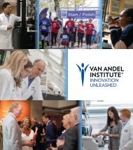
Microinjection of embryonic stem cells<br />
In these photos, technicians are microinjecting embryonic stem cells into mouse blastocysts for gene targeting studies.<br />
(Photos by Julie Koeman)<br />
23
Vivarium and Laboratory of Transgenics<br />
Bryn Eagleson, A.A., RLATG<br />
Bryn Eagleson began her career in laboratory animal services in 1981 with Litton<br />
Bionetics at the National Cancer Institute’s Frederick Cancer Research and<br />
Development Center (NCI-FCRDC) in Maryland. In 1983, she joined the Johnson &<br />
Johnson Biotechnology Center in San Diego, California. In 1988, she returned to the<br />
NCI-FCRDC, where she continued to develop her skills in transgenic technology and<br />
managed the transgenic mouse colony. During this time Ms. Eagleson attended<br />
Frederick Community College and Hood College in Frederick, Maryland. In 1999,<br />
she joined VARI as the Vivarium Director and Transgenics Special Program Manager.<br />
Managerial staff<br />
Jason Martin, RLATG<br />
Laboratory Members<br />
Technical staff<br />
Dawna Dylewski, B.S.<br />
Audra Guikema, B.S., L.V.T.<br />
Elissa Boguslawksi, RALAT<br />
Jamie Bondsfield<br />
Sylvia Marinelli<br />
Vivarium staff<br />
Lisa DeCamp, B.S.<br />
Laura Sixburry, B.S.<br />
Crystal Brady<br />
Kathy Geil<br />
Jarred Grams<br />
Tina Schumaker<br />
Research Interests<br />
T<br />
he<br />
goal of the vivarium and the<br />
transgenics laboratory is to develop,<br />
provide, and support high-quality mouse<br />
modeling services for the Van Andel Research<br />
Institute investigators, Michigan Technology Tri-<br />
Corridor collaborators, and the greater research<br />
community. We use two Topaz Technologies<br />
software products, Granite and Scion, for<br />
integrated management of the vivarium finances,<br />
the mouse breeding colony, and the Institutional<br />
Animal Care and Use Committee (IACUC)<br />
protocols and records. Imaging equipment, such<br />
as the PIXImus mouse densitometer and the<br />
Acuson Sequoia 512 ultrasound machine, is<br />
available for noninvasive imaging of mice.<br />
VetScan blood chemistry and hematology<br />
analyzers are now available for blood analysis.<br />
Also provided by the vivarium technical staff are<br />
an extensive xenograft model development and<br />
analysis service, rederivation, surgery, dissection,<br />
necropsy, breeding, and health-status monitoring.<br />
Transgenics<br />
Fertilized eggs contain two pronuclei, one that<br />
is derived from the egg and contains the maternal<br />
genetic material and one derived from the sperm<br />
that contains the paternal genetic material.<br />
As development proceeds, these two pronuclei<br />
fuse, the genetic material mixes, and the cell<br />
proceeds to divide and develop into an embryo.<br />
Transgenic mice are produced by injecting small<br />
quantities of foreign DNA (the transgene) into a<br />
pronucleus of a one-cell fertilized egg.<br />
DNA microinjected into a pronucleus randomly<br />
integrates into the mouse genome and will<br />
theoretically be present in every cell of the<br />
resulting organism. Expression of the transgene is<br />
controlled by elements called promoters that are<br />
genetically engineered into the transgenic DNA.<br />
Depending on the selection of the promoter, the<br />
transgene can be expressed in every cell of the<br />
mouse or in specific cell populations such as<br />
neurons, skin cells, or blood cells.<br />
Temporal expression of the transgene during<br />
development can also be controlled by genetic<br />
engineering. These transgenic mice are excellent<br />
models for studying the expression and function of<br />
the transgene in vivo.<br />
24
Standing from left to right: Bondsfield, Sixbury, Grams, DeCamp, Brady, Guikema, Martin<br />
seated from left to right: Schumaker, Dylewski, Marinelli, Eagleson, Boguslawski<br />
25
Bioinformatics Special Program<br />
Kyle A. Furge, Ph.D.<br />
Dr. Furge received his Ph.D. in biochemistry from the Vanderbilt University School<br />
of Medicine in 2000. Prior to obtaining his degree, he worked as a software<br />
engineer at YSI, Inc., where he wrote operating systems for embedded computer<br />
devices. Dr. Furge did his postdoctoral work in the laboratory of Dr. George Vande<br />
Woude and became a Bioinformatics Scientist at VARI in June of 2001.<br />
Laboratory Members<br />
Staff<br />
Karl Dykema, B.A.<br />
Research Interests<br />
Ashigh-throughput biotechnologies such<br />
as DNA sequencing, gene expression<br />
microarrays, and genotyping become<br />
more available to researchers, analysis of the data<br />
produced by these technologies becomes<br />
increasingly difficult. Disciplines such as<br />
bioinformatics and computational biology have<br />
recently emerged to help develop methods that<br />
assist in the storage, distribution, integration, and<br />
analysis of these data sets. The bioinformatics<br />
program at VARI is currently focused on using<br />
computational approaches to understand how<br />
cancer cells differ from normal cells at the<br />
molecular level. In addition, VARI is also part of<br />
the overall bioinformatics effort in the state of<br />
Michigan through the Michigan Center for<br />
Biological Information.<br />
Laboratory members from the bioinformatics<br />
program have worked on a wide variety of projects<br />
to further the research efforts at VARI in 2004.<br />
Recently, we constructed a program to identify<br />
short sequences within genes that are likely to be<br />
responsive to siRNA interference. siRNAs are<br />
short, double-stranded DNA sequences that when<br />
introduced into living cells bind to the RNA<br />
produced from a gene of interest and inhibit<br />
expression of the gene. As such, the introduction<br />
of siRNAs is becoming a more widely used<br />
technique to examine the role of individual genes<br />
in both cancerous and noncancerous cells.<br />
The program we developed contained a new<br />
algorithm, developed by one of the VARI<br />
investigators, to identify potential sites within<br />
genes that would be sensitive to siRNAs. In<br />
another project, we assisted the Microarray<br />
Technology Laboratory in the placement of quality<br />
control markers on gene expression microarrays.<br />
These microarrays contain more than 20,000<br />
unique DNA fragments that are robotically placed<br />
on a small glass slide. In order to ensure the DNA<br />
fragments are placed correctly, the quality control<br />
markers are robotically placed on the arrays in a<br />
very specific pattern as the arrays are constructed.<br />
As each array is produced, a quick visual<br />
inspection of the pattern of quality control markers<br />
can be used to verify that all the DNA fragments<br />
were placed on the arrays correctly.<br />
In addition to assisting other VARI research<br />
labs, our group has a special focus on<br />
understanding how cytogenetic abnormalities<br />
influence cancer development and progression.<br />
Many cancer types, including liver and kidney<br />
cancers, are associated with defined sets of DNA<br />
gains and losses. For example, the majority of<br />
hepatocellular carcinomas contain extra copies of<br />
chromosome 1p and lack copies of chromosome<br />
4q. In contrast, the majority of clear cell renal cell<br />
carcinomas contain an extra copy of chromosome<br />
5q and lack a copy of chromosome 3p.<br />
Interestingly, we and other groups have noticed<br />
that transcription is dramatically disrupted within<br />
regions of DNA copy number change. We are<br />
currently developing and testing a number of<br />
different algorithms to identify these disrupted<br />
regions using gene expression data. In addition,<br />
we are developing methods to identify key<br />
regulatory genes that lie within the abnormal<br />
region and may be involved in tumor development<br />
and/or progression.<br />
26
External Collaborators<br />
Xin Chen, Stanford University, Stanford, California<br />
Recent Publications<br />
Yang, X.J., M.-H. Tan, H.L. Kim, J.A. Ditlev, M.W. Betten, C.E. Png, E.J. Kort, K. Futami, K.J. Dykema,<br />
K.A. Furge, M. Takahashi, H. Kanayama, P.H. Tan, B.S. Teh, C. Luan, et al. In press. A molecular<br />
classification of papillary renal cell carcinoma. Cancer Research.<br />
Dykema, K.J., and K.A. Furge. 2004. Diminished transcription of chromosome 4q is inversely related to<br />
local spread of hepatocellular carcinoma. Genes, Chromosomes and Cancer 41(4): 390–394.<br />
Furge, Kyle A., Kerry A. Lucas, Masayuki Takahashi, Jun Sugimura, Eric J. Kort, Hiro-omi Kanayama,<br />
Susumu Kagawa, Philip Hoekstra, John Curry, Ximing J. Yang, and Bin T. Teh. 2004. Robust<br />
classification of renal cell carcinoma based on gene expression data and predicted cytogenetic<br />
profiles. Cancer Research 64(12): 4117–4121.<br />
Haven, C.J., V.M. Howell, P.H.C. Eilers, R. Dunne, M. Takahashi, M. van Puijenbroek, K. Furge, J. Kievit,<br />
M.-H. Tan, G.J. Fleuren, B.G. Robinson, L.W. Delbridge, J. Philips, A.E. Nelson, U. Krause, et al.<br />
2004. Gene expression of parathyroid tumors: molecular subclassification and identification of the<br />
potential malignant phenotype. Cancer Research 64(20): 7405–7411.<br />
Lindvall, Charlotta, Kyle Furge, Magnus Björkholm, Xiang Guo, Brian Haab, Elisabeth Blennow, Magnus<br />
Nordenskjöld, and Bin Tean Teh. 2004. Combined genetic and transcriptional profiling of acute<br />
myeloid leukemia with normal and complex karyotypes. Haematologica 89(9): 1072–1081.<br />
Sugimura, Jun, Richard S. Foster, Oscar W. Cummings, Eric J. Kort, Masayuki Takahashi, Todd T. Lavery,<br />
Kyle A. Furge, Lawrence H. Einhorn, and Bin Tean Teh. 2004. Gene expression profiling of earlyand<br />
late-relapse nonseminomatous germ cell tumor and primitive neuroectodermal tumor of the testis.<br />
Clinical Cancer Research 10(7): 2368–2378.<br />
Tan, Min-Han, Craig G. Rogers, Jeffrey T. Cooper, Jonathon A. Ditlev, Thomas J. Maatman, Ximing Yang,<br />
Kyle A. Furge, and Bin Tean Teh. 2004. Gene expression profiling of renal cell carcinoma. Clinical<br />
Cancer Research 10(18): 6315S–6321S.<br />
Yang, Ximing J., Jun Sugimura, Maria S. Tretiakova, Kyle Furge, Gregory Zagaja, Mitchell Sokoloff,<br />
Michael Pins, Raymond Bergan, David J. Grignon, Walter M. Stadler, Nicholas J. Vogelzang, and Bin<br />
Tean Teh. 2004. Gene expression profiling of renal medullary carcinoma: potential clinical relevance.<br />
Cancer 100(5): 976–985.<br />
From left to right: Furge, Dykema<br />
27
Laboratory of Cancer Immunodiagnostics<br />
Brian B. Haab, Ph.D.<br />
Dr. Haab obtained his Ph.D. in chemistry from the University of California at<br />
Berkeley in 1998. He then served as a postdoctoral fellow in the laboratory of<br />
Patrick Brown in the Department of Biochemistry at Stanford University. Dr. Haab<br />
joined VARI as a Special Program Investigator in May 2000.<br />
Staff<br />
Songming Chen, Ph.D.<br />
Michael Shafer, Ph.D.<br />
Sara Forrester, B.S.<br />
Darren Hamelinck, B.S.<br />
Randall Orchekowski, B.S.<br />
Laboratory Members<br />
Students<br />
Thomas LaRoche<br />
Richard Schildhouse<br />
Research Interests<br />
Many cancers are difficult to detect at<br />
early stages and are often diagnosed too<br />
late to allow curative treatment. Earlier<br />
and more accurate detection of cancer could lead<br />
to better outcomes for many patients. A greater<br />
knowledge of the molecular changes associated<br />
with the development of cancer could lead to a<br />
better understanding of disease mechanisms and<br />
improved diagnostic tests. The Haab laboratory<br />
is developing novel experimental approaches to<br />
gather such molecular information and to use it<br />
for the diagnosis of cancer. Our goal is that these<br />
studies will produce measurable benefits to<br />
cancer patients.<br />
We are taking a variety of approaches to<br />
identify changes in blood protein composition that<br />
define cancer and that could be diagnostically<br />
useful. Microarray methods have features that are<br />
particularly useful for this research. We have been<br />
developing several related antibody and protein<br />
microarray methods—such as two-color<br />
competition assays, sandwich assays, glycan<br />
detection, and antigen detection of antibodies—to<br />
analyze serum samples from cancer patients and<br />
controls. With these methods (Fig. 1), we can<br />
efficiently probe the binding to many different<br />
antibodies and proteins and explore the use of<br />
multiple measurements for classifying samples.<br />
Previous technological developments that are now<br />
in routine use include a high-sensitivity detection<br />
method (two-color rolling-circle amplification)<br />
and a new method for isolating multiple<br />
microarrays on a single slide, allowing highthroughput<br />
sample processing. Our research also<br />
makes use of gel and chromatographic separations<br />
and mass spectrometry to provide complementary<br />
experimental information.<br />
An ongoing study of protein profiles from the<br />
sera of pancreatic cancer patients and controls (in<br />
collaboration with Randall Brand and George<br />
Vande Woude) shows the value of antibody<br />
microarrays for diagnostics research. Protein<br />
profiles from antibody microarrays targeting a<br />
wide variety of proteins—such as those previously<br />
associated with cancer, involved in biological<br />
systems altered by cancer, or having elevated levels<br />
in the tumor environment—are revealing many<br />
proteins at either higher or lower abundances in the<br />
cancer patients. Some of the proteins were not<br />
known to be associated with pancreatic cancer and<br />
may contribute to improved early detection of the<br />
disease. The serum samples can be classified as<br />
from cancer or control patients using the antibody<br />
measurements. The accuracy of the classifications<br />
was greatly improved with multiple measurements<br />
in combination (relative to using individual<br />
measurements); we need to further develop this<br />
approach for cancer diagnostics.<br />
A recent modification to this technology<br />
measures alterations in the glycosylation state of<br />
the proteins. Glycosylation—the attachment of<br />
specific carbohydrate structures to proteins—<br />
plays a major role in determining protein function,<br />
and glycosylation alterations have been associated<br />
with the development and progression of cancer.<br />
The ability to conveniently measure changes in<br />
specific carbohydrates on different proteins could<br />
be valuable in identifying the changes most<br />
28
associated with cancer and thus of use for<br />
diagnostics. We have methods for detecting<br />
changes in glycosylation on proteins captured by<br />
antibody microarrays (see Fig. 1C) and are using<br />
those methods to profile the glycosylation<br />
alterations on serum proteins from cancer patients<br />
and control subjects.<br />
We use protein microarrays (see Fig. 1D) to<br />
gather additional information about changes<br />
occurring in the blood of cancer patients. Some<br />
tumor proteins elicit the production of antibodies<br />
targeting those proteins. The identification and<br />
measurement of tumor-recognizing antibodies<br />
could provide information about molecular<br />
alterations in the tumor and be valuable in cancer<br />
detection. The protein microarray is an ideal<br />
screening tool for that purpose. In collaboration<br />
with Gilbert Omenn and Samir Hanash,<br />
microarrays of tumor-derived proteins from<br />
cancer cell lines are probed with sera from cancer<br />
patients to identify proteins recognized by the<br />
patients’ antibodies. An ongoing study of serum<br />
from prostate cancer patients (in collaboration<br />
with Alan Partin) has identified several proteins<br />
that could be involved in an immune response.<br />
We are characterizing the nature of the responses<br />
and the proteins involved.<br />
The above methods may be applied in a novel<br />
way to further their effectiveness. We have begun<br />
to develop and improve existing diagnostic markers<br />
through the use of longitudinal measurements<br />
(serial measurements over time). In a collaboration<br />
with William Catalona, Robert Vessella, and Ziding<br />
Feng, we are looking at changes over time in the<br />
concentrations of several serum proteins leading up<br />
to disease recurrence in prostate cancer patients.<br />
We hypothesize that the use of individualized<br />
thresholds defining abnormal protein levels,<br />
defined by each person’s history of measurements,<br />
will yield improved diagnostic accuracy over the<br />
use of single, population-wide thresholds. Our<br />
hypothesis has been supported in some individual<br />
demonstrations, and we now have an experimental<br />
system for systematically exploring it for a large<br />
number of proteins and many patients.<br />
Figure 1. Antibody and protein microarray<br />
formats. A) Two-color competition assay. Two pools<br />
of proteins, respectively labeled with biotin and<br />
digoxigenin tags, are mixed and co-incubated on<br />
antibody microarrays. The relative amount of binding<br />
to each antibody from the two pools is determined<br />
through detection of the biotin and digoxigenin tags.<br />
B) Sandwich assay. After incubation of a protein<br />
sample on an antibody microarray, the amount of<br />
protein binding to each antibody is measured using a<br />
second antibody that targets the captured proteins.<br />
C) Glycan detection. A pool of digoxigenin-labeled<br />
proteins is incubated on antibody microarrays, and the<br />
level of protein binding at each antibody is determined<br />
by detection of the digoxigenin tag. The amount of a<br />
particular glycan on the captured proteins is detected<br />
using a biotin-labeled protein that specifically binds to<br />
that glycan. D) Protein array detection of antibodies.<br />
Serum samples are incubated on arrays containing a<br />
variety of tumor-derived proteins. Antibodies in the<br />
serum samples that recognize and bind to the<br />
proteins are detected using a secondary antibody that<br />
binds to human antibodies.<br />
We are seeking to apply the methods<br />
described above in ways that will have a positive<br />
result in terms of cancer care. The incorporation<br />
of mass spectrometry methods, through<br />
collaborations with Greg Cavey at VARI and<br />
members of the Michigan Proteome Consortium,<br />
will provide more opportunities for discovery.<br />
The further testing of the value of these tools is<br />
being pursued through local clinical oncology<br />
programs. The access to clinical practice is<br />
especially valuable for translating the<br />
development and discovery in our laboratory into<br />
benefits for cancer patients.<br />
External Collaborators<br />
Phil Andrews, Gilbert Omenn, and Diane Simeone, University of Michigan, Ann Arbor<br />
Randall Brand, Evanston Northwestern Healthcare, Illinois<br />
E. Brian Butler and Bin S. Teh, Baylor College of Medicine, Houston, Texas<br />
William Catalona, John Grayhack, and Anthony Schaeffer, Northwestern University, Evanston, Illinois<br />
29
Jose Costa and Paul Lizardi, Yale University School of Medicine, New Haven, Connecticut<br />
Deborah Dillon, Brigham and Women’s Hospital, Boston, Massachusetts<br />
Ziding Feng and Samir Hanash, Fred Hutchinson Cancer Research Center, Seattle, Washington<br />
Jorge Marrero, University of Michigan Hospital, Ann Arbor<br />
Alan Partin, Johns Hopkins University, Baltimore, Maryland<br />
Peter Schirmacher, University of Cologne, Germany<br />
Robert Vessella, University of Washington, Seattle<br />
Cornelius Verweij, University of Amsterdam, The Netherlands<br />
Recent Publications<br />
Hamelinck, D., H. Zhou, L. Li, Z. Feng, C. Verweij, D. Dillon, J. Costa, and B.B. Haab. In press.<br />
“Optimized normalization for antibody microarrays and the identification of serum protein<br />
alterations associated with pancreatic cancer.” Molecular & Cellular Proteomics.<br />
Haab, B.B., and P.M. Lizardi. In press. “RCA-enhanced protein detection arrays.” Methods in<br />
Molecular Biology.<br />
Breuhahn, Kai, Sebastian Vreden, Ramsi Haddad, Susanne Beckebaum, Dirk Stippel, Peer Flemming,<br />
Tanja Nussbaum, Wolfgang H. Caselmann, Brian B. Haab, and Peter Schirmacher. 2004.<br />
Molecular profiling of human hepatocellular carcinoma defines mutually exclusive interferon<br />
regulation and insulin-like growth factor II overexpression. Cancer Research 64(17): 6058–6064.<br />
Konwinski, R., R. Haddad, J.A. Chun, S. Klenow, S. Larson, B.B. Haab, and L.L. Furge. 2004.<br />
Oltipraz, 3H-1,2-dithiole-3-thione and sulforaphane induce overlapping and protective antioxidant<br />
responses in murine microglial cells. Toxicology Letters 153(3): 343–355.<br />
Lindvall, Charlotta, Kyle Furge, Magnus Björkholm, Xiang Guo, Brian Haab, Elisabeth Blennow,<br />
Magnus Nordenskjöld, and Bin Tean Teh. 2004. Combined genetic and transcriptional profiling<br />
of acute myeloid leukemia with normal and complex karyotypes. Haematologica 89(9):<br />
1072–1081.<br />
Qiu, Ji, Juan Madoz-Gurpide, David E. Misek, Rork Kuick, Dean E. Brenner, George Michailidis,<br />
Brian B. Haab, Gilbert S. Omenn, and Sam Hanash. 2004. Development of natural protein<br />
microarrays for diagnosing cancer based on an antibody response to tumor antigens. Journal of<br />
Proteome Research 3(2): 261–267.<br />
Zhou, Heping, Kerri Bouwman, Mark Schotanus, Cornelius Verweij, Jorge A. Marrero, Deborah<br />
Dillon, Jose Costa, Paul Lizaardi, and Brian B. Haab. 2004. Two-color, rolling-circle<br />
amplification on antibody microarrays for sensitive, multiplexed serum-protein measurements.<br />
Genome Biology 5(4): R28.<br />
From left to right: Haab, Hamelinck, Orchekowski, Chen, Shafer, Forrester<br />
30
Molecular Medicine and Virology Group<br />
Sheri L. Holmen, Ph.D.<br />
Dr. Holmen received her M.S. in biomedical science from Western Michigan University<br />
in 1995 and her Ph.D. in tumor biology from the Mayo Clinic College of Medicine in<br />
2000. She did her postdoctoral work at VARI in the laboratory of Bart Williams from<br />
2000 to 2003 and became a Junior Investigator at VARI in December 2003.<br />
Laboratory Members<br />
Staff<br />
Marleah Russo<br />
Research Interests<br />
T<br />
he<br />
primary focus of the Molecular<br />
Medicine and Virology Group is to<br />
identify molecules that can be effective<br />
targets for cancer therapy, with the goal of<br />
developing better therapies with fewer side effects.<br />
The sequencing of the human genome has yielded<br />
a wealth of biological data, but we still know<br />
relatively little about which genes are causally<br />
associated with tumor development and which are<br />
only markers of cancer. Because of the high cost<br />
of developing new therapies, it is important that we<br />
identify which genetic changes can be<br />
productively targeted. We are concentrating our<br />
initial efforts on melanoma and glioblastoma,<br />
tumors which demonstrate constitutive activation<br />
of Ras signaling.<br />
The RCAS system<br />
We use a series of replication-competent<br />
retroviral vectors based on the SR-A strain of Rous<br />
sarcoma virus (RSV), a member of the avian<br />
leukosis virus (ALV) family, to study the roles of<br />
different genes in tumor initiation and progression.<br />
RSV is the only known naturally occurring,<br />
replication-competent retrovirus that carries an<br />
oncogene, src. In the RCAS vectors, the region<br />
encoding src (which is dispensable for viral<br />
replication) has been replaced by a synthetic DNA<br />
linker. Foreign genes inserted into this linker are<br />
expressed from the viral LTR promoter via a<br />
subgenomic splice site (just as src is in RSV).<br />
RCAN vectors differ from RCAS vectors in that<br />
they lack the src splice acceptor; the gene of<br />
interest is inserted along with an internal promoter.<br />
Higher-titer viruses subsequently have been<br />
generated by replacing the RSV SR-A pol gene<br />
with the pol gene of the Bryan strain of RSV.<br />
These vectors are termed RCASBP or RCANBP.<br />
The ability of these vectors to infect non-avian<br />
cells relies on expression of the corresponding<br />
receptor on the cell surface. The viral receptor is<br />
typically introduced into mammalian cells (or<br />
mice) via an inducible and/or tissue-specific<br />
transgene. Therefore, this system allows for tissueand<br />
cell-specific targeted infection of mammalian<br />
cells through ectopic expression of the viral<br />
receptor. Alternatively, when targeted infection of<br />
mammalian cells is not required (e.g., in cell<br />
culture), infection can be achieved through the use<br />
of non-avian envelopes, such as the amphotropic<br />
envelope from murine leukemia virus. The<br />
receptor for this envelope is endogenously<br />
expressed on almost all mammalian cells.<br />
We have used the RCASBP/RCANBP family<br />
of retroviral vectors extensively in both cultured<br />
cells and live animals for studies of viral<br />
replication and of cancer modeling in mice. Most<br />
of these studies have analyzed gain-of-function<br />
phenotypes by delivering and overexpressing a<br />
particular gene of interest. Recently, we<br />
engineered the RCANBP vector to reduce the<br />
expression of specific genes through the delivery<br />
of short hairpin RNA sequences. We also<br />
engineered this vector to control the expression of<br />
the inserted gene using the tetracycline (tet)-<br />
regulated system. Sequences inserted into this<br />
region are transcribed from a tet-responsive<br />
element and not the viral LTR. This virus allows<br />
inserted genes to be turned on and off in order to<br />
determine if expression of the gene is required for<br />
tumor initiation, maintenance, and progression.<br />
The ability to turn off gene expression will help<br />
determine if that gene is a good target for therapy.<br />
31
Melanoma<br />
Melanoma is the most rapidly increasing<br />
malignancy among young people in the United<br />
States. If detected early, the disease is easily<br />
treated, but once the disease has metastasized it<br />
has a high mortality rate. Current systemic<br />
therapy for advanced, metastatic melanoma<br />
includes dacarbazine (DTIC) chemotherapy,<br />
either alone or in combination with other agents,<br />
and biological therapy using recombinant<br />
interferon-α (IFN-α) and/or interleukin-2 (IL-2).<br />
However, except on rare occasions, none of these<br />
treatments has produced long-term control of the<br />
disease, and cytokine therapy is associated with<br />
significant toxicities.<br />
We have developed a mouse model of<br />
melanoma based on the avian RCAS/TVA system.<br />
In this model, the retroviral receptor TVA is<br />
expressed under the control of the tyrosinaserelated<br />
protein 2 (TRP2) promoter, which is<br />
expressed in melanocytes. In replicating<br />
mammalian cells that express TVA, the viral vector<br />
is capable of stably integrating into the DNA and<br />
expressing the experimental gene at high levels,<br />
but the virus is replication-defective because viral<br />
RNA and proteins are inefficiently produced.<br />
Therefore, the viral vectors cannot spread in the<br />
target animals; in addition, since little viral<br />
envelope protein is produced, there is no<br />
interference to superinfection. Theoretically, there<br />
is no limit to the number of experimental genes<br />
that can be introduced into a TVA-expressing<br />
mammalian cell. The ability of these cells to be<br />
infected by multiple viruses allows efficient<br />
modeling of melanoma, because multiple<br />
oncogenic alterations can be introduced into the<br />
same cell or animal without the costs associated<br />
with mating multiple strains of transgenic mice.<br />
Activated ras oncogenes, which turn on<br />
mitogen-activated protein kinase (MAPK)<br />
signaling, are detected in approximately 20% of<br />
human melanomas. Recently, activating mutations<br />
in the B-raf gene, which also activate MAPK<br />
signaling, have been found in more than 65% of<br />
malignant melanomas. With mutually exclusive<br />
mutations in ras and B-raf, the MAPK signaling<br />
pathway is constitutively activated in over 85% of<br />
cases of malignant melanoma, indicating its<br />
importance. Overexpression of activated H-ras<br />
(G12V) specifically in melanocytes of Ink4adeficient<br />
mice results in the development of<br />
multiple spontaneous cutaneous melanomas.<br />
However, unlike in the human disease, these<br />
tumors fail to metastasize. A conditional Ink4a/Arf<br />
knockout allele, Ink4a-lox, has been introduced<br />
into the germline of FVB/N mice. The lox sites<br />
flank exons 2 and 3 of this locus such that Cremediated<br />
excision eliminates both p16 INK4A and<br />
p19 ARF . We have recently obtained two<br />
homozygous Ink4a-lox/lox breeding pairs, which<br />
will be crossed to our TRP2-TVA transgenic mice.<br />
We plan to use the mice to study the role of<br />
different genes in melanoma initiation,<br />
maintenance, and progression.<br />
Glioblastoma<br />
Glioblastoma multiforme (GBM) is the most<br />
common and aggressive primary brain tumor. It is<br />
also the most fatal: mean survival is less than one<br />
year from the time of diagnosis, with less than 10%<br />
survival after two years. Despite major<br />
improvements in imaging, radiation, and surgery,<br />
the prognosis for patients with this disease has not<br />
changed in the last 20 years. Recently, genes that<br />
are differentially expressed in tumor tissue relative<br />
to normal brain tissue have been found. However,<br />
those that can be productively targeted for<br />
therapeutic intervention in human patients remain<br />
to be identified.<br />
A mouse model of human GBM based on the<br />
avian RCAS/TVA system has been developed by<br />
Eric Holland. In this model, the retroviral receptor<br />
TVA is expressed under the control of the Nestin<br />
promoter, which is active in neural and glial<br />
progenitors. Intracranial infection of Nestin-TVA<br />
mice with RCASBP(A)-Akt and RCASBP(A)-<br />
KRas induces glioblastomas that are histologically<br />
similar to human GBM. We have generated an<br />
RCANBP(A)TRE-KRas virus in which expression<br />
of K-Ras can be controlled post-delivery. We are<br />
using the Nestin-TVA model to test the function of<br />
this virus in vivo. We plan to use these vectors to<br />
determine if K-Ras expression is required for<br />
tumor maintenance in this system and to identify<br />
which genes are appropriate targets for therapy.<br />
Antiviral strategies<br />
A second, related focus of our group is the<br />
identification of effective antiviral strategies using<br />
the RCASBP/RCANBP family of retroviral<br />
vectors as a model system. Viral diseases pose a<br />
32
major risk to the food supply and to animal<br />
welfare, especially in today’s high-intensity animal<br />
agriculture. Many viruses are highly<br />
communicable and are capable of rapid mutation<br />
to escape immune surveillance; few effective antiviral<br />
drugs are available. Over the past several<br />
years, we have developed strategies aimed at<br />
conferring dominant resistance to viral pathogens<br />
in chickens. These strategies have focused on<br />
manipulating viral (“pathogen-derived resistance”)<br />
and/or cellular genes (“host-derived resistance”) to<br />
express new proteins capable of disrupting the<br />
viral life cycle. The results have provided valuable<br />
information on the biology of avian viruses, as<br />
well as having the potential for practical<br />
application. We are now working to adapt the new<br />
RNA interference (RNAi) technology to the<br />
development of new anti-viral strategies for two<br />
important chicken pathogens, ALV and Marek’s<br />
Disease virus (MDV). These two targets have<br />
been chosen because both are economically<br />
important and because they have distinctly<br />
different infectious cycles that provide different<br />
challenges for RNAi.<br />
External Collaborators<br />
Jerry Dodgson, Michigan State University, East Lansing<br />
Henry Hunt and Huanmin Zhang, Avian Disease and Oncology Laboratory, East Lansing, Michigan<br />
Recent Publications<br />
Ai, Minrong, Sheri L. Holmen, Wim van Hul, Bart O. Williams, and Matthew W. Warman. <strong>2005</strong>.<br />
Reduced affinity to and inhibition by DKK1 form a common mechanism by which high bone<br />
mass–associated missense mutations in LRP5 affect canonical Wnt signaling. Molecular and<br />
Cellular Biology 25(12): 4946–4955.<br />
Holmen, Sheri L., Scott A. Robertson, Cassandra R. Zylstra, and Bart O. Williams. <strong>2005</strong>.<br />
Wnt-independent activation of ß-catenin mediated by a Dkk-1-Frizzled 5 fusion protein.<br />
Biochemical and Biophysical Research Communications 328(2): 533–539.<br />
Holmen, Sheri L., Cassandra R. Zylstra, Aditi Mukherjee, Robert Sigler, Marie-Claude Faugere,<br />
Mary Bouxsein, Lianfu Deng, Thomas Clemens, and Bart O. Williams. <strong>2005</strong>. Essential role of<br />
ß-catenin in postnatal bone acquisition. Journal of Biological Chemistry 280(22): 21162–21168.<br />
Sanchez-Perez, Luis, Timothy Kottke, Rosa Maria Diaz, Atique Ahmed, Jill Thompson, Heung<br />
Chong, Alan Melcher, Sheri Holmen, Gregory Daniels, and Richard G. Vile. <strong>2005</strong>. Potent<br />
selection of antigen loss variants of B16 melanoma following inflammatory killing of<br />
melanocytes in vivo. Cancer Research 65(5): 2009–2017.<br />
Bromberg-White, Jennifer L., Craig P. Webb, Veronique<br />
S. Patacsil, Cindy K. Miranti, Bart O. Williams, and<br />
Sheri L. Holmen. 2004. Delivery of short hairpin<br />
RNA sequences by using a replication-competent<br />
avian retroviral vector. Journal of Virology 78(9):<br />
4914–4916.<br />
Holmen, Sheri L., Troy A. Giambernardi, Cassandra R.<br />
Zylstra, Bree D. Buckner-Berghuis, James H. Resau,<br />
J. Fred Hess, Vaida Glatt, Mary L. Bouxsein,<br />
Minrong Ai, Matthew L. Warman, and Bart O.<br />
Williams. 2004. Decreased BMD and limb<br />
deformities in mice carrying mutations in both Lrp5<br />
and Lrp6. Journal of Bone and Mineral Research<br />
19(12): 2033–2040.<br />
From left to right: Russo, Holmen<br />
33
Laboratory of Integrin Signaling and Tumorigenesis<br />
Cindy K. Miranti, Ph.D.<br />
Dr. Miranti received her M.S. in microbiology from Colorado State University in 1982<br />
and her Ph.D. in biochemistry from Harvard Medical School in 1995. She was a<br />
postdoctoral fellow in the laboratory of Dr. Joan Brugge at ARIAD Pharmaceuticals,<br />
Cambridge, Mass., from 1995 to 1997 and in the Department of Cell Biology at<br />
Harvard Medical School from 1997 to 2000. Dr. Miranti joined VARI as a <strong>Scientific</strong><br />
Investigator in January 2000. She is also an Adjunct Assistant Professor in the<br />
Department of Physiology at Michigan State University.<br />
Laboratory Members<br />
Staff<br />
Suganthi Chinnaswamy, Ph.D.<br />
Mathew Edick, Ph.D.<br />
Robert Long, B.A.<br />
Veronique V. Schulz, B.S.<br />
Student<br />
Erik Freiter<br />
Research Interests<br />
Our laboratory is interested in<br />
understanding the mechanisms by which<br />
integrin receptors, interacting with the<br />
extracellular matrix, regulate cell processes<br />
involved in the development of cancer. Using<br />
tissue culture models, biochemistry, molecular<br />
genetics, and mouse models, we are defining the<br />
cellular and molecular events of integrindependent<br />
adhesion and downstream signaling that<br />
are important in melanoma and prostate<br />
tumorigenesis and metastasis.<br />
Integrins are transmembrane proteins that<br />
serve as receptors for extracellular matrix (ECM)<br />
proteins. By interacting with the ECM, integrins<br />
stimulate intracellular signaling transduction<br />
pathways that regulate cell shape, proliferation,<br />
migration, survival, gene expression, and<br />
differentiation. Integrins do not act autonomously;<br />
they are involved in “crosstalk” with receptor<br />
tyrosine kinases (RTKs) to regulate many cellular<br />
processes. Studies in our lab, for example, indicate<br />
that integrin-mediated adhesion to ECM proteins<br />
activates the epidermal growth factor receptors<br />
EGFR and ErbB2 and the HGF/SF receptor Met.<br />
Integrin-mediated activation of these RTKs is<br />
ligand-independent and is required for activation<br />
of a subset of intracellular signaling molecules in<br />
response to cell adhesion.<br />
The prostate gland and cancer<br />
Tumors that develop in cells of epithelial origin,<br />
i.e., carcinomas, represent the largest tumor burden<br />
in the United States. Prostate cancer is the most<br />
frequently diagnosed cancer in U.S. men and the<br />
second leading cause of cancer death in men.<br />
Eighty percent of human prostate tumors arise in the<br />
peripheral zone of the gland and are primarily<br />
confined to the intermediate basal and secretory<br />
epithelial cells. Patients who at the time of diagnosis<br />
have androgen-dependent and organ-confined<br />
prostate cancer are relatively easy to cure through<br />
radical prostatectomy or localized radiotherapy.<br />
However, patients with aggressive and metastatic<br />
disease have fewer options. Androgen ablation can<br />
significantly reduce the tumor burden in these<br />
patients, but the potential for relapse and the<br />
development of androgen-independent cancer is<br />
high. Currently there are no effective treatments for<br />
patients who reach this stage of disease.<br />
In the human prostate secretory glands, basal<br />
epithelial cells form a contiguous layer adjacent to<br />
the basement membrane. Upon them rests a layer of<br />
secretory luminal cells, forming a stratified<br />
epithelium. The basal cells express a broad<br />
repertoire of integrins, including α2, α3, α6, β1, and<br />
β4. The secretory cells express primarily α6 and β1,<br />
with some α2. In vivo, basal cells secrete and<br />
organize a laminin 5–containing basement<br />
membrane that also contains collagens IV and VII<br />
and laminin 10. The basal cells bind to laminin 5<br />
and collegen VII through α6β4 to form<br />
hemidesmosomal complexes at the basal surface.<br />
Basal cells adhering to this matrix respond to growth<br />
factors secreted by the surrounding stroma,<br />
including EGF and HGF. They proliferate and give<br />
rise to the nonproliferating secretory cells through<br />
34
the generation of an intermediate, transiently<br />
amplifying cell population that has traits of both cell<br />
types. In primary prostate tumors, α6β4 integrin<br />
and its ligands, laminin 5 and collagen VII, are lost.<br />
The tumor cells, unlike normal secretory cells,<br />
develop the ability to adhere, via α2β1 and α6β1, to<br />
an altered basement membrane consisting of<br />
collagen IV and laminin 10. Thus, tumor cells have<br />
characteristics of both basal and secretory cells, but<br />
not all the properties of either.<br />
Whether the tumor cells are derived from the<br />
transient differentiating population of basal cells or<br />
from differentiated secretory cells has not been<br />
unequivocally determined, but it is clear that the<br />
way in which these cells interact with the ECM has<br />
been changed. If tumor cells are derived from<br />
basal cells, they are now interacting with an altered<br />
matrix and using different integrins to engage it. If<br />
they are derived from the secretory cells, the tumor<br />
cells are now engaging a matrix, which they did<br />
not do previously. A fundamental question in our<br />
lab is whether the changes in integrin/matrix<br />
interactions that occur in tumor cells are required<br />
for or help to drive the survival of tumor cells.<br />
The role of integrins and RTKs in<br />
prostate epithelial cell survival<br />
Increased cell survival due to resistance to cell<br />
death is a prerequisite for tumorigenesis. Several<br />
reports have suggested that the signaling pathways<br />
that regulate cell survival in normal prostate<br />
epithelial cells are different from those in prostate<br />
tumor cells. How integrin engagement of different<br />
ECMs regulates survival pathways is not known.<br />
We have recently found that integrin-induced<br />
activation of both EGFR and c-Met in primary<br />
prostate epithelial cells is required for cell survival<br />
in the absence of growth factors. Our goal is to<br />
determine how integrin activation of EGFR and<br />
c-Met regulate cell survival.<br />
We have previously shown that integrin<br />
activation of EGFR is required for integrinmediated<br />
induction of the Ras/Erk and PI3K/Akt<br />
signaling pathways. Recent studies indicate that<br />
integrin activation of Src depends on c-Met.<br />
Interestingly, inhibition of either Src or Ras/Erk<br />
signaling—but not PI3K/Akt signaling—induces<br />
cell death in primary prostate cells even when they<br />
are still adherent to matrix. Together these data<br />
indicate that Src signals generated by c-Met, as well<br />
as Ras/Erk signals generated by EGFR, are driving<br />
cell survival in primary cells (Fig. 1). We are<br />
currently exploring the downstream events that Src<br />
and Erk regulate to maintain cell survival on ECM.<br />
Figure 1. Met and EGFR both act independently<br />
to regulate integrin-mediated survival of primary<br />
prostate epithelial cells. Activation of Src and Erk<br />
through Met and EGFR, respectively, are proposed to<br />
be involved in integrin-mediated survival.<br />
We are also exploring the mechanisms by<br />
which integrins activate EGFR and ErbB2. This<br />
activation is ligand independent, requires only the<br />
cytoplasmic domain of EGFR, and stimulates the<br />
phosphorylation of only a subset of sites on EGFR.<br />
ErbB2 activation depends on EGFR, suggesting<br />
that integrins induce the formation of an<br />
EGFR/ErbB2 heterodimer. Integrin activation of<br />
Src or FAK is not required, but integrin activation<br />
of a phosphatase may be involved; we are currently<br />
investigating the role of several phosphatases.<br />
Integrin and RTK crosstalk in<br />
prostate cancer metastasis<br />
Death from prostate cancer is due to the<br />
development of metastatic disease, which is<br />
difficult to control. The mechanisms involved in<br />
progression to metastatic disease are not<br />
understood. Our approach is to characterize genes<br />
that are specifically associated with metastatic<br />
prostate cancer. CD82/KAI1 is a metastasis<br />
suppressor gene whose expression is specifically<br />
lost in metastatic cancer but not in primary tumors.<br />
CD82/KAI1 is known to associate with both<br />
integrins and RTKs. Our goal has been to<br />
determine how loss of CD82/KAI1 expression<br />
promotes metastasis by studying the role of<br />
CD82/KAI1 in integrin and RTK crosstalk.<br />
35
During prostate cancer progression there is a<br />
shift in the expression of laminin-specific integrins:<br />
β4 integrins are lost, and there is a concomitant<br />
increase in α6β1 and α3β1, which both interact<br />
with CD82/KAI1. We predict that the loss of<br />
CD82/KAI1 alters the function of α6β1 and α3β1.<br />
Using primary prostate epithelial cells, which<br />
express high levels of CD82/KAI1, as well as<br />
several prostate tumor cell lines that do not, we are<br />
exploring the role of CD82/KAI1 in regulating<br />
α6β1- and α3β1-mediated cell adhesion, migration,<br />
and integrin signaling. We have found that<br />
overexpression of CD82/KAI1 in tumor cells<br />
suppresses laminin-specific migration and invasion;<br />
integrin-induced EGFR and c-Met receptor<br />
activation; Src and Lyn activation; and activation of<br />
the Src/Lyn substrates Cas and FAK. Furthermore,<br />
integrin activation of Src is dependent on c-Met, and<br />
laminin-mediated invasion depends on both Src and<br />
c-Met. Together these data indicate that<br />
CD82/KAI1 normally acts to regulate integrin<br />
signaling to c-Met such that upon its loss of<br />
expression in tumor cells, signaling through c-Met<br />
to Src is increased, leading to increased motility and<br />
invasion (Fig. 2). We are currently determining the<br />
mechanism by which CD82/KAI1 down-regulates<br />
c-Met signaling. In reciprocal experiments, we are<br />
inhibiting the expression of CD82/KAI1 in primary<br />
cells using siRNA and mouse models.<br />
Integrin regulation of melanoma<br />
progression by PKC<br />
The incidence of melanoma has been steadily<br />
increasing over the last 10 years. If diagnosed at<br />
an early stage, melanoma is curable, but once it has<br />
become invasive, it progresses very rapidly and is<br />
virtually untreatable. Metastasis and invasion by<br />
tumor cells require the activity of integrins, and in<br />
melanoma, expression of the αvβ3 integrin is<br />
induced during the development of invasive<br />
disease. Therefore, an understanding of how the<br />
αvβ3 integrin functions to regulate invasion will<br />
help our understanding of melanoma metastasis.<br />
We have been focusing our attention on two<br />
signaling molecules, PKCα and Src, both of which<br />
are regulated by the αvβ3 integrin and whose<br />
activities are enhanced in metastatic melanoma.<br />
Adhesion of normal melanocytes to extracellular<br />
matrix induces the formation of focal adhesion<br />
complexes and actin stress fibers. However, in a<br />
highly invasive metastatic melanoma cell line,<br />
C8161.9, these structures are absent. We have<br />
shown that the levels of Src activity and PKCα<br />
protein are elevated in these cells, and<br />
overexpression of PKCα in immortalized normal<br />
melanocytes is sufficient to confer an invasive<br />
phenotype in vitro. We have found that the activity<br />
of Rac, a small GTPase that is required for the<br />
formation of lamellipodia, is elevated in these cells<br />
and that inhibition of PKCα blocks Rac activity.<br />
The activity of the small GTPase Rho, which is<br />
involved in stress fiber and focal adhesion<br />
formation, is negatively regulated by active Src.<br />
Thus PKCα, acting to enhance Rac, and active Src,<br />
acting to inhibit Rho, together drive the formation<br />
of lamellipodia and the dissolution of stress fibers<br />
and focal adhesions, leading to increased motility<br />
(Fig. 3). We are currently exploring how Src is<br />
activated in melanoma cells, as well as the effects<br />
of blocking Src and PKCα expression with siRNA<br />
on migration and invasion.<br />
Figure 2. CD82 reexpression in prostate tumor<br />
cells inhibits invasion by blocking integrinmediated<br />
signaling to Met and Src.<br />
Figure 3. PKCa and Src cooperate to enhance<br />
migration and invasion in melanoma cells by<br />
differentially targeting Rac and Rho.<br />
36
External Collaborators<br />
Beatrice Knudsen, Fred Hutchinson Cancer Research Center, Seattle, Washington<br />
Senthil Muthuswamy, Cold Spring Harbor Laboratory, New York<br />
Recent Publications<br />
Sridhar, S.C., and C.K. Miranti. In press. Tumor metastasis suppressor KAI1/CD82 is a tetraspanin.<br />
In Contemporary Cancer Research: Metastasis, C. Rinker-Schaeffer, M. Sokoloff, and D. Yamada,<br />
eds. Totowa, N.J.: Humana Press.<br />
Bill, Heather M., Beatrice Knudsen, Sheri L. Moores, Senthil K. Muthuswamy, Vikram R. Rao, Joan S.<br />
Brugge, and Cindy K. Miranti. 2004. Epidermal growth factor receptor–dependent regulation of<br />
integrin-mediated signaling and cell cycle entry in epithelial cells. Molecular and Cellular Biology<br />
24(19): 8586–8599.<br />
Lee, Chong-Chou, Andrew J. Putnam, Cindy K. Miranti, Margaret Gustafson, Ling-Mei Wang, George F.<br />
Vande Woude, and Chong-Feng Gao. 2004. Overexpression of sprouty-2 inhibits HGF/SF-mediated<br />
cell growth, invasion, migration, and cytokinesis. Oncogene 23(30): 5193–5202.<br />
From left to right: Schulz, Edick, Miranti, Long, Freiter, Sridhar<br />
37
Laboratory of Analytical, Cellular, and Molecular Microscopy<br />
and<br />
Laboratory of Microarray Technology and Molecular Diagnostics<br />
James H. Resau, Ph.D.<br />
Dr. Resau received his Ph.D. from the University of Maryland School of Medicine in<br />
1985. He has been involved in clinical and basic science imaging and pathologyrelated<br />
research since 1972. Between 1968 and 1994, he was in the U.S. Army<br />
(active duty and reserve) and served in Vietnam. From 1985 until 1992, Dr. Resau<br />
was a tenured faculty member at the University of Maryland School of Medicine,<br />
Department of Pathology. Dr. Resau was the Director of the Analytical, Cellular and<br />
Molecular Microscopy Laboratory in the Advanced BioScience Laboratories–Basic<br />
Research Program at the National Cancer Institute–Frederick Cancer Research and<br />
Development Center, Maryland, from 1992 to 1999. He joined VARI as a Special<br />
Program Senior <strong>Scientific</strong> Investigator in June 1999 and in 2003 was promoted to Deputy Director. In 2004, Dr.<br />
Resau assumed as well the direction of the Laboratory of Microarray Technology to consolidate the imaging and<br />
quantification of clinical samples in a CLIA-type research laboratory program.<br />
Staff<br />
Eric Kort, M.S.<br />
Bree Berghuis, B.S.,<br />
HTL (ASCP), QIHC<br />
Pete Haak, B.S.<br />
Eric Hudson, B.S.<br />
Paul Norton, B.S.<br />
J.C. Goolsby<br />
Laboratory Members<br />
Consulting Veterinarian<br />
Robert Sigler, D.V.M., Ph.D.<br />
Students<br />
Hien Dang<br />
Brandon Leeser<br />
Amy Percival<br />
Huang Tran<br />
Research Interests<br />
T<br />
he<br />
Laboratory of Analytical, Cellular,<br />
and Molecular Microscopy (ACMM)<br />
and the Laboratory of Microarray<br />
Technology and Molecular Diagnostics (MTMD)<br />
are organized and equipped to produce highresolution<br />
images, genomic arrays, and<br />
bioinformatics data that support the cellular and<br />
molecular biology programs of the Institute. Our<br />
laboratories collaborate with the investigators to<br />
improve the understanding, diagnosis, and<br />
characterization of disease, injury, and<br />
differentiation. Although we primarily study<br />
cancer, we work with investigators in and out of<br />
the Institute on a variety of diseases and<br />
processes. As examples, during the past year, we<br />
have used our microscopes to produce intravital<br />
images of the Met protein in living cells and<br />
animals; localized and quantified a unique<br />
transcriptional protein in human cells; worked<br />
closely with VARI’s Bart Williams to characterize<br />
the phenotypic changes in transgenic mice<br />
expressing mutations related to Wnt and the<br />
Lrp5/Lrp6 genes; began to image Art Alberts’<br />
small GTPase proteins in living cells; and<br />
collaborated in studies of the expression of c-Met<br />
in a series of primary human breast cancers.<br />
During 2004, we added two significant<br />
instruments to the ACMM laboratory. The first is<br />
the Aperio Scanscope, which enables us to<br />
digitize a 1 × 3-inch microscope slide in full<br />
color such that it can be analyzed, quantified, and<br />
shared throughout the digital network. The<br />
second is the Ventana automated immunostainer,<br />
which can carry out FISH, ISH, IHC, and<br />
array spotting in a programmed, specific, and<br />
accurate fashion. These two instruments have<br />
allowed us to increase our productivity, shorten<br />
our turnaround time, and improve the quality of<br />
our preparations without any increase in<br />
personnel. In the last calendar year, we<br />
processed 403 requests for histopathologic<br />
services that required 4,100 blocks and more<br />
than 31,600 glass slides. Using the Scanscope<br />
during December 2004, we scanned over 300<br />
cases in high resolution for inclusion in the<br />
VATR database. We anticipate generating digital<br />
Scanscope files for 3,000–4,000 cases in <strong>2005</strong><br />
that will be available on network servers. The<br />
MTMD laboratory is preparing gene expression<br />
data from cells and tissues and is correlating that<br />
with histology, tissue volume, and nuclear<br />
density to determine an effective and accurate<br />
38
screen for applications in molecular diagnostic<br />
assays. We have developed a QC and QA<br />
protocol for evaluating specimens for array<br />
analysis. During the past year we have prepared<br />
1,500 cDNA arrays for 20 collaborators from<br />
both in and outside the Institute.<br />
Our laboratories are primarily responsible for<br />
the archived clinical histopathology program<br />
called the Van Andel Tissue Repository (VATR).<br />
This program allows investigators to use existing<br />
human clinical samples to assess the expression<br />
of proteins. We also use these blocks to prepare a<br />
wide variety of tissue microarrays for research.<br />
We have increased the number of specimens in<br />
VATR to nearly 200,000 tissue samples. We are<br />
continuously entering data from the 200,000<br />
blocks and now have 54,473 reports that further<br />
explain and describe the archives. The reports are<br />
not directly linked to any personal identifiers or<br />
names and meet all HIPAA/CLIA regulations.<br />
During this year we have begun the process of<br />
imaging representative blocks from the cases and<br />
adding demographic data. The material from<br />
future years will have digital information on age,<br />
sex, and diagnosis and will be linked to image<br />
files. These samples will be used in cellular and<br />
molecular protocols approved by our Institutional<br />
Review Board. Since its inception, the VATR<br />
program has been used by 24 registered users<br />
who have submitted 534 requests for searches and<br />
101 subsequent tissue requests.<br />
In collaboration with Rick Hay, we have<br />
augmented the VATR program with freshly frozen<br />
tissues from the tissue acquisition program. This<br />
HIPAA-compliant program involves the active<br />
cooperation of patients, surgeons, and pathologists<br />
from area hospitals, and the surgical tissues<br />
collected are used primarily in cDNA and<br />
Affymetrix gene expression studies. This<br />
collection also provides the participating<br />
physicians with access to research collaborations,<br />
with the aim of facilitating the translation of<br />
research results into clinical practice. The goal of<br />
this project is to develop genetics-based<br />
diagnostic classification of human disease. There<br />
is a <strong>Scientific</strong> Advisory Board for this project<br />
comprising members of VARI and of the<br />
Spectrum Health pathology, surgical and medical<br />
oncology, and surgery departments.<br />
Original research within our labs focuses on<br />
quantification of images or arrays and the<br />
development of objective measurable data from<br />
images. A recent paper by Rozenblatt-Rosen et<br />
al. used a program written by Eric Kort for<br />
determining the co-localization of pixels that<br />
express unique fluorescent properties as well as<br />
the number of lumens or vessels in IHC-stained<br />
preparations. This is a direct extension of our<br />
Cytometry paper of 2003.<br />
We have recently obtained funding to<br />
advance our understanding of breast cancer in<br />
collaboration with Ilan Tsarfaty, George Vande<br />
Woude, and Craig Webb. We have begun<br />
molecular imaging of breast cancer and<br />
correlation of the findings with Affymetrix gene<br />
expression. Other collaborations within VARI<br />
involve Met and HGF/SF in cells and tissues; the<br />
location of gene-targeted proteins in rodents; and<br />
the evaluation of monoclonal antibodies as<br />
diagnostic reagents. We have been funded by the<br />
Michigan Technology Tri-Corridor to develop<br />
the commercialization of diagnostic gene<br />
expression (collaboration with Bin Teh), imaging<br />
of primary tumors (Rick Hay), and expression of<br />
mRNA in human sperm as an indicator of<br />
fertility or sterility (Stephen Krawetz). We are in<br />
the early stages of developing a program in<br />
neuropathology with a concentration on<br />
Parkinson’s disease, starting with the<br />
morphological and genomic characterization of<br />
human adult stem-cell populations of specific<br />
neurons.<br />
This year we have a student from Bath<br />
University in the U.K. The Bridges to the<br />
Baccalaureate program is a collaboration to<br />
support the recruitment of women and minorities<br />
into science careers; Dr. Resau is a coinvestigator<br />
and the VAI site coordinator for the<br />
program. In addition this year, we partnered with<br />
the Grand Rapids Area Pre-College Engineering<br />
Program (GRAPCEP) to build a school within a<br />
school for science education and instruction in<br />
Creston High School of the Grand Rapids Public<br />
School system. Our GRAPCEP mentorship<br />
program continues to be funded by Pfizer for a<br />
fifth year. Seven high school students have<br />
trained in the laboratory and are now in<br />
baccalaureate programs.<br />
39
External Collaborators<br />
Eric Arnoys and John Ubels, Calvin College, Grand Rapids, Michigan<br />
Stephan Baldus, University of Cologne, Germany<br />
Lonson Barr, Marcos Dantus, and Matti Koeppel, Michigan State University, East Lansing<br />
Nadia Harbeck, Ludwig-Maximilians-Universität, Munich, Germany<br />
Christine Hughes and O. Orit Rosen, Harvard University, Cambridge, Massachusetts<br />
Sylvia Kachalsky, Linkagene, Lod, Israel<br />
Iafa Keydar, Tel Aviv University, Israel<br />
Stephen Krawetz, Wayne State University, Detroit, Michigan<br />
Ernst Lengyel, University of Chicago, Illinois<br />
Maria Roberts, National Cancer Institute, Frederick, Maryland<br />
Rulong Shen, Ohio State University, Columbus<br />
Ilan Tsarfaty, Tel Aviv University, Israel<br />
Recent Publications<br />
Kort, E.J., M.R. Moore, E.A. Hudson, B. Leeser, G.M. Yeruhalmi, R. Leibowitz-Amit, G. Tsarfaty, I.<br />
Tsarfaty, S. Moshkovitz, and J.H. Resau. In press. Use of organ explant and cell culture. In<br />
Mechanisms of Carcinogenesis, Hans Kaiser, ed. Dordrecht, The Netherlands: Kluwer Academic.<br />
Lengyel, Ernst, Dieter Prechtel, James H. Resau, Katja Gauger, Anita Welk, Kristina Lindemann,<br />
Georgia Salanti, Thomas Richter, Beatrice Knudsen, George F. Vande Woude, and Nadia Harbeck.<br />
<strong>2005</strong>. c-Met overexpression in node-positive breast cancer identifies patients with poor clinical<br />
outcome independent of Her2/neu. International Journal of Cancer 113(4): 678–682.<br />
Rozenblatt-Rosen, Orit, Christina M. Hughes, Suraj J. Nannepaga, Kalai Selvi Shanmugam, Terry D.<br />
Copeland, Tad Guszczynski, James H. Resau, and Matthew Meyerson. <strong>2005</strong>. The parafibromin<br />
tumor suppressor protein is part of a human Paf1 complex. Molecular and Cellular Biology 25(2):<br />
612–620.<br />
Seals, Darren F., Eduardo F. Azucena, Jr., Ian Pass, Lia Tesfay, Rebecca Gordon, Melissa Woodrow,<br />
James H. Resau, and Sara A. Courtneidge. <strong>2005</strong>. The adaptor protein Tks5/Fish is required for<br />
podosome formation and function, and for the protease-driven invasion of cancer cells. Cancer<br />
Cell 7(2): 155–165.<br />
Akervall, Jan, Xiang Guo, Chao-Nan Qian, Jacqueline Schoumans, Brandon Leeser, Eric Kort,<br />
Andrew Cole, James Resau, Carol Bradford, Thomas Carey, Johan Wennerberg, Harald Anderson,<br />
Jan Tennvall, and Bin T. Teh. 2004. Genetic and expression profiles of squamous cell carcinoma<br />
of the head and neck correlate with cisplatin sensitivity and resistance in cell lines and patients.<br />
Clinical Cancer Research 10(24): 8204–8213.<br />
Birchenall-Roberts, Maria C., Tao Fu, Ok-sun Bang, Michael Dambach, James H. Resau, Cari L.<br />
Sadowski, Daniel C. Bertolette, Ho-Jae Lee, Seong-Jin Kim, and Francis W. Ruscetti. 2004.<br />
Tuberous sclerosis complex 2 gene product interacts with human SMAD proteins. Journal of<br />
Biological Chemistry 279(24): 25605–25613.<br />
Holmen, Sheri L., Troy A. Giambernardi, Cassandra R. Zylstra, Bree D. Buckner-Berghuis, James H.<br />
Resau, J. Fred Hess, Vaida Glatt, Mary L. Bouxsein, Minrong Ai, Matthew L. Warman, and Bart<br />
O. Williams. 2004. Decreased BMD and limb deformities in mice carrying mutations in both<br />
Lrp5 and Lrp6. Journal of Bone and Mineral Research 19(12): 2033–2040.<br />
Tan, Min-Han, Carl Morrison, Pengfei Wang, Ximing Yang, Carola J. Haven, Chun Zhang, Ping Zhao,<br />
Maria S. Tretiakova, Eeva Korpi-Hyovalti, John R. Burgess, Khee Chee Soo, Wei-Keat Cheah,<br />
Brian Cao, James Resau, Hans Morreau, and Bin Tean Teh. 2004. Loss of parafibromin<br />
immunoreactivity is a distinguishing feature of parathyroid carcinoma. Clinical Cancer Research<br />
10(19): 6629–663<br />
40
From left to right: Resau, Goolsby, Haak, Berghuis, Norton, Percival, Hudson, Ferrell<br />
41
Laboratory of Germline Modification<br />
Pamela J. Swiatek, Ph.D., M.B.A.<br />
Dr. Swiatek received her M.S. (1984) and Ph.D. (1988) degrees in pathology from<br />
Indiana University. From 1988 to 1990, she was a postdoctoral fellow at the Tampa<br />
Bay Research Institute. From 1990 to 1994, she was a postdoctoral fellow at the<br />
Roche Institute of Molecular Biology in the laboratory of Tom Gridley. From 1994 to<br />
2000, Dr. Swiatek was a Research Scientist and Director of the Transgenic Core<br />
Facility at the Wadsworth Center in Albany, N.Y., and an Assistant Professor in the<br />
Department of Biomedical Sciences at the State University of New York at Albany.<br />
She joined VARI as a Special Program Investigator in August 2000. She has been<br />
the chair of the Institutional Animal Care and Use Committee since 2002 and is an<br />
Adjunct Assistant Professor in the College of Veterinary Medicine at Michigan State<br />
University. Dr. Swiatek received her M.B.A. in <strong>2005</strong> from Krannert School of<br />
Management at Purdue University.<br />
Staff<br />
Julie Koeman, B.S.<br />
Kellie Sisson, B.S.<br />
Juraj Zahatnansky, B.S.<br />
Laboratory Members<br />
IACUC Coordinator<br />
Kaye Johnson, B.S.<br />
Research Interests<br />
The germline modification lab is a fullservice<br />
lab that functions at the levels of<br />
service, research, and teaching to<br />
develop, analyze, and maintain mouse models of<br />
human disease. Our lab applies a business<br />
philosophy to core service offerings and we focus<br />
on scientific innovation, customer satisfaction,<br />
and service excellence. Mouse models are<br />
produced using gene-targeting technology, a wellestablished,<br />
powerful method for inserting<br />
specific genetic changes into the mouse genome.<br />
The resulting mice can be used to study the effects<br />
of these changes in the complex biological<br />
environment of a living organism. The genetic<br />
changes can include the introduction of a gene<br />
into a specific site in the genome, (gene “knockin”)<br />
or the inactivation of a gene already in the<br />
genome (gene “knock-out”). Since these<br />
mutations are introduced into the reproductive<br />
cells known as the germline, they can be used to<br />
study the developmental aspects of gene function<br />
associated with inherited genetic diseases.<br />
In addition to traditional gene-targeting<br />
technologies, the germline modification lab can<br />
produce mouse models in which the gene of<br />
interest is inactivated in a target organ or cell line<br />
instead of in the entire animal. These types of<br />
mouse models, known as conditional knock-outs,<br />
are particularly useful in studying genes that, if<br />
missing, cause the mouse to die as an embryo.<br />
The lab also has the capability to produce mutant<br />
embryos that have a wild-type placenta using<br />
tetraploid embryo technology. This technique is<br />
useful when the gene-targeted mutation prevents<br />
implantation of the mouse embryo in the uterus.<br />
We also assist in the development of embryonic<br />
stem (ES) or fibroblast cell lines from mutant<br />
embryos, which allows for in vitro studies of the<br />
gene mutation.<br />
Our gene-targeting service encompasses<br />
three major procedures: DNA electroporation,<br />
clone expansion and cryopreservation, and<br />
microinjection. Gene targeting procedures are<br />
initiated by mutating the genomic DNA of interest<br />
and inserting it into ES cells using the<br />
electroporation technique. The mutated gene<br />
integrates into the genome of the ES cells and, by<br />
a process called homologous recombination,<br />
replaces one of the two wild-type copies of the<br />
gene in the cells. Clones are identified, isolated,<br />
and cryopreserved, and genomic DNA is extracted<br />
from each clone and delivered to the client for<br />
analysis. Correctly targeted ES cell clones are<br />
thawed, established into tissue culture, and<br />
cryopreserved in liquid nitrogen. Gene-targeting<br />
mutations are introduced into the mouse by<br />
microinjection of the pluripotent ES cell clones<br />
into 3.5-day-old mouse embryos (blastocysts).<br />
These embryos, containing a mixture of wild-type<br />
42
and mutant ES cells, develop into mice called<br />
chimeras. The offspring of chimeras that inherit<br />
the mutated gene are heterozygotes, because they<br />
possess one copy of the mutated gene. The<br />
heterozygous mice are bred together to produce<br />
mice that completely lack the normal gene. These<br />
homozygous mice have two copies of the mutant<br />
gene and are called knock-out mice.<br />
Once gene targeting mice are produced, our<br />
lab assists in developing breeding schemes and<br />
provides for complete analysis of the mutants.<br />
The efficiency of mutant mouse production and<br />
analysis is enhanced by the AutoGenprep 960, a<br />
robotic, high-throughput DNA isolation machine.<br />
Tail biopsies from genetically engineered mice are<br />
processed in a 96-well format and the DNA<br />
samples are delivered to the client for analysis. It<br />
is our future plan that the DNA analysis be fully<br />
automated, with samples moving directly from the<br />
AutoGenprep 960 to a high-throughput<br />
genotyping platform, eliminating the need for<br />
clients to perform this labor-intensive analysis.<br />
The germline modification lab also directs the<br />
VARI cytogenetics core, which offers a variety of<br />
custom services. Mouse, rat, and human cell lines<br />
derived from tumors, fibroblasts, blood, or ES<br />
cells can be grown in tissue culture, growtharrested,<br />
fixed, and spread onto glass slides.<br />
Karyotyping of chromosomes using Leishman- or<br />
Giemsa-stained (G-banded) chromosomes is our<br />
basic service. However, spectral karyotyping<br />
(SKY) analysis of metaphase chromosome<br />
spreads, using high-quality, 24-color, wholechromosome<br />
fluorescent paints, can aid in the<br />
detection of subtle and complex chromosomal<br />
rearrangements. Fluorescence in situ<br />
hybridization (FISH) analysis, using indirectly or<br />
directly labeled bacterial artificial chromosome<br />
(BAC) or plasmid probes, can also be performed<br />
on metaphase spreads or on interphase nuclei<br />
derived from tissue touch preps or nondividing<br />
cells. Sequential staining of identical metaphase<br />
spreads using FISH and SKY can assist in<br />
identifying the chromosome integration site of a<br />
randomly integrated transgene.<br />
Finally the germline modification lab<br />
provides cryopreservation services for archiving<br />
and reconstituting valuable mouse strains. These<br />
cost-effective procedures decrease the need to<br />
continuously breed valuable mouse models, and<br />
they provide added insurance against the loss of<br />
custom mouse lines due to disease outbreak or a<br />
catastrophic event. Eight-cell mouse embryos or<br />
mouse sperm can be cryopreserved and stored in<br />
liquid nitrogen; they can be reconstituted by<br />
implantation into the oviducts of recipient mice or<br />
by in vitro fertilization of oocytes, respectively.<br />
The VARI germline modification lab directs<br />
the Michigan Animal Model Consortium<br />
(MAMC) of the Core Technology Alliance (CTA)<br />
Corp. MAMC is one of six collaborative core<br />
facilities located at the University of Michigan,<br />
Michigan State University, Wayne State<br />
University, Kalamazoo Community College, and<br />
VARI, offering research services in proteomics,<br />
structural biology, genomics, bioinformatics,<br />
high-throughput compound screening, and animal<br />
modeling. These labs receive funding from the<br />
Michigan Economic Development Corporation to<br />
efficiently provide mouse modeling services to<br />
researchers studying human diseases and to<br />
promote the commercialization of the core<br />
services in order to stimulate the development of<br />
biomedical research in Michigan.<br />
The MAMC services are classified into three<br />
major categories: mouse model development;<br />
analysis; and maintenance and preservation.<br />
Model development services consist of gene<br />
targeting, transgenic, TVA transgenic, and<br />
xenotransplantation procedures. Analytical<br />
procedures are performed on the animal models to<br />
determine the nature and extent of any phenotypic<br />
consequences in the models and their correlation<br />
with human disease. Mouse analysis consists of<br />
histology, necropsy, veterinary pathology,<br />
cytogenetics, blood chemistry, blood hematology,<br />
and imaging. The DNA isolation service supports<br />
the genotyping analysis of the models, and the<br />
monoclonal antibody core supports the molecular<br />
analysis of the mutant mice and xenotransplantation<br />
models. Finally, the maintenance<br />
and preservation services include a mouse<br />
repository, mouse breeding services, mouse<br />
rederivation, and embryo/sperm cryopreservation.<br />
These services are described more completely on<br />
the MAMC website at .<br />
43
Recent Publications<br />
Robertson, Scott A., Jacqueline Schoumans, Brendan D. Looyenga, Jason A. Yuhas, Cassandra R.<br />
Zylstra, Julie M. Koeman, Pamela J. Swiatek, Bin T. Teh, and Bart O. Williams. <strong>2005</strong>. Spectral<br />
karyotyping of sarcomas and fibroblasts derived from Ink4a/Arf-deficient mice reveals chromosomal<br />
instability in vitro. International Journal of Oncology 26(3): 629–634.<br />
Wu, Lin, Jun Gu, Huadong Cui, Qing-Yu Zhang, Melissa Behr, Cheng Fang, Yan Weng, Kerri<br />
Kluetzman, Pamela J. Swiatek, Weizhu Yang, Laurence Kaminsky, and Xinxin Ding. <strong>2005</strong>.<br />
Transgenic mice with a hypomorphic NADPH-cytochrome P450 reductase gene: effects on<br />
development, reproduction, and microsomal cytochrome P450. Journal of Pharmacology and<br />
Experimental Therapeutics 312(1): 35–43.<br />
Graveel, Carrie, Yanli Su, Julie Koeman, Ling-Mei Wang, Lino Tessarollo, Michelle Fiscella, Carmen<br />
Birchmeier, Pamela Swiatek, Roderick Bronson, and George Vande Woude. 2004. Activating Met<br />
mutations produce unique tumor profiles in mice with selective duplication of the mutant allele.<br />
Proceedings of the National Academy of Sciences U.S.A. 101(49): 17198–17203.<br />
From left to right: Swiatck, Koeman, Zahatnansky, Sisson<br />
44
Laboratory of Cancer Genetics<br />
Bin T. Teh, M.D., Ph.D.<br />
Dr. Teh obtained his M.D. from the University of Queensland, Australia, in 1992, and<br />
his Ph.D. from the Karolinska Institute, Sweden, in 1997. Before joining the Van<br />
Andel Research Institute (VARI), he was an Associate Professor of medical<br />
genetics at the Karolinska Institute. Dr. Teh joined VARI as a Senior <strong>Scientific</strong><br />
Investigator in January 2000. Dr. Teh’s research mainly focuses on kidney cancer,<br />
and he is currently on the Medical Advisory Board of the Kidney Cancer Association.<br />
He became VARI’s Deputy Director for Research Operations in the fall of 2003 and<br />
was promoted to Distinguished <strong>Scientific</strong> Investigator in <strong>2005</strong>.<br />
Staff<br />
Miles Chao-Nan Qian, M.D., Ph.D.<br />
Pengfei Wang, M.D., Ph.D.<br />
Xin Yao, M.D., Ph.D.<br />
Jindong Chen, Ph.D.<br />
Kunihiko Futami, Ph.D.<br />
Laboratory Members<br />
Sok Kean Khoo, Ph.D.<br />
Douglas Luccio-Camelo, Ph.D.<br />
David Petillo, Ph.D.<br />
Eric Kort, M.S.<br />
Stephanie Potter, M.S.<br />
Jeff Bates, B.S.<br />
Timothy Yaw Bediako, B.S.<br />
Mark Betten, B.S.<br />
Aaron Massie, B.S.<br />
Research Interests<br />
Kidney cancer, or renal cell carcinoma<br />
(RCC), is the tenth most common<br />
cancer in the United States (34,000 new<br />
cases and more than 12,000 deaths a year). Its<br />
incidence has been increasing, a phenomenon<br />
that cannot be accounted for by the wider use of<br />
imaging procedures. We have established a<br />
comprehensive and integrated kidney research<br />
program, and our major research goals are 1) to<br />
identify the molecular signatures of different<br />
subtypes of kidney tumors, both hereditary and<br />
sporadic, and to understand how these genes<br />
function and interact in giving rise to the tumors<br />
and their progression; 2) to identify and develop<br />
novel biomarkers and key drug targets; and 3) to<br />
generate animal models for drug testing and<br />
preclinical bioimaging.<br />
Our program to date has established a<br />
worldwide network of collaborators, a tissue bank<br />
containing fresh-frozen tumor pairs (over 700<br />
cases) and serum, and a gene expression profiling<br />
database of 500 tumors with long-term clinical<br />
follow-up information for half of them. Our<br />
program includes positional cloning of hereditary<br />
RCC syndromes and functional studies of their<br />
related genes, microarray and bioinformatic<br />
analysis, and generation of RCC mouse models.<br />
Hereditary RCC syndromes – positional<br />
cloning and functional studies<br />
Following the identification of the HRPT2,<br />
BHD, and two familial RCC breakpoint genes<br />
(NORE1 and LSAMP) by us and others, we are<br />
now focusing on functional studies of these genes,<br />
including generating mouse models that harbor<br />
mutations of these genes (see below). We<br />
established the role of HRPT2 in parathyroid<br />
carcinoma by mutation analysis and<br />
immunohistochemical staining. We have shown<br />
that its protein, parafibromin, accumulates<br />
predominantly in the nucleus, and this nuclear<br />
localization is essential to maintaining its antiproliferative<br />
function.<br />
Microarray gene expression profiling<br />
and bioinformatics<br />
Based on microarray profiling of 400 kidney<br />
tumors using both our own spotted arrays and<br />
Affymetrix microarrays, we have identified the<br />
molecular signatures for 1) different subtypes of<br />
kidney tumors; 2) the prognosis for clear cell RCC<br />
and papillary RCC; and 3) the prediction of drug<br />
response (immunotherapy). Our studies have been<br />
validated by RT-PCR and immunohistochemical<br />
staining. We have also been working closely with<br />
VARI’s Kyle Furge in analyzing our microarray<br />
data. Using a program developed by his team,<br />
comparative genomic microarray analysis<br />
(CGMA), we correlated gene expression profiles<br />
with the predicted chromosomal imbalances for<br />
different RCC histological subtypes, which may<br />
facilitate our efforts in identifying RCC-related<br />
genes from these chromosomal regions. More<br />
information and details can be found in several of<br />
our review articles and book chapters.<br />
45
Mouse models of kidney cancer<br />
In collaboration with VARI’s Bart Williams<br />
and Pam Swiatek, we have set up several mouse<br />
models for kidney tumors. These include<br />
conventional and conditional knock-outs for<br />
BHD, HRPT2, VHL, PTEN, and APC. For the<br />
latter three cases, we collaborated with Peter<br />
Igarashi of the University of Texas Southwestern<br />
Medical Center and obtained from him<br />
Ksp1.3/Cre transgenic mice expressing Cre<br />
recombinase exclusively in the kidney and<br />
developing GU tract, which can mediate<br />
epithelial-specific Cre/lox recombination in these<br />
tissues. We then crossed these mice with the<br />
APC-, VHL-, and PTEN-floxed mice. To date, we<br />
have completed the studies on APC conditional<br />
knock-outs, which give the phenotype of<br />
polycystic kidney disease and renal adenoma.<br />
We are currently characterizing the phenotypes of<br />
the other mice. In addition, we have established<br />
xenograft subcapsular models using five different<br />
RCC cell lines. The natural course of these<br />
models has been documented and studied by<br />
microarrary gene expression profiling.<br />
External Collaborators<br />
We have extensive collaborations with researchers and clinicians in the United States and overseas.<br />
Recent Publications<br />
Morris, M.R., D. Gentle, M. Abdulrahman, E.N. Maina, K. Gupta, R.E. Banks, M.S. Wiesener, T.<br />
Kishida, M. Yao, B. Teh, F. Latif, and E.R. Maher. In press. Tumor suppressor activity and<br />
epigenetic inactivation of hepatocyte growth factor activator inhibitor type 2 (HAI-2/SPINT2) in<br />
papillary and clear cell renal cell carcinoma. Cancer Research.<br />
Takahashi, M., X.J. Yang, S. McWhinney, N. Sano, C.H. Eng, S. Kagawa, B.T. Teh, and H. Kanayama.<br />
In press. cDNA microarray analysis assists in diagnosis of malignant intrarenal pheochromocytoma<br />
originally masquerading as a renal cell carcinoma. Journal of Medical Genetics.<br />
Tretiakova, M., M. Turkyilmaz, T. Grushko, M. Kocherginsky, C. Rubin, O. Olopade, B.T. Teh, and<br />
X.J. Yang. In press. Topoisomerase II[α] expression in Wilms tumors. Clinical Cancer Research.<br />
Wang, P., M.-H. Tan, C. Zhang, H. Morreau, and B.T. Teh. In press. HRPT2, a tumor suppressor gene<br />
for hyperparathyroidism-jaw tumor syndrome. Hormone and Metabolic Research.<br />
Yang, X.J., M.-H. Tan, H.L. Kim, J.A. Ditlev, M.W. Betten, C.E. Png, E.J. Kort, K. Futami, K.J.<br />
Dykema, K.A. Furge, M. Takahashi, H. Kanayama, P.H. Tan, B.S. Teh, C. Luan, et al. In press.<br />
A molecular classification of papillary renal cell carcinoma. Cancer Research.<br />
Cardinal, J., L. Bergman, N. Hayward, A. Sweet, J. Warner, L. Marks, D. Learoyd, T. Dwight, B.<br />
Robinson, M. Epstein, M. Smith, B.T. Teh, D. Cameron, and J. Prins. <strong>2005</strong>. A report of a national<br />
mutation testing service for the MEN1 gene: clinical presentations and implications for mutation<br />
testing. Journal of Medical Genetics 42(1): 69–74.<br />
Chuang, S.T., P. Chu, J. Sugimura, M.S. Tretiakova, V. Papavero, K. Wang, M.-H. Tan, F. Lin, B.T.<br />
Teh, and X.J. Yang. <strong>2005</strong>. Overexpression of glutathione S-transferase α in clear cell renal cell<br />
carcinoma. American Journal of Clinical Pathology 123(3): 421–429.<br />
Lee, Youn-Soo, Alexander O. Vortmeyer, Irina A. Lubensky, Timothy W.A. Vogel, Barbara Ikejiri,<br />
Sophie Ferlicot, Gérard Benoît, Sophie Giraud, Edward H. Oldfield, W. Marston Linehan, Bin T.<br />
Teh, Stéphane Richard, and Zhengping Zhuang. <strong>2005</strong>. Coexpression of erythropoietin and<br />
erythropoietin receptor in Von Hippel-Lindau disease–associated renal cysts and renal cell<br />
carcinoma. Clinical Cancer Research 11(3): 1059–1064.<br />
Qian, Chao-Nan, Jared Knol, Peter Igarashi, Fangmin Lin, Uko Zylstra, Bin Tean Teh, and Bart O.<br />
Williams. <strong>2005</strong>. Cystic renal neoplasia following conditional inactivation of Apc in mouse renal<br />
tubular epithelium. Journal of Biological Chemistry 280(5): 3938–3945.<br />
Robertson, Scott A., Jacqueline Schoumans, Brendan D. Looyenga, Jason A. Yuhas, Cassandra R.<br />
Zylstra, Julie M. Koeman, Pamela J. Swiatek, Bin T. Teh, and Bart O. Williams. <strong>2005</strong>. Spectral<br />
46
karyotyping of sarcomas and fibroblasts derived from Ink4a/Arf-deficient mice reveals<br />
chromosomal instability in vitro. International Journal of Oncology 26(3): 629–634.<br />
Rogers, C., M.-H. Tan, and B.T. Teh. <strong>2005</strong>. Gene expression profiling of renal cell carcinoma and<br />
clinical implications. Urology 65(2): 231–237.<br />
Schoumans, Jacqueline, Ann Nordgren, Claudia Ruivenkamp, Karen Brøndum-Nielsen, Bin Tean Teh,<br />
Göran Annéren, Eva Holmberg, Magnus Nordenskjöld, and Britt-Marie Anderlid. <strong>2005</strong>.<br />
Genome-wide screening using array-CGH does not reveal microdeletions/microduplications in<br />
children with Kabuki syndrome. European Journal of Human Genetics 13(2): 260–263.<br />
Takahashi, Masayuki, Veronica Papavero, Jason Yuhas, Eric Kort, Hiro-omi Kanayama, Susumu Kagawa,<br />
R.C. Baxter, Ximing J. Yang, Steven G. Gray, and Bin T. Teh. <strong>2005</strong>. Altered expression of members<br />
of the IGF-axis in clear cell renal cell carcinoma. International Journal of Oncology 26(4): 923–931.<br />
Akervall, Jan, Xiang Guo, Chao-Nan Qian, Jacqueline Schoumans, Brandon Leeser, Eric Kort,<br />
Andrew Cole, James Resau, Carol Bradford, Thomas Carey, Johan Wennerberg, Harald Anderson,<br />
Jan Tennvall, and Bin T. Teh. 2004. Genetic and expression profiles of squamous cell carcinoma<br />
of the head and neck correlate with cisplatin sensitivity and resistance in cell lines and patients.<br />
Clinical Cancer Research 10(24): 8204–8213.<br />
Atkins, Michael B., David E. Avigan, Ronald M. Bukowski, Richard W. Childs, Janice P. Dutcher, Tim<br />
G. Eisen, Robert A. Figlin, James H. Finke, Robert C. Flanigan, Daniel J. George, S. Nahum<br />
Goldberg, Michael S. Gordon, Othon Iliopoulos, William G. Kaelin, Jr., W. Marston Linehan, et<br />
al. 2004. Innovations and challenges in renal cancer: consensus statement. Clinical Cancer<br />
Research 10(18, Pt.2): 6277S–6281S.<br />
Calender, A., C. Morrison, P. Komminoth, J.Y. Scaoazec, K. Sweet, and B.T. Teh. 2004. Multiple<br />
endocrine neoplasia type 1. In Pathology and Genetics of Tumours of the Endocrine Organs,<br />
DeLellis, Heitz, Lloyd, and Eng, eds. WHO Classification of Tumours series, Vol. 8. Lyon,<br />
France: IARC Press, pp. 218–227.<br />
Furge, Kyle A., Kerry A. Lucas, Masayuki Takahashi, Jun Sugimura, Eric J. Kort, Hiro-omi<br />
Kanayama, Susumu Kagawa, Philip Hoekstra, John Curry, Ximing J. Yang, and Bin T. Teh. 2004.<br />
Robust classification of renal cell carcinoma based on gene expression data and predicted<br />
cytogenetic profiles. Cancer Research 64(12): 4117–4121.<br />
Haven, Carola J., Viive M. Howell, Paul H.C. Eilers, Robert Dunne, Masayuki Takahashi, Marjo van<br />
Puijenbroek, Kyle Furge, Job Kievit, Min-Han Tan, Gert Jan Fleuren, Bruce G. Robinson, Leigh<br />
W. Delbridge, Jeanette Philips, Anne E. Nelson, Ulf Krause, et al. 2004. Gene expression of<br />
parathyroid tumors: molecular subclassification and identification of the potential malignant<br />
phenotype. Cancer Research 64(20): 7405–7411.<br />
Lindvall, Charlotta, Kyle Furge, Magnus Björkholm, Xiang Guo, Brian Haab, Elisabeth Blennow,<br />
Magnus Nordenskjöld, and Bin Tean Teh. 2004. Combined genetic and transcriptional profiling of<br />
acute myeloid leukemia with normal and complex karyotypes. Haematologica 89(9): 1072–1081.<br />
Luccio-Camelo, Douglas C., Karina N. Une, Rafael E.S. Ferreira, Sok Kean Khoo, Radoslav Nickolov,<br />
Marcello D. Bronstein, Mario Vaisman, Bin Tean Teh, Lawrence A. Frohman, Berenice B.<br />
Mendonca, and Monica R. Gadelha. 2004. A meiotic recombination in a new isolated familial<br />
somatotropinoma kindred. European Journal of Endocrinology 150(5): 643–648.<br />
Marsh, Deborah J., Hans Morreau, and Bin T. Teh. 2004. HRPT2 and parathyroid cancer. Lancet<br />
Oncology 5(2): 78.<br />
Morrison, C., K. Sweet, and B.T. Teh. 2004. Hyperthyroidism-jaw tumor syndrome. In Pathology<br />
and Genetics of Tumours of the Endocrine Organs, DeLellis, Heitz, Lloyd, and Eng, eds. WHO<br />
Classification of Tumours series, Vol. 8. Lyon, France: IARC Press, pp. 228–237.<br />
Patton, Kurt T., Maria S. Tretiakova, Jorge L. Yao, Veronica Papavero, Lei Huo, Brian P. Adley, Guan<br />
Wu, Jiaoti Huang, Michael R. Pins, Bin T. Teh, and Ximing J. Yang. 2004. Expression of RON<br />
proto-oncogene in renal oncocytoma and chromophobe renal cell carcinoma. American Journal<br />
of Surgical Pathology 28(8): 1045–1050.<br />
47
Stephens, P., C. Hunter, G. Bignell, S. Edkins, H. Davies, J. Teague, C. Stevens, S. O’Meara, R. Smith,<br />
A. Parker, A. Barthorpe, M. Blow, L. Brackenbury, A. Butler, O. Clarke, et al. 2004. Lung cancer:<br />
intragenic ERBB2 kinase mutations in tumours. Nature 431(7008): 525–526.<br />
Sugimura, Jun, Richard S. Foster, Oscar W. Cummings, Eric J. Kort, Masayuki Takahashi, Todd T.<br />
Lavery, Kyle A. Furge, Lawrence H. Einhorn, and Bin Tean Teh. 2004. Gene expression profiling<br />
of early- and late-relapse nonseminomatous germ cell tumor and primitive neuroectodermal tumor<br />
of the testis. Clinical Cancer Research 10(7): 2368–2378.<br />
Sugimura, Jun, Ximing J. Yang, Maria S. Tretiakova, Masayuki Takahashi, Eric J. Kort, Barbara<br />
Fulton, Tomoaki Fujioka, Nicholas J. Vogelzang, and Bin Tean Teh. 2004. Gene expression<br />
profiling of mesoblastic nephroma and Wilms tumors—comparison and clinical implications.<br />
Urology 64(2): 362–368.<br />
Tan, Min-Han, Carl Morrison, Pengfei Wang, Ximing Yang, Carola J. Haven, Chun Zhang, Ping Zhao,<br />
Maria S. Tretiakova, Eeva Korpi-Hyovalti, John R. Burgess, Khee Chee Soo, Wei-Keat Cheah,<br />
Brian Cao, James Resau, Hans Morreau, and Bin Tean Teh. 2004. Loss of parafibromin<br />
immunoreactivity is a distinguishing feature of parathyroid carcinoma. Clinical Cancer Research<br />
10(19): 6629–6637.<br />
Tan, Min-Han, Craig G. Rogers, Jeffrey T. Cooper, Jonathon A. Ditlev, Thomas J. Maatman, Ximing<br />
Yang, Kyle A. Furge, and Bin Tean Teh. 2004. Gene expression profiling of renal cell carcinoma.<br />
Clinical Cancer Research 10(18): 6315S–6321S.<br />
Tan, M.-H., and B.T. Teh. 2004. Renal neoplasia in the hyperparathyroidism-jaw tumor syndrome.<br />
Current Molecular Medicine 4(8): 895–897.<br />
Teh, B.T. 2004. Gene expression profiling begins to fulfill promise in differential diagnosis and<br />
prognosis of renal cell carcinomas. Kidney Cancer Journal 2(3): 15–18.<br />
Standing, left to right: Qian, Wang, Massie, Petillo, Bates, Bediako, Chen<br />
seated, left to right: Betten, Khoo, Antio, Potter, Teh, Futami<br />
48
Laboratory of Molecular Oncology<br />
George F. Vande Woude, Ph.D.<br />
Dr. Vande Woude received his M.S. (1962) and Ph.D. (1964) from Rutgers<br />
University. From 1964–1972, he served first as a postdoctoral research associate,<br />
then as a research virologist for the U.S. Department of Agriculture at Plum Island<br />
Animal Disease Center. In 1972, he joined the National Cancer Institute as Head<br />
of the Human Tumor Studies and Virus Tumor Biochemistry sections and, in 1980,<br />
was appointed Chief of the Laboratory of Molecular Oncology. In 1983, he became<br />
Director of the Advanced Bioscience Laboratories–Basic Research Program at the<br />
National Cancer Institute’s Frederick Cancer Research and Development Center, a<br />
position he held until 1998. From 1995, Dr. Vande Woude first served as Special<br />
Advisor to the Director, and then as Director for the Division of Basic Sciences at the National Cancer Institute.<br />
In 1999, he was recruited to the Directorship of the Van Andel Research Institute in Grand Rapids, Michigan.<br />
Staff<br />
George Vande Woude, Ph.D.<br />
Rick Hay, Ph.D., M.D.<br />
Yu-Wen Zhang, M.D., Ph.D.<br />
Chongfeng Gao, Ph.D.<br />
Carrie Graveel, Ph.D.<br />
Laboratory Members<br />
Sharon Moshkovitz, Ph.D.<br />
Qian Xie, Ph.D.<br />
Dafna Kaufman, M.Sc.<br />
Lia Tesfay, M.S.<br />
Matt VanBrocklin, M.S.<br />
Benjamin Staal, B.S.<br />
Yanli Su, A.M.A.T.<br />
Visiting Scientists<br />
Galia Tsarfaty, M.D.<br />
Ilan Tsarfaty, Ph.D.<br />
Student<br />
Jack DeGroot<br />
Research Interests<br />
Activating mutations in Met are found in<br />
human kidney and gastric cancers,<br />
providing compelling genetic evidence<br />
that Met is an important oncogene<br />
(http://www.vai.org/vari/metandcancer/). To study<br />
how activating mutations are involved in tumor<br />
development, we generated mice bearing Met<br />
with mutations representing both inherited and<br />
sporadic mutations found in human cancers. The<br />
different mutant Met lines developed unique<br />
tumor profiles including carcinomas, sarcomas,<br />
and lymphomas. Cytogenetic analysis of the<br />
tumors in Met mutant mice shows that in all cases<br />
amplification of the mutant met allele is observed.<br />
Selective chromosomal amplification is also<br />
found in patients with renal cancer, indicating<br />
that amplification of the mutant met allele may be<br />
required for tumor progression.<br />
The differences in tumor types and latency,<br />
depending on the mutation, may be due to<br />
signaling differences triggered by the specific<br />
mutation. Studies are underway to identify the<br />
signaling pathways selectively activated by these<br />
mutations. Understanding the signaling specificity<br />
of these mutations is essential to identifying and<br />
developing successful therapeutics. Our mutant<br />
mice provide a valuable model for testing Met<br />
inhibitors and for understanding the molecular<br />
events critical for Met-mediated tumorigenesis.<br />
Proliferation and invasion<br />
We are interested in how HGF/SF-induced<br />
proliferation and invasion contribute to tumor<br />
progression. We have established in vitro methods<br />
to select highly proliferative or invasive cell<br />
populations that may mimic the in vivo process of<br />
clonal selection during tumor progression. Our<br />
studies show that most tumor cells display both<br />
invasive and proliferative phenotypes and that they<br />
can reversibly change from invasive to proliferative<br />
phenotypes. We are currently studying the genetic<br />
and epigenetic factors that determine the different<br />
cellular responses.<br />
HGF/SF-Met-mediated tumor angiogenesis<br />
HGF/SF acts as an angiogenic switch by<br />
simultaneously up-regulating vascular endothelial<br />
growth factor (VEGF) and down-regulating<br />
thrombospondin 1 (TSP-1) expression in the same<br />
tumor cells, mediated through the MAPK<br />
pathway. In addition, HGF/SF also downregulates<br />
TSP-1 expression in normal human<br />
umbilical vascular endothelial cells, in which<br />
VEGF expression is undetectable. TSP-1 and<br />
inhibitors of VEGF (such as Avastin) have been<br />
shown to inhibit tumor angiogenesis and growth.<br />
Our data raise the question of whether TSP-1 in<br />
combination with VEGF inhibitors would<br />
enhance inhibition of HGF/SF-Met-mediated<br />
49
tumor angiogenesis and growth relative to each<br />
tested separately, and whether these inhibitors are<br />
better than those specifically directed against<br />
HGF/SF or Met receptor. We are testing these<br />
approaches.<br />
Immunocompromised transgenic<br />
mice with Met-expressing xenografts<br />
We have generated a severe combined<br />
immune deficiency (SCID) mouse strain carrying<br />
a human HGF/SF transgene. This mouse<br />
provides a species-compatible ligand for<br />
propagating human tumor cells expressing human<br />
Met receptors. The growth of Met-expressing<br />
human tumor xenografts can be significantly<br />
enhanced in this transgenic mouse relative to<br />
those in nontransgenic hosts. Our data strongly<br />
suggest that this immunocompromised strain will<br />
be a useful tool for investigating the role of Met<br />
in tumor malignancy. Currently, we are testing<br />
experimental metastases and orthotopic<br />
xenografts of human tumor cells. This model will<br />
also be used for preclinical testing of drugs or<br />
compounds targeting the HGF/SF-Met complex<br />
and downstream signaling pathways.<br />
Geldanamycin inhibits tumor cell<br />
invasion at femtomolar concentrations<br />
Our lab has been studying the mechanism of<br />
geldanamycin (GA) inhibition of urokinase<br />
activation of plasmin from plasminogen (uPA).<br />
Previously, we have shown that a subset of GA<br />
derivatives at femtomolar concentrations (fM-<br />
GAi) inhibit HGF/SF-induced activation of<br />
plasmin in canine MDCK cells. We have<br />
recently found that such inhibition also occurs in<br />
several human glioblastoma cell lines (DBTRG,<br />
U373, and SNB19) and in SK-LMS-1 human<br />
leiomyosarcoma cells. Curiously, fM-GAi drugs<br />
only inhibit HGF/SF-induced uPA activity, and<br />
only when the magnitude of HGF/SF-uPA<br />
induction is above 1.5 times basal uPA activity.<br />
These fM-GAi derivatives also block MDCK cell<br />
scattering and glioblastoma tumor cell invasion<br />
in vitro at concentrations well below those<br />
required to exhibit a measurable effect on Met<br />
expression. Other experiments using radicicol<br />
and macbecin II provide circumstantial evidence<br />
for a novel non-HSP90 molecular target that is<br />
involved in HGF/SF-mediated tumor cell<br />
invasion.<br />
Imaging of Met oncogene activation<br />
We have generated mice carrying a murine<br />
GFP-Met transgene that emits intense<br />
fluorescence to reveal where in the animal Met is<br />
expressed. Five founder mice were selected that<br />
had GFP-Met expression levels from high to<br />
moderate. All of the transgenic GFP-Met male<br />
mice—but no females—develop tumors in the<br />
preputial sebaceous glands. Moreover, mice<br />
expressing the highest levels of the GFP-Met<br />
transgene develop tumors earlier. However,<br />
tumors originating from the different founder<br />
lines had similar pathological phenotypes; the<br />
mice presented with cystic sebaceous tumors<br />
having a rapid growth rate. GFP-Met expression<br />
is always higher in the sebaceous gland tumors<br />
relative to normal skin, with tumors covering the<br />
spectrum of adenomas, adenocarcinomas, and<br />
angiosarcomas. Metastases developed in 71% of<br />
GFP-Met transgenic mice with adenocarcinoma<br />
and in 18% of mice with angiosarcoma. The<br />
metastases were found locally in the skin and in<br />
distant organs such as liver, lung, and kidney.<br />
Image analyses of unfixed frozen tumor<br />
metastases showed large numbers of cells<br />
overexpressing GFP-Met, and tissue arrays of<br />
these tumors revealed higher GFP-Met levels<br />
relative to the primary tumors.<br />
MAPK in melanoma<br />
Constitutive activation of MAPK signaling<br />
contributes to many human cancers, including<br />
melanoma, with activating mutations in Nras or<br />
BRAF found in 80% of the tumors. While most<br />
cancer cells exhibit a cytostatic response to<br />
disruption of MAPK signaling, melanomas show<br />
a cytotoxic response. The Koo laboratory<br />
showed that inhibition of MAPK signaling<br />
efficiently triggers cell death by apoptosis in<br />
human melanoma cells. Normal epidermal<br />
melanocytes, however, do not undergo apoptosis<br />
in response to MAPK inhibition, but arrest in the<br />
G1 phase of the cell cycle. Moreover, in vivo<br />
interference of MAPK signaling produces either<br />
significant or complete regression of human<br />
melanoma xenograft tumors in athymic nude<br />
mice. These results indicate that the MAPK<br />
pathway represents tumor-specific survival<br />
signaling in melanoma, and that inhibition of this<br />
pathway may be a therapeutic strategy.<br />
50
Nuclear oncology<br />
During the past year our laboratory has made<br />
progress in the area of nuclear oncology on two<br />
fronts: the continued development of full-length<br />
anti-Met monoclonal antibodies (mAbs) for<br />
radioimmunodiagnostic and therapeutic applications<br />
and the new development of genetically<br />
engineered Met-directed antibody fragments.<br />
We call our biological agents that recognize<br />
human Met “MetSeek” and we continue to<br />
evaluate two full-length mAbs, Met3 and Met5,<br />
in animal models of cancer. Met3 recognizes an<br />
epitope on the extracellular domain of human<br />
Met, but not on canine Met, whereas Met5<br />
recognizes an epitope common to human and<br />
canine Met. Experiments on the usefulness of<br />
radiolabeled Met3 for imaging human<br />
glioblastoma multiforme tumor xenografts<br />
(because of their exquisite sensitivity to<br />
geldanamycins) are in progress. We have a new<br />
collaboration with David Schteingart to study<br />
Met expression by human adrenocortical<br />
carcinomas and to evaluate MetSeek mAbs as<br />
diagnostic tools in this setting. Preliminary<br />
experiments (performed in collaboration with<br />
David Wenkert and Milton Gross) have shown<br />
that radiometal chelation (e.g., with Tc-99m<br />
pertechnetate) may be used instead of direct<br />
radioiodination for labeling MetSeek mAbs. In<br />
collaboration with David Waters, we have<br />
conducted a short-term analysis of nonradioactive<br />
Met5 in normal dogs. No clinical,<br />
hematological, or chemical evidence of toxicity<br />
has been found, and histopathological analysis of<br />
tissues harvested at necropsy is pending.<br />
We have launched a collaborative project to<br />
develop and evaluate genetically engineered<br />
Met-directed antibody fragments as alternatives<br />
to full-length MetSeek mAbs for diagnostic and<br />
therapeutic applications. Single-chain versions<br />
of Met3 and Met5 (scFv) are under construction<br />
at ApoLife, Inc., a biotechnology firm in Detroit,<br />
and we have shown that a new Met-directed<br />
human-Fab-expressing phage display product<br />
can be used to image autocrine human<br />
leiomyosarcoma (SK-LMS-1/HGF) tumor<br />
xenografts in nude mice.<br />
External Collaborators<br />
Milton Gross, Department of Veterans Affairs Healthcare System, Ann Arbor, Michigan<br />
Nadia Harbeck, Technische Universität, Munich, Germany<br />
Beatrice Knudsen, Fred Hutchinson Cancer Research Center, Seattle, Washington<br />
Ernest Lengyel, University of Chicago, Illinois<br />
Alnawaz Rehemtulla, Brian Ross, and David Schteingart, University of Michigan, Ann Arbor<br />
Yuehai Shen and David Wenkert, Michigan State University, East Lansing<br />
Olga Volpert, Northwestern University, Evanston, Illinois<br />
David Waters, Gerald P. Murphy Cancer Foundation, West Lafayette, Indiana<br />
Robert Wondergem, East Tennessee State University, Johnson City<br />
Recent Publications<br />
Jiao, Y., P. Zhao, J. Zhu, T. Grabinski, Z. Feng, X. Guan, R.S. Skinner, M.D. Gross, Y. Su, G.F. Vande<br />
Woude, R.V. Hay, and B. Cao. In press. Construction of human naïve Fab library and<br />
characterization of anti-Met Fab fragment generated from the library. Molecular Biotechnology.<br />
Gao, Chong Feng, and George F. Vande Woude. <strong>2005</strong>. HGF/SF signaling in tumor progression. Cell<br />
Research 15(1): 49–51.<br />
Islam, Azharul, Yoko Sakamoto, Kazuhisa Kosaka, Satoshi Yoshitome, Isamu Sugimoto, Kazuo<br />
Yamada, Ellen Shibuya, George F. Vande Woude, and Eikichi Hashimoto. <strong>2005</strong>. The distinct<br />
stage-specific effects of 2-(p-amylcinnamoyl)amino-4-chlorobenzoic acid on the activation of<br />
MAP kinase and Cdc2 kinase in Xenopus oocyte maturation. Cellular Signalling 17(4): 507–523.<br />
Lengyel, Ernst, Dieter Prechtel, James H. Resau, Katja Gauger, Anita Welk, Kristina Lindemann,<br />
Georgia Salanti, Thomas Richter, Beatrice Knudsen, George F. Vande Woude, and Nadia Harbeck.<br />
51
<strong>2005</strong>. c-Met overexpression in node-positive breast cancer identifies patients with poor clinical<br />
outcome independent of Her2/neu. International Journal of Cancer 113(4): 678–682.<br />
Zhang, Yu-Wen, Yanli Su, Nathan Lanning, Margaret Gustafson, Nariyoshi Shinomiya, Ping Zhao,<br />
Brian Cao, Galia Tsarfaty, Ling-Mei Wang, Rick Hay, and George F. Vande Woude. <strong>2005</strong>.<br />
Enhanced growth of human Met-expressing xenografts in a new strain of immunocompromised<br />
mice transgenic for human hepatocyte growth factor/scatter factor. Oncogene 24(1): 101–106.<br />
Graveel, Carrie, Yanli Su, Julie Koeman, Ling-Mei Wang, Lino Tessarollo, Michelle Fiscella, Carmen<br />
Birchmeier, Pamela Swiatek, Roderick Bronson, and George Vande Woude. 2004. Activating Met<br />
mutations produce unique tumor profiles in mice with selective duplication of the mutant allele.<br />
Proceedings of the National Academy of Sciences U.S.A. 101(49): 17198–17203.<br />
Kelloff, Gary J., Robert C. Bast, Jr., Donald S. Coffey, Anthony V. D’Amico, Robert S. Kerbel, John<br />
W. Park, Raymond W. Ruddon, Gordon J.S. Rustin, Richard L. Schilsky, Caroline C. Sigman, and<br />
George F. Vande Woude. 2004. Biomarkers, surrogate end points, and the acceleration of drug<br />
development for cancer prevention and treatment: an update. Clinical Cancer Research 10(11):<br />
3881–3884.<br />
Lee, Chong-Chou, Andrew J. Putnam, Cindy K. Miranti, Margaret Gustafson, Ling-Mei Wang,<br />
George F. Vande Woude, and Chong-Feng Gao. 2004. Overexpression of sprouty-2 inhibits<br />
HGF/SF-mediated cell growth, invasion, migration, and cytokinesis. Oncogene 23(30):<br />
5193–5202.<br />
Shinomiya, Nariyoshi, Chong Feng Gao, Qian Xie, Margaret Gustafson, David J. Waters, Yu-Wen<br />
Zhang, and George F. Vande Woude. 2004. RNA interference reveals that ligand-independent<br />
Met activity is required for tumor cell signaling and survival. Cancer Research 64(21):<br />
7962–7970.<br />
Vande Woude, George F., Gary J. Kelloff, Raymond W. Ruddon, Han-Mo Koo, Caroline C. Sigman,<br />
J. Carl Barrett, Robert W. Day, Adam P. Dicker, Robert S. Kerbel, David R. Parkinson, and<br />
William J. Slichenmyer. 2004. Reanalysis of cancer drugs: old drugs, new tricks. Clinical<br />
Cancer Research 10(11): 3897–3907.<br />
Zhang, Yu-Wen, Carrie Graveel, Nariyoshi Shinomiya, and George F. Vande Woude. 2004.<br />
Met decoys: will cancer take the bait? Cancer Cell 6(1): 5–6.<br />
From left to right, back row: Zhang, Su, Gao, DeGroot, Staal<br />
middle row: Hay, Xie, Tesfay, Kaufman, Moshkovitz<br />
front row: Reed, Vande Woude, Graveel<br />
52
Laboratory of Tumor Metastasis and Angiogenesis<br />
Craig P. Webb, Ph.D.<br />
Dr. Webb received his Ph.D. in cell biology from the University of East Anglia,<br />
England, in 1995. He then served as a postdoctoral fellow in the laboratory of<br />
George Vande Woude in the Molecular Oncology Section of the Advanced<br />
BioScience Laboratories–Basic Research Program at the National Cancer<br />
Institute–Frederick Cancer Research and Development Center, Maryland<br />
(1995–1999). Dr. Webb joined VARI as a <strong>Scientific</strong> Investigator in October 1999.<br />
Staff<br />
Jennifer Bromberg-White, Ph.D.<br />
Jeremy Miller, Ph.D.<br />
David Monsma, Ph.D.<br />
Emily Eugster, M.S.<br />
Sujata Srikanth, M.Phil.<br />
Meghan Sheehan, B.S.<br />
Laboratory Members<br />
Visiting Scientist<br />
Gustavo Cumbo-Nacheli, M.D.<br />
Research Interests<br />
W<br />
e<br />
use model systems ranging from cellbased<br />
assays to preclinical animal<br />
models and clinical subjects to identify<br />
and validate biomarkers and therapeutic targets of<br />
aggressive cancer. The ability to navigate<br />
seamlessly across these diverse model systems<br />
(for example, comparing data from mouse<br />
models to that from human clinical trials) and<br />
between the various molecular platforms is<br />
essential in optimizing the translational research<br />
pipeline (Fig. 1).<br />
translate our investigational findings<br />
into clinical practice, initially in the<br />
areas of improved diagnostics and<br />
pharmacogenomics. Our discoveries<br />
of new, potentially “drugable”<br />
targets is being coupled with siRNA<br />
technologies to verify the functional<br />
validity of targets in preclinical<br />
models. Companion diagnostics are<br />
also being developed that could be<br />
used to assess drug efficacy in<br />
patients. We are beginning to partner<br />
with pharmaceutical companies to<br />
validate new therapeutic areas for<br />
existing drugs, as well as to test<br />
predictions of optimal drug<br />
combinations. At some point in the<br />
near future, accurate diagnosis of<br />
disease will naturally transition to<br />
appropriate treatment.<br />
Our goal is to efficiently<br />
Tumor metastasis<br />
Metastasis accounts for the majority of<br />
cancer-related mortalities. The active recruitment<br />
of tumor vasculature, termed angiogenesis,<br />
is integral to both tumor growth and metastasis.<br />
Our laboratory currently uses both in vitro and in<br />
vivo systems to study metastasis and angiogenesis.<br />
Using a variety of molecular technologies, we<br />
have identified genomic and proteomic correlates<br />
of metastatic disease that may be future<br />
biomarkers and/or molecular targets for diagnosis<br />
Figure 1. Overview of translational research. Commencing with<br />
human research subjects, we can readily transition through specimen<br />
and data collection, data analysis, preclinical models, and ultimately<br />
clinical trials.<br />
53
and treatment. Using our proprietary informatics<br />
system (described below), we have identified<br />
several genes that distinguish normal from<br />
abnormal colon tissue and predict the metastatic<br />
outcome of patients with colorectal cancer.<br />
The potential diagnostic applications of these data<br />
are currently being pursued in a larger cohort of<br />
patients. Our findings to date suggest we can<br />
accurately diagnose colon cancer in pathological<br />
samples and, moreover, predict the likelihood<br />
of metastatic relapse well in advance of<br />
clinical presentation.<br />
In addition, we are using laser capture<br />
microdissection in conjunction with genomic and<br />
proteomic technologies to identify key tumor-host<br />
interactions during metastatic progression, with<br />
emphasis on identifying the molecular factors that<br />
contribute to metastatic dormancy in the liver. A<br />
number of candidate genes are now being pursued<br />
as potential targets of future treatments. For this<br />
purpose, we have developed retroviral and<br />
lentiviral systems for delivering small hairpin RNA<br />
(shRNA) molecules that target candidate genes.<br />
We are knocking-out the expression of several<br />
potential mediators of the metastatic phenotype in<br />
human tumor cell lines and assessing the effects in<br />
orthotopic murine xenografts.<br />
Multiple myeloma<br />
Some 40,000 Americans are living with<br />
multiple myeloma, and deaths are over 11,000 per<br />
year, usually within three years of diagnosis.<br />
Treatment of this highly aggressive cancer is<br />
extremely limited. Without in-depth study into the<br />
molecular causes of multiple myeloma, near-term<br />
advances in diagnosis and treatment are unlikely.<br />
At the end of 2002, with the generous support<br />
of the McCarty Foundation and Ralph Hauenstein,<br />
we initiated the development of a dedicated<br />
multiple myeloma research laboratory (MMRL).<br />
Our specific goal is to use our unique integrated<br />
approach to identify optimal treatments for<br />
patients. We are collaborating with Keith Stewart<br />
at the Princess Margaret Hospital, Toronto, as well<br />
as with local hematologists, oncologists,<br />
pathologists, and other specialists. Through the<br />
collection of detailed clinical information such as<br />
treatment response (efficacy and toxicity), coupled<br />
with single nucleotide polymorphism, gene<br />
expression, and proteomic analysis of collected<br />
samples, we aim to identify molecular correlates of<br />
therapeutic response and disease progression.<br />
Systems biology<br />
XenoBase (patent pending) is a fully integrated<br />
genomic/proteomic/medical informatics system<br />
that includes analysis and annotation tools. Raw<br />
data from molecular analyses can be associated<br />
with specimens and subjects of interest, and<br />
comparative analysis can be performed on data<br />
across platforms and species. XenoBase allows for<br />
direct correlation between the subject, sample, and<br />
experimental parameters and the molecular data.<br />
Literature-based and gene ontology annotation<br />
software have been incorporated, along with<br />
specific metrics for biomarker and target discovery.<br />
Current therapeutics that specifically target<br />
molecular aberrations can also readily be identified<br />
(Fig. 2). Thus, XenoBase represents an integrated<br />
system for basic bimolecular research, clinical<br />
diagnostics, and new pharmacogenomic strategies<br />
for the future.<br />
XenoBase is written primarily in C#.NET and<br />
uses a rich Windows GUI for robust client<br />
interaction. All statistical algorithms are written<br />
in C++ and optimized for a Windows 32-bit<br />
operating system. This approach combines the<br />
speed of C++ with the easy maintenance of the<br />
.NET framework. The application itself is<br />
structured into four primary layers. The GUI layer<br />
allows for client interaction with the system. The<br />
Controller layer provides the bulk of the code and<br />
is responsible for controlling interactions between<br />
the GUI, the algorithms, and the database. The<br />
Algorithm components are part of the controller<br />
layer but are segmented to work independently<br />
and are optimized for performance using C++.<br />
Finally, the Database layer houses the database<br />
I/O code and “plumbing” necessary to keep the<br />
application database independent and allow the<br />
transactional objects to interact without the<br />
overhead of database-specific calls. The code<br />
itself uses a standard .NET exception handling<br />
framework and is documented internally using<br />
code-blocks and inline comments. The use of<br />
C#.NET allows for the automatic generation of<br />
both an object model and API documentation.<br />
The system was built with expandability in<br />
mind. The layered approach allows new user<br />
interfaces (Internet application, PDA, web<br />
54
Figure 2. Molecular<br />
networks associated<br />
with aggressive<br />
mesothelioma.<br />
This figure shows the<br />
interconnectivity<br />
between genes<br />
associated with rapidly<br />
progressing<br />
mesothelioma.<br />
Mapping genomic<br />
correlates of disease to<br />
highly curated<br />
molecular pathways can<br />
identify the underlying<br />
molecular mechanisms<br />
of the disease. This<br />
information could be<br />
used for diagnostic<br />
applications, as well as<br />
identification of key<br />
steps that may<br />
represent intervention<br />
points for treatment.<br />
Here, EGFR was<br />
identified as a key<br />
convergence point that<br />
appears hyperactivated<br />
in aggressive<br />
mesothelioma. Pathway<br />
mapping was generated<br />
using MetaCore<br />
(GeneGo, Inc., St.<br />
Joseph, MI). For more<br />
information on this tool,<br />
see http://genego.com).<br />
services, etc.) to be developed without<br />
redeveloping core functionality. The .NET<br />
framework provides ready access to development<br />
tools and plug-ins, speeding development of new<br />
features and allowing easy integration with<br />
virtually any software platform. The use of<br />
ADO.NET allows for seamless integration with a<br />
variety of databases, including Microsoft SQL<br />
Server, IBM DB/2, Oracle, and MySQL. Finally,<br />
the system makes use of a database-driven storage<br />
system and is database-independent. Its design<br />
includes scripts for database creation and baseline<br />
data setup and uses ODBC (OLEDB) for rapid<br />
execution and flexible database binding.<br />
External Collaborators<br />
James R. Baker, University of Michigan Health System, Ann Arbor, Michigan<br />
Lonson L. Barr, Michigan State University College of Osteopathic Medicine, Grand Rapids, Michigan<br />
Andrej Bugrim, GeneGo, Inc., St. Joseph, Michigan<br />
Alan D. Campbell, Spectrum Health Cancer Center, Grand Rapids, Michigan<br />
Sandra L. Cottingham, Pamela G. Kidd, Susan M. Mammina, and Timothy J. Pelkey, Spectrum Health,<br />
Pathology & Laboratory Medicine, Grand Rapids, Michigan<br />
Alan T. Davis, Michigan State University and Spectrum Health, Grand Rapids, Michigan<br />
Michael Dobbs, Anthony J. Foster, Pamela Grady, Thomas J. Monroe, Linda C. Pool, Deborah Ritz-<br />
Holland, Marcy Ross, Angela R. Tiberio, and Michael J. Warzynski, Spectrum Health, Grand<br />
Rapids, Michigan<br />
55
Timothy Fitzgerald, St. Mary’s Mercy Medical Center, Grand Rapids, Michigan<br />
Neal Goodwin, ProNAi, Kalamazoo, Michigan<br />
Jason Joseph, Order Streams Management, Inc., Grand Rapids, Michigan<br />
Donald G. Kim and Martin A. Luchtefeld, The Ferguson Clinic, Grand Rapids, Michigan<br />
David E. Langholz, Richard F. McNamara, and Timothy C. Vander Meulen, West Michigan Heart,<br />
Grand Rapids, Michigan<br />
Eric P. Lester, Oncology Care Associates, P.L.L.C., St. Joseph, Michigan<br />
Martin McMahon, University of California, San Francisco<br />
John B. O’Donnell, Grand Rapids Medical Education & Research Center, Grand Rapids, Michigan<br />
Gilbert S. Omenn, University of Michigan Medical School, Ann Arbor, Michigan<br />
Leon Oostendorp, Towers Surgeons, P.C., Grand Rapids, Michigan<br />
Timothy J. O’Rourke, Cancer & Hematology Centers of Western Michigan, P.C., Grand Rapids,<br />
Michigan<br />
Harvey I. Pass, Barbara Ann Karmanos Cancer Institute, Detroit, Michigan<br />
Keith Stewart, McLaughlin Centre for Molecular Medicine, Toronto, Canada<br />
Annette Thelen, Michigan State University, East Lansing<br />
Guenter Tusch, Grand Valley State University, Allendale, Michigan<br />
Recent Publications<br />
Creighton, C.J., J.L. Bromberg-White, D. Misek, D. Monsma, F. Brichory, R. Kuick, T.J. Giordano,<br />
W. Gao, G.S. Omenn, C.P. Webb, and S.M. Hanash. In press. Reciprocal tumor-host<br />
interactions revealed by gene expression profiling of lung adenocarcinoma xenografts.<br />
Molecular Cancer Therapeutics.<br />
Bromann, Paul A., Hasan Korkaya, Craig P. Webb, Jeremy Miller, Tammy L. Calvin, and Sara A.<br />
Courtneidge. <strong>2005</strong>. Platelet-derived growth factor stimulates Src-dependent mRNA<br />
stabilization of specific early genes in fibroblasts. Journal of Biological Chemistry 280(11):<br />
10253–10263.<br />
Webb, Craig P., and Harvey I. Pass. 2004. Translational research: from accurate diagnosis to<br />
appropriate treatment. Journal of Translational Medicine 2: 22 pp.<br />
From left to right: Bromberg-White, Monsma, Sheehan, Cumbo-Nacheli, Miller,<br />
Eugster, Srikanth, Webb<br />
56
High-throughput processing of antibody microarrays.<br />
Microscope slides containing 12 (A) or 48 (B) identical microarrays per slide were produced, and each<br />
microarray was incubated with a different sample. The insets show microarrays containing 288 spots (from A)<br />
and 96 spots (from B). The arrays were separated by a hydrophobic wax border that was imprinted onto the<br />
slide using a custom-built device (patent pending). The device uses an interchangable printing cartridge that<br />
can be designed to imprint any user-defined pattern onto a slide. The ability to process multiple samples on one<br />
slide increases the throughput and decreases the cost of the experiments and enables large-scale projects such as<br />
clinical biomarker studies.<br />
(Brian Haab)<br />
57
Laboratory of Chromosome Replication<br />
Michael Weinreich, Ph.D.<br />
Dr. Weinreich received his Ph.D. in biochemistry from the University of<br />
Wisconsin–Madison in 1993. He then was a postdoctoral fellow in the laboratory of<br />
Bruce Stillman, director of the Cold Spring Harbor Laboratory, New York, from 1993<br />
to 2000. Dr. Weinreich joined VARI as a <strong>Scientific</strong> Investigator in March 2000.<br />
Staff<br />
Carrie Gabrielse, B.S.<br />
Jeffrey Kasperski, B.S.<br />
Laboratory Members<br />
Jessica Lanning, B.S.<br />
Students<br />
Charles Miller, B.S.<br />
Amber Crampton<br />
Research Interests<br />
Figure 1. Model for the initiation of DNA replication.<br />
We are studying the initiation of<br />
chromosomal DNA replication in<br />
budding yeast and in human cells. The<br />
initiation of DNA synthesis occurs at multiple<br />
replication origins throughout the genome within<br />
S-phase, but each replication origin is activated<br />
only once per cell cycle. Initiation is precisely<br />
controlled because errors (such as initiating<br />
replication multiple times from a single origin)<br />
would cause genome amplification and instability.<br />
Since DNA replication is essential for cell division<br />
and genome instability is a property of many<br />
cancer cells, we are also investigating the aberrant<br />
regulation of initiation factors in cancer.<br />
Initiation occurs at very well defined<br />
sequences in the budding yeast, so we are using<br />
this organism as a genetic model. Initiation<br />
requires at least three steps (see Fig. 1). The first<br />
is origin marking by ORC, a six-subunit complex<br />
that recognizes conserved DNA sequence elements<br />
in all origins. The second step is the assembly of a<br />
large macromolecular complex called the prereplicative<br />
complex, or pre-RC. ORC, Cdc6p,<br />
Cdt1p, and the MCM complex are together<br />
required to form the pre-RC. Additional proteins<br />
associate with the origin during G1, and then this<br />
large complex of proteins is activated to form bidirectional<br />
replication forks by the Cdc7p-Dbf4p<br />
protein kinase. The process of origin activation is<br />
not understood.<br />
Does chromatin structure affect initiation?<br />
Cdc6p is a critical, limiting factor for assembly<br />
of the pre-RC. We have previously shown that<br />
Cdc6p interacts with ORC and that its essential<br />
activity requires a functional ATP-binding motif. A<br />
cdc6-4 mutant that alters the ATP binding domain is<br />
temperature sensitive (ts), and we have used this<br />
property to isolate suppressors of the cdc6-4 ts.<br />
Loss-of-function alleles within the silent<br />
information regulators (SIR2–4) suppress the cdc6-<br />
4 ts in varying degrees. Deletion of SIR2 gives the<br />
best suppression (Fig. 2). We have also shown that<br />
the loss of SIR2 suppresses several replication<br />
initiation mutants but that its function is originspecific.<br />
Sir2p is a histone<br />
deacetylase required for the<br />
formation of an altered<br />
(“silenced”) chromatin domain at<br />
several transcriptionally silent loci<br />
in the budding yeast. Therefore,<br />
one model to explain our finding<br />
proposes that histone acetylation<br />
stimulates pre-RC assembly. We<br />
are currently investigating the<br />
potential mechanism by which<br />
Sir2p inhibits Cdc6p function and<br />
also how the Sir2p origin<br />
specificity is achieved.<br />
58
CDC7 kinase is required for DNA<br />
replication and repair<br />
Another effort in the lab is to<br />
understand how the two-subunit<br />
Cdc7p-Dbf4p kinase triggers<br />
replication initiation. We have<br />
isolated mutants of the kinase that are<br />
proficient in DNA replication but<br />
defective in DNA repair. Dbf4p is<br />
phosphorylated following inhibition<br />
of DNA replication in a MEC1- and<br />
RAD53-dependent manner and this<br />
phosphorylation appears to inhibit<br />
Cdc7p-Dbf4p kinase activity. MEC1<br />
is a homologue of the human<br />
ATM/ATR (PI 3<br />
-related) kinases that<br />
are key regulators of the response to<br />
DNA damage. RAD53 is the homologue<br />
of the human Cds1/Chk2<br />
checkpoint kinase. Genetic data<br />
suggest that RAD53 is required for<br />
survival of our Cdc7p-Dbf4p kinase<br />
mutants and may promote an<br />
alternative function of this kinase,<br />
i.e., to aid recovery from DNA<br />
damage-induced replication arrest.<br />
Determining how the DNA damage<br />
checkpoint pathway alters the activity<br />
of Cdc7p-Dbf4p kinase or modulates<br />
DNA replication and repair is an<br />
ongoing and exciting area of research.<br />
A.<br />
B.<br />
WT<br />
cdc6-4<br />
cdc6-4 orc1∆N<br />
cdc6-4 sir1∆<br />
cdc6-4 sir2∆<br />
cdc6-4 sir3∆<br />
cdc6-4 sir4∆<br />
25˚C<br />
37˚C<br />
Figure 2. A) Representation of Cdc6p showing the conservation of eight<br />
motifs (II through SensII) within the AAA+ family of ATP binding proteins.<br />
A change of K114A within the “Walker A” ATP binding motif results in the<br />
cdc6-4 ts allele. B) Deletion of SIR2, SIR3, or SIR4 suppresses the<br />
cdc6-4 ts. Serial dilutions of wild-type, cdc6-4, and various cdc6-4<br />
double-mutant strains were spotted onto plates at low and high<br />
temperature to determine growth.<br />
From left to right, standing: Kasperski, Weinreich, Miller; seated: Crampton, Gabrielse<br />
59
Laboratory of Cell Signaling and Carcinogenesis<br />
Bart O. Williams, Ph.D.<br />
Dr. Williams received his Ph.D. in biology from Massachusetts Institute of<br />
Technology in 1996. For three years, he was a postdoctoral fellow at the National<br />
Institutes of Health in the laboratory of Harold Varmus, former Director of NIH.<br />
Dr. Williams joined VARI as a <strong>Scientific</strong> Investigator in July 1999.<br />
Laboratory Members<br />
Staff<br />
Charlotta Lindvall-Weinreich, M.D., Ph.D.<br />
Troy A. Giambernardi, Ph.D.<br />
Katia Bruxvoort, B.S.<br />
Holli Charbonneau, B.S.<br />
Cassandra Zylstra, B.S.<br />
Students<br />
Nicole Evans<br />
Jose Toro<br />
Research Interests<br />
My laboratory is interested in<br />
understanding how alterations in the<br />
Wnt signaling pathway cause human<br />
disease. Wnt signaling is an evolutionarily<br />
conserved process that has been adapted to<br />
function in the differentiation of most tissues<br />
within the body. One of the long-term goals of<br />
our laboratory is to understand how specificity is<br />
generated for the different signaling pathways.<br />
Recently, we have focused on understanding<br />
the role of Wnt signaling in bone formation. We<br />
are interested not only in normal bone<br />
development, but also in whether aberrant Wnt<br />
signaling plays a role in the predisposition of<br />
some common tumor types (for example prostate,<br />
breast, lung, and renal tumors) to metastasize to<br />
and grow in the bone. The long-term goal is to<br />
provide insights that could be used in developing<br />
strategies to lessen the morbidity and mortality<br />
associated with skeletal metastasis.<br />
Wnt signaling in normal bone development<br />
Mutations in the Wnt receptor, Lrp5, have<br />
been causally linked to alterations in human bone<br />
development. We have characterized a mouse<br />
strain deficient in Lrp5 that recapitulates the lowbone-density<br />
phenotype seen in human patients<br />
having this deficiency. We have further shown<br />
that mice carrying mutations in both Lrp5 and<br />
the related Lrp6 protein have even more-severe<br />
defects in bone density.<br />
We also tested whether Lrp5 deficiency<br />
causes changes in bone density due to aberrant<br />
signaling through β-catenin (Fig. 1). To do this,<br />
we created mice carrying an osteoblast-specific<br />
deletion of β-catenin. These mice die within five<br />
weeks of birth due to profound deficiencies in<br />
bone development. A reciprocal experiment, in<br />
which mice missing the Apc gene specifically in<br />
osteoblasts (and therefore expressing elevated<br />
levels of β-catenin), was also performed. These<br />
mutant mice again died very early after birth, and<br />
Figure 1. Bone defects in mice carrying<br />
osteoblast-specific mutations in β-catenin and/or<br />
Apc. Top set of three: micro-CT of femurs from<br />
a) a 30-day-old wild-type mouse, b) a mouse with an<br />
osteoblast-specific deletion of β-catenin or c) a mouse<br />
lacking both Apc and β-catenin in osteoblasts. Lower<br />
two: micro-CT of femurs from d) 12-day-old mice<br />
either wild type or e) carrying an osteoblast-specific<br />
deletion of Apc. Note the presence of severe<br />
osteoporosis in mice lacking β-catenin (b) and<br />
osteopetrosis in those lacking Apc in osteoblasts (e).<br />
The image in (c) demonstrates that loss of β-catenin<br />
is epistatic to the loss of Apc in osteoblasts.<br />
60
they showed a dramatic overgrowth of bone to the<br />
point where very little marrow cavity was present.<br />
Our current work in this project is aimed at<br />
addressing the molecular mechanisms that<br />
underlie these phenotypes. We have isolated<br />
osteoblasts from these mice and are analyzing<br />
them in tissue culture to determine their abilities<br />
to produce and mineralize osteoid. Also, we are<br />
identifying potential downstream mediators of<br />
Wnt signaling in osteoblasts via microarraybased<br />
expression analysis. As a result of this<br />
work (done in collaboration with Tom Clemens),<br />
we found that alterations in Wnt/β-catenin<br />
signaling in osteoblasts led to changes in the<br />
expression of RANKL and osteoprotegerin<br />
(OPG). Consistent with this, histomorphometric<br />
evaluation of bone in the mice with osteoblastspecific<br />
deletions of either Apc or β-catenin<br />
revealed significant alterations in osteoclastogenesis.<br />
We are currently working to address<br />
how other genetic alterations linked to Wnt/βcatenin<br />
signaling affect bone development and<br />
osteoblast function.<br />
Wnt signaling in cancer<br />
Activation of the Wnt signaling pathway<br />
occurs in a significant percentage of prostate<br />
carcinomas. In some cases this is associated with<br />
activating mutations in the β-catenin genes, while<br />
in others there is a loss of APC. Two hallmarks<br />
of advanced prostate cancer are skeletal<br />
osteoblastic metastasis and the androgenindependent<br />
survival of tumor cells. The<br />
association of Wnt signaling with bone growth,<br />
plus the fact that β-catenin can bind to the<br />
androgen receptor and make it more susceptible<br />
to activation with steroid hormones other than<br />
dihydrotestosterone, make Wnt signaling an<br />
attractive candidate to explain some phenotypes<br />
associated with advanced prostate cancer.<br />
We have created mice with a prostatespecific<br />
deletion of the Apc gene. These mice<br />
develop fully penetrant prostate hyperplasia by<br />
four months of age, and these tumors progress to<br />
frank carcinomas by seven months. We are<br />
currently assessing whether these tumors are<br />
androgen-independent, and we are evaluating<br />
whether loss of Apc can synergize with other<br />
genetic alterations to produce higher-grade<br />
prostate carcinoma. It is our ultimate goal to test<br />
whether loss of activation of β-catenin signaling<br />
can lead to a mouse model of prostate cancer<br />
with skeletal metastases.<br />
We are also examining the roles of Lrp5 and<br />
Lrp6 in normal mammary development. We have<br />
found that mice lacking these genes have delays in<br />
normal mammary development. We are currently<br />
assessing the temporal and spatial organization of<br />
Lrp6 and Lrp5 expression during this process.<br />
We are also addressing the relative roles of<br />
Lrp6 and Lrp5 in Wnt1-induced mammary<br />
carcinogenesis. We have found that deficiency in<br />
Lrp5 dramatically inhibits the development of<br />
mammary tumors in this context. The tumors<br />
that do develop have altered morphology. We are<br />
testing the hypothesis that this inhibition of<br />
tumorigenesis is associated with changes in<br />
mammary stem cells.<br />
VARI mutant mouse repository<br />
With partial support from the Michigan<br />
Animal Models Consortium, my laboratory<br />
maintains a repository of mutant mouse strains to<br />
support the general development of animal<br />
models of human disease. The repository<br />
includes strains that can be used with the<br />
RCAS/TVA retroviral gene targeting system,<br />
which my laboratory has used to develop more<br />
efficient ways of accurately and efficiently<br />
modeling human diseases in mice.<br />
External Collaborators<br />
Caroline Alexander, University of Wisconsin–Madison<br />
Mary Bouxsein, Beth Israel Deaconess Medical Center, Boston, Massachusetts<br />
Thomas Clemens, University of Alabama–Birmingham<br />
Marie-Claude Faugere, University of Kentucky, Lexington<br />
Peter Igarashi, University of Texas–Southwestern Medical School, Dallas<br />
Michael T. Lewis and Yi Li, Baylor Breast Center, Houston, Texas<br />
Matthew Warman, Case Western Reserve University, Cleveland, Ohio<br />
61
Recent Publications<br />
Ai, Minrong, Sheri L. Holmen, Wim van Hul, Bart O. Williams, and Matthew W. Warman. <strong>2005</strong>.<br />
Reduced affinity to and inhibition by DKK1 form a common mechanism by which high bone<br />
mass–associated missense mutations in LRP5 affect canonical Wnt signaling. Molecular and<br />
Cellular Biology 25(12): 4946–4955.<br />
Holmen, Sheri L., Scott A. Robertson, Cassandra R. Zylstra, and Bart O. Williams. <strong>2005</strong>.<br />
Wnt-independent activation of ß-catenin mediated by a Dkk-1-Frizzled 5 fusion protein.<br />
Biochemical and Biophysical Research Communications 328(2): 533–539.<br />
Holmen, Sheri L., Cassandra R. Zylstra, Aditi Mukherjee, Robert Sigler, Marie-Claude Faugere,<br />
Mary Bouxsein, Lianfu Deng, Thomas Clemens, and Bart O. Williams. <strong>2005</strong>. Essential role of<br />
ß-catenin in postnatal bone acquisition. Journal of Biological Chemistry 280(22): 21162–21168.<br />
Qian, Chao-Nan, Jared Knol, Peter Igarashi, Fangmin Lin, Uko Zylstra, Bin Tean Teh, and Bart O.<br />
Williams. <strong>2005</strong>. Cystic renal neoplasia following conditional inactivation of Apc in mouse<br />
renal tubular epithelium. Journal of Biological Chemistry 280(5): 3938–3945.<br />
Robertson, Scott A., Jacqueline Schoumans, Brendan D. Looyenga, Jason A. Yuhas, Cassandra R.<br />
Zylstra, Julie M. Koeman, Pamela J. Swiatek, Bin T. Teh, and Bart O. Williams. <strong>2005</strong>. Spectral<br />
karyotyping of sarcomas and fibroblasts derived from Ink4a/Arf-deficient mice reveals<br />
chromosomal instability in vitro. International Journal of Oncology 26(3): 629–634.<br />
Holmen, Sheri L., Troy A. Giambernardi, Cassandra R. Zylstra, Bree D. Buckner-Berghuis, James<br />
H. Resau, J. Fred Hess, Vaida Glatt, Mary L. Bouxsein, Minrong Ai, Matthew L. Warman, and<br />
Bart O. Williams. 2004. Decreased BMD and limb deformities in mice carrying mutations in<br />
both Lrp5 and Lrp6. Journal of Bone and Mineral Research 19(12): 2033–2040.<br />
Spike, Benjamin T., Alexandra Dirlam, Benjamin C. Dibling, James Marvin, Bart O. Williams, Tyler<br />
Jacks, and Kay F. Macleod. 2004. The Rb tumor suppressor is required for stress<br />
erythropoiesis. EMBO Journal 23(21): 4319–4329.<br />
From left to right: Williams, Charbonneau, Toro, Zylstra, Giambernardi, Bruxvoort<br />
62
Laboratory of Structural Sciences<br />
H. Eric Xu, Ph.D.<br />
Dr. Xu went to Duke University and the University of Texas Southwestern Medical<br />
Center, where he earned his Ph.D. in molecular biology and biochemistry.<br />
Following a postdoctoral fellowship with Carl Pabo at MIT, he moved to<br />
GlaxoWellcome in 1996 as a research investigator of nuclear receptor drug<br />
discovery. Dr. Xu joined VARI as a Senior <strong>Scientific</strong> Investigator in July 2002.<br />
Staff<br />
Schoen Kruse, Ph.D.<br />
Yong Li, Ph.D.<br />
David Tolbert, Ph.D.<br />
X. Edward Zhou, Ph.D.<br />
Laboratory Members<br />
Jennifer Daughtery, B.S.<br />
Amanda Kovach, B.S.<br />
Kelly Suino, B.S.<br />
Tricia Velting, B.S.<br />
Visiting Scientist<br />
Ross Reynolds, Ph.D.<br />
Research Interests<br />
Our laboratory is using x-ray<br />
crystallography and molecular biology<br />
to study structures and functions of key<br />
protein complexes that are important in basic<br />
biology and in drug discovery relevant to human<br />
diseases such as cancers and diabetes. Currently<br />
we are focusing on nuclear hormone receptors<br />
and the Met tyrosine kinase receptor.<br />
The nuclear hormone receptors<br />
Nuclear hormone receptors form a large<br />
family of ligand-regulated and DNA-binding<br />
transcriptional factors, which include receptors<br />
for classic steroid hormones (such as estrogen,<br />
progesterone, androgens, and glucocorticoids),<br />
as well as receptors for peroxisome proliferator<br />
activators, vitamin D, vitamin A, and thyroid<br />
hormones. These classic receptors are among the<br />
most successful targets in the history of drug<br />
discovery. Almost every one has one or more<br />
synthetic ligands currently being used as<br />
medicines. In the last two years, we have<br />
developed the following projects centering on the<br />
structural biology of nuclear receptors.<br />
Peroxisome proliferator–activated receptors<br />
The peroxisome proliferator–activated<br />
receptors (PPARα, δ, and γ) are the key regulators<br />
of glucose and fatty acid homeostasis and as such<br />
are important therapeutic targets for cardiovascular<br />
disease, diabetes, and cancer.<br />
To understand the molecular basis of ligandmediated<br />
signaling, we have determined crystal<br />
structures of each PPAR’s ligand-binding domain<br />
(LBD) bound to diverse ligands including fatty<br />
acids, the lipid-lowering fibrate drugs, and the<br />
new anti-diabetic drugs called glitazones. We<br />
have also determined crystal structures of these<br />
receptors bound to co-activators or co-repressors.<br />
These structures have provided a framework for<br />
understanding the mechanisms of agonists and<br />
antagonists, as well as the mechanisms for<br />
recruitment of co-activators and co-repressors by<br />
nuclear receptors. We are now developing this<br />
project beyond the structures of the ligandbinding<br />
domains and into defining the structures<br />
of large PPAR fragment/DNA complexes.<br />
The human glucocorticoid receptor<br />
The human glucocorticoid receptor (GR) is a<br />
key regulator of energy metabolism and of<br />
homeostasis of the immune system. The GR is<br />
also a classic target of drug discovery due to its<br />
association with numerous pathological<br />
conditions. There are more than 10 GR ligands<br />
(including dexamethasone) that are currently<br />
used for treating such diverse medical conditions<br />
as asthma, allergy, autoimmune diseases, and<br />
cancer. At the molecular level, GR can function<br />
either as a transcriptional activator or repressor.<br />
Both functions are tightly regulated by small<br />
ligands that bind to the GR LBD. To explore the<br />
molecular mechanisms of GR ligand binding and<br />
signaling, we determined a crystal structure of<br />
the GR LBD bound to dexamethasone and a coactivator<br />
motif from TIF2. The structure<br />
revealed a novel LBD-LBD dimer interface, an<br />
unexpected charge clamp responsible for<br />
sequence-specific binding of co-activators, and a<br />
unique ligand-binding pocket that accounts for<br />
63
the specific recognition of diverse GR ligands.<br />
Currently we are studying receptor-ligand<br />
interactions by crystallizing GR with various<br />
steroid or nonsteroid molecules.<br />
The human androgen receptor<br />
The androgen receptor (AR) is the central<br />
molecule in the development and progression of<br />
prostate cancer, and as such it serves as the<br />
molecular target of anti-androgen therapy.<br />
However, the majority of prostate cancer patients<br />
develop resistance to anti-androgen therapy, mostly<br />
due to mutations in this hormone receptor that alter<br />
the three-dimensional structure of the receptor.<br />
These hormone-independent cells are highly<br />
aggressive and are responsible for most deaths from<br />
prostate cancer. In this project, we are aiming to<br />
determine the structures of the mutated AR proteins<br />
that alter the response to anti-hormone therapy. In<br />
collaboration with Donald MacDonnell, we are<br />
working on the crystal structure of the full-length<br />
AR/DNA complex.<br />
Structural genomics of nuclear receptor<br />
ligand binding domains<br />
The LBD of nuclear receptors contains key<br />
structural elements that mediate the receptors’<br />
ligand-dependent regulation, and as such, the<br />
LBD has been the focus of intense structural<br />
studies. There are only a few nuclear receptors for<br />
which the LBD structure remains unsolved. In the<br />
past two years, we have focused on structural<br />
characterization of two of these “orphan”<br />
receptors: constitutive androstane receptor (CAR)<br />
and steroidogenic factor-1 (SF-1). The CAR<br />
structure reveals a compact LBD fold that<br />
contains a small pocket only half the size of the<br />
pocket in PXR, a closely related receptor (Fig. 1).<br />
The constitutive activity of CAR appears to be<br />
mediated by a novel linker helix between the C-<br />
terminal AF-2 helix and helix 10.<br />
On the other hand, SF-1 is regarded as a<br />
ligand-independent receptor, but its LBD<br />
structure reveals the presence of a phospholipid<br />
ligand in a surprisingly large pocket, more than<br />
twice the size of the pocket in the mouse LRH-1,<br />
a closely related receptor (Fig. 2). The bound<br />
phospholipid is readily exchanged and modulates<br />
SF-1 interactions with co-activators. Mutations<br />
designed to reduce the size of the SF-1 pocket or<br />
to disrupt hydrogen bonds with the phospholipid<br />
Figure 1. Structural comparison of CAR vs. PXR.<br />
The ligand binding pockets are shown as the pink<br />
surface. The AF-2 helix of the receptors is in red and<br />
the co-activator helix is in purple.<br />
abolish SF-1/co-activator interactions and reduce<br />
SF-1 transcriptional activity. These findings<br />
establish that SF-1 is a ligand-dependent receptor<br />
and suggest an unexpected link between nuclear<br />
receptors and phospholipid signaling pathways.<br />
We can expect more surprises as structural work<br />
continues on the remaining orphan receptors.<br />
The Met tyrosine kinase receptor<br />
Met is a tyrosine kinase receptor that is<br />
activated by hepatocyte growth factor/scatter<br />
factor (HGF/SF). Aberrant activation of the Met<br />
receptor has been linked with the development<br />
and metastasis of many types of solid tumors and<br />
has been correlated with poor clinical prognosis.<br />
HGF/SF has a modular structure with an N-<br />
terminal domain, four kringle domains, and an<br />
inactive serine protease domain. The structure of<br />
the N-terminal domain with a single kringle<br />
domain (NK1) has been determined; less is<br />
known about the structure of the Met<br />
Figure 2. Ribbon representation of the SF-1/<br />
phospholipid/SHP complex in two views separated<br />
by 90˚. SF-1 is in red and the SHP ID1 motif is in<br />
yellow. The bound phospholipid ligand is shown in a<br />
space-filling representation, with carbon, oxygen,<br />
nitrogen, and phosphate depicted as green, red, blue,<br />
and purple, respectively.<br />
64
extracellular domain. The molecular basis of the<br />
Met-HGF/SF interaction and the activation of<br />
Met signaling by this interaction remain poorly<br />
understood. In collaboration with George Vande<br />
Woude and Ermanno Gherardi, we are<br />
developing this project to solve the crystal<br />
structure of the Met receptor/HGF complex.<br />
External Collaborators<br />
Doug Engel, University of Michigan, Ann Arbor<br />
Ermanno Gherardi, University of Cambridge, U.K.<br />
Steve Kliewer, University of Texas Southwestern Medical Center, Dallas<br />
Millard Lambert, GlaxoSmithKline Inc., Research Triangle Park, North Carolina<br />
Donald MacDonnell, Duke University, Durham, North Carolina<br />
Stoney Simmons, National Institutes of Health, Bethesda, Maryland<br />
Scott Thacher, Orphagen Pharmaceuticals, San Diego, California<br />
Brad Thompson, University of Texas Medical Branch at Galveston<br />
Ming-Jer Tsai, Baylor College of Medicine, Houston, Texas<br />
Recent Publications<br />
Li, Y., M. Choi, K. Suino, A. Kovach, J. Daugherty, S.A. Kliewer, and H.E. Xu. In press. Structural<br />
and biochemical basis for selective repression of the orphan nuclear receptor LRH-1 by SHP.<br />
Proceedings of the National Academy of Sciences U.S.A.<br />
Li, Yong, Mihwa Choi, Greg Cavey, Jennifer Daugherty, Kelly Suino, Amanda Kovach, Nathan C.<br />
Bingham, Steven A. Kliewer, and H. Eric Xu. <strong>2005</strong>. Crystallographic identification and<br />
functional characterization of phospholipids as ligands for the orphan nuclear receptor<br />
steroidogenic factor-1. Molecular Cell 17(4): 491–502.<br />
Haffner, Curt D., James M. Lenhard, Aaron B. Miller, Darryl L. McDougals, Kate Dwornik, Olivia R.<br />
Ittoop, Robert T. Gampe, Jr., H. Eric Xu, Steve Blanchard, Valerie G. Montana, Tom G. Consler,<br />
Randy K. Bledsoe, Andrea Ayscue, and Dallas Croom. 2004. Structure-based design of potent<br />
retinoid X receptor α agonists. Journal of Medicinal Chemistry 47(8): 2010–2029.<br />
Suino, Kelly, Li Peng, Ross Reynolds, Yong Li, Ji-Young Cha, Joyce J. Repa, Steven A. Kliewer, and<br />
H. Eric Xu. 2004. The nuclear xenobiotic receptor CAR: structural determinants of constitutive<br />
activation and heterodimerization. Molecular Cell 16(6): 893–906.<br />
From left to right: Li, Zhou, Tolbert, Daugherty, Xu, Velting, Kruse, Suino, Kovach<br />
65
Laboratory of Mammalian Developmental Genetics<br />
Nian Zhang, Ph.D.<br />
Dr. Zhang received his M.S. in entomology from Southwest Agricultural University,<br />
People’s Republic of China, in 1985 and his Ph.D. in molecular biology from the<br />
University of Edinburgh, Scotland, in 1992. From 1992 to 1996, he was a<br />
postdoctoral fellow at the Roche Institute of Molecular Biology. He next served as a<br />
postdoctoral fellow (1996) and a Research Associate (1997–1999) in the laboratory<br />
of Tom Gridley in mammalian developmental genetics at the Jackson Laboratory, Bar<br />
Harbor, Maine. Dr. Zhang joined VARI as a <strong>Scientific</strong> Investigator in December 1999.<br />
Staff<br />
Wei Ma, Ph.D.<br />
Lisheng Zhang, Ph.D.<br />
Kate Groh, B.S.<br />
Liang Kang<br />
Laboratory Members<br />
Student<br />
William Bond<br />
Research Interests<br />
We are interested in understanding the<br />
cellular and molecular mechanisms<br />
underlying pattern formation during<br />
embryonic development. We previously cloned<br />
and targeted the mouse Lunatic fringe (Lfng) gene,<br />
which plays an important role in embryo<br />
segmentation. Mice homozygous for the Lfng<br />
mutation suffer from severe malformation of their<br />
axial skeleton as a result of irregular somite<br />
formation during embryonic development. Lfng<br />
encodes a secreted signaling molecule essential for<br />
regulating the Notch signaling pathway in mice.<br />
We showed that Lfng expression was in response to<br />
a biological clock that oscillated once during the<br />
formation of each segment, and the failure of the<br />
Lfng mutants in responding to this clock resulted in<br />
the abnormal segmentation phenotype. We want to<br />
understand how the rhythmic expression of Lfng is<br />
controlled. Our recent studies indicate that the<br />
cyclic expression of Lfng is controlled by a<br />
negative feedback loop. The signals transmitted<br />
through the loop are mediated by the components<br />
in the Notch signaling pathway. We found that<br />
Hes7 can directly bind to the N-boxes in the 5′-<br />
regulatory region of the Lfng gene and its own<br />
promoter and repress their transcription in vitro.<br />
Hes7 can also override the Notch-mediated<br />
activation of the Lfng promoter. However, our<br />
results suggest that Hes7 needs a co-factor to<br />
achieve this suppression in vivo. Our data suggests<br />
that when NOTCH1 is modified by LFNG, it<br />
becomes receptive to the signal from DLL3, thus<br />
activating its downstream target. This target<br />
interacts with Hes7 to turn down Hes7 and Lfng.<br />
When the level of HES7 is down, it relieves its<br />
repression on Lfng and its own promoter, and thus<br />
the next cycle begins. We have also demonstrated<br />
that the 3′-untranslated region (UTR) is important<br />
for the rapid degradation of Lfng mRNA, which<br />
ensures accurate oscillation.<br />
Germ cell development<br />
The second focus of our laboratory is on germ<br />
cell development, particularly the mechanisms that<br />
govern germ cell migration, survival,<br />
spermatogonial stem cell renewal, and<br />
differentiation, as well as their implications for<br />
human disease. It is unclear how spermatogonial<br />
stem cells are regenerated during the entire<br />
reproductive life in mammals. Previous studies on<br />
the nematode Caenorhabditis elegans have shown<br />
that the Notch/lin12-mediated signal transduction<br />
pathway is important if germ cells are to remain in<br />
an undifferentiated state. Mutations that<br />
compromise this pathway force germ cells to enter<br />
meiosis earlier than normal. A constitutively<br />
activated signal prevents germ cells from entering<br />
meiosis, resulting in overproliferation of germ<br />
cells, a phenotype called “germ cell tumor.” Given<br />
the fact that some members in the Notch signaling<br />
pathway are expressed in the testis, we speculate<br />
that Notch signaling may play a similar role in<br />
spermatogonial differentiation in mammals. We<br />
will further examine the role Notch signaling may<br />
play during spermatogenesis using transgenic<br />
animals and conditional gene targeting.<br />
66
We are also studying spontaneous mutations<br />
that cause sterility. In mice, primordial germ cells<br />
(PGCs) are differentiated from the epiblasts during<br />
an early embryonic stage. After their formation,<br />
they migrate through the dorsal mesentery and<br />
enter the genital ridge, where they collaborate with<br />
the somatic gonad cells to form the gonads. The<br />
PGCs proliferate during their migration. We are<br />
studying the spontaneous mutation atrichosis (at),<br />
which causes male and female sterility.<br />
Preliminary data suggest that this mutation affects<br />
fetal germ cell proliferation. We found that by 12.5<br />
dpc, there already are significantly fewer germ<br />
cells in the gonads of atrichosis embryos relative to<br />
the wild type. We have mapped the atrichosis<br />
mutation to a 270-kb region on chromosome 10.<br />
We are now screening the candidate genes by<br />
transgenic rescue and direct sequencing.<br />
Another mutation we are studying is in the sks<br />
gene; this mutation affects normal meiosis in both<br />
sexes. Our results indicate that sks is required for<br />
the metaphase/anaphase transition in meiosis I. In<br />
the sks mutant, homologous chromosomes fail to<br />
separate; therefore, meiosis is stopped at MI. We<br />
have demonstrated that this failure is due to the<br />
inability of the sks mutant to degrade securin in the<br />
primary spermatocytes and possibly in the oocytes.<br />
From left to right, back row: Kang, Ma, L. Zhang, Groh;<br />
front row: N. Zhang, Bond<br />
67
Double staining of protein and RNA in mouse testis<br />
This is a section of mouse testis stained with anti-MPM-2 antibody (green) and an RNA probe (red) to label the<br />
spermatocytes. This image is from studies of germ cell development and the implications for human disease.<br />
(Nian Zhang)<br />
68
Daniel Nathans Memorial Award
Daniel Nathans Memorial Award<br />
The Daniel Nathans Memorial Award was established in memory<br />
of Dr. Daniel Nathans, a distinguished member of our scientific<br />
community and a founding member of VARI’s Board of <strong>Scientific</strong><br />
Advisors. We established this award to recognize individuals who<br />
emulate Dan and his contributions to biomedical and cancer<br />
research. It is our way of thanking and honoring him for his help<br />
and guidance in bringing Jay and Betty Van Andel’s dream to<br />
reality. The Daniel Nathans Memorial Award was announced at<br />
our inaugural symposium, “Cancer & Molecular Genetics in the<br />
Twenty-First Century,” in September 2000.<br />
2004 Award to<br />
Dr. Brian Druker<br />
The 2004 recipient of the Daniel Nathans Memorial Award is Brian Druker, M.D., of the Oregon<br />
Health & Science University Cancer Institute and the Howard Hughes Medical Institute. Dr. Druker’s<br />
research focuses on translating knowledge of the molecular pathogenesis of cancer into specific<br />
therapies, and he is investigating the optimal use of molecularly targeted agents. Dr. Druker will visit<br />
Grand Rapids in September <strong>2005</strong> to deliver two lectures, one directed to a scientific audience and the<br />
second directed to the general public.<br />
Previous Award Recipients<br />
2000 Richard D. Klausner, M.D.<br />
2001 Francis S. Collins, M.D., Ph.D.<br />
2002 Lawrence H. Einhorn, M.D.<br />
2003 Robert A. Weinberg, Ph.D.<br />
71
Postdoctoral Fellowship Program
Postdoctoral Fellowship Program<br />
The Van Andel Research Institute provides postdoctoral training opportunities to Ph.D. scientists<br />
beginning their research careers. The fellowships help promising scientists advance their knowledge<br />
and research experience while at the same time supporting the research endeavors of VARI. The<br />
fellowships are funded in three ways: 1) by the laboratories to which the fellow is assigned; 2) by the<br />
VARI Office of the Director; or 3) by outside agencies. Each fellow is assigned to a scientific<br />
investigator who oversees the progress and direction of research. Fellows who worked in VARI<br />
laboratories in 2004 and early <strong>2005</strong> are listed below.<br />
Eduardo Azucena<br />
Wayne State University, Detroit, Michigan<br />
VARI mentor: Sara Courtneidge<br />
Paul Bromann<br />
Northwestern University, Evanston, Illinois<br />
VARI mentor: Sara Courtneidge<br />
Jennifer Bromberg-White<br />
Pennsylvania State University College of<br />
Medicine, Hershey<br />
VARI mentor: Craig Webb<br />
Jun Chen<br />
West China University of Medical Sciences,<br />
Chengdu, China<br />
VARI mentor: Nian Zhang<br />
Yunju Chen<br />
University of Glasgow, U.K.<br />
VARI mentor: Arthur Alberts<br />
Philippe Depeille<br />
University of Montpellier, France<br />
VARI mentor: Nicholas Duesbery<br />
Mathew Edick<br />
University of Tennessee, Memphis<br />
VARI mentor: Cindy Miranti<br />
Kathryn Eisenmann<br />
University of Minnesota, Minneapolis<br />
VARI mentor: Arthur Alberts<br />
Kunihiko Futami<br />
Tokyo University of Fisheries, Japan<br />
VARI mentor: Bin Teh<br />
Chongfeng Gao<br />
Tokyo Medical and Dental University, Japan<br />
VARI mentor: George Vande Woude<br />
Troy Giambernardi<br />
University of Texas Health Science Center, San<br />
Antonio<br />
VARI mentor: Bart Williams<br />
Carrie Graveel<br />
University of Wisconsin – Madison<br />
VARI mentor: George Vande Woude<br />
Holly Holman<br />
University of Glasgow, U.K.<br />
VARI mentor: Arthur Alberts<br />
Hasan Korkaya<br />
International Center for Genetic Engineering<br />
and Biotechnology, New Delhi, India<br />
VARI mentor: Sara Courtneidge<br />
Schoen Kruse<br />
University of Colorado, Boulder<br />
VARI mentor: Eric Xu<br />
Xudong Liang<br />
Qinghai Medical University, Xining, China<br />
VARI mentor: Nicholas Duesbery<br />
Phumzile Loudidi<br />
Cambridge University, England<br />
VARI mentor: Eric Xu<br />
Wei Ma<br />
Chinese Academy of Science, Beijing<br />
VARI mentor: Nian Zhang<br />
75
Donald Pappas, Jr.<br />
Louisiana State University, Baton Rouge<br />
VARI mentor: Michael Weinreich<br />
Ian Pass<br />
University of Dundee, Scotland<br />
VARI mentor: Sara Courtneidge<br />
Michael Shafer<br />
Michigan State University, East Lansing<br />
VARI mentor: Brian Haab<br />
Muthu Shanmugam<br />
National University of Singapore, Singapore<br />
VARI mentor: Brian Haab<br />
Paul Spilotro<br />
St. George University, Grenada<br />
VARI mentor: Nicholas Duesbery<br />
Suganthi Sridhar<br />
Southern Illinois University, Carbondale<br />
VARI mentor: Cindy Miranti<br />
Jun Sugimura<br />
University of Morioka Medical School, Japan<br />
VARI mentor: Bin Teh<br />
Min-Han Tan<br />
National University of Singapore, Singapore<br />
VARI mentor: Bin Teh<br />
Rebecca Uzarski<br />
Michigan State University, East Lansing<br />
VARI mentor: Sara Courtneidge<br />
Pengfei Wang<br />
Fourth Military Medical University, Xian, China<br />
VARI mentor: Bin Teh<br />
Qian Xie<br />
Fudan University, Shanghai, China<br />
VARI mentor: George Vande Woude<br />
Chun Zhang<br />
Tokyo Medical and Dental University, Japan<br />
VARI mentor: Bin Teh<br />
Lisheng Zhang<br />
Chinese Academy of Science, Beijing<br />
VARI mentor: Nian Zhang<br />
Xiaoyin Zhou<br />
University of Alabama, Birmingham<br />
VARI mentor: Eric Xu<br />
76
Student Programs
Grand Rapids Area Pre-College Engineering Program<br />
The Grand Rapids Pre-College Engineering Program (GRAPCEP) is administered by Davenport<br />
College and jointly sponsored and funded by Pfizer, Inc., and VARI. The program is designed to<br />
provide selected high school students, who have plans to major in science or genetic engineering in<br />
college, the opportunity to work in a research laboratory. In addition to training in research methods,<br />
the students also learn workplace success skills such as teamwork and leadership. The three 2004<br />
GRAPCEP students were<br />
Lynda Gladding (Resau/Duesbery)<br />
Union High School<br />
Ricky Gonzales (Resau/Duesbery)<br />
Ottawa Hills High School<br />
Mehreteab Mengsteab (Xu)<br />
Creston High School<br />
79
Summer Student Internship Program<br />
The VARI student internships were established to provide college students with an opportunity<br />
to work with professional researchers in their fields of interest, to use state-of-the-art equipment and<br />
technologies, and to learn valuable people and presentation skills. At the completion of the 10-week<br />
program, the students summarize their projects in an oral presentation.<br />
From May 2004 to March <strong>2005</strong>, VARI hosted 44 students from 17 colleges and universities in<br />
formal summer internships under the Frederik and Lena Meijer Student Internship Program and in<br />
other student positions during the year. An asterisk (*) indicates a Meijer student intern.<br />
Aquinas College, Grand Rapids, Michigan<br />
Brent Goslin* (Weinreich)<br />
Calvin College, Grand Rapids, Michigan<br />
Arianne Folkema* (Williams)<br />
Jason Koning (Williams)<br />
Cornell University, Ithaca, New York<br />
Susan Kloet (Teh)<br />
Dalhousie University, Nova Scotia<br />
Jasmine Belanger* (Haab)<br />
Grand Rapids Community College, Michigan<br />
Jose Toro (Williams)<br />
Grand Valley State University,<br />
Allendale, Michigan<br />
Timothy Bearup* (Cao)<br />
Jack DeGroot (Vande Woude)<br />
Nicole Evans (Williams)<br />
Erik Freiter (Miranti)<br />
Lisa Orcasitas (Duesbery)<br />
Benjamin Staal* (Vande Woude)<br />
Neil Swanson (Duesbery)<br />
Kelli VanDussen (Weinreich)<br />
Tricia Velting* (Xu)<br />
Grove City College, Pennsylvania<br />
Sarah Feenstra (Vande Woude)<br />
Hope College, Holland, Michigan<br />
Marie Graves (Cavey)<br />
Wendy Johnson (Cavey)<br />
Tom LaRoche (Haab)<br />
Richard Schildhouse (Haab)<br />
Mary VerHeulen (Vande Woude)<br />
Michigan State University, East Lansing<br />
Aaron DeWard (Weinreich/Alberts)<br />
Stephanie Ellison* (Vande Woude)<br />
Mia Hemmes* (Duesbery)<br />
Jennifer Kaufman (Resau)<br />
Yaojian Liu (Alberts)<br />
Charles Miller (Weinreich)<br />
Michigan Technological University, Houghton<br />
Hien Dang (Resau)<br />
Nanjing Medical University, China<br />
Yong-jun Jiao (Cao)<br />
Jin Zhu (Cao)<br />
Xin Wang (Cao)<br />
University of Bath, United Kingdom<br />
William Bond (Zhang)<br />
Katharine Collins (Alberts)<br />
Amber Crampton (Weinreich)<br />
Victoria Hammond (Weinreich)<br />
Amy Percival (Resau)<br />
80
University of Detroit Mercy<br />
Brandon Leeser (Resau)<br />
University of Illinois at Urbana-Champaign<br />
Huang Tran (Resau)<br />
University of Michigan, Ann Arbor<br />
Benjamin Briggs* (Furge)<br />
Michael Wells* (Teh)<br />
Natalie Wolters* (Courtneidge)<br />
University of Richmond, Virginia<br />
Jeremiah NcNamara* (Webb)<br />
Western Michigan University, Kalamazoo<br />
Kenneth Olinger* (Resau)<br />
Nicole Repair* (Miranti)<br />
81
Han-Mo Koo Memorial Seminar Series
Han-Mo Koo Memorial Seminar Series<br />
This seminar series is dedicated to the memory of Dr. Han-Mo Koo, who was a VARI <strong>Scientific</strong><br />
Investigator from 1999 until his passing in May of 2004.<br />
2004<br />
January<br />
Karen Vousden, Beatson Institute for Cancer Research, Glasgow, Scotland<br />
“The p53 pathway as a therapeutic target”<br />
Patrick Brophy, University of Michigan, Ann Arbor<br />
“Ureteric budding: controlling two ends of the event”<br />
February<br />
Jeffrey Settleman, Harvard Medical School, Boston, Massachusetts<br />
“Rho GTPase signaling in development”<br />
Josef Prchal, Baylor College of Medicine, Houston, Texas<br />
“Tumors, hypoxia, and polycythemic disorders”<br />
March<br />
Robert Weinberg, Whitehead Institute for Biomedical Research, Cambridge, Massachusetts<br />
The Daniel Nathans Lecture: “Mechanisms of human tumor formation”<br />
“How cancer begins” (lay audience)<br />
Mike Caliguiri, Ohio State University, Columbus<br />
“Natural killer cells: biology and clinical implications”<br />
James Basilion, Massachusetts General Hospital, Charlestown, Massachusetts<br />
“Noninvasive imaging of gene expression: imaging multiple targets simultaneously”<br />
David Frank, Harvard University, Cambridge, Massachusetts<br />
“STAT signal transduction in the pathogenesis and treatment of cancer”<br />
April<br />
Ernst-Robert Lengyel, University of Chicago, Illinois<br />
“Regulation of proteolysis by adhesion receptors”<br />
Kathleen Siminovitch, Mount Sinai Hospital, Toronto, Canada<br />
“The Wiskott-Aldrich syndrome protein: forging the link between actin and T cell activation”<br />
Jose Cibelli, Michigan State University, East Lansing<br />
“Embryonic stem cells by parthenogenesis in mammals”<br />
85
May<br />
Francesco Marincola, National Institutes of Health, Bethesda, Maryland<br />
“Anti-cancer vaccines: from bench to bedside and from bedside to bench”<br />
Robert Gallo, University of Maryland, Baltimore<br />
“HIV in the third decade — some lessons from past experiences and future prospects”<br />
June<br />
Cynthia Wetmore, Mayo Clinic, Rochester, Minnesota<br />
“Implications of Ptc haploinsufficiency for the proliferation and cell fate of neuronal precursors”<br />
July<br />
Arnold Glazier, Drug Innovation and Design, Newton, Massachusetts<br />
“Pattern recognition tumor targeting”<br />
Renata Pasqualini, M.D. Anderson Cancer Center, Houston, Texas<br />
“Translating function protein interactions into targeted therapies for cancer and obesity”<br />
September<br />
Douglas Hanahan, University of California, San Francisco<br />
“Mechanisms of angiogenesis, and anti-angiogenic therapies in mouse models of cancer”<br />
Henry Higgs, Dartmouth Medical School, Hanover, New Hampshire<br />
“Comparative molecular physiology of mammalian formin proteins: potent actin assembly factors”<br />
October<br />
Janice P. Dutcher, Our Lady of Mercy Medical Center, New York<br />
“Clinical features and treatment of metastatic renal cell cancer”<br />
Robert L. Heinrikson, Proteos, Kalamazoo, Michigan<br />
“Innovation in design and production of proteins and peptides — the Proteos approach”<br />
Qing-Xiang Sang, Florida State University, Tallahassee, Florida<br />
“Metalloproteases and nonprotease factors in early stages of tumor invasion: new hypotheses on<br />
the mutated stem cells and cancer drug resistance”<br />
November<br />
John Carpten, Translational Genomics Research Institute, Phoenix, Arizona<br />
“Searching for prostate cancer in tumor suppressor genes”<br />
December<br />
Marcos Dantus, Michigan State University, East Lansing<br />
“The development of future biomedical applications using coherent laser control”<br />
Roland S. Annan, GlaxoSmithKline, King of Prussia, Pennsylvania<br />
“A qualitative and quantitative view of phosphorylation-dependent biological function”<br />
86
<strong>2005</strong><br />
January<br />
Ali Shilatifard, St. Louis University, Missouri<br />
“A COMPASS and a GPS in defining molecular machinery in histone modifications,<br />
transcriptional regulation, and human cancer: the coordinates of the genome”<br />
February<br />
Hsueh-Chia Chang, University of Notre Dame, Indiana<br />
“Microfluidic technologies for cancer detection and drug delivery”<br />
Paul A. Krieg, University of Arizona, Tucson<br />
“Growth factor regulation of vascular development”<br />
March<br />
Max Wicha, University of Michigan, Ann Arbor<br />
“Stem cells in normal human breast development and cancer”<br />
Judah Folkman, Harvard Medical School, Boston, Massachusetts<br />
“Platelet angiogenic profile as an early biomarker for cancer”<br />
Robert J. Amato, The Methodist Hospital, Houston, Texas<br />
“Therapeutic updates for the management of patients with renal cell carcinoma”<br />
Aaron M. Zorn, Cincinnati Children’s Hospital Research Foundation, Ohio<br />
“Sox17 and β-catenin signaling in development”<br />
April<br />
Martin E. Hemler, Harvard Medical School and Dana-Farber Cancer Institute, Boston,<br />
Massachusetts<br />
“Cell surface molecular networking: the role of tetraspanin-enriched microdomains”<br />
Alan Mackay-Sim, Griffith University, Australia<br />
“Adult stem cells from olfactory mucosa”<br />
Ravi Salgia, University of Chicago, Illinois<br />
“Role of c-Met in lung cancer”<br />
87
Organization
Van Andel Research Institute Boards<br />
VARI Board of Trustees<br />
David L. Van Andel, Chairman and CEO<br />
Christian Helmus, M.D.<br />
Fritz M. Rottman, Ph.D.<br />
James B. Wyngaarden, M.D.<br />
David L. Van Andel<br />
Board of <strong>Scientific</strong> Advisors<br />
The Board of <strong>Scientific</strong> Advisors advises the CEO and the Board of Trustees, providing<br />
recommendations and suggestions regarding the overall goals and scientific direction of VARI.<br />
The members are<br />
Michael S. Brown, M.D., Chairman<br />
Richard Axel, M.D.<br />
Joseph L. Goldstein, M.D.<br />
Tony Hunter, Ph.D.<br />
Phillip A. Sharp, Ph.D.<br />
<strong>Scientific</strong> Advisory Board<br />
The <strong>Scientific</strong> Advisory Board advises the VARI Director, providing recommendations and<br />
suggestions specific to the ongoing research, especially in the areas of cancer, genomics, and genetics.<br />
It also coordinates and oversees the scientific review process for the Institute’s research programs.<br />
The members are<br />
Alan Bernstein, Ph.D.<br />
Malcolm Brenner, M.D., Ph.D.<br />
Patrick O. Brown, M.D., Ph.D.<br />
Joan Brugge, Ph.D.<br />
Webster Cavenee, Ph.D.<br />
Frank McCormick, Ph.D.<br />
Davor Solter, M.D., Ph.D.<br />
Bruce Stillman, Ph.D.<br />
91
Van Andel Research Institute<br />
Office of the Director<br />
Director<br />
George Vande Woude, Ph.D.<br />
Deputy Director<br />
for Clinical Programs<br />
Deputy Director<br />
for Special Programs<br />
Rick Hay, M.D., Ph.D.<br />
James H. Resau, Ph.D.<br />
Deputy Director<br />
for Research Operations<br />
Director for<br />
Research Administration<br />
Bin T. Teh, M.D., Ph.D.<br />
Roberta Jones<br />
92
Van Andel Research Institute<br />
Administrator to<br />
the Director<br />
Science Editor<br />
Michelle Reed<br />
David E. Nadziejka<br />
Administration Group<br />
From left to right, back row: Dingman, Holman, Stougaard, Ferrell, Carrigan;<br />
front row: Antio, Koo, McGrail, Novakowski<br />
93
Van Andel Institute Administrative Organization<br />
The organizational units listed below provide administrative support to both the Van Andel<br />
Research Institute and the Van Andel Education Institute.<br />
Executive<br />
Steven R. Heacock, Chief Administrative<br />
Officer and General Counsel<br />
R. Jack Frick, Chief Financial Officer<br />
Ann Schoen, Executive Assistant<br />
Communications and Development<br />
Patrick Kelly, Vice President<br />
John Van Fossen<br />
Dianna Davidson<br />
Andrea Nielsen<br />
Margo Pratt<br />
Information Technology<br />
Bryon Campbell, Ph.D.,<br />
Chief Information Officer<br />
David Drolett, Manager<br />
Michael Roe, Manager<br />
Tom Barney<br />
Phil Bott<br />
Kenneth Hoekman<br />
Kimberlee Jeffries<br />
Theo Pretorius<br />
Russell Vander Mey<br />
Candy Wilkerson<br />
Human Resources<br />
Linda Zarzecki, Director<br />
Margie Hoving<br />
Pamela Murray<br />
Angela Plutschouw<br />
Grants and Contracts<br />
Carolyn W. Witt, Director<br />
Rob Junge<br />
Sara O’Neal<br />
David Ross<br />
Finance<br />
Timothy Myers, Controller<br />
Heather Ly, Supervisor<br />
Richard Herrick<br />
Keri Jackson<br />
Angela Lawrence<br />
Susan Raymond<br />
Kevin Tefelsky<br />
Jamie VanPortfleet<br />
Purchasing<br />
Richard Disbrow, Manager<br />
Chris Kutchinski<br />
Amy Poplaski<br />
John Waldon<br />
Facilities<br />
Samuel Pinto, Manager<br />
Jason Dawes<br />
Richard Sal<br />
Richard Ulrich<br />
Security<br />
Kevin Denhof, CPP, Chief<br />
Christen Dingman<br />
Sandra Folino<br />
Emily Young<br />
Glass Washing/ Media Preparation<br />
Heather Frazee<br />
Marlene Sal<br />
Contract Support<br />
Valeria Long, Librarian<br />
(Grand Valley State University)<br />
Jim Kidder, Safety Manager<br />
(Michigan State University)<br />
Raymond Rupp<br />
Patty Sund<br />
Al Troupe<br />
94
95
Van Andel Institute<br />
Van Andel Research Institute<br />
96
Recent VARI Photos
99
100
Back cover photo: Spectral karyotyping of a tumor cell line<br />
Fluorescence microscopy for spectral karyotyping (SKY) analysis of a human tumor cell line. The red light<br />
helps reduce fading in the fluorescent slides, which are light sensitive.<br />
(Julie Koeman)<br />
The Van Andel Institute and/or its affiliated organizations (VARI and VAEI), through its responsible managers,<br />
recruits, hires, upgrades, trains, and promotes in all job titles without regard to race, color, religion, sex,<br />
national origin, age, height, weight, marital status, disability, pregnancy, or veteran status, except where an<br />
accommodation is unavailable and/or it is a bone fide occupational qualification.<br />
Typesetting and printing by RiverRun Press, Parchment, Michigan.