Dental Asia November/December 2021
For more than two decades, Dental Asia is the premium journal in linking dental innovators and manufacturers to its rightful audience. We devote ourselves in showcasing the latest dental technology and share evidence-based clinical philosophies to serve as an educational platform to dental professionals. Our combined portfolio of print and digital media also allows us to reach a wider market and secure our position as the leading dental media in the Asia Pacific region while facilitating global interactions among our readers.
For more than two decades, Dental Asia is the premium journal in linking dental innovators
and manufacturers to its rightful audience. We devote ourselves in showcasing the latest dental technology and share evidence-based clinical philosophies to serve as an educational platform to dental professionals. Our combined portfolio of print and digital media also allows us to reach a wider market and secure our position as the leading dental media in the Asia Pacific region while facilitating global interactions among our readers.

You also want an ePaper? Increase the reach of your titles
YUMPU automatically turns print PDFs into web optimized ePapers that Google loves.
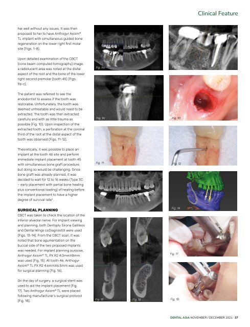
Clinical Feature<br />
her well without any issues. It was then<br />
proposed to her to have Anthogyr Axiom ®<br />
TL implant with simultaneous guided bone<br />
regeneration on the lower right first molar<br />
site (Figs. 1-8).<br />
Upon detailed examination of the CBCT<br />
(cone beam computed tomography) image,<br />
a radiolucent area was noted at the distal<br />
aspect of the root and the bone of the lower<br />
right second premolar (tooth 45) (Figs.<br />
9a-c).<br />
Fig. 9a<br />
Fig. 9b<br />
The patient was referred to see the<br />
endodontist to assess if the tooth was<br />
restorable. Unfortunately, the tooth was<br />
deemed untreatable and would need to be<br />
extracted. The tooth was then extracted<br />
carefully and with as little trauma as<br />
possible (Fig. 10). Upon inspection of the<br />
extracted tooth, a perforation at the coronal<br />
third of the root at the distal aspect of the<br />
tooth was observed (Figs. 11-12).<br />
Fig. 9c<br />
Fig. 10<br />
Theoretically, it was possible to place an<br />
implant at the tooth 46 site and perform<br />
immediate implant placement at tooth 45<br />
with simultaneous bone graft procedure,<br />
but doing so would be challenging. Since<br />
bone graft was already planned, it was<br />
decided to wait for 12 to 16 weeks (Type 3C<br />
– early placement with partial bone healing<br />
plus conventional loading) of healing before<br />
the implant placement to have a higher<br />
degree of survival rate 2 .<br />
Fig. 11<br />
Fig. 12<br />
SURGICAL PLANNING<br />
CBCT was taken to check the location of the<br />
inferior alveolar nerve. For implant viewing<br />
and planning, both Dentsply Sirona Gallileos<br />
and <strong>Dental</strong> Wings coDiagnostiX were used<br />
(Figs. 13-14). From the CBCT scan, it was<br />
noted that bone agumentation on the<br />
buccal side of the two proposed implants<br />
was needed. For implant planning purpose,<br />
Anthogyr Axiom ® TL PX R2 4.0mmX8mm<br />
was used (Fig. 15). At tooth 46, Anthogyr<br />
Axiom ® TL PX R2 4.6mmX6.5mm was used<br />
for surgical planning (Fig. 16).<br />
Fig. 13 Fig. 14<br />
Fig. 17<br />
On the day of surgery, a surgical stent was<br />
used to aid the implant placement (Fig.<br />
17). Two Anthogyr Axiom ® TL were placed<br />
following manufacturer’s surgical protocol<br />
(Fig. 18).<br />
Fig. 15 Fig. 16<br />
Fig. 18<br />
DENTAL ASIA NOVEMBER / DECEMBER <strong>2021</strong> 37