Dental Asia November/December 2021
For more than two decades, Dental Asia is the premium journal in linking dental innovators and manufacturers to its rightful audience. We devote ourselves in showcasing the latest dental technology and share evidence-based clinical philosophies to serve as an educational platform to dental professionals. Our combined portfolio of print and digital media also allows us to reach a wider market and secure our position as the leading dental media in the Asia Pacific region while facilitating global interactions among our readers.
For more than two decades, Dental Asia is the premium journal in linking dental innovators
and manufacturers to its rightful audience. We devote ourselves in showcasing the latest dental technology and share evidence-based clinical philosophies to serve as an educational platform to dental professionals. Our combined portfolio of print and digital media also allows us to reach a wider market and secure our position as the leading dental media in the Asia Pacific region while facilitating global interactions among our readers.

Create successful ePaper yourself
Turn your PDF publications into a flip-book with our unique Google optimized e-Paper software.
User Report<br />
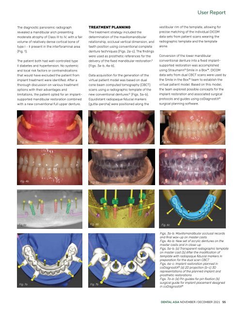
The diagnostic panoramic radiograph<br />
revealed a mandibular arch presenting<br />
moderate atrophy of Class III to IV, with a fair<br />
volume of relatively dense cortical bone of<br />
type I - II present in the interforaminal area<br />
(Fig. 1).<br />
The patient both had well-controlled type<br />
II diabetes and hypertension. No systemic<br />
and local risk factors or contraindications<br />
that would have excluded the patient from<br />
implant treatment were identified. After a<br />
thorough discussion on various treatment<br />
options with their advantages and<br />
limitations, the patient opted for an implantsupported<br />
mandibular restoration combined<br />
with a new conventional full upper denture.<br />
TREATMENT PLANNING<br />
The treatment strategy included the<br />
determination of the maxillomandibular<br />
relationship, occlusal vertical dimension, and<br />
teeth position using conventional complete<br />
denture techniques (Figs. 2a-c). The findings<br />
were used as prosthetic references for the<br />
delivery of the fixed mandibular restoration 11<br />
(Figs. 3a-b, 4a-b).<br />
Data acquisition for the generation of the<br />
virtual patient model was based on dual<br />
cone beam computed tomography (CBCT)<br />
scans using a radiographic template of the<br />
new conventional dentures 12 (Figs. 5a-b).<br />
Equidistant radiopaque fiducial markers<br />
(gutta-percha) were positioned along the<br />
vestibular rim of the template, allowing for<br />
precise matching of the individual DICOM<br />
data sets from patient scans wearing the<br />
radiographic template and the template<br />
alone.<br />
Conversion of the lower mandibular<br />
conventional denture into a fixed implantsupported<br />
restoration was accomplished<br />
using Straumann ® Smile in a Box. DICOM<br />
data sets from dual CBCT scans were used by<br />
the Smile in the Box team to establish the<br />
virtual patient model. Based on this model,<br />
the team explored possible concepts for the<br />
implant restoration and associated surgical<br />
protocols and guides using coDiagnostiX ®<br />
surgical planning software.<br />
Fig. 3a Fig. 3b Fig. 4a<br />
Fig. 4b<br />
Fig. 5a<br />
Fig. 5b<br />
Fig.6a<br />
Fig. 6b<br />
Fig. 6c<br />
Fig. 7a<br />
Fig. 7b<br />
Figs. 3a-b: Maxillomandibular occlusal records<br />
and final wax-up on master casts<br />
Figs. 4a-b: New set of acrylic dentures on the<br />
master casts and in close-up<br />
Figs. 5a-b: (a) Transparent radiographic template<br />
on master cast (b) After the modification of<br />
template with radiopaque fiducial markers in<br />
preparation for the dual scan CBCT<br />
Figs. 6a-c: Implant restoration planned in<br />
coDiagnostiX ® (a) 2D projection (b-c) 3D<br />
representations of the planned implant and<br />
prosthetic restorations<br />
Figs. 7a-b: (a) Pin guides for pin fixation (b)<br />
surgical guide for implant placement designed<br />
in coDiagnostiX ®<br />
DENTAL ASIA NOVEMBER / DECEMBER <strong>2021</strong> 55