T Cell Research Brochure - BD Biosciences
T Cell Research Brochure - BD Biosciences
T Cell Research Brochure - BD Biosciences

You also want an ePaper? Increase the reach of your titles
YUMPU automatically turns print PDFs into web optimized ePapers that Google loves.
PHOSPHORYLATION<br />
<strong>BD</strong> Phosfl ow Technology: Detecting Transient<br />
Phosphorylation Events<br />
Innovative <strong>BD</strong> Phosfl ow technology is the fi rst complete<br />
fl ow cytometry solution to reveal intracellular data on<br />
basal and induced protein phosphorylation events in both<br />
cell lines and primary cells. The <strong>BD</strong> Phosfl ow approach is<br />
especially informative with T cells, in which phosphorylation<br />
of signaling pathway proteins—such as Stat transcription<br />
factors—leads to the expression of particular T-cell<br />
phenotypes.<br />
<strong>BD</strong> <strong>Biosciences</strong> provides reagents and kits for the study of<br />
protein phosphorylation by fl ow cytometry, including the<br />
<strong>BD</strong> Phosfl ow T <strong>Cell</strong> Activation Kit, as well as <strong>BD</strong> Phosfl ow<br />
antibodies such as anti-Stat3 (pY705).<br />
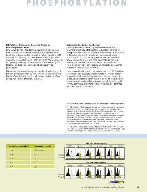
Activator (response modifi er) Phosphorylation marker<br />
PMA ERK 1/2, p38MAPK<br />
hIFN-α Stat1<br />
hIL-6 Stat3<br />
hIL-2 Stat5<br />
hIL-4 Stat6<br />
Control<br />
Treated<br />
Control<br />
Treated<br />
Specialized Antibodies and Buffers<br />
Developed collaboratively by <strong>BD</strong> and researchers at<br />
Stanford University, <strong>BD</strong> Phosfl ow technology consists of<br />
phosphoprotein-specifi c, fl uorochrome-labeled, monoclonal<br />
antibodies, along with a system of optimized buffers.<br />
These buffers fi x the cellular proteins to maintain their<br />
phosphorylation state, and then permeabilize the cell<br />
membrane to allow the antibodies to be introduced.<br />
Flow cytometry can then capture an intracellular snapshot<br />
of protein phosphorylation events.<br />
Used in combination with cell surface markers, <strong>BD</strong> Phosfl ow<br />
technology can evaluate phosphoprotein activation from<br />
populations within heterogenous samples, such as whole<br />
blood. It is an ideal solution when removal of red blood cells<br />
may unintentionally activate stress-induced signaling (p38/<br />
MAPK) pathways. Even rare cell subtypes can be identifi ed<br />
without upfront enrichment.<br />
T-cell activation profi les monitored with the <strong>BD</strong> Phosfl ow T <strong>Cell</strong> Activation Kit<br />
The <strong>BD</strong> Phosfl ow T <strong>Cell</strong> Activation Kit is a comprehensive research system that<br />
uses fl ow cytometry to reliably determine the level of key phosphorylated<br />
signaling proteins involved in T-cell activation. The ready-to-use kit includes<br />
pre-titrated fl uorochrome-conjugated antibodies, optimized buffers,<br />
experiment setup beads, lyophilized human control cells, validated protocols,<br />
and access to Cytobank software for storing, sharing, and analysis of data.<br />
The kit offers a total solutions approach that minimizes assay-to-assay<br />
variability to help both experienced and novice fl ow cytometry users produce<br />
more dependable, comparable results faster.<br />
The histogram overlays below (using Cytobank software) show CD4 + and CD8 +<br />
T-cell signaling responses to treatment, monitored using the <strong>BD</strong> Phosfl ow T <strong>Cell</strong><br />
Activation Kit. Response modifi ers are described in the chart to the left.<br />
p-ERK<br />
p-ERK<br />
CD4<br />
p-p38 p-Stat1<br />
+ cells, activation profiles<br />
p-Stat3 p-Stat5 p-Stat6<br />
CD8<br />
p-p38 p-Stat1<br />
+ cells, activation profiles<br />
p-Stat3 p-Stat5 p-Stat6<br />
Fold Change<br />
-25.0 -12.5 0.0<br />
12.5 25.0<br />
For <strong>Research</strong> Use Only. Not for use in diagnostic or therapeutic procedures.<br />
11