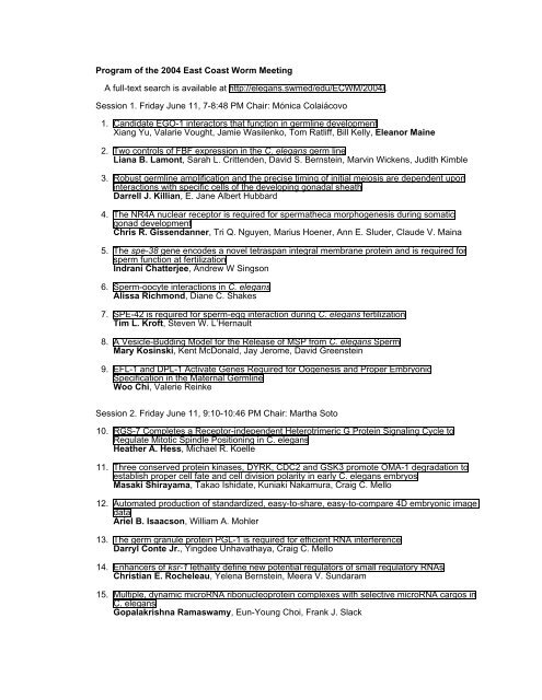
Program of the 2004 East Coast Worm Meeting - Caenorhabditis ...
Program of the 2004 East Coast Worm Meeting - Caenorhabditis ...
Program of the 2004 East Coast Worm Meeting - Caenorhabditis ...

Create successful ePaper yourself
Turn your PDF publications into a flip-book with our unique Google optimized e-Paper software.
<strong>Program</strong> <strong>of</strong> <strong>the</strong> <strong>2004</strong> <strong>East</strong> <strong>Coast</strong> <strong>Worm</strong> <strong>Meeting</strong><br />
A full-text search is available at http://elegans.swmed/edu/ECWM/<strong>2004</strong>/.<br />
Session 1. Friday June 11, 7-8:48 PM Chair: Mónica Colaiácovo<br />
1. Candidate EGO-1 interactors that function in germline development<br />
Xiang Yu, Valarie Vought, Jamie Wasilenko, Tom Ratliff, Bill Kelly, Eleanor Maine<br />
2. Two controls <strong>of</strong> FBF expression in <strong>the</strong> C. elegans germ line<br />
Liana B. Lamont, Sarah L. Crittenden, David S. Bernstein, Marvin Wickens, Judith Kimble<br />
3. Robust germline amplification and <strong>the</strong> precise timing <strong>of</strong> initial meiosis are dependent upon<br />
interactions with specific cells <strong>of</strong> <strong>the</strong> developing gonadal sheath<br />
Darrell J. Killian, E. Jane Albert Hubbard<br />
4. The NR4A nuclear receptor is required for sperma<strong>the</strong>ca morphogenesis during somatic<br />
gonad development<br />
Chris R. Gissendanner, Tri Q. Nguyen, Marius Hoener, Ann E. Sluder, Claude V. Maina<br />
5. The spe-38 gene encodes a novel tetraspan integral membrane protein and is required for<br />
sperm function at fertilization<br />
Indrani Chatterjee, Andrew W Singson<br />
6. Sperm-oocyte interactions in C. elegans<br />
Alissa Richmond, Diane C. Shakes<br />
7. SPE-42 is required for sperm-egg interaction during C. elegans fertilization<br />
Tim L. Kr<strong>of</strong>t, Steven W. L’Hernault<br />
8. A Vesicle-Budding Model for <strong>the</strong> Release <strong>of</strong> MSP from C. elegans Sperm<br />
Mary Kosinski, Kent McDonald, Jay Jerome, David Greenstein<br />
9. EFL-1 and DPL-1 Activate Genes Required for Oogenesis and Proper Embryonic<br />
Specification in <strong>the</strong> Maternal Germline<br />
Woo Chi, Valerie Reinke<br />
Session 2. Friday June 11, 9:10-10:46 PM Chair: Martha Soto<br />
10. RGS-7 Completes a Receptor-independent Heterotrimeric G Protein Signaling Cycle to<br />
Regulate Mitotic Spindle Positioning in C. elegans<br />
Hea<strong>the</strong>r A. Hess, Michael R. Koelle<br />
11. Three conserved protein kinases, DYRK, CDC2 and GSK3 promote OMA-1 degradation to<br />
establish proper cell fate and cell division polarity in early C. elegans embryos<br />
Masaki Shirayama, Takao Ishidate, Kuniaki Nakamura, Craig C. Mello<br />
12. Automated production <strong>of</strong> standardized, easy-to-share, easy-to-compare 4D embryonic image<br />
data<br />
Ariel B. Isaacson, William A. Mohler<br />
13. The germ granule protein PGL-1 is required for efficient RNA interference<br />
Darryl Conte Jr., Yingdee Unhavathaya, Craig C. Mello<br />
14. Enhancers <strong>of</strong> ksr-1 lethality define new potential regulators <strong>of</strong> small regulatory RNAs<br />
Christian E. Rocheleau, Yelena Bernstein, Meera V. Sundaram<br />
15. Multiple, dynamic microRNA ribonucleoprotein complexes with selective microRNA cargos in<br />
C. elegans<br />
Gopalakrishna Ramaswamy, Eun-Young Choi, Frank J. Slack
16. eak (enhancer-<strong>of</strong>-akt-1) genes encode membrane-associated proteins that potentiate AKT-1<br />
signaling in <strong>the</strong> C. elegans XXX cells<br />
Patrick J. Hu, Jinling Xu, Gary Ruvkun<br />
17. Notch function in differentiated neurons is required to maintain dauer<br />
Jimmy Ouellet, Richard Roy<br />
Session 3. Saturday June 12, 8:30-10:06 AM Chair: Siu Sylvia Lee<br />
18. Control <strong>of</strong> aging and developmental arrest by TGFbeta and insulin pathways during C.<br />
elegans diapause<br />
Manjing Pan, Li Sun da Graca, Tao Liu, Garth I. Patterson<br />
19. Life span regulation by JNK MAP kinase in C. elegans: a novel input into daf-16<br />
Seung Wook Oh, Nenad Svrzikapa, Heidi A. Tissenbaum<br />
20. Regulation <strong>of</strong> chemoreceptor gene expression by MEF-2 and class II HDACs in C. elegans<br />
Alexander M van der Linden, Katie Nolan, Piali Sengupta<br />
21. mig-10 functions downstream <strong>of</strong> unc-6 and slt-1 to mediate axon guidance<br />
Christopher C. Quinn, Elizabeth Stovall, Elizabeth F. Ryder, William G. Wadsworth<br />
22. Genes Involved In Serotonergic Neurotransmission<br />
Megan Higginbotham, Bob Horvitz<br />
23. Ryanodine Receptors Regulate Neurotransmitter Release at <strong>the</strong> C. elegans Neuromuscular<br />
Junction<br />
Qiang Liu, Michael Nonet, Lawrence Salk<strong>of</strong>f, Zhao-Wen Wang<br />
24. KEL-8, a novel Kelch-like protein, is required for glutamate receptor degradation<br />
Henry Schaefer, Christopher Rongo<br />
25. Knockout <strong>of</strong> GLT-3 C. elegans Glutamate Transporter: A Genetic Approach to Study<br />
Excitotoxic Neurodegeneration<br />
Itzhak Mano, Sarah Straud, Monica Driscoll<br />
Session 4. Saturday June 12, 10:40 AM-12:16 PM Chair: Peter Roy<br />
26. A non-developmental role for lin-12 Notch signaling in <strong>the</strong> C. elegans adult nervous system<br />
Michael Y. Chao, Jonah Larkins-Ford, Anne C. Hart<br />
27. Calcium permeability <strong>of</strong> death-inducing DEG/ENaC ion channel MEC-4(d)<br />
Laura Bianchi, Wei-Hsiang Lee, Gargi Mukherjee, Beate Gerstbrein, Dewey Royal,<br />
Maryanne Royal, Jian Xue, Monica Driscoll<br />
28. UNC-55, a Nuclear Receptor, is Essential for Male Mating<br />
Ge Shan, Bill Walthall<br />
29. Genes Controlling Sensory Axon Patterning in <strong>the</strong> C. elegans Male Tail<br />
Lingyun Jia, Scott W. Emmons<br />
30. A genomic approach to <strong>the</strong> development and function <strong>of</strong> <strong>the</strong> C. elegans male tail rays<br />
Douglas S. Portman, Daryl D. Hurd, Nicole Juskiw, Kwi Yeon Lee, William R. Mowrey,<br />
Carolyn Tyler, Hai Wu<br />
31. <strong>Worm</strong>Base: What’s New and What’s Next?<br />
Nansheng Chen, Lincoln D. Stein, <strong>Worm</strong>Base Consortium
32. Frogs and snails and puppy dog tails? <strong>Worm</strong>atlas launches a guide to what boy worms are<br />
made <strong>of</strong><br />
Robyn Lints, Zeynep F. Altun, Huawei Weng, Gloria Stephney, Maurice Volaski, David H.<br />
Hall<br />
33. A New Phylogeny Reveals Frequent Loss <strong>of</strong> Introns During Nematode Evolution<br />
Ronald E Ellis, Soochin Cho<br />
Session 5. Saturday June 12, 5:30-7:06 PM Chair: Landon Moore<br />
34. <strong>Caenorhabditis</strong> phylogeny predicts convergence <strong>of</strong> hermaphroditism and extensive intron<br />
loss<br />
Karin C. Kiontke, Nicholas P. Gavin, Yevgeniy Raynes, Casey Roehrig, Fabio Piano, David<br />
H. A. Fitch<br />
35. Evolutionary innovation <strong>of</strong> excretory system in <strong>Caenorhabditis</strong> elegans<br />
Xiaodong Wang, Helen M. Chamberlin<br />
36. cdc-14 regulates cki-1 to control cell-cycle arrest<br />
R. Mako Saito, Audrey Perreault, Bethan Peach, John S. Satterlee, Sander van den Heuvel<br />
37. Transcriptional regulation <strong>of</strong> Hox gene lin-39 during vulval cell fate specification<br />
Javier A. Wagmaister, Julie E. Gleason, Corey A. Morris, Leilani M. Miller, Ginger R. Miley,<br />
Kerry Kornfeld, David M. Eisenmann<br />
38. EPS-8 regulates LET-23/EGFR localization during C. elegans vulval development<br />
Attila Stetak, Assunta Croce, Giuseppe Cassata, Pier P. DiFiore, Erika Fröhli Hoier, Alex<br />
Hajnal<br />
39. The C-terminal sequence <strong>of</strong> C. elegans smad/SMA-3 has multiple roles<br />
Jianjun Wang, Cathy Savage-Dunn<br />
40. PKC2 A Calcium-Diacylglcerol Kinase that Runs Hot and Cold<br />
Marianne Land, Charles S. Rubin<br />
41. Regulation <strong>of</strong> a Conserved Oxidative Stress Defense by GSK-3 and p38 signaling in C.<br />
elegans<br />
Jae Hyung An, Riva Oliveira, Rosanna Baker, Kelly Vranas, Hideki Inoue, Naoki Hisamoto,<br />
Yanxia Bei, Craig C. Mello, Kunihiro Matsumoto, T. Keith Blackwell<br />
Session 6. Sunday June 13, 8:30-9:42 AM Chair: Laura Mathies<br />
42. LIN-28 and LIN-46 converge at a branchpoint in <strong>the</strong> heterochronic pathway<br />
Eric G. Moss, Keven Kemper<br />
43. The role <strong>of</strong> daf-6 and cell-cell interactions in amphid morphogenesis<br />
Elliot A. Perens, Shai Shaham<br />
44. An FGF Signaling Pathway Regulates Membrane Extensions from <strong>the</strong> Body Wall Muscles in<br />
C. elegans<br />
Scott Dixon, Raynah Fernandes, Peter J. Roy<br />
45. EGL-15 FGF Receptor Is<strong>of</strong>orms Play Different Roles in SM Migration<br />
Te-Wen Lo, Ca<strong>the</strong>rine S. Branda, Peng Huang, Isaac E. Sasson, S. Jay Goodman, Michael<br />
J. Stern<br />
46. MLS-2, an HMX class homeodomain protein essential for mesodermal patterning and cell<br />
fate specification<br />
Yuan Jiang, Jun Kelly Liu
47. Regulation <strong>of</strong> TRA-1 by sex specific proteolytic processing and localization<br />
Mara Schvarzstein, Laura Mathies, Andrew Spence<br />
Session 7. Sunday June 13, 10:10 AM-12:10 PM Chair: Barth Grant<br />
48. Mutations in him-8 suppress developmental defects <strong>of</strong> egl-13 mutants<br />
Brian L. Nelms, Wendy Hanna-Rose<br />
49. Generating a more comprehensive picture <strong>of</strong> apoptosis using multiple functional genomic<br />
techniques<br />
Stuart Milstein, Pierre-Olivier Vidalain, Siming Li, David Hill, Marc Vidal<br />
50. Characterization and cloning <strong>of</strong> a novel component in <strong>the</strong> cell-corpse engulfment pathway<br />
Xiaomeng Yu, Xiaohong Leng, Chin-Hua Chuang, Sampeter Odera, H. Robert Horvitz,<br />
Zheng Zhou<br />
51. Characterization <strong>of</strong> <strong>the</strong> Cell Deaths Caused by Mutations in lin-24 and lin-33<br />
Brendan Galvin, Saechin Kim, Erika Hartwieg, Bob Horvitz<br />
52. RME-6 is a new regulator <strong>of</strong> Rab5-mediated endocytosis<br />
Miyuki Sato, Ken Sato, Paul Andre Fonarev, Barth Grant<br />
53. Septins function in morphogenesis <strong>of</strong> <strong>the</strong> C. elegans pharynx<br />
Fern P. Finger<br />
54. An Essential Role for HTP-3, a HIM-3 Paralog, in Mediating Meiotic Chromosome Behaviour<br />
and Structure<br />
William Goodyer, Monique Zetka<br />
55. HDA-1 regulates C. elegans embryogenesis: a potential role for a ubiquitous chromatin<br />
modifier in regulating tissue-specific gene expression and patterning<br />
Johnathan R. Whetstine, Julian Ceron, Valerie Reinke, Yang Shi<br />
56. Genetics <strong>of</strong> telomere replication in C. elegans<br />
Bettina Meier, Sarah Mense, Yan Zhao, Shawn Ahmed<br />
57. Centromere resolution is inhibited by cohesin proteins and requires condensin II<br />
components, HCP-6 and Mix-1<br />
Landon L. Moore, Matt Stankiewicz, David Rosen, Tovah Day<br />
Poster session: Saturday June 12, 1:45-5:30 PM<br />
58. Identification and Characterization <strong>of</strong> C. elegans Amine-gated Chloride Channels<br />
Namiko Abe, Niels Ringstad, Bob Horvitz<br />
59. Linker cell death may be caspase-independent<br />
Mary C. Abraham, Shai Shaham<br />
60. Octopamine Inhibits Pharyngeal Pumping and Egg Laying and Stimulates Locomotion<br />
Mark Alkema, Niels Ringstad, Bob Horvitz<br />
61. Establishment <strong>of</strong> anteroposterior neuronal polarity<br />
Eleanor Allen, Anastasia Bakoulis, Dave Hunt, Scott Clark<br />
62. The let-7 and mir-35 Families <strong>of</strong> MicroRNAs Each Act Redundantly in C. elegans<br />
Ezequiel Alvarez-Saavedra, Eric A Miska, Allison L Abbott, Nelson C Lau, David P Bartel,<br />
Victor Ambros, Bob Horvitz
63. Characterization <strong>of</strong> <strong>the</strong> Syn<strong>the</strong>tic Multivulva Suppressor Gene isw-1, a Homolog <strong>of</strong> <strong>the</strong><br />
Drosophila Chromatin-Remodeling ATPase ISWI<br />
Erik Andersen, Xiaowei Lu, Scott Clark, Bob Horvitz<br />
64. met-1 and met-2, Two Putative Histone Methyltransferases, May Act as Syn<strong>the</strong>tic Multivulva<br />
Genes<br />
Erik Andersen, Bob Horvitz<br />
65. Generation <strong>of</strong> left/right asymmetry in <strong>the</strong> nervous system <strong>of</strong> C. elegans<br />
Celia Antonio, Oliver Hobert<br />
66. Alterations <strong>of</strong> <strong>the</strong> C. elegans Excretory Duct in Dauer Larvae<br />
Kristin R. Armstrong*, Helen M. Chamberlin*<br />
67. Sheath cell-dependent maintenance <strong>of</strong> AWC dendritic morphology<br />
Taulant Bacaj, Shai Shaham<br />
68. Association <strong>of</strong> Oscheius species with millipedes in <strong>the</strong> Wright State University Woods<br />
Scott Everet Baird, Christine M. Spice<br />
69. Females, Hermaphrodites and Developmental Bias<br />
Chris Baldi, Soochin Cho, Ronald E Ellis<br />
70. Genome-wide RNAi screen to identify novel regulators <strong>of</strong> membrane trafficking<br />
Zita Balklava, Barth D. Grant<br />
71. Molecular connections between developmental timing and circadian timing: The C. elegans<br />
homolog <strong>of</strong> <strong>the</strong> circadian gene doubletime regulates post-embryonic developmental timing<br />
Diya Banerjee, Alvin Kwok, Shin-Yi Lin, Frank Slack<br />
72. Towards cloning mutations isolated in a genetic screen for hyperactive egg-laying mutants<br />
I. Amy Bany, Michael R. Koelle<br />
73. C. elegans HDACs, CBP, and CREB play roles in polyglutamine neurotoxicity<br />
Emily A. Bates, Cindy Voisine, Martin Victor, Yang Shi, Anne Hart<br />
74. Study <strong>of</strong> <strong>the</strong> genetic and cellular bases <strong>of</strong> ventral nerve cord maintenance<br />
Claire Benard, Oliver Hobert<br />
75. A Genome-Wide RNAi Screen for Components Affecting Sex Muscle Differentiation<br />
Daniel C. Bennett, Isaac E. Sasson, Michael J. Stern<br />
76. A Genetic Screen for New Genes Involved in Aging<br />
Ala Berdichevsky, Leonard Guarente, Bob Horvitz<br />
77. Towards understanding <strong>the</strong> role <strong>of</strong> <strong>the</strong> stomatin-like protein UNC-24 in <strong>the</strong> process <strong>of</strong> gentle<br />
touch sensation: evidence for functional interaction with <strong>the</strong> mechanosensory ion channel<br />
complex MEC-4/MEC-10<br />
Laura Bianchi, Wei-Hsiang Lee, Dan Slone, Julie Y. Koh, David M. Miller III, Monica Driscoll<br />
78. A germline-specific RNA-binding protein required for germ cell survival and cytokinesis<br />
Peter R, Boag, T. Keith Blackwell<br />
79. Identification <strong>of</strong> C. elegans spindle assembly checkpoint components<br />
Mike Boxem, Marc Vidal<br />
80. Development <strong>of</strong> high-throughput sublehtal toxicity tests using <strong>Caenorhabditis</strong> elegans<br />
Windy A. Boyd, Sandra J. McBride, Julie R. Rice, Jonathan H. Freedman
81. Using enriched populations <strong>of</strong> single neuron types and microarrays to identify sensory<br />
neuron-specific genes<br />
Marc Colosimo, Adam Brown, Anne Lanjuin, Saikat Mukhopadhyay, Piali Sengupta<br />
82. Modulations <strong>of</strong> <strong>the</strong>rmotactic behavior by food<br />
Adam Brown, Damon Clark, Chris Gabel, Steven Lin, Ares Perides, Piali Sengupta, Aravi<br />
Samuel<br />
83. A Systematic Genome-Wide Genetic Interaction Analysis <strong>of</strong> Signaling Pathways in C.<br />
elegans<br />
Alexandra Byrne, Scott J. Dixon, Jason M<strong>of</strong>fat, Peter J. Roy<br />
84. Temporal regulation <strong>of</strong> postmitotic neural differentiation by heterochronic genes including <strong>the</strong><br />
microRNA lin-4<br />
Ka<strong>the</strong>rine O. Carter, Kristy Reinert, Shin-Yi Lin, Frank Slack<br />
85. Identification <strong>of</strong> genes acting redundantly with lin-35 Rb<br />
Julian Ceron, Abha Chandra, Khursheed Wani, Jean-Francois Rual, Marc Vidal, Sander van<br />
den Heuvel<br />
86. Functional analysis <strong>of</strong> AMPA-type glutamate receptor tail sequences in C. elegans.<br />
Howard Chang, Chris Rongo<br />
87. Feeding status and serotonin modulate a chemosensory circuit in C. elegans<br />
Michael Y. Chao, Hidetoshi Komatsu, Hana S. Fukuto, Hea<strong>the</strong>r M. Dionne, Anne C. Hart<br />
88. D1- and D2-like dopamine receptors antagonistically modulate C. elegans behavior through<br />
Galphaq and Galphao signaling<br />
Daniel Chase, Judy Pepper, Michael Koelle<br />
89. Germline establishment and maintenance in <strong>the</strong> early embryo<br />
Paula M. Checchi, Christine E. Schaner, William G. Kelly<br />
90. GUM-1, a protein affecting <strong>the</strong> subcellular localization <strong>of</strong> RME-1, is required for endocytic<br />
recycling in <strong>the</strong> C. elegans intestine<br />
Carlos Chih-Hsiung, Chen, Peter Schweinsberg, Eric Lambie, Barth Grant<br />
91. Translational control in <strong>the</strong> germ plasm: nos-2 RNA regulation.<br />
Pei-Lung Chen, Ingrid D’Agostino, Geraldine Seydoux<br />
92. Translational control in <strong>the</strong> germ plasm: nos-2 RNA regulation<br />
Pei-Lung Chen, Ingrid D’Agostino, Geraldine Seydoux<br />
93. Age-associated feeding decline in C. elegans can be modulated by genetic and<br />
environmental inputs<br />
David K. Chow, Ca<strong>the</strong>rine A. Wolkow<br />
94. An Investigation into <strong>the</strong> potential regulatory relationships between components <strong>of</strong> a network<br />
specified by pal-1<br />
Julia M. Claggett, L. Ryan Baugh, Craig P. Hunter<br />
95. Genome-wide RNAi analysis <strong>of</strong> <strong>Caenorhabditis</strong> elegans distal tip cell migration<br />
Erin J. Cram, Jean E. Schwarzbauer<br />
96. Characterization <strong>of</strong> <strong>the</strong> DTC niche and germline stem cells in C. elegans<br />
Sarah L. Crittenden, Dana Byrd, Kim Leonhard, Judith Kimble<br />
97. Structure/function studies <strong>of</strong> <strong>the</strong> polarity regulator PAR-1 in C. elegans<br />
Adrian Cuenca, Geraldine Seydoux
98. Experimental Design for C. elegans Microarray<br />
Yuxia Cui, Sandra J. McBride, Jonathan H. Freedman<br />
99. Tracking <strong>the</strong> mid-life crisis <strong>of</strong> C. elegans<br />
Diana David-Rus, Peter J. Schmeissner, Beate Hartmann, Christophe Grundschober, Uri<br />
Einav, Eytan Domany, Patrick Nef, Garth Patterson, Monica Driscoll<br />
100. An HCP-6 Suppression Screen for Genes Involved in Centromere Resolution<br />
Tovah A. Day, Landon Moore<br />
101. Analysis <strong>of</strong> sel-2, an enhancer <strong>of</strong> lin-12 activity in vulval precursor cells<br />
Natalie de Souza, Laura G. Vallier, Hanna Fares, Iva Greenwald<br />
102. Function <strong>of</strong> a novel protein EFF-1 in cell fusion<br />
Jacob J del Campo, Ariel B Issacson, Morgan Tucker, Min Han, William A Mohler<br />
103. Degenerate binding sites for <strong>the</strong> FAX-1 nuclear receptor predict potential downstream target<br />
genes<br />
Stephen DeMeo, Rebecca Lombel, Danielle Snowflack, Aaron Wagner, Eric Smith, Sheila<br />
Clever, Bruce Wightman<br />
104. Mapping transcription regulatory networks in C. elegans<br />
B. Deplancke, D. Dupuy, M. Vidal, A.J. Marian Walhout<br />
105. SYP-3, a coiled-coil protein required for chromosome synapsis and chiasma formation in C.<br />
elegans<br />
Andreas Eizinger, Allison Hurlburt, JoAnne Engebrecht, Kirthi Reddy, Anne Villeneuve,<br />
Mónica Colaiácovo<br />
106. A genetic screen for mutants defective in male leaving, a mate-searching behavior <strong>of</strong> C.<br />
elegans<br />
Chunhui Fang, Rajarshi Ghosh, Scott W. Emmons<br />
107. DKF-2 is a Novel Target-Effector <strong>of</strong> Protein Kinase C<br />
Hui Feng, Min Ren, Charles S. Rubin<br />
108. A novel mutant that partially suppresses <strong>the</strong> daf-2 (e1370) Daf-c phenotype<br />
Manuel A. Fidalgo, Manuel J. Munoz<br />
109. Specificity <strong>of</strong> <strong>the</strong> C. elegans Putative Transmembrane Channel SID-1<br />
Michael C. Fitzgerald, Craig P. Hunter<br />
110. SMA-9, a Protein Involved in Patterning <strong>of</strong> <strong>the</strong> C.elegans M Lineage<br />
Marisa L Foehr, Ming Xu, Jun Liu<br />
111. Cloning and characterization <strong>of</strong> <strong>the</strong> C. elegans post-embryonic cytokinesis gene unc-85<br />
Iwen Fu, Jason W. Reuter, Fern P. Finger<br />
112. CeMyoD(hlh-1) in embryonic muscle fate determination<br />
Tetsunari Fukushige, Joan McDermott, Thomas Brodigan, Michael Krause<br />
113. Quantification <strong>of</strong> electrotaxis<br />
Chris Gabel, Albert Kao, Dmitri Pavlichin, Aravi Samuel<br />
114. Barotaxis<br />
Chris Gabel, Alex Dahlen, Aravi Samuel<br />
115. Genetic analysis <strong>of</strong> mutations that suppress dauer arrest in age-1/PI3 kinase mutants<br />
Minaxi S. Gami, Keaton Hanselman, Ca<strong>the</strong>rine A. Wolkow
116. spe-19, a Gene Affecting Spermiogenesis<br />
Brian Geldziler, Andy Singson<br />
117. Death defying acts: RNAi screen for genes influencing neuronal necrosis<br />
Beate Gerstbrein, Vienna Lo, Monica Driscoll<br />
118. An in vivo analysis <strong>of</strong> age-related biomarkers in C. elegans<br />
Beate Gerstbrein, Georgios Stamatas, Nikiforos Kollias, Monica Driscoll<br />
119. Serotonin and octopamine modulate thrashing behavior <strong>of</strong> C elegans<br />
Rajarshi Ghosh, Scott W. Emmons<br />
120. Functional Characterization <strong>of</strong> <strong>the</strong> Vertebrate Homologs <strong>of</strong> LIN-10: Mint 1, 2, and 3<br />
Doreen R. Glodowski, Bonnie L. Firestein, Christopher Rongo<br />
121. The Myt1 ortholog in C. elegans is essential for oocyte maturation<br />
Andy Golden<br />
122. RecQ Helicases, Genomic Stability and Lifespan in C. elegans<br />
Melissa M. Grabowski, Nenad Svrzikapa, Heidi Tissenbaum<br />
123. The temporal patterning microRNA let-7 controls multiple transcription factors including <strong>the</strong><br />
nuclear hormone receptor DAF-12<br />
Helge Grosshans, Ted Johnson, Mark Gerstein, Frank J. Slack<br />
124. The genes tra-4 and mog-7 are necessary to ensure hermaphrodite development in <strong>the</strong><br />
soma and in <strong>the</strong> germline<br />
Phillip Grote, Claudia Huber, Barbara Conradt<br />
125. SMA-10 is a novel extracellular regulator <strong>of</strong> <strong>the</strong> Sma/Mab TGF-beta pathway in C. elegans<br />
Tina L. Gumienny, Cole M. Zimmerman, Andrew F. Roberts, Huang Wang, Lena Chin,<br />
Richard W. Padgett<br />
126. LON-2 is a glypican heparan sulfate proteoglycan that regulates <strong>the</strong> Sma/Mab TGF-beta<br />
pathway in C. elegans<br />
Tina L. Gumienny, Huang Wang, Richard W. Padgett<br />
127. Reproductive isolation <strong>of</strong> C. briggsae haplotypes<br />
Rachael M. Hampton, Scott E. Baird<br />
128. High Throughput TILLING, Ecotilling, Genotyping, and Sequencing Instrumentation<br />
Jeff Harford<br />
129. Analysis <strong>of</strong> synMuv Protein Complexes in vivo and Characterization <strong>of</strong> <strong>the</strong> Class B synMuv<br />
Gene lin-61<br />
Melissa M Harrison, Xiaowei Lu, Bob Horvitz<br />
130. Visualizing activity <strong>of</strong> C. elegans interneurons<br />
Gal Haspel, Anne C. Hart<br />
131. Multiple factors act in concert to initiate <strong>the</strong> cell death <strong>of</strong> <strong>the</strong> NSM sister cells<br />
Julia Hatzold, Barbara Conradt<br />
132. An RNAi-based suppressor screen for components <strong>of</strong> <strong>the</strong> Aurora B kinase pathway<br />
Todd R. Heallen, Jill M. Schumacher<br />
133. Characterization <strong>of</strong> tissue-specific suppressors <strong>of</strong> cdc-25.1(gf)<br />
Michael Hebeisen, Roshni Basu, Richard Roy
134. Form and function <strong>of</strong> glia-neuron interactions<br />
Maxwell G. Heiman, Shai Shaham<br />
135. Vulval and uterine development are not temporally coordinated in ku212 mutants<br />
Li Huang, Wendy Hanna-Rose<br />
136. COMPUTER MODELING, SIMULATION AND ANALYSIS OF C. elegans VULVAL<br />
INDUCTION<br />
Na’aman Kam, Jasmin Fisher, David Harel, Amir Pnueli, Michael J. Stern, E. Jane Albert<br />
Hubbard<br />
137. A Screen for Genes Syn<strong>the</strong>tically Lethal with lin-35 Rb<br />
Mike Hurwitz, Bob Horvitz<br />
138. Genetic variation reveals differences among C. elegans isolates for ASH mediated behaviors<br />
Rhonda Hyde, Anne C. Hart<br />
139. Study <strong>of</strong> <strong>the</strong> Effects <strong>of</strong> Oxidative Damage Repair on Aging and Muscle HealthSpan<br />
Carolina Ibanez-Ventoso, Samuel Bassous, Peter J. Schmeisser, Suzhen Guo, Monica<br />
Driscoll<br />
140. Guidance and cell-matching <strong>of</strong> a migrating epidermal sheet during ventral enclosure by<br />
MAB-20 and PLX-2<br />
Richard Ikegami, Kristin Simokat, Louise Dixon, Jeff Hardin, Joseph Culotti<br />
141. Identification <strong>of</strong> Wnt Pathway Target Genes in C. elegans by Microarray Analysis<br />
Belinda M. Jackson, David M. Eisenmann<br />
142. Combinatorial control <strong>of</strong> lin-48 expression in <strong>the</strong> C. elegans excretory duct cell is mediated<br />
through Pax and bZip transcription factors<br />
Hongtao Jia, Xiaodong Wang, Helen M. Chamberlin<br />
143. Structural and functional studies <strong>of</strong> <strong>the</strong> C.elegans Hsp90 ortholog DAF21<br />
Dayadevi Jirage, Harold Smith<br />
144. GNA-2, Chitin and <strong>the</strong> Functionally-Redundant CEJ-1/B0280.5 are Required for <strong>the</strong><br />
Syn<strong>the</strong>sis <strong>of</strong> a Lipophobic Extraembryonic Matrix (EEM) and are Essential for Development and<br />
Polarity in <strong>the</strong> One-cell Embryo<br />
Wendy L. Johnston, Aldis Krizus, James W. Dennis<br />
145. Biochemical and structure/function analysis <strong>of</strong> DGK-1, a neuronal diacylglycerol kinase and<br />
putative downstream effector <strong>of</strong> Gapha o signaling<br />
Antony Jose, Michael Koelle<br />
146. Cytological screening <strong>of</strong> <strong>the</strong> germ lines <strong>of</strong> sterile mutants for meiotic defects<br />
Malek Jundi, Monique C. Zetka<br />
147. EGL-26 belongs to <strong>the</strong> NlpC/P60 superfamily <strong>of</strong> putative enzymes and is closely related to a<br />
mammalian acyl transferase<br />
Rasika Kalamegham, Wendy Hanna-Rose<br />
148. Genes controlling <strong>the</strong> developmental response to nutrients in L1 larvae<br />
Gautam Kao, Peter Naredi, Simon Tuck<br />
149. Characterization <strong>of</strong> F11A10.3, a RING domain protein that interacts with <strong>the</strong> transcription<br />
factor UNC-3<br />
Ozgur Karakuzu, Brinda Prasad, Scott Cameron
150. Characterization <strong>of</strong> sel-6, a suppressor <strong>of</strong> lin-12 gain-<strong>of</strong>-function mutants<br />
Iskra Katic, Iva Greenwald<br />
151. How Does Ivermectin Induce Cell Death?<br />
Aamna Kaul, Joseph Dent<br />
152. Expression, function and regulation <strong>of</strong> gon-2<br />
Ben Kemp, Rachel West, Diane Church, Samantha Schilling, Janet Lee, Robert Bruce III,<br />
Mat<strong>the</strong>w Ambros, Eric Lambie<br />
153. Centrosome Maturation and Duplication In C. elegans Require <strong>the</strong> Coiled-Coil Protein SPD-2<br />
Ca<strong>the</strong>rine A Kemp, Kevin R. Kopish, Peder Zipperlen, Julie Ahringer, Kevin F. O’Connell<br />
154. nhr-67 and nhr-111, two NR2E nuclear receptors that may function in nervous system<br />
development<br />
Ryan Kennedy, Kristy Reinert, Genna Albert, Chris Gissendanner, Ann Sluder, Bruce<br />
Wightman<br />
155. The lin-4 homologue, mir-237, directs proper vulva and gonad development in C. elegans<br />
Aurora Esquela Kerscher, Lei Bai, Frank J. Slack<br />
156. Sex-specific centrosome inheritance requires cki-2 in C. elegans<br />
Dae Young Kim, Richard Roy<br />
157. Roles <strong>of</strong> PAR-3 and PKC-3 in establishment and maintenance <strong>of</strong> epi<strong>the</strong>lial cell polarity in C.<br />
elegans<br />
Heon S. Kim<br />
158. The Regulation <strong>of</strong> <strong>the</strong> CDC-6 Replication Licensing by CUL-4<br />
Jihyun Kim, Hui Feng, Edward T. Kipreos<br />
159. The O/E transcription factor UNC-3 specifies <strong>the</strong> identities <strong>of</strong> <strong>the</strong> ASI chemosensory neurons<br />
via cell-specific repression and activation mechanisms<br />
Kyuhyung Kim, Marc Colosimo, Piali Sengupta<br />
160. Identification <strong>of</strong> CUL-4 complex components<br />
Youngjo Kim, Edward T. Kipreos<br />
161. Genetic pathways that affect C. elegans leaving, a mate searching behavior<br />
Gunnar A. Kleemann, Ling yun Jia, Johnathan O .Lipton, Scott W. Emmons<br />
162. A screen for suppressors <strong>of</strong> cyclin-D1 in C. elegans<br />
John Koreth, Mike Boxem, Huihong Xu, Stuart H. Orkin, Sander van den Heuvel<br />
163. Attempts to develop molecular genetic tools to study parasitic nematodes<br />
Kelly Kraus, Meera Sundaram<br />
164. Reiteration <strong>of</strong> a lineage branch generating right-sided amphid neurons in ref-1 bHLH mutants<br />
Anne Lanjuin, Julia K. Thompson, Piali Sengupta<br />
165. Identification and Characterization <strong>of</strong> Suppressors <strong>of</strong> him-3<br />
Ka-Lun Law, Monique Zetka<br />
166. A him-8 mutation suppresses <strong>the</strong> PIE-1-induced synMuv defect<br />
Jungsoon Lee, Prashant Raghavan, Byung-Jae Park, Tae Ho Shin<br />
167. Interaction between <strong>the</strong> SEK-1-PMK-1 p38 MAPK and DAF-2/DAF-16 insulin signaling<br />
pathways mediating pathogen resistance and longevity in C. elegans<br />
Dennis H. Kim, Valerie Reinke, Danielle A. Garsin, Gary Ruvkun, Siu Sylvia Lee, Frederick<br />
M. Ausubel
168. Global transcriptional changes caused by cognition enhancing compounds in C. elegans N2<br />
French A. Lewis, III, Brian A. Dougherty<br />
169. Study <strong>of</strong> par-3 function in C. elegans<br />
Bingsi Li<br />
170. sma-9, A Gene that Regulates Body Size Development in C. elegans<br />
Jun Liang, Ling Yu, Cathy Savage-Dunn<br />
171. Toward Identifying Targets <strong>of</strong> MAP Kinase During C. elegans germline Development Using<br />
Functional Proteomic Approaches<br />
Baiqing Lin, Valerie Reinke<br />
172. CaM KII Regulates Neurotransmitter Release at <strong>the</strong> C. elegans Neuromuscular Junction<br />
Qiang Liu, Zhao-Wen Wang<br />
173. Control <strong>of</strong> aging and developmental arrest by TGF and insulin pathways during C.<br />
elegansdiapause<br />
Tao Liu, Manjing Pan, Garth Patterson<br />
174. End-to-end chromosome fusions in C elegans<br />
Mia R. Lowden, Bettina Meier, Shawn C. Ahmed<br />
175. An intracellular serpin, srp-6, is required for survival from hypoosmotic shock in<br />
<strong>Caenorhabditis</strong> elegans<br />
Cliff J. Luke, Stephen C. Pak, Vasantha Kumar, Sule Çataltepe, Carmen M. Knebel,<br />
Anthony Clark, Deiter Brömme, Gary A. Silverman<br />
176. Genetic Analysis <strong>of</strong> <strong>the</strong> Putative SUP-9/SUP-10/UNC-93 Two-Pore Domain K + Channel<br />
Complex<br />
Long Ma, Bob Horvitz<br />
177. Modeling and simulation <strong>of</strong> <strong>the</strong> behavior <strong>of</strong> proliferating C. elegans germ cells<br />
John Maciejowski, Nadia Ugel, Marco Isopi, Bud Mishra, E. Jane Albert Hubbard<br />
178. Nog mutants and early germline proliferation in C. elegans<br />
John Maciejowski, Giselle Cipriani, Ji-Inn Lee, James Ahn, Kerry Donny-Clark, E. Jane<br />
Albert Hubbard<br />
179. DNA replication and <strong>the</strong> proliferation versus meiotic development decision<br />
Valarie Vought, Min-Ho Lee, Larissa Wirlo, Katy Michalak, Deborah Springer, Ying Liu,<br />
Valerie Reinke, Tim Schedl, Eleanor Maine<br />
180. The molecular analysis <strong>of</strong> ego-2<br />
Ying Liu, Deborah Swenton, Dave Hansen, Eleanor Maine<br />
181. Control <strong>of</strong> lipid accumulation by ciliated neurons in C. elegans<br />
Ho Yi Mak, Gary Ruvkun<br />
182. n3263 is a mutant with persistent cell corpses that defines a candidate new engulfment gene<br />
Paolo M. Mangahas, H. Robert Horvitz, Zheng Zhou<br />
183. Localization <strong>of</strong> APH-1 Protein in Embryos<br />
David McGaughey, Valerie Hale, Caroline Goutte<br />
184. EGL-32 Functions in Sperm to Regulate Egg-Laying through <strong>the</strong> TGF-beta Pathway in<br />
C.elegans<br />
Marie McGovern, Ling Yu, Cathy Savage-Dunn
185. sns-10, <strong>the</strong> C. elegans ortholog <strong>of</strong> Aristaless/ARX, regulates sensory and motor neuron<br />
development<br />
Tal J. Melkman, Piali Sengupta<br />
186. him genes and X chromosome meiosis<br />
Philip M. Meneely, Joshua Havassy, Kathryn Crozier<br />
187. Characterization <strong>of</strong> an hlh-8 mutant<br />
Stephany G. Meyers, Ann K. Corsi<br />
188. Functional Analysis <strong>of</strong> <strong>the</strong> MicroRNA Genes <strong>of</strong> C. elegans<br />
Eric A Miska, Ezequiel Alvarez-Saavedra, Allison L Abbott, Andrew B Hellman, Nelson C<br />
Lau, David P Bartel, Victor Ambros, Bob Horvitz<br />
189. Genetic and molecular analysis <strong>of</strong> <strong>the</strong> C2H2 zinc-finger gene ehn-3<br />
Kristin M. Morphy, Judith Kimble, Laura D. Mathies<br />
190. Analysis <strong>of</strong> an UNC-13 protein expressed from an internal promoter<br />
Theresa Moser, Bethany Stitt, Kristin Servent, Brooke Swalm, Lydia Sanchez, Monique<br />
Spencer, Rebecca Kohn<br />
191. cwp-4, a novel male-specific C. elegans gene with a potential role in mating behavior<br />
William R. Mowrey, Douglas S. Portman<br />
192. Identification <strong>of</strong> genes regulating chemosensory neuron-specific morphologies<br />
Saikat Mukhopadhyay, Anne Lanjuin, Piali Sengupta<br />
193. The Role <strong>of</strong> PDZ Domain Proteins in GLR-1 Localization<br />
Vidhya Munnamalai, Christopher Rongo<br />
194. Uncovering <strong>the</strong> role for sperm contributed SCU-1 in regulating meiotic exit and axis formation<br />
in <strong>the</strong> early <strong>Caenorhabditis</strong> elegans embryo<br />
MaryAnn Murrow, Anna Mazor, Rebecca Lyczak<br />
195. Characterization <strong>of</strong> <strong>the</strong> identity and specificty <strong>of</strong> RGS protein targets in C. elegans<br />
Edith M. Myers, Michael R. Koelle<br />
196. Evidence that lin-35 Rb functions in hyp7 to inhibit vulval fates<br />
Toshia R Myers, Iva Greenwald<br />
197. RNAi-mediated screen for meiotic genes in C. elegans<br />
Sandra Nagl, Allison Hurlburt, Mónica Colaiácovo<br />
198. A germline-specific cell cycle inhibitor involved in dauer and adult lifespan<br />
Patrick Narbonne, Richard Roy<br />
199. The C. elegans pumilio ortholog, puf-9, controls <strong>the</strong> timing <strong>of</strong> development<br />
Mona J. Nolde, Kristy Reinert, Nazli Saka, Frank J. Slack<br />
200. High throughput genetic screen for suppressors <strong>of</strong> necrotic cell death<br />
Yury O. Nunez, Dewey Royal, MaryAnn Royal, Michael Lizzio Jr., Monica Driscoll<br />
201. Activation <strong>of</strong> SKN-1 stress response by a chemoprotective antioxidant<br />
Riva P. Oliveira, Jae Hyung An, Rosana P. Baker, Hideki Inoue, Kunihiro Matsumoto, T.<br />
Keith Blackwell<br />
202. Some Dauer Formation, Social Feeding, and Chemotaxis Mutants are Abnormal in <strong>the</strong><br />
Enhanced Slowing Response<br />
Daniel Omura, Bob Horvitz
203. Transcriptional regulation and stochasticity <strong>of</strong> eff-1 in fusing cell types<br />
Eugene Opoku-Serebuoh, Victoria L. Scranton, William A. Mohler<br />
204. Characterization <strong>of</strong> mutants defective in intestinal nuclear division<br />
Jimmy Ouellet, Richard Roy<br />
205. SRP-2 is an intracellular serpin that participates in postembryonic development<br />
Stephen C. Pak, Vasantha Kumar, Christopher Tsu, Cliff J. Luke, Yuko S. Askew, David J.<br />
Askew, David R. Mills, Anthony C. Clark, Gary Silverman<br />
206. The Identification <strong>of</strong> Factors Mediating <strong>the</strong> Downregulation <strong>of</strong> MEP-1 Function by PIE-1<br />
Byung-Jae Park, Prashant Raghavan, Seung-Il Kim, Jungsoon Lee, Keunhee Park, Tae Ho<br />
Shin<br />
207. A Screen for Mutants Resistant to Serotonin: a Search for Genes Involved in<br />
Neurotransmitter Signaling and Centrosome Movement<br />
Judy S. Pepper, Michael R. Koelle<br />
208. Investigating interacting partners <strong>of</strong> CeTwist<br />
Mary C. Philogene, Ann Corsi<br />
209. Genome wide RNAi screen for new synMuv genes<br />
Gino Poulin, Yan Dong, Andrew Fraser, Neil Hopper, Julie Ahringer<br />
210. A screen for axon branching and guidance mutants<br />
Brinda Prasad, Scott Clark<br />
211. Identification <strong>of</strong> Genetic Pathways Dependent on Protein N-glycosylation by GlcNAc-TV<br />
Justin M. Prien, Justin M. Crocker, Aldis Krizus, James W. Dennis, Charles E. Warren<br />
212. Components <strong>of</strong> <strong>the</strong> dosage compensation machinery antagonize <strong>the</strong> MEP-1 complex<br />
function<br />
Prashant Raghavan, Jungsoon Lee, Byun-Jae Park, Seun-il Kim, Keunhee Park, Tae Ho<br />
Shin<br />
213. Behavioral quiescence during <strong>the</strong> L1 stage and its alteration in eat-7 mutants<br />
David M. Raizen, Meera Sundaram, Allan I. Pack<br />
214. Characterization <strong>of</strong> UNC-43/CaMKII transport and activity in neurons<br />
Paris Rapp, Toru Umemura, Christopher Rongo<br />
215. Identification <strong>of</strong> factors required for germline silencing<br />
Tom Ratliff, Karissa McClinic, David Han, Bill Kelly<br />
216. Histone variant H2A.Z is essential for development in C.elegans<br />
Brianne J. Ray, William G. Kelly, Adam Raymond<br />
217. A Mos1 transposon mutagenesis screen for suppressors <strong>of</strong> <strong>the</strong> let-7 microRNA<br />
K. Reinert, F. J. Slack<br />
218. DKF-1 is A Diacylglycerol-regulated Kinase Involved in C.elegans Motility<br />
Min Ren, Hui Feng, Charles S. Rubin<br />
219. Modulation <strong>of</strong> C. elegans Egg-laying Behavior by <strong>the</strong> Environment and Experience<br />
Niels Ringstad, Bob Horvitz<br />
220. Dissecting <strong>the</strong> role <strong>of</strong> CNK-1 in LIN-45 Raf activation<br />
Christian E. Rocheleau, Meera V. Sundaram
221. Progress Towards <strong>the</strong> Cloning <strong>of</strong> lin-38 and Identification <strong>of</strong> Novel Class A SynMuv Genes<br />
Adam Saffer, Ewa Davison, Bob Horvitz<br />
222. C. elegans rme-3 encodes a clathrin heavy chain required for embryogenesis and<br />
neuro-muscular function<br />
Ken Sato, Chih-Hsiung Chen, Miyuki Sato, Barth D. Grant<br />
223. Characterization and mapping <strong>of</strong> sig-1, a new gene involved in germline-specific silencing <strong>of</strong><br />
extrachromosomal transgenes<br />
Christine E. Schaner, William G. Kelly<br />
224. The mutation bc202 blocks physiological as well as non-physiological germ cell death in <strong>the</strong><br />
adult hermaphrodite gonad<br />
Claus Schertel, Barbara Conradt<br />
225. Thrashing in liquid as a quantifiable measurement <strong>of</strong> aging<br />
Peter J. Schmeissner, Suzhen Guo, Shih-Hung Yu, Monica Driscoll<br />
226. The BarH Class Homeodomain Gene ceh-30 is Directly Regulated by tra-1 to Specify <strong>the</strong><br />
Sexually Dimorphic Survival <strong>of</strong> <strong>the</strong> CEM Neurons<br />
Hillel Schwartz, Bob Horvitz<br />
227. The "Green Pharynx" Phenotype <strong>of</strong> Transgene Misexpression Yields New Insight into <strong>the</strong><br />
synMuv Genes<br />
Hillel Schwartz, Dawn Wendell, Bob Horvitz<br />
228. Toward expression and biochemical characterization <strong>of</strong> EFF-1<br />
Victoria L. Scranton, William A. Mohler<br />
229. atx-2 Promotes Germline Proliferation and <strong>the</strong> Female Fate<br />
Xingyu She, Valarie E. Vought, Dave Hansen, Deborah Springer, Eleanor M. Maine<br />
230. The Unfolded Protein Response Regulates Glutamate Receptor Export from <strong>the</strong> ER<br />
Jaegal Shim, Toru Umemura, Erika Nothstein, Christopher Rongo<br />
231. Microarrays analysis <strong>of</strong> two essential factors required for RNAi, RDE-1 and RDE-4<br />
Martin J. Simard, Darryl Conte Jr, Jennifer A. Keys, Juerg Straubhaar, Danila Ulyanov,<br />
Craig C. Mello<br />
232. Regulation <strong>of</strong> gene expression by <strong>the</strong> Pax factor EGL-38<br />
Sama F. Sleiman, Helen M. Chamberlin<br />
233. C.elegans recognizes protons as a nociceptive stimulus through <strong>the</strong> DEG/EnaC and TRP<br />
channel<br />
Alfonso J. Apicella, Robert D. Slone, Monica Driscoll, William R. Schafer<br />
234. Move or Die: Epidermal Migration in <strong>the</strong> <strong>Caenorhabditis</strong> elegans Embryo<br />
Esteban Chen*, Michael M. S. Huang*, Veronica Zappi, Martha C. Soto<br />
235. Phenotypic characterization <strong>of</strong> egg-1, a molecule involved in fertilization<br />
Pavan Kadandale, Allison Stewart, Richard Klancer, Barth Grant, Andrew Singson<br />
236. How are cytoplasmic asymmetries achieved? In vivo studies <strong>of</strong> germ plasm localization<br />
dynamics during early embryogenesis<br />
Michael L. Stitzel, Denis Wirtz, Geraldine Seydoux<br />
237. Screen for Enhancers <strong>of</strong> ksr-2 Lethality<br />
Craig Stone, Meera Sundaram
238. EGL-26 controls vulF morphogenesis<br />
Hongliu Sun, Rita Sharma, Wendy Hanna-Rose<br />
239. Development <strong>of</strong> <strong>Caenorhabditis</strong> elegans in CeHR Axenic Medium<br />
Maria Szilagyi, Hugh F. LaPenotiere, Eric D. Clegg<br />
240. The Wnt genes egl-20 and cwn-1 are redundantly required for proper vulval cell fate<br />
specification<br />
Elizabeth Szyleyko, Julie E. Gleason, David M. Eisenmann<br />
241. The G proteins GOA-1 and EGL-30 function antagonistically in <strong>the</strong> HSN neurons that<br />
regulate egg-laying behavior in C. elegans<br />
Jessica E. Tanis, James J. Moresco, Robert A. Lindquist, Michael R. Koelle<br />
242. SRY-box containing protein SOX-2 directly binds to <strong>the</strong> promoter <strong>of</strong> Hox gene egl-5 and<br />
negatively regulates its expression<br />
Yingqi Teng, Scott W. Emmons<br />
243. Identification <strong>of</strong> genes involved in <strong>the</strong> specification <strong>of</strong> <strong>the</strong> sexually dimorphic CEMs<br />
Tatiana Tomasi, Stefanie Löser, Phillip Grote, Barbara Conradt<br />
244. Genetic screen for factors functioning with EGL-38 Pax to regulate lin-48 expression in<br />
<strong>Caenorhabditis</strong> elegans<br />
Rong-Jeng Tseng, Helen M. Chamberlin<br />
245. Transcriptional specification <strong>of</strong> neural subtype in <strong>the</strong> C. elegans male tail<br />
Carolyn Tyler, Nicole Juskiw, Douglas S. Portman<br />
246. Suppressors <strong>of</strong> pha-4/FoxA loss <strong>of</strong> function mutations define potential pha-4 regulators<br />
Dustin L. Updike, Susan E. Mango<br />
247. Identification <strong>of</strong> genes involved in cell fate specification <strong>of</strong> <strong>the</strong> gonadal sheath cells in<br />
<strong>Caenorhabditis</strong> elegans<br />
Laura G. Vallier, Helaina Skop, Lindsay Eisemann<br />
248. Genetic Screens for Suppressors <strong>of</strong> <strong>the</strong> ceh-30(n3714gf) Phenotype <strong>of</strong> Inappropriate<br />
Survival <strong>of</strong> <strong>the</strong> Male-Specific CEM Neurons in Hermaphrodites<br />
Johanna Varner, Hillel Schwartz, Bob Horvitz<br />
249. Screens for suppressors <strong>of</strong> cul-2 and zyg-11<br />
Srividya Vasudevan, Edward T. Kipreos<br />
250. Half-molecule ATP-binding cassette transporter, CeHMT1, is required for PC-dependent<br />
heavy metal detoxification in <strong>Caenorhabditis</strong> elegans<br />
Olena K. Vatamaniuk, Elizabeth A. Bucher, Meera V. Sundaram, Philip A. Rea<br />
251. How are apoptotic cells recognized by <strong>the</strong>ir phagocytes?<br />
Victor Venegas, Zheng Zhou<br />
252. Modifiers <strong>of</strong> polyglutamine-mediated neurodegeneration<br />
Cindy, Voisine, Adriana, K., Jones, Anne, C., Hart<br />
253. Disruption <strong>of</strong> germline pattern by forward and reverse genetics<br />
Roumen V. Voutev, E. Jane Albert Hubbard<br />
254. Regulation <strong>of</strong> Cell Death in <strong>the</strong> C. elegans Tail Spike Cell<br />
Carine Waase, Shai Shaham<br />
255. A suppressor screen for genes involved in UNC-6 mediated guidance<br />
Gauri Kulkarni, Chaunte Cannon, William G. Wadsworth
256. Regulation <strong>of</strong> RNA Polymerase II in C. elegans embryos and germline<br />
Amy K. Walker, T. Keith Blackwell<br />
257. Identification and characterization <strong>of</strong> <strong>the</strong> downstream target genes <strong>of</strong> CeTwist and its partner<br />
CeE/DA<br />
Peng Wang, Ann Corsi<br />
258. Identification <strong>of</strong> regulatory sequences necessary for egl-1 function in <strong>the</strong> ventral nerve cord<br />
Erin Webster, Tamara Strauss, Kelly Liu, Scott Cameron<br />
259. Identification and Characterization <strong>of</strong> MAPK Signaling Targets in <strong>the</strong> C. elegans Germline<br />
Stefanie West, Valerie Reinke<br />
260. The nuclear receptor gene fax-1 and homeobox gene unc-42 coordinate interneuron identity<br />
by regulating <strong>the</strong> expression <strong>of</strong> glutamate receptor subunits and o<strong>the</strong>r neuron-specific genes<br />
Bruce Wightman, Sheila Clever<br />
261. Characterization <strong>of</strong> loci that control or depend upon N-glycosylation in C. elegans<br />
William C. Wiswall Jr, Kristin M.D. Shaw, Weston B. Struwe, Charles E. Warren<br />
262. sid-5 is required for robust environmental RNAi<br />
Amanda J. Wright, Craig P. Hunter<br />
263. LIN-10 inhibits <strong>the</strong> synaptic delivery <strong>of</strong> GLR-1<br />
Tricia Wright, Henry Schaefer, Keri Martinowich, Howard Chang, Douglas Beach,<br />
Christopher Rongo<br />
264. MSP signals microtubule reorganization in C. elegans oocytes prior to fertilization<br />
Jana E. Harris, Ikuko Yamamoto, David Greenstein<br />
265. The conserved DEAD-box helicase CGH-1 negatively regulates MAP kinase activation in C.<br />
elegans oocytes<br />
Ikuko Yamamoto, David I. Greenstein<br />
266. Functional genomic characterization <strong>of</strong> germline stem cells in C. elegans<br />
Zhang Yang, Michelle Banfill, Valerie Reinke<br />
267. The Function <strong>of</strong> rde-1 homologs in C. elegans<br />
Erbay Yigit, Martin Simard, Ka Ming Pang, Ngan K. Vo, Shih-Chang Tsai, Shohei Mitani,<br />
Craig C. Mello<br />
268. Alteration <strong>of</strong> Pax protein DNA binding properties affects tissue-specific activities <strong>of</strong> <strong>the</strong> C.<br />
elegans gene egl-38<br />
Guojuan Zhang, Rong-Jeng Tseng, Helen M. Chamberlin<br />
269. The regulation <strong>of</strong> egl-5 expression in C. elegans by sop-2<br />
Hongjie Zhang, Yingqi Teng, Scott Emmons<br />
270. Characterizing <strong>the</strong> Cell Biology <strong>of</strong> mec-4(d)-induced Necrotic Cell Death in C. elegans<br />
Wenying Zhang, Monica Driscoll<br />
271. The Roles <strong>of</strong> Chitin and Chitin Synthases in C. elegans and Parasitic Nematodes<br />
Yinhua Zhang, Jeremy Foster, Laura Nelson, Dong Ma, Clotilde Carlow<br />
272. Identification and characterization <strong>of</strong> a mutation which causes arg-1 to be expressed<br />
ectopically in C. elegans<br />
Jie Zhao, Ann Corsi<br />
273. Neuropeptide modulation <strong>of</strong> C. elegans male mating behavior<br />
Tiewen Liu, Maureen M. Barr
274. Tracking <strong>the</strong> mid-life crisis <strong>of</strong> C. elegans<br />
Diana David-Rus, Peter J. Schmeissner, Beate Hartmann, Christophe Grundschober, Uri<br />
Einav, Eytan Domany, Patrick Nef, Garth Patterson, Monica Driscoll<br />
275. Isolation <strong>of</strong> Mutations that Cause Mini-Chromosome Loss<br />
Christine Barbishs, Sandi-Jo Galati, Stephanie Keller, Stacey Eggert, Mary Howe<br />
276. Abstract for Leica Microsystems Inc.<br />
Lon Nelson<br />
The author index follows <strong>the</strong> abstracts.
1. Candidate EGO-1 interactors that function in germline development.<br />
Xiang Yu 1 , Valarie Vought 1 , Jamie Wasilenko 1 , Tom Ratliff 2 , Bill Kelly 2 , Eleanor Maine 1<br />
1Department <strong>of</strong> Biology, Syracuse University, Syracuse, NY<br />
2Biology Department, Emory University, Atlanta, GA<br />
ego-1 was identified in a genetic screen for enhancers <strong>of</strong> glp-1 (Qiao et al.1995). Later studies<br />
showed that ego-1 mutants have defects in germline proliferation, meiotic progression,<br />
gametogenesis and RNA interference (RNAi) (Smardon et al. 2000). EGO-1 belongs to <strong>the</strong><br />
RNA-dependent RNA Polymerase (RdRP) family. RdRP family members have been shown to<br />
function in RNA silencing in many organisms, although <strong>the</strong>ir specific role is unclear. Our working<br />
model is that <strong>the</strong> ego-1 developmental defects result from defects in RNA metabolism, perhaps<br />
specifically from <strong>the</strong> failure to produce or amplify small, non-coding RNA important for various<br />
cellular processes. Two approaches that we have taken to understanding <strong>the</strong> role <strong>of</strong> EGO-1 in<br />
germline development are to screen for interaction partners using <strong>the</strong> yeast two-hybrid system<br />
and to investigate chromatin structure in ego-1 mutant germ lines.<br />
EGO-1 is predicted to be a 1632 amino acid (aa) protein with a conserved RdRP domain (813<br />
aa) located between N-terminal (491 aa) and C-terminal (328 aa) domains. We used both<br />
N-terminal and C-terminal domains as "bait" in two-hybrid screens. We <strong>the</strong>n tested candidate<br />
interactors by RNAi to identify those that function in <strong>the</strong> germ line, and particularly those whose<br />
germline function might be related to ego-1. We identified three genes that bind <strong>the</strong> EGO-1<br />
N-terminal domain and have severe proliferation defects in RNAi injection experiments. We <strong>the</strong>n<br />
used tempered RNAi to produce milder proliferation defects, and checked for genetic interactions<br />
with glp-1. We find that R08C7.12 RNAi causes a meiotic progression defect similar to ego-1<br />
mutants, but does not enhance glp-1(ts). In contrast, ZC518.2 RNAi causes a masculinized germ<br />
line (a Mog defect) in wildtype animals and enhances glp-1(ts). Finally, <strong>the</strong> Y54F10BM.2 RNAi<br />
proliferation defect is more severe in a glp-1(ts) background than in a wildtype background,<br />
suggesting that it is enhanced by glp-1(ts). We are now extending <strong>the</strong>se RNAi studies to include<br />
o<strong>the</strong>r relevant genes, and are collaborating with <strong>the</strong> Conradt lab (Dartmouth College) to recover<br />
deletion alleles <strong>of</strong> all three genes. To complement <strong>the</strong>se studies, we are using a directed yeast<br />
two-hybrid approach to better define <strong>the</strong> physical interactions between EGO-1 and <strong>the</strong>se three<br />
proteins.<br />
Studies in S. pombe have shown that chromatin silencing modifications depend, at least in part,<br />
on components <strong>of</strong> <strong>the</strong> RNAi machinery, including <strong>the</strong> pombe RdRP, RdP1 (Volpe et al., 2002).<br />
For example, accumulation <strong>of</strong> a silencing modification on centromeres, methylation <strong>of</strong> histone H3<br />
on <strong>the</strong> lysine 9 residue (H3meK9), is defective in RdP1 mutants. In <strong>the</strong> C. elegans germ line,<br />
H3meK9 silencing modifications accumulate on <strong>the</strong> X chromosome during male meiosis and on<br />
o<strong>the</strong>r unpaired DNA (Bean et al., <strong>2004</strong>). We are investigating whe<strong>the</strong>r EGO-1 function is<br />
important for this aspect <strong>of</strong> germline chromatin assembly. Preliminary data suggest that silencing<br />
modifications on <strong>the</strong> male X are dependent on EGO-1 function.<br />
Bean et al. (<strong>2004</strong>) Nature Genetics 36, 100-105<br />
Qiao et al. (1995) Genetics 141, 551-569<br />
Smardon et al. (2000) Current Biology 10, 169-178<br />
Volpe et al. (2002) Science 297, 1833-1837.
2. Two controls <strong>of</strong> FBF expression in <strong>the</strong> C. elegans germ line<br />
Liana B. Lamont 1 , Sarah L. Crittenden 2 , David S. Bernstein 1 , Marvin Wickens 1 , Judith<br />
Kimble 1,2<br />
1Department <strong>of</strong> Biochemistry, University <strong>of</strong> Wisconsin-Madison, Madison, Wisconsin 53706<br />
2Howard Hughes Medical Institute, University <strong>of</strong> Wisconsin-Madison, Madison, Wisconsin 53706<br />
The GLP-1 (Notch) signaling pathway and FBF RNA-binding proteins are crucial regulators <strong>of</strong><br />
germline mitoses and germline stem cell (GSC) proliferation. The FBF proteins are Puf<br />
translational repressors and homologs <strong>of</strong> Drosophila Pumilio. FBF is encoded by two nearly<br />
identical genes, fbf-1 and fbf-2. An fbf-1 fbf-2 double mutant is sterile and lacks GSC, but fbf-1 or<br />
fbf-2 single deletion mutants are similar to wild-type. Therefore, <strong>the</strong> nearly identical FBF-1 and<br />
FBF-2 proteins can promote mitosis interchangeably.<br />
Although most fbf-1 and fbf-2 single mutants are fertile, rare animals are sterile. Surprisingly,<br />
<strong>the</strong>se rare sterile mutants have opposite defects: fbf-1 single mutants can have a masculinized<br />
Mog germ line and make only sperm, and fbf-2 single mutants can have a feminized Fog germ<br />
line and make only oocytes. Therefore, fbf-1 and fbf-2 have distinct, albeit subtle, roles in<br />
controlling germ line fates.<br />
While analyzing <strong>the</strong> individual characteristics <strong>of</strong> fbf-1 and fbf-2, we uncovered two types <strong>of</strong> FBF<br />
control. First, FBF-1 protein increases in an fbf-2 deletion mutant, and FBF-2 protein increases in<br />
an fbf-1 deletion mutant. One simple model is that FBF directly controls its own expression.<br />
Indeed, FBF binding sites are present in both fbf-1 and fbf-2 3’UTRs, as assessed by both yeast<br />
three-hybrid and gel shift experiments. Therefore, in <strong>the</strong> wild-type germ line, FBF levels appear to<br />
be restricted by negative auto-regulation.<br />
We also noticed that <strong>the</strong> fbf-2 5’flanking region possesses four consensus LAG-1 binding sites,<br />
while fbf-1 has none. This suggested to us that fbf-2 might be a direct target <strong>of</strong> GLP-1 signaling.<br />
We completed several experiments to test this idea. Gel shift assays demonstrate that predicted<br />
LAG-1 sites upstream <strong>of</strong> fbf-2 can bind purified LAG-1. In contrast, an fbf-1 region that differs by<br />
only a single base pair does not bind LAG-1. Both fbf-2 mRNA and FBF-2 protein localize to <strong>the</strong><br />
distal-most germ line, whereas fbf-1 products are more broadly distributed. Moreover, <strong>the</strong> level <strong>of</strong><br />
FBF-2, but not FBF-1, responds to changes in GLP-1 signaling. We attempted to generate<br />
multiple fbf-2 transcriptional reporters without success. None<strong>the</strong>less, our cumulative data<br />
supports <strong>the</strong> idea that GLP-1 signaling controls fbf-2 transcription directly.<br />
We will propose a model in which GLP-1 signaling and cross-regulation between <strong>the</strong> fbf genes<br />
work toge<strong>the</strong>r to establish <strong>the</strong> normal pattern <strong>of</strong> GSC proliferation and differentiation.
3. Robust germline amplification and <strong>the</strong> precise timing <strong>of</strong> initial meiosis are dependent<br />
upon interactions with specific cells <strong>of</strong> <strong>the</strong> developing gonadal sheath<br />
Darrell J. Killian, E. Jane Albert Hubbard<br />
New York University, Department <strong>of</strong> Biology<br />
An early step in animal development is <strong>the</strong> establishment <strong>of</strong> <strong>the</strong> germ line, an "immortal" cell<br />
lineage that produces gametes as a means to contribute genetic information to <strong>the</strong> next<br />
generation. In animals as diverse as flies, nematodes, and mammals, <strong>the</strong> adult germ line is a<br />
spatial gradient <strong>of</strong> differentiation from germline stem cells progressing through meiosis to gamete<br />
formation. However, germline development begins from a pool <strong>of</strong> presumably equivalent<br />
primordial germ cells (PGCs). The development <strong>of</strong> <strong>the</strong> germ line from PGCs to a patterned adult<br />
tissue is dependent upon interactions with <strong>the</strong> developing somatic gonad. We are interested in<br />
somatic gonad/germline interactions that influence proliferation and differentiation <strong>of</strong> <strong>the</strong><br />
developing germ line.<br />
In C. elegans, soma/germline interactions occur throughout germline development and can be<br />
studied through genetic and anatomical perturbations. The PGCs, Z2 and Z3, associate with <strong>the</strong><br />
somatic gonad precursor cells Z1 and Z4. This interaction is essential for initial germ cell divisions<br />
and <strong>the</strong>ir competence to differentiate. Later in development, germline proliferation is dependent<br />
on <strong>the</strong> somatic distal tip cell (DTC). Ablation <strong>of</strong> <strong>the</strong> DTC or disruption <strong>of</strong> <strong>the</strong> underlying<br />
LAG-2/GLP-1 signaling pathway results in a loss <strong>of</strong> germline stem cells to meiosis (Kimble and<br />
White 1981; Austin and Kimble 1987). Conversely, constitutive activation <strong>of</strong> GLP-1 leads to<br />
germline hyper-proliferation at <strong>the</strong> expense <strong>of</strong> differentiation (Berry et al., 1997). Yet ano<strong>the</strong>r<br />
germline pattern defect is proximal proliferation (Pro phenotype) characterized by ectopic<br />
germline proliferation in <strong>the</strong> proximal region <strong>of</strong> <strong>the</strong> germ line. Pro mutant germ lines contain, from<br />
distal to proximal, germline stem cells, meiotic cells, gametes, and a proximal germline tumor. In<br />
several Pro mutants <strong>the</strong> germline tumor is derived <strong>of</strong> a subset <strong>of</strong> <strong>the</strong> pre-meiotic germ line that<br />
failed to differentiate (Seydoux et al., 1990; Pepper et al., 2003; Killian and Hubbard <strong>2004</strong>).<br />
We have shown that a reduction-<strong>of</strong>-function mutation in pro-1 results in weak distal germline<br />
proliferation, delayed meiosis, and a highly penetrant Pro phenotype. These phenotypes are due<br />
to loss <strong>of</strong> pro-1 activity in <strong>the</strong> sheath/sperma<strong>the</strong>ca (SS) lineage <strong>of</strong> <strong>the</strong> somatic gonad, not <strong>the</strong><br />
germ line. Consistent with this finding, <strong>the</strong> earliest germline defects in pro-1 mutants are detected<br />
just following <strong>the</strong> division <strong>of</strong> <strong>the</strong> SS cells. Fur<strong>the</strong>rmore, a stronger reduction <strong>of</strong> pro-1 function by<br />
RNAi deletes <strong>the</strong> SS lineage, fur<strong>the</strong>r impairs germline proliferation, but does not cause a Pro<br />
phenotype. pro-1 encodes a member <strong>of</strong> a highly conserved but poorly characterized sub-family <strong>of</strong><br />
<strong>the</strong> WD-repeat containing proteins (Killian and Hubbard <strong>2004</strong>).<br />
Our studies with pro-1 prompted an investigation <strong>of</strong> early SS lineage/germline interactions with<br />
respect to germline proliferation, <strong>the</strong> timing <strong>of</strong> initial meiosis, and <strong>the</strong> molecular function <strong>of</strong><br />
PRO-1. Our time-course analysis <strong>of</strong> coordinate somatic gonad/germline development and our<br />
cell-killing experiments complement and extend earlier findings by McCarter et al. (1997). We find<br />
that <strong>the</strong> distal SS daughter, sheath 1, is in direct contact with mitotic germ cells in <strong>the</strong> L3 and L4<br />
and this contact is crucial for robust proliferation. Sheath 1 does not contact <strong>the</strong> mitotic germ line<br />
in adults (Hall et al., 1999) when <strong>the</strong> germline is in a steady state. In addition, we find that<br />
ablation <strong>of</strong> <strong>the</strong> proximal daughter <strong>of</strong> <strong>the</strong> SS cell, <strong>the</strong> precursor to sheath 2-5 and sperma<strong>the</strong>cal<br />
cells, delays <strong>the</strong> onset <strong>of</strong> meiosis in <strong>the</strong> germ line. Toge<strong>the</strong>r, our results support important and<br />
antagonistic roles <strong>of</strong> <strong>the</strong> SS daughter cells in early germline pattern formation. We are also<br />
investigating <strong>the</strong> role <strong>of</strong> PRO-1 with respect to <strong>the</strong> function <strong>of</strong> <strong>the</strong> SS lineage cells. The S.<br />
cerevisiae ortholog <strong>of</strong> pro-1, Ipi3, has been implicated in <strong>the</strong> rRNA processing step <strong>of</strong> ribosome.<br />
Our genetic analysis supports an analogous role for PRO-1. Up-regulation <strong>of</strong> ribosome<br />
biogenesis via mutation in ei<strong>the</strong>r ncl-1 or lin-35/Rb significantly rescues pro-1(na48) germline<br />
pattern defects. Defects in ribosome biogenesis have been linked to both inhibition <strong>of</strong> cell growth<br />
and cell cycle arrest. We are currently investigating a potential developmental control <strong>of</strong> ribosome<br />
biogenesis related to <strong>the</strong> functions <strong>of</strong> <strong>the</strong> SS lineage cells and germline patterning.
4. The NR4A nuclear receptor is required for sperma<strong>the</strong>ca morphogenesis during somatic<br />
gonad development<br />
Chris R. Gissendanner 1 , Tri Q. Nguyen 2 , Marius Hoener 3 , Ann E. Sluder 4 , Claude V. Maina 1<br />
1New England Biolabs, Beverly, MA<br />
2Develogen AG, Goettingen, Germany<br />
3F. H<strong>of</strong>fmann-La Roche Ltd., Basel, Switzerland<br />
4Cambria Biosciences, Woburn, MA<br />
The NR4A group <strong>of</strong> nuclear receptor (NR) transcription factors regulates a wide variety <strong>of</strong><br />
biological processes in <strong>the</strong> metazoa. The vertebrate NR4A paralogs (NGFI-Balpha, beta, and<br />
gamma) predominately have neuronal functions but also regulate o<strong>the</strong>r processes such as<br />
embryonic development, organogenesis, and apoptosis. The insect NR4A NR, DHR38, has been<br />
proposed to have a complex function in ecdysone signaling during metamorphosis. The C.<br />
elegans genome also encodes an NR4A NR gene, nhr-6. We previously reported that nhr-6 is<br />
required for C. elegans reproduction (IWM 2003). Animals homozygous for a null allele <strong>of</strong> nhr-6,<br />
as well as animals subjected to nhr-6 RNAi, have ovulation defects and lay abnormally shaped<br />
eggs. Fur<strong>the</strong>r analysis has demonstrated that <strong>the</strong> ovulation defects are due to abnormal<br />
sperma<strong>the</strong>ca development in nhr-6 mutant animals. The sperma<strong>the</strong>cae <strong>of</strong> nhr-6 mutants are<br />
severely malformed and exhibit cytoskeletal defects. In particular, <strong>the</strong> sperma<strong>the</strong>cal valves<br />
display <strong>the</strong> most severe morphological phenotype. nhr-6 is expressed in <strong>the</strong> somatic gonad<br />
beginning in L3 and becomes restricted to <strong>the</strong> sperma<strong>the</strong>cal region in L4 animals. The highest<br />
levels <strong>of</strong> nhr-6 expression are in cells that likely form <strong>the</strong> sperma<strong>the</strong>ca-uterine junction. We are<br />
currently investigating <strong>the</strong> molecular mechanisms <strong>of</strong> NHR-6 function including <strong>the</strong> identification <strong>of</strong><br />
potential target genes, characterization <strong>of</strong> <strong>the</strong> NHR-6 binding sites, and <strong>the</strong> regulation <strong>of</strong> nhr-6<br />
expression. Our analysis suggests that NHR-6 can be utilized as a useful model system for<br />
dissecting <strong>the</strong> complex developmental functions <strong>of</strong> nuclear receptors.
5. The spe-38 gene encodes a novel tetraspan integral membrane protein and is required<br />
for sperm function at fertilization.<br />
Indrani Chatterjee, Andrew W Singson<br />
Waksman Instiute, Rutgers University, Piscataway, NJ 08854<br />
Fertilization involves <strong>the</strong> union <strong>of</strong> haploid gametes; a sperm and an oocyte to give rise to a<br />
diploid zygote. C.elegans serves as an excellent model system to identify and study genes that<br />
are required for <strong>the</strong> process <strong>of</strong> fertilization. Mutations in <strong>the</strong> spe-38 gene result in both male and<br />
hermaphrodite sterility due to a sperm specific defect. Sperm produced by spe-38 mutants are<br />
morphologically identical to wild type sperm. They have normal motility and can make contact<br />
with oocytes without fertilizing <strong>the</strong>m. spe-38 sperm can also stimulate ovulation and can indulge<br />
in sperm competition. We cloned <strong>the</strong> spe-38 gene in order to determine its molecular function in<br />
fertilization. The spe-38 gene encodes a novel tetraspan integral membrane protein. O<strong>the</strong>r<br />
structurally similar tetraspan molecules have been implicated in processes such as gamete<br />
adhesion/fusion in mammals, membrane adhesion/fusion during yeast mating, and <strong>the</strong> formation<br />
<strong>of</strong> tight-junctions in metazoa. This suggests that SPE-38 is required for gamete adhesion and/or<br />
fusion at fertilization. The ongoing characterization <strong>of</strong> SPE-38 will lead to a better understanding<br />
<strong>of</strong> C. elegans fertilization and complement work in o<strong>the</strong>r organism.
6. Sperm-oocyte interactions in C. elegans<br />
Alissa Richmond, Diane C. Shakes<br />
Department <strong>of</strong> Biology, College <strong>of</strong> William and Mary<br />
Fertilization is <strong>the</strong> key event that initiates <strong>the</strong> developmental program <strong>of</strong> any organism. Gamete<br />
interactions have been studied extensively in organisms such as sea urchin, Xenopus, and<br />
mammals. In C. elegans, much is known about gametogenesiss and early embryonic<br />
development but fertilization itself remains poorly understood. For many organisms, fertilization<br />
occurs through <strong>the</strong> fusion <strong>of</strong> <strong>the</strong> oocyte and sperm plasma membranes. In contrast, TEM studies<br />
in <strong>the</strong> filarial nematode, Dir<strong>of</strong>ilaria immitis, suggest that nematode sperm is engulfed by <strong>the</strong><br />
oocyte (Sacchi et al., 2002). To determine <strong>the</strong> mechanism (fusion or endocytosis) <strong>of</strong> fertilization in<br />
C. elegans, we have analyzed <strong>the</strong> localization <strong>of</strong> various sperm markers immediately after<br />
fertilization. Our analysis <strong>of</strong> two classes <strong>of</strong> sperm membrane proteins strongly suggests that in C.<br />
elegans, <strong>the</strong> oocyte and sperm membranes fuse. Most convincingly, staining <strong>of</strong> newly fertilized<br />
embryos using <strong>the</strong> 1CB4 monoclonal antibody revealed a tight patch <strong>of</strong> staining at <strong>the</strong> point <strong>of</strong><br />
sperm entry.
7. SPE-42 is required for sperm-egg interaction during C. elegans fertilization<br />
Tim L. Kr<strong>of</strong>t, Steven W. L’Hernault<br />
Department <strong>of</strong> Biology, Emory University, Atlanta, GA 30322<br />
We exploit <strong>the</strong> unusual reproductive biology <strong>of</strong> C. elegans to discover mutants that are<br />
defective in fertilization. We generate spermatogenesis defective (Spe) mutants by mutagenizing<br />
hermaphrodites and selecting self-sterile worms that lay oocytes. Self-sterile hermaphrodites can<br />
be mated to wild type males, and mutants that produce cross progeny are usually defective only<br />
in spermatogenesis. Spe mutant sperm are examined and only mutants with cytologically normal<br />
spermatozoa are chosen for fur<strong>the</strong>r study. Identification, cloning and analysis <strong>of</strong> genes whose<br />
protein products function during fertilization will lead to a better understanding <strong>of</strong> sperm-egg<br />
interactions necessary for fertilization and cell-cell communication in general. Here we report our<br />
analyses <strong>of</strong> spe-42, a late-acting fertilization defective mutant. Spermatids from spe-42 mutant<br />
animals activate normally when exposed to <strong>the</strong> protease pronase and form spermatozoa that are<br />
morphologically indistinguishable from wild type by light microscopy. A sensitive mating assay<br />
showed that spe-42 males can mate normally and produce apparently wild type seminal fluid that<br />
is competent to activate spermatids. spe-42 was placed on <strong>the</strong> genetic map by three-point,<br />
deficiency and single nucleotide polymorphism mapping techniques. A cosmid within <strong>the</strong> genetic<br />
interval defined by <strong>the</strong>se mapping techniques restores self-fertility to spe-42 mutants. Both alleles<br />
(eb5 and tn1231) have been sequenced and <strong>the</strong> underlying genetic lesions have been<br />
determined. eb5is an opal nonsense mutation within exon 10 and tn1231 is a splice donor mutant<br />
following exon 12. spe-42 consists <strong>of</strong> 15 exons and encodes a predicted transmembrane protein.<br />
We have identified SPE-42 homologs in a number <strong>of</strong> species including mosquitoes, fruit flies,<br />
mice, rats and humans.
8. A Vesicle-Budding Model for <strong>the</strong> Release <strong>of</strong> MSP from C. elegans Sperm<br />
Mary Kosinski 1 , Kent McDonald 2 , Jay Jerome 1 , David Greenstein 1<br />
1Department <strong>of</strong> Cell and Developmental Biology, Vanderbilt University, Nashville, TN USA<br />
2The University <strong>of</strong> California-Berkely, CA USA<br />
Oocyte meiotic maturation is essential to prepare <strong>the</strong> oocyte for fertilization and embryonic<br />
development. C. elegans sperm stimulate oocyte meiotic maturation and gonadal sheath cell<br />
contraction using major sperm protein (MSP) as a signaling molecule. MSP promotes meiotic<br />
maturation and activates MAP kinase in oocytes in part by binding <strong>the</strong> VAB-1 Eph receptor<br />
protein-tyrosine kinase. The discovery <strong>of</strong> MSP’s signaling role raised <strong>the</strong> question <strong>of</strong> how sperm<br />
release MSP to signal oocytes and sheath cells at a distance. MSP is a cytoskeletal protein that<br />
nematode sperm utilize for motility and it does not possess a hydrophobic leader sequence. In<br />
addition, C. eleganssperm lack many standard secretory components, such as ribosomes, ER, or<br />
Golgi. Thus, <strong>the</strong> release <strong>of</strong> MSP may depend on a novel mechanism.<br />
Using specific antibodies, we detect MSP as far as 90 microns outside <strong>of</strong> spermatids and<br />
spermatozoa in vivo,consistent with its function as an extracellular signal. Labeling with vital dyes<br />
and sperm specific antibodies rules out sperm lysis as a potential mechanism. Wide-field and<br />
confocal microscopy shows extracellular MSP to be punctate with large (
9. EFL-1 and DPL-1 Activate Genes Required for Oogenesis and Proper Embryonic<br />
Specification in <strong>the</strong> Maternal Germline<br />
Woo Chi, Valerie Reinke<br />
Department <strong>of</strong> Genetics Yale University School <strong>of</strong> Medicine 333 Cedar Street New Haven, CT<br />
06520<br />
E2F, a heterodimer consisting <strong>of</strong> an E2F and a DP subunit, is a sequence-specific transcription<br />
factor that regulates genes required for DNA syn<strong>the</strong>sis and cell proliferation during <strong>the</strong> G1-S<br />
transition in <strong>the</strong> mammalian cell cycle. In C. elegans, dpl-1(DP-like) and efl-1(E2F-like) have been<br />
shown to function in both embryonic development and vulval development (Page et al., 2001;<br />
Coel and Horvitz, 2001). A null mutation in dpl-1 results in embryonic lethality at (or prior to) <strong>the</strong><br />
one-cell stage, while a strong loss-<strong>of</strong>-function or null mutation <strong>of</strong> efl-1 causes embryonic lethality<br />
with apparent defects in DNA replication and cell division. We wished to identify transcriptional<br />
target genes <strong>of</strong> dpl-1 and efl-1 in <strong>the</strong> maternal germline and to investigate <strong>the</strong>ir roles in germline<br />
and embryonic development.<br />
Microarray analysis comparing dissected young adult gonads from dpl-1(n3316) or efl-1(n3639)<br />
to wild type show a highly significant target gene overlap (p
10. RGS-7 Completes a Receptor-independent Heterotrimeric G Protein Signaling Cycle to<br />
Regulate Mitotic Spindle Positioning in C. elegans<br />
Hea<strong>the</strong>r A. Hess, Michael R. Koelle<br />
Yale University<br />
Heterotrimeric G proteins promote astral microtubule forces on centrosomes to position mitotic<br />
spindles properly during asymmetric cell division in C. elegans embryos. While all previously<br />
studied G protein functions require activation by seven-transmembrane receptors, this function<br />
appears to be receptor-independent, and <strong>the</strong> active form <strong>of</strong> <strong>the</strong> G proteins remains unclear. We<br />
obtained mutants for all 13 C. elegans regulators <strong>of</strong> G protein signaling and found that one,<br />
RGS-7, decreases <strong>the</strong> speed and magnitude <strong>of</strong> centrosome movements. The effects <strong>of</strong> RGS-7<br />
require two redundant Gao-related G proteins and <strong>the</strong>ir non-receptor activators. Using<br />
recombinant proteins, we found that <strong>the</strong> non-receptor activator RIC-8 stimulates GTP binding by<br />
Gao, and <strong>the</strong> RGS domain <strong>of</strong> RGS-7 stimulates GTP hydrolysis by Gao. These results<br />
demonstrate that <strong>the</strong> active species in <strong>the</strong> receptor-independent G protein cycle is Gao·GTP, and<br />
that RGS-7 completes <strong>the</strong> cycle by driving Gao to its inactive, GDP-bound form.
11. Three conserved protein kinases, DYRK, CDC2 and GSK3 promote OMA-1 degradation<br />
to establish proper cell fate and cell division polarity in early C. elegans embryos<br />
Masaki Shirayama, Takao Ishidate, Kuniaki Nakamura, Craig C. Mello<br />
UMass Medical School, Worcester, MA 01605<br />
The proper establishment <strong>of</strong> cell polarity and identity underlies development in all multicellular<br />
animals. In C. elegans, <strong>the</strong> 4-cell stage blastomere, EMS receives inductive signals that orient its<br />
division axis and specify <strong>the</strong> endoderm fate. GSK-3 is positively required for Wnt-mediated EMS<br />
cell fate determination and mitotic spindle alignment in EMS. Paradoxically, despite lacking<br />
E-derived endoderm, gsk-3 mutants frequently produce ectopic endoderm from <strong>the</strong> C blastomere.<br />
Thus GSK-3 functions to promote endoderm development in E and simultaneously functions to<br />
repress endoderm development in C. In our screens for temperature-sensitive mutants, we<br />
isolated 5 mutants that (like gsk-3 mutants) produce ectopic endoderm derived from <strong>the</strong> C<br />
blastomere and are defective in EMS cell polarity. These mutations include one allele <strong>of</strong> cdk-1,<br />
one allele <strong>of</strong> its binding partner, cks-1, one allele <strong>of</strong> <strong>the</strong> dyrk-family kinase member mbk-2, and<br />
two alleles <strong>of</strong> <strong>the</strong> zinc finger gene oma-1. Previous work has linked OMA-1 destruction to C-cell<br />
fate specification and EMS polarity1), <strong>the</strong>refore, we tested whe<strong>the</strong>r cdk-1, mbk-2 and gsk-3<br />
mutants exhibit defects in OMA-1 degradation. We found that OMA-1 protein persists ectopically<br />
in all <strong>of</strong> <strong>the</strong>se mutants. We have also found that MBK-2 directly phosphorylates OMA-1 protein<br />
and this phosphorylation is abolished in <strong>the</strong> ne411and zu405 OMA-1 mutant protein due to a<br />
mutation in a consensus MBK-2 site. We are currently testing <strong>the</strong> model that sequential<br />
phosphorylation <strong>of</strong> OMA-1 by MBK-2, CDK-1 and GSK-3 kinases is required for <strong>the</strong> timely<br />
degradation <strong>of</strong> OMA-1 protein.<br />
1) Lin, R. Dev. Biol. 258 (2003) 226
12. Automated production <strong>of</strong> standardized, easy-to-share, easy-to-compare 4D embryonic<br />
image data.<br />
Ariel B. Isaacson, William A. Mohler<br />
Dept. <strong>of</strong> Genetics and Developmental Biology, UConn Health Center, Farmington, CT<br />
06030-3301<br />
We are developing s<strong>of</strong>tware tools for production <strong>of</strong> "publishable" 4D movie data from<br />
microscope recordings <strong>of</strong> GFP expression in embryos. Raw image stacks comprising a field <strong>of</strong><br />
several randomly-oriented embryos are typically processed to produce three useful standardized<br />
data types for each embryo: sliced-4D QuickTimeVR (QTVR) movies, stereo-4D QTVR movies,<br />
and embryonic developmental expression chronograms (EDECs). A finished sliced-4D QTVR<br />
allows free animation through focus and over time for an individual embryo, re-aligned into <strong>the</strong><br />
canonical anterior (left), posterior (right), dorsal(up), ventral(down) orientation for display <strong>of</strong> C.<br />
elegans embryos. A stereo-4D QTVR allows free rotational animation <strong>of</strong> 3D volume<br />
reconstructions (maximum point projection) around <strong>the</strong> X and Y-axes, as well as animation<br />
through time. By keying on <strong>the</strong> onset <strong>of</strong> embryonic movement, each movie isolated from an<br />
asynchronous field <strong>of</strong> embryos can be properly annotated as to developmental time. Both<br />
sliced-4D and stereo-4D QTVRs benefit from several aspects <strong>of</strong> <strong>the</strong> established QuickTimeVR<br />
data format: facile navigation <strong>of</strong> <strong>the</strong> movie using <strong>the</strong> computer mouse or keyboard, availability <strong>of</strong><br />
free viewing s<strong>of</strong>tware and web browser plug-ins, streaming-download web access to large<br />
movies, and flexible choices for data compression. The third data type, an EDEC, represents <strong>the</strong><br />
full 4D data set in a 2-dimensional summary image - averaged fluorescence intensity along <strong>the</strong><br />
one dimension <strong>of</strong> <strong>the</strong> anterior-posterior axis is plotted against time. Each <strong>of</strong> <strong>the</strong>se formats has<br />
unique advantages in analysis and comparison among expression patterns for different gene<br />
products. All three should be applicable to <strong>the</strong> "super-imposable" comparison and correlation <strong>of</strong><br />
<strong>the</strong> expression/localization patterns <strong>of</strong> different gene products.
13. The germ granule protein PGL-1 is required for efficient RNA interference<br />
Darryl Conte Jr. 1 , Yingdee Unhavathaya 1 , Craig C. Mello 1,2<br />
1<strong>Program</strong> in Molecular Medicine, University <strong>of</strong> Massachusetts Medical School, Worcester, MA<br />
01605<br />
2Howard Hughes Medical Institute<br />
In C. elegans, very small quantities <strong>of</strong> dsRNA are sufficient to generate RNA interference not<br />
only in <strong>the</strong> animal that is exposed to dsRNA but also in subsequent generations (1, 2). These<br />
findings indicate that amplification and inheritance <strong>of</strong> a silencing agent(s) (such as siRNA) are<br />
important to <strong>the</strong> mechanism <strong>of</strong> RNAi. P-granules, <strong>the</strong> worm equivalent <strong>of</strong> germ granules, are<br />
expressed in <strong>the</strong> maternal germline, deposited into early embryos and are implicated in<br />
translational regulation <strong>of</strong> maternal mRNAs. Therefore, we hypo<strong>the</strong>sized that <strong>the</strong> germline<br />
P-granules <strong>of</strong> C. elegans may be important for <strong>the</strong> transmission <strong>of</strong> a silencing factor and hence<br />
for RNAi.<br />
An examination <strong>of</strong> mutations in known P-granule components revealed that <strong>the</strong> PGL-1 protein<br />
is required for RNAi. siRNAs failed to accumulate in pgl-1 mutant worms exposed to dsRNA<br />
targeting a maternal mRNA, while siRNAs appeared to accumulate in <strong>the</strong> embryos <strong>of</strong> wild-type<br />
worms. Remarkably, animals challenged with dsRNA targeting zygotic mRNAs expressed in<br />
somatic tissues produced progeny with a reduced penetrance <strong>of</strong> RNAi phenotypes. These data<br />
are consistent with <strong>the</strong> hypo<strong>the</strong>sis that PGL-1 may be important for efficient transmission <strong>of</strong> a<br />
silencing agent to <strong>the</strong> progeny <strong>of</strong> animals exposed to dsRNA.<br />
PGL-1 encodes a protein with an RGG-box found in RNA-binding proteins and is required for<br />
fertility in C. elegans (3). An attractive scenario is that PGL-1 interacts with and transmits siRNA<br />
to <strong>the</strong> progeny <strong>of</strong> animals exposed to dsRNA. Alternatively, PGL-1 may regulate <strong>the</strong> activity <strong>of</strong> <strong>the</strong><br />
RNAi pathway by modulating <strong>the</strong> accessibility, localization or expression <strong>of</strong> RNAi pathway<br />
components or intermediates. We will present our recent efforts to elucidate <strong>the</strong> function <strong>of</strong> PGL-1<br />
in RNAi and <strong>the</strong> relationship to <strong>the</strong> role <strong>of</strong> PGL-1 in development.<br />
1) Fire et al. (1998) Nature 391:806-811<br />
2) Grishok, Tabara and Mello (2000) Science 287:2494-2497<br />
3) Kawasaki et al. (1998) Cell 94:635-645<br />
This work is funded by National Institutes <strong>of</strong> Health grants GM058800 (to C.C.M.) and<br />
GM063348 (to D.C.)
14. Enhancers <strong>of</strong> ksr-1 lethality define new potential regulators <strong>of</strong> small regulatory RNAs<br />
Christian E. Rocheleau, Yelena Bernstein, Meera V. Sundaram<br />
Department <strong>of</strong> Genetics, University <strong>of</strong> Pennsylvania, Philadelphia, PA<br />
Ras signaling is required for multiple cell fate decisions during C. elegans development<br />
including specification <strong>of</strong> <strong>the</strong> excretory duct cell. Strong mutations in let-60 ras result in distinct<br />
rod-like larval lethal phenotype owing to loss <strong>of</strong> <strong>the</strong> excretory duct cell. Mutations in positive<br />
regulators ksr-1 and cnk-1 display little or no rod-like lethality (90% rod-like larval phenotype (1). To identify additional positive regulators <strong>of</strong><br />
Ras signaling, we performed a RNAi screen for enhancers <strong>of</strong> ksr-1 rod-like lethality. From a<br />
screen <strong>of</strong> chromosome I we identified eleven ekl genes (enhancers <strong>of</strong> ksr-1 lethality).<br />
Several observations suggest that at least a subset <strong>of</strong> <strong>the</strong> ekl genes may function toge<strong>the</strong>r in<br />
<strong>the</strong> generation or function <strong>of</strong> small regulatory RNAs. First, <strong>the</strong> molecular identities <strong>of</strong> three strong<br />
enhancers ego-1 (a putative RNA-dependent RNA polymerase), ekl-1 (a tudor domain protein),<br />
and ekl-3 (a Dicer-related helicase), are consistent with an RNA based mechanism. Second, two<br />
additional ekl genes may be co-regulated with one or more <strong>of</strong> <strong>the</strong> above genes based on <strong>the</strong><br />
microarray topology map (2), and Gene Recommender algorhythm (3). Third, RNAi <strong>of</strong> ekl-4 (a<br />
conserved SANT domain protein) has embryonic lethal and heterochronic related phenotypes<br />
similar to those described for alg-1/alg-2 and dcr-1 (4), suggestive <strong>of</strong> a role in microRNA<br />
processing. These cumulative observations suggest that small regulatory RNAs may be<br />
cooperating with Ras signaling in specification <strong>of</strong> <strong>the</strong> excretory duct cell fate.<br />
We are in <strong>the</strong> process <strong>of</strong> confirming that ekl-4(RNAi) phenotypes are due to heterochronic<br />
defects and if ekl-4 is required for miRNA processing. In addition, we are determining if <strong>the</strong> o<strong>the</strong>r<br />
ekl genes have heterochronic phenotypes and if loss <strong>of</strong> alg-1/alg-2 can enhance <strong>the</strong> ksr-1 ras-like<br />
lethal phenotype. The results <strong>of</strong> <strong>the</strong>se experiments will be presented at <strong>the</strong> meeting.<br />
1. see poster by Rocheleau and Sundaram 2. Kim et al., Science (2001) 3. Owen et al.,<br />
Genome Biology (2003) 4. Grishok et al., Cell (2001)
15. Multiple, dynamic microRNA ribonucleoprotein complexes with selective microRNA<br />
cargos in C. elegans<br />
Gopalakrishna Ramaswamy, Eun-Young Choi, Frank J. Slack<br />
Department <strong>of</strong> Molecular, Cellular and Developmental Biology, Yale University, P.O. Box 208103,<br />
New Haven, CT 06520<br />
Hundreds <strong>of</strong> small, non-translated regulatory RNAs, known as microRNAs (miRNAs) are<br />
expressed from <strong>the</strong> genomes <strong>of</strong> animals and plants. miRNAs as well as small interfering RNAs<br />
(siRNAs) are associated with protein components <strong>of</strong> <strong>the</strong> RNA induced silencing complex (RISC),<br />
including Argonaute and VIG proteins, but little is known about miRNA mechanism <strong>of</strong> action or<br />
miRNA ribonucleoprotein (miRNP) assembly. Here we demonstrate <strong>the</strong> presence <strong>of</strong> multiple,<br />
distinct miRNPs in C. elegans extracts. These complexes form in a temporal sequence, which<br />
suggests that a series <strong>of</strong> smaller intermediates form into a larger, functional miRNP. All <strong>of</strong> <strong>the</strong>se<br />
complexes require Argonaute-like-2 (ALG-2) to form, and are thus, likely to be related to RISC.<br />
We detected two large complexes (~ 500 kDa and > 669 kDa), which are able to bind both let-7<br />
and lin-4 miRNAs, while two smaller complexes <strong>of</strong> ~160 kDa and ~250 kDa, show selectivity and<br />
bind let-7, but not lin-4. We identified two proteins, p41 and p14 that bind directly to <strong>the</strong> let-7<br />
miRNA in all complexes, suggesting that <strong>the</strong>se might be core miRNP factors. p41 is similar in size<br />
to C. elegans VIG-1 and we demonstrate that VIG-1 can bind directly to let-7 miRNA. We<br />
conclude that multiple miRNP complexes exist in C. elegans, and that <strong>the</strong>se may have different<br />
miRNA cargos and roles.
16. eak (enhancer-<strong>of</strong>-akt-1) genes encode membrane-associated proteins that potentiate<br />
AKT-1 signaling in <strong>the</strong> C. elegans XXX cells.<br />
Patrick J. Hu 1,2 , Jinling Xu 2 , Gary Ruvkun 2<br />
1Division <strong>of</strong> Hematology/Oncology, Massachusetts General Hospital<br />
2Department <strong>of</strong> Molecular Biology, Massachusetts General Hospital<br />
Akt/PKB proteins transduce signals that regulate growth and apoptosis in mammals.<br />
Dysregulation <strong>of</strong> Akt/PKB signaling plays prominent roles in <strong>the</strong> pathogenesis <strong>of</strong> cancer and<br />
diabetes in humans. In C. elegans, AKT-1 prevents dauer formation by transducing insulin-like<br />
signals from <strong>the</strong> DAF-2 insulin/IGF-1 receptor to inhibit <strong>the</strong> FoxO transcription factor DAF-16.<br />
In order to identify novel components <strong>of</strong> DAF-2 signaling, we performed an Eak<br />
(enhancer-<strong>of</strong>-akt-1) screen. We have isolated 21 independent Eak mutants defining 7<br />
complementation groups from a screen <strong>of</strong> approximately 20,000 haploid genomes. eak mutants<br />
have weak dauer-constitutive phenotypes and strongly enhance <strong>the</strong> weak dauer-constitutive<br />
phenotype <strong>of</strong> akt-1 loss-<strong>of</strong>-function mutants. eak phenotypes are suppressed by daf-16 null<br />
mutants, suggesting that EAK proteins function in DAF-2 signaling. Interestingly, all eak alleles<br />
assayed thus far have normal lifespans.<br />
4 eak genes have been cloned. eak-3 and eak-4 encode novel proteins; eak-5 and eak-6<br />
encode proteins with similarity to protein tyrosine phosphatases. Analysis <strong>of</strong> transgenic animals<br />
expressing transcriptional and translational fluorescent protein reporter constructs indicates that<br />
all 4 EAK proteins are predominantly expressed in and localize to <strong>the</strong> plasma membrane <strong>of</strong> <strong>the</strong><br />
XXX cells. Expression <strong>of</strong> AKT-1 in <strong>the</strong> XXX cells fully rescues <strong>the</strong> dauer phenotype <strong>of</strong> eak;akt-1<br />
double mutants, suggesting that <strong>the</strong> XXX cells are a major site <strong>of</strong> AKT-1 function in dauer<br />
regulation. Taken toge<strong>the</strong>r, <strong>the</strong> data support a model whereby AKT-1 and EAK proteins function<br />
in parallel in <strong>the</strong> XXX cells to prevent dauer formation during larval development.
17. Notch function in differentiated neurons is required to maintain dauer.<br />
Jimmy Ouellet, Richard Roy<br />
Department <strong>of</strong> Biology, McGill University, Montréal, Québec<br />
Appropriate cell fate specification is highly dependent on cell-cell communication and <strong>the</strong><br />
interplay between different pathways provides <strong>the</strong> developmental complexity typical <strong>of</strong> higher<br />
animals. In <strong>the</strong> case <strong>of</strong> dauer formation in C. elegans, three conserved signaling pathways<br />
(TGF-β, Insulin-like, and cGMP signaling pathways) regulate <strong>the</strong> complex morphological and<br />
metabolic changes typical <strong>of</strong> this stage, and many <strong>of</strong> <strong>the</strong>se changes must occur globally in <strong>the</strong><br />
animal. Evidence suggests that <strong>the</strong>se signaling pathways are required in a small subset <strong>of</strong><br />
neurons, implicating that a second signal (probably neuroendocrine) is produced in <strong>the</strong>se neurons<br />
to cause <strong>the</strong>se global changes. Although <strong>the</strong> Notch signaling pathway has not been involved in<br />
dauer formation, we have shown that when <strong>the</strong> Delta-like ligand (LAG-2) or <strong>the</strong> Notch receptors<br />
<strong>the</strong>mselves (LIN-12 and GLP-1) are mutated, dauer larvae are unable to maintain this stage and<br />
<strong>the</strong>refore recover prematurely. When we tested <strong>the</strong> downstream effector <strong>of</strong> Notch signaling, <strong>the</strong><br />
transcription factor lag-1, for recovery in <strong>the</strong> daf-7 mutant background we noticed that <strong>the</strong> animals<br />
maintained <strong>the</strong> dauer stage better than <strong>the</strong> single mutant daf-7. This suggests that <strong>the</strong><br />
downstream genes activated by Notch, which are required for dauer maintenance, are<br />
’’derepressed’’ in <strong>the</strong> lag-1mutant background and are rendered independent <strong>of</strong> Notch signaling.<br />
We also found, through <strong>the</strong> analysis <strong>of</strong> <strong>the</strong> different double mutants, that <strong>the</strong> Insulin-like pathway<br />
was absolutely required for <strong>the</strong> premature dauer recovery caused by mutation in components <strong>of</strong><br />
<strong>the</strong> Notch signaling pathway. This suggests that downstream Notch targets may repress<br />
Insulin-like signaling in order to maintain <strong>the</strong> dauer stage. More interestingly, all <strong>the</strong>se interactions<br />
occur in a small collection <strong>of</strong> neurons in <strong>the</strong> head <strong>of</strong> <strong>the</strong> animal. We are currently trying to<br />
determine which neurons are involved in Notch signaling during dauer.<br />
Animals harboring a lag-2::GFP transgene express GFP in <strong>the</strong> IL1 head neurons at <strong>the</strong> onset<br />
and throughout <strong>the</strong> dauer stage. Although <strong>the</strong>se neurons were never shown to be required for<br />
dauer development, <strong>the</strong>y are none<strong>the</strong>less among <strong>the</strong> subset <strong>of</strong> neurons remodeled during <strong>the</strong><br />
dauer stage. To determine if <strong>the</strong>se neurons play a role in dauer development, we used <strong>the</strong> mutant<br />
background deg-1(u38), which causes <strong>the</strong> degeneration <strong>of</strong> <strong>the</strong> IL1 neurons as well as <strong>the</strong><br />
motorneurons AVD, AVG and PVC. We found that <strong>the</strong>se dauer animals are incapable <strong>of</strong><br />
maintaining dauer, as was observed in daf-7; lag-2 mutants, suggesting that <strong>the</strong>se neurons are<br />
required for dauer maintenance. Our analysis <strong>of</strong> <strong>the</strong> lag-2 promoter indicated that 3 forkhead<br />
binding sites are sufficient for <strong>the</strong> dauer-specific expression in <strong>the</strong>se head neurons. We are<br />
currently testing <strong>the</strong> 15 predicted forkhead transcription factors (Fkh) identified from <strong>the</strong> C.<br />
elegans genome database to determine which Fkh binds to <strong>the</strong>se sites to initiate <strong>the</strong> Notch<br />
cascade at <strong>the</strong> onset <strong>of</strong> dauer. Based on our genetic studies, it appears that <strong>the</strong> Fkh DAF-16 is<br />
not involved.<br />
We propose that <strong>the</strong> pathways required for dauer formation (TGF-β and/or <strong>the</strong> Insulin-like<br />
pathway) activate <strong>the</strong> Notch ligand in head neurons specifically during dauer through a Fkh.<br />
Ligand binding to <strong>the</strong> nearby Notch receptors leads to <strong>the</strong> expression <strong>of</strong> genes required for <strong>the</strong><br />
inhibition <strong>of</strong> <strong>the</strong> Insulin-like pathway, probably through a neuroendocrine mechanism since <strong>the</strong>se<br />
changes are rapid and must occur in all cells throughout <strong>the</strong> animal. Therefore, our results<br />
provide a link between three important signaling pathways, as well as implicating Notch signaling<br />
in a novel function, perhaps different than its more canonical role in binary cell fate specification.
18. Control <strong>of</strong> aging and developmental arrest by TGFbeta and insulin pathways during C.<br />
elegans diapause<br />
Manjing Pan, Li Sun da Graca, Tao Liu, Garth I. Patterson<br />
Department <strong>of</strong> Molecular Biology and Biochemistry, Rutgers University, Piscataway, NJ<br />
When resources are scant, C. elegans larvae arrest as non-aging dauers under <strong>the</strong> control <strong>of</strong><br />
insulin- and TGFbeta-related signaling pathways. Recent experiments suggest that insulin- and<br />
TGFbeta-related pathways function in neurons to control <strong>the</strong> dauer decision (Wolkow et al., 2000<br />
Science. 290:147; Inoue & Thomas 2000 Dev Biol 217:192; da Graca et al. 2003 Dev 131:435;<br />
Libina et al, 2003 Cell 115:489). Many genes in <strong>the</strong> TGFbeta pathway are broadly expressed in<br />
<strong>the</strong> nervous system, but daf-5, a putative transcriptional coregulator that is negatively regulated<br />
by <strong>the</strong> TGFbeta pathway, is strongly expressed in only about a dozen neurons. These neurons<br />
include four pairs <strong>of</strong> neurons that innervate <strong>the</strong> amphid sensilla, which makes <strong>the</strong>m very good<br />
candidates for being <strong>the</strong> initial source <strong>of</strong> dauer signal in <strong>the</strong> nervous system.<br />
We have used microarray analysis to identify many genes that are regulated by <strong>the</strong> TGFbeta<br />
pathway (see abstract by Liu et al). We find that many insulin-related ligands show<br />
TGFbeta-pathway dependent regulation. C. elegans has over thirty insulin-related ligands with<br />
diverse structures, but only one known insulin receptor kinase. The abundance <strong>of</strong> ligands is<br />
puzzling, and genetic analysis <strong>of</strong> <strong>the</strong>se diverse ligands has shed light on <strong>the</strong>ir function. RNAi<br />
analysis has shown that four <strong>of</strong> <strong>the</strong> ligands promote dauer and five promote reproductive growth.<br />
Of <strong>the</strong>se, only ins-7 has been previously shown to play this role. Ano<strong>the</strong>r clue to <strong>the</strong> puzzle <strong>of</strong> <strong>the</strong><br />
seemingly excessive number <strong>of</strong> ligands is <strong>the</strong> presence <strong>of</strong> a large family <strong>of</strong> genes with a small<br />
degree <strong>of</strong> sequence similarity to <strong>the</strong> extracellular, ligand-binding domain <strong>of</strong> insulin and o<strong>the</strong>r<br />
tyrosine-kinase receptors. Unlike daf-2, which is <strong>the</strong> only known insulin receptor in C. elegans,<br />
<strong>the</strong>se novel genes have no kinase domain. We have performed RNAi on a subset <strong>of</strong> this large<br />
family <strong>of</strong> genes, and find that one promotes dauer formation, and three promote reproductive<br />
growth. We will present our functional analysis <strong>of</strong> <strong>the</strong>se and o<strong>the</strong>r TGFbeta-regulated genes.
19. Life span regulation by JNK MAP kinase in C. elegans: a novel input into daf-16<br />
Seung Wook Oh, Nenad Svrzikapa, Heidi A. Tissenbaum<br />
<strong>Program</strong> in Molecular Medicine, University <strong>of</strong> Massachusetts Medical School, Aaron Lazare<br />
Research Building, 364 Plantation Street, Worcester, MA 01605<br />
The insulin-like signaling plays a pivotal role in life span regulation in many diverse organisms.<br />
In C. elegans, mutations attenuating daf-2, an ortholog <strong>of</strong> <strong>the</strong> mammalian insulin and insulin-like<br />
growth factor-1 (IGF-1) receptor, or age-1, <strong>the</strong> PI 3-kinase catalytic subunit (p110), extend life<br />
span and confer stress resistance. These phenotypes require <strong>the</strong> FOXO transcription factor<br />
ortholog, DAF-16, which is down regulated by this pathway. Here we show <strong>the</strong> C. elegans JNK<br />
MAP kinase as a novel input into daf-16 for life span regulation. Overexpression <strong>of</strong> jnk-1 extends<br />
life span by up to 45%, which requires two upstream kinases, jkk-1 and mek-1. Genetic analysis<br />
indicates that life span extension by jnk-1 overexpression is suppressed by daf-16 RNAi and<br />
synergizes with a mutation in daf-2. In addition, overexpression <strong>of</strong> jnk-1 promotes resistance to<br />
oxidative and heat stress. Taken toge<strong>the</strong>r, <strong>the</strong>se findings define a new role for <strong>the</strong> JNK MAP<br />
kinase signaling pathway in life span regulation and identify a new pathway that targets daf-16.
20. Regulation <strong>of</strong> chemoreceptor gene expression by MEF-2 and class II HDACs in C.<br />
elegans<br />
Alexander M van der Linden, Katie Nolan, Piali Sengupta<br />
Brandeis University, Department <strong>of</strong> Biology, MS008, 405 South Street, MA02454, Waltham<br />
C. elegans is able to regulate <strong>the</strong> expression <strong>of</strong> chemoreceptors in response to environmental<br />
cues. This provides a simple mechanism by which C. elegans can rapidly respond to changing<br />
environmental conditions. How is <strong>the</strong> expression <strong>of</strong> chemoreceptor genes regulated, both during<br />
development and in response to environmental signals? Previously, we identified a role for <strong>the</strong><br />
Ser/Thr-kinase KIN-29 in regulating <strong>the</strong> efficient expression <strong>of</strong> a subset <strong>of</strong> candidate olfactory<br />
receptors in <strong>the</strong> chemosensory system [1]. kin-29 mutants exhibit reduced expression <strong>of</strong> <strong>the</strong> str-1<br />
receptor in <strong>the</strong> AWB olfactory neurons and <strong>the</strong> sra-6 receptor in <strong>the</strong> ASH sensory neurons. In<br />
addition, kin-29 mutants exhibit a reduced body-size and are hypersensitive to dauer pheromone,<br />
indicative <strong>of</strong> alterations in <strong>the</strong> ability to correctly sense environmental conditions. All phenotypes<br />
can be rescued by kin-29 expression in chemosensory neurons, suggesting that modulation <strong>of</strong><br />
receptor expression may be crucial for regulating body-size and pheromone responses. However,<br />
<strong>the</strong> mechanisms through which KIN-29 functions to regulate gene expression are unknown.<br />
In order to identify targets <strong>of</strong> KIN-29, we identified suppressor mutations in <strong>the</strong> MADS box<br />
transcription factor mef-2 (MEF2) and <strong>the</strong> class II histone deacetylase hda-4. All phenotypes <strong>of</strong><br />
kin-29 mutants are suppressed by mutations in mef-2 and hda-4. MEF2 and class II HDACs have<br />
previously been shown to regulate gene expression in response to intracellular signaling and<br />
electrical activity in muscles and neurons. Cell-specific expression indicates that mef-2 functions<br />
in <strong>the</strong> nervous system to regulate both str-1::gfp expression and body-size. Promoter deletion<br />
experiments suggest that sequences required for developmentally regulated str-1 expression in<br />
<strong>the</strong> AWB neurons are distinct from sequences required for KIN-29-regulated modulation <strong>of</strong><br />
expression levels. There are several potential MEF2 binding sequences within <strong>the</strong> str-1 proximal<br />
promoter, indicating that KIN-29 may function via MEF-2 to modify <strong>the</strong> direct transcription <strong>of</strong> str-1.<br />
All toge<strong>the</strong>r, our findings suggest an intriguing model for a remodeling <strong>of</strong> chromatin structure in<br />
regulating chemoreceptor gene expression in response to environmental signals in C. elegans.<br />
[1] Lanjuin & Sengupta (2002), Regulation <strong>of</strong> chemosensory receptor expression and sensory<br />
signaling by <strong>the</strong> KIN-29 Ser/Thr kinase, Neuron, Vol. 33, 369-381.
21. mig-10 functions downstream <strong>of</strong> unc-6 and slt-1 to mediate axon guidance.<br />
Christopher C. Quinn 1 , Elizabeth Stovall 2 , Elizabeth F. Ryder 2 , William G. Wadsworth 1<br />
1Dept. <strong>of</strong> Pathology, Robert Wood Johnson Medical School, Piscataway, NJ<br />
2Biology and Biotechnology, Worcester Polytechnic Institutute, Worcester, MA<br />
Migrating axons are guided through <strong>the</strong> developing nervous system by extracellular guidance<br />
cues. These cues interact with transmembrane receptors, causing an attractive or repulsive<br />
response to <strong>the</strong> guidance cue. While many extracellular guidance cues and <strong>the</strong>ir receptors have<br />
been identified, <strong>the</strong> intracellular mechanisms that mediate <strong>the</strong> response to <strong>the</strong> guidance cues are<br />
largely unknown.<br />
The ventral migrations <strong>of</strong> <strong>the</strong> AVM and PVM axons are guided by attraction to netrin/UNC-6<br />
and repulsion from slit/SLT-1 (Gitai et al, 2002, Neuron 37,53-65; C.C.Q, unpublished<br />
observations). We have found that MIG-10 is expressed in <strong>the</strong> AVM and PVM axons where it<br />
functions cell-autonomously to mediate <strong>the</strong> axon’s response to both netrin/UNC-6 and slit/SLT-1.<br />
This function occurs in parallel to UNC-34/enabled, a regulator <strong>of</strong> actin polymerization that acts<br />
downstream <strong>of</strong> netrin/UNC-6 and slit/SLT-1 (Yu et al, 2002, Nat. Neurosci. 5, 1147-1154). MIG-10<br />
is a cytoplasmic protein that includes several proline rich domains as well as a Ras Association<br />
domain and a Plekstrin Homology domain. We propose that MIG-10 serves as a<br />
signaling/adaptor protein that can integrate <strong>the</strong> repulsive and attractive guidance signals received<br />
by <strong>the</strong>se axons.
22. Genes Involved In Serotonergic Neurotransmission<br />
Megan Higginbotham, Bob Horvitz<br />
MIT Biology, 77 Massachusetts Ave., Cambridge MA 02139<br />
Wild-type animals that have been acutely food deprived slow <strong>the</strong>ir locomotory rate upon<br />
encountering bacteria more than do well-fed animals. This behavior, called <strong>the</strong> enhanced slowing<br />
response, is serotonin (5-HT) dependent. Animals mutant for <strong>the</strong> 5-HT reuptake transporter<br />
mod-5 slow even more than wild-type animals because endogenous serotonin activity is<br />
potentiated. We call this behavior <strong>the</strong> hyperenhanced slowing response. mod-5 animals are<br />
hypersensitive to immobilization by exogenous 5-HT. To identify additional genes involved in<br />
5-HT signaling and possibly in <strong>the</strong> enhanced slowing response, we screened for suppressors <strong>of</strong><br />
<strong>the</strong> 5-HT hypersensitivity <strong>of</strong> mod-5 animals. We also used a candidate gene approach, testing for<br />
5-HT resistance <strong>of</strong> strains containing deletions in genes that encode proteins similar to<br />
metabotropic serotonin receptors.<br />
Using Mos1 transposon mutagenesis (Bessereau et al. Nature, 413: 70-74, 2001), we<br />
screened worms corresponding to 46,200 haploid genomes and identified three suppressors.<br />
Two contain insertions in genes that when mutated are known to suppress mod-5 for both <strong>the</strong><br />
exogenous 5-HT hypersensitivity and <strong>the</strong> hyperenhanced slowing response. One <strong>of</strong> <strong>the</strong>se<br />
suppressors is an allele <strong>of</strong> mod-1, which encodes a 5-HT-gated chloride channel, and <strong>the</strong> o<strong>the</strong>r is<br />
an allele <strong>of</strong> goa-1, a predicted alpha subunit <strong>of</strong> a heterotrimeric G-protein. The third suppressor,<br />
n4094, partially suppresses <strong>the</strong> 5-HT hypersensitivity <strong>of</strong> mod-5 animals but does not suppress <strong>the</strong><br />
hyperenhanced slowing response <strong>of</strong> mod-5 animals. n4094 animals contain an insertion in a<br />
gene with similarity to bicarbonate transporters. A deletion allele <strong>of</strong> this gene phenocopies<br />
n4094. Experiments are currently underway to determine whe<strong>the</strong>r this deletion mutant displays<br />
o<strong>the</strong>r defects and to determine <strong>the</strong> expression pattern <strong>of</strong> this gene.<br />
Using our candidate gene approach, we found a deletion, ser-4(ok512),that confers resistance<br />
to 5-HT and defects in <strong>the</strong> enhanced slowing response. ser-4 likely encodes a metabotropic<br />
serotonin receptor (Olde and McCombie, J. Mol. Neurosci., 8:53-62, 1997). ser-4(ok512)<br />
suppressed both <strong>the</strong> 5-HT hypersensitivity and <strong>the</strong> hyperenhanced slowing response <strong>of</strong> mod-5<br />
animals. We will place ser-4 in a genetic pathway with o<strong>the</strong>r genes known to function in <strong>the</strong><br />
hyperenhanced slowing response, including mod-1, goa-1, and dgk-1 (diacylglycerol kinase).<br />
We also hope to define <strong>the</strong> neural circuit(s) through which mod-5 and suppressors <strong>of</strong> mod-5 act<br />
to affect <strong>the</strong> enhanced slowing response. Using antibodies raised against MOD-5,we have<br />
identified two pairs <strong>of</strong> head neurons in which MOD-5 is expressed: <strong>the</strong> NSMs and ei<strong>the</strong>r <strong>the</strong><br />
AIMs or <strong>the</strong> AIYs. Additionally, a translational mod-1::rfp reporter has been constructed and<br />
studied (by Eric Miska), and <strong>the</strong> expression pattern <strong>of</strong> SER-4 has been reported (Tsalik et al.,<br />
Dev. Biol., 263(1): 81-102, 2003) using a translational GFP reporter. We will confirm <strong>the</strong>se<br />
expression patterns by raising antibodies against <strong>the</strong>se proteins. We will <strong>the</strong>n express <strong>the</strong>se<br />
genes in subsets <strong>of</strong> <strong>the</strong> neurons in which <strong>the</strong>y are expressed to determine where <strong>the</strong>se genes are<br />
required to function for a normal enhanced slowing response.
23. Ryanodine Receptors Regulate Neurotransmitter Release at <strong>the</strong> C. elegans<br />
Neuromuscular Junction<br />
Qiang Liu 1 , Michael Nonet 2 , Lawrence Salk<strong>of</strong>f 2 , Zhao-Wen Wang 1<br />
1Department <strong>of</strong> Neuroscience, University <strong>of</strong> Connecticut Health Center, Farmington, CT 06030<br />
2Department <strong>of</strong> Anatomy and Neurobiology, Washington University School <strong>of</strong> Medicine, St.<br />
Louis, MO 63110<br />
Traditionally, calcium influx through voltage-gated calcium channels was thought as <strong>the</strong> sole<br />
source <strong>of</strong> calcium for synaptic release. Emerging evidence suggests that calcium release from<br />
intracellular stores may also play a role. We investigated a potential role <strong>of</strong> presynaptic ryanodine<br />
receptors (RYRs), which are calcium release channels in <strong>the</strong> endoplasmic reticulum membrane,<br />
in spontaneous and evoked synaptic exocytosis using C. elegans as a model system. The<br />
existence <strong>of</strong> a single RYR gene (ryr-1) and <strong>the</strong> availability <strong>of</strong> ryr-1 mutants in C. elegans make<br />
<strong>the</strong> analysis convenient. The potential role <strong>of</strong> RYRs was investigated by analyzing miniature and<br />
evoked postsynaptic currents (mPSCs and ePSCs) at <strong>the</strong> neuromuscular junction. In ryr-1<br />
loss-<strong>of</strong>-function mutants, both <strong>the</strong> frequency and mean amplitude <strong>of</strong> mPSCs were significantly<br />
reduced compared with <strong>the</strong> wild-type. These changes appeared due to presynaptic RYR-1 defect<br />
since <strong>the</strong> sensitivity <strong>of</strong> body-wall muscle cells to exogenously applied neurotransmitters was<br />
similar to that <strong>of</strong> <strong>the</strong> wild-type. Acute pharmacological blockade <strong>of</strong> RYRs produced similar<br />
changes in mPSCs as <strong>the</strong> ryr-1 mutations, suggesting that a secondary developmental defect<br />
was not involved. In wild-type preparations, elimination <strong>of</strong> extracellular calcium reduced mPSC<br />
frequency by ~70%. By contrast, similar treatment essentially abolished mPSCs in <strong>the</strong> ryr-1<br />
mutant. Thus, calcium influx and RYR-mediated calcium release are likely <strong>the</strong> exclusive sources<br />
<strong>of</strong> calcium for spontaneous synaptic release. The ryr-1 mutant also showed a significant reduction<br />
in ePSC amplitude. However, <strong>the</strong> quantal content (ePSC current integral divided by <strong>the</strong> mean<br />
mPSC current integral) did not change, suggesting that RYRs regulate ePSCs by controlling<br />
quantal size but not quantal number. Quantal number appeared to be solely determined by<br />
depolarization-mediated calcium influx. Given <strong>the</strong>ir large effects on mPSCs and ePSCs, RYRs<br />
may play an important role in synaptic plasticity.
24. KEL-8, a novel Kelch-like protein, is required for glutamate receptor degradation<br />
Henry Schaefer, Christopher Rongo<br />
The Waksman Institute, Department <strong>of</strong> Genetics, Rutgers University, Piscataway, NJ 08854.<br />
In <strong>the</strong> central nervous system, glutamate receptor insertion into and removal from <strong>the</strong><br />
postsynaptic membrane is an important mechanism for changes in synaptic strength. Glutamate<br />
receptor localization is a stepwise process that includes transport from <strong>the</strong> cell body to<br />
postsynaptic specializations in <strong>the</strong> neurite, exocytosis into <strong>the</strong> membrane, endocytosis, and<br />
eventual degradation. Using a visual-based, forward genetic screen with animals expressing a<br />
fluorescent-tagged receptor subunit, we isolated mutants with mislocalized glutamate receptors.<br />
One <strong>of</strong> <strong>the</strong> genes found to be required for receptor turnover produces a Kelch-like protein we are<br />
calling KEL-8. In kel-8 mutants, unlocalized receptor fills <strong>the</strong> ventral nerve cord. O<strong>the</strong>r synaptic<br />
proteins are properly localized in kel-8 mutants, suggesting that KEL-8 is not required for general<br />
cell polarity or synapse formation per se. A possible actin-binding protein, KEL-8 has previously<br />
been found to bind proteasome subunits(1). We find that KEL-8 is expressed in neurons and is<br />
localized to punctate structures in <strong>the</strong> neurite. It has been shown that ubiquitination <strong>of</strong> glutamate<br />
receptors causes <strong>the</strong>ir degradation, and overexpression <strong>of</strong> monoubiquitin reduces <strong>the</strong> abundance<br />
<strong>of</strong> glutamate receptor in <strong>the</strong> neurite(2). However, <strong>the</strong> accumulation <strong>of</strong> receptor in kel-8 mutants is<br />
resistant to this ubiquitin-mediated turnover. This evidence suggests a novel function for a<br />
Kelch-like protein in <strong>the</strong> degradation <strong>of</strong> glutamate receptors by <strong>the</strong> proteasome.<br />
1. A. Davy et al., EMBO Rep 2, 821-8 (Sep, 2001).<br />
2. M. Burbea, L. Dreier, J. S. Dittman, M. E. Grunwald, J. M. Kaplan, Neuron 35, 107-20 (Jul 3,<br />
2002).
25. Knockout <strong>of</strong> GLT-3 C. elegans Glutamate Transporter: A Genetic Approach to Study<br />
Excitotoxic Neurodegeneration<br />
Itzhak Mano 1 , Sarah Straud 2 , Monica Driscoll 1<br />
1Mol Biol & Biochem, Rutgers University, Piscataway, NJ<br />
2Mol Biol, University <strong>of</strong> Texas Southwestern Medical Center, Dallas, TX<br />
In stroke, injury, and several neurodegenerative diseases such as ALS, over-stimulation <strong>of</strong><br />
neurons by glutamate (Glu) hyperactivates postsynaptic Glu receptors (GluRs) and triggers a<br />
process <strong>of</strong> necrotic neuronal death termed excitotoxicity. Malfunction <strong>of</strong> surface glutamate<br />
transporters (sGluTs), which normally remove glutamate from <strong>the</strong> synapse, makes critical<br />
contributions to this process. To model sGluT malfunction in an in vivo physiological context and<br />
apply <strong>the</strong> power <strong>of</strong> genetics to <strong>the</strong> study <strong>of</strong> excitotoxicity we generated sGluT knockout strains in<br />
C. elegans. After establishing that sGluT knockout causes Glu overstimulation, we document <strong>the</strong><br />
first definitive case <strong>of</strong> excitotoxic-like condition in C. elegans. We show that disruption <strong>of</strong> <strong>the</strong><br />
sGluT gene glt-3 acts synergistically with activated GalphaS* signaling to promote<br />
neurodegeneration. We demonstrate that <strong>the</strong> Glu-dependent effect is mediated through GluRs <strong>of</strong><br />
<strong>the</strong> AMPA subtype and a Type 9 adenylyl cyclase, assigning <strong>the</strong> latter a novel role in an<br />
excitotoxic-like condition. Our observations suggest that in addition to <strong>the</strong> o<strong>the</strong>r well-appreciated<br />
mechanisms, AMPA receptor-cAMP synergism may be critical to excitotoxicity, a hypo<strong>the</strong>sis with<br />
significant clinical implications.
26. A non-developmental role for lin-12 Notch signaling in <strong>the</strong> C. elegans adult nervous<br />
system<br />
Michael Y. Chao 1,2 , Jonah Larkins-Ford 1 , Anne C. Hart 1,2<br />
1Massachusetts General Hospital Center for Cancer Research, 149-7202 13th Street,<br />
Charlestown, MA 02129<br />
2Dept. <strong>of</strong> Pathlogy, Harvard Medical School, Boston, MA<br />
The Notch signaling pathway is conserved between species and plays important roles in cell<br />
fate determination during development <strong>of</strong> many tissue types, including <strong>the</strong> nervous system. Here<br />
we describe a non-developmental role for <strong>the</strong> C. elegans Notch homolog lin-12 in <strong>the</strong> adult<br />
nervous system. C. elegans predominantly moves forward but intermittently initiates short<br />
spontaneous reversals. lin-12 gain and loss <strong>of</strong> function mutant animals both had increased<br />
spontaneous reversal rates compared to wild type. Overexpressing lin-12 and knocking down<br />
lin-12 function by RNAi in o<strong>the</strong>rwise wild type animals led to similar results. To rule out cell fate<br />
changes, we observed <strong>the</strong> cellular expression patterns <strong>of</strong> several reporter genes and saw no<br />
significant differences between lin-12 and wild type animals in AVA, AVB, AVD, AVC, or AIY,<br />
interneurons important for locomotion and initiation <strong>of</strong> reversals. These results suggested that <strong>the</strong><br />
behavioral defects we observed in lin-12 mutants were not due to developmental changes in<br />
neurons.<br />
Instead, using a conditional, cold-sensitive gain <strong>of</strong> function lin-12 allele, we found that<br />
increasing lin-12 activity in adults (i.e., after nervous system development is complete) was<br />
sufficient to confer increased reversal rates. Cellular ablations, cell type specific expression<br />
experiments, and epistatic analysis suggested that both gain and loss <strong>of</strong> function lin-12 activity in<br />
a subset <strong>of</strong> neurons (AVA, AVB, and AVG) was sufficient to confer increased reversals.<br />
Previous studies have implicated AMPA-class glutamate gated cation channels in regulating<br />
reversals. We <strong>the</strong>refore examined <strong>the</strong> role <strong>of</strong> <strong>the</strong> C. elegans AMPA receptor homolog glr-1.<br />
Reversals caused by lin-12 gain or loss <strong>of</strong> function were strongly suppressed in a glr-1 mutant<br />
background. We also observed nose touch defects in lin-12 gain <strong>of</strong> function mutants that were<br />
consistent with defects in glr-1 function. However, it is as yet unclear if lin-12 regulates <strong>the</strong><br />
function and/or expression <strong>of</strong> glr-1 or o<strong>the</strong>r glutamate-gated ion channels. Our results<br />
demonstrate that lin-12 Notch signaling regulates behavior and thus neuronal activity in <strong>the</strong> adult<br />
nervous system <strong>of</strong> C. elegans.
27. Calcium permeability <strong>of</strong> death-inducing DEG/ENaC ion channel MEC-4(d)<br />
Laura Bianchi, Wei-Hsiang Lee, Gargi Mukherjee, Beate Gerstbrein, Dewey Royal, Maryanne<br />
Royal, Jian Xue, Monica Driscoll<br />
Department <strong>of</strong> Molecular Biology and Biochemistry, Rutgers University, Piscataway, NJ.<br />
The C. elegans ion channel complex formed by <strong>the</strong> DEG/ENaC MEC-4 and MEC-10 subunits,<br />
exclusively assembled in six specialized touch sensing neurons, is thought to constitute <strong>the</strong> core<br />
<strong>of</strong> a voltage-independent Na + -selective mechanosensory ion channel. In a screen for touch<br />
insensitive nematodes, a very interesting mec-4 mutant was isolated. This mec-4(d) mutation<br />
specified a large side-chain amino acid substitution near <strong>the</strong> channel pore and induces<br />
degeneration <strong>of</strong> touch neurons through a mechanism that appears to involve channel<br />
hyperactivation. Analysis <strong>of</strong> mec-4(d)-induced neurodegeneration, however, indicated that large<br />
increase <strong>of</strong> [Ca 2+ ] i is one <strong>of</strong> <strong>the</strong> molecular requirements for cell death. While Ca 2+ release from<br />
ER-based stores is certainly occurring in mec-4(d)-induced cell-death, <strong>the</strong> triggering factor<br />
remains unknown. We speculated that Ca 2+ could permeate through MEC-4(d) channels to<br />
perhaps induce Ca 2+ -induced Ca 2+ release from stores.<br />
P> P><br />
We analyzed Ca 2+ permeability <strong>of</strong> MEC-4(d) channels reconstituted in Xenopus oocytes and<br />
found that <strong>the</strong>y are permeable to Ca 2+ (P Ca/P Na ~ 0.22). Interestingly, Ca 2+ currents through<br />
MEC-4(d) channels display <strong>the</strong> same sensitivity to amiloride that Na + currents do, with Ki for <strong>the</strong><br />
drug being reduced by co-expression with MEC-10(d). We also found that Ca 2+ permeability is<br />
independent <strong>of</strong> co-expression with MEC-10(d), MEC-2 or MEC-6, and appears to be determined<br />
by MEC-4(d) residues. We are currently searching for residues involved in Ca 2+ permeability, by<br />
introducing second site mutations in and near <strong>the</strong> pore, identified in our screen for suppressors <strong>of</strong><br />
mec-4(d)-induced cell death. In addition we are evaluating <strong>the</strong> intracellular pathway activated by<br />
entry <strong>of</strong> Ca 2+ through MEC-4(d) that leads to massive release <strong>of</strong> Ca 2+ from <strong>the</strong> stores that<br />
ultimately results in cell death.
28. UNC-55, a Nuclear Receptor, is Essential for Male Mating<br />
Ge Shan, Bill Walthall<br />
Department <strong>of</strong> Biology, Georgia State University, Atlanta, GA 30303.<br />
unc-55has a sexually dimorphic expression pattern: it is necessary for<br />
organizing <strong>the</strong> synaptic pattern <strong>of</strong> <strong>the</strong> VD motor neurons, which are<br />
involved in locomotion in both males and hermaphrodites; and is<br />
necessary for successful male copulation. unc-55 is a nuclear receptor<br />
gene in <strong>the</strong> coup-tf gene family. Transgenic males express unc-55::gfp<br />
reporters in a male specific sensory neuron and structural cells<br />
associated with <strong>the</strong> spicules. Developmentally staged RNA interference<br />
experiments show that UNC-55 in <strong>the</strong> male tail cells is required for<br />
successful copulation but expression in <strong>the</strong> motor neurons is not. There<br />
are two mRNA is<strong>of</strong>orms (unc-55a and unc-55b) detected throughout<br />
postembryonic development in males, whereas only unc-55a is detected in<br />
hermaphrodites. The difference between <strong>the</strong> two is<strong>of</strong>orms is 12 amino<br />
acids that link <strong>the</strong> DNA binding domain to <strong>the</strong> ligand-binding domain. Two<br />
fusion genes were constructed one from each <strong>of</strong> <strong>the</strong> is<strong>of</strong>orms, both under<br />
<strong>the</strong> control <strong>of</strong> <strong>the</strong> unc-55 promoter and both with <strong>the</strong> gfp cassette on <strong>the</strong><br />
3’ end. In an unc-55 mutant background <strong>the</strong> gfp expression pattern in <strong>the</strong><br />
motor neurons was <strong>the</strong> same for both, however, locomotion defects were<br />
rescued only by UNC-55A, <strong>the</strong> is<strong>of</strong>orm presnt in both males and<br />
hermaphrodites. Although <strong>the</strong>re were subtle differences in <strong>the</strong> gfp<br />
expression pattern; both is<strong>of</strong>orms were capable <strong>of</strong> rescuing male mating<br />
in unc-55 mutants. unc-55b appears to sustain male mating ability as<br />
<strong>the</strong>y age. Additional experiments will address <strong>the</strong> genetic program to<br />
which unc-55 contributes in male mating.
29. Genes Controlling Sensory Axon Patterning in <strong>the</strong> C. elegans Male Tail<br />
Lingyun Jia, Scott W. Emmons<br />
Albert Einstein College <strong>of</strong> Medicine, Bronx, 10461, USA<br />
The C. elegans male exhibits sex-specific behaviors like mate searching and copulation. The<br />
generation and modification <strong>of</strong> such behaviors depends on <strong>the</strong> precise connections between<br />
male-specific neurons. However, how <strong>the</strong> male-specific circuits are established during<br />
development is not well known. We initiated a genetic analysis by focusing on sensory neurons <strong>of</strong><br />
<strong>the</strong> rays to identify genes involved in this process. We first demonstrated by using fluorescent<br />
protein that each ray axon takes a distinct but stereotyped pathway into <strong>the</strong> preanal ganglion and<br />
is highly branched <strong>the</strong>re, identical with previous study by Sulston(1980). In a genetic screen for<br />
mutations with abnormal ray axon morphology, we isolated mutations in nine genes that cause a<br />
diversity <strong>of</strong> ray axon morphology defects. A subset <strong>of</strong> <strong>the</strong>se mutations define two non-sex-specific<br />
genes: unc-27 and sax-2, and two potentially new genes: egl-35 and rax-2 with sex-specific<br />
functions.<br />
unc-27 encodes troponin I, a subunit <strong>of</strong> Troponin Complex. Our results show that UNC-27/TnI<br />
controls axon pathway by ordering muscle position and <strong>the</strong> substrate for <strong>the</strong> axons to migrate<br />
along, indicating that nerves and muscles are spatially coincidently regulated during<br />
development. unc-27 loss <strong>of</strong> function results in axon wandering. Polarized microscope analysis<br />
demonstrated that mutations in unc-27 distort <strong>the</strong> evenly spaced dense bodies, detaching muscle<br />
from basement membrane and epidermis. Mutations in most genes encoding my<strong>of</strong>ilament<br />
proteins do not affect axon guidance except for mup-2/TnT, ano<strong>the</strong>r subunit <strong>of</strong> <strong>the</strong> Troponin<br />
Complex that is also required for muscle cell positioning during development. We showed by<br />
mosaic analysis that UNC-27 expression in muscles but not in neurons or hypodermis rescued<br />
<strong>the</strong> axon wandering; suggesting muscles provide a scaffold for axon pathfinding.<br />
sax-2 encodes <strong>the</strong> C.elegans homologue <strong>of</strong> <strong>the</strong> drosophila furry, a conserved protein without<br />
any known functional domains. sax-2 plays an important role in maintaining axon morphology.<br />
The mutation sax-2(bx130) causes ray neurons to send supernumerary processes in <strong>the</strong> adult<br />
stage and reduces <strong>the</strong> density <strong>of</strong> <strong>the</strong> presyanptic vesicles in <strong>the</strong> PAG. It also affects <strong>the</strong> stability<br />
<strong>of</strong> amphid sensory neurons, as do o<strong>the</strong>r sax-2 alleles (Zallen et al, 1999). O<strong>the</strong>r studies have<br />
shown that furry is required for cellular morphogenesis and <strong>the</strong> polarity <strong>of</strong> cell division in <strong>the</strong> fly<br />
and yeast. The disruption <strong>of</strong> neuronal morphology indicates furry is also required for maintaining<br />
integrity <strong>of</strong> <strong>the</strong> mature nervous system. Consistently, <strong>the</strong> reduced density <strong>of</strong> presynaptic vesicles<br />
and <strong>the</strong> low mating efficiency <strong>of</strong> males suggest that sax-2 may play a role in establishing neural<br />
circuits important for copulation.<br />
Finally, two genes, egl-35 and rax-1 (ray axon defect), specifically control ray axon guidance<br />
and also appear to affect sex behaviors. Mutations in egl-35 and rax-1 cause ray axons to fail to<br />
project into <strong>the</strong> PAG, but do not affect <strong>the</strong> non-sex- specific axons, consistent with normal<br />
non-sex-specific behaviors like locomotion. However, egl-35(bx129) males loose attraction to<br />
hermaphrodites and fail to initiate any steps <strong>of</strong> <strong>the</strong> mating program. Our assay for mate searching<br />
(sexual motivation) suggests that bx129 males may have reduced sex drive. Males <strong>of</strong><br />
rax-1(bx132) also exhibit reduced mating frequency. Currently, we are cloning <strong>the</strong>m and<br />
analyzing <strong>the</strong> genetic interaction with known guidance factors and o<strong>the</strong>r rax genes.
30. A genomic approach to <strong>the</strong> development and function <strong>of</strong> <strong>the</strong> C. elegans male tail rays<br />
Douglas S. Portman 1,2 , Daryl D. Hurd 1,3 , Nicole Juskiw 4 , Kwi Yeon Lee 1 , William R. Mowrey 5 ,<br />
Carolyn Tyler 5 , Hai Wu 6<br />
1 Center for Aging and Developmental Biology, University <strong>of</strong> Rochester Medical Center,<br />
Rochester, NY 14642<br />
2 Department <strong>of</strong> Biomedical Genetics, University <strong>of</strong> Rochester Medical Center, Rochester, NY<br />
14642<br />
3 Biology Dept., St. John Fisher College, Rochester, NY 14618<br />
4 GEBS Summer Scholar <strong>Program</strong>, University <strong>of</strong> Rochester Medical Center, Rochester, NY<br />
14642<br />
5 Neuroscience Graduate Cluster, University <strong>of</strong> Rochester Medical Center, Rochester, NY 14642<br />
6 Genetics, Genomics and Development Graduate Cluster, University <strong>of</strong> Rochester Medical<br />
Center, Rochester, NY 14642<br />
The rays <strong>of</strong> <strong>the</strong> C. elegans male tail present an ideal opportunity to understand <strong>the</strong> genetic<br />
mechanisms by which distinct neural subtypes arise from neuroblast precursors. As sensory<br />
organs, <strong>the</strong> rays are also a model to address <strong>the</strong> means by which chemo- and mechanosensation<br />
regulate behavior. Each <strong>of</strong> <strong>the</strong> eighteen rays develops during L3 and L4 from a single ray<br />
precursor cell (Rn). Triggered by <strong>the</strong> action <strong>of</strong> <strong>the</strong> proneural bHLH gene lin-32, each ray<br />
precursor executes <strong>the</strong> ray sublineage to generate two neurons <strong>of</strong> distinct subtype (RnA and<br />
RnB), <strong>the</strong> ray structural cell (Rnst), and a cell that undergoes programmed cell death. To better<br />
understand how <strong>the</strong> ray sublineage generates <strong>the</strong>se distinct cell fates, we carried out microarray<br />
experiments designed to identify new ray-expressed genes. In <strong>the</strong>se studies, we compared total<br />
patterns <strong>of</strong> gene expression between hlh-2; lin-32 and lin-22 adult males, which respectively lack<br />
and have supernumerary rays. By testing candidate ray genes with reporter constructs, we<br />
identified 17 genes whose ray expression was not previously known.<br />
Among <strong>the</strong>se new ray-expressed genes are several transcription factors, including egl-46,<br />
hlh-10, lim-7 and an uncharacterized DM domain gene. These represent excellent candidates to<br />
act downstream <strong>of</strong> or in parallel with lin-32 to specify individual ray cell fates. We have found that<br />
mutations in egl-46 and hlh-10 do not seem to have an effect on ray development; lim-7 and <strong>the</strong><br />
DM gene are currently being characterized. We also found a beta-tubulin is<strong>of</strong>orm, tbb-4, that is<br />
expressed in all ray neurons. Preliminary results indicate that this tubulin may be present in all<br />
ciliated sensory neurons and could contribute to <strong>the</strong> structure <strong>of</strong> <strong>the</strong> cilium itself. Finally, we have<br />
identified five novel genes, cwp-1 through -5, that share <strong>the</strong> highly-specific expression pattern <strong>of</strong><br />
lov-1 and pkd-2, two genes known to be essential for male mating behavior. lov-1, pkd-2 and <strong>the</strong><br />
five cwp genes are expressed in all RnB neurons (except <strong>the</strong> R6Bs), <strong>the</strong> hook sensory neuron<br />
HOB and <strong>the</strong> male-specific CEM head sensory neurons. We are currently testing <strong>the</strong> possibility<br />
that <strong>the</strong> cwp genes function in a sensory pathway with lov-1 and pkd-2. In addition, by defining<br />
minimal elements from <strong>the</strong>se genes that are sufficient to drive RnB expression, we hope to<br />
characterize <strong>the</strong> regulatory mechanisms downstream <strong>of</strong> lin-32 that implement specific gene<br />
expression in <strong>the</strong> RnB neural subtype.
31. <strong>Worm</strong>Base: What’s New and What’s Next?<br />
Nansheng Chen 1 , Lincoln D. Stein 1 , <strong>Worm</strong>Base Consortium 2<br />
1 Cold Spring Harbor Laboratory, 1 Bungtown Road, Cold Spring Harbor, NY 11724<br />
2 Californica Institute <strong>of</strong> Technology, Wellcome Trust Sanger Center, Washingtown University at<br />
St. Louis, Cold Spring Harbor Laboratory<br />
As a data and s<strong>of</strong>tware resource for nematode biology and genomics, <strong>Worm</strong>Base<br />
(http://www.wormbase.org) continues to grow and improve. In this presentation, I will highlight <strong>the</strong><br />
growth and improvement in <strong>the</strong> past year from three major aspects: <strong>the</strong> database, user interface,<br />
and bioinformatics support. The growth <strong>of</strong> <strong>the</strong> database was highlighted by <strong>the</strong> incorporation,<br />
expansion and integration <strong>of</strong> new large-scale datasets including microarray, SAGE, interactome,<br />
3D protein structure, ORFeome and RNAi data. Significant changes also included our continued<br />
effort in curation <strong>of</strong> sequence data, literature, gene expression and antibodies, gene ontology,<br />
phenotype ontology, and anatomy ontology. On <strong>the</strong> user interface side, much effort was made to<br />
make various web pages uniform, informative and stable. Textpresso, a literature search tool, is a<br />
new and useful feature added last year. New bioinformatics support includes batch downloads<br />
(’Batch Genes’ and ’Batch Sequences’ pages), remote access to <strong>Worm</strong>Base databases (ACeDB<br />
database and Bio::DB::GFF MySQL databases), and periodic genome freezes. Looking ahead,<br />
<strong>Worm</strong>Base will continue to grow and improve in all <strong>the</strong>se aspects. <strong>Worm</strong>Base data models and<br />
s<strong>of</strong>tware architecture are being reorganized and optimized to accommodate <strong>the</strong> genomics and<br />
biology data from more nematode species, including <strong>the</strong> three <strong>Caenorhabditis</strong> species whose<br />
genomes are currently sequenced.
32. Frogs and snails and puppy dog tails? <strong>Worm</strong>atlas launches a guide to what boy worms<br />
are made <strong>of</strong>.<br />
Robyn Lints, Zeynep F. Altun, Huawei Weng, Gloria Stephney, Maurice Volaski, David H. Hall<br />
<strong>Worm</strong>atlas consortium, Center for C. elegans Anatomy, Department <strong>of</strong> Neuroscience, Albert<br />
Einstein College <strong>of</strong> Medicine, 1410 Pelham Parkway S., Bronx, NY 10164, USA.<br />
C. elegans is sexually dimorphic, producing hermaphrodite and male sexes that differ from one<br />
ano<strong>the</strong>r in both <strong>the</strong>ir morphology and behavior. The hermaphrodite has been studied extensively<br />
and over <strong>the</strong> past three years we have developed a web-based guide to its anatomy<br />
(www.wormatlas.org) designed to assist researchers with interests ranging from gene<br />
characterization to computational modeling. In addition, The Handbook, Glossary and selected<br />
Slideable <strong>Worm</strong> electron micrographs (EMs) from <strong>the</strong> site are to be published as a laboratory<br />
handbook in <strong>the</strong> coming year (Cold Spring Harbor Laboratory Press, NY).<br />
As part <strong>of</strong> our long-term objective to describe C. elegans developmental stages and sexes, we<br />
are now embarking on a description <strong>of</strong> <strong>the</strong> male anatomy. Males differ from hermaphrodites<br />
primarily in <strong>the</strong> reproductive tract and in <strong>the</strong> tail, which bears <strong>the</strong> male copulatory apparatus (1,<br />
2). The male has a single-armed gonad containing a germ line that produces only sperm. The<br />
tract opens to <strong>the</strong> exterior at <strong>the</strong> male anus (cloaca) via a modified rectal epi<strong>the</strong>lial chamber<br />
called <strong>the</strong> proctodeum. The tail copulatory apparatus is organized around this opening and<br />
consists <strong>of</strong> <strong>the</strong> copulatory spicules, several types <strong>of</strong> male-specific external sensory organs,<br />
interneurons, motor neurons and muscles. Most male-specific cells arise post-embryonically<br />
through sex-specific division <strong>of</strong> precursors common to both sexes. Thus, establishing <strong>the</strong> adult<br />
male form involves <strong>the</strong> generation and organization <strong>of</strong> a large number <strong>of</strong> male-specific cells and<br />
<strong>the</strong>ir integration into an existing framework <strong>of</strong> non-sex-specific tissues.<br />
Several studies have assigned roles for male-specific cells in fascinating sex-specific behaviors<br />
such as mate-searching (3) and copulation (4). However, a full understanding <strong>of</strong> <strong>the</strong>se behaviors<br />
requires a detailed knowledge <strong>of</strong> <strong>the</strong> cellular substrates underlying <strong>the</strong>ir expression, both at <strong>the</strong><br />
level <strong>of</strong> individual cells and <strong>the</strong> functional units <strong>the</strong>y form through <strong>the</strong>ir interconnection. In<br />
contrast to <strong>the</strong> hermaphrodite, <strong>the</strong> male anatomy is only partially described and, in particular, <strong>the</strong><br />
connectivity <strong>of</strong> many male neurons is still unknown. To resolve this problem, we are currently<br />
collaborating with <strong>the</strong> Emmons lab to describe <strong>the</strong> fine structure and connectivity <strong>of</strong> individual<br />
neurons and o<strong>the</strong>r cell types in <strong>the</strong> male. This project resumes <strong>the</strong> effort initiated by <strong>the</strong> MRC in<br />
<strong>the</strong> 1970s to reconstruct <strong>the</strong> male posterior nervous system from <strong>the</strong> N2Y EM series. Emerging<br />
connectivity data will be incorporated into a web-based guide to <strong>the</strong> male anatomy. The male<br />
anatomy will be presented in a format similar to that used for <strong>the</strong> hermaphrodite. Additional<br />
features we hope to incorporate in <strong>the</strong> future include a Slideable Male <strong>Worm</strong>, 3D models <strong>of</strong> cell<br />
shapes generated from EM serial reconstructions and quick-time movies <strong>of</strong> those important<br />
events in a male’s life. We anticipate that <strong>the</strong> male web pages will provide a comparative basis for<br />
gene expression or developmental studies and represent a significant advancement on current<br />
descriptions <strong>of</strong> this sex. It is our hope that, through this multi-tiered web-based description and<br />
an accompanying handbook, <strong>the</strong> workings <strong>of</strong> at least this male sex will become less <strong>of</strong> a<br />
mystery.<br />
1. Horvitz and Sulston (1977) Dev. Biol. 56, 110-156.<br />
2. Sulston et al.(1980) Dev. Biol. 78, 542-576.<br />
3. Lipton and Emmons (2003) J. Neurobiol 54: 93-110.<br />
4. Sternberg and Emmons (1997). In C. elegans II (Eds. Riddle et al., CSHL Press, NY), pp.<br />
295-334.
33. A New Phylogeny Reveals Frequent Loss <strong>of</strong> Introns During Nematode Evolution<br />
Ronald E Ellis 1 , Soochin Cho 2<br />
1Department <strong>of</strong> Molecular Biology, UMDNJ - SOM, Stratford, NJ 08084<br />
2Department <strong>of</strong> EEB, University <strong>of</strong> Michigan, Ann Arbor, MI 48109<br />
Since introns were discovered 26 years ago, people have wondered how changes in<br />
intron/exon structure occur, and what role <strong>the</strong>se changes play in evolution. To answer <strong>the</strong>se<br />
questions, we have begun studying gene structure in nematodes related to C. elegans. As a first<br />
step, we cloned a set <strong>of</strong> five genes from six different <strong>Caenorhabditis</strong> species, and used <strong>the</strong>ir<br />
amino acid sequences to construct a detailed phylogeny <strong>of</strong> <strong>the</strong> genus. Our results show that C.<br />
briggsae and C. remanei are sister species, and imply that mating systems have changed<br />
frequently during recent nematode evolution.<br />
Using this phylogeny, and <strong>the</strong> species C. sp. PS1010 as an outgroup, we were able to<br />
determine which changes in intron/exon structure were caused by <strong>the</strong> deletion <strong>of</strong> ancestral<br />
introns, and which were caused by <strong>the</strong> insertion <strong>of</strong> new introns. Our results show that nematode<br />
introns are lost at a very high rate during evolution, almost 400-fold higher than in mammals.<br />
These losses do not occur randomly, but instead favor some introns and do not affect o<strong>the</strong>rs. By<br />
contrast, intron gains are far less common than losses in <strong>the</strong>se genes.<br />
For years, people have focused on recombination between a gene and a cDNA copy <strong>of</strong> its<br />
transcript as <strong>the</strong> primary cause <strong>of</strong> intron loss. However, we found that adjacent introns were not<br />
likely to be lost toge<strong>the</strong>r, as one might expect from this model, which implies that o<strong>the</strong>r<br />
mechanisms are also important. Based on sequence data, we suggest that both simple deletions<br />
and mutations at splice donor sites might play a significant role in causing <strong>the</strong> loss <strong>of</strong> introns<br />
during evolution. Fur<strong>the</strong>rmore, each <strong>of</strong> <strong>the</strong>se species has small introns, much like those in C.<br />
elegans. The small size <strong>of</strong> <strong>the</strong>se introns should increase <strong>the</strong> rate at which each type <strong>of</strong> loss<br />
occurs, and could account for <strong>the</strong> dramatic difference in loss rate between nematodes and<br />
mammals.<br />
Because <strong>of</strong> <strong>the</strong> wealth <strong>of</strong> genomic data that will be generated for <strong>the</strong>se species, we should<br />
soon be able to expand our studies and determine <strong>the</strong> relative importance <strong>of</strong> <strong>the</strong>se mechanisms<br />
during evolution.
34. <strong>Caenorhabditis</strong> phylogeny predicts convergence <strong>of</strong> hermaphroditism and extensive<br />
intron loss<br />
Karin C. Kiontke, Nicholas P. Gavin, Yevgeniy Raynes, Casey Roehrig, Fabio Piano, David H.<br />
A. Fitch<br />
Department <strong>of</strong> Biology, New York University, New York, NY 10003<br />
Despite <strong>the</strong> prominence <strong>of</strong> <strong>Caenorhabditis</strong> elegans as a major developmental and genetic<br />
model system, its phylogenetic relationship to its closest relatives has not been resolved.<br />
Resolution <strong>of</strong> <strong>the</strong>se relationships is necessary for studying <strong>the</strong> steps that underlie life history,<br />
genomic, and morphological evolution <strong>of</strong> this important system. Using nucleotide sequence data<br />
from five different nuclear genes [small subunit ribosomal RNA gene, large subunit ribosomal<br />
RNA gene, gene for <strong>the</strong> largest subunit <strong>of</strong> RNA polymerase II (RNAP2), par-6 and pkc-3] from 10<br />
<strong>Caenorhabditis</strong> species currently in culture, we find a well-resolved phylogeny: C. briggsae is <strong>the</strong><br />
sister species <strong>of</strong> C. remanei, C. elegans is <strong>the</strong> sister species <strong>of</strong> a clade consisting <strong>of</strong> C. briggsae,<br />
C. remanei and C. sp. CB5161. These 4 species are representatives <strong>of</strong> <strong>the</strong> Elegans group <strong>of</strong><br />
<strong>Caenorhabditis</strong>, <strong>the</strong> sister species <strong>of</strong> which is C. japonica. The next branches down <strong>the</strong> tree are<br />
formed by C. sp. PS1010, C. drosophilae plus C. sp. DF5070, C. plicata and C. sp. SB341.<br />
RNAP2 analyzed separately, however, supports a clade consisting <strong>of</strong> C. sp. PS1010 and <strong>the</strong> pair<br />
<strong>of</strong> sister species, C. drosophilae and C. sp. DF5070.<br />
Our phylogeny reveals three striking patterns in <strong>the</strong> evolution <strong>of</strong> <strong>Caenorhabditis</strong>: (1)<br />
Hermaphroditism has evolved independently in C. elegans and its close relative C. briggsae. (2)<br />
In <strong>the</strong> RNAP2 gene <strong>the</strong>re is a large degree <strong>of</strong> intron turnover within <strong>Caenorhabditis</strong> and intron<br />
losses are much more frequent than intron gains. Toge<strong>the</strong>r with data from 28 o<strong>the</strong>r species this<br />
also indicates that while in some lineages intron losses are more frequent than gains, in o<strong>the</strong>r<br />
lineages intron gains exceed <strong>the</strong> losses. (3) Despite <strong>the</strong> lack <strong>of</strong> marked morphological diversity in<br />
<strong>the</strong>se nematodes, more genetic disparity is present within this one genus than has occurred<br />
within all vertebrates and possibly within all arthropods.
35. Evolutionary innovation <strong>of</strong> excretory system in <strong>Caenorhabditis</strong> elegans<br />
Xiaodong Wang, Helen M. Chamberlin<br />
Department <strong>of</strong> Molecular Genetics, Ohio State University, Columbus, OH 43210<br />
Changes in developmental gene regulation can result in physiological and morphological<br />
differences between species. To understand <strong>the</strong> molecular changes responsible for <strong>the</strong> evolution<br />
<strong>of</strong> gene expression patterns, our lab studies <strong>the</strong> excretory system from several <strong>Caenorhabditis</strong><br />
species. We have found that <strong>the</strong>re are unique features <strong>of</strong> <strong>the</strong> C. elegans excretory system, and<br />
that <strong>the</strong>se changes result from <strong>the</strong> evolution <strong>of</strong> novel regulation <strong>of</strong> <strong>the</strong> zinc-finger transcription<br />
factor gene lin-48.<br />
Previously, we showed that <strong>the</strong> morphology <strong>of</strong> <strong>the</strong> C. elegans excretory system differed from<br />
that <strong>of</strong> C. briggsae, and that this difference resulted from <strong>the</strong> expression <strong>of</strong> lin-48 in <strong>the</strong> C.<br />
elegans excretory duct cell (Wang and Chamberlin, 2002). To investigate whe<strong>the</strong>r this difference<br />
resulted from <strong>the</strong> gain <strong>of</strong> lin-48 regulatory elements in C. elegans <strong>of</strong> loss from C. briggsae, we<br />
have studied excretory system structure and function in o<strong>the</strong>r <strong>Caenorhabditis</strong> species. We have<br />
found that <strong>the</strong> C. elegans excretory system exhibits unique structural and functional features. The<br />
morphology <strong>of</strong> <strong>the</strong> C. elegans excretory system differs from o<strong>the</strong>r <strong>Caenorhabditis</strong> species, and C.<br />
elegans animals show greater tolerance to a high-salt environment compared to o<strong>the</strong>r species.<br />
Evolution <strong>of</strong> <strong>the</strong> regulation <strong>of</strong> lin-48 expression is a key event in producing <strong>the</strong>se unique features.<br />
For example, The C. elegans gene contains critical CES-2-responsive regulatory sequences,<br />
whereas <strong>the</strong>se sequences are absent from <strong>the</strong> lin-48 gene <strong>of</strong> C. briggsae, C. remanei, and C. sp<br />
CB5161. Likewise, <strong>the</strong> C. elegans lin-48 mutant phenotype can be rescued by lin-48 cDNA from<br />
each species when it is expressed under control <strong>of</strong> <strong>the</strong> C. elegans lin-48 regulatory sequences.<br />
Finally, we have shown that ectopic expression <strong>of</strong> lin-48 in <strong>the</strong> C. briggsae excretory duct can<br />
confer C. elegans morphology on an o<strong>the</strong>rwise normal C. briggsae animal. Taken toge<strong>the</strong>r, our<br />
results illustrate how increased organism complexity can result from <strong>the</strong> evolution <strong>of</strong> novel gene<br />
regulatory features.<br />
To better understand o<strong>the</strong>r molecular changes responsible for species differences, we are<br />
conducting two genetic screens for increased salt tolerance in C. briggsae. The screens may<br />
allow us to identify LIN-48-responsive genes that mediate higher salt tolerance, as well as o<strong>the</strong>r<br />
genes that act independent <strong>of</strong> LIN-48.<br />
Wang, X. & Chamberlin, H. M. Genes Dev. 16, 2345-2349 (2002).
36. cdc-14 regulates cki-1 to control cell-cycle arrest<br />
R. Mako Saito, Audrey Perreault, Bethan Peach, John S. Satterlee, Sander van den Heuvel<br />
MGH Cancer Center, Charlestown, MA 02129<br />
The development <strong>of</strong> multicellular organisms requires proper temporal and spatial control <strong>of</strong> cell<br />
divisions. To reveal underlying mechanisms, we screened for cell-cycle mutants that disrupt <strong>the</strong><br />
reproducible pattern <strong>of</strong> somatic divisions in <strong>the</strong> nematode C. elegans. The screen for mutations<br />
that allowed extra divisions <strong>of</strong> <strong>the</strong> vulva precursor cells (VPC) led to <strong>the</strong> identification <strong>of</strong> genes<br />
that are required for <strong>the</strong>ir developmental arrest.<br />
Surprisingly, we found a novel role for <strong>the</strong> cdc-14 phosphatase in establishing <strong>the</strong> temporary<br />
quiescence <strong>of</strong> <strong>the</strong> VPCs. While budding yeast Cdc14p is essential for mitotic exit, inactivation <strong>of</strong><br />
C. elegans cdc-14 resulted in extra cell divisions within multiple lineages, with no apparent<br />
defects in mitosis or cell-fate determination. The localization <strong>of</strong> a functional CDC-14 reporter was<br />
dynamic and cell-cycle dependent. Several lines <strong>of</strong> genetic evidence suggest that loss <strong>of</strong> cdc-14<br />
function disrupts a cki-1 dependent pathway to arrest cell divisions during development.<br />
Moreover, we show that cdc-14 acts upstream <strong>of</strong> cki-1 and elevates CKI-1 nuclear accumulation.<br />
These data demonstrate that cdc-14 contributes to developmental regulation <strong>of</strong> cell-cycle arrest<br />
by promoting cki-1 activity. If conserved, a role for Cdc14 in Cip/Kip stabilization may have<br />
important implications for human cancer.
37. Transcriptional regulation <strong>of</strong> Hox gene lin-39 during vulval cell fate specification<br />
Javier A. Wagmaister 1 , Julie E. Gleason 1 , Corey A. Morris 2 , Leilani M. Miller 2 , Ginger R.<br />
Miley 3 , Kerry Kornfeld 3 , David M. Eisenmann 1<br />
1Biological Sciences, University <strong>of</strong> Maryland, Baltimore County, Baltimore, MD<br />
2Department <strong>of</strong> Biology, Santa Clara University, Santa Clara, CA<br />
3Molecular Biology and Pharmacology, Washington University School <strong>of</strong> Medicine, St. Louis, MO<br />
We are interested in understanding how extracellular signaling processes regulate Hox gene<br />
activity and cell fate specification during C. elegans vulval development. The vulva arises from six<br />
Vulval Precursor Cells (P3.p-P8.p) located in <strong>the</strong> ventral midline <strong>of</strong> <strong>the</strong> body. Vulval specification<br />
requires <strong>the</strong> activity <strong>of</strong> Wnt, Notch, and RTK/RAS signaling pathways and <strong>the</strong> expression <strong>of</strong><br />
transcription factors including <strong>the</strong> Hox gene lin-39. LIN-39 protein levels increase specifically in<br />
P6.p at <strong>the</strong> time <strong>of</strong> vulval induction 1 and lin-39 levels are regulated by Wnt 2 and Ras 1 signaling<br />
pathways. However, it is not clear whe<strong>the</strong>r regulation is at <strong>the</strong> transcriptional or<br />
post-transcriptional level. Our goals are to understand how lin-39 integrates information from Wnt<br />
and Ras signaling pathways, and to study <strong>the</strong> interaction <strong>of</strong> lin-39 with <strong>the</strong> regulatory network<br />
participating in VPC fate specification.<br />
Using transcriptional and translational lin-39::GFP fusions containing <strong>the</strong> entire lin-39 genomic<br />
sequence, we expanded <strong>the</strong> previous analysis <strong>of</strong> lin-39expression throughout development in<br />
ventral cord neurons, sex myoblast descendants, and <strong>the</strong> VPCs. We found that at <strong>the</strong> time <strong>of</strong><br />
vulval induction GFP levels increased in P6.p from both constructs, indicating that lin-39 is<br />
regulated at <strong>the</strong> transcriptional level. Transcriptional upregulation is dependent on Ras signaling<br />
and <strong>the</strong> zinc-finger transcription factor sem-4, but it can be independent <strong>of</strong> <strong>the</strong> Forkhead<br />
transcription factor lin-31. We are currently analyzing <strong>the</strong> interaction <strong>of</strong> lin-39 with o<strong>the</strong>r members<br />
<strong>of</strong> <strong>the</strong> transcriptional regulatory apparatus like eor-1, eor-2, and lin-25.<br />
In an effort to understand <strong>the</strong> details <strong>of</strong> lin-39 transcriptional regulation, we isolated from <strong>the</strong><br />
lin-39 promoter one fragment responsive to Ras (TCF3) and ano<strong>the</strong>r element required for <strong>the</strong><br />
initiation and maintenance <strong>of</strong> lin-39 expression in a subset <strong>of</strong> VPCs (TCF4D). TCF3 is located<br />
approximately 6 kb upstream <strong>of</strong> <strong>the</strong> transcription starting site and is sufficient for <strong>the</strong> expression<br />
<strong>of</strong> GFP in P6.p at <strong>the</strong> time <strong>of</strong> vulval induction. Fur<strong>the</strong>rmore, GFP expression driven by TCF3 is<br />
expanded to <strong>the</strong> six VPCs in a let-60(g<strong>of</strong>) background. TCF3 contains putative LIN-31 and LIN-1<br />
binding sites and <strong>the</strong>se two transcription factors are able to bind to several regions in TCF3. The<br />
fragment TCF4D drives GFP expression mainly before vulval induction in P5.p to P8.p, starting<br />
early during embryogenesis in <strong>the</strong> P cells. The pattern <strong>of</strong> GFP expression is stronger in P7-8.p<br />
than in P5-6.p and absent in P3-4.p suggesting that a gradient coming from <strong>the</strong> posterior might<br />
be implicated in regulating lin-39 expression through TCF4D. We identified a 300 bp element<br />
sufficient for <strong>the</strong> expression <strong>of</strong> GFP, and a 20 bp region conserved in C. briggsae that is<br />
necessary for GFP expression. Considering <strong>the</strong> specific pattern <strong>of</strong> TCF4D expression we propose<br />
that TCF4D might be regulated by <strong>the</strong> Wnt pathway.<br />
1 Malo<strong>of</strong>, J. N. and Kenyon C. (1998). ÒThe Hox gene lin-39 is required during C.<br />
elegansvulval induction to select <strong>the</strong> outcome <strong>of</strong> Ras signaling.Ó Development 125(2): 181-90.<br />
2 Eisenmann, D. M., et al. (1998). ÒThe beta-catenin homolog BAR-1 and LET-60 Ras<br />
coordinately regulate <strong>the</strong> Hox gene lin-39 during <strong>Caenorhabditis</strong> elegans vulval development.Ó<br />
Development 125(18): 3667-80.
38. EPS-8 regulates LET-23/EGFR localization during C. elegans vulval development<br />
Attila Stetak 1 , Assunta Croce 2 , Giuseppe Cassata 2 , Pier P. DiFiore 2 , Erika Fröhli Hoier 1 , Alex<br />
Hajnal 1<br />
1University <strong>of</strong> Zürich, Inst. <strong>of</strong> Zoology, Winterthurerstr. 190, 8057 Zürich, Switzerland<br />
2IFOM-FIRC Institute <strong>of</strong> Molecular Oncology, Via Adamello, 16, I-20139 Milano, Italy<br />
During <strong>the</strong> development <strong>of</strong> <strong>the</strong> hermaphrodite vulva, three out <strong>of</strong> six equivalent vulval precursor<br />
cells (P3.p to P8.p, <strong>the</strong>VPCs) are induced by <strong>the</strong> gonadal anchor cell (AC) to adopt vulval cell<br />
fates. The anchor cell signal activates in P6.p <strong>the</strong> conserved EGFR/RAS/MAPK pathway. For <strong>the</strong><br />
proper signalling in P6.p, <strong>the</strong> EGF-receptor (LET-23) has to be localized at <strong>the</strong> baso-lateral<br />
surface <strong>of</strong> <strong>the</strong> cell. A ternary complex composed <strong>of</strong> LIN-7, LIN-2 and LIN-10 has been identified<br />
that anchors LET-23 on <strong>the</strong> baso-lateral cell surface. Mutations in lin-2, lin-7 or lin-10 cause<br />
receptor mislocalisation and a vulvaless phenotype. However, it is unknown how this ternary<br />
complex is anchored to <strong>the</strong> baso-lateral surface, and how receptor-endocytosis influences <strong>the</strong><br />
status and composition <strong>of</strong> this complex. To address <strong>the</strong>se questions, we looked for interactors <strong>of</strong><br />
<strong>the</strong> LIN-7/LIN-2/LIN-10 complex in <strong>the</strong> ORFeome database and depleted <strong>the</strong> candidates using<br />
RNAi. We found that one <strong>of</strong> <strong>the</strong> putative interactors, <strong>the</strong> C. elegans homologue <strong>of</strong> <strong>the</strong> mammalian<br />
EGF-receptor substratre 8 (Y57G11C.24) interacts genetically with <strong>the</strong> EGFR/RAS/MAPK<br />
pathway and co-precipitates with LIN-2/CASK in a stimulation dependent manner. Using yeast<br />
two-hybrid and GST pull-down experiments we demonstrate that C. elegans EPS-8 binds<br />
exclusively to <strong>the</strong> C-terminal L27-domain <strong>of</strong> LIN-2, while LIN-7 binds to <strong>the</strong> N-terminal<br />
L27-domain. The eps-8 promoter is active in <strong>the</strong> VPCs, and it is positively regulated by <strong>the</strong><br />
EGFR/RAS/MAPK pathway. Overexpression <strong>of</strong> EPS-8 in <strong>the</strong> VPCs transforms <strong>the</strong> fate <strong>of</strong> P5.p<br />
and P7.p from 2° to 1° fate. Genetic analysis revealed that eps-8 is a positive regulator <strong>of</strong> <strong>the</strong><br />
EGFR/RAS/MAPK pathway that acts at <strong>the</strong> level <strong>of</strong> LIN-2. Fur<strong>the</strong>rmore, overexpression <strong>of</strong> EPS-8<br />
restores baso-lateral LET-23 localization in LIN-7 but not in LIN-2 mutant animals. In summary,<br />
we identified EPS-8 as a positive regulator <strong>of</strong> <strong>the</strong> EGFR/RAS/MAPK pathway that links receptor<br />
endocytosis to vulval cell fate specification.
39. The C-terminal sequence <strong>of</strong> C. elegans smad/SMA-3 has multiple roles<br />
Jianjun Wang, Cathy Savage-Dunn<br />
Department <strong>of</strong> Biology, Queens College and GSUC-CUNY<br />
TGF-beta pathways have been extensively studied in both vertebrate and invertebrate animals.<br />
The signaling begins when <strong>the</strong> ligand (TGF-beta/BMP) binds on <strong>the</strong> type I and type II receptors.<br />
The type I receptor is phosphorylated by type II receptor. And <strong>the</strong> activated type I receptor<br />
phosphorylates intracellular transducers, smads. The smads form a complex and go into <strong>the</strong><br />
nucleus. They form special structure complexes with transcription factors and regulate target<br />
gene expression.<br />
In C. elegans, <strong>the</strong>re are at least two TGF-beta related pathways, <strong>the</strong> Dauer pathway and <strong>the</strong><br />
Sma/Mab pathway. Our lab focuses primarily on <strong>the</strong> Sma/Mab pathway. The mutants have small<br />
body size and male tail defects, fused rays and crumpled spicules. There are three smad<br />
proteins, SMA-2, SMA-3 and SMA-4. Using a GFP reporter, we find <strong>the</strong> sma-3 gene expresses in<br />
pharynx, intestine and hypodermis. We fur<strong>the</strong>r demonstrated that hypodermal expression <strong>of</strong><br />
sma-3 is necessary and sufficient for body size rescue. The results by o<strong>the</strong>r investigators<br />
studying DAF-4, SMA-6 and LON-1 are consistent with <strong>the</strong> conclusion that <strong>the</strong> hypodermis is <strong>the</strong><br />
essential tissue to control body size. To study <strong>the</strong> role <strong>of</strong> phosphorylation in Smad activity in vivo,<br />
we designed several kinds <strong>of</strong> C-terminal mutations <strong>of</strong> SMA-3. The amino acids SMT were<br />
changed into DME, AMA, YMY or deleted. Instead <strong>of</strong> being constitutively active, <strong>the</strong><br />
pseudophosphorylation mutation DME is dominant negative and nonfunctional in body size<br />
regulation. Except for YMY, <strong>the</strong> remaining mutations are also dominant negative in body size.<br />
Using a GFP-tagged SMA-3 reporter construct, we find that <strong>the</strong> dominant negative mutations<br />
could turn on a feedback loop leading to a reduction in SMA-3(+) protein accumulation.<br />
Interestingly, all <strong>of</strong> <strong>the</strong> mutations are still partially functional in rescuing male tail ray fusions<br />
except YMY. Finally, only DME could rescue <strong>the</strong> crumpled spicules, a result consistent with our<br />
original speculation. The results from yeast two hybridization show that SMA-3, but not SMA-2,<br />
interacts with <strong>the</strong> forkhead transcription factor, LIN-31, which is important for spicule<br />
formation.The results suggest that SMA-3 has multiple targets in different cell types and that <strong>the</strong>y<br />
have differential requirements for SMA-3 phosphorylation.
40. PKC2 A Calcium-Diacylglcerol Kinase that Runs Hot and Cold<br />
Marianne Land, Charles S. Rubin<br />
Dept. Mol Pharmacology, Albert Einstein Col. Med., Bronx NY 10461<br />
PKC2/KIN11 is <strong>the</strong> only diacylglycerol (DAG) and Ca 2+ protein kinase C is<strong>of</strong>orm in C.elegans.<br />
pkc-2 null worms wander randomly at temperatures higher and lower than <strong>the</strong>ir cultivation<br />
temperature when placed in a <strong>the</strong>rmal gradient. These a<strong>the</strong>rmotactic worms were rescued with<br />
<strong>the</strong> 30 kbp kin-11 gene (containing 3 promoters previously characterized in this lab). Expression<br />
<strong>of</strong> PKC2 protein at <strong>the</strong> wild type level (in stable transgenic integrants) normalized <strong>the</strong> mutant<br />
phenotype. High level PKC2 expression caused worms to migrate to regions where temperatures<br />
are lower than <strong>the</strong>ir cultivation temperature (cryophilic phenotype). pkc-2 null animals are unable<br />
to transmit signals generated by binding <strong>of</strong> various receptors with classical model odorants.<br />
High-level expression <strong>of</strong> PKC2 in worms carrying an inactivating mutation [tax-6(p695)] in <strong>the</strong><br />
catalytic domain <strong>of</strong> PP2B (calcineurin) converts <strong>the</strong>rmophilic worms to cryophiles.<br />
Overexpression <strong>of</strong> PKC2 might cause chronic phosphorylation <strong>of</strong> a target-effector, <strong>the</strong>reby<br />
locking temperature perception circuitry in a one state and bypassing tax-6. Ano<strong>the</strong>r possibility is<br />
that TAX-6 may be involved in up-regulation <strong>of</strong> a channel or receptor that elevates DAG and/or<br />
Ca 2+ . A high concentration <strong>of</strong> PKC2 could overcome inactive TAX-6 by enabling kinase activation<br />
at basal levels <strong>of</strong> DAG and Ca 2+ .<br />
tax-4 or tax-2, which are loss <strong>of</strong> function mutants that encode defective subunits <strong>of</strong> a cGMP<br />
gated Ca 2+ channel, exhibit an a<strong>the</strong>rmotactic phenotype (V Komatsu H, Mori I, Rhee J-S, Akaike<br />
N, Ohshima Y, Neuron 17: 707-718 1996) in <strong>the</strong> absence or presence <strong>of</strong> over expression <strong>of</strong><br />
PKC-2. TAX-2 and/or TAX-4 may be substrates <strong>of</strong> PKC2 , regulated by a PKC2 effector or<br />
generate Ca 2+ that could activate PKC2. N and C terminal portions <strong>of</strong> TAX-4 and TAX-2,<br />
generated as His 6-fusion proteins in E.coli, were not phosphorylated by PKC2 in vitro. Thus it is<br />
more likely that <strong>the</strong> TAX-2/TAX-4 channel imports Ca 2+ that activates PKC2.<br />
Analysis <strong>of</strong> functions <strong>of</strong> PKC2 exons (and corresponding protein domains) is under<br />
investigation. Large segments <strong>of</strong> genomic DNA upstream from C and B promoters were deleted<br />
from <strong>the</strong> 30kbp kin-11 gene. The modified DNA was sufficient to drive normal PKC2 expression<br />
and rescue <strong>the</strong> a<strong>the</strong>rmotactic phenotype <strong>of</strong> null worms. Like mammalian PKC beta, PKC2 uses<br />
alternative splicing <strong>of</strong> <strong>the</strong> final and penultimate exons to generate potentially functionally<br />
divergent is<strong>of</strong>orms. Expression <strong>of</strong> PKC2 cDNA containing only <strong>the</strong> penultimate exon is capable <strong>of</strong><br />
rescuing <strong>the</strong> a<strong>the</strong>rmotactic phenotype in kin-11 -/- worms. Its ability to rescue <strong>the</strong> chemotactic<br />
response <strong>of</strong> PKC2 null worms is being tested. We are currently utilizing genetic and proteomic<br />
analysis to identify PKC2 substrates.
41. Regulation <strong>of</strong> a Conserved Oxidative Stress Defense by GSK-3 and p38 signaling in C.<br />
elegans<br />
Jae Hyung An 1 , Riva Oliveira 1 , Rosanna Baker 1 , Kelly Vranas 1 , Hideki Inoue 2 , Naoki<br />
Hisamoto 2 , Yanxia Bei 3 , Craig C. Mello 3 , Kunihiro Matsumoto 2 , T. Keith Blackwell 1<br />
1 Joslin Diabetes Center, One Joslin Place, Boston, MA 02215<br />
2 Division <strong>of</strong> Biological Science, Graduate School <strong>of</strong> Science, Nagoya University, Chikusa-ku,<br />
Nagoya 464-8602, JAPAN<br />
3 <strong>Program</strong> in Molecular Medicine, University <strong>of</strong> Massachusetts Medical School, 373 Plantation<br />
Street, Worcester, MA 01605 USA<br />
Oxidative stress is a central etiologic factor in diabetes, cardiovascular disease, reperfusion<br />
injury, and various o<strong>the</strong>r pathologies. In vertebrates a major defense against oxidative and<br />
xenobiotic stress is orchestrated by <strong>the</strong> two Nrf bZIP transcription factors, which induce<br />
expression <strong>of</strong> a battery <strong>of</strong> Phase II detoxification enzymes. These enzymes syn<strong>the</strong>size<br />
glutathione and scavenge free radicals directly. This stress response system is also activated by<br />
chemoprotective antioxidants that are produced by many plants, and can inhibit chemical<br />
carcinogenesis in mice. We have determined that in C. elegans, this oxidative stress defense is<br />
mediated by <strong>the</strong> transcription factor SKN-1 (An and Blackwell (2003) Genes Dev., 17, 1882).<br />
SKN-1 is distantly related to Nrf proteins but binds DNA through a unique monomeric mechanism,<br />
indicating that this detoxification system has been conserved despite a dramatic divergence in<br />
DNA recognition. Previous work showed that in <strong>the</strong> embryo, maternally expressed SKN-1 initiates<br />
formation <strong>of</strong> <strong>the</strong> entire digestive system and o<strong>the</strong>r mesendodermal tissues. We have found that<br />
during postembryonic stages, zygotically expressed SKN-1 accumulates in intestinal nuclei in<br />
response to oxidative stress and directly regulates a key Phase II gene through constitutive and<br />
stress-inducible mechanisms in different tissues. In contrast, SKN-1 is constitutively nuclear and<br />
active in <strong>the</strong> ASI neurons. skn-1 mutants are sensitive to oxidative stress, and age prematurely.<br />
We believe that <strong>the</strong> developmental function <strong>of</strong> SKN-1 arose from this ancient conserved stress<br />
response.<br />
We are now investigating how this detoxification response is regulated in <strong>the</strong> C. elegans<br />
intestine. The Matsumoto lab has shown that a p38-like stress-activated kinase (PMK-1)<br />
phosphorylates SKN-1, and p38 signaling is required for SKN-1 to accumulate in nuclei or<br />
activate target genes in response to oxidative stresses. We have found that <strong>the</strong> C. elegans<br />
glycogen synthase kinase-3 (GSK-3) alpha/beta ortholog gsk-3 prevents constitutive SKN-1<br />
activation. When gsk-3 is inhibited by RNAi, SKN-1 is localized to intestinal nuclei without<br />
oxidative stress. A direct GSK-3 phosphorylation site and putative priming kinase site that we<br />
have identified are each required to prevent constitutive localization <strong>of</strong> SKN-1 to intestinal nuclei.<br />
The p38 pathway regulates SKN-1 epistatically to GSK-3, indicating that it is required for SKN-1<br />
function independently <strong>of</strong> mechanisms that overcome effects <strong>of</strong> GSK-3 on SKN-1. Our<br />
experiments have identified novel components <strong>of</strong> this conserved oxidative stress response, and<br />
have shown that oxidative stress resistance is modulated by a kinase (GSK-3) that has many key<br />
functions in metabolism, growth control, and differentiation. We believe that in <strong>the</strong> intestine SKN-1<br />
integrates multiple redox and metabolic inputs. Mechanisms through which <strong>the</strong>se and o<strong>the</strong>r<br />
signals may regulate SKN-1 are under investigation.
42. LIN-28 and LIN-46 converge at a branchpoint in <strong>the</strong> heterochronic pathway<br />
Eric G. Moss, Keven Kemper<br />
Dept. <strong>of</strong> Molecular Biology, UMDNJ, Stratford, NJ<br />
LIN-28 acts early in larval development to determine what developmental events happen in <strong>the</strong><br />
L2 stage and later. LIN-46 acts antagonistically at a step immediately downstream <strong>of</strong> LIN-28. Our<br />
genetic analysis has revealed that LIN-46 independently affects cell fates at <strong>the</strong> L2/L3 transition<br />
and <strong>the</strong> larval-to-adult (L/A) switch. Thus, LIN-28 and LIN-46 converge at a branch-point in <strong>the</strong><br />
heterochronic pathway. LIN-28 is a predominantly cytoplasmic protein that is present in<br />
complexes with non-polysomal mRNAs. LIN-28 is conserved in evolution to humans and its<br />
expression is governed by microRNAs. The genetic analysis <strong>of</strong> LIN-28 has indicated it is a highly<br />
specific regulator, and is <strong>the</strong>refore likely to bind to specific RNA targets. We have isolated<br />
LIN-28-RNA complexes using Tandem Affinity Purification for <strong>the</strong> purpose <strong>of</strong> identifying those<br />
targets. Our molecular analysis <strong>of</strong> LIN-46 has suggested that this protein acts as a scaffold for a<br />
multi-protein complex. Some <strong>of</strong> LIN-28’s targets may encode components <strong>of</strong> this complex. The<br />
major question is: Are <strong>the</strong> components <strong>of</strong> this developmental timing complex (and LIN-28’s<br />
targets) animal-wide regulators (as are LIN-28 and LIN-46) or do <strong>the</strong>y act in specific tissues to<br />
control cell type developmental events? The identity <strong>of</strong> LIN-28’s targets, <strong>the</strong>ir expression patterns<br />
and phenotypes will answer this question. Our purification procedure and <strong>the</strong> characterization <strong>of</strong><br />
<strong>the</strong> candidate targets will be presented.
43. The role <strong>of</strong> daf-6 and cell-cell interactions in amphid morphogenesis<br />
Elliot A. Perens, Shai Shaham<br />
Rockefeller University, 1230 York Ave., New York , NY 10021<br />
Sensory organs are composed <strong>of</strong> sensory neuron dendrites surrounded by ensheathing<br />
glial-like cells. We are interested in understanding sensory organ morphogenesis. The generation<br />
<strong>of</strong> sensory organ morphology is dependent on <strong>the</strong> formation <strong>of</strong> a tube by <strong>the</strong> ensheathing cells<br />
and <strong>the</strong> coordination <strong>of</strong> this lumen with <strong>the</strong> sensory neuron processes. To understand how tube<br />
morphogenesis is coupled to sensory neuron development, we are studying <strong>the</strong> morphogenesis<br />
<strong>of</strong> <strong>the</strong> C. elegans amphid sensory organ.<br />
To gain insight into <strong>the</strong> genetic basis <strong>of</strong> sensory organ morphogenesis, we characterized daf-6.<br />
daf-6 encodes a sterol-sensing domain containing protein related to Drosophila and vertebrate<br />
Patched. Microscopy analysis suggests that daf-6 sensory defects are due to a failure to generate<br />
a proper tube within <strong>the</strong> amphid sheath cells. Mosaic analysis demonstrated that daf-6 functions<br />
in <strong>the</strong> amphid sheath cells(1), and heat shock rescue experiments revealed that daf-6 is required<br />
in <strong>the</strong> amphid during a narrow window <strong>of</strong> embryogenesis when <strong>the</strong> amphid sheath lumen forms.<br />
DAF-6 is expressed in and lines <strong>the</strong> luminal surfaces <strong>of</strong> <strong>the</strong> amphid sheath and socket cells, as<br />
well as o<strong>the</strong>r tube cells, including <strong>the</strong> phasmid sheath and socket cells, excretory system cells,<br />
vulval cells, and rectal cells, suggesting that daf-6 may play a general role in lumen formation. In<br />
animals mutant for both daf-6 and che-14, a C. elegans Dispatched homolog, <strong>the</strong> excretory canal<br />
lumen is malformed, and embryos do not pass through <strong>the</strong> vulva opening. Thus, daf-6 functions<br />
with che-14 to regulate proper lumen formation.<br />
To address <strong>the</strong> coordination <strong>of</strong> amphid sheath lumen development with formation <strong>of</strong> <strong>the</strong> ciliated<br />
ending <strong>of</strong> <strong>the</strong> sensory neurons, we examined daf-19 mutants, which lack sensory neuron cilia.<br />
We have observed several sheath cell defects, most notably an alteration in <strong>the</strong> pattern <strong>of</strong><br />
DAF-6::GFP localization in <strong>the</strong> amphid sheath tube. Currently, we are attempting to determine<br />
whe<strong>the</strong>r this defect is due to a defect in amphid lumen formation or a more selective defect in<br />
DAF-6 subcellular localization.<br />
We are also interested in identifying additional genes that function with daf-6 to regulate<br />
amphid morphogenesis. To do so, we are carrying out genetic screens to identify daf-6<br />
suppressors or enhancers. Our results will be presented at <strong>the</strong> meeting.<br />
(1) Herman R.K. (1987). Mosaic analysis <strong>of</strong> two genes that affect nervous system structure in<br />
<strong>Caenorhabditis</strong> elegans. Genetics 116, 377-388.
44. An FGF Signaling Pathway Regulates Membrane Extensions from <strong>the</strong> Body Wall<br />
Muscles in C. elegans<br />
Scott Dixon, Raynah Fernandes, Peter J. Roy<br />
Department <strong>of</strong> Molecular and Medical Genetics, University <strong>of</strong> Toronto, Toronto, Canada<br />
In C. elegans, membrane projections from <strong>the</strong> body wall muscles (BWMs), called arms, form<br />
<strong>the</strong> post-synaptic element <strong>of</strong> <strong>the</strong> neuromuscular junction. To characterize muscle arm<br />
development we generated transgenic worms expressing membrane-anchored yellow fluorescent<br />
protein in <strong>the</strong> BWMs and observed that individual muscles extend a stereotypical number <strong>of</strong><br />
arms. Fur<strong>the</strong>rmore, we noted a burst <strong>of</strong> muscle arm extension late in <strong>the</strong> first larval stage that is<br />
coincident with, and at least partially dependent upon, motor neuron proliferation. In a screen for<br />
genes required for normal muscle arm development we isolated sem-5, which encodes a small<br />
adapter protein that functions in receptor tyrosine kinase signaling. We determined that <strong>the</strong><br />
number <strong>of</strong> muscle arm projections to <strong>the</strong> nerve cord is decreased, and <strong>the</strong> number <strong>of</strong> aberrant<br />
membrane extensions (AMEs) from <strong>the</strong> BWMs increased, in a sem-5(RNAi) background<br />
compared to controls. Moreover, we show that BWM expression <strong>of</strong> sem-5 is both necessary and<br />
sufficient to prevent AME formation. We have determined that disruption in any component <strong>of</strong> a<br />
conserved FGF/MAPK signaling pathway, comprising <strong>the</strong> genes let-756, egl-15, sem-5, soc-2,<br />
sos-1, let-60, mek-2 and mpk-1 results in <strong>the</strong> AME phenotype. Finally, we show that expression<br />
<strong>of</strong> <strong>the</strong> LET-756 ligand from several distinct cell types can rescue <strong>the</strong> AME phenotype conferred<br />
by a let-756 null mutation. Our results suggest a non-instructive role for FGF signaling in <strong>the</strong><br />
control <strong>of</strong> muscle membrane extensions in C. elegans.
45. EGL-15 FGF Receptor Is<strong>of</strong>orms Play Different Roles in SM Migration<br />
Te-Wen Lo 1 , Ca<strong>the</strong>rine S. Branda 1 , Peng Huang 1 , Isaac E. Sasson 1 , S. Jay Goodman 2 ,<br />
Michael J. Stern 1<br />
1Department <strong>of</strong> Genetics, Yale University<br />
2Department <strong>of</strong> Cell Biology, Yale University<br />
Fibroblast growth factor receptors (FGF receptors) are transmembrane tyrosine kinases that<br />
can initiate many different cellular processes as a response to <strong>the</strong>ir activation. The C. elegans<br />
FGF receptor, EGL-15, is similarly involved in multiple biological processes. These processes<br />
include an essential function and a chemoattraction <strong>of</strong> <strong>the</strong> migrating sex myoblasts (SMs) to flank<br />
<strong>the</strong> center <strong>of</strong> <strong>the</strong> gonad. To carry out <strong>the</strong>se functions, EGL-15 has two functionally distinct<br />
is<strong>of</strong>orms resulting from alternative splicing <strong>of</strong> two distinct fifth exons. These exons encode an<br />
alternative EGL-15-specific domain which lies N-terminal to <strong>the</strong> major ligand-binding domain in<br />
<strong>the</strong> extracellular region <strong>of</strong> <strong>the</strong> receptor. These is<strong>of</strong>orms, termed EGL-15(5A) and EGL-15(5B),<br />
mediate two major functions <strong>of</strong> EGL-15: 5A is necessary for SM chemoattraction, while 5B is<br />
required for viability.<br />
Structural as well as expression differences contribute to <strong>the</strong> different roles <strong>of</strong> <strong>the</strong> is<strong>of</strong>orms.<br />
Immunohistochemistry with EGL-15-specific antibodies have shown that <strong>the</strong>se is<strong>of</strong>orms are<br />
expressed in a tissue-specific manner. 5A is predominantly expressed in <strong>the</strong> M lineage, which<br />
gives rise to <strong>the</strong> migrating SMs and <strong>the</strong>ir sex muscle descendants. By contrast, 5B is<br />
predominantly expressed in <strong>the</strong> hypodermis. Tissue-specific expression, however, explains only<br />
part <strong>of</strong> <strong>the</strong> functional differences between <strong>the</strong>se two receptor is<strong>of</strong>orms. Ectopic expression<br />
experiments have demonstrated that 5A can carry out <strong>the</strong> reciprocal essential function <strong>of</strong> 5B<br />
when expressed appropriately, but that 5B is incapable <strong>of</strong> carrying out <strong>the</strong> SM chemoattraction<br />
function normally mediated by 5A. These data indicate that <strong>the</strong> structural differences between<br />
<strong>the</strong>se two is<strong>of</strong>orms contribute to <strong>the</strong>ir functional differences.<br />
Although <strong>the</strong> 5B is<strong>of</strong>orm does not function in SM chemoattraction, it does play a role in SM<br />
migration. In <strong>the</strong> absence <strong>of</strong> 5A-mediated chemoattraction, a chemorepulsion by <strong>the</strong> gonad is<br />
revealed, resulting in posteriorly displaced SMs. Three lines <strong>of</strong> evidence implicate <strong>the</strong><br />
involvement <strong>of</strong> <strong>the</strong> 5B is<strong>of</strong>orm in this chemorepulsion. These include a mosaic analysis <strong>of</strong> egl-15<br />
and two transgenic approaches that eliminate all EGL-15 expression in <strong>the</strong> migrating SMs. All<br />
three approaches resulted in centrally-distributed final SM positions. SMs in control animals that<br />
retained <strong>the</strong> 5B is<strong>of</strong>orm remained repelled to posterior distributions. These data, <strong>the</strong>refore,<br />
indicate a role for 5B in SM repulsion, suggesting that structural differences in <strong>the</strong> extracellular<br />
domains <strong>of</strong> <strong>the</strong>se two is<strong>of</strong>orms can help specify ei<strong>the</strong>r attraction to or repulsion from <strong>the</strong> gonad.
46. MLS-2, an HMX class homeodomain protein essential for mesodermal patterning and<br />
cell fate specification<br />
Yuan Jiang, Jun Kelly Liu<br />
Biotech. Building Rm. 441, cornell University. Ithaca, NY 14853<br />
We are interested in understanding mesodermal patterning and fate specification by studying<br />
<strong>the</strong> C. elegans postembryonic mesodermal lineage, <strong>the</strong> M lineage. The M lineage is derived from<br />
a single precursor cell, <strong>the</strong> M mesoblast, and gives rise to six cell types: striated bodywall<br />
muscles (BWMs), nonmuscle coelomocytes (CCs), and four classes <strong>of</strong> non-striated sex muscles<br />
which are descendants <strong>of</strong> <strong>the</strong> sex myoblasts (SMs). We are studying <strong>the</strong> function <strong>of</strong> <strong>the</strong> mls-2<br />
(mesodermal lineage specification) gene in M lineage patterning and fate specification.<br />
The mls-2(cc615) mutation causes randomization <strong>of</strong> division planes in <strong>the</strong> M lineage, and<br />
subsequent fate transformation <strong>of</strong> CCs and BWMs to SMs. In addition, cc615mutants have<br />
defects in SM migration and show some larval and adult lethality. We have cloned <strong>the</strong> wild type<br />
mls-2 gene (C39E6.4). mls-2 encodes a homeodomain protein that belongs to <strong>the</strong> HMX family <strong>of</strong><br />
homeodomain proteins that are also present in sea urchin, Drosophila and vertebrates., We<br />
examined <strong>the</strong> expression pattern <strong>of</strong> mls-2 using both functional mls-2::gfp fusion construct and<br />
affinity purified anti-MLS-2 antibodies. We found that <strong>the</strong> MLS-2 protein is localized in nuclei <strong>of</strong><br />
early M lineage cells and a subset <strong>of</strong> head neurons. Fur<strong>the</strong>rmore, mls-2 expression in <strong>the</strong> M<br />
lineage and <strong>the</strong> head neurons appears to require distinct cis-acting elements. Overexpression <strong>of</strong><br />
mls-2 in <strong>the</strong> early M lineage where mls-2 is normally expressed caused a variety <strong>of</strong> defects in <strong>the</strong><br />
M lineage. Forced expression <strong>of</strong> mls-2 in <strong>the</strong> later M lineage such as in SMs where mls-2 is not<br />
normally expressed resulted in extra rounds <strong>of</strong> divisions <strong>of</strong> SMs. This suggests that mls-2 may<br />
have multiple roles in <strong>the</strong> M lineage and that MLS-2 protein level is critical for <strong>the</strong> correct<br />
patterning <strong>of</strong> <strong>the</strong> M lineage. The M lineage defects <strong>of</strong> mls-2(cc615) are very similar to those <strong>of</strong><br />
mab-5, hlh-1, egl-27 and egl-20 mutants. We are currently carrying out molecular and genetic<br />
epistasis experiments to investigate <strong>the</strong> relationship between mls-2 and those factors.
47. Regulation <strong>of</strong> TRA-1 by sex specific proteolytic processing and localization.<br />
Mara Schvarzstein 1 , Laura Mathies 2 , Andrew Spence 1<br />
1Department <strong>of</strong> Molecular and Medical Genetics, University <strong>of</strong> Toronto, Toronto, Ontario, M5S<br />
1A8, Canada<br />
2Department <strong>of</strong> Genetics, North Carolina State University, Raleigh, NC<br />
TRA-1A is <strong>the</strong> terminal global regulator <strong>of</strong> sex determination in C. elegans. Its activity is<br />
required to specify all somatic female cell fates. In <strong>the</strong> germline, active TRA-1A plays a minor role<br />
in maintaining spermatogenesis. In addition, TRA-1A has a role in patterning <strong>the</strong> somatic gonad<br />
in both sexes. TRA-1A is a member <strong>of</strong> <strong>the</strong> Gli/Ci family <strong>of</strong> zinc finger transcription factors.<br />
TRA-1A regulation appears to be post-transcriptional because <strong>the</strong> tra-1 transcripts are present at<br />
equal levels in both males and hermaphrodites. To study TRA-1 regulation in detail, we<br />
generated an antibody against TRA-1A. Western blot analysis <strong>of</strong> nematode lysates revealed that<br />
while both males and hermaphrodites have similar levels <strong>of</strong> full length TRA-1A, hermaphrodites<br />
accumulate a set <strong>of</strong> smaller, more abundant TRA-1 is<strong>of</strong>orms. We suggest that <strong>the</strong>se<br />
hermaphrodite-specific TRA-1 is<strong>of</strong>orms are generated by proteolytic processing <strong>of</strong> <strong>the</strong> full length<br />
protein. O<strong>the</strong>rs have shown that TRA-1 promotes certain female cell fates by repressing<br />
transcription <strong>of</strong> genes required for male cell fates. We hypo<strong>the</strong>size that <strong>the</strong><br />
hermaphrodite-specific TRA-1 is<strong>of</strong>orms that we observe are transcriptional repressors<br />
responsible for promoting female differentiation.<br />
The observation that several feminizing tra-1 alleles encode truncated derivatives <strong>of</strong> TRA-1A<br />
provides genetic support for our hypo<strong>the</strong>sis. To test it directly, I am mapping <strong>the</strong> endpoints <strong>of</strong> <strong>the</strong><br />
hermaphrodite-specific is<strong>of</strong>orms <strong>of</strong> TRA-1A and using that information to produce transgenes that<br />
encode only <strong>the</strong>se is<strong>of</strong>orms. I will <strong>the</strong>n test <strong>the</strong>se transgenes for feminizing activity.<br />
TRA-1 immunostaining revealed differences in <strong>the</strong> expression pattern <strong>of</strong> TRA-1 in males and<br />
hermaphrodites. In <strong>the</strong> embryo I detect TRA-1 in <strong>the</strong> somatic gonad precursor cells (Z1 and Z4) in<br />
both sexes, consistent with evidence that tra-1 is required in both sexes to establish <strong>the</strong> two-fold<br />
symmetry <strong>of</strong> <strong>the</strong> gonad primordium. Early in <strong>the</strong> first larval (L1) stage TRA-1 is nuclear in<br />
hermaphrodite Z1 and Z4, but it is evenly distributed in <strong>the</strong> nucleus and cytosol <strong>of</strong> <strong>the</strong>se cells in<br />
<strong>the</strong> male. TRA-1 expression persists in <strong>the</strong> descendants <strong>of</strong> Z1 and Z4 in <strong>the</strong> hermaphrodite but<br />
not in <strong>the</strong> male. By adulthood TRA-1 is expressed in <strong>the</strong> nuclei <strong>of</strong> many different tissues in <strong>the</strong><br />
hermaphrodite. In adult males, TRA-1 is detected only weakly in a few unidentified somatic cells<br />
and in <strong>the</strong> germline, consistent with its requirement for maintaining spermatogenesis.<br />
Our analysis suggests that TRA-1 activity is regulated by sex-specific proteolytic processing<br />
and by differential localization. Thus <strong>the</strong> regulation <strong>of</strong> TRA-1 may be analogous to that <strong>of</strong> Ci/Gli,<br />
which undergo regulated proteolysis to yield transcriptional repressors.
48. Mutations in him-8 suppress developmental defects <strong>of</strong> egl-13 mutants<br />
Brian L. Nelms, Wendy Hanna-Rose<br />
Penn State University, 406 Althouse Lab, University Park, PA 16802<br />
EGL-13 is a Sox domain transcription factor that is required for <strong>the</strong> maintenance <strong>of</strong> uterine<br />
seam cell fate. Mutant alleles <strong>of</strong> egl-13 cause connection-<strong>of</strong>-gonad and egg-laying defects in C.<br />
elegans hermaphrodites. By looking for genetic interactors <strong>of</strong> egl-13, we hope to gain insight into<br />
how Sox family members, which are important in many aspects <strong>of</strong> metazoan development, are<br />
regulated and function. We performed a screen for suppressors <strong>of</strong> <strong>the</strong> developmental defects <strong>of</strong><br />
egl-13 mutants and isolated two such suppressors.<br />
Multiple recessive mutations in <strong>the</strong> gene him-8 partially suppress <strong>the</strong> defects caused by<br />
incompletely penetrant mutant alleles <strong>of</strong> egl-13. We tested several o<strong>the</strong>r him genes, and none <strong>of</strong><br />
<strong>the</strong>se mutants suppressed <strong>the</strong> defects <strong>of</strong> mutant egl-13, suggesting that suppression is not an<br />
indirect effect <strong>of</strong> <strong>the</strong> Him phenotype in general. The him-8 gene product is haploinsufficient for<br />
suppression <strong>of</strong> egl-13, and we conclude that normal HIM-8 protein acts antagonistically to<br />
EGL-13. Because null alleles <strong>of</strong> egl-13 cannot be suppressed, we conclude that this antagonistic<br />
interaction most likely occurs ei<strong>the</strong>r upstream <strong>of</strong> EGL-13 or more directly on EGL-13 protein itself.<br />
RNAi <strong>of</strong> egl-13 in suppressed animals abrogates suppression, consistent with <strong>the</strong> conclusion that<br />
mutant HIM-8 does not bypass <strong>the</strong> requirement for EGL-13. Fur<strong>the</strong>rmore, <strong>the</strong> addition <strong>of</strong> multiple<br />
copies <strong>of</strong> <strong>the</strong> egl-13 promoter via an integrated transgene array can also abrogate <strong>the</strong><br />
suppression <strong>of</strong> egg-laying defects by him-8 mutations. We are currently investigating <strong>the</strong><br />
molecular mechanism <strong>of</strong> suppression and <strong>the</strong> role <strong>of</strong> <strong>the</strong> egl-13 promoter.<br />
In addition to him-8, we have isolated a second suppressor <strong>of</strong> egl-13 that shows genetic<br />
behavior similar to that <strong>of</strong> suppression by him-8, but does not exhibit a Him phenotype. We are<br />
currently in <strong>the</strong> process <strong>of</strong> mapping and cloning this second gene.
49. Generating a more comprehensive picture <strong>of</strong> apoptosis using multiple functional<br />
genomic techniques<br />
Stuart Milstein 1,2 , Pierre-Olivier Vidalain 1 , Siming Li 1 , David Hill 1 , Marc Vidal 1<br />
1Center for Cancer Systems Biology and Deparmtent <strong>of</strong> Cancer Biology Dana-Farber Cancer<br />
Institute Boston MA<br />
2stuart_milstein@dfic.harvard.edu<br />
In C. elegans 131 somatic cells and several hundred germ cells undergo programmed cell<br />
death, or apoptosis. Through a combination <strong>of</strong> forward and reverse genetics, a total <strong>of</strong> 21 genes<br />
have been shown to be involved in C. elegans apoptosis. In addition, 13 additional genes might<br />
have a role in apoptosis because <strong>the</strong>ir predicted products share significant homology to proteins<br />
already known to be involved in <strong>the</strong> process in o<strong>the</strong>r organisms. Our ultimate goal is to develop a<br />
predictive model <strong>of</strong> apoptosis in C. elegans by identifying most components involved and a nearly<br />
complete set <strong>of</strong> functional interactions between <strong>the</strong>m. Our first step is <strong>the</strong> generation <strong>of</strong> a physical<br />
protein-protein interaction, or interactome map, starting with <strong>the</strong> product <strong>of</strong> each one <strong>of</strong> <strong>the</strong> 34<br />
known or suspected apoptosis genes.<br />
We performed yeast two-hybrid screens using 31 proteins known or suspected to act in C.<br />
elegans apoptosis and combined <strong>the</strong> resulting map with WI5 (worm interactome version 5), our<br />
recently published interactome dataset consisting <strong>of</strong> ~2,900 proteins connected by nearly 5,500<br />
potential interactions. After filtering WI5 for <strong>the</strong> highest quality interactions we generated an<br />
apoptosis interactome map consisting <strong>of</strong> 218 potential interactions involving 20known or<br />
suspected apopotosis proteins and 167 new potential components. We have been able to<br />
confirme <strong>the</strong> majority <strong>of</strong> <strong>the</strong> interactions that we have tested so far (14/17) by co-affinity<br />
purification using GST and Myc tagged proteins in a mammalian cell line.<br />
We are currently using this interactome map to derive a more comprehensive model <strong>of</strong><br />
apoptosis in C. elegans. Our strategy is two-fold. First we perturb <strong>the</strong> system by systematically<br />
removing gene products using RNAi, ei<strong>the</strong>r in wild-type or in apoptotic mutant animals. Second,<br />
we are beginning to develop reagents to test <strong>the</strong> consequences <strong>of</strong> removing individual<br />
interactions, ra<strong>the</strong>r than removing proteins entirely, by generating interaction defective alleles<br />
using <strong>the</strong> reverse yeast two-hybrid system. Such alleles will <strong>the</strong>n be tested back in various<br />
backgrounds to determine phenotype <strong>of</strong> <strong>the</strong>se interaction defective alleles. Using <strong>the</strong>se methods<br />
we hope to develop a more complete and predictive model <strong>of</strong> apoptosis.
50. Characterization and cloning <strong>of</strong> a novel component in <strong>the</strong> cell-corpse engulfment<br />
pathway<br />
Xiaomeng Yu 1 , Xiaohong Leng 1 , Chin-Hua Chuang 1 , Sampeter Odera 1 , H. Robert Horvitz 2 ,<br />
Zheng Zhou 1,3<br />
1 Verna and Marrs McLean Department <strong>of</strong> Biochemistry & Molecular Biology., Baylor College <strong>of</strong><br />
Medicine, Houston, TX 77030<br />
2 Department <strong>of</strong> Biology, Massachusetts Institute <strong>of</strong> Technology, Cambridge, MA 02139<br />
3 <strong>Program</strong> in Developmental Biology, Baylor College <strong>of</strong> Medicine, Houston, TX 77030<br />
In multicellular organisms, cells that undergo apoptosis are swiftly removed by o<strong>the</strong>r cells via<br />
<strong>the</strong> process <strong>of</strong> phagocytosis. This is an evolutionarily conserved process <strong>of</strong> which many<br />
components identified so far are shared throughout animal kingdom. During <strong>the</strong> development <strong>of</strong><br />
C. elegans, 131 somatic cells undergo apoptosis and are removed by <strong>the</strong>ir neighboring cells.<br />
Genetic studies have identified at least seven genes that are required for efficient engulfment <strong>of</strong><br />
<strong>the</strong>se apoptotic cells. These seven genes define two parallel and partially redundant pathways. In<br />
one pathway, ced-1, -6 and -7 function toge<strong>the</strong>r to promote <strong>the</strong> recognition <strong>of</strong> cell-corpse by <strong>the</strong><br />
engulfing cell. CED-1 functions as a phagocytic receptor. It is majorly expressed in and presented<br />
onto <strong>the</strong> surface <strong>of</strong> engulfing cells and has been shown to cluster around cell-corpses during<br />
engulfment. CED-6, an adaptor protein that also contains PTB domain, potentially relays <strong>the</strong><br />
CED-1 signaling downstream. CED-7, an ABC transporter protein, may function in presenting <strong>the</strong><br />
CED-1 ligand onto <strong>the</strong> surface <strong>of</strong> dying cells, since CED-1 clustering around dying cell is greatly<br />
diminished in ced-7 mutant. In <strong>the</strong> o<strong>the</strong>r pathway, CED-2/CrkII, CED-5/DOCK180 and<br />
CED-12/ELMO1 form a protein complex that activates CED-10/Rac1 GTPase. Activation <strong>of</strong><br />
CED-10/Rac-1 leads to <strong>the</strong> rearrangement <strong>of</strong> cytoskeleton, an event required for <strong>the</strong> extension <strong>of</strong><br />
pseudopods surrounding cell-corpse.<br />
P> P><br />
Defects in <strong>the</strong> removal <strong>of</strong> apoptotic cells have been associated with multiple human diseases.<br />
In worms, however, this process may not be essential, since mutants that are strongly defective<br />
in <strong>the</strong> engulfment <strong>of</strong> cell-corpses are still viable. From screens that we recently conducted,<br />
however, 17 new alleles were discovered that cause lethality in addition to <strong>the</strong> persistent<br />
cell-corpse phenotype, which suggests that genes defined by <strong>the</strong>se mutants have o<strong>the</strong>r essential<br />
roles in worm development. These 17 mutants define at least three complementation groups. I<br />
am particularly interested in one group which includes 14 alleles. By studying one representative<br />
allele in this group, n4039, I found that <strong>the</strong> gene affected in this mutant is a novel component that<br />
has not been characterized in <strong>the</strong> engulfment process. This gene functions in <strong>the</strong> ced-1, -6 and -7<br />
pathway and may also be required for CED-1 function, since CED-1 clustering is diminished in<br />
n4039 mutant. However, it is unlikely that this gene functions in <strong>the</strong> presentation <strong>of</strong> CED-1 ligand<br />
as has been suggested for CED-7, since tissue-specific expression <strong>of</strong> this gene in engulfing but<br />
not dying cells is sufficient to rescue <strong>the</strong> engulfment defect in n4039 mutant. In this meeting, I will<br />
present <strong>the</strong> molecular characterization <strong>of</strong> <strong>the</strong> gene defined by n4039 and 13 o<strong>the</strong>r alleles<br />
regarding its function in cell-corpse engulfment.
51. Characterization <strong>of</strong> <strong>the</strong> Cell Deaths Caused by Mutations in lin-24 and lin-33<br />
Brendan Galvin, Saechin Kim, Erika Hartwieg, Bob Horvitz<br />
HHMI, Dept. <strong>of</strong> Biology, MIT, Cambridge, MA 02139, USA<br />
Mutations in <strong>the</strong> genes lin-24 and lin-33 can semidominantly cause <strong>the</strong> inappropriate deaths <strong>of</strong><br />
Pn.p cells. Some Pn.p cells are vulval precursor cells, and lin-24 and lin-33 mutations can result<br />
in a vulvaless phenotype. The Pn.p cell deaths caused by lin-24 and lin-33 mutations are<br />
morphologically distinct from both programmed cell deaths and <strong>the</strong> necrotic deaths observed in<br />
strains containing certain mutant alleles <strong>of</strong> genes that encode degenerin ion channels (mec-4,<br />
deg-1, mec-10, and unc-8). Interestingly, <strong>the</strong> inappropriate deaths <strong>of</strong> <strong>the</strong> Pn.p cells in lin-24 and<br />
lin-33 mutants require a subset <strong>of</strong> <strong>the</strong> genes necessary for programmed cell death, including at<br />
least some <strong>of</strong> <strong>the</strong> genes required for corpse engulfment.<br />
We have cloned lin-24 and lin-33. lin-24 encodes a protein containing an Aerolysin toxin<br />
domain, while lin-33 encodes a novel protein. Aerolysin-like toxins are cytolytic toxins made<br />
predominantly by bacteria and act by destroying <strong>the</strong> membrane permeability barrier <strong>of</strong> eukaryotic<br />
cells and causing osmotic lysis.<br />
A deletion allele we isolated <strong>of</strong> lin-24 and an intragenic suppressor we isolated <strong>of</strong> lin-33(n1043)<br />
each result in a wild-type phenotype. Genetic analyses suggest that <strong>the</strong> deaths caused by a<br />
semidominant mutation in one gene require <strong>the</strong> function <strong>of</strong> <strong>the</strong> o<strong>the</strong>r gene. Additionally, <strong>the</strong><br />
deletion allele <strong>of</strong> lin-24 can dominantly suppress <strong>the</strong> semidominant Pn.p death phenotypes <strong>of</strong><br />
both lin-24 and lin-33 mutants. To characterize <strong>the</strong> lin-24 and lin-33 Pn.p cell deaths, we are<br />
analyzing <strong>the</strong>se deaths by electron microscopy. We are also analyzing <strong>the</strong> expression <strong>of</strong> <strong>the</strong><br />
lin-24 and lin-33 genes using gfp reporter constructs and an antibody raised against LIN-24. The<br />
results <strong>of</strong> ectopic expression studies, mosaic analysis, and dosage studies <strong>of</strong> <strong>the</strong>se genes will be<br />
presented.
52. RME-6 is a new regulator <strong>of</strong> Rab5-mediated endocytosis<br />
Miyuki Sato, Ken Sato, Paul Andre Fonarev, Barth Grant<br />
Dept. <strong>of</strong> Molecular Biology and Biochemistry, Rutgers University, 604 Allison Road, Piscataway,<br />
NJ 08854<br />
Endocytosis is an essential process required for key cellular functions such as <strong>the</strong> uptake <strong>of</strong><br />
nutrients, recycling <strong>of</strong> membranes and regulation <strong>of</strong> signaling pathways. Using a GFP fusion <strong>of</strong><br />
YP170, a C. elegans yolk protein, we have isolated rme mutants that show defects in<br />
receptor-mediated endocytosis. This genetic screen was successful in identifying 11 new genes,<br />
including several that encode novel conserved proteins required for endocytic traffic.<br />
One such rme mutant, rme-6, shows a particularly strong yolk uptake defect in oocytes. In<br />
rme-6 mutants, yolk receptors accumulate to abnormally high levels on <strong>the</strong> cell surface,<br />
suggesting that receptor internalization is blocked. rme-6 also causes defect in fluid-phase<br />
endocytosis in coelomocytes, indicating that rme-6 is required for at least two classes <strong>of</strong><br />
endocytosis in at least two cell types. The rme-6 gene encodes a novel protein that is conserved<br />
from worms to humans. RME-6 has a predicted Vps9 domain at its C-terminus and a<br />
RasGAP-like domain at its N-terminus. The Vps9 domain is a known motif conserved among<br />
guanine nucleotide exchange factors (GEFs) <strong>of</strong> <strong>the</strong> small GTPase Rab5. Rab5 is a key regulator<br />
<strong>of</strong> <strong>the</strong> early endocytic pathway in mammalian cells required for transport from <strong>the</strong> plasma<br />
membrane to <strong>the</strong> early endosome, and homotypic fusion <strong>of</strong> early endosomes with one-ano<strong>the</strong>r.<br />
rme-6 mutants display several phenotypes that specifically indicate low Ce-RAB5 activity, as<br />
would be expected if RME-6 functions as a Ce-RAB5 exchange factor. These phenotypes include<br />
very small early endosomes and failure to recruit <strong>the</strong> Rab5 effector EEA1 to early endosomal<br />
membranes. Fur<strong>the</strong>rmore, double knockdown <strong>of</strong> rme-6 and <strong>the</strong> apparent C. elegans orthologue <strong>of</strong><br />
<strong>the</strong> canonical Rab5 exchange factor rabex-5 (Y39A1A.5a) results in complete dispersal <strong>of</strong><br />
Ce-RAB5 from endosomes to <strong>the</strong> cytoplasm and loss <strong>of</strong> embryo viability. A rescuing GFP::rme-6<br />
transgene is broadly expressed and <strong>the</strong> GFP::RME-6 fusion protein is enriched in cortical<br />
structures <strong>of</strong> oocytes and coleomocytes, showing significant spatial overlap with <strong>the</strong> coated-pit<br />
protein clathrin and markers <strong>of</strong> <strong>the</strong> early endosome. Finally we found that RME-6 interacts<br />
physically with <strong>the</strong> GDP-form <strong>of</strong> Ce-RAB5 in two-hybrid and co-immunoprecipitation assays.<br />
These results demonstrate that RME-6 is a new regulator <strong>of</strong> Rab5 and indicate that it is a key<br />
regulator <strong>of</strong> endocytosis in metazoans.
53. Septins function in morphogenesis <strong>of</strong> <strong>the</strong> C. elegans pharynx<br />
Fern P. Finger<br />
Biology Department, Rensselaer Polytechnic Institute, Troy, NY 12180<br />
Septins are a family <strong>of</strong> GTPases involved in cytokinesis in diverse organisms. They are also<br />
expressed in post-mitotic cells, suggestive <strong>of</strong> o<strong>the</strong>r cellular functions. C. elegans have two<br />
septins, encoded by <strong>the</strong> unc-59 and unc-61 genes. We have recently described novel roles for<br />
septins in cellular and axonal migration (1). Loss <strong>of</strong> septin function also results in up to 50%<br />
lethality in <strong>the</strong> first larval (L1) stage due to defective formation <strong>of</strong> <strong>the</strong> pharynx. Some pharynges<br />
appear to not have properly elongated and are not attached to <strong>the</strong> buccal cavity, while o<strong>the</strong>rs<br />
appear morphologically normal, but are also unattached. The range <strong>of</strong> defects seen in <strong>the</strong> septin<br />
mutants suggests that some pharynges may not elongate properly, whereas o<strong>the</strong>rs may attach to<br />
<strong>the</strong> buccal cavity and <strong>the</strong>n snap back under <strong>the</strong> stress <strong>of</strong> embryonic elongation. Cuticle and<br />
basement membrane are produced, and <strong>the</strong> mutant pharynges contract, indicating epi<strong>the</strong>lial and<br />
neuromuscular function, suggesting that all <strong>of</strong> <strong>the</strong> normal types <strong>of</strong> pharyngeal cells are present.<br />
The pharynges <strong>of</strong> septin mutants expressing a plasma membrane-localized GFP,<br />
pha-4::GFP::PM (2), do not contain any unusually large cells, suggesting that no early cytokinesis<br />
failures have occurred, as is suggested by <strong>the</strong> normal embryonic lineages in previous analysis <strong>of</strong><br />
unc-59(e1005) (3). In indirect immun<strong>of</strong>luorescence studies, UNC-59 appears to be associated<br />
with junctional complexes containing AJM-1 in <strong>the</strong> embryonic pharynx, and UNC-59 is able to<br />
localize correctly in pharynges <strong>of</strong> <strong>the</strong> null mutant unc-61(e228). This suggests that UNC-59<br />
localization is independent <strong>of</strong> UNC-61. Expression <strong>of</strong> both septins is observed in <strong>the</strong> pharynx<br />
using previously made septin::GFP fusions. My working hypo<strong>the</strong>sis for septin function in<br />
pharyngeal morphogenesis is that <strong>the</strong> septins may organize or stabilize <strong>the</strong> actomyosin-rich<br />
apical domain <strong>of</strong> <strong>the</strong> pharynx during <strong>the</strong> contraction phase <strong>of</strong> pharyngeal attachment (2),<br />
analogous to <strong>the</strong>ir potential role in organizing <strong>the</strong> contractile ring during cytokinesis. This<br />
potential function <strong>of</strong> <strong>the</strong> C. elegans septins may not be restricted to <strong>the</strong> pharynx, as we also<br />
detect septins at o<strong>the</strong>r epi<strong>the</strong>lial junctions that are subject to considerable mechanical stress.<br />
1. Finger, F. P., Kopish, K. R., and White, J. G. A role for septins in cellular and axonal<br />
migration in C. elegans. Dev. Biol., 261: 220-234, 2003.<br />
2. Portereiko, M. F. and Mango, S. E. Early morphogenesis <strong>of</strong> <strong>the</strong> <strong>Caenorhabditis</strong> elegans<br />
pharynx. Dev. Biol., 233: 482-494, 2001.<br />
3. Sulston, J. and Horvitz, H. Abnormal cell lineages in mutants <strong>of</strong> <strong>the</strong> nematode C.<br />
elegans. Dev. Biol., 82: 41-55, 1981.
54. An Essential Role for HTP-3, a HIM-3 Paralog, in Mediating Meiotic Chromosome<br />
Behaviour and Structure<br />
William Goodyer, Monique Zetka<br />
Department <strong>of</strong> Biology, McGill University, Quebec, Canada, H3A 1B1<br />
Meiosis is essential for <strong>the</strong> creation <strong>of</strong> genetic variation and <strong>the</strong> accurate transmission <strong>of</strong><br />
genetic material in sexually reproducing organisms. Throughout this process, chromosomes<br />
undergo complex structural rearrangements whose relationship to universally conserved meiotic<br />
processes is poorly understood. The him-3 gene encodes a meiosis-specific structural<br />
component <strong>of</strong> chromosome cores and is required for several crucial meiotic events including<br />
chromosome pairing, <strong>the</strong> nuclear reorganization that accompanies <strong>the</strong> onset <strong>of</strong> homolog<br />
alignment, synapsis, and recombination 1 ,2 . Three paralogs <strong>of</strong> him-3, known as <strong>the</strong> him-three<br />
paralogs (htp-1, 2, and 3), have been identified, raising <strong>the</strong> possibility that <strong>the</strong>y constitute a family<br />
<strong>of</strong> genes involved in mediating chromosome structure and behaviour.<br />
We are investigating <strong>the</strong> localization and function <strong>of</strong> HTP-3, one <strong>of</strong> <strong>the</strong>se paralogs, and<br />
its interaction with <strong>the</strong> him-3 family <strong>of</strong> proteins. Yeast two-hybrid screening data suggests that<br />
HTP-3 interacts directly with both HIM-3 3 and HTP-1 4 . Stainings with anti-HTP-3 antibodies<br />
have revealed that HTP-3 localizes to all germ line nuclei. HTP-3 is diffusely present within <strong>the</strong><br />
nucleoplasm <strong>of</strong> mitotic nuclei, but like HIM-3, HTP-3 localizes to chromosome axes in meiotic<br />
nuclei. HTP-3 localization is independent <strong>of</strong> both HIM-3 and HTP-1. However, consistent with an<br />
interaction between HTP-3 and HIM-3, HTP-3 is required for <strong>the</strong> localization <strong>of</strong> HIM-3 to meiotic<br />
chromosome cores. Additionally, htp-3 (RNAi) hermaphrodites exhibit <strong>the</strong> most severe meiotic<br />
defects <strong>of</strong> him-3 mutants. Given <strong>the</strong>se results, HTP-3 is hypo<strong>the</strong>sized to have a primary meiotic<br />
function in direct recruitment <strong>of</strong> HIM-3 to chromosome axes. However, due to <strong>the</strong> mitotic<br />
localization <strong>of</strong> HTP-3 and o<strong>the</strong>r meiotic defects in <strong>the</strong> htp-3 (RNAi) background unique from him-3<br />
mutants, HTP-3 likely has additional roles.<br />
Fur<strong>the</strong>r cytological and genetic analyses <strong>of</strong> HTP-3 are in progress and will be<br />
presented. Our goal is to ultimately understand how HTP-3, in conjunction with its paralogs,<br />
mediates <strong>the</strong>se essential meiotic processes as well as o<strong>the</strong>r germ line events.<br />
Supported by NSERC and CIHR.<br />
1 Zetka et al. 1999. Genes and Dev. 12: 2258, 2 Couteau et al.. <strong>2004</strong>. Curr Biol. 14: 585<br />
3 Zetka, unpublished results, 4 Walhout, et al. 2002. Curr Biol. 12: 1952
55. HDA-1 regulates C. elegans embryogenesis: a potential role for a ubiquitous chromatin<br />
modifier in regulating tissue-specific gene expression and patterning.<br />
Johnathan R. Whetstine 1 , Julian Ceron 1 , Valerie Reinke 2 , Yang Shi 1<br />
1Harvard Medical School, Department <strong>of</strong> Pathology<br />
2Yale School <strong>of</strong> Medicine, Department <strong>of</strong> Genetics<br />
In order for an organism to properly develop, a series <strong>of</strong> complex signals must be deciphered<br />
and processed into extra- and intracellular signals that will result in <strong>the</strong> temporal and spatial<br />
modulation <strong>of</strong> gene expression. The modification <strong>of</strong> <strong>the</strong> DNA environment is one such intracellular<br />
signal. DNA is organized and compartmentalized into a complex structure called chromatin. The<br />
chromatin contains both chromosomal and histone proteins (H2A, H2B, H3, and H4), and <strong>the</strong><br />
post-translational modifications <strong>of</strong> <strong>the</strong>se histone proteins (i.e. , phosphorylation, acetylation,<br />
ubiquitination, and methylation) serve as critical marks for a series <strong>of</strong> cellular processes (i.e. ,<br />
transcription). Therefore, understanding <strong>the</strong> enzymes responsible for <strong>the</strong>se modifications is<br />
critical in understanding how multi-cellular organisms develop. For this reason, a gene expression<br />
pr<strong>of</strong>ile was generated for a highly conserved histone deacetylase (HDA-1) that results in<br />
embryonic lethality when depleted by RNAi. N2 worms were fed a potent hda-1 RNAi vector that<br />
resulted in a dramatic depletion <strong>of</strong> hda-1 transcripts and protein. The transcripts from RNAi<br />
treated animals were hybridized to a microarray containing ~94% <strong>of</strong> <strong>the</strong> C. elegans genome.<br />
Interestingly, <strong>the</strong> microarray data revealed that certain clusters <strong>of</strong> genes are enriched in <strong>the</strong>se<br />
RNAi treated embryos (i.e. , intestine related genes). In fact, many <strong>of</strong> <strong>the</strong> targets were confirmed<br />
by RT-PCR. The confirmation was <strong>the</strong>n followed up by assessing <strong>the</strong> occupancy <strong>of</strong> HDA-1 and<br />
acetylation status <strong>of</strong> several target promoters using chromatin immunoprecipitation assays.<br />
HDA-1 was found at <strong>the</strong> target promoters and was associated with decreased acetylation status.<br />
In addition to identifying key genes downstream <strong>of</strong> HDA-1, <strong>the</strong> clusters from <strong>the</strong> microarray<br />
suggested that certain target organs are highly influenced by HDA-1 during early embryogenesis.<br />
These observations were <strong>the</strong>n confirmed by using a series <strong>of</strong> tissue-specific markers. Overall, our<br />
data demonstrated that HDA-1 directly regulated specific sub-sets <strong>of</strong> genes that are<br />
tissue-specifically expressed. Future studies will focus on <strong>the</strong> role <strong>of</strong> various gene families<br />
identified in this study and <strong>the</strong>ir contribution to <strong>the</strong> hda-1 RNAi treated embryos.
56. Genetics <strong>of</strong> telomere replication in C. elegans<br />
Bettina Meier 1 , Sarah Mense 2 , Yan Zhao 2 , Shawn Ahmed 1,2<br />
1Department <strong>of</strong> Genetics, University <strong>of</strong> North Carolina, Chapel Hill, NC 27599-3280, USA<br />
2Department <strong>of</strong> Biology, University <strong>of</strong> North Carolina, Chapel Hill, NC 27599-3280, USA<br />
Replication <strong>of</strong> chromosome ends by telomerase is pivotal to maintain immortality <strong>of</strong> <strong>the</strong><br />
germline in many organisms. Telomerase, a reverse transcriptase, adds multiple repeats <strong>of</strong> a 6<br />
bp sequence to <strong>the</strong> end <strong>of</strong> <strong>the</strong> chromosomes, <strong>the</strong> telomeres, and <strong>the</strong>reby confers stable<br />
chromosome length. The telomerase catalytic subunit, TERT, and it’s RNA component have been<br />
shown to be sufficient for <strong>the</strong> addition <strong>of</strong> repetitive sequences to chromosome ends in vitro,<br />
however several additional proteins are required to ensure telomerase function in vivo. We are<br />
interested in studying telomerase function in C. elegans.<br />
We are currently characterizing strains with different mutant alleles <strong>of</strong> <strong>the</strong> catalytic subunit <strong>of</strong><br />
telomerase, trt-1. These mutants become sterile after multiple generations and show variable<br />
germline phenotypes that might mirror a late proliferation defect. In addition, we are studying a<br />
telomere replication mutant, mrt-1, which maps nearby trt-1. mrt-1 mutants are not rescued by an<br />
extragenic trt-1 gene. Analysis <strong>of</strong> cosmids spanning <strong>the</strong> map position <strong>of</strong> mrt-1 identified no<br />
obvious candidate gene, suggesting that mrt-1 may be a novel factor required for telomere<br />
replication in C. elegans, and we are in <strong>the</strong> process <strong>of</strong> cloning this gene.<br />
To identify genes that syn<strong>the</strong>tically interact with trt-1, transient gene knockdowns <strong>of</strong> all<br />
essential genes by RNA interference have been performed in a trt-1 mutant background. We<br />
have identified several essential genes whose RNAi phenotypes are more severe in <strong>the</strong> absence<br />
<strong>of</strong> trt-1. Among <strong>the</strong>se candidates are genes required for cell-cycle control, chromatin structure,<br />
and nucleic acid metabolism. The specificity <strong>of</strong> <strong>the</strong>se genetic interactions in <strong>the</strong> context <strong>of</strong><br />
different trt-1 alleles or o<strong>the</strong>r telomere replication mutants, such as mrt-1, may help to define <strong>the</strong><br />
nature <strong>of</strong> <strong>the</strong> telomere replication defects in <strong>the</strong>se mutants.
57. Centromere resolution is inhibited by cohesin proteins and requires condensin II<br />
components, HCP-6 and Mix-1<br />
Landon L. Moore, Matt Stankiewicz, David Rosen, Tovah Day<br />
Department <strong>of</strong> Genetics and Genomics, Boston University School <strong>of</strong> Medicine, Boston, MA 02118<br />
Centromere maturation, <strong>the</strong> processes <strong>of</strong> organizing centromeric chromatin, resolving sister<br />
centromeres and assembling sister kinetochores, is essential for maintaining chromosome<br />
stability. The centromere protein HCP-4/CeCENP-C is required for centromere resolution and<br />
kinetochore assembly. Prior to microtubule capture, sister centromeres resolve from one ano<strong>the</strong>r,<br />
coming to rest on opposite surfaces <strong>of</strong> <strong>the</strong> condensing chromosomes. Despite <strong>the</strong> importance <strong>of</strong><br />
this process in chromosome segregation, centromere resolution remains poorly understood. To<br />
better understand <strong>the</strong> process <strong>of</strong> centromere resolution and CeCENP-C’s role, we took<br />
advantage <strong>of</strong> <strong>the</strong> holocentric chromosomes present in C. elegans. Because centromere<br />
resolution resembles <strong>the</strong> resolution <strong>of</strong> sister chromatids that occurs during prophase <strong>of</strong><br />
monocentric chromosomes, we investigated <strong>the</strong> involvement <strong>of</strong> proteins required for cohesion in<br />
centromere resolution. While removal <strong>of</strong> individual cohesin proteins (CeSCC1, CeSCC3,<br />
CeSMC1) or a genetic loss-<strong>of</strong>-function mutation in CeSMC1 did not abrogate <strong>the</strong> close<br />
association <strong>of</strong> sister chromatids in mitosis, it did suppress <strong>the</strong> requirement for CeCENP-C in<br />
resolution. In addition, to <strong>the</strong> cohesin complex proteins, we observed that loss <strong>of</strong> CeSCC2, which<br />
in yeast is necessary to recruit <strong>the</strong> cohesin complex to DNA, and CeTRF4, an alternative DNA<br />
polymerase required for establishment <strong>of</strong> cohesion, were required to inhibit centromere<br />
resolution. O<strong>the</strong>r proteins involved in cohesion, such as EVL-14/PDS-5 however, did not appear<br />
to be involved. Interestingly, <strong>the</strong> restored centromere resolution correlated with <strong>the</strong> recruitment <strong>of</strong><br />
HCP-6, a component <strong>of</strong> <strong>the</strong> condensin II complex. We found that condensin II proteins<br />
Mix-1/CeSMC2 and HCP-6 toge<strong>the</strong>r with <strong>the</strong> removal <strong>of</strong> cohesin-mediated inhibition are required<br />
for centromere resolution to occur prior to microtubule capture. Additionally, defects in<br />
centromere resolution timing led to aberrant kinetochore microtubule interactions. These results<br />
support <strong>the</strong> idea that centromere resolution involves an organized pathway that is regulated by<br />
<strong>the</strong> CeCENP-C mediated release <strong>of</strong> cohesion.
58. Identification and Characterization <strong>of</strong> C. elegans Amine-gated Chloride Channels<br />
Namiko Abe, Niels Ringstad, Bob Horvitz<br />
HHMI, Dept. Biology, MIT, Cambridge, MA 02139 USA<br />
Biogenic amines generally function as neuromodulators by activating metabotropic G<br />
protein-coupled receptors. In vertebrates, <strong>the</strong> biogenic amine serotonin (5-HT) also acts as a<br />
fast-acting excitatory neurotransmitter by activating <strong>the</strong> ionotropic 5-HT 3 receptor, a non-selective<br />
cation channel. In C. elegans, 5-HT may function as a fast-acting inhibitory neurotransmitter<br />
through MOD-1, a 5-HT-gated chloride channel (Ranganathan, Cannon, and Horvitz, Nature 408:<br />
470-475, 2000).<br />
To determine whe<strong>the</strong>r <strong>the</strong>re are o<strong>the</strong>r MOD-1-like receptors in C. elegans, we searched <strong>the</strong> C.<br />
elegans genome for sequences similar to <strong>the</strong> mod-1 coding sequence and found 26<br />
uncharacterized genes predicted to encode ligand-gated chloride channels. We are expressing<br />
each <strong>of</strong> <strong>the</strong>se genes in Xenopus oocytes and testing <strong>the</strong>m for amine receptor activity. So far,<br />
this approach has identified three novel ionotropic receptors gated by biogenic amines. One <strong>of</strong><br />
<strong>the</strong>se receptors is activated most potently by dopamine, ano<strong>the</strong>r by tyramine, and one receptor is<br />
weakly activated by 5-HT and not by o<strong>the</strong>r known amines. Ion-replacement studies suggest that,<br />
like MOD-1, <strong>the</strong>se channels selectively pass chloride ions. We are characterizing <strong>the</strong><br />
pharmacological properties <strong>of</strong> <strong>the</strong>se channels. We have found that some dopamine receptor<br />
antagonists, including chloropromazine, haloperidol, spiperone, raclopride, and SCH 23390<br />
antagonize <strong>the</strong> current carried by <strong>the</strong> putative dopamine recepor. To understand how <strong>the</strong>se<br />
receptors function in vivo, we have isolated deletion mutants in <strong>the</strong>se three genes and are<br />
determining if any <strong>of</strong> <strong>the</strong>se mutants are abnormal for any behaviors. The mutant with a deletion<br />
in <strong>the</strong> putative tyramine receptor gene has a phenotype consistent with behaviors known to be<br />
mediated by tyramine signaling (Alkema et al., IWM Abstract no. 14, 2003).
59. Linker cell death may be caspase-independent<br />
Mary C. Abraham, Shai Shaham<br />
The Rockefeller University, 1230 York Avenue, New York, NY 10021<br />
<strong>Program</strong>med cell death is important for development and inappropriate programmed cell death<br />
can contribute to disease. An important breakthrough in <strong>the</strong> understanding <strong>of</strong> cell death in C.<br />
elegans was <strong>the</strong> identification <strong>of</strong> <strong>the</strong> caspase ced-3 as a key regulator <strong>of</strong> apoptosis. Caspases<br />
also play crucial roles in <strong>the</strong> execution <strong>of</strong> cell death across many species. Are caspases<br />
absolutely required for programmed cell death, or do caspase-independent mechanisms exist<br />
enabling cells to die in a programmed way? The C. elegans linker cell, which leads <strong>the</strong> migration<br />
<strong>of</strong> <strong>the</strong> male gonad, has been identified as a rare example <strong>of</strong> a C. elegans cell that can die in a<br />
ced-3 mutant background (1). We have studied linker cell death by microscopy studies <strong>of</strong> a GFP<br />
marked linker cell in a number <strong>of</strong> mutant animals including animals harboring mutations in core<br />
apoptotic pathway genes such as ced-3 and ced-4 and also in engulfment genes. None <strong>of</strong> <strong>the</strong><br />
known cell death genes that we have tested appear to play roles in linker cell death, suggesting<br />
that <strong>the</strong> mechanism for linker cell death involves unknown cell death molecules. For example,<br />
within a 3-hour time window when <strong>the</strong> linker cell in a wild-type animal undergoes <strong>the</strong><br />
morphological hallmarks <strong>of</strong> <strong>the</strong> final stages <strong>of</strong> cell death, 32/41 ced-4 animals and 43/57 ced-3<br />
animals undergo <strong>the</strong> same morphological changes. Apart from ced-3, <strong>the</strong>re are three caspase<br />
related genes in C. elegans (2), and we have investigated <strong>the</strong> potential involvement <strong>of</strong> <strong>the</strong>se<br />
o<strong>the</strong>r caspases in linker cell death by use <strong>of</strong> p35 (a broad spectrum caspase inhibitor) or RNAi<br />
techniques against <strong>the</strong> caspase related gene csp-3. To date, no evidence for caspase<br />
involvement in linker cell death has been observed. It had previously been reported that linker cell<br />
death requires a "murder" signal from a U cell descendant to die. This hypo<strong>the</strong>sis was based on<br />
reports <strong>of</strong> linker cell survival ei<strong>the</strong>r after U cell ablation or in a linker cell migration defective strain<br />
in which <strong>the</strong> linker cell failed to reach its normal position adjacent to <strong>the</strong> U cell descendants (3).<br />
Our investigation <strong>of</strong> U cell involvement in <strong>the</strong> death <strong>of</strong> <strong>the</strong> linker cell presented here suggests that<br />
<strong>the</strong> U cell descendants may not be required for <strong>the</strong> linker cell to die after all. To identify genes<br />
that are responsible for linker cell death, we are conducting a visual genetic screen for animals in<br />
which <strong>the</strong> linker cell fails to die.<br />
References:<br />
(1) Ellis H.M. and Horvitz H.R. (1986) Genetic Control <strong>of</strong> <strong>Program</strong>med Cell Death in <strong>the</strong><br />
Nematode C. elegans. Cell, 44:817-29.<br />
(2) Shaham S. (1998) Identification <strong>of</strong> multiple <strong>Caenorhabditis</strong> elegans caspases and <strong>the</strong>ir<br />
potential roles in proteolytic cascades. J. Cell Biol., 273:35109-35117.<br />
(3) Sulston J.E., Albertson D.G. and Thompson J.N. (1980) The <strong>Caenorhabditis</strong> elegans male:<br />
postembryonic development <strong>of</strong> nongonadal structures. Dev Biol.,78:542-76.
60. Octopamine Inhibits Pharyngeal Pumping and Egg Laying and Stimulates Locomotion.<br />
Mark Alkema, Niels Ringstad, Bob Horvitz<br />
HHMI, Dept. Biology, MIT, Cambridge, MA 02139, USA<br />
Octopamine is a biogenic amine implicated in several invertebrate behaviors. Octopamine<br />
biosyn<strong>the</strong>sis requires a tyrosine decarboxylase (TDC) to convert tyrosine to tyramine and a<br />
tyramine beta-hydroxylase (TBH) to convert tyramine to octopamine. We characterized a C.<br />
elegans tyrosine decarboxylase gene (tdc-1) and a tyramine-beta-hydroxylase gene (tbh-1). We<br />
showed that tdc-1 deletion mutants lack tyramine and octopamine, whereas tbh-1 deletion<br />
mutants lack only octopamine. This observation indicates that tdc-1 is required for tyramine<br />
biosyn<strong>the</strong>sis and that tdc-1 and tbh-1 are required for octopamine biosyn<strong>the</strong>sis. tdc-1 and tbh-1<br />
expression overlap in <strong>the</strong> RIC interneurons and <strong>the</strong> gonadal sheath cells. tdc-1, but not tbh-1, is<br />
expressed in <strong>the</strong> RIM motor neurons and four uterine cells. These expression patterns suggest<br />
that <strong>the</strong> RIC interneurons and gonadal sheath cells are octopaminergic and <strong>the</strong> RIM motor<br />
neurons and UV cells are tyraminergic.<br />
tdc-1 mutants have several behavioral defects that are not shared by tbh-1mutants, suggesting<br />
a role for tyramine in <strong>the</strong> suppression <strong>of</strong> head oscillations upon anterior touch and in reversal<br />
behavior (Alkema et al. 14 IWM 2003). tbh-1 and tdc-1 mutants share several behavioral defects:<br />
<strong>the</strong>y fail to properly inhibit egg laying and pharyngeal pumping in <strong>the</strong> absence <strong>of</strong> food and move<br />
more slowly than wild-type animals. tbh-1 and tdc-1 mutants are also hypersensitive to<br />
exogenous serotonin in assays <strong>of</strong> locomotion, pharyngeal pumping and egg laying. Our results<br />
indicate that endogenous octopamine inhibits pharyngeal pumping and egg laying and stimulates<br />
locomotion and thus acts antagonistically to serotonin in <strong>the</strong>se behaviors. Mutations in tdc-1 and<br />
tbh-1 suppress <strong>the</strong> pharyngeal pumping defect but not <strong>the</strong> egg-laying defect <strong>of</strong> serotonin-deficient<br />
tph-1 mutants. Food-deprived tbh-1 and tdc-1 mutants, much like food-deprived animals mutant<br />
for <strong>the</strong> serotonin-reuptake transporter MOD-5, become almost immobilized when <strong>the</strong>y encounter<br />
a bacterial lawn. tbh-1 deletions can suppress <strong>the</strong> resistance to exogenous serotonin in<br />
locomotion assays <strong>of</strong> animals mutant for <strong>the</strong> serotonin-gated chloride channel MOD-1.<br />
Fur<strong>the</strong>rmore, mod-1; tbh-1 double mutants display a hyperenhanced slowing response similar to<br />
that <strong>of</strong> <strong>the</strong> tbh-1 single mutants. These data suggest that tbh-1acts downstream <strong>of</strong> or in parallel to<br />
mod-1 in <strong>the</strong> modulation <strong>of</strong> locomotion. mod-1 is expressed in a small number <strong>of</strong> motor neurons<br />
and interneurons, including <strong>the</strong> octopaminergic RIC neurons (Eric Miska, unpublished<br />
observation). This finding suggests that serotonin release can hyperpolarize <strong>the</strong> RIC interneurons<br />
and <strong>the</strong>reby inhibit octopamine signaling.
61. Establishment <strong>of</strong> anteroposterior neuronal polarity<br />
Eleanor Allen, Anastasia Bakoulis, Dave Hunt, Scott Clark<br />
Skirball Institute, NYU School <strong>of</strong> Medicine, New York, New York<br />
Each neuron type in C. elegans projects axons with characteristic trajectories along <strong>the</strong><br />
dorsoventral and/or anteroposterior body axes. Axons extend from <strong>the</strong> cell body at distinct<br />
orientations, revealing an intrinsic cellular polarity. Although evident in a mature neuron, when or<br />
how neuronal polarity is established is unclear. In addition, <strong>the</strong> processes that direct growth cone<br />
extensions along <strong>the</strong> anteroposterior body axis are poorly understood. To gain insight into <strong>the</strong><br />
molecular mechanisms that determine neuronal polarity and anteroposterior axon guidance, we<br />
have begun screens for mutants with defects in <strong>the</strong> growth <strong>of</strong> <strong>the</strong> AVG axons. AVG is located in<br />
<strong>the</strong> retrovesicular ganglion and projects a very short anteriorly directed process and a long<br />
posteriorly directed process that extends to <strong>the</strong> tail. AVG pioneers <strong>the</strong> right ventral nerve bundle<br />
and is thought to provide cues that promote <strong>the</strong> proper assembly and organization <strong>of</strong> <strong>the</strong> ventral<br />
nerve cord.<br />
We isolated several mutations that alter AVG axonal outgrowth. zd101 caused <strong>the</strong> anteriorly<br />
directed AVG axon to over extend and enter <strong>the</strong> nerve ring; <strong>the</strong> posteriorly directed axon was<br />
unaffected. zd101 is an allele <strong>of</strong> klp-7, which encodes a kinesin-related protein involved in <strong>the</strong><br />
regulation <strong>of</strong> microtubules. We had found previously that klp-7 mutations cause neurons that<br />
normally extend only a single axon to project a second axon. Toge<strong>the</strong>r <strong>the</strong>se observations<br />
indicate that <strong>the</strong> regulation <strong>of</strong> microtubules can influence both <strong>the</strong> initiation and termination <strong>of</strong><br />
axon extension.<br />
Several mutations were recovered that cause <strong>the</strong> apparent reversal <strong>of</strong> AVG anteroposterior<br />
polarity, as defined by <strong>the</strong> lengths <strong>of</strong> anterior and posterior axons. These mutants exhibited a<br />
range <strong>of</strong> three AVG axonal phenotypes: animals with a wildtype anterior axon and a posterior<br />
axon shorter than wild type, animals with anterior and posterior processes about equal in length<br />
and animals with a very long anterior axon and a very short or no posterior axon. When <strong>the</strong> axons<br />
were about equal in length, <strong>the</strong> anterior process terminated in <strong>the</strong> nerve ring and <strong>the</strong> posterior<br />
process stopped before <strong>the</strong> vulva. However, when <strong>the</strong> posterior axon was absent and <strong>the</strong> anterior<br />
axon was very long, <strong>the</strong> long axon typically entered <strong>the</strong> nerve ring and <strong>the</strong> looped back to <strong>the</strong> tail<br />
via <strong>the</strong> dorsal cord or lateral fascicle. These observations suggest that if <strong>the</strong> neuronal polarity <strong>of</strong><br />
AVG is completely reversed, <strong>the</strong> long axon can be guided to <strong>the</strong> tail and that <strong>the</strong> AVG guidance<br />
cues are not restricted to <strong>the</strong> ventral nerve cord. Fur<strong>the</strong>rmore, <strong>the</strong>se cues might only influence <strong>the</strong><br />
direction <strong>of</strong> growth and not extension per se. We also found several genes that influence <strong>the</strong><br />
anteroposterior polarity <strong>of</strong> o<strong>the</strong>r neurons, suggesting that multiple processes are involved in <strong>the</strong><br />
establishment <strong>of</strong> anteroposterior neuronal polarity.
62. The let-7 and mir-35 Families <strong>of</strong> MicroRNAs Each Act Redundantly in C. elegans<br />
Ezequiel Alvarez-Saavedra 1 , Eric A Miska 1 , Allison L Abbott 2 , Nelson C Lau 3 , David P<br />
Bartel 3 , Victor Ambros 2 , Bob Horvitz 1<br />
1HHMI, Dept Biology, MIT, Cambridge, MA 02139, USA<br />
2Dept. Genetics, Dartmouth Medical School, Hanover, NH 03755, USA<br />
33Whitehead Institute for Biomedical Research and Dept. Biology, MIT, Cambridge, MA 02139,<br />
USA<br />
From our efforts to obtain deletion alleles for all microRNA genes in C. elegans (see abstract by<br />
Miska et al.) we have identified and are characterizing two families <strong>of</strong> microRNAs <strong>the</strong> members <strong>of</strong><br />
each <strong>of</strong> which appear to act redundantly.<br />
The let-7 microRNA regulates <strong>the</strong> larval-to-adult transition. mir-48, mir-84, and mir-241 encode<br />
microRNAs similar in sequence to let-7. To study <strong>the</strong>ir functions we obtained strains with<br />
deletions in <strong>the</strong>se microRNA genes by screening a library <strong>of</strong> mutagenized worms. Strains with<br />
single mutations in mir-48, mir-84or mir-241 have a wild-type phenotype. However, mir-48;<br />
mir-84double mutants undergo an additional molt in <strong>the</strong> adult stage. <strong>Worm</strong>s mutant for both<br />
mir-48 and mir-241 generate extra seam cells in <strong>the</strong> third and fourth larval stages, probably as a<br />
consequence <strong>of</strong> reiterations <strong>of</strong> <strong>the</strong> second larval stage developmental program. These findings<br />
suggest functional redundancy amonglet-7 family members and roles for <strong>the</strong> let-7 family in <strong>the</strong><br />
control <strong>of</strong> <strong>the</strong> L2-to-L3 transition and <strong>the</strong> larval-to-adult transition.<br />
The mir-35 genomic cluster <strong>of</strong> microRNAs consists <strong>of</strong> seven genes, mir-35 through mir-41, that<br />
share closely related sequences. These microRNAs are expressed only during embryogenesis,<br />
as assayed by nor<strong>the</strong>rn blot and reporter GFP constructs. A deletion that removes all seven <strong>of</strong><br />
<strong>the</strong>se microRNAs results in a temperature-sensitive late embryonic lethal phenotype, while a<br />
deletion that affects only mir-37 to mir-41 does not cause lethality. The embryonic lethality can be<br />
rescued by expression <strong>of</strong> mir-35and mir-36. We are conducting a screen for suppressors <strong>of</strong> <strong>the</strong><br />
temperature-sensitive lethality to seek targets <strong>of</strong> this family <strong>of</strong> microRNAs.
63. Characterization <strong>of</strong> <strong>the</strong> Syn<strong>the</strong>tic Multivulva Suppressor Gene isw-1, a Homolog <strong>of</strong> <strong>the</strong><br />
Drosophila Chromatin-Remodeling ATPase ISWI<br />
Erik Andersen, Xiaowei Lu, Scott Clark, Bob Horvitz<br />
HHMI, Dept. Biology, MIT, Cambridge, MA 02139, USA<br />
The syn<strong>the</strong>tic Multivulva (synMuv) genes are grouped into at least three functionally redundant<br />
classes, A, B, and C, that negatively regulate <strong>the</strong> induction <strong>of</strong> vulval cell fates. Animals with a<br />
mutation in one or more genes within <strong>the</strong> same class are non-Muv. Animals with mutations in<br />
genes within any two classes are Muv. Among some <strong>of</strong> <strong>the</strong> identified class B gene products are<br />
counterparts <strong>of</strong> a transcriptional repression complex, and among <strong>the</strong> identified class C gene<br />
products are homologs <strong>of</strong> a putative transcriptional activation complex.<br />
To identify genes that interact genetically with <strong>the</strong> synMuv genes, we performed two screens<br />
for synMuv suppressors. From <strong>the</strong>se screens, 166 suppressors <strong>of</strong> <strong>the</strong> synMuv phenotype <strong>of</strong><br />
lin-15AB(n765ts) animals and 43 suppressors <strong>of</strong> <strong>the</strong> synMuv phenotype <strong>of</strong> lin-53(n833);<br />
lin-15A(n767) animals were isolated. We cloned one lin-53(n833); lin-15A(n767) synMuv<br />
suppressor. It encodes a homolog <strong>of</strong> <strong>the</strong> chromatin-remodeling ATPase ISWI. We named this<br />
gene isw-1. RNAi or a presumptive null allele <strong>of</strong> isw-1 can suppress <strong>the</strong> Muv phenotype <strong>of</strong> most,<br />
if not all, synMuv mutant combinations. By contrast, loss <strong>of</strong> isw-1 function did not suppress <strong>the</strong><br />
Muv phenotype <strong>of</strong> a null mutant <strong>of</strong> lin-1, a RTK/Ras pathway effector. The loss-<strong>of</strong>-function<br />
Vulvaless phenotype <strong>of</strong> mutants in <strong>the</strong> RTK/Ras pathway is epistatic to <strong>the</strong> synMuv phenotype.<br />
We conclude that isw-1 is likely to act downstream <strong>of</strong> or in parallel to <strong>the</strong> synMuv genes and<br />
upstream <strong>of</strong> or in parallel to <strong>the</strong> RTK/Ras pathway.<br />
Immunohistochemistry using antisera specific to ISW-1 suggests that ISW-1 is in <strong>the</strong> nuclei <strong>of</strong><br />
most if not all cells throughout development. Levels <strong>of</strong> ISW-1 are not significantly changed in<br />
synMuv mutants as compared to <strong>the</strong> wild type based on quantitative western blotting. We are<br />
currently trying to identify in which cells and during which developmental times isw-1 is required<br />
to allow <strong>the</strong> synMuv phenotype.<br />
ISWI has been biochemically purified from Drosophila embryo extracts as a component <strong>of</strong><br />
multiple complexes, including <strong>the</strong> Nucleosome Remodeling Factor (NURF), <strong>the</strong> Chromatin<br />
Accessibility Complex (CHRAC), and <strong>the</strong> ATP-utilizing Chromatin assembly and remodeling<br />
Factor (ACF). RNAi <strong>of</strong> C. elegans homologs <strong>of</strong> NURF, CHRAC, and ACF complex members in a<br />
lin-15AB(n765ts) background was used to test for suppression <strong>of</strong> <strong>the</strong> synMuv phenotype. A<br />
NURF301 homolog and <strong>the</strong> NURF38 homolog dhp-2 were identified as synMuv suppressor<br />
genes likely to act with isw-1 to regulate vulval development. Additionally, a deletion allele <strong>of</strong> <strong>the</strong><br />
NURF301 homolog can suppress <strong>the</strong> synMuv phenotype <strong>of</strong> lin-15AB(n765ts) animals. Thus,<br />
ISW-1 may be acting as a component <strong>of</strong> a C. elegans NURF-like complex to antagonize <strong>the</strong><br />
synMuv phenotype. We are mapping and cloning additional mutationally-defined suppressors <strong>of</strong><br />
<strong>the</strong> synMuv phenotype in an effort to identify o<strong>the</strong>r factors that may act with a putative C. elegans<br />
NURF-like complex or in parallel processes to antagonize <strong>the</strong> synMuv phenotype.
64. met-1 and met-2, Two Putative Histone Methyltransferases, May Act as Syn<strong>the</strong>tic<br />
Multivulva Genes<br />
Erik Andersen, Bob Horvitz<br />
HHMI, Dept. Biology, MIT, Cambridge, MA 02139, USA<br />
Repression <strong>of</strong> euchromatic gene transcription may occur through <strong>the</strong> specific covalent<br />
modification <strong>of</strong> histone tails leading to <strong>the</strong> recruitment <strong>of</strong> trans-acting repressive factors. In one<br />
model, through <strong>the</strong> actions <strong>of</strong> <strong>the</strong> Nucleosome Remodeling and Deacetylase (NuRD) complex,<br />
lysine 9 <strong>of</strong> histone H3 (H3K9) is deacetylated and methylated by a histone methyltransferase<br />
(HMTase), which ultimately leads to recruitment <strong>of</strong> heterochromatin protein 1 (HP1) and a<br />
transcriptionally silenced state.<br />
Among <strong>the</strong> proteins encoded by <strong>the</strong> class B syn<strong>the</strong>tic Multivulva (synMuv) genes are homologs<br />
<strong>of</strong> some NuRD complex members, including LIN-53 p48, HDA-1 HDAC, and LET-418<br />
Mi-2. Within <strong>the</strong> Pn.p cells fated to adopt a non-vulval cell fate, a putative LIN-53/HDA-1/LET-418<br />
transcriptional repression complex may be recruited to genes to be repressed through interaction<br />
with LIN-35 Rb and <strong>the</strong> sequence-specific heterodimeric transcription factor DPL-1 DP/EFL-1<br />
E2F. Through <strong>the</strong> actions <strong>of</strong> <strong>the</strong> histone deacetylase HDA-1, histones may be deacetylated.<br />
After subsequent methylation <strong>of</strong> histones, <strong>the</strong> C. elegans HP1 homolog (and synMuv protein)<br />
HPL-2 may be recruited, leading to <strong>the</strong> repression <strong>of</strong> genes that would normally promote a vulval<br />
fate. Given this model, we decided to seek HMTase-encoding genes that interact genetically with<br />
<strong>the</strong> synMuv genes.<br />
All known HMTases specific for modifying histone tail residues have an enzymatic SET<br />
domain. Through BLAST, Pfam, and SMART database searches we identified and classified <strong>the</strong><br />
31 genes predicted to encode SET-domain-containing proteins in C. elegans. The HMTases<br />
predicted to modify histone H3 lysines 4, 9, or 36 have conserved domains flanking <strong>the</strong> SET<br />
domain. There are nine genes in C. elegans predicted to encode such HMTases. Using existing<br />
mutant alleles or deletion alleles that we have generated, we tested all nine <strong>of</strong> <strong>the</strong>se HMTases for<br />
a synMuv phenotype with mutations in synMuv class A or B genes. Additionally, we injected<br />
dsRNA to inactivate each <strong>of</strong> <strong>the</strong> 31 genes predicted to encode SET domain containing genes and<br />
tested for a synMuv phenotype with mutations in synMuv class A or B genes.<br />
Mutations in two genes, met-1 and met-2, cause a synMuv phenotype in combination with<br />
mutations in all synMuv class A genes but not with mutations in synMuv class B genes. Based<br />
upon sequence similarity, met-1 is predicted to encode a histone H3 lysine 36 (H3K36) HMTase.<br />
In S. cerevisiae, methylation <strong>of</strong> H3K36 may play a role in transcriptional elongation or repression.<br />
We are currently testing whe<strong>the</strong>r perturbations in transcriptional elongation can cause a synMuv<br />
phenotype. met-2 is predicted to encode a histone H3K9 HMTase. We are testing whe<strong>the</strong>r<br />
met-2 plays a role in <strong>the</strong> recruitment <strong>of</strong> HPL-2. Interestingly, strains doubly mutant for met-1 and<br />
met-2 are syn<strong>the</strong>tically Muv. We will discuss our model for <strong>the</strong> redundant actions <strong>of</strong> <strong>the</strong>se<br />
putative histone methyltransferases and <strong>the</strong>ir interactions with <strong>the</strong> products <strong>of</strong> <strong>the</strong> synMuv genes.
65. Generation <strong>of</strong> left/right asymmetry in <strong>the</strong> nervous system <strong>of</strong> C. elegans<br />
Celia Antonio, Oliver Hobert<br />
Department <strong>of</strong> biochemistry and molecular biophysics, Columbia University, 701W 168th street<br />
HHSC724, New York, NY-10032<br />
Although <strong>the</strong> nervous system <strong>of</strong> most animals is superficially bilaterally symmetric, <strong>the</strong>re is<br />
some degree <strong>of</strong> left-right (L/R) asymmetry. The asymmetry could be at <strong>the</strong> level <strong>of</strong> anatomical<br />
size difference <strong>of</strong> particular structures, patterns <strong>of</strong> gene expression and/or functional output <strong>of</strong><br />
those neurons. The main taste receptors in <strong>the</strong> nematode <strong>Caenorhabditis</strong> elegans, <strong>the</strong> ASE left<br />
(ASEL) and right (ASER) neurons, are an example <strong>of</strong> cells that although bilaterally symmetric in<br />
respect to all morphological criteria, show different patterns <strong>of</strong> gene expression. The putative<br />
guanyl cyclase receptors gcy-6 and gcy-7 are expressed only in ASEL while gcy-5 is only<br />
expressed in ASER leading to different sensitivities <strong>of</strong> <strong>the</strong> two neurons to soluble compounds<br />
such as sodium and potassium.<br />
Previously in <strong>the</strong> lab, a forward genetic screen was performed to find <strong>the</strong> genes involved in<br />
restricting <strong>the</strong> expression <strong>of</strong> <strong>the</strong> receptors to one side <strong>of</strong> <strong>the</strong> animal. Through this screen, several<br />
transcription factors and a microRNA were found and shown to act in a regulatory cascade to<br />
promote <strong>the</strong> asymmetric gene expression in <strong>the</strong> ASE neurons. This cascade has however already<br />
an asymmetric activity with its most genetically upstream component, <strong>the</strong> zinc-finger transcription<br />
factor die-1, only expressed in ASEL. Since <strong>the</strong> original screen was not saturated, I performed a<br />
new screen with <strong>the</strong> aim to identify <strong>the</strong> molecular nature <strong>of</strong> <strong>the</strong> initial process that leads to <strong>the</strong><br />
asymmetries observed in <strong>the</strong> ASE neurons. Using <strong>the</strong> promotor region <strong>of</strong> gcy-5 fused to green<br />
fluorescent protein as a reporter I have screened 19,800 haploid genomes and retrieved 26<br />
mutants. These mutants were divided into three classes: class I, in which ASER now expresses<br />
<strong>the</strong> left cell markers (2 ASEL); class II, in which <strong>the</strong> right cell markers are now also expressed in<br />
<strong>the</strong> ASEL (2 ASER) and finally class III, in which <strong>the</strong> ASE cells form but fail to express most <strong>of</strong> <strong>the</strong><br />
identity determining markers. The mutants retrieved were called, as previously, lsy (pronounced<br />
lousy) for lateral symmetry mutants. Complementation tests and sequencing analysis have shown<br />
that at least 1 class I gene is not allelic to any <strong>of</strong> <strong>the</strong> known class I genes and is <strong>the</strong>refore a new<br />
lsy mutant. Four <strong>of</strong> <strong>the</strong> class II mutants failed to complement with known genes and are <strong>the</strong>refore<br />
also new lsy genes. Three <strong>of</strong> <strong>the</strong> retrieved mutants are allelic to che-1, <strong>the</strong> only member <strong>of</strong> class<br />
III known until now. After grouping all <strong>the</strong> mutants into complementation groups <strong>the</strong> new lsy<br />
mutants will be mapped giving priority to <strong>the</strong> mutants that affect die-1 expression and <strong>the</strong>ir role in<br />
<strong>the</strong> L/R asymmetry <strong>of</strong> <strong>the</strong> ASE neurons will be examined.
66. Alterations <strong>of</strong> <strong>the</strong> C. elegans Excretory Duct in Dauer Larvae<br />
Kristin R. Armstrong*, Helen M. Chamberlin*<br />
*Department <strong>of</strong> Molecular Genetics, Ohio State University, Columbus, OH 43210<br />
We are interested in studying organ development and function in order to understand both <strong>the</strong><br />
normal processes and how improper organ development and function can lead to birth defects or<br />
cancer. The research in this project focuses on how an organ’s structure, function, and response<br />
can be modified by external stimuli, such as environmental conditions. The C. elegans excretory<br />
system is a good model for organ development and function because <strong>of</strong> its simplicity. The<br />
excretory system is composed <strong>of</strong> only four cell types, yet it performs all <strong>of</strong> <strong>the</strong> necessary functions<br />
<strong>of</strong> osmotic regulation and waste removal, similar to mammalian kidneys. The simple structure <strong>of</strong><br />
<strong>the</strong> excretory system makes it possible to study development and function at <strong>the</strong> cellular level.<br />
Dauer formation is an example <strong>of</strong> a change induced by environmental conditions in C. elegans.<br />
During dauer formation, <strong>the</strong> morphology <strong>of</strong> numerous cells and organs is altered, including <strong>the</strong><br />
pharynx, hypodermis, and excretory system (Riddle, Blumenthal, Meyer, and Priess, 1997). It<br />
was previously shown that <strong>the</strong> excretory system might play a role in <strong>the</strong> C. elegans’<br />
morphological transition into dauer state (Nelson and Riddle, 1984). We are following up on this<br />
research using GFP markers to help visualize each component <strong>of</strong> <strong>the</strong> excretory system. We<br />
currently have markers for <strong>the</strong> excretory cell and excretory duct and are working on markers for<br />
<strong>the</strong> excretory pore and gland cells. Using <strong>the</strong> markers, we are able to observe <strong>the</strong> morphology <strong>of</strong><br />
<strong>the</strong> excretory system during its development.<br />
As an initial experiment, we compared <strong>the</strong> morphology <strong>of</strong> <strong>the</strong> excretory duct in normal L3 larvae<br />
and L3 larvae that had entered dauer. Our studies showed <strong>the</strong>re is a difference in <strong>the</strong> excretory<br />
duct morphology between dauer and non-dauer larvae. In dauer larvae, <strong>the</strong> duct’s position had<br />
shifted, <strong>the</strong> duct cell body was elongated, and <strong>the</strong> appendages were hyper-extended as<br />
compared to normal L3 larvae. We plan to expand <strong>the</strong>se studies to o<strong>the</strong>r excretory cell types, and<br />
to investigate how <strong>the</strong>se changes alter <strong>the</strong> excretory system function.<br />
Riddle, D.L., Blumenthal, T., Meyer, B.J., and Priess, J.R., C. elegans II (1997): 739-768<br />
Nelson, F.K and Riddle, D.L., J. Exp. Zool. (1984) Jul; 231 (1): 45-56
67. Sheath cell-dependent maintenance <strong>of</strong> AWC dendritic morphology<br />
Taulant Bacaj, Shai Shaham<br />
Laboratory <strong>of</strong> Developmental Genetics, The Rockefeller University, New York, NY 10021<br />
A complex set <strong>of</strong> interactions takes place between neurons and <strong>the</strong>ir associated glial cells.<br />
Glia provide <strong>the</strong> tracks and cues along which axons migrate, <strong>the</strong>y are responsible for axon<br />
myelination as well as neuronal synapse ensheathment. We have chosen <strong>the</strong> amphid sensory<br />
structures in C. elegans as a model for studying <strong>the</strong> pathways that underlie <strong>the</strong> glia-neuron<br />
dialog. Each <strong>of</strong> <strong>the</strong> two bilaterally symmetric amphid structures is composed <strong>of</strong> a glial-like sheath<br />
cell, a glial-like socket cell, and twelve neurons that endow C. elegans with many <strong>of</strong> its sensory<br />
modalities.<br />
To test whe<strong>the</strong>r neuronal structure is regulated by its associated glia we have focused on <strong>the</strong><br />
AWC olfactory neuron. At <strong>the</strong> tip <strong>of</strong> its dendrite, AWC possesses an elaborate wing-like structure<br />
that is completely enshea<strong>the</strong>d by <strong>the</strong> sheath cell.<br />
We ablated sheath cells on one side <strong>of</strong> L1 larvae in a strain expressing <strong>the</strong> sheath cell marker<br />
vap-1::GFP and <strong>the</strong> AWC marker odr-1::RFP using a laser microbeam. Operated animals were<br />
<strong>the</strong>n imaged at <strong>the</strong> L4 or adult stages using a spinning-disk confocal microscope. The shape <strong>of</strong><br />
<strong>the</strong> AWC wing-like projection was dramatically different in laser operated amphids (as determined<br />
by loss <strong>of</strong> GFP expression) as compared to non-operated control amphids. Thus, in 12/15 L4<br />
larvae and 36/37 adults scored, <strong>the</strong> ablated amphids had lost <strong>the</strong> wing-like structure, and instead,<br />
a bright spherical structure or a finger-like protrusion was <strong>of</strong>ten observed. In contrast, 52/52<br />
non-ablated amphids displayed wild-type AWC morpholgy.<br />
These results suggest that sheath cell-dependent signals may be required for active<br />
maintenance <strong>of</strong> AWC dendritic morphology. Interestingly, odr-1::RFP is faintly expressed in <strong>the</strong><br />
AWB neuron which ends in a V-like dendritic process. Our preliminary findings suggest that <strong>the</strong><br />
morphology <strong>of</strong> this neuron remained unchanged in ablated amphids. Thus, it is possible that <strong>the</strong><br />
maintenance <strong>of</strong> some dendritic structures within <strong>the</strong> amphid are not dependent on an intact<br />
sheath cell.<br />
Our goal is to determine if mutations exist that phenocopy <strong>the</strong> laser ablated amphids. We<br />
reason that mutant animals with abnormal AWC morphology will be defective in AWC olfaction.<br />
To identify mutants, we will first visualize <strong>the</strong> AWC dendritic processes in known mutants<br />
defective in AWC olfaction. In addition, we will perform a saturation screen for mutants that fail to<br />
chemotax toward AWC-mediated attractants. These chemotaxis-defective mutants will be<br />
screened for abnormal AWC morphology.
68. Association <strong>of</strong> Oscheius species with millipedes in <strong>the</strong> Wright State University Woods.<br />
Scott Everet Baird, Christine M. Spice<br />
Department <strong>of</strong> Biological Sciences, Wright State University, Dayton OH 45435<br />
Rhabditid nematodes commonly form phoretic or necromenic associations with o<strong>the</strong>r soil<br />
invertebrates. These associations typically are as dauers although although associations <strong>of</strong> o<strong>the</strong>r<br />
larval stages also have been reported. Two hermaphroditic species <strong>of</strong> Oscheius, O. myriophila<br />
and an isolate tenatively identified as O. necromena, have been reported as necromenic<br />
associates <strong>of</strong> millipedes. Both <strong>of</strong> <strong>the</strong>se species have been found in association with <strong>the</strong> millipede<br />
Pseudopolydesmus serratus in <strong>the</strong> Biology Preserve on <strong>the</strong> campus <strong>of</strong> Wright State University. A<br />
third as yet unnamed hermaphroditic species <strong>of</strong> Oscheius was found in association with a second<br />
millipede, Oxidus gracilis. O. myriophila, O. necromena, and O. sp. were found at <strong>the</strong> same<br />
collection site, i.e. <strong>the</strong>y were sympatric. O. myriophila never was found in association with Ox.<br />
gracilis despite this millipede being its type host. When first observed, all collected nematodes<br />
were L4s, consistant with a previous report <strong>of</strong> <strong>the</strong> association <strong>of</strong> O. myriophila L4s with<br />
millipedes. Relationships among <strong>the</strong>se species were investigated through sequence comparisons<br />
<strong>of</strong> 18S rDNA. O. myriophila, O. necromena, and O. sp. clustered toge<strong>the</strong>r as a monophyletic<br />
clade within <strong>the</strong> insectivora-group <strong>of</strong> Oscheius. Thus, association with millipedes and <strong>the</strong>ir<br />
hermaphroditic mode <strong>of</strong> reproduction may be ancestral characters <strong>of</strong> this group <strong>of</strong> species.
69. Females, Hermaphrodites and Developmental Bias<br />
Chris Baldi 1 , Soochin Cho 2 , Ronald E Ellis 1<br />
1Department <strong>of</strong> Molecular Biology, UMDNJ - SOM, Stratford, NJ 08084<br />
2Department <strong>of</strong> EEB, University <strong>of</strong> Michigan, Ann Arbor, MI 48109<br />
A developmental bias is a restriction in <strong>the</strong> direction <strong>of</strong> evolution caused by <strong>the</strong> nature <strong>of</strong><br />
development itself. For example, which digits are lost during frog or salamander evolution is very<br />
predictable, and appears to be determined by <strong>the</strong> way <strong>the</strong>se digits are patterned during<br />
development. Although such biases are likely to influence <strong>the</strong> direction <strong>of</strong> evolutionary change,<br />
<strong>the</strong>y have been difficult to study, and <strong>the</strong> best models involve physical restrictions imposed by<br />
patterning mechanisms.<br />
Mating systems provide an excellent model for studying how <strong>the</strong> structure <strong>of</strong> genetic regulatory<br />
pathways might create developmental biases. Although self-fertilizing hermaphrodites are ideally<br />
suited for colonizing new environments, species that contain such hermaphrodites are common in<br />
nematodes, but absent from insects or vertebrates. To learn why, we are studying <strong>the</strong> regulation<br />
<strong>of</strong> female and hermaphroditic mating systems in C. elegans and related nematodes.<br />
We recently prepared a detailed phylogeny for <strong>Caenorhabditis</strong>. Our results show that <strong>the</strong> two<br />
male/hermaphrodite species are not sisters. Along with work by T. Schedl, this phylogeny<br />
indicates that hermaphroditism might have evolved independently in C. elegans and C. briggsae,<br />
which implies that underlying features <strong>of</strong> <strong>the</strong> sex-determination system are likely to favor such<br />
changes.<br />
What are <strong>the</strong>se features? The key difference between hermaphrodites and females is that <strong>the</strong><br />
former produce sperm and oocytes, but <strong>the</strong> latter make only ooctytes. Since fog-1 and fog-3<br />
control which cells produce sperm in C. elegans, we cloned <strong>the</strong>ir homologs from o<strong>the</strong>r<br />
nematodes. The sequences <strong>of</strong> both genes have been diverging rapidly, but RNA-mediated<br />
interference shows that <strong>the</strong>ir functions are completely conserved. Inactivating ei<strong>the</strong>r fog-1 or fog-3<br />
causes germ cells to become oocytes, but has no o<strong>the</strong>r effects on development.<br />
Why, <strong>the</strong>n, do fog-1 and fog-3 cause XX animals to make sperm in some species, but not in<br />
o<strong>the</strong>rs? We observed significant levels <strong>of</strong> fog-1 expression at all times and in both sexes <strong>of</strong> C.<br />
briggsae and C. remanei. By contrast, we found that fog-3 expression peaks during larval<br />
development in C. briggsae hermaphrodites, disappears in adults, and is never observed in C.<br />
remanei females. Thus, expression <strong>of</strong> fog-3 is strongly correlated with spermatogenesis.<br />
Fur<strong>the</strong>rmore, in both species fog-3 expression is controlled by sex-determination genes, which<br />
may act through conserved TRA-1A binding sites in <strong>the</strong> fog-3 promoters. We suggest that<br />
changes in <strong>the</strong> regulation <strong>of</strong> fog-3 by upstream genes could explain why C. briggsae produces<br />
XX hermaphrodites, but C. remanei makes XX females.<br />
We are now studying <strong>the</strong>se upstream regulators. In C. elegans,mutations in fog-2 transform<br />
XX animals into true females. We isolated a mutation with a similar phenotype, glf-1(v35), in C.<br />
briggsae, and are now characterizing <strong>the</strong> gene. Preliminary results suggest it is probably not<br />
related to known C. elegans sex-determination genes. In addition, we have begun <strong>the</strong> process <strong>of</strong><br />
manipulating fog-3 levels in C. remanei by cloning and characterizing <strong>the</strong> tra-1 gene.<br />
Surprisingly, lowering tra-1 activity by RNAi causes a decrease in fog-3 transcript levels and<br />
prevents spermatogenesis. Thus, tra-1 plays a role in male spermatogenesis in C. remanei, just<br />
as it does in C. elegans.<br />
Our results suggest that several factors allow nematode mating systems to undergo rapid<br />
evolution. First, in nematodes, <strong>the</strong> decision to produce sperm or oocytes is controlled by two<br />
genes, fog-1 and fog-3, that lack pleiotropic effects. By contrast, mutations that alter germline<br />
sex in fruit flies also cause tumor formation or cell death. Second, mutations in any <strong>of</strong> a number<br />
<strong>of</strong> upstream regulators could change <strong>the</strong> activity <strong>of</strong> fog-3 during development, and thus switch<br />
mating systems. In fact, <strong>the</strong> nature <strong>of</strong> <strong>the</strong>se regulatory mechanisms might differ between C.<br />
elegans and C. briggsae. Third, once nematodes develop a male/hermaphroditic mating system,<br />
it might be more stable than in o<strong>the</strong>r species, because males will always be produced by<br />
non-disjunction, and <strong>the</strong>ir presence should prevent inbreeding depression.
70. Genome-wide RNAi screen to identify novel regulators <strong>of</strong> membrane trafficking.<br />
Zita Balklava, Barth D. Grant<br />
Department <strong>of</strong> Molecular Biology and Biochemistry, Rutgers University, 604 Allison Road, Nelson<br />
Biological Laboratories, Piscataway, NJ 08854<br />
Endocytosis is <strong>the</strong> vesicle-mediated cellular process by which membranes and extracellular<br />
macromolecules are internalized and sorted in <strong>the</strong> cell. Endocytic trafficking is essential to many<br />
aspects <strong>of</strong> eukaryotic life, including nutrient uptake, recycling <strong>of</strong> membranes and membrane<br />
proteins, antigen processing by <strong>the</strong> immune system, synaptic vesicle recycling by <strong>the</strong> nervous<br />
system, and growth factor receptor regulation during development. This work is focused on<br />
identifying all <strong>of</strong> <strong>the</strong> components required for membrane traffic in multicellular organisms. To<br />
study <strong>the</strong> mechanisms <strong>of</strong> membrane transport we have utilized an in vivo endocytosis assay,<br />
where we follow <strong>the</strong> trafficking <strong>of</strong> a yolk protein-green fluorescent protein chimera (YP170::GFP)<br />
in transgenic C. elegans strains. We have shown before that C. elegans yolk is endocytosed by<br />
receptors <strong>of</strong> <strong>the</strong> LDL receptor superfamily on <strong>the</strong> surface <strong>of</strong> growing oocytes, sorted in <strong>the</strong><br />
endosomal system, and stored in vesicles. Since <strong>the</strong> basic components and pathways <strong>of</strong><br />
endocytic trafficking are conserved between C. elegans and vertebrates, this system can be used<br />
to identify completely novel proteins and to test <strong>the</strong> endocytic functions <strong>of</strong> any gene <strong>of</strong> interest.<br />
We have combined RNAi with <strong>the</strong> YP170::GFP trafficking assay to systematically screen <strong>the</strong><br />
whole C.elegans genome for all regulators <strong>of</strong> this membrane trafficking pathway.<br />
We have completed screening five <strong>of</strong> <strong>the</strong> six C. elegans chromosomes (>13,000 genes out <strong>of</strong><br />
18,000 total) and have identified >600 candidate regulators <strong>of</strong> membrane traffic. Significant part<br />
<strong>of</strong> candidate genes we have identified so far encode novel proteins, or <strong>the</strong>y encode proteins that<br />
have been previously studied in o<strong>the</strong>r contexts such as actin dynamics but had not been<br />
previously connected to <strong>the</strong> regulation <strong>of</strong> membrane traffic. We are currently developing<br />
secondary assays in C. elegans that will allow us to determine <strong>the</strong> specific steps in membrane<br />
traffic regulated by each new protein.
71. Molecular connections between developmental timing and circadian timing: The C.<br />
elegans homolog <strong>of</strong> <strong>the</strong> circadian gene doubletime regulates post-embryonic<br />
developmental timing<br />
Diya Banerjee, Alvin Kwok, Shin-Yi Lin, Frank Slack<br />
Dept. <strong>of</strong> Molecular, Cellular and Developmental Biology, Yale University, PO Box 208103, New<br />
Haven, CT 06520<br />
Spatial patterning during animal development is genetically controlled by genes such as <strong>the</strong><br />
Hoxcluster. Similarly, <strong>the</strong> temporal aspect <strong>of</strong> developmental patterning is under <strong>the</strong> genetic<br />
control <strong>of</strong> heterochronic genes. The most extensively investigated heterochronic pathway defines<br />
a somatic clock that controls <strong>the</strong> timing <strong>of</strong> cell fate determination during C. elegans<br />
post-embryonic development. Although a number <strong>of</strong> key heterochronic genes have been<br />
identified, <strong>the</strong> molecular mechanisms that underlie temporal control <strong>of</strong> cell division and<br />
proliferation remain elusive. The heterochronic gene lin-42 is <strong>the</strong> C. elegans homolog <strong>of</strong><br />
Drosophila and mammalian period, key regulators <strong>of</strong> circadian rhythms, which specify behavior<br />
and physiology over a 24 hour day/night cycle. lin-42 thus defines a molecular link between two<br />
different types <strong>of</strong> biological clocks: developmetal timing and circadian timing. In addition to lin-42,<br />
<strong>the</strong> C. elegans genome contains homologs <strong>of</strong> a number <strong>of</strong> core circadian clock genes. We have<br />
investigated <strong>the</strong> possible function <strong>of</strong> <strong>the</strong>se homologs in control <strong>of</strong> developmental timing by using<br />
RNA interference (RNAi) to knock-down gene expression. We describe <strong>the</strong> identification and<br />
characterization <strong>of</strong> two genes that interact with <strong>the</strong> developmental clock: kin-20 (casein kinase<br />
Iε/CKIε) and kin-19 (casein kinase Iα/CKΙα), which are C. eleganshomologs <strong>of</strong> Drosophila<br />
doubletime. kin-19/CKΙαand kin-20/CKIε antagonistically regulate cell fate decisions during late<br />
larval development. kin-19 is downstream <strong>of</strong> and negatively regulated by kin-20, lin-42, lin-41<br />
and hbl-1, which ei<strong>the</strong>r work toge<strong>the</strong>r or in parallel. We have also found that <strong>the</strong> homologs to <strong>the</strong><br />
Drosophila clock genes timeless, casein kinase IIα, protein phosphatase 2A, vrille, and pdp1<br />
appear to function in <strong>the</strong> developmental clock. Our results, showing function <strong>of</strong> multiple circadian<br />
gene homologs in <strong>the</strong> developmental clock, suggest that circadian and developmental timing<br />
pathways may utilize conserved mechanisms <strong>of</strong> temporal regulation. Our work also suggests that<br />
one or more <strong>of</strong> <strong>the</strong> heterochronic genes already identified, including CKΙα and microRNAs, may<br />
play a role in circadian timing. Fur<strong>the</strong>rmore, our findings point to circadian genes being<br />
regulators <strong>of</strong> cell fate timing, and support reports that circadian genes can function as oncogenes<br />
in mammals.
72. Towards cloning mutations isolated in a genetic screen for hyperactive egg-laying<br />
mutants<br />
I. Amy Bany, Michael R. Koelle<br />
Yale University School <strong>of</strong> Medicine<br />
Genetic studies in C. elegans indicate that neurotransmitter(s) signal through <strong>the</strong> neural<br />
Galpha(o) protein GOA-1 to inhibit egg laying, and this inhibition is opposed by signaling through<br />
<strong>the</strong> neural Galpha(q) protein EGL-30. To gain a more complete understanding <strong>of</strong> <strong>the</strong> mechanisms<br />
by which Galpha(o)/Galpha(q) signaling regulates egg-laying behavior, we have screened 39,000<br />
mutagenized haploid genomes for mutations that confer a hyperactive egg-laying phenotype<br />
similar to that caused by loss-<strong>of</strong>-function mutations in goa-1. We isolated 17 mutants that display<br />
a strongly hyperactive egg-laying phenotype, including mutations in <strong>the</strong> known G protein signaling<br />
genes eat-16 and dgk-1. We also identified one allele <strong>of</strong> <strong>the</strong> homeodomain transcription factor<br />
unc-4. UNC-4 is necessary for acetylcholine expression in <strong>the</strong> VC neurons, and acetylcholine<br />
from <strong>the</strong> VC neurons can inhibit egg-laying behavior 1 . The remaining eleven mutants appear to<br />
identify eleven different genes. Eight <strong>of</strong> <strong>the</strong>se isolates belong to a new class <strong>of</strong> mutants that are<br />
hyperactive for egg laying but, unlike goa-1 mutants, are not hyperactive for locomotion. We are<br />
pursuing two <strong>of</strong> <strong>the</strong> mutations isolated in this screen: vs25 and vs33.<br />
The strongest mutation isolated in <strong>the</strong> screen was vs25. vs25 is semi-dominant: <strong>the</strong><br />
homozygous mutants lay virtually all <strong>of</strong> <strong>the</strong>ir eggs at an early developmental stage and <strong>the</strong><br />
heterozygotes lay approximately 50% early-stage eggs. vs25 is wild-type for locomotion and<br />
gross body morphology. Mutants hyperactive for only <strong>the</strong>ir egg-laying behavior have never been<br />
identified before this study, and might represent elements <strong>of</strong> <strong>the</strong> GOA-1 pathway that specifically<br />
regulate egg-laying behavior. Alternatively, <strong>the</strong>se mutants might identify a new pathway that<br />
inhibits egg-laying behavior. vs25 has been mapped to a 200kb interval on <strong>the</strong> left arm <strong>of</strong> <strong>the</strong> X<br />
chromosome between <strong>the</strong> visible markers unc-10 and dpy-7, and transformation rescue<br />
experiments are ongoing.<br />
Ano<strong>the</strong>r mutant identified in <strong>the</strong> screen, vs33, closely phenocopies goa-1 mutants. In addition<br />
to being strongly hyperactive for egg laying, vs33 mutants are hyperactive for locomotion; <strong>the</strong>y<br />
initiate body bends and back up more frequently, and <strong>the</strong>ir body bends are deeper than those <strong>of</strong><br />
wild type. vs33 animals also appear thin and pale, a body morphology typical <strong>of</strong> <strong>the</strong> previously<br />
known hyperactive mutants, presumably due to defects in eating behavior. The pleiotropies vs33<br />
shares with goa-1 mutants suggest that it represents a new component <strong>of</strong> <strong>the</strong> GOA-1 signaling<br />
pathway. We mapped vs33 to a 1.3 cM interval on <strong>the</strong> X chromosome, between <strong>the</strong> visible<br />
markers dpy-8 and unc-97. SNP mapping has begun to narrow <strong>the</strong> interval containing vs33.<br />
Studying <strong>the</strong> genes identified by <strong>the</strong>se mutations should help us gain a more complete cellular<br />
and molecular picture <strong>of</strong> how neurotransmitters, acting through G proteins, regulate behavior.<br />
1 Bany, I.A., Dong, M-Q., Koelle, M.R. J Neurosci. 23, 8060-8069 (2003).
73. C. elegans HDACs, CBP, and CREB play roles in polyglutamine neurotoxicity<br />
Emily A. Bates 1 , Cindy Voisine 1 , Martin Victor 1 , Yang Shi 2 , Anne Hart 1<br />
1Massachusetts General Hospital, 149 13th St., Charlestown, MA 02129<br />
2Harvard Medical School, New Research Building, 854, 77Louis Pasteur Ave, Boston, MA 02115<br />
The expansion <strong>of</strong> a polyglutamine (polyQ) tract in <strong>the</strong> huntingtin protein causes neuronal death<br />
in Huntington’s disease, but <strong>the</strong> molecular basis <strong>of</strong> cell death is unknown. Transcriptional<br />
regulation via histone acetylase (HAT) activity and histone deacetylase (HDAC) activity is critical<br />
to many cellular processes. Some results from Drosophila and vertebrate systems suggest that<br />
expanded polyQ tracts may alter transcription by sequestering glutamine rich transcriptional<br />
regulatory proteins. CREB binding protein (CBP), a glutamine rich histone acetyltransferase, has<br />
been found in expanded polyQ aggregates in patient tissue and cell culture models. HDAC<br />
inhibitors reduce polyQ induced death in cell culture and Drosophila. This transcriptional<br />
hypo<strong>the</strong>sis for polyQ toxicity suggests that 1) overexpression <strong>of</strong> CBP and CREB or loss <strong>of</strong> HDAC<br />
function will protect cells from polyQ-induced degeneration and 2) CBP (cbp-1) or CREB (crh-1)<br />
loss <strong>of</strong> function mutations will enhance polyQ-induced neurodegeneration.<br />
We are testing this hypo<strong>the</strong>sis in a C. elegans model <strong>of</strong> polyQ toxicity. N-terminal fragments <strong>of</strong><br />
human huntingtin with varying lengths <strong>of</strong> glutamine tracts are expressed in C. elegans ASH<br />
sensory neurons. At least 30% <strong>of</strong> <strong>the</strong> ASH neurons expressing mutant huntingtin fragment<br />
degenerate in 8 day old animals. Putative roles <strong>of</strong> crh-1 (Alkema & Horvitz), CBP, and HDAC in<br />
polyQ-induced neurodegeneration were examined using RNA interference and loss <strong>of</strong> function<br />
alleles. Deletion <strong>of</strong> CREB enhances polyQ toxicity in ASH neurons. Similarly, deletion <strong>of</strong> one copy<br />
<strong>of</strong> cbp-1 enhances polyQ toxicity. Loss <strong>of</strong> function alleles and RNA interference were used to<br />
systematically reduce function <strong>of</strong> each C. elegans HDAC in our model <strong>of</strong> polyQ toxicity.<br />
Knockdown <strong>of</strong> most CeHDACs enhanced polyglutamine toxicity; only one CeHDAC knockdown<br />
resulted in suppressed polyQ toxicity. Additional experiments are underway to determine if<br />
overexpression <strong>of</strong> <strong>the</strong>se proteins modulates polyQ toxicity. These studies will fur<strong>the</strong>r define <strong>the</strong><br />
role <strong>of</strong> <strong>the</strong> CREB/CBP pathway in polyQ expansion diseases.
74. Study <strong>of</strong> <strong>the</strong> genetic and cellular bases <strong>of</strong> ventral nerve cord maintenance<br />
Claire Benard, Oliver Hobert<br />
Dept <strong>of</strong> Biochemistry and Molecular Biophysics, Center for Neurobiology and Behavior, Columbia<br />
University, 701 W., 168th St, HHSC 724, New York, NY 10032<br />
The correct wiring <strong>of</strong> <strong>the</strong> nervous system requires not only that axons navigate to <strong>the</strong>ir targets,<br />
but also that <strong>the</strong>y maintain <strong>the</strong>ir correct position in axon fascicles after termination <strong>of</strong> axon<br />
outgrowth. Previous work in <strong>the</strong> laboratory uncovered <strong>the</strong> existence <strong>of</strong> a novel mechanism that<br />
ensures that axons maintain <strong>the</strong>ir correct positioning in <strong>the</strong> axonal tracts <strong>of</strong> <strong>the</strong> ventral nerve cord<br />
<strong>of</strong> C. elegans. Aurelio et al. (2002) showed that microsurgical removal <strong>of</strong> <strong>the</strong> neuron PVT in <strong>the</strong><br />
first larval stage leads to <strong>the</strong> inability <strong>of</strong> embryonically generated axons to maintain <strong>the</strong>ir proper<br />
position within <strong>the</strong> ventral nerve cord. Aurelio et al. (2002) fur<strong>the</strong>r discovered that <strong>the</strong> PVT neuron<br />
expresses <strong>the</strong> zig genes, which encode a family <strong>of</strong> proteins containing two immunoglobulin<br />
domains. Six family members are expressed in PVT, five <strong>of</strong> which show a precisely timed onset<br />
<strong>of</strong> expression in PVT at <strong>the</strong> first larval stage. In addition, <strong>the</strong> knockout <strong>of</strong> zig-4 leads to defects in<br />
<strong>the</strong> maintenance <strong>of</strong> axon positioning in <strong>the</strong> VNC (Aurelio et al., 2002). Also, abrogating <strong>the</strong> proper<br />
expression <strong>of</strong> <strong>the</strong> zig genes in PVT in lim-6 ceh-14 or in lin-14 mutants leads to ventral nerve cord<br />
maintenance defects (Aurelio et al., 2003). Thus, <strong>the</strong> role <strong>of</strong> PVT in maintaining proper axon<br />
positioning in <strong>the</strong> ventral nerve cord appears to be mediated by <strong>the</strong> temporally controlled<br />
secretion <strong>of</strong> 2-immunoglobulin domain proteins during <strong>the</strong> first larval stage. One attractive<br />
possibility is that ZIG-4 functions as an anti-adhesive between contralateral neurons. Additional<br />
work in <strong>the</strong> lab has recently implicated egl-15, which encodes <strong>the</strong> fibroblast growth factor<br />
receptor, in <strong>the</strong> maintenance <strong>of</strong> axons in <strong>the</strong> ventral midline (Bulow et al., in press).<br />
To fur<strong>the</strong>r our understanding <strong>of</strong> <strong>the</strong> cellular and genetic bases <strong>of</strong> maintenance <strong>of</strong> axon<br />
positioning in <strong>the</strong> ventral nerve cord we are carrying out laser ablations <strong>of</strong> <strong>the</strong> neurons that flip<br />
over <strong>the</strong> midline in <strong>the</strong> absence <strong>of</strong> <strong>the</strong> zig- and egl-15-mediated maintenance. We expect that this<br />
analysis will reveal <strong>the</strong> type <strong>of</strong> cellular interactions that exist between <strong>the</strong> neurons that fail to<br />
maintain <strong>the</strong>ir position in <strong>the</strong> ventral nerve cord. We are also undertaking a screen for mutants<br />
defective in maintenance in <strong>the</strong> ventral nerve cord. Progress will be discussed.<br />
Aurelio O, Hall D H, Hobert O (2002). Science 295, 686.<br />
Aurelio O, Hobert O (2002). Development 130, 599.<br />
Bulow H, Boulin T, Hobert O (<strong>2004</strong>). Neuron, in press.
75. A Genome-Wide RNAi Screen for Components Affecting Sex Muscle Differentiation<br />
Daniel C. Bennett, Isaac E. Sasson, Michael J. Stern<br />
Department <strong>of</strong> Genetics, Yale University, New Haven, CT, USA<br />
FGF receptor signaling plays a role in many varied biological processes including cell viability,<br />
cell migration, and cell differentiation. The C. elegans FGF receptor is encoded by a single gene,<br />
EGL-15. The functions <strong>of</strong> EGL-15, like mammalian FGF receptors, are biologically diverse. The<br />
5A is<strong>of</strong>orm <strong>of</strong> EGL-15 functions in <strong>the</strong> M lineage <strong>of</strong> <strong>the</strong> developing hermaphrodite to regulate<br />
migration <strong>of</strong> <strong>the</strong> sex muscle precursors, and can also play a role in <strong>the</strong> subsequent differentiation<br />
<strong>of</strong> <strong>the</strong> sex muscles.<br />
Hyperactivation <strong>of</strong> <strong>the</strong> C. elegans FGFR inhibits muscle differentiation. Our lab has generated<br />
a hyperactivated EGL-15(5A) is<strong>of</strong>orm bearing <strong>the</strong> transmembrane region <strong>of</strong> oncogenic<br />
NEU/Her-2. This construct is known as egl-15(5A*). The egl-15(5A*) construct causes<br />
constitutive ligand-independent dimerization <strong>of</strong> <strong>the</strong> receptor and stimulation <strong>of</strong> downstream<br />
signaling. The sex muscle precursors in egl-15(5A*) animals fail to differentiate to functional sex<br />
muscles, and <strong>the</strong> animals are severely egg-laying defective. This failure <strong>of</strong> <strong>the</strong> sex muscles to<br />
differentiate can be visualized by <strong>the</strong> lack <strong>of</strong> muscle actin filaments in <strong>the</strong>se cells and by<br />
disorganized expression <strong>of</strong> <strong>the</strong> vulval muscle-specific marker 16Nde::RFP (modified from Harfe<br />
and Fire, 1998 1 ). The downstream effectors <strong>of</strong> EGL-15 in <strong>the</strong> M-lineage and <strong>the</strong> mechanism by<br />
which egl-15(5A*) inhibits sex muscle differentiation remain unknown.<br />
To identify additional factors which influence sex muscle differentiation, we are looking for<br />
genes which, when compromised, can restore sex muscle differentiation in <strong>the</strong> presence <strong>of</strong><br />
egl-15(5A*). To do this, we are utilizing <strong>the</strong> recently available genome-wide RNA interference<br />
library 2 to perform a suppressor screen. To simplify <strong>the</strong> screening process, we have generated a<br />
strain bearing an integrated array containing <strong>the</strong> vulval muscle-specific marker 16Nde::RFP, <strong>the</strong><br />
egl-15(5A*) construct, and <strong>the</strong> marker <strong>of</strong> differentiated body wall muscles P myo-3::GFP-NLS. This<br />
strain allows rapid visual scoring <strong>of</strong> vulval muscle and body wall muscle differentiation in live<br />
animals using a dissecting microscope and appropriate fluorescent light filters.<br />
Sex muscle differentiation in C. elegans acts as a model for <strong>the</strong> differentiation <strong>of</strong> mammalian<br />
muscle cells in vitro. In mammalian muscle precursors, activation <strong>of</strong> <strong>the</strong> FGF receptor can inhibit<br />
muscle differentiation, whereas <strong>the</strong> PI3-kinase pathway is thought to promote muscle<br />
differentiation. A candidate gene approach has already shown that activation <strong>of</strong> <strong>the</strong> C. elegans<br />
PI3-kinase signaling pathway can partially suppress muscle differentiation in <strong>the</strong> presence <strong>of</strong><br />
egl-15(5A*). Thus, sex muscle differentiation in C. elegans is regulated by pathways similar to<br />
those that affect mammalian sex muscle differentiation. The suppressors <strong>of</strong> egl-15(5A*)are<br />
expected to include both <strong>the</strong> components <strong>of</strong> <strong>the</strong> egl-15(5A*) pathway responsible for inhibiting<br />
muscle differentiation, and signaling components in o<strong>the</strong>r myogenic pathways. Candidate<br />
pathways are those involved in muscle differentiation in mammalian systems, including <strong>the</strong> p38<br />
MAP kinase and p70S6-kinase pathways.<br />
Suppressors will be tested for potential roles in EGL-15 signaling with respect to sex myoblast<br />
migration, sex muscle differentiation, and o<strong>the</strong>r FGF signaling-dependent processes.<br />
Suppressors will also be tested for roles in regulating <strong>the</strong> differentiation <strong>of</strong> o<strong>the</strong>r muscle types in<br />
C. elegans. Thus, this project provides an opportunity to study both FGF signaling in <strong>the</strong> M<br />
lineage and <strong>the</strong> process <strong>of</strong> muscle differentiation in general.<br />
1. Harfe, BD., and Fire, A. 1998 Development 125: 421-429.<br />
2. Kamath RS., et al. 2003 Nature. 421: 231-237.
76. A Genetic Screen for New Genes Involved in Aging<br />
Ala Berdichevsky 1 , Leonard Guarente 2 , Bob Horvitz 1<br />
1HHMI, Dept. Biology, MIT, Cambridge, MA 02139, USA<br />
2Dept. Biology, MIT, Cambridge, MA 02139, USA<br />
Over <strong>the</strong> last decade, studies <strong>of</strong> C. elegans and o<strong>the</strong>r organisms have resulted in progress<br />
towards understanding molecular mechanisms <strong>of</strong> aging. Mutations in genes involved in <strong>the</strong><br />
insulin-like pathway or in mitochondrial function significantly affect C. elegans lifespan. Research<br />
in <strong>the</strong> field has largely focused on genes that normally function to reduce longevity, that is, genes<br />
that when mutated extend lifespan. This focus is primarily a consequence <strong>of</strong> <strong>the</strong> difficulties in<br />
distinguishing accelerated aging from sickness in short-lived mutants. Recently, Garigan et al.(1)<br />
and Herndon et al.(2) described characteristics <strong>of</strong> aging C. elegans, including decline in<br />
locomotion, muscle deterioration, and accumulation <strong>of</strong> aut<strong>of</strong>luorescence in <strong>the</strong> intestine <strong>of</strong> aging<br />
animals. Such observations help distinguish aged from sick worms and <strong>the</strong>refore facilitate <strong>the</strong><br />
study <strong>of</strong> <strong>the</strong> aging process in C. elegans.<br />
We are interested in identifying and studying genes that function to prevent or delay <strong>the</strong> aging<br />
<strong>of</strong> C. elegans. Loss-<strong>of</strong>-function mutations <strong>of</strong> such genes should result in accelerated aging,<br />
similar to human progeria. We are performing a genetic screen for mutants that age prematurely,<br />
using accumulation <strong>of</strong> intestinal aut<strong>of</strong>luorescence as a marker.<br />
The accumulation <strong>of</strong> a lip<strong>of</strong>uscin-like fluorescent pigment in <strong>the</strong> gut <strong>of</strong> old worms has long been<br />
known to be a marker <strong>of</strong> aging. The amount <strong>of</strong> pigment is consistent among wild-type animals <strong>of</strong><br />
<strong>the</strong> same age and gradually increases throughout adulthood. We measured <strong>the</strong> spectrum <strong>of</strong> <strong>the</strong><br />
aut<strong>of</strong>luorescence <strong>of</strong> old worms and found that emission peaks at 420 nm upon excitation at 350<br />
nm. We are using a filter set with similar excitation and emission values to visualize lip<strong>of</strong>uscin and<br />
to screen for mutants showing early accumulation <strong>of</strong> this pigment.<br />
So far we have screened worms corresponding to 20,000 mutagenized haploid genomes and<br />
identified 12 isolates that exhibit premature accumulation <strong>of</strong> gut aut<strong>of</strong>luorescence. Eight <strong>of</strong> <strong>the</strong>se<br />
isolates have significantly shorter lifespans than <strong>the</strong> wild type. We are currently mapping <strong>the</strong><br />
mutations and characterizing <strong>the</strong> mutants with respect to o<strong>the</strong>r aging-related characteristics, such<br />
as behavioral decline and tissue deterioration.<br />
1. Garigan, Hsu, Fraser, Kamath, Ahringer and Kenyon, Genetics 161: 1101-1112 (2002).<br />
2. Herndon, Schmeissner, Dudaronek, Brown, Listner, Sakano, Paupard, Hall and Driscoll,<br />
Nature 419: 808-814 (2002).
77. Towards understanding <strong>the</strong> role <strong>of</strong> <strong>the</strong> stomatin-like protein UNC-24 in <strong>the</strong> process <strong>of</strong><br />
gentle touch sensation: evidence for functional interaction with <strong>the</strong> mechanosensory ion<br />
channel complex MEC-4/MEC-10<br />
Laura Bianchi 1 , Wei-Hsiang Lee 1 , Dan Slone 1 , Julie Y. Koh 2 , David M. Miller III 2 , Monica<br />
Driscoll 1<br />
1Dept. <strong>of</strong> Mol. Biol. and Biochem., Rutgers University, Piscataway, NJ<br />
2Dept. <strong>of</strong> Cell. and Developmental Biology, Vanderbilt University Med. Ctr., Nashville, TN<br />
unc-24 encodes a 415 aa long protein endowed with a stomatin-like domain and a lipid transfer<br />
domain. unc-24 is expressed in many C. elegans neurons including ventral cord neurons and<br />
touch neurons, and unc-24 mutants are forward Uncs, indicating that UNC-24 is required for<br />
normal function <strong>of</strong> <strong>the</strong> forward movement motor circuit. Recent work has shown that in ventral<br />
cord neurons UNC-24 is required for stability and postraslational processing <strong>of</strong> UNC-1, ano<strong>the</strong>r<br />
stomatin-like protein (Sedensky et al., 2001 and <strong>2004</strong>). In addition UNC-24 was shown to be<br />
required for localization <strong>of</strong> UNC-1 in specialized membrane microdomains called lipid rafts. Since<br />
genetic as well as biochemical experiments have detected interactions between UNC-1 and<br />
UNC-8, ano<strong>the</strong>r DEG/ENaC ion channel subunit, UNC-24 may be important for <strong>the</strong> processing<br />
and/or stability <strong>of</strong> DEG/ENaC ion channel complexes.<br />
In touch neurons, <strong>the</strong> DEG/ENaC Na + channel subunits MEC-4 and MEC-10 form a<br />
heteromeric channel postulated to constitute <strong>the</strong> core <strong>of</strong> a mechanosensory complex. The<br />
membrane-anchored stomatin-like protein MEC-2 is required for MEC-4/MEC-10 channel<br />
function, but it does not influence MEC-4 cellular localization (Goodman et al., 2002). Based on<br />
previous work as well as our observation that UNC-24 is expressed in touch neurons, we<br />
postulated that it may be important for cellular processing and/or function <strong>of</strong> <strong>the</strong> mechanosensory<br />
ion channel complex.<br />
When we assayed unc-24(0) mutants sensitivity to gentle touch stimulation, we found that <strong>the</strong>y<br />
are strikingly touch insensitive at <strong>the</strong> L1 stage, but recover to full touch sensitivity by L2/L3. In <strong>the</strong><br />
attempt to determine <strong>the</strong> mechanism by which UNC-24 affects sensitivity to gentle touch, we<br />
assayed cellular localization <strong>of</strong> MEC::GFP proteins in unc-24(0) mutant nematodes as well as<br />
cellular localization <strong>of</strong> UNC-24::GFP in mec mutant worms, during development. We found that<br />
while MEC-10::GFP localization was defective in unc-24(0) adult worms but not in L1,<br />
UNC-24::GFP was mislocalized in L1 as well as in adult mec-2(0) worms, suggesting physical<br />
and/or functional interaction between UNC-24 and MEC-2 and implying a possible role <strong>of</strong> UNC-24<br />
in controlling <strong>the</strong> channel processing or function. To determine if UNC-24 affected channel<br />
function, we co-expressed it in Xenopusoocytes with <strong>the</strong> MEC channel complex and found an<br />
effect on current amplitude that was time dependent. Our results propose a developmentally<br />
regulated role <strong>of</strong> UNC-24 in controlling <strong>the</strong> function <strong>of</strong> <strong>the</strong> mechanosensory ion channel complex.
78. A germline-specific RNA-binding protein required for germ cell survival and<br />
cytokinesis<br />
Peter R Boag 1,2 , T. Keith Blackwell 1,2<br />
1Joslin Center for Diabetes, One Joslin Place, Boston, MA 02215<br />
2Department <strong>of</strong> Pathology, Harvard medical School<br />
During oogenesis in C. elegans approximately half <strong>of</strong> <strong>the</strong> developing germ cells undergo<br />
apoptosis at <strong>the</strong> pachytene stage <strong>of</strong> meiosis I. This process has been termed physiological<br />
apoptosis and is genetically distinct from both somatic and genotoxic programmed cell death,<br />
although <strong>the</strong> core apoptotic components ced-3, ced-4 and ced-9 are shared. The observation<br />
that <strong>the</strong> apoptotic germ cells ÒdonateÓ <strong>the</strong>ir cytoplasm to <strong>the</strong> germline syncytium suggest <strong>the</strong>y<br />
may be acting as nurse cells (Gumienny, et al., Development 126, 1011-22,1999). The<br />
mechanism through which physiological cell death is initiated is unclear, however <strong>the</strong> requirement<br />
<strong>of</strong> <strong>the</strong> conserved germline helicase CGH-1 for oocyte survival suggests RNA metabolism could<br />
be involved (Navarro, et al., Development 128, 3221-32, 2001). In Drosophila, clam and<br />
Xenopus <strong>the</strong> CGH-1 orthologs are associated with maternal RNAs and <strong>the</strong>ir translational control,<br />
suggesting that a similar mechanism may function in C. elegans.<br />
To determine if o<strong>the</strong>r RNA interacting proteins are associated with physiological germ cell<br />
death, we used RNAi to test selected germline enriched RNA-binding proteins. We identified <strong>the</strong><br />
C. elegans homolog <strong>of</strong> a small family <strong>of</strong> conserved RNA-binding proteins, which included<br />
oocyte-specific proteins from amphibians. The 340 amino acid germline-specific protein contains<br />
three conserved regions, including a RNA-binding domain (RGG box) at <strong>the</strong> carboxyl terminus.<br />
We have named this gene rsc-1 (RNA binding, germ cell survival and cytokinesis).<br />
Immunostaining localised RSC-1 to <strong>the</strong> germline syncytium and P granules, and to <strong>the</strong> early<br />
embryonic germline precursor cells. Low level staining was also detected in early somatic<br />
lineages and were lost in a pattern reminiscent <strong>of</strong> class II RNAs. Co-immunostaining with RSC-1and<br />
CGH-1-specific antibodies indicates that <strong>the</strong> two proteins have overlapping localisation in<br />
both <strong>the</strong> germline and embryo. RSC-1 and CGH-1 co-immunoprecipitated from adult<br />
hermaphrodite protein lysates, however if <strong>the</strong> lysate was pre-treated with RNAse A this was<br />
abolished. This indicates <strong>the</strong> two proteins are part <strong>of</strong> a germline ribonucleoprotein complex<br />
(RNP).<br />
An elevated level <strong>of</strong> ced-3 and ced-9(1950gf) dependent, but P53 independent death was<br />
evident in rsc-1(RNAi) hermaphrodites, indicating that germ cell death was regulated by <strong>the</strong><br />
physiological apoptosis pathway. Interestingly, while cgh-1 RNAi resulted in increased<br />
physiological germ cell death and sterility, rsc-1(RNAi) hermaphrodites were fertile, although<br />
maternal affect lethal. rsc-1(RNAi) animals also displayed germline and embryonic cytokinesis<br />
defects. Small spherical bodies (SSB) accumulated in <strong>the</strong> proximal gonad in rsc-1(RNAi)<br />
hermaphrodites. These bodies first appear immediately after <strong>the</strong> loop region, contained <strong>the</strong> yolk<br />
receptor protein RME-2, but no DNA or GLD-1, <strong>the</strong> translational repressor <strong>of</strong> rme-2. Formation <strong>of</strong><br />
<strong>the</strong> SSB was independent <strong>of</strong> apoptosis and required germ cell exit from pachytene. Taken<br />
toge<strong>the</strong>r, this suggests that SSBs arise through defects in oocyte formation as <strong>the</strong> germ cell exit<br />
pachytene. Embryonic cytokinesis failed and resulted in multinucleated embryos. Embryos<br />
displayed defects in pronuclei fusion and failed cleavage furrow closure.<br />
Our data indicates rsc-1 is required for cytokinesis in <strong>the</strong> germline and embryo, and for normal<br />
regulation <strong>of</strong> physiological apoptosis. The RSC-1/CGH-1 RNP complex provides a link between<br />
germline RNA dynamics, celluarization and physiological cell death, and is consistent with <strong>the</strong><br />
model that this apoptosis contributes to germline homeostasis.
79. Identification <strong>of</strong> C. elegans spindle assembly checkpoint components<br />
Mike Boxem, Marc Vidal<br />
Dana Farber Cancer Institute and Department <strong>of</strong> Genetics, Harvard Medical School, 44 Binney<br />
Street, Boston, MA 02115, USA.<br />
The spindle assembly checkpoint is an essential mechanism to prevent cells from exiting<br />
mitosis until all chromosomes have attached to both spindle poles and are properly aligned at <strong>the</strong><br />
metaphase plate. We are combining high-throughput yeast two-hybrid (HT-Y2H) screens with<br />
reverse genetic approaches to study <strong>the</strong> spindle assembly checkpoint in <strong>the</strong> metazoan<br />
<strong>Caenorhabditis</strong> elegans.<br />
Our laboratory recently mapped a large fraction <strong>of</strong> C. elegans protein interactions by HT-Y2H * .<br />
The overall quality <strong>of</strong> this Y2H dataset was validated experimentally by independent coaffinity<br />
purification assays. Toge<strong>the</strong>r with already described interactions and interactions from o<strong>the</strong>r<br />
species potentially conserved in C. elegans (interologs), <strong>the</strong> current <strong>Worm</strong> Interactome map<br />
(WI5) contains ~5,500 interactions.<br />
From WI5, smaller subnetworks, or "modules", can be derived which represent particular<br />
biological processes. The subnetwork surrounding <strong>the</strong> C. elegans homologs <strong>of</strong> <strong>the</strong> known spindle<br />
assembly checkpoint components Mad1-3p, Bub1-3p, Rod, and ZW10 consists <strong>of</strong> 62 interactions.<br />
Of <strong>the</strong>se, 53 are novel interactions, and 9 are interologs or previously described interactions.<br />
Following <strong>the</strong> "guilt by association" reasoning, <strong>the</strong> 57 proteins involved in <strong>the</strong>se interactions have<br />
a relatively high probability <strong>of</strong> being involved in some aspect <strong>of</strong> <strong>the</strong> spindle assembly checkpoint.<br />
We are using RNA interference in conjunction with a number <strong>of</strong> assays to identify checkpoint<br />
functions for <strong>the</strong> corresponding genes. By studying a limited set <strong>of</strong> candidate genes in detail, we<br />
may uncover gene functions that would be difficult to identify in genome wide genetic or RNAi<br />
screens, for example because <strong>the</strong>y have a partially penetrant phenotype, or because <strong>the</strong>y are<br />
partially redundant with ano<strong>the</strong>r gene.<br />
* Li, S. et al. (<strong>2004</strong>). A map <strong>of</strong> <strong>the</strong> interactome network <strong>of</strong> <strong>the</strong> metazoan C. elegans. Science<br />
303, 540-3.
80. Development <strong>of</strong> high-throughput sublehtal toxicity tests using <strong>Caenorhabditis</strong> elegans<br />
Windy A. Boyd, Sandra J. McBride, Julie R. Rice, Jonathan H. Freedman<br />
Nicholas School <strong>of</strong> <strong>the</strong> Environment and Earth Sciences, Duke University, Durham, NC 27708<br />
The National Toxicology <strong>Program</strong> (NTP), a division <strong>of</strong> <strong>the</strong> Department <strong>of</strong> Health and Human<br />
Services, is responsible for <strong>the</strong> development <strong>of</strong> sound scientific tests designed to estimate <strong>the</strong><br />
effects <strong>of</strong> chemicals on human health. Recently, <strong>the</strong> NTP and o<strong>the</strong>r regulatory agencies have<br />
recognized <strong>the</strong> need for alternative toxicological methods and models to decrease <strong>the</strong> time and<br />
expense associated with <strong>the</strong> current testing protocols. The numerous advantages <strong>of</strong> using<br />
<strong>Caenorhabditis</strong> elegans as a model organism have been well documented in <strong>the</strong> fields <strong>of</strong><br />
developmental and molecular biology and genetics. Advantages such as short life cycles, easy<br />
and inexpensive maintenance and culture, and detailed biological knowledge have led to a rise in<br />
<strong>the</strong> use <strong>of</strong> C. elegans as a toxicity testing organism. This has allowed for <strong>the</strong> development <strong>of</strong><br />
rapid, low-cost toxicity tests that readily lend <strong>the</strong>mselves to mechanistic studies <strong>of</strong> toxicant<br />
actions in a multi-cellular organism. Through collaboration with <strong>the</strong> NTP, our lab is developing <strong>the</strong><br />
infrastructure to examine at least 200 potential neurological and developmental toxicants using C.<br />
elegans. To this point, we have developed automated systems to measure sublethal toxicity<br />
endpoints including growth, reproduction, movement, and feeding. These automated systems<br />
include two robotic laboratory workstations for liquid handling and pipetting; a Complex Object<br />
Parametric Analyzer and Sorter (COPAS) BIOSORT, for dispensing and analyzing nematode<br />
length and fluorescence; and an imaging workstation, which includes a computer-controlled<br />
microscope interfaced with a CCD camera for motion tracking and multidimensional image<br />
analysis. To increase <strong>the</strong> rate and efficiency <strong>of</strong> chemical screens, a 96-well plate format is being<br />
used for dispensing <strong>of</strong> test organisms, and quantification <strong>of</strong> specific toxicological endpoints. As C.<br />
elegans are dispensed into wells, <strong>the</strong> time <strong>of</strong> flight (TOF), extinction (EXT), and green and red<br />
fluorescence are measured for each nematode, and data are automatically recorded for later<br />
analysis. In a typical toxicological test, gravid adult C. elegans are bleached, and <strong>the</strong>n <strong>the</strong> eggs<br />
are ei<strong>the</strong>r transferred directly to plates seeded with OP50 E. coli or allowed to hatch overnight in<br />
M9 buffer. C. elegans at specific developmental stages are used for each test: L1s for 72-h<br />
growth, L4s for 48-h reproduction, and 3-day-old adults for 4-h or 24-h movement and feeding<br />
assays. To determine <strong>the</strong> effects <strong>of</strong> various concentrations <strong>of</strong> toxicant on growth, reproduction, or<br />
feeding, <strong>the</strong> characteristics <strong>of</strong> <strong>the</strong> nematode population in each well are measured using <strong>the</strong><br />
COPAS BIOSORT following toxicant exposure. To monitor feeding, immediately after toxicant<br />
exposure, red fluorescent microspheres (0.5 µm) are added to <strong>the</strong> wells. To assess <strong>the</strong> effects <strong>of</strong><br />
toxicants on C. elegans movement, a series <strong>of</strong> images <strong>of</strong> toxicant-exposed nematodes are<br />
collected and <strong>the</strong>n analyzed for changes in distance traveled and velocity, using commercially<br />
available motion tracking s<strong>of</strong>tware. For each test, <strong>the</strong> effective concentration that results in a 50%<br />
reduction in response relative to controls (EC50) is <strong>the</strong>n calculated. Four chemicals (cadmium<br />
chloride, acetaminophen, ferric citrate, and 1-methyl-3-nitro-1-nitroso-guanidine) are currently<br />
being used as preliminary test chemicals for development <strong>of</strong> <strong>the</strong> screens. All <strong>of</strong> <strong>the</strong> toxicity tests<br />
have been completed with cadmium chloride, and <strong>the</strong>se data will be presented to illustrate <strong>the</strong><br />
technology.
81. Using enriched populations <strong>of</strong> single neuron types and microarrays to identify sensory<br />
neuron-specific genes<br />
Marc Colosimo 1,2 , Adam Brown 1 , Anne Lanjuin 1 , Saikat Mukhopadhyay 1 , Piali Sengupta 1<br />
1Brandeis University, 415 South St., Waltham, MA 02454<br />
2Current address: MITRE, 202 Burlington Road, Bedford, MA 01730<br />
To gain a better understanding <strong>of</strong> <strong>the</strong> gene sets that are expressed in and required for <strong>the</strong><br />
functions <strong>of</strong> specific sensory neuron types, our lab utilized a previously described C. elegans<br />
primary cell culture technique (1) coupled with FACS sorting to obtain enriched collections <strong>of</strong><br />
single neuron types. We extracted RNA from <strong>the</strong>se populations and hybridized to <strong>the</strong> C. elegans<br />
Affymetrix Genechips to compare <strong>the</strong> gene expression pr<strong>of</strong>iles <strong>of</strong> <strong>the</strong> aversive olfactory neuron<br />
type, AWB, and <strong>the</strong> primary <strong>the</strong>rmosensory neuron type, AFD. We identified a list <strong>of</strong><br />
approximately 250 genes with at least a 2 fold change in one neuron type as compared to <strong>the</strong><br />
o<strong>the</strong>r. The expression predictions were fur<strong>the</strong>r validated via real-time PCR and in vivo expression<br />
analyses using gfp fusion constructs. Expression patterns <strong>of</strong> several genes were confirmed by<br />
green fluorescent protein (gfp) fusion constructs. Genes analyzed represent a diverse array <strong>of</strong><br />
gene families encoding sensory receptors, kinases, and transcription factors.<br />
We have fur<strong>the</strong>r characterized a subset <strong>of</strong> genes identified in <strong>the</strong>se experiments (also see<br />
abstract by Brown et al). In particular, we have shown that dac-1, <strong>the</strong> C. elegans dachshund<br />
homolog, is expressed in AFD, as well as weakly in <strong>the</strong> neurons ASK, ASE, and AWC.<br />
dachshund belongs to <strong>the</strong> SKI/SNO family <strong>of</strong> proteins, and encodes a nuclear factor involved in<br />
<strong>the</strong> development <strong>of</strong> several organ types, including <strong>the</strong> Drosophila eye. Interestingly, a dac-1<br />
knockout allele, dac-1(gk211), exhibits a cryophilic phenotype when placed on a linear<br />
temperature gradient, similar to ttx-1, an Otd/otx transcription factor required for AFD<br />
development. This implicates dac-1 in <strong>the</strong> development and/or function <strong>of</strong> <strong>the</strong> AFD<br />
<strong>the</strong>rmosensory neurons.<br />
These experiments validate <strong>the</strong> use <strong>of</strong> cell culture and microarrays in C. elegans to identify new<br />
genes required for development and function <strong>of</strong> specific neuronal subtypes. Current experiments<br />
are aimed at fur<strong>the</strong>r analyzing <strong>the</strong> functions <strong>of</strong> genes identified by <strong>the</strong>se experiments, and<br />
application <strong>of</strong> this technology to identify genes expressed in additional sensory neuron types.<br />
1.) Christensen M, Estevez A, Yin X, Fox R, Morrison R, McDonnell M, Gleason C, Miller DM<br />
3rd, Strange K. (2002). A primary culture system for functional analysis <strong>of</strong> C. elegans neurons<br />
and muscle cells. Neuron 33, 503-14.
82. Modulations <strong>of</strong> <strong>the</strong>rmotactic behavior by food<br />
Adam Brown 1 , Damon Clark 2 , Chris Gabel 2 , Steven Lin 2 , Ares Perides 2 , Piali Sengupta 1 ,<br />
Aravi Samuel 2<br />
1Biology Department, Brandeis University<br />
2Physics Department, Harvard University<br />
When navigating at temperatures above <strong>the</strong>ir cultivation temperature, worms move down<br />
spatial <strong>the</strong>rmal gradients. However, at or near <strong>the</strong>ir cultivation temperature, worms track<br />
iso<strong>the</strong>rms. We have shown that while <strong>the</strong> mechanism for iso<strong>the</strong>rmal tracking works both in <strong>the</strong><br />
presence and absence <strong>of</strong> food, migration down gradients is suppressed in <strong>the</strong> presence <strong>of</strong> food.<br />
Therefore, food appears to specifically inhibit only one <strong>of</strong> <strong>the</strong> two <strong>the</strong>rmotactic behavioral modes.<br />
Since <strong>the</strong> positions <strong>of</strong> iso<strong>the</strong>rmal tracks reflect <strong>the</strong> memory <strong>of</strong> cultivation temperature, this<br />
provides us with a simple assay to measure <strong>the</strong>rmal memory. In this assay, we impose a linear<br />
<strong>the</strong>rmal gradient from 15C-25C on a plate containing a population <strong>of</strong> worms on a bacterial lawn,<br />
calculate <strong>the</strong> trajectories <strong>of</strong> crawling worms using video microscopy and particle tracking, and<br />
score <strong>the</strong> positions <strong>of</strong> iso<strong>the</strong>rmal tracks. We have used this memory assay to determine <strong>the</strong> rates<br />
and characteristics <strong>of</strong> learning in normal and mutant worms. N2 worms originally cultivated at 15C<br />
and shifted as adults to 25C will adjust <strong>the</strong> locations <strong>of</strong> <strong>the</strong>ir iso<strong>the</strong>rmal tracks to reflect <strong>the</strong> new<br />
temperature in ~3 hours. However, animals originally cultivated at 25C and shifted to 15C require<br />
~4-6 hours to ’learn’ <strong>the</strong> new temperature. Using enriched populations <strong>of</strong> AFD neurons and<br />
microarrays, we identified <strong>the</strong> diacylglycerol kinase gene dgk-3 which is expressed strongly in <strong>the</strong><br />
AFD neuron and more weakly in additional sensory neuron types. dgk-3 mutants exhibit wild-type<br />
behaviors on spatial and temporal <strong>the</strong>rmal gradients. Interestingly, while dgk-3 mutant adults<br />
shifted from 25C to 15C learn <strong>the</strong> new temperature at <strong>the</strong> same rate as wild-type animals (~4-6<br />
hours), <strong>the</strong>y require ~10-12 hours to learn <strong>the</strong> new temperature when shifted from 15C to 25C.<br />
These data suggest that <strong>the</strong> pathways required for resetting <strong>the</strong>rmal memory upon temperature<br />
upshifts and downshifts may be distinct, and that dgk-3 might play a specific role in memory<br />
formation or resetting upon upshifts. We are exploring <strong>the</strong> cellular basis <strong>of</strong> <strong>the</strong> memory formation<br />
and investigating <strong>the</strong> role <strong>of</strong> DGK-3 mediated pathways in <strong>the</strong>rmotactic behavioral plasticity.
83. A Systematic Genome-Wide Genetic Interaction Analysis <strong>of</strong> Signaling Pathways in C.<br />
elegans<br />
Alexandra Byrne, Scott J. Dixon, Jason M<strong>of</strong>fat, Peter J. Roy<br />
Dept. <strong>of</strong> Molecular and Medical Genetics, University <strong>of</strong> Toronto, Toronto, Canada<br />
We are systematically creating a genome-wide network <strong>of</strong> genetic interactions in C. elegans<br />
focused on signaling pathways <strong>of</strong> universal importance to animal development and pathogenesis.<br />
In Saccharomyces cerevisiae, Syn<strong>the</strong>tic Genetic Array (SGA) analysis relies on <strong>the</strong> principle that<br />
if two genes function redundantly in an essential process, disrupting both in <strong>the</strong> same cell will<br />
result in a new (syn<strong>the</strong>tic) lethal phenotype 1 . While <strong>the</strong>re is no comprehensive library <strong>of</strong> C.<br />
elegans deletion mutants, we can systematically disrupt gene function by RNA-interference<br />
(RNAi) in <strong>the</strong> background <strong>of</strong> hypomorphic mutations and search for syn<strong>the</strong>tic genetic interactions.<br />
By screening viable hypomorphic mutants whose null phenotypes are lethal, we will identify<br />
interactions as a result <strong>of</strong> genetic enhancement as well as syn<strong>the</strong>tic genetic interactions. In<br />
deference to yeast SGA analysis, we call our approach "worm Systematic Syn<strong>the</strong>tic Genetic<br />
Analysis" (worm SSGA). We are focusing on seven conserved signaling pathways, namely <strong>the</strong><br />
Insulin, EGF, FGF, Ros, Notch, Wingless and TGF-beta signaling pathways. We are also<br />
including two components <strong>of</strong> <strong>the</strong> DNA-damage response (DDR) pathway to serve as a global<br />
control, since this pathway is expected to have minimal links to <strong>the</strong> o<strong>the</strong>r pathways. We have<br />
begun to build <strong>the</strong> network <strong>of</strong> genetic interactions amongst genes that are known or predicted to<br />
function in <strong>the</strong> aforementioned signaling pathways. In doing so, we obtained variable success<br />
with positive controls for six <strong>of</strong> seven pathways: Insulin (100%), EGF (56%), FGF (63%), Ros<br />
(100%), Notch (30%), and Wingless (18%). No positive controls were available for <strong>the</strong> TGF-beta<br />
pathway since analyses <strong>of</strong> single and double mutants have not revealed any embryonic or<br />
essential functions 2 . We have also uncovered previously unknown links between well<br />
characterized pathways. Our hope is that SSGA will define new function for many<br />
uncharacterized genes and reveal several new interactions between classic signaling pathways<br />
important in animal development and pathogenesis.<br />
1 Tong, A. H. et al. Systematic genetic analysis with ordered arrays <strong>of</strong> yeast deletion mutants.<br />
Science 294, 2364-8 (2001). 2 Patterson, G. I. & Padgett, R. W. TGF beta-related pathways.<br />
Roles in <strong>Caenorhabditis</strong> elegans development. Trends Genet 16, 27-33 (2000).
84. Temporal regulation <strong>of</strong> postmitotic neural differentiation by heterochronic genes<br />
including <strong>the</strong> microRNA lin-4<br />
Ka<strong>the</strong>rine O. Carter 1 , Kristy Reinert 1 , Shin-Yi Lin 2 , Frank Slack 3<br />
1 Yale University, Department <strong>of</strong> Molecular, Cellular and Developmental Biology, KBT 938, PO<br />
Box 208103, New Haven CT 06520<br />
2 Princeton University, Department <strong>of</strong> Molecular Biology, Washington Road, Princeton, NJ<br />
08544-1014<br />
3 Yale University, Department <strong>of</strong> Molecular, Cellular and Developmental Biology, KBT 936, PO<br />
Box 208103, New Haven CT 06520<br />
In <strong>the</strong> two hermaphrodite specific neurons (HSNs) <strong>of</strong> C. elegans, axon outgrowth and serotonin<br />
syn<strong>the</strong>sis occur at fixed times during larval development, but <strong>the</strong> mechanisms underlying <strong>the</strong>ir<br />
temporal regulation are largely unknown. Genes that control developmental timing in hypodermal<br />
tissues have been identified and ordered into a dedicated pathway for temporal control, <strong>the</strong><br />
heterochronic pathway. One <strong>of</strong> <strong>the</strong>se genes, lin-14, has been shown to regulate timing <strong>of</strong><br />
postmitotic development in <strong>the</strong> DD neuron subtype, suggesting that heterochronic genes could<br />
also control temporal patterning in neural tissues. In order to test this possibility, timing <strong>of</strong> axon<br />
outgrowth and initiation <strong>of</strong> serotonin syn<strong>the</strong>sis in <strong>the</strong> HSNs were scored using integrated<br />
unc-86::myr-GFP and tph-1::GFP markers in heterochronic mutant backgrounds. Of <strong>the</strong> nine<br />
heterochronic genes tested, four have caused significant defects in timing <strong>of</strong> HSN differentiation,<br />
including <strong>the</strong> microRNA lin-4 as well as hbl-1, lin-29, and lin-28. lin-4 mutants display retarded<br />
HSN differentiation, while hbl-1, lin-29, and lin-28 result in partially penetrant precocious<br />
phenotypes. The lin-4 defects are partially rescued by hbl-1 RNAi knockdown, suggesting that<br />
hbl-1 acts downstream <strong>of</strong> lin-4. Since both lin-4 and hbl-1 are expressed in <strong>the</strong> HSNs and normal<br />
hbl-1 down-regulation requires its 3’UTR, this interaction could be direct. Based on <strong>the</strong>se data, it<br />
appears that <strong>the</strong> hypodermis and HSNs utilize some but not all <strong>of</strong> <strong>the</strong> same heterochronic genes<br />
to regulate timing <strong>of</strong> developmental events, suggesting that temporal patterning may be controlled<br />
by distinct tissue-specific mechanisms.
85. Identification <strong>of</strong> genes acting redundantly with lin-35 Rb<br />
Julian Ceron 1 , Abha Chandra 1 , Khursheed Wani 1 , Jean-Francois Rual 2 , Marc Vidal 2 , Sander<br />
van den Heuvel 1<br />
1MGH Cancer Center and Department <strong>of</strong> Pathology, Harvard Medical School, Charlestown, MA<br />
02129,USA<br />
2Dana-Faber Cancer Institute and Department <strong>of</strong> Genetics, Harvard Medical School, Boston, MA<br />
02115, USA<br />
lin-35 encodes <strong>the</strong> single C. elegans representative <strong>of</strong> <strong>the</strong> Retinoblastoma (Rb) protein family,<br />
which consist <strong>of</strong> three members in mammals (pRb, p107, p130). Rb has been shown to act in <strong>the</strong><br />
transcriptional repression <strong>of</strong> genes with roles in cell cycle control, differentiation and apoptosis.<br />
Importantly, components <strong>of</strong> <strong>the</strong> "Rb pathway" are mutated or deregulated in most human<br />
cancers.<br />
Although lin-35 loss <strong>of</strong> function results in viable animals that predominantly show a reduced<br />
brood size, <strong>the</strong>re are additional defects that become apparent when o<strong>the</strong>r genes are inactivated.<br />
For instance, a multivulva phenotype is observed when lin-35 loss <strong>of</strong> function is combined with<br />
inactivation <strong>of</strong> genes <strong>of</strong> a second class. Therefore, lin-35 functions as a SynMuv (syn<strong>the</strong>tic<br />
multivulva) class B gene to repress vulval cell fates redundantly with SynMuv class A genes.<br />
More recently, redundant functions <strong>of</strong> lin-35 in cell-cycle control and pharyngeal morphogenesis<br />
have also been discovered (Boxem M and van den Heuvel S, 2001; Fay DS et al, 2003). In order<br />
to find additional genes that genetically interact with lin-35, we are carrying out novel forward and<br />
reverse genetic screens for phenotypes that are syn<strong>the</strong>tic, suppressed or enhanced in <strong>the</strong> context<br />
<strong>of</strong> lin-35 loss <strong>of</strong> function. In <strong>the</strong>se screens, we may identify genes that are required for cell<br />
survival in <strong>the</strong> absence <strong>of</strong> lin-35 function, revealing potential targets for anti-cancer <strong>the</strong>rapeutics.<br />
In addition, genes may be found that act to regulate lin-35 function, or that act in parallel to lin-35.<br />
In <strong>the</strong> forward genetic screen <strong>of</strong> ~2850 haploid genomes, we isolated 20 mutants that showed<br />
interaction with lin-35 Rb. These included 6 SynMuv class A mutants, which validates <strong>the</strong> screen.<br />
For <strong>the</strong> reverse genetic approach, we have screened more than 10,000 RNAi feeding clones <strong>of</strong><br />
<strong>the</strong> ORFeome RNAi library. This RNAi screen has identified a large number <strong>of</strong> genes, most <strong>of</strong><br />
which display a feeding RNAi phenotype in lin-35 mutants but not in wild-type animals. We are<br />
using different genetic backgrounds and o<strong>the</strong>r gene inactivation methods in secondary screens to<br />
fur<strong>the</strong>r select true candidates. Encouragingly, cki-1 (known to interact genetically with lin-35) has<br />
already been identified in <strong>the</strong> reverse screen.
86. Functional analysis <strong>of</strong> AMPA-type glutamate receptor tail sequences in C. elegans.<br />
Howard Chang, Chris Rongo<br />
Waksman Institute, Rutgers University, Piscataway, NJ<br />
The modulation <strong>of</strong> AMPA-type glutamate receptor localization to central nervous system<br />
synapses is an important component <strong>of</strong> synaptic plasticity, and can be triggered and regulated by<br />
LTP (Long-Term Potentiation) and CaMKII (Type II calcium-calmodulin-dependent protein kinase)<br />
activity. In mammalian hippocampal neurons, <strong>the</strong> different AMPA receptor subunits GluR1, GluR2<br />
and GluR3 are proposed to form heterotetramers, and individual subunits can confer localization<br />
specificity to <strong>the</strong> tetrameric channels that <strong>the</strong>y comprise. One likely explanation for such subunit<br />
specificity is that <strong>the</strong> targeting <strong>of</strong> AMPA receptors to <strong>the</strong> synapses is probably due to <strong>the</strong><br />
interaction <strong>of</strong> PDZ domain-containing proteins and <strong>the</strong> receptor subunit cytosolic tail sequences.<br />
In C. elegans, <strong>the</strong>re are four AMPA/kainate-like receptor subunits that are expressed in <strong>the</strong><br />
command interneurons, including, GLR-1, GLR-2, GLR-4 and GLR-5. GLR-1 has C-terminal<br />
sequences that contributes to channel turnover. We have identified that <strong>the</strong> GLR-2 subunit<br />
cytosolic tail domain appears to be instructive for channel localization. We will perform a<br />
yeast-two-hybrid screen using GLR-2 tail as a bait. Hopefully, we can identify <strong>the</strong> direct binding<br />
partner <strong>of</strong> AMPA-type glutamate receptors in C. elegans. These results should increase our<br />
understanding <strong>of</strong> <strong>the</strong> localization and regulation <strong>of</strong> AMPA-type glutamate receptors in vivo.
87. Feeding status and serotonin modulate a chemosensory circuit in C. elegans<br />
Michael Y. Chao 1,2 , Hidetoshi Komatsu 1,3 , Hana S. Fukuto 1,2 , Hea<strong>the</strong>r M. Dionne 1 , Anne C.<br />
Hart 1,2<br />
1Massachusetts General Hospital Center for Cancer Research, 149-7202 13th Street,<br />
Charlestown, MA 02129<br />
2Dept. <strong>of</strong> Pathlogy, Harvard Medical School, Boston, MA<br />
3Current address: Takeda Chemical Industries Ltd., Ibaraki 300-4293, Japan<br />
C. elegans responds to <strong>the</strong> odorant octanol by initiating backward movement. This behavioral<br />
response is modulated by food. Response to octanol is concentration dependent, and animals<br />
are consistently more responsive on food than <strong>of</strong>f food. The biogenic amine serotonin (5-HT) is<br />
thought to signal <strong>the</strong> presence <strong>of</strong> food in C. elegans, in part because many behaviors that are<br />
modulated by food are also modulated by 5-HT. We found that tph-1 animals, which lack 5-HT,<br />
are normal in <strong>the</strong>ir response to undiluted octanol but fail to respond to diluted octanol both on and<br />
<strong>of</strong>f food. Response is restored by exogenous 5-HT. Consistent with a role for 5-HT in modulating<br />
<strong>the</strong> sensitivity <strong>of</strong> <strong>the</strong> behavioral response, mod-5 animals, which are defective for a 5-HT<br />
selective reuptake transporter and consequently have increased 5-HT signaling, are<br />
hypersensitive to octanol.<br />
We have previously found that three pairs <strong>of</strong> amphid neurons, ASH, ADL, and AWB, contribute<br />
to octanol detection. ASH neurons are primarily responsible for octanol detection on food, while<br />
all three neurons contribute <strong>of</strong>f food. These experiments addressed <strong>the</strong> role <strong>of</strong> 5-HT on a neural<br />
circuit at large, but did not address whe<strong>the</strong>r <strong>the</strong> focus <strong>of</strong> 5-HT activity is pre- or post-synaptic.<br />
Behavioral (i.e., reversals) and physiological (i.e., Ca 2+ influx; Hilliard and Schafer, submitted)<br />
response to nose touch, an ASH mediated behavior, depended on <strong>the</strong> presence <strong>of</strong> food or 5-HT,<br />
suggesting that 5-HT may be acting pre-synaptically. However, mutants that are defective for<br />
postsynaptic glutamate gated cation channels (glr-1, glr-2, and nmr-1) all had varying degrees <strong>of</strong><br />
octanol response defects, suggesting that <strong>the</strong> postsynaptic command interneurons may also be a<br />
target for 5-HT modulation.<br />
A genetic screen is planned in which mutants that are hypersensitive to octanol are isolated.<br />
This approach should lead to <strong>the</strong> identification <strong>of</strong> genes that play a role in 5-HT signal<br />
transduction (ala mod-5), as well as general negative regulators <strong>of</strong> sensory transduction such as<br />
tax-6.
88. D1- and D2-like dopamine receptors antagonistically modulate C. elegans behavior<br />
through Galphaq and Galphao signaling<br />
Daniel Chase, Judy Pepper, Michael Koelle<br />
Yale University School <strong>of</strong> Medicine<br />
Defects in signaling by <strong>the</strong> neurotransmitter dopamine underlie schizophrenia and drug<br />
addiction and loss <strong>of</strong> dopamine signaling causes Parkinson’s disease. Dopamine controls <strong>the</strong><br />
activity <strong>of</strong> neurons by acting through two classes <strong>of</strong> G protein-coupled receptors known as D1-like<br />
and D2-like. Activation <strong>of</strong> D1- and D2-like receptors have opposing effects on behavior, but <strong>the</strong><br />
signaling mechanisms underlying this antagonism remain debated. We analyzed <strong>the</strong> mechanisms<br />
<strong>of</strong> dopamine signaling genetically in C. elegans. Knocking out a D2-like receptor, DOP-3, caused<br />
behavioral defects similar to those observed in animals lacking dopamine. Knocking out a D1-like<br />
receptor, DOP-1, reversed <strong>the</strong> defects seen in <strong>the</strong> DOP-3 knockout. Thus in C. elegans, as in<br />
mammals, D1- and D2-like signaling antagonize each o<strong>the</strong>r to control behavior. We identified <strong>the</strong><br />
physiological signaling pathways responsible for this antagonism in C. elegans using a genetic<br />
screen for mutants unable to respond to dopamine. The screen identified four genes that encode<br />
components <strong>of</strong> <strong>the</strong> antagonistic Gα o and Gα q signaling pathways, including Gα o itself and two<br />
subunits <strong>of</strong> <strong>the</strong> RGS complex that inhibits Gα q. Thus dopamine regulates behavior in C. elegans<br />
through D1- and D2-like receptors that activate <strong>the</strong> antagonistic Gα q and Gα o signaling<br />
pathways, respectively. While <strong>the</strong> antagonistic Gα q and Gα o signaling pathways have been<br />
well-characterized in C. elegans this mechanism has not been previously implicated in dopamine<br />
signaling. Each <strong>of</strong> <strong>the</strong> signaling components identified in C. elegansis conserved in mammals and<br />
expressed in <strong>the</strong> brain. If <strong>the</strong> mechanism <strong>of</strong> Gα q and Gα o signaling is also conserved from C.<br />
elegans to mammals, signaling by D1-like receptors through Gα q and D2-like receptors through<br />
Gα o could explain many <strong>of</strong> <strong>the</strong> observed antagonistic effects <strong>of</strong> <strong>the</strong>se receptors in mammals.
89. Germline establishment and maintenance in <strong>the</strong> early embryo.<br />
Paula M. Checchi 1,2 , Christine E. Schaner 1,2 , William G. Kelly 1<br />
1Biology, Emory University, Atlanta, GA 30306<br />
2<strong>Program</strong> in Biochemistry, Cell and Developmental Biology<br />
In C. elegans, embryogenesis is orchestrated by a well-characterized series <strong>of</strong> cellular<br />
divisions, resulting in both a somatic lineage as well as <strong>the</strong> germline, which will later give rise to<br />
<strong>the</strong> eggs and sperm. In <strong>the</strong> early embryo, a number <strong>of</strong> protective mechanisms exist to shield<br />
germline blastomeres from inductive signals that specify <strong>the</strong> fates <strong>of</strong> somatic cells. Specifically,<br />
germ cells are kept transcriptionally quiescent by <strong>the</strong> activity <strong>of</strong> <strong>the</strong> maternally loaded CCCH<br />
protein PIE-1. However, upon <strong>the</strong> birth <strong>of</strong> <strong>the</strong> germ cell precursors Z2 and Z3, PIE-1 disappears.<br />
The mechanisms regulating <strong>the</strong> spatial and temporal expression <strong>of</strong> PIE-1 are incompletely<br />
understood. In <strong>the</strong> early embryo, <strong>the</strong> Seydoux lab has demonstrated that PIE-1 is degraded from<br />
<strong>the</strong> somatic blastomeres by <strong>the</strong> zinc finger protein ZIF-1. While <strong>the</strong> factors mediating PIE-1<br />
degradation in Z2/Z3 have not yet been identified, data from our lab suggest that ZIF-1 may also<br />
play a role in degrading PIE-1 from <strong>the</strong> germ cell precursors. In addition, we have previously<br />
demonstrated that <strong>the</strong>re is a chromatin based repressive mechanism that succeeds PIE-1<br />
degradation, effectively marking and maintaining <strong>the</strong> germ lineage. The germ cell precursors<br />
Z2/Z3 are <strong>the</strong> only cells that lose <strong>the</strong> histone modification H3 K4 dimethylation, a conserved<br />
marker for transcriptionally competent chromatin. The loss <strong>of</strong> this modification temporally<br />
coincides with <strong>the</strong> disappearance <strong>of</strong> PIE-1 in Z2/Z3, but it is not yet known whe<strong>the</strong>r <strong>the</strong>re is a<br />
mechanistic link between PIE-1 degradation and chromatin remodeling. In zif-1(RNAi) embryos,<br />
PIE-1 is abnormally stabilized in Z2/Z3 and H3 K4 dimethylation is inappropriately retained,<br />
suggesting that failure to degrade PIE-1 may prevent chromatin remodeling in <strong>the</strong> germ cell<br />
precursors. Fur<strong>the</strong>rmore, we have demonstrated that two temperature sensitive embryonic arrest<br />
mutants emb-4(hc60) and mal-2(qm31) have aberrant patterns <strong>of</strong> both PIE-1 expression and H3<br />
K4 dimethylation, both <strong>of</strong> which abnormally persist in Z2/Z3. Using polymorphisms, we have<br />
physically mapped <strong>the</strong>se loci to a small interval on LGV, and we are currently determining <strong>the</strong><br />
molecular identity <strong>of</strong> <strong>the</strong>se mutants and <strong>the</strong>ir potential role in regulating loss <strong>of</strong> PIE-1 and/or <strong>the</strong><br />
onset <strong>of</strong> chromatin remodeling at germline restriction.
90. GUM-1, a protein affecting <strong>the</strong> subcellular localization <strong>of</strong> RME-1, is required for<br />
endocytic recycling in <strong>the</strong> C. elegans intestine<br />
Carlos Chih-Hsiung Chen 1 , Peter Schweinsberg 1 , Eric Lambie 2 , Barth Grant 1<br />
1Department <strong>of</strong> Molecular Biology and Biochemistry, Rutgers University, Piscataway, NJ 08854<br />
2Department <strong>of</strong> Biological Sciences, Dartmouth College, Hanover, NH 03755<br />
The endocytic pathway <strong>of</strong> eukaryotes is essential for <strong>the</strong> internalization and trafficking <strong>of</strong><br />
macromolecules, fluid, membranes, and membrane proteins. One <strong>of</strong> <strong>the</strong> most enigmatic aspects<br />
<strong>of</strong> this process is endocytic recycling, <strong>the</strong> return <strong>of</strong> macromolecules (<strong>of</strong>ten receptors) and fluid<br />
from endosomes to <strong>the</strong> plasma membrane. A mechanistic understanding <strong>of</strong> this critical transport<br />
step will require <strong>the</strong> identification <strong>of</strong> <strong>the</strong> proteins that regulate it. C. elegans genetics has allowed<br />
us to identify several new factors that function in this portion <strong>of</strong> <strong>the</strong> endocytic pathway.<br />
We have shown that a new receptor-mediated endocytosis (rme) gene identified in our genetic<br />
screens, rme-1, is a critical regulator <strong>of</strong> endocytic recycling in worms and mammalian systems.<br />
RME-1 protein is normally localized to <strong>the</strong> limiting membrane <strong>of</strong> endosomes in many cell-types.<br />
rme-1 mutants display endocytic defects in several tissues including strongly reduced uptake <strong>of</strong><br />
yolk proteins by oocytes, due to poor recycling <strong>of</strong> yolk receptors, and <strong>the</strong> formation <strong>of</strong> gigantic<br />
endosomes in <strong>the</strong> basolateral intestine, due to defective recycling <strong>of</strong> pseudocoelomic fluid.<br />
Recently we found ano<strong>the</strong>r mutant, gum-1,that displays rme-1-like intestinal morphology.<br />
Fur<strong>the</strong>rmore in gum-1 mutants RME-1 protein appears diffuse in <strong>the</strong> cytoplasm and lacks its<br />
normal localization to endosomes, indicating that gum-1 and rme-1 function in <strong>the</strong> same pathway.<br />
Two likely scenarios are suggested by <strong>the</strong>se findings. Ei<strong>the</strong>r GUM-1 functions toge<strong>the</strong>r with<br />
RME-1 in recycling endosome to plasma membrane transport, and in gum-1 mutants RME-1 fails<br />
to be recruited to recycling endosomes, or GUM-1 functions earlier in <strong>the</strong> pathway in early<br />
endosome to recycling endosome transport, and in gum-1 mutants recycling endosomes are<br />
missing altoge<strong>the</strong>r. We are currently addressing <strong>the</strong>se possibilities by examining <strong>the</strong> localization<br />
<strong>of</strong> GUM-1 and RME-1 relative to one-ano<strong>the</strong>r, and examining <strong>the</strong> affects <strong>of</strong> <strong>the</strong>se mutants on <strong>the</strong><br />
structure and function <strong>of</strong> each endosomal compartment using a set <strong>of</strong> GFP markers we have<br />
developed for worm intestinal endosomes. Preliminary analysis indicates that gum-1 mutants<br />
have abnormally abundant and abnormally large early endosomes, consistent with our second<br />
model where GUM-1 and RME-1 function in sequential steps <strong>of</strong> endosome transport.<br />
We have cloned GUM-1 and found that it encodes <strong>the</strong> C. elegans homologue <strong>of</strong> a small rab<br />
GTPase <strong>of</strong> <strong>the</strong> ras superfamily. A variety <strong>of</strong> small GTPases are known to be master regulators <strong>of</strong><br />
membrane transport, but <strong>the</strong> function <strong>of</strong> GUM-1 and its orthologues was unknown until this work.<br />
Using a gum-1p::gfp::gum-1(+) reporter transgene, we found that gum-1 is broadly expressed<br />
and is localized to cytoplasmic puncta that likely represent endosomes. Colocalization<br />
experiments are underway that should identify <strong>the</strong> specific identity <strong>of</strong> <strong>the</strong>se GUM-1 positive<br />
structures. Results will be presented at <strong>the</strong> meeting.
91. Translational control in <strong>the</strong> germ plasm: nos-2 RNA regulation.<br />
Pei-Lung Chen, Ingrid D’Agostino, Geraldine Seydoux<br />
Department <strong>of</strong> Molecular Biology and Genetics, Johns Hopkins School <strong>of</strong> Medicine, Baltimore MD<br />
Nanos is a conserved regulator <strong>of</strong> germ cell differentiation. In many organisms, nanos RNA is<br />
maternally inherited and is translated only in <strong>the</strong> germ plasm, a specialized cytoplasm that<br />
segregates with <strong>the</strong> germline during early embryogenesis. How nanos translation is activated<br />
specifically in <strong>the</strong> germ plasm is poorly understood.<br />
We are investigating this question using <strong>the</strong> C. elegans nanoshomolog nos-2. nos-2 encodes a<br />
maternal RNA that is enriched on P granules (germ plasm organelles) and is translated<br />
specifically in <strong>the</strong> germline founder cell, P 4, and its daughters <strong>the</strong> primordial germ cells Z2 and<br />
Z3. This expression depends on <strong>the</strong> nos-2 3’UTR. A GFP:Histone H2B:nos-2 3’UTR transgene<br />
(driven by <strong>the</strong> pie-1 promoter) is expressed only in P 4 and Z2 and Z3.<br />
To begin to identify <strong>the</strong> transacting factors that act on <strong>the</strong> nos-2 3’UTR, we have examined <strong>the</strong><br />
expression <strong>of</strong> <strong>the</strong> GFP: H2B: nos-23’UTR transgene in 2 classes <strong>of</strong> mutants: mutants in P<br />
granule components and mutants in <strong>the</strong> RNAi pathway. Our initial results indicate that certain P<br />
granules components are needed to repress ectopic nos-2 translation, whereas o<strong>the</strong>rs are<br />
needed to activate nos-2 translation in P 4.
92. Translational control in <strong>the</strong> germ plasm: nos-2 RNA regulation.<br />
Pei-Lung Chen, Ingrid D’Agostino, Geraldine Seydoux<br />
Department <strong>of</strong> Molecular Biology and Genetics, Johns Hopkins School <strong>of</strong> Medicine, Baltimore<br />
MD.<br />
Nanos is a conserved regulator <strong>of</strong> germ cell differentiation. In many organisms, nanos RNA is<br />
maternally inherited and is translated only in <strong>the</strong> germ plasm, a specialized cytoplasm that<br />
segregates with <strong>the</strong> germline during early embryogenesis. How nanos translation is activated<br />
specifically in <strong>the</strong> germ plasm is poorly understood.<br />
We are investigating this question using <strong>the</strong> C. elegans nanoshomolog nos-2. nos-2 encodes a<br />
maternal RNA that is enriched on P granules (germ plasm organelles) and is translated<br />
specifically in <strong>the</strong> germline founder cell, P 4, and its daughters <strong>the</strong> primordial germ cells Z2 and<br />
Z3. This expression depends on <strong>the</strong> nos-2 3’UTR. A GFP:Histone H2B:nos-2 3’UTR transgene<br />
(driven by <strong>the</strong> pie-1 promoter) is expressed only in P 4 and Z2 and Z3.<br />
To begin to identify <strong>the</strong> transacting factors that act on <strong>the</strong> nos-2 3’UTR, we have examined <strong>the</strong><br />
expression <strong>of</strong> <strong>the</strong> GFP: H2B: nos-23’UTR transgene in 2 classes <strong>of</strong> mutants: mutants in P<br />
granule components and mutants in <strong>the</strong> RNAi pathway. Our initial results indicate that certain P<br />
granules components are needed to repress ectopic nos-2 translation, whereas o<strong>the</strong>rs are<br />
needed to activate nos-2 translation in P 4.
93. Age-associated feeding decline in C. elegans can be modulated by genetic and<br />
environmental inputs.<br />
David K. Chow, Ca<strong>the</strong>rine A. Wolkow<br />
National Institute on Aging Gerontology Research Center, 5600 Nathan Shock Dr. Rm. 4-E-10<br />
Baltimore, MD 21224<br />
Our goal was to study organ aging using <strong>the</strong> C. elegans pharynx as a model. The pharynx is a<br />
neuromuscular organ that primarily functions to ingest and crush bacterial food. This process is<br />
marked by a pumping movement that can be observed within <strong>the</strong> pharynx. The rate at which <strong>the</strong><br />
pharynx pumps is well-known to decline over lifespan. Our experiments explored genetic and<br />
environmental inputs that modulate <strong>the</strong> age-related decline <strong>of</strong> pharynx function.<br />
We considered two models <strong>of</strong> pharyngeal aging that could account for <strong>the</strong> decrease in pumping<br />
rate across lifespan. The first suggests that muscle usage over lifespan leads to mechanical<br />
damage, <strong>the</strong> accumulation <strong>of</strong> which impairs pumping. The second model proposes that efficiency<br />
<strong>of</strong> cellular function decreases as <strong>the</strong> animal ages, in turn, decreasing function. We found that<br />
dramatic reductions <strong>of</strong> pumping rate in youth appeared to protect from declines <strong>of</strong> pharynx<br />
function in old age, although mild pumping rate reductions in youth were not apparently<br />
protective. These results tentatively support our second model <strong>of</strong> pharyngeal aging where<br />
reduced cellular function during aging leads to age-related declines in pharynx function.
94. An Investigation into <strong>the</strong> potential regulatory relationships between components <strong>of</strong> a<br />
network specified by pal-1.<br />
Julia M. Claggett, L. Ryan Baugh, Craig P. Hunter<br />
Department <strong>of</strong> Molecular and Cellular Biology, Harvard University, Cambridge, MA 02138.<br />
PAL-1 protein, contributed both maternally and zygotically, is necessary and sufficient to<br />
specify and maintain <strong>the</strong> C blastomere lineage in <strong>the</strong> C. elegans embryo (Hunter and Kenyon,<br />
1996). A number <strong>of</strong> this master regulator’s targets were identified by microarrays comparing <strong>the</strong><br />
transcript abundance in wild-type and mutant embryos ei<strong>the</strong>r lacking or containing extra C<br />
blastomeres. Fur<strong>the</strong>rmore, we collected <strong>the</strong>se embryos at defined time points, thus additionally<br />
providing temporal information. Target genes could <strong>the</strong>n be separated by <strong>the</strong>ir transcriptional<br />
initiation into four consecutive temporal phases defined by a singular cell cycle beginning with <strong>the</strong><br />
2C-cell stage (Baugh et al, 2003). Using reporter YFP constructs for thirteen <strong>of</strong> <strong>the</strong> targets and a<br />
volume-rendering program, <strong>the</strong> 3D spatial expression pattern <strong>of</strong> each target gene was<br />
established. On <strong>the</strong> basis <strong>of</strong> this spatial information and knowledge <strong>of</strong> <strong>the</strong> temporal phase to<br />
which each target belongs, we have proposed a set <strong>of</strong> regulatory relationships between <strong>the</strong><br />
components. We are currently testing <strong>the</strong>se hypo<strong>the</strong>ses by disrupting potential ÒupstreamÓ<br />
regulators via RNAi and/or mutation and ei<strong>the</strong>r observing <strong>the</strong> effect on individual ÒdownstreamÓ<br />
reporters or analyzing <strong>the</strong> effect on transcript abundance using QPCR. We hope that such<br />
measurements will give us insight into how <strong>the</strong> genes within <strong>the</strong> pal-1 network regulate each<br />
o<strong>the</strong>r in order to establish and maintain <strong>the</strong> various cell fates within <strong>the</strong> C blastomere lineage.<br />
Hunter, C.P. and Kenyon, C. (1996). Spatial and temporal controls target pal-1<br />
blastomere-specification activity to a single blastomere lineage in C. elegans embryos. Cell 87,<br />
217-26.<br />
Baugh, L.R., Hill, A.A., Slonim, D.K., Brown, E.L. and Hunter, C.P. (2003). Composition and<br />
dynamics <strong>of</strong> <strong>the</strong> <strong>Caenorhabditis</strong> elegans early embryonic transcriptome. Development 130,<br />
889-900.
95. Genome-wide RNAi analysis <strong>of</strong> <strong>Caenorhabditis</strong> elegans distal tip cell migration<br />
Erin J. Cram, Jean E. Schwarzbauer<br />
Princeton University Department <strong>of</strong> Molecular Biology Princeton, NJ 08544<br />
Integrin receptors for extracellular matrix transmit mechanical and biochemical information<br />
through molecular connections to <strong>the</strong> actin cytoskeleton and to a number <strong>of</strong> intracellular signaling<br />
pathways. In C. elegans, integrins (pat-2, pat-3, and ina-1) are essential for embryonic<br />
development, muscle cell adhesion and contraction, and migration <strong>of</strong> nerve cell axons and<br />
gonadal distal tip cells. To identify key components involved in distal tip cell migration, I am using<br />
an RNA interference (RNAi)-based genetic screen for deformities in gonad morphogenesis. We<br />
have obtained a library <strong>of</strong> most C. elegans open reading frames, each contained in a vector<br />
optimized for bacterial mediated RNAi. I have surveyed 5540 clones or 32% <strong>of</strong> <strong>the</strong> RNAi library.<br />
So far, 42 clones (0.75%) cause highly penetrant DTC migration defects. Some <strong>of</strong> <strong>the</strong>se genes<br />
include talin, various tubulins (eg tba-1, tba-2), Rac (ced-10), daughterless (hlh-2), and <strong>the</strong> plakin<br />
shortstop (vab-10). Information from <strong>the</strong>se screens will identify a set <strong>of</strong> genes required for distal<br />
tip cell migration in vivo, and may advance our knowledge <strong>of</strong> <strong>the</strong> role <strong>of</strong> integrins in cell responses<br />
to <strong>the</strong> extracellular environment.
96. Characterization <strong>of</strong> <strong>the</strong> DTC niche and germline stem cells in C. elegans<br />
Sarah L. Crittenden 1 , Dana Byrd 1 , Kim Leonhard 2 , Judith Kimble 1,2<br />
1Howard Hughes Medical Institute, University <strong>of</strong> Wisconsin-Madison, Madison, Wisconsin 53706<br />
2Department <strong>of</strong> Biochemistry, University <strong>of</strong> Wisconsin-Madison, Madison, Wisconsin 53706<br />
The mitotic region <strong>of</strong> <strong>the</strong> C. elegans germ line includes stem cells, as defined by its ability to<br />
maintain a proliferating population <strong>of</strong> germ cells as well as to generate gametes. [Note that all<br />
"cells" in <strong>the</strong> germ line are part <strong>of</strong> a syncytium; we call <strong>the</strong>m "cells" for simplicity; all are partially<br />
enclosed by membranes and appear to behave as individuals within <strong>the</strong> mitotic region.] In young<br />
wild-type adults, <strong>the</strong> mitotic region is composed <strong>of</strong> about 225 germ cells that extend 18-20 germ<br />
cell diameters along <strong>the</strong> distal-proximal axis. The somatic distal tip cell (DTC) promotes<br />
proliferation by GLP-1/Notch signaling and provides <strong>the</strong> "stem cell niche".<br />
To ask whe<strong>the</strong>r contact between <strong>the</strong> DTC and <strong>the</strong> germ cells defines <strong>the</strong> extent <strong>of</strong> <strong>the</strong> mitotic<br />
region, we examined <strong>the</strong> DTC and its processes using a lag-2::GFP reporter. In animals <strong>of</strong><br />
different ages and in mutants with longer and shorter mitotic regions we found that DTC process<br />
length does not correlate with <strong>the</strong> length <strong>of</strong> <strong>the</strong> mitotic region. These findings extend <strong>the</strong> work <strong>of</strong><br />
Hall et al. (1999) and confirm <strong>the</strong> idea that <strong>the</strong> length <strong>of</strong> DTC processes does not define <strong>the</strong><br />
extent <strong>of</strong> <strong>the</strong> mitotic region.<br />
In an attempt to identify classical germline stem cells, we are characterizing cell cycles and<br />
orientation <strong>of</strong> mitotic spindles within <strong>the</strong> germline mitotic region. Preliminary results using BrdU,<br />
Cy3-dUTP and anti-PH3 reveal no differences along <strong>the</strong> distal-proximal axis with respect to<br />
frequency <strong>of</strong> mitosis or time from BrdU incorporation to M phase. In addition, <strong>the</strong> orientation <strong>of</strong><br />
mitotic spindles appears random throughout <strong>the</strong> region. We suggest that stem cells in <strong>the</strong> C.<br />
elegans germ line may be controlled at a population level ra<strong>the</strong>r than by asymmetric divisions.<br />
Although we see no difference in cell cycle timing or orientation <strong>of</strong> cell divisions along <strong>the</strong><br />
distal-proximal axis, germ cells in <strong>the</strong> mitotic region are not uniform by o<strong>the</strong>r criteria. First, cells in<br />
<strong>the</strong> adult mitotic region do not respond uniformly to removal <strong>of</strong> GLP-1 signaling (shift <strong>of</strong> glp-1(ts)<br />
from permissive to restrictive temperature). Instead, entry into meiosis occurs in a wave that<br />
begins with <strong>the</strong> most proximal cells and progresses to <strong>the</strong> most distal cells. Second, cells within<br />
<strong>the</strong> mitotic region do not express regulators <strong>of</strong> <strong>the</strong> mitosis/meiosis decision uniformly (e.g., see<br />
Hansen et al., <strong>2004</strong>). Our current model is that <strong>the</strong> mitotic region <strong>of</strong> <strong>the</strong> germ line includes distal<br />
cells that reside within <strong>the</strong> DTC niche and remain undifferentiated, while more proximal cells that<br />
have left <strong>the</strong> niche begin <strong>the</strong> transition into differentiation. Before <strong>the</strong> more proximal cells can<br />
leave <strong>the</strong> mitotic cell cycle and enter meiosis, <strong>the</strong>y must achieve a critical level <strong>of</strong> regulatory<br />
activity that drives <strong>the</strong>m forward into <strong>the</strong> meiotic cell cycle.<br />
Hall et al. (1999) Developmental Biology 212, 101-123<br />
Hansen et al. (<strong>2004</strong>), Developmental Biology 268, 342-357
97. Structure/function studies <strong>of</strong> <strong>the</strong> polarity regulator PAR-1 in C. elegans.<br />
Adrian Cuenca, Geraldine Seydoux<br />
Dept. <strong>of</strong> Molecular Biology and Genetics, Johns Hopkins U. School <strong>of</strong> Medicine, Baltimore MD<br />
21205<br />
Polarization <strong>of</strong> <strong>the</strong> C. elegans zygote along <strong>the</strong> anterior-posterior axis depends on a signal from<br />
<strong>the</strong> sperm asters, which causes <strong>the</strong> cortically-enriched PAR proteins to relocalize to distinct<br />
anterior and posterior domains. The sperm asters exclude <strong>the</strong> PAR-3/PAR-6/PKC3 complex from<br />
<strong>the</strong> nearby cortex, allowing PAR-1 and PAR-2 to accumulate <strong>the</strong>re (Goldstein and Hird, 1996;<br />
O’Connell et al., 2000; Wallenfang and Seydoux, 2000; Cuenca et al., 2003).<br />
We are investigating how <strong>the</strong> serine-threonine kinase PAR-1 localizes to <strong>the</strong> cortex. A<br />
GFP:PAR-1 fusion accumulates around <strong>the</strong> sperm-derived centrosomes and on <strong>the</strong> nearby<br />
posterior cortex during polarization. We have identified a domain at <strong>the</strong> carboxy-terminal end <strong>of</strong><br />
PAR-1 necessary and sufficient for both <strong>of</strong> <strong>the</strong>se localizations. This domain is conserved in<br />
PAR-1 kinases from o<strong>the</strong>r organisms and includes a region that binds to <strong>the</strong> non-muscle myosin<br />
NMY-2 (Guo and Kemphues, 1996). Consistent with earlier results (Boyd et al., 1996), analysis <strong>of</strong><br />
GFP fused to <strong>the</strong> localization domain in zygotes depleted for PAR-2 or PAR-3 suggest that<br />
PAR-1 can associate with <strong>the</strong> cortex in <strong>the</strong> absence <strong>of</strong> o<strong>the</strong>r PAR proteins, but requires PAR-2<br />
and PAR-3 to accumulate specifically on <strong>the</strong> posterior cortex. We are currently constructing<br />
PAR-1 transgenes lacking <strong>the</strong> localization domain to investigate its relevance to PAR-1 polarity<br />
function in vivo.
98. Experimental Design for C. elegans Microarray<br />
Yuxia Cui, Sandra J. McBride, Jonathan H. Freedman<br />
Nicholas School <strong>of</strong> <strong>the</strong> Environment and Earth Sciences, Duke University, Durham, NC 27708<br />
To determine <strong>the</strong> optimal conditions to be used in future C. elegans microarray analyses, <strong>the</strong><br />
effects <strong>of</strong> different RNA isolation protocols on microarray data quality were examined. Total RNA<br />
was collected from cadmium-treated C. elegans (100uM CdCl2, 24-h exposure) and non-treated<br />
nematodes. Total RNA was labeled and <strong>the</strong>n hybridized to a printed 96-oligonuclotide array. The<br />
96 genes are part <strong>of</strong> Qiagen C. elegans Array-Ready Oligo set. Three RNA isolation procedures<br />
were tested in this study. Data analysis showed gene specific dye effect on <strong>the</strong> relative<br />
expression levels <strong>of</strong> <strong>the</strong> gene. It was also found that <strong>the</strong> RNA isolation procedure affects<br />
microarray hybridization quality in terms <strong>of</strong> signal/background ratio (one-way ANOVA F-test alpha<br />
= 0.05). The highest signal/background ratio (mean signal/background ratio across arrays = 3.73,<br />
95% C.I. from 1.99 to 5.47 for green channel signal, and mean signal/background ratio across<br />
arrays = 4.21, 95% C.I. from 1.42 to 7.00 for red channel signal) was obtained when frozen<br />
nematodes were ground prior to Trizol extraction. In order to determine <strong>the</strong> major source <strong>of</strong><br />
variance in C. elegans microarray, variance components <strong>of</strong> data were estimated by ANOVA<br />
analysis and variance caused by arrays contributes to <strong>the</strong> majority <strong>of</strong> data variance. To calculate<br />
<strong>the</strong> reasonable minimum sample size, <strong>the</strong> median standard deviation over all genes was<br />
estimated and <strong>the</strong> optimal subsampling method was determined.
99. Tracking <strong>the</strong> mid-life crisis <strong>of</strong> C. elegans<br />
Diana David-Rus 1 , Peter J. Schmeissner 1 , Beate Hartmann 2 , Christophe Grundschober 2 , Uri<br />
Einav 3 , Eytan Domany 3 , Patrick Nef 4 , Garth Patterson 1 , Monica Driscoll 1<br />
1Rutgers University, Piscataway, New Jersey<br />
2H<strong>of</strong>fman-LaRoche, Basel, Switzerland<br />
3Weizmann Institute <strong>of</strong> Science, Rehovot, Israel<br />
4H<strong>of</strong>fmann-LaRoche, Basel, Switzerland<br />
In an effort to better understand <strong>the</strong> biology <strong>of</strong> aging with an emphasis on mid-life changes that<br />
influence healthspan, we have undertaken a DNA microarray analysis <strong>of</strong> global gene expression<br />
pr<strong>of</strong>iles over time using Affymetrix gene chip arrays. Our experiment includes time points<br />
including <strong>the</strong> reproductive and post-reproductive periods, with a series <strong>of</strong> consecutive mid-life<br />
time points being covered. Studies in our lab and o<strong>the</strong>rs have suggested that critical events<br />
during <strong>the</strong> mid-life <strong>of</strong> <strong>the</strong> nematode can influence <strong>the</strong> aging <strong>of</strong> this organism. For our analyses, we<br />
used supervised and unsupervised methods, including a clustering method developed in <strong>the</strong><br />
Domany lab. Interestingly, we find a sharp change in <strong>the</strong> transcriptional levels <strong>of</strong> numerous genes<br />
at about 10 days post egg-lay. This abrupt change in gene expression on day 10 is consistent<br />
with a window <strong>of</strong> time identified previously by our lab as a time when mutations in <strong>the</strong> age-1 gene<br />
are able to delay <strong>the</strong> deterioration <strong>of</strong> muscle tissue (Herndon et al., 2002, Nature, 419: 808-14).<br />
The abrupt change also correlates with a transition point at which aut<strong>of</strong>luorescent biomarkers<br />
accumulate (see abstract by Gerstbrein et al., this volume). Since two similar microarray<br />
experiments already have been performed (Lund et al., 2001, Curr Biol. 12(18): 1566-73; Murphy<br />
et al., 2003, Nature, 424(6946):277-83), we attempted a detailed cross-comparison between data<br />
from all three experiments, using non-parametric techniques. Again, a dramatic change in gene<br />
expression was observed for some genes at time points corresponding to our day 10 time point.<br />
Approximately 100 genes show significant changes in gene expression over adulthood in all three<br />
studies. These expression changes might be relevant to rapid end-stage deterioration in old<br />
nematodes.
100. An HCP-6 Suppression Screen for Genes Involved in Centromere Resolution<br />
Tovah A. Day, Landon Moore<br />
Department <strong>of</strong> Genetics and Genomics, Boston University School <strong>of</strong> Medicine, Boston, MA 02118<br />
Holocentric centromere protein (HCP)-6 was originally discovered in a screen for aberrant<br />
chromosome segregation. A temperature sensitive mutation, hcp-6(mr17), causes embryonic<br />
lethality as a result <strong>of</strong> defects in chromosome segregation. Although <strong>the</strong> mechanism <strong>of</strong> HCP-6<br />
action remains unclear, hcp-6(mr17) chromosomes are characterized by a failure to properly<br />
condense during prophase. When HCP-6 function is compromised, defects in both centromere<br />
resolution and twisting lead to merotelic orientation, attachment <strong>of</strong> a single kinetochore to<br />
microtubules from both centromeres. Seemingly, HCP-6 is pleiotropic, exhibiting additional<br />
phenotypes that range from everted vulva to genomic instability. In order to distinguish <strong>the</strong><br />
requirements for HCP-6 in <strong>the</strong>se different processes, we designed a suppressor screen that<br />
depended upon <strong>the</strong> temperature sensitive nature <strong>of</strong> allele mr17. Screening for growth at <strong>the</strong><br />
non-permissive temperature allowed identification <strong>of</strong> mutations that suppressed embryonic<br />
lethality. We screened 2000 EMS-mutagenized haploid genomes for growth at 26 degrees and<br />
identified one putative suppressor, Suppressor <strong>of</strong> HCP-6 Embryonic Lethality (SHL) -1. Although<br />
<strong>the</strong> initial screen was performed at 26 degrees, suppression was only weakly penetrant at this<br />
temperature. Because HCP-6 retains a strong embryonic lethal phenotype at 23 degrees, we<br />
examined SHL-1 at 23 degrees and observed more robust suppression at <strong>the</strong> lower temperature.<br />
Interestingly, <strong>the</strong> suppressor strain maintains <strong>the</strong> phenotype <strong>of</strong> everted vulva and as such may be<br />
able to help us differentiate <strong>the</strong> various functions <strong>of</strong> HCP-6. Future work will include microscopic<br />
characterization <strong>of</strong> <strong>the</strong> suppressor’s chromosome phenotype and SNP mapping to determine its<br />
identity.
101. Analysis <strong>of</strong> sel-2, an enhancer <strong>of</strong> lin-12 activity in vulval precursor cells<br />
Natalie de Souza 1 , Laura G. Vallier 2 , Hanna Fares 3 , Iva Greenwald 1<br />
1 HHMI/Dept. <strong>of</strong> Biochemistry and Mol. Biophysics, Columbia University College <strong>of</strong> Physicians<br />
and Surgeons, 701 W. 168th St., New York, NY 10032<br />
2 Biology Dept., 114 H<strong>of</strong>stra University, Hempstead, NY 11549<br />
3 University <strong>of</strong> Arizona, Dept. <strong>of</strong> Mol. and Cellular Biology, 1007 E. Lowell St., Tucson, AZ 85721<br />
LIN-12 mediates intercellular signaling in several developmental decisions in C. elegans,<br />
including lateral specification during vulval precursor cell (VPC) patterning. Elucidation <strong>of</strong> <strong>the</strong><br />
function <strong>of</strong> sel genes (suppressor or enhancer <strong>of</strong> lin-12), has fur<strong>the</strong>red our understanding <strong>of</strong><br />
LIN-12/Notch signaling at <strong>the</strong> molecular level. An apparent null allele <strong>of</strong> sel-2 enhances lin-12<br />
activity in <strong>the</strong> VPCs: lin-12(n302) hermaphrodites lack an anchor cell and do not display any<br />
vulval induction, but sel-2(0)lin-12(n302) hermaphrodites are Multivulva, with all VPCs adopting<br />
<strong>the</strong> secondary fate (<strong>the</strong> fate associated with LIN-12 activation). sel-2 was cloned and found to<br />
encode a protein with a mammalian ortholog that has been implicated in intracellular traffic in<br />
various polarized cell types. As <strong>the</strong> VPCs are also polarized cells, studying <strong>the</strong> role <strong>of</strong> sel-2 in <strong>the</strong><br />
VPCs <strong>of</strong>fers <strong>the</strong> possibility to understand fur<strong>the</strong>r <strong>the</strong> mechanisms <strong>of</strong> polarized traffic in C.elegans<br />
and in o<strong>the</strong>r organisms. In wild type hermaphrodites, LIN-12::GFP is visualized in <strong>the</strong> apical<br />
domain <strong>of</strong> <strong>the</strong> VPCs, but in a sel-2 mutant, LIN-12 can be visualized throughout <strong>the</strong> cells. The<br />
distribution <strong>of</strong> LET-23, which is normally basolateral, appears not to be affected. Experiments to<br />
investigate <strong>the</strong> role <strong>of</strong> sel-2 in LIN-12 subcellular localization and/or turnover in <strong>the</strong> VPCs are in<br />
progress.
102. Function <strong>of</strong> a novel protein EFF-1 in cell fusion.<br />
Jacob J del Campo 1 , Ariel B Issacson 1 , Morgan Tucker 2 , Min Han 2 , William A Mohler 1<br />
1University <strong>of</strong> Connecticut Health Center Farmington, CT<br />
2University <strong>of</strong> Colorado Boulder, CO<br />
The mechanism <strong>of</strong> cell membrane fusion remains a mystery in every organism in which it is<br />
observed. Forward genetics has identified a single gene (eff-1) required specifically for cell fusion<br />
in C. elegans. Mutations within <strong>the</strong> extracellular domain <strong>of</strong> EFF-1 do not seem to affect prefusion<br />
events (differentiation, adhesion, etc.). However, in embryos, eff-1 mutants cannot fuse dorsal or<br />
ventral hypodermis. eff-1 mutant larvae can also be phenotyped by a small ball at <strong>the</strong> end <strong>of</strong> <strong>the</strong><br />
tail, presumably resulting from a failed embryonic fusion <strong>of</strong> two tail spike cells. As <strong>the</strong> worm<br />
matures and elongates, o<strong>the</strong>r morphological structures are subject to cell fusions (i.e. vulva,<br />
seam cells, pharynx). Although eff-1 mutant worms fail to fuse cells within <strong>the</strong>se tissues, <strong>the</strong>y are<br />
remarkably viable and are self-fertile.<br />
EFF-1 is a predicted type I integral membrane glycoprotein. BLAST searches indicate that<br />
EFF-1 is not a member <strong>of</strong> any known protein family. We are employing two approaches to<br />
deduce <strong>the</strong> functional domains and motifs <strong>of</strong> EFF-1. (1) We are changing putative structural and<br />
functional domains (phospholipase A2 active site, putative fusion peptide), via site directed<br />
mutagenesis and testing for rescue in eff-1 mutant worms. (2) We have isolated several new<br />
alleles with EMS, including several probable eff-1 null mutations. To observe EFF-1 localization in<br />
vivo, EFF-1 was tagged with GFP at <strong>the</strong> C-terminus. EFF-1::GFP expression precedes embryonic<br />
cell fusion events, and <strong>the</strong> labeled protein localizes dynamically and specifically to fusion<br />
competent cell contacts. Mutant EFF-1::GFP constructs show atypical localization. As ectopic<br />
expression <strong>of</strong> EFF-1 in worms produces extra cell fusions, we are currently testing whe<strong>the</strong>r EFF-1<br />
can induce cell fusion in cells <strong>of</strong> o<strong>the</strong>r species.
103. Degenerate binding sites for <strong>the</strong> FAX-1 nuclear receptor predict potential downstream<br />
target genes<br />
Stephen DeMeo, Rebecca Lombel, Danielle Snowflack, Aaron Wagner, Eric Smith, Sheila<br />
Clever, Bruce Wightman<br />
Biology Department, Muhlenberg College, Allentown, PA 18104<br />
The fax-1 gene encodes a nuclear hormone receptor that regulates neuron specification during<br />
embryogenesis by orchestrating <strong>the</strong> coordinated transcription <strong>of</strong> neuron-specific gene products.<br />
The human ortholog <strong>of</strong> fax-1, PNR, functions in <strong>the</strong> generation <strong>of</strong> photoreceptor cells in <strong>the</strong> retina.<br />
Mutations in human PNR are a cause <strong>of</strong> inherited retinal degeneration and blindness. The human<br />
diseases caused by PNR mutations include Enhanced S-cone Sensitivity, in which S-cones are<br />
over-represented in <strong>the</strong> retina at <strong>the</strong> expense <strong>of</strong> rods and o<strong>the</strong>r cone types. This defect in neuron<br />
specification is reminiscent <strong>of</strong> <strong>the</strong> incorrect specification <strong>of</strong> neurons in fax-1 mutants (see<br />
Wightman et al. abstract). We are studying <strong>the</strong> DNA-binding properties <strong>of</strong> FAX-1 towards <strong>the</strong> goal<br />
<strong>of</strong> predicting <strong>the</strong> candidate regulatory target genes that mediate neuron specification. Like o<strong>the</strong>r<br />
nuclear receptors, FAX-1 demonstrates selective binding to hexameric half-sites. Using yeast<br />
one-hybrid and gel-shift assay techniques, we have shown preferential binding <strong>of</strong> FAX-1 to two<br />
dimeric half sites, AAGTCA, a known target <strong>of</strong> PNR and also AGGTCA, a sequence not<br />
previously shown to be a target <strong>of</strong> <strong>the</strong> NR2E class <strong>of</strong> nuclear hormone receptors. Optimal binding<br />
occurs when <strong>the</strong> directly-repeated hexamers are separated by a single base-pair (e.g.,<br />
AAGTCANAAGTCA). FAX-1 does not bind monomeric hexamer sites, but <strong>the</strong> existence <strong>of</strong> a<br />
strong binding site allows dimerization and binding to an adjacent weak binding site. FAX-1<br />
exhibits only marginal preference for purines at <strong>the</strong> second position <strong>of</strong> hexamers, suggesting that<br />
<strong>the</strong> protein is relatively indifferent to <strong>the</strong> bases at that position. There are approximately 130 sites<br />
in <strong>the</strong> C. elegans genome with <strong>the</strong> sequence ANGTCANANGTCA, <strong>the</strong> predicted FAX-1 binding<br />
site from our analysis. We are evaluating candidate downstream genes by creating<br />
promoter::GFP fusions and testing <strong>the</strong>m for dependence on fax-1.<br />
P> P><br />
Our studies also have implications for amino acid-base pair contacts in DNA-binding by FAX-1.<br />
In crystal structures, <strong>the</strong> second base-pair <strong>of</strong> each hexamer DNA site is contacted by amino acid<br />
19 <strong>of</strong> <strong>the</strong> nuclear receptor DNA-binding domain. Both FAX-1 and PNR have asparagines at this<br />
position, while Tailless and Tlx, two nuclear receptors that discriminate between A and G at<br />
hexamer second base positions have aspartates. Thus <strong>the</strong> asparagines at amino acid 19 may<br />
mediate relaxed DNA-binding specificity for FAX-1 and PNR. Because FAX-1 and PNR have<br />
identical P-boxes, <strong>the</strong> short α-helical region that directly contact <strong>the</strong> DNA base-pairs, we expect<br />
that core hexameric binding properties <strong>of</strong> both proteins will be very similar or identical. Therefore,<br />
<strong>the</strong> investigation <strong>of</strong> FAX-1 DNA-binding properties may be predictive for identifying candidate<br />
targets <strong>of</strong> FAX-1 in C. elegans and PNR in humans.
104. Mapping transcription regulatory networks in C. elegans<br />
B. Deplancke 1 , D. Dupuy 2 , M. Vidal 2 , A.J. Marian Walhout 1<br />
1<strong>Program</strong> in Gene Function and Expression, University <strong>of</strong> Massachusetts Medical School,<br />
Worcester, MA<br />
2Dana-Farber Cancer Institute and Harvard Medical School, Boston, MA<br />
Since <strong>the</strong> advent <strong>of</strong> microarrays, large amounts <strong>of</strong> gene expression data have become<br />
available for C. elegans. However, <strong>the</strong> transcription regulatory mechanisms that dictate <strong>the</strong> gene<br />
expression pr<strong>of</strong>iles observed frequently remain elusive. This is mainly because <strong>the</strong> identity <strong>of</strong> <strong>the</strong><br />
regulatory transcription factors (TFs) that control differential gene expression are difficult to infer<br />
from such datasets. Regulatory TFs activate or repress transcription <strong>of</strong> <strong>the</strong>ir target genes by<br />
binding to cis-regulatory elements that are frequently located in or near a gene’s promoter. Thus,<br />
in order to experimentally "match" a regulatory TF with its target genes, assays are needed that<br />
demonstrate a physical interaction between regulatory TFs and <strong>the</strong> cis-regulatory elements or<br />
promoters <strong>the</strong>y bind to.<br />
To facilitate <strong>the</strong> large-scale identification <strong>of</strong> TF-promoter interactions, we developed a<br />
Gateway-compatible version <strong>of</strong> <strong>the</strong> yeast one-hybrid (Y1H) system. This system enables <strong>the</strong><br />
rapid identification <strong>of</strong> TF-DNA interactions using both small (i.e. repeats <strong>of</strong> DNA elements <strong>of</strong><br />
interest) and large DNA fragments (i.e. gene promoters). The possibility to generate many Y1H<br />
DNA bait constructs simultaneously by Gateway cloning makes this system amenable to<br />
high-throughput settings. Here, we present Y1H data obtained using various DNA fragments as<br />
baits including four intact C. elegans promoters. These DNA baits were screened versus a<br />
Y1H-compatible worm cDNA library and a newly generated worm TF mini-library consisting <strong>of</strong><br />
675 predicted TF-encoding full-length open reading frames in <strong>the</strong> right orientation and frame, and<br />
in roughly equimolar amounts. The TF mini-library has a highly reduced complexity compared to<br />
<strong>the</strong> cDNA library, which increases both <strong>the</strong> throughput <strong>of</strong> <strong>the</strong> Y1H assay and <strong>the</strong> probability to<br />
detect TF-DNA interactions with TFs that are relatively underrepresented in cDNA libraries.<br />
Because <strong>of</strong> <strong>the</strong> ability to associate C. elegans TFs with <strong>the</strong>ir respective target genes, <strong>the</strong><br />
high-throughput Y1H system will be a valuable tool to decipher <strong>the</strong> transcription regulatory<br />
networks that control differential gene expression during worm development.
105. SYP-3, a coiled-coil protein required for chromosome synapsis and chiasma<br />
formation in C. elegans<br />
Andreas Eizinger 1 , Allison Hurlburt 1 , JoAnne Engebrecht 2 , Kirthi Reddy 3 , Anne Villeneuve 3 ,<br />
Mónica Colaiácovo 1<br />
1Department <strong>of</strong> Genetics, Harvard Medical School, Boston, MA 02115<br />
2Department <strong>of</strong> Pharmacological Sciences, State University <strong>of</strong> New York, Stony Brook, NY<br />
11794-8651<br />
3Department <strong>of</strong> Developmental Biology, Stanford University, Stanford, CA 94305<br />
Meiosis is <strong>the</strong> form <strong>of</strong> cell division, essential for sexually reproducing organisms, that leads to<br />
<strong>the</strong> formation <strong>of</strong> haploid gametes. During prophase <strong>of</strong> <strong>the</strong> first meiotic division, a proteinaceous<br />
structure called <strong>the</strong> synaptonemal complex (SC) forms between paired and aligned homologous<br />
chromosomes. The SC is an evolutionarily conserved structure consisting, in C. elegans, <strong>of</strong> axial<br />
components such as HIM-3, and central region components, SYP-1 and SYP-2. Analysis <strong>of</strong> syp-1<br />
and syp-2 mutants revealed that initial homolog pairing unfolds independent <strong>of</strong> <strong>the</strong> presence <strong>of</strong><br />
central region components, but that <strong>the</strong>se are necessary for fur<strong>the</strong>r stabilization <strong>of</strong> pairing<br />
interactions as well as for completion <strong>of</strong> crossover recombination.<br />
We now report on <strong>the</strong> analysis <strong>of</strong> syp-3, which encodes a 213 aa protein containing a<br />
coiled-coil domain. syp-3 was identified in an RNAi based screen for genes that affect meiosis<br />
and matched to me42, identified through a genetic screen for meiotic mutants. syp-3 (me42)<br />
mutants exhibit increased progeny lethality (96%) associated with a high incidence <strong>of</strong> male<br />
progeny (36%) indicative <strong>of</strong> problems with both autosomal and X chromosome segregation.<br />
Analysis <strong>of</strong> meiotic chromosome morphology in syp-3 (me42) indicates that chromosomes fail<br />
to fully synapse at pachytene and lack chiasmata at diakinesis. This lack <strong>of</strong> chiasmata does not<br />
reflect an inability to initiate meiotic double-strand breaks (DSB) given that RAD-51, a protein<br />
involved in strand exchange during DSB repair, associates with both wild type kinetics and levels<br />
in syp-3(me42). Fur<strong>the</strong>rmore, processing <strong>of</strong> DSBs is not impaired in syp-3(me42) as determined<br />
by lack <strong>of</strong> chromosome fragmentation at diakinesis, normal turnover <strong>of</strong> RAD-51 foci and <strong>the</strong> lack<br />
<strong>of</strong> activation <strong>of</strong> a late pachytene DNA damage checkpoint. Taken toge<strong>the</strong>r, <strong>the</strong>se results along<br />
with <strong>the</strong> lack <strong>of</strong> proximity to a homologous partner from which to repair DSBs and <strong>the</strong> absence <strong>of</strong><br />
functional chiasmata, suggest repair might be unfolding between sister chromatids.<br />
Localization studies indicate that SYP-3 first associates with chromosomes during transition<br />
zone <strong>of</strong> meiotic prophase, and localizes in a continuous fashion between synapsed chromosomes<br />
at pachytene (a pattern reminiscent <strong>of</strong> SYP-1 and SYP-2 localization). However, unlike SYP-1<br />
and SYP-2, SYP-3 persists associated onto chromosomes throughout diakinesis.<br />
Preliminary data suggests that HIM-3 and <strong>the</strong> meiotic cohesin REC-8 do not require SYP-3 for<br />
proper chromosomal association and dissociation. However, SYP-1 and SYP-2 are delayed in<br />
localizing onto chromosomes upon entrance into meiosis in <strong>the</strong> syp-3 (me42) mutant, and localize<br />
continuously along unsynapsed chromosomes at pachytene. Fur<strong>the</strong>rmore, SYP-3 localization in<br />
syp-1 and syp-2 mutants is strongly reduced. Altoge<strong>the</strong>r, <strong>the</strong>se results implicate SYP-3 as a<br />
possible regulator <strong>of</strong> <strong>the</strong> assembly <strong>of</strong> SYP-1 and SYP-2 on properly synapsed homologous<br />
chromosomes.
106. A genetic screen for mutants defective in male leaving, a mate-searching behavior <strong>of</strong><br />
C. elegans<br />
Chunhui Fang, Rajarshi Ghosh, Scott W. Emmons<br />
Department <strong>of</strong> Molecular Genetics, Albert Einstein College <strong>of</strong> Medicine, Bronx, NY 10461<br />
When an individual adult <strong>of</strong> <strong>the</strong> nematode C. elegans is cultured on a small patch <strong>of</strong> bacteria, it<br />
will display a sexually dimorphic behavior we termed as leaving behavior: males will wander away<br />
from <strong>the</strong> food source as time goes by while hermaphrodites will usually stay. This is a putative<br />
male mate-searching behavior, since <strong>the</strong> presence <strong>of</strong> hermaphrodite can fully suppress this<br />
behavior. The successful execution <strong>of</strong> leaving requires sensory input from external environment<br />
(<strong>the</strong> presence <strong>of</strong> food and absence <strong>of</strong> hermaphrodite), integration <strong>of</strong> sensory information and<br />
locomotion output. Mutations that disrupt any <strong>of</strong> <strong>the</strong>se steps can cause defective leaving<br />
behavior. While we have previously shown that mutations that disrupt sensory input or locomotion<br />
output will alter <strong>the</strong> leaving behavior, little is known about mutations that affect <strong>the</strong> process <strong>of</strong><br />
information processing.<br />
To identify genes involved in <strong>the</strong> execution <strong>of</strong> this behavior, we performed a genetic screen for<br />
mutations that alter <strong>the</strong> leaving behavior <strong>of</strong> males. him-5 (e1490) hermaphrodites were<br />
mutagenized at L4 stage with PLACE>EMSPLACE> and <strong>the</strong> leaving behavior <strong>of</strong> male progeny <strong>of</strong><br />
F2 animals was scored. Mutations that produce apparent morphological or o<strong>the</strong>r behavioral<br />
phenotypes such as dpy orunc were discarded. After screening about 1200 genomes, we<br />
recovered 20 Las (Leaving assay defective) mutations that render males to stay instead <strong>of</strong> leave.<br />
Four <strong>of</strong> <strong>the</strong>m, bx121, bx137, bx138 and bx139, were chosen to characterize fur<strong>the</strong>r due to <strong>the</strong>ir<br />
strong Las phenotype. These mutant males have normal ray morphology and can mate<br />
efficiently. One <strong>of</strong> <strong>the</strong>se mutants, bx137, has a higher reversal frequency on food than wildtype<br />
animals, but it is currently unknown whe<strong>the</strong>r this is <strong>the</strong> underlying reason for <strong>the</strong> defective leaving<br />
behavior. No apparent phenotype has been identified for <strong>the</strong> o<strong>the</strong>r 3 mutants. In no case is <strong>the</strong><br />
leaving behavior <strong>of</strong> hermaphrodites <strong>of</strong> <strong>the</strong>se mutants altered. In order to fur<strong>the</strong>r characterize <strong>the</strong><br />
genes defined by <strong>the</strong>se mutations, we are currently mapping <strong>the</strong>m using SNP-based method.
107. DKF-2 is a Novel Target-Effector <strong>of</strong> Protein Kinase C<br />
Hui Feng, Min Ren, Charles S. Rubin<br />
Department <strong>of</strong> Molecular Pharmacology, Albert Einstein College <strong>of</strong> Medicine, Bronx, NY 10461<br />
Extensive knowledge <strong>of</strong> structures, modes <strong>of</strong> activation and properties <strong>of</strong> protein kinase C<br />
is<strong>of</strong>orms (PKCs) is available. In contrast, information about downstream substrate-effectors and<br />
<strong>the</strong> molecular and cellular basis for physiological roles <strong>of</strong> PKCs is limited. Recently-discovered<br />
protein kinase D (PKD) is<strong>of</strong>orms may contribute significantly to diversification and integration in<br />
DAG-controlled signal transduction. PKDs have PKC-like DAG binding sites coupled to novel<br />
catalytic domains. Moreover, PKDs are candidate target-effectors for PKCs in signaling<br />
pathways. Consequently, characterization <strong>of</strong> structure, function, regulatory properties, substrate<br />
specificities, intracellular locations, molecular genetics and physiological roles <strong>of</strong> PKDs will<br />
advance <strong>the</strong> quest to comprehend molecular principles underlying DAG-mediated signal<br />
transduction.<br />
A cDNA that encodes a novel C. elegans PKD (named D kinase family-2 or DKF-2) was<br />
isolated and characterized. DKF-2 (1068 amino acids, Mr ~ 120,000) protein contains tandem<br />
DAG-binding and PH regulatory domains. Incubation <strong>of</strong> transfected cells with TPA (a phorbol<br />
ester tumor promoter that mimics DAG) elicits translocation <strong>of</strong> DKF-2 from cytoplasm to plasma<br />
membrane, <strong>the</strong>reby enabling kinase activation via upstream activators. Subsequently, DKF-2 is<br />
internalized and degraded in a phosphorylation-dependent manner. PKC inhibitors (which do not<br />
diminish DKF-2 kinase activity in vitro) abolish DKF-2 activation in intact cells. Thus, PKCs control<br />
DKF2 in vivo. A centrally-located PH domain is not essential for targeting DKF-2 to plasma<br />
membrane. Mutation <strong>of</strong> several conserved residues within <strong>the</strong> PH module or PH domain deletion<br />
have little effect on DKF-2 kinase activity. However, substitution <strong>of</strong> W717 with Ala decreases total<br />
DKF-2 phosphotransferase activity ~ 70%. The data document a novel regulatory property for a<br />
PKD and suggest that <strong>the</strong> C-terminal region <strong>of</strong> <strong>the</strong> PH domain may co-operate with adjacent<br />
downstream amino acids to occlude <strong>the</strong> PKD catalytic cleft. Ser925 and Ser929 in <strong>the</strong> DKF-2<br />
activation loop regulate <strong>the</strong> activity and stability <strong>of</strong> <strong>the</strong> enzyme. Application <strong>of</strong> phospho-specific<br />
antibodies, PKC inhibitors and kinase assays demonstrated that phosphorylation <strong>of</strong> Ser925 by a<br />
PKC is essential for DKF-2 activation by growth factors. Phosphorylation <strong>of</strong> additional sites on<br />
DKF-2 is also required for expression <strong>of</strong> maximal enzymic activity. S925A substitution decreases<br />
phosphotransferase activity on a model substrate by 75%, while double Ala mutations (S925A,<br />
S929A) render <strong>the</strong> kinase inactive. The S929A mutant kinase is very stable because it is resistant<br />
to proteasome-mediated degradation. Down-regulation <strong>of</strong> DKF-2 is triggered by phosphorylation<br />
<strong>of</strong> Ser929. Reversal or suppression <strong>of</strong> Ser929 phosphorylation generates a stable,<br />
proteasome-resistant kinase. Cellular and temporal patterns <strong>of</strong> DKF-2 gene transcription were<br />
determined to provide insights into physiological functions <strong>of</strong> <strong>the</strong> kinase. A 1.4 kbp DNA fragment<br />
(promoter/enhancer) that flanks <strong>the</strong> 5’ end <strong>of</strong> <strong>the</strong> dkf-2 gene was inserted upstream from a<br />
nucleus-directed, GFP reporter gene in a C. elegans expression vector (designated dkf2P::GFP).<br />
Transgenic C. elegans that carry a chimeric dkf2P::GFP gene were created and assayed by<br />
immun<strong>of</strong>luorescence microscopy. High level dkf-2 promoter activity was evident principally in<br />
nuclei <strong>of</strong> intestinal cells. The dkf-2 gene promoter sequence contains a tandem pair <strong>of</strong> GATA<br />
regulatory elements (CTGATAA) that are separated by 25 nts. Thus, dkf-2 expression may be<br />
partly regulated via <strong>the</strong> gut-specific transcriptional activator, elt-2. RNAi and gene disruption<br />
experiments were used to elucidate in vivo functions for DKF-2. dkf-2 null mutants live 30%<br />
longer than wild type worms. In a population <strong>of</strong> dkf-2 null animals, <strong>the</strong> first occurrence <strong>of</strong> death<br />
and <strong>the</strong> day at which 50% survival occurs are increased by 4 days relative to N2 worms. The<br />
maximum life span <strong>of</strong> dfk-2 null worms is 5-6 days longer than <strong>the</strong> maximal lifetime <strong>of</strong> wild type C.<br />
elegans. Thus, DKF-2 may function in regulating feeding, energy metabolism, digestion or<br />
responses to toxic stress.
108. A novel mutant that partially suppresses <strong>the</strong> daf-2 (e1370) Daf-c phenotype<br />
Manuel A. Fidalgo, Manuel J. Munoz<br />
Centro Andaluz de Biologia del Desarrollo. Univ. Pablo de Olavide. Ctra. de Utrera, Km. 1.<br />
41013. SEVILLE (SPAIN)<br />
Mutations in <strong>the</strong> IGF/insulin like receptor daf-2 confer a daf-c phenotype toge<strong>the</strong>r with an<br />
increase <strong>of</strong> longevity in <strong>the</strong> adult.<br />
Suppressors <strong>of</strong> <strong>the</strong> daf-c phenotype <strong>of</strong> daf-2(e1370) mutant have been isolated. Mutations in<br />
<strong>the</strong> daf-16 or daf-18 genes suppress daf-c phenotype <strong>of</strong> e1370, making <strong>the</strong> animal enter to adult.<br />
In <strong>the</strong> o<strong>the</strong>r hand, mutations in <strong>the</strong> daf-12 gene make <strong>the</strong> e1370 mutant arrest in L2d, a larval<br />
stage previous to dauer. We have found a novel mutant (named pv12) that suppresses <strong>the</strong> daf-c<br />
phenotype but <strong>the</strong>y do not let <strong>the</strong> animal to develop to adult. Most <strong>of</strong> worms from <strong>the</strong> daf-2<br />
(e1370); pv12 double showed an L3-L4 arrest. Preliminary result suggest that pv12 works<br />
downstream <strong>of</strong> daf-16 because is enable to relocate <strong>the</strong> DAF-16 out <strong>of</strong> <strong>the</strong> nucleus in an insulin<br />
signaling defective mutant background. This new weak suppressor can help to understand how<br />
<strong>the</strong> insulin-signaling pathway regulates dauer formation. The mutant itself is short lived and also<br />
has a reduction in <strong>the</strong> brood size. Mapping and fur<strong>the</strong>r characterization <strong>of</strong> this mutant is under<br />
progress.
109. Specificity <strong>of</strong> <strong>the</strong> C. elegans Putative Transmembrane Channel SID-1<br />
Michael C. Fitzgerald, Craig P. Hunter<br />
Department <strong>of</strong> Molecular and Cellular Biology, Harvard University, 16 Divinity Ave, Cambridge,<br />
MA 02138<br />
SID-1 was identified in a genetic screen for mutants capable <strong>of</strong> cell autonomous RNAi but<br />
deficient for systemic RNAi (Winston et al., 2002). The systemic RNAi defect is likely due to <strong>the</strong><br />
inability <strong>of</strong> cells lacking SID-1 to import double-stranded RNA (dsRNA) from neighboring cells.<br />
The nature <strong>of</strong> SID-1 activity was elucidated using a heterologous system whereby ei<strong>the</strong>r wild-type<br />
SID-1 or, as a negative control, a missense mutant form <strong>of</strong> SID-1 was transiently expressed in<br />
Drosophila S2 cells (Feinberg & Hunter 2003). These investigations showed that SID-1 enables<br />
efficient RNAi by adding dsRNA to <strong>the</strong> media <strong>of</strong> transfected S2 cells (soaking RNAi) and uptake<br />
<strong>of</strong> labeled dsRNA into cells. Fur<strong>the</strong>rmore, long dsRNA was shown to be more effective than short<br />
dsRNA for SID-1 mediated soaking RNAi in S2 cells and for systemic RNAi in C. elegans.<br />
We are investigating <strong>the</strong> length dependence <strong>of</strong> silencing as well as substrate specificity <strong>of</strong> <strong>the</strong><br />
SID-1 channel. We have shown that SID-1 is extremely efficient, enabling soaking RNAi with less<br />
than one molecule <strong>of</strong> dsRNA per transfected cell and will present evidence that transport is<br />
extremely rapid. We will also report <strong>the</strong> results <strong>of</strong> ongoing analyses <strong>of</strong> length-dependent<br />
transport and channel selectivity, which may have implications for <strong>the</strong> use <strong>of</strong> SID-1 as a tool for<br />
molecular biology.<br />
W. M. Winston, C. Molodowitch, C. P. Hunter, Science 295, 2456-59 (2002). Systemic RNAi in<br />
C. elegans requires <strong>the</strong> putative transmembrane protein SID-1.<br />
E. H. Feinberg, C. P. Hunter, Science 301, 1545-7 (2003). Transport <strong>of</strong> dsRNA into cells by<br />
<strong>the</strong> transmembrane protein SID-1.
110. SMA-9, a Protein Involved in Patterning <strong>of</strong> <strong>the</strong> C.elegans M Lineage<br />
Marisa L Foehr, Ming Xu, Jun Liu<br />
Department <strong>of</strong> Molecular Biology and Genetics. Cornell University, Ithaca, NY 14853<br />
We are interested in understanding mesodermal patterning and cell fate specification using <strong>the</strong><br />
C. elegans postembryonic mesodermal lineage, <strong>the</strong> M lineage, as a model system. The M lineage<br />
is derived from a single precursor cell (M), which undergoes a series <strong>of</strong> asymmetric divisions to<br />
generate a number <strong>of</strong> differentiated cell types. In particular, <strong>the</strong> dorsal daughter <strong>of</strong> M produces<br />
two coelomocytes (CCs), while <strong>the</strong> ventral daughter produces two sex myoblasts (SMs). The<br />
mechanisms underlying this asymmetry are not clear. We are interested in a molecule, SMA-9,<br />
that functions in establishing this polarity and in regulating cell fate specification in <strong>the</strong> M lineage.<br />
sma-9 mutants generate a duplication <strong>of</strong> <strong>the</strong> ventral descendants <strong>of</strong> <strong>the</strong> M lineage. This results<br />
in an end phenotype in which <strong>the</strong> M derived CCs are missing and extra SMs are generated. In<br />
addition, sma-9 mutant animals are smaller than wild type and males have tail patterning defects<br />
(Liang et al, 2003 1 ). In order to understand how sma-9 functions in regulating <strong>the</strong> dorsal/ventral<br />
asymmetry <strong>of</strong> <strong>the</strong> M lineage, we set out to determine <strong>the</strong> expression pattern <strong>of</strong> sma-9. sma-9 is a<br />
highly complex molecule that has multiple splicing is<strong>of</strong>orms. We are interested in dissecting <strong>the</strong><br />
expression patterns <strong>of</strong> <strong>the</strong> different is<strong>of</strong>orms, as well as understanding which is<strong>of</strong>orms are<br />
specifically functioning in <strong>the</strong> M lineage. To this end, we have generated is<strong>of</strong>orm specific<br />
antibodies to two <strong>of</strong> <strong>the</strong> three SMA-9 C-terminal is<strong>of</strong>orms and examined <strong>the</strong> expression pattern <strong>of</strong><br />
SMA-9 using <strong>the</strong>se antibodies. Additionally, GFP fusion constructs linking <strong>the</strong> genomic N<br />
terminus to <strong>the</strong> different C terminal ends have been made. We are currently generating<br />
transgenic lines carrying <strong>the</strong>se fusion constructs.<br />
sma-9 encodes <strong>the</strong> C. elegans homolog <strong>of</strong> <strong>the</strong> Drosophila protein Schnurri, a known regulator<br />
<strong>of</strong> <strong>the</strong> TGF-beta signaling pathway. While sma-9 functions in <strong>the</strong> TGF-beta signaling pathway in<br />
regulating body size and male tail patterning (Liang et al, 2003), it does not appear to function in<br />
<strong>the</strong> TGF-beta signaling pathway in patterning <strong>the</strong> M lineage. Instead, we have found some<br />
intriguing evidence that sma-9 genetically interacts with <strong>the</strong> lin-12/Notch signaling pathway in<br />
patterning <strong>of</strong> <strong>the</strong> M lineage.<br />
1. J.Liang et al. Development 130, 6453-6464. 2003.
111. Cloning and characterization <strong>of</strong> <strong>the</strong> C. elegans post-embryonic cytokinesis gene<br />
unc-85<br />
Iwen Fu 1 , Jason W. Reuter 2 , Fern P. Finger 1<br />
1Biology Department, Rensselaer Polytechnic Institute, Troy, NY 12180<br />
2Laboratory <strong>of</strong> Molecular Biology, University <strong>of</strong> Wisconsin, Madison, WI 53706<br />
Cytokinesis, <strong>the</strong> physical division <strong>of</strong> a mo<strong>the</strong>r cell into two daughter cells, is <strong>the</strong> final stage <strong>of</strong><br />
<strong>the</strong> cell cycle.We are studying <strong>the</strong> unc-85 gene, which is required for post-embryonic cytokinesis<br />
in C. elegans. Consistent with its known role in post-embryonic cytokinesis (1, 2), we find little<br />
lethality in unc-85(e1414) mutants during embryogenesis and larval development. The<br />
uncoordinated phenotype <strong>of</strong> unc-85(e1414) mutants has been attributed to cell division failures <strong>of</strong><br />
post-embryonic ventral cord neuronal precursors (1, 2). However, assays <strong>of</strong> locomotory behavior<br />
reveal that approximately 45% <strong>of</strong> newly hatched unc-85 mutants are uncoordinated. Because all<br />
<strong>of</strong> <strong>the</strong> embryonic ventral cord neurons are present, <strong>the</strong> uncoordination cannot be attributed to<br />
cytokinesis failures in <strong>the</strong> ventral cord neuronal precursors. This suggests an additional function<br />
for unc-85 in neuronal morphogenesis or function. We are cloning <strong>the</strong> unc-85 gene to determine<br />
its molecular identity. Its genetic location has previously been mapped to a 664 kb region on<br />
chromosome II between vab-1 and pho-1. Using a combination <strong>of</strong> deficiency and snip-SNP<br />
mapping, we have narrowed <strong>the</strong> position <strong>of</strong> unc-85 to a 225 kb region between F59A6.3 and<br />
pho-1, a region spanned by eight cosmids, and we are continuing to narrow <strong>the</strong> unc-85region by<br />
additional SNP mapping. We will attempt to rescue unc-85(e1414) mutants by injection <strong>of</strong><br />
cosmids, and we will also use RNAi to test candidate genes in <strong>the</strong> unc-85 region for<br />
unc-85(e1414) phenocopy.<br />
1. Sulston, J. and Horvitz, H. Abnormal cell lineages in mutants <strong>of</strong> <strong>the</strong> nematode C.<br />
elegans. Dev. Biol., 82:41-55, 1981.<br />
2. White, J. G., Horvitz, H. R., and Sulston, J. E. Neurone differentiation in cell lineage<br />
mutants <strong>of</strong> <strong>Caenorhabditis</strong> elegans. Nature, 297: 584-587, 1982.
112. CeMyoD(hlh-1) in embryonic muscle fate determination.<br />
Tetsunari Fukushige, Joan McDermott, Thomas Brodigan, Michael Krause<br />
Laboratory <strong>of</strong> Molecular Biology, NIDDK, NIH, Be<strong>the</strong>sda, Maryland, 20892<br />
The MyoD family <strong>of</strong> basic helix-loop-helix transcription factors regulates skeletal muscle<br />
formation and differentiation in mammals. The lone C. elegans member <strong>of</strong> this family, CeMyoD<br />
(encoded by hlh-1), is <strong>the</strong> earliest known factor identified in <strong>the</strong> body wall muscle cell lineage<br />
consistent with an important role in fate specification. However, mutant analysis has shown that<br />
CeMyoD activity is not required for body wall muscle formation in C. elegans. The correct number<br />
<strong>of</strong> embryonic muscle cells (81) are formed in hlh-1 loss-<strong>of</strong>-function mutants and many terminal<br />
muscle markers are expressed at apparently normal levels. These mutant animals do have a<br />
severe muscle phenotype; hlh-1 mutant muscle functions poorly and results in severe<br />
morphogenesis defects and larval lethality.<br />
To test if CeMyoD is sufficient for muscle cell fate determination, we generated an integrated<br />
transgenic worm carrying a heat-shock promoter::hlh-1 construct. Ectopically expressed hlh-1 in<br />
<strong>the</strong> early stages <strong>of</strong> embryogenesis results in widespread conversion <strong>of</strong> many embryonic cell<br />
lineages to muscle as assayed by terminal muscle markers. Markers for o<strong>the</strong>r cell fates, such as<br />
3ENB12 (pharyngeal cells), ELT-2 (gut cells) and LIN-26 (hypodermal cells), are not detected in<br />
embryos strongly expressing ectopic hlh-1. These results demonstrate that CeMyoD alone is<br />
sufficient to activate <strong>the</strong> striated muscle cell fate. We have begun to address how <strong>the</strong> hlh-1 gene<br />
is activated during embryogenesis. The early presence <strong>of</strong> CeMyoD protein in cells restricted to<br />
<strong>the</strong> body wall muscle fate suggests that maternal factors may be regulating hlh-1 expression. This<br />
is particularly true in <strong>the</strong> C and D lineages in which formation <strong>of</strong> <strong>the</strong> founder cell is only one or two<br />
divisions prior to <strong>the</strong> onset <strong>of</strong> CeMyoD accumulation. Using RNAi <strong>of</strong> maternal components<br />
important for early cell determination, we are trying to identify factors that are necessary and<br />
sufficient to activate hlh-1.
113. Quantification <strong>of</strong> electrotaxis<br />
Chris Gabel, Albert Kao, Dmitri Pavlichin, Aravi Samuel<br />
Physics Department, Harvard University<br />
In an electric field, <strong>the</strong> worm has preferred trajectories for forward crawling movement that<br />
depend on <strong>the</strong> direction and magnitude <strong>of</strong> <strong>the</strong> electric field. In weak fields (5 V/cm, worms crawl towards <strong>the</strong> negative<br />
pole in trajectories at an angle to <strong>the</strong> direction <strong>of</strong> <strong>the</strong> field. The angle <strong>of</strong> approach towards <strong>the</strong><br />
negative pole increases with field strength, rising from 0 degrees (parallel to <strong>the</strong> field lines)<br />
towards 90 degrees (perpendicular to <strong>the</strong> field lines). We have quantified <strong>the</strong>se electrotactic<br />
movements by tracking wild-type and mutant worms crawling over agar surfaces containing<br />
different types and concentrations <strong>of</strong> ions while responding to electric fields varied in amplitude,<br />
frequency, and direction.
114. Barotaxis<br />
Chris Gabel, Alex Dahlen, Aravi Samuel<br />
Physics Department, Harvard University<br />
Touching <strong>the</strong> worm on <strong>the</strong> nose or tail is transduced reflexively through a mechanosensory<br />
circuit into forward or backward movement. But what happens when a worm is touched all over at<br />
once? Can <strong>the</strong> mechanosensory circuit process a spatially isotropic stimulus that varies only in<br />
time, e.g., hydrostatic pressure? For <strong>the</strong>se experiments, we use a computer-controlled hyperbaric<br />
chamber that allows us to deliver arbitrary waveforms <strong>of</strong> hydrostatic pressure, and we monitor<br />
and quantify worm movements using video microscopy and machine vision. Analyzing <strong>the</strong><br />
movements <strong>of</strong> worms responding to sinusoidal, triangle waves, and step changes in pressure is<br />
revealing a sophisticated mechanism for <strong>the</strong> detection <strong>of</strong> pressure waveforms at least as small as<br />
0.01 psi/sec. For example, triangle waves stimulate redirections during upramps and stimulate<br />
forward movement during downramps with a behavioral hysteresis <strong>of</strong> ~8 seconds delaying <strong>the</strong><br />
switch to <strong>the</strong> new pattern <strong>of</strong> movement after <strong>the</strong> change in stimulus direction. This hysteresis is<br />
symptomatic <strong>of</strong> an algorithm that performs temporal comparisons in a recently elapsed window <strong>of</strong><br />
time to compute whe<strong>the</strong>r ambient pressure is rising or falling. By correlating response patterns to<br />
stimulus patterns, we are uncovering <strong>the</strong> quantitative structure <strong>of</strong> this algorithm. The actual<br />
detection <strong>of</strong> hydrostatic pressure also presents a puzzle. Since worms are essentially bags <strong>of</strong><br />
water and water is nearly incompressible how is hydrostatic pressure converted into mechanical<br />
sensation? We are sensitive to pressure variations in our eardrums because gas is compressible,<br />
but worms lack ears. However, worms might be exploiting <strong>the</strong>ir resting internal pressurization:<br />
any curved interface can support a pressure difference proportional to surface tension divided by<br />
radius <strong>of</strong> curvature. A sensor associated with any curved interfaces between <strong>the</strong> inside and<br />
outside <strong>of</strong> <strong>the</strong> worm is thus a candidate pressure transducer. We hypo<strong>the</strong>sized that overwhelming<br />
<strong>the</strong> transducer by pulling a sudden vacuum might disrupt barotactic signal transduction. We<br />
suddenly pulled a brief vacuum on worms, and while many behaviors are completely unaffected<br />
(including avoidances to osmolarity and body touch) <strong>the</strong> barotactic response is completely wiped<br />
out. After ~2 hours, vacuum-treated worms recover <strong>the</strong>ir barotactic response, so if we are<br />
damaging <strong>the</strong> transducer, it is capable <strong>of</strong> repair. We are now investigating <strong>the</strong> neuronal correlates<br />
<strong>of</strong> <strong>the</strong> barotactic response using mutant analysis and laser dissection.
115. Genetic analysis <strong>of</strong> mutations that suppress dauer arrest in age-1/PI3 kinase mutants.<br />
Minaxi S. Gami, Keaton Hanselman, Ca<strong>the</strong>rine A. Wolkow<br />
National Institute <strong>of</strong> Aging, Laboratory <strong>of</strong> Invertebrate Molecular Genetics Unit, Baltimore,<br />
Maryland. USA<br />
The DAF 2/insulin-like signaling pathway regulates lifespan and dauer arrest in C. elegans .<br />
Ligand activation <strong>of</strong> <strong>the</strong> insulin/IGF1-receptor like protein DAF-2, activates AGE-1, which is<br />
homologous to <strong>the</strong> catalytic subunit <strong>of</strong> phosphatidylinositol 3-kinase (PI-3 kinase). Activated<br />
AGE-1/PI3 kinase generates 3-phosphoinositides (PIPs), lipid secondary messengers that in turn<br />
activate serine threonine kinases AKT-1, AKT-1 and PDK-1. PIP levels are negatively regulated<br />
by <strong>the</strong> PTEN tumor suppressor homologue DAF-18. Activated AKT-1 and AKT-2 phosphorylate<br />
DAF-16, <strong>the</strong> ultimate output <strong>of</strong> <strong>the</strong> DAF-2 /insulin-like signaling. DAF-16 encodes a fork-head<br />
transcription factor that is required in dauer formation and longevity in age-1 mutants. In order to<br />
identify additional components <strong>of</strong> <strong>the</strong> age-1 pathway, a genetic screen was performed for<br />
suppressors <strong>of</strong> <strong>the</strong> constitutive dauer arrest phenotype in age-1 (mg44) null mutants. 40 mutants<br />
were identified from approximately 20, 000 haploid genomes screened, including alleles <strong>of</strong> daf-16<br />
and daf-18. In addition, several mutations appear to represent previously unknown components<br />
<strong>of</strong> <strong>the</strong> age-1 pathway. We are currently mapping and characterizing <strong>the</strong>se mutations.
116. spe-19, a Gene Affecting Spermiogenesis<br />
Brian Geldziler, Andy Singson<br />
Waksman Institute, Rutgers University, Piscataway, New Jersey<br />
A primary goal <strong>of</strong> our lab is <strong>the</strong> characterization <strong>of</strong> genes required by sperm for fertilization.<br />
Toward that end, we have been studying spe-19, and will present our ongoing characterization.<br />
SNP mapping has allowed us to localize spe-19 between cosmids M162 and ZC15 on <strong>the</strong> far<br />
right arm <strong>of</strong> chromosome V. Hermaphrodites homozygous in ei<strong>the</strong>r <strong>of</strong> our two alleles <strong>of</strong> spe-19<br />
ovulate at rates comparable to wild-type, but are self-sterile. Outcross progeny can be produced<br />
by crossing to wild-type males. Homozygous male spe-19 worms are fertile. Their sperm engage<br />
in sperm competition as do wild-type, and sire broods <strong>of</strong> comparable size to wild-type males.<br />
Our phenotypic analysis suggests that spe-19 functions in spermiogenesis (sperm activation).<br />
spe-19 mutant worms produce normal numbers <strong>of</strong> sperm but become sperm-depleted more<br />
quickly than wild type. Although <strong>the</strong>y produce normal looking spermatids, <strong>the</strong>se spermatids fail to<br />
complete spermiogenesis in vitro, with most arresting at <strong>the</strong> spiked intermediate stage.<br />
Unlike a number <strong>of</strong> well-characterized spermiogenesis-defective mutants in C. elegans, spe-19<br />
mutant hermaphrodites exhibit little or no spermatid activation when exposed to male seminal<br />
fluid, and thus spe-19 may encode a novel component <strong>of</strong> <strong>the</strong> spermiogenesis pathway. spe-19<br />
sterility is suppressed in spe-19 spe-6 double mutants, suggesting that spe-19 functions<br />
upstream <strong>of</strong> spe-6, in <strong>the</strong> same genetic pathway as spes 8, 12, 27 and 29.
117. Death defying acts: RNAi screen for genes influencing neuronal necrosis<br />
Beate Gerstbrein, Vienna Lo, Monica Driscoll<br />
Rutgers University, Dept. <strong>of</strong> Molecular Biology and Biochemistry, 604 Allison Road, Piscataway,<br />
NJ, 08855<br />
Understanding <strong>the</strong> molecular mechanisms <strong>of</strong> neuronal degeneration is a prerequisite for <strong>the</strong><br />
development <strong>of</strong> successful pharmaceutical interventions to prevent or cure inappropriate cell<br />
death in conditions such as stroke or neuronal injury. C. elegans lends itself as a model for<br />
elaborating mechanisms <strong>of</strong> necrotic neuronal death because <strong>of</strong> <strong>the</strong> availibility <strong>of</strong> mutations that<br />
induce cell death with morphological characteristics <strong>of</strong> mammalian necrosis. Molecular<br />
requirements, such as <strong>the</strong> rise in intracellular calcium concentration and roles for calpains, are<br />
conserved from nematodes to humans. C. elegans necrosis is clearly distinct from apoptosis in<br />
both molecular mechanism and morphology.<br />
In <strong>the</strong> necrosis model we use most extensively, touch neurons undergo necrotic degeneration<br />
as a consequence <strong>of</strong> expression <strong>of</strong> a hyperactive ion channel, similar to excitotoxic cell death in<br />
mammals. Healthy touch receptor neurons sense external touch stimuli through a<br />
mechanosensory protein complex localized in <strong>the</strong> plasma membrane, which includes <strong>the</strong> ion<br />
channel subunit MEC-4. A specific amino acid substitution (MEC-4(d)) near <strong>the</strong> channel pore<br />
hyperactivates <strong>the</strong> channel and causes necrotic cell death, leaving worms insensitive to gentle<br />
touch. Besides mec-4(d) hyperactivating mutations affecting <strong>the</strong> G-protein subunit Galpha s, and<br />
<strong>the</strong> acetylcholine receptor subunit deg-3 also induce necrotic-like cell death.<br />
Our previous work has identified Ca 2+ -dependent proteins calreticulin and calpains as well as<br />
ca<strong>the</strong>psin proteases to be required for <strong>the</strong> efficient execution <strong>of</strong> mec-4(d)-induced cell death. We<br />
had not, however, conducted a saturation genetic screen for all mutations that could block<br />
mec-4(d) dependent death in <strong>the</strong> touch neurons (but see also abstract by Nunez et al., this<br />
volume). In order to identify additional genes needed for <strong>the</strong> efficient progression through<br />
necrosis, we knocked down <strong>the</strong> function <strong>of</strong> 2280 genes on chromosome I using <strong>the</strong> RNAi library<br />
constructed by Ahringer et al. We found that <strong>the</strong> reduction <strong>of</strong> function <strong>of</strong> about 40 genes<br />
decreases <strong>the</strong> extent <strong>of</strong> degeneration. We are testing <strong>the</strong> effect <strong>of</strong> RNAi-mediated knock down <strong>of</strong><br />
<strong>the</strong> mec-4(d) suppressors on o<strong>the</strong>r cell death inducers, as well as <strong>the</strong>ir effect on oxidative stress<br />
and beta amyloid-mediated toxicity. This will reveal if <strong>the</strong> cell death suppressors identified in our<br />
screen are required for a "death pathway" common to different cell death inducers, and if <strong>the</strong>y<br />
affect stress conditions o<strong>the</strong>r than necrotic injury.<br />
We have also tested specific genes/mutations affecing ion homeostasis for <strong>the</strong>ir involvement in<br />
mec-4(d) induced degeneration. We identified a K + - channel which, when mutated, functions as a<br />
strong cell death suppressor. The control <strong>of</strong> K + homeostasis plays an important role in <strong>the</strong><br />
regulation <strong>of</strong> cell volume and is a key factor in apoptosis in higher organisms. We are<br />
investigating <strong>the</strong> effect <strong>of</strong> this mutation on different cell death inducers by itself and in<br />
combination with o<strong>the</strong>r cell death suppressors.
118. An in vivo analysis <strong>of</strong> age-related biomarkers in C. elegans<br />
Beate Gerstbrein 1 , Georgios Stamatas 2 , Nikiforos Kollias 2 , Monica Driscoll 1<br />
1Rutgers University, Dept. <strong>of</strong> Molecular Biology and Biochemistry, 604 Allison Road, Piscataway,<br />
NJ<br />
2Methods and Models Development, Johnson & Johnson, 199 Grandview Road, Skillman, NJ<br />
The rapidly growing elderly human population confronts an ever-increasing risk for several<br />
debilitating diseases, possible cognitive decline, and decline in muscle strength over time--all <strong>of</strong><br />
which adds up to serious health and economic concerns for both <strong>the</strong> individual and society as a<br />
whole. A critical priority in aging research is thus to understand <strong>the</strong> basic biology <strong>of</strong> aging and to<br />
define strategies for <strong>the</strong> delay/prevention <strong>of</strong> components <strong>of</strong> age-related decline. Given <strong>the</strong><br />
existence <strong>of</strong> several conserved mechanisms <strong>of</strong> aging, simple models such as C. elegans have<br />
become important tools for elaborating <strong>the</strong> basic biology <strong>of</strong> aging and might be exploited to<br />
provide insight into <strong>the</strong> problem <strong>of</strong> aging gracefully.<br />
Our recent focus is on defining <strong>the</strong> genetics <strong>of</strong> nematode healthspan, describing genetic<br />
interventions that enable animals to live healthier for longer, delaying <strong>the</strong> age-related decline<br />
phase. For this reason, we have been carefully analyzing biomarkers <strong>of</strong> aging.<br />
One "universal" feature <strong>of</strong> aging shared by nematodes and humans is <strong>the</strong> accumulation <strong>of</strong><br />
aut<strong>of</strong>luorescent compounds over time. In humans, a heterogenous group <strong>of</strong> such compounds,<br />
referred to as AGEs (advanced glycation endproducts), is also found in elevated concentrations<br />
in some neurodegenerative disease conditions and in diabetic patients, adding to complications<br />
associated with <strong>the</strong> disease. The chemistry <strong>of</strong> <strong>the</strong> generation <strong>of</strong> AGEs involves non-enzymatic<br />
reactions between sugar groups and proteins, resulting in modifications that impair protein<br />
function and turnover.<br />
We measured aut<strong>of</strong>luorescence in live C. elegans using a fiber-optic probe coupled to a<br />
spectr<strong>of</strong>luorimetry. Living C. elegans displays a characteristic aut<strong>of</strong>luorescence<br />
excitation/emission spectrum with 2 distinct major bands: The first one is <strong>the</strong> excitation/emission<br />
pair 290 nm/330 nm, which corresponds to <strong>the</strong> aromatic amino acid tryptophan (TRP peak). This<br />
peak value measures overall protein content. The second excitation/emission pair 340 nm/430<br />
nm can be attributed to lip<strong>of</strong>uscin and advanced glycation endproducts (AGE peak).<br />
Our data show that <strong>the</strong> TRP peak does not change significantly over time in adult worms,<br />
indicating that protein levels stay relatively constant in mature nematodes. In marked contrast<br />
however, AGE fluorescence increases over adult lifespan. The increase does not occur at a<br />
uniform rate, however. Ra<strong>the</strong>r, at a midlife timepoint <strong>of</strong> about 10 days <strong>of</strong> adulthood, <strong>the</strong>re is a shift<br />
to an accelerated accumulation rate.<br />
We assayed how AGE fluorescence changes over time in <strong>the</strong> background <strong>of</strong> various<br />
lifespan-extending mutations. In age-1 mutants, <strong>the</strong> shift to accelerated accumulation is delayed<br />
for a few days whereas with daf-2, a generally low rate <strong>of</strong> accumulation is maintained. By<br />
contrast, daf-16 mutations markedly increase accumulation rates. However, not all short-lived<br />
nematodes accumulate high levels <strong>of</strong> AGE products--nematodes with shortened life span due to<br />
mutations in <strong>the</strong> respiratory chain did not show increased aut<strong>of</strong>luorescence as compared to wild<br />
type.<br />
We also sorted same-age animals into groups that appear to move well and age well, to be in<br />
moderate decline, or to have aged very poorly (ABC locomotory classes described in Herndon et<br />
al, 2002). Unexpectedly, we found that class C animals have greatly enhanced levels <strong>of</strong> AGE<br />
products as compared to <strong>the</strong>ir same-age siblings that appear to have aged better. This indicates<br />
that AGE levels may report physiological aging ra<strong>the</strong>r than chronological age. In vivo AGE<br />
biomarker analysis may be used to screen for genetic influences on healthspan.<br />
We found <strong>the</strong> lowest AGE fluorescence in nematodes starved ei<strong>the</strong>r by growth in liquid culture<br />
or by eat mutations. Interestingly, <strong>the</strong> AGE fluorescence maximum peak is shifted in starved<br />
worms, a result never observed in any <strong>of</strong> <strong>the</strong> numerous o<strong>the</strong>r experiments we performed. This<br />
suggests <strong>the</strong>re may be a "signature" <strong>of</strong> <strong>the</strong> calorically restricted physiological state that could be<br />
used to genetically dissect controls on this important process.
119. Serotonin and octopamine modulate thrashing behavior <strong>of</strong> C elegans<br />
Rajarshi Ghosh, Scott W. Emmons<br />
Albert Einstein College <strong>of</strong> Medicine 1300, Morris Park Avenue Bronx New York 10461<br />
Certain mutants defective in male leaving behavior (<strong>the</strong>se males, unlike wild type ones, do not<br />
leave a patch <strong>of</strong> food over a period <strong>of</strong> time) were also serotonin hypersensitive i.e. <strong>the</strong>y got<br />
paralyzed in a 10mM solution <strong>of</strong> serotonin in M9 buffer. While studying <strong>the</strong> serotonin<br />
hypersensitivity at this concentration we found that ~70% <strong>of</strong> wild type worms were in a paralyzed<br />
state after 15 minutes <strong>of</strong> thrashing while 50% were in a paralyzed state after 20 minutes, implying<br />
that 20% <strong>of</strong> worms came out <strong>of</strong> paralysis state. We hypo<strong>the</strong>sized that in liquid <strong>the</strong> worms might<br />
be going into a resting state and eventually coming out <strong>of</strong> it, and that this pattern is modulated by<br />
serotonin. To investigate such a phenomenon we studied thrashing in greater details.<br />
We found that in contrast to locomotion in agar plates, <strong>the</strong> body bends/20 seconds <strong>of</strong> wild type<br />
worms is ~ 4 times greater in M9. This is consistent with a previous observation that <strong>the</strong> worms<br />
exhibited a forward bias in <strong>the</strong>ir movement i.e. waves passed backwards ~4 times more<br />
frequently in M9 than when worms were moving on agar plates (Croll, 1975). Most interestingly,<br />
we observed that this thrashing pattern was interrupted by bouts <strong>of</strong> rest periods (~6-7 minutes),<br />
which for <strong>the</strong> wild type worm occurred after a period <strong>of</strong> ~ 50 minutes <strong>of</strong> thrashing from <strong>the</strong> time<br />
<strong>the</strong>y were put in M9. The worms <strong>the</strong>n come out <strong>of</strong> <strong>the</strong> resting state and continued thrashing.<br />
Given <strong>the</strong> striking differences in locomotion we wondered what <strong>the</strong> neural correlate <strong>of</strong> transition<br />
from slow (on plates) to fast (in M9) movement might be and what genes might be involved in<br />
such a process? In addition we also wanted to address <strong>the</strong> question <strong>of</strong> how such a behavior<br />
comes to a halt and <strong>the</strong>n resumes again?<br />
To answer <strong>the</strong>se questions we devised an assay where single worms were put into micro titer<br />
wells containing 200microliters <strong>of</strong> M9 buffer and watched every minute for 60 minutes and <strong>the</strong>ir<br />
thrash/rest pattern were recorded. We noted several interesting properties <strong>of</strong> thrashing.<br />
We found that <strong>the</strong> rearing temperature has no obvious detectable influence on <strong>the</strong> thrash/rest<br />
cycle but <strong>the</strong> assay temperature is critical for <strong>the</strong> wild type thrash/rest pattern. By using sensory<br />
pathway mutants like che-2(e1033), osm-5(p118) and unc-86(sm117) we found that sensory<br />
input is essential for exhibiting wild type thrashing behavior. These mutants show very slow<br />
thrashing and <strong>the</strong> wild-type thrash-rest pattern is remarkably disrupted.<br />
We also found that <strong>the</strong> wild type pattern <strong>of</strong> thrash/rest cycle is disrupted by exogenous<br />
serotonin and octopamine. Wild type worms go more frequently into paralysis with increasing<br />
concentration <strong>of</strong> exogenous serotonin. Octopamine has <strong>the</strong> opposite effect i.e. it increases <strong>the</strong><br />
thrash periods but has a moderate effect on paralysis periods. We found that like <strong>the</strong> null allele, a<br />
novel, mis-sense allele <strong>of</strong> mod-5 (<strong>the</strong> serotonin reuptake transporter in C.elegans.) exhibits<br />
defective thrashing behavior. Interestingly <strong>the</strong> thrashing defect <strong>of</strong> <strong>the</strong> mis-sense mutation can be<br />
supressed by exogenous octopamine. We are characterizing <strong>the</strong> genetic complexity <strong>of</strong> mod-5<br />
locus with respect to thrashing behavior. Using mutants in <strong>the</strong> serotonergic pathway we also<br />
found that serotonin is required for wild-type thrash- rest cycle and excess serotonin tend to<br />
disrupt this pattern. We are characterizing <strong>the</strong> modulation <strong>of</strong> thrashing behavior by serotonin and<br />
octopamine in greater details.
120. Functional Characterization <strong>of</strong> <strong>the</strong> Vertebrate Homologs <strong>of</strong> LIN-10: Mint 1, 2, and 3<br />
Doreen R. Glodowski 1 , Bonnie L. Firestein 2 , Christopher Rongo 1<br />
1Waksman Institute, Rutgers University, 190 Frelinghuysen Rd, Piscataway, NJ 08854<br />
2Dept. <strong>of</strong> Cell Biology and Neuroscience, Rutgers University, 604 Allison Rd, Piscataway, NJ<br />
08854<br />
The C. elegans protein, LIN-10, functions in neurons to direct postsynaptic localization <strong>of</strong><br />
AMPA-type glutamate receptors (AMPARs) and in epi<strong>the</strong>lial cells to direct basolateral localization<br />
<strong>of</strong> <strong>the</strong> epidermal growth factor receptor (EGFR) protein, LET-23 1,2 . There are three vertebrate<br />
homologs <strong>of</strong> LIN-10: Mint 1, 2, and 3. Sequence comparison between LIN-10 and <strong>the</strong> Mint<br />
proteins, which revealed <strong>the</strong> presence <strong>of</strong> a highly divergent amino terminal region, led to <strong>the</strong><br />
hypo<strong>the</strong>sis that LIN-10 evolved in higher eukaryotes into a family <strong>of</strong> proteins with distinct cellular<br />
functions mediated by <strong>the</strong>ir amino terminal domains. To test this hypo<strong>the</strong>sis, <strong>the</strong> activities <strong>of</strong> each<br />
Mint protein were assayed in lin-10 mutant strains <strong>of</strong> C. elegans, which display distinct<br />
phenotypes for mislocalization <strong>of</strong> AMPARs and EGFRs. Significantly, <strong>of</strong> <strong>the</strong> Mint proteins, only<br />
Mint 2 rescued mislocalization <strong>of</strong> AMPARs, demonstrating that <strong>the</strong> Mint proteins have distinct<br />
activities. Experiments testing <strong>the</strong> ability <strong>of</strong> each Mint protein to rescue mislocalization <strong>of</strong> EGFRs<br />
are ongoing and results will be presented. Fur<strong>the</strong>r functional characterization <strong>of</strong> <strong>the</strong> Mint proteins<br />
is being performed in a vertebrate system by observing <strong>the</strong> effects <strong>of</strong> altered expression <strong>of</strong> Mint 1<br />
and/or Mint 2 in cultured hippocampal neurons. Preliminary experiments show that a<br />
snRNA-mediated approach 3 can be used to attenuate expression <strong>of</strong> Mint 1 and/or Mint 2 in<br />
cultured hippocampal neurons.<br />
1. Rongo, C., Whitfield, C.W., Rodal, A., Kim, S.K., and Kaplan, J.M. (1998) Cell: 94, 751-759.<br />
2. Whitfield, C.W., Benard, C., Barnes, T., Hekimi, S., and Kim, S.K. (1999) Mol Biol Cell: 10,<br />
2087-2100.<br />
3. Akum, B.F., Chen, M., Gunderson, S.I., Riefler, G.M., Scerri-Hansen, M.M., and Firestein,<br />
B.L. (<strong>2004</strong>) Nature Neurosci: 2, 145-152.
121. The Myt1 ortholog in C. elegans is essential for oocyte maturation.<br />
Andy Golden<br />
Laboratory <strong>of</strong> Biochemistry and Genetics, NIDDK, NIH, Be<strong>the</strong>sda, Maryland 20892<br />
Myt1, a member <strong>of</strong> <strong>the</strong> WEE1 kinase family, is an essential negative regulator <strong>of</strong> <strong>the</strong><br />
CDK/cyclin complex that acts during <strong>the</strong> G2/M transition <strong>of</strong> <strong>the</strong> meiotic cell cycle in Xenopus<br />
oocytes. Using RNA interference (RNAi), we have shown C. elegans oocytes depleted <strong>of</strong> <strong>the</strong><br />
Myt1 homolog, WEE-1.3, fail to develop and mature normally within <strong>the</strong> oviduct <strong>of</strong> <strong>the</strong><br />
hermaphrodite. At <strong>the</strong> earliest stages <strong>of</strong> WEE-1.3 depletion, <strong>the</strong> oocytes appear to mature<br />
precociously. Despite this precocious maturation, oocytes are ovulated normally. However, such<br />
WEE-1.3-depleted oocytes are fertilization-incompetent in <strong>the</strong> presence <strong>of</strong> wild-type sperm and<br />
appear to remain in an extended M-phase state within <strong>the</strong> uterus. With longer exposure to<br />
wee-1.3 RNAi, <strong>the</strong> ability <strong>of</strong> <strong>the</strong> hermaphrodite germline to produce normal oocytes becomes<br />
greatly compromised. Our explanation is that CDK-1, <strong>the</strong> presumed WEE-1.3 target, is an<br />
abundant maternal protein that must to be kept inactive during critical phases <strong>of</strong> oocyte<br />
development. Presumably, in <strong>the</strong> absence <strong>of</strong> WEE-1.3, CDK-1 is precociously activated in<br />
immature oocytes. This model is streng<strong>the</strong>ned by our observation that CDK-1 depletion<br />
suppresses <strong>the</strong> wee-1.3 RNAi maturation/fertilization defects. Our experimental results confirm<br />
that WEE-1.3 acts through CDK-1 in <strong>the</strong> proper regulation <strong>of</strong> <strong>the</strong> G2/M transition during C.<br />
elegans oocyte development and maturation.
122. RecQ Helicases, Genomic Stability and Lifespan in C. elegans<br />
Melissa M. Grabowski, Nenad Svrzikapa, Heidi Tissenbaum<br />
<strong>Program</strong> in Gene Function and Expression, University <strong>of</strong> Massachusetts Medical School, Aaron<br />
Lazare Research Building, 364 Plantation Street, Worcester MA 01605<br />
Bloom and Werner syndrome are heritable syndromes caused by mutations in <strong>the</strong> RecQ<br />
helicases BLM and WRN respectively. While humans have 5 RecQ helicases (three <strong>of</strong> which are<br />
responsible for disease phenotypes characterized by genomic instability), C. elegans has 4 RecQ<br />
helicase family memebers. To investigate <strong>the</strong> relationship between genomic stability and<br />
organismal lifespan we are studying <strong>the</strong> RecQ helicase family in C. elegans .<br />
The four members <strong>of</strong> <strong>the</strong> C. elegans RecQ family are T04A11.6 (him-6), F18C5.3 (wrn-1),<br />
E03A3.2 (rcq-5), and K02F3.1. HIM-6 is homologous to human BLM, and WRN-1 is homolgous to<br />
human WRN. The remaining two helicases RCQ-5 and K02F3.1 are homologous to human<br />
RecQL and RecQL5 respectively. We have obtained knock out strains <strong>of</strong> all four C. elegans<br />
RecQ helicases (him-6, wrn-1, rcq-5, K02F3.1) from <strong>the</strong> CGC.<br />
We are currently characterizing <strong>the</strong> phenotypes <strong>of</strong> <strong>the</strong> deletion strains. Preliminary results<br />
indicate wrn-1 and rcq-5 show no effect on brood size but him-6 shows a significant decrease in<br />
brood size. Interestingly, thus far single mutations <strong>of</strong> <strong>the</strong>se genes seem to have no significant<br />
effect on lifespan. In <strong>the</strong> case <strong>of</strong> him-6 this may be due to <strong>the</strong> fact that at least 50% <strong>of</strong> <strong>the</strong><br />
progeny die as embryos and we are only able to perform life spans on <strong>the</strong> surviving worms. We<br />
are currently examining <strong>the</strong> effects <strong>of</strong> X-ray exposure and <strong>the</strong> role <strong>of</strong> <strong>the</strong>se genes during S-phase.<br />
These studies should provide fur<strong>the</strong>r insight into <strong>the</strong> role <strong>of</strong> genomic instability and aging.
123. The temporal patterning microRNA let-7 controls multiple transcription factors<br />
including <strong>the</strong> nuclear hormone receptor DAF-12<br />
Helge Grosshans, Ted Johnson, Mark Gerstein, Frank J. Slack<br />
Yale University, New Haven, CT 06520-8103, USA. Email: helge.grosshans@yale.edu<br />
The C. elegans let-7 noncoding RNA is a member <strong>of</strong> <strong>the</strong> large family <strong>of</strong> microRNAs (miRNAs),<br />
which posttranscriptionally regulate gene expression in plants and animals. let-7 is<br />
phylogenetically conserved and its expression is temporally regulated in many animals. In C.<br />
elegans, let-7 controls terminal differentiation in <strong>the</strong> seam cells as a part <strong>of</strong> <strong>the</strong> heterochronic<br />
temporal patterning pathway. To understand <strong>the</strong> role <strong>of</strong> let-7 in temporal control <strong>of</strong> worm<br />
development, we used a combination <strong>of</strong> computational sequence analysis and reverse genetics<br />
to identify and validate new target genes <strong>of</strong> <strong>the</strong> let-7miRNA. Strikingly, <strong>the</strong> DAF-12 nuclear<br />
hormone receptor, which has previously been reported to act upstream <strong>of</strong> let-7 (A. Antebi et al.,<br />
1998), is among <strong>the</strong> newly identified targets. We show that daf-12 is epistatic to let-7 and,<br />
importantly, that its expression is under posttranscriptional control, mediated by its 3’ untranslated<br />
region and let-7. A strong enrichment <strong>of</strong> transcription factors among <strong>the</strong> nine novel let-7 targets<br />
supports <strong>the</strong> notion that let-7 acts as master regulator <strong>of</strong> temporal patterning, allowing <strong>the</strong> cell to<br />
connect miRNA-mediated translational control to transcriptional control <strong>of</strong> a larger number <strong>of</strong><br />
more down-stream genes.
124. The genes tra-4 and mog-7 are necessary to ensure hermaphrodite development in<br />
<strong>the</strong> soma and in <strong>the</strong> germline<br />
Phillip Grote, Claudia Huber, Barbara Conradt<br />
Department <strong>of</strong> Genetics, Dartmouth Medical School, 7400 Remsen, Hanover, NH USA<br />
During C. elegans development, <strong>the</strong> decision to develop into a hermaphrodite (XX) or a male<br />
(XO) is determined by <strong>the</strong> ratio <strong>of</strong> <strong>the</strong> number <strong>of</strong> X chromosomes to <strong>the</strong> number <strong>of</strong> sets <strong>of</strong><br />
autosomes (X:A ratio). In <strong>the</strong> soma <strong>the</strong> signal induced by this ratio is transmitted through a<br />
cascade <strong>of</strong> interacting genes, which determines <strong>the</strong> activity <strong>of</strong> <strong>the</strong> most downstream factor <strong>of</strong> this<br />
cascade, <strong>the</strong> transcription factor TRA-1A (tra, transformer). The same genes are involved in<br />
determining sexual fate in <strong>the</strong> germline; however, additional genes are required for this process.<br />
The determination <strong>of</strong> <strong>the</strong> sexual fate in <strong>the</strong> germline is a complex regulatory process because<br />
self-fertilizing hermaphrodites produce sperm during <strong>the</strong> last larval stage (L4) but have to switch<br />
to <strong>the</strong> production <strong>of</strong> oocytes after becoming adults.<br />
Strong loss-<strong>of</strong>-function (lf) mutations in genes required for feminization (tra genes) can lead to<br />
<strong>the</strong> development <strong>of</strong> completely transformed animals: XX animals develop into phenotypic males.<br />
Weak lf mutations in tra genes can cause a weak masculinization: XX animals lack <strong>the</strong><br />
hermaphrodite-specific neurons (HSNs), have <strong>the</strong> male-specific cephalic companion neurons<br />
(CEMs) and show additional defects indicative <strong>of</strong> masculinization. In a forward genetic screen<br />
originally designed to identify factors involved in <strong>the</strong> regulation <strong>of</strong> <strong>the</strong> sexually dimorphic death <strong>of</strong><br />
<strong>the</strong> CEMs, we identified a gene, which when mutated causes a weak masculinization <strong>of</strong> XX<br />
animals, suggesting that it is required for feminization. We <strong>the</strong>refore named this gene tra-4. tra-4<br />
encodes a protein with seven C2H2 type zinc-finger domains, which are known to interact with<br />
DNA and/or RNA. Fur<strong>the</strong>rmore we identified a homologue <strong>of</strong> tra-4 in <strong>the</strong> C. elegans genome.<br />
Hermaphrodites homozygous for a lf mutation in <strong>the</strong> tra-4 homologue fail to switch from sperm<br />
production to oocyte production after <strong>the</strong> L4 stage. We <strong>the</strong>refore refer to this gene as mog-7<br />
(mog, male-only gonad).<br />
We are currently investigating <strong>the</strong> roles <strong>of</strong> tra-4 and mog-7 in <strong>the</strong> regulation <strong>of</strong> sex<br />
determination in <strong>the</strong> soma and in <strong>the</strong> germline, respectively. We propose that tra-4 is necessary<br />
for female development in <strong>the</strong> soma and that mog-7 fulfills a similar function in <strong>the</strong> germline.<br />
Fur<strong>the</strong>rmore, while tra4 and mog-7 seem to play similar but independent roles in sex<br />
determination, <strong>the</strong>y appear to have a redundant function in an essential process, since tra-4(lf);<br />
mog-7(lf) double mutants are non-viable.
125. SMA-10 is a novel extracellular regulator <strong>of</strong> <strong>the</strong> Sma/Mab TGF-beta pathway in C.<br />
elegans<br />
Tina L. Gumienny, Cole M. Zimmerman, Andrew F. Roberts, Huang Wang, Lena Chin, Richard<br />
W. Padgett<br />
Waksman Institute, Rutgers University, 190 Frelinghuysen Road, Piscataway, New Jersey<br />
08854-8020<br />
Body size in C. elegans is regulated by a TGF-beta-like signaling pathway that controls both<br />
animal length (Sma) and male tail development (Mab). The Sma/Mab pathway is activated when<br />
its ligand, DBL-1, binds to a heteromeric complex <strong>of</strong> transmembrane receptor serine kinases<br />
(RSKs), DAF-4 and SMA-6, resulting in phosphorylation and nuclear accumulation <strong>of</strong><br />
transcriptional regulators known as Smads, SMA-2, SMA-3, and SMA-4. While <strong>the</strong> ligand DBL-1<br />
is expressed in nerve cells, <strong>the</strong> focus <strong>of</strong> this pathway appears to act in <strong>the</strong> hypodermis, where <strong>the</strong><br />
receptors and Smads are required.<br />
We isolated five alleles <strong>of</strong> sma-10 in a screen to identify novel members <strong>of</strong> this pathway.<br />
Mutant animals range in size from 60 to 75% <strong>the</strong> length <strong>of</strong> wild-type worms. Genetic analyses<br />
indicate that it functions between dbl-1 (ligand) and sma-6 (receptor), suggesting that SMA-10<br />
may function at <strong>the</strong> cell surface, upstream <strong>of</strong> SMA-6.<br />
To fur<strong>the</strong>r elucidate its molecular function, we have cloned sma-10 using SNP mapping and<br />
germline transformation rescue. sma-10 encodes a protein with several leucine-rich repeats, 3<br />
IG-like domains, a single transmembrane domain, and an intracellular domain <strong>of</strong> only 19 amino<br />
acids. This structure suggests that SMA-10 and its homologs may function as extracellular<br />
cell-surface binding proteins for TGF-beta-related ligands or receptors. Predicted proteins with<br />
significant similarity to SMA-10 have been identified in animal genomes from fly to human, but<br />
none <strong>of</strong> <strong>the</strong>se provides any additional insight into <strong>the</strong> possible function <strong>of</strong> SMA-10. The<br />
Drosophila gene rescues <strong>the</strong> phenotype <strong>of</strong> sma-10(lf). Notably, <strong>the</strong> gene for a closely related<br />
human protein has been mapped to a region that is deleted in several tumor lines, similar to o<strong>the</strong>r<br />
members <strong>of</strong> TGF signaling pathways that function as tumor suppressors.<br />
The rescuing translational fusion sma-10p::sma-10:gfp fluoresces brightly in <strong>the</strong> pharynx and<br />
weakly in <strong>the</strong> hypodermis. However, rescue <strong>of</strong> <strong>the</strong> body size defect is achieved with a<br />
hypodermal promoter.<br />
From <strong>the</strong>se results, we propose that SMA-10 is a novel, conserved positive regulator <strong>of</strong><br />
TGF-beta signaling.
126. LON-2 is a glypican heparan sulfate proteoglycan that regulates <strong>the</strong> Sma/Mab<br />
TGF-beta pathway in C. elegans<br />
Tina L. Gumienny, Huang Wang, Richard W. Padgett<br />
Waksman Institute, Rutgers University, 190 Frelinghuysen Road, Piscataway, New Jersey<br />
08854-8020<br />
The TGF-beta growth factor superfamily and its downstream core signaling components are<br />
found in all multicellular animal phyla studied, from nematodes to mammals, and are responsible<br />
for controlling several different developmental processes. They regulate pathways controlling cell<br />
cycle progression, apoptosis and immune functions, and suppress some types <strong>of</strong> cell growth.<br />
Because <strong>of</strong> such roles, <strong>the</strong>y are frequently mutated in metastases. While <strong>the</strong> core components <strong>of</strong><br />
this pathway have been identified and well-analyzed, <strong>the</strong> extracellular regulators <strong>of</strong> this pathway<br />
are not as well understood.<br />
Our lab focuses on <strong>the</strong> TGF-beta pathway that regulates body size and male tail ray<br />
development (<strong>the</strong> Sma/Mab pathway). Mutations in this pathway that prevent signal transduction<br />
result in small animals. lon-2(lf) mutant animals, however, are longer than <strong>the</strong> wild type.<br />
lon-2 acts genetically upstream <strong>of</strong> <strong>the</strong> TGF-beta superfamily member dbl-1. The protein<br />
contains <strong>the</strong> sequence motifs characteristic <strong>of</strong> glypican heparan sulfate proteoglycans, including<br />
an N-terminal signal sequence, 12 <strong>of</strong> <strong>the</strong> 14 conserved cysteines, a glycosaminoglycan<br />
attachment site, and a glycosyl phosphatidylinositol (GPI) linkage site. Unique to LON-2, though,<br />
is an RGD integrin-binding motif. It is closest in sequence homology to Drosophila dally (division<br />
abnormally delayed ) and mammalian Glypican-3, which have been shown to modulate TGF-beta<br />
superfamily member signaling. dally rescues <strong>the</strong> Lon phenotype <strong>of</strong> lon-2(lf) animals. The lon-2<br />
promoter driving GFP expresses in <strong>the</strong> intestine, as do <strong>the</strong> TGF-beta receptor genes sma-6 and<br />
daf-4 and <strong>the</strong> Smad gene sma-3 (though none is required in <strong>the</strong> intestine). However, a rescuing<br />
LON-2 translational fusion with GFP fluoresces in neurons in <strong>the</strong> head and ventral nerve cord,<br />
similar to dbl-1.<br />
Based on genetic, sequence, and functional analyses, we propose that LON-2 acts at <strong>the</strong><br />
extracellular surface to negatively regulate DBL-1 signaling.
127. Reproductive isolation <strong>of</strong> C. briggsae haplotypes.<br />
Rachael M. Hampton, Scott E. Baird<br />
Department <strong>of</strong> Biological Sciences, Wright State University, Dayton OH<br />
Sequence comparisons <strong>of</strong> Cb-glp-1, Cb-tra-2, and Cb-COII have shown that C. briggsae<br />
strains tend to cluster into two distinct haplotypes with strains AF16 and VT847 comprising<br />
haplotype I and strains HK104, HK105, and PB800 grouping into a separate haplotype. We have<br />
confirmed this haplotype structure through sequence analyses at eleven additional nuclear loci for<br />
<strong>the</strong>se strains. Evidence for reproductive isolation between haplotypes I and II has also been<br />
obtained from reciprocal crosses between strains AF16 and HK104. The intrinsic rate <strong>of</strong> increase,<br />
or r max, was used as a measure <strong>of</strong> fitness and <strong>the</strong> reproductive schedules <strong>of</strong> F1 and F2<br />
hermaphrodites were determined. Approximately one third <strong>of</strong> all F2 hybrids exhibit up to a 25%<br />
reduction in r max relative to <strong>the</strong> parental and F1 values. This decrease in fitness results from a<br />
delay in development. High frequencies <strong>of</strong> delayed F2s are generally obtained between AF16<br />
(haplotype I) and any strain in haplotype II. Loss <strong>of</strong> fitness in F2 hybrids is expected to result from<br />
dysgenic interactions among alleles in two or more genes, one <strong>of</strong> which is linked to Cb-egl-5.<br />
Recombinant inbred lines from AF16 and HK104 crosses selecting for rapid development have<br />
shown a significant skewing <strong>of</strong> <strong>the</strong> HK104 allele for Cb-egl-5 (p = 0.00016). The association <strong>of</strong><br />
<strong>the</strong> AF16 allele <strong>of</strong> Cb-egl-5 with a loss <strong>of</strong> fitness has also been confirmed by directly genotyping<br />
23 delayed F2s. Of <strong>the</strong>se, 14 were homozygous for AF16 and 9 were heterozygous. Additional<br />
genes involved in <strong>the</strong>se interactions will be identified through bulk segregant analysis.
128. High Throughput TILLING, Ecotilling, Genotyping, and Sequencing Instrumentation<br />
Jeff Harford 1,2<br />
1LI-COR Biosciences, 4308 Progressive Avenue, Lincoln, NE 68504<br />
2email: jharford@licor.com<br />
Targeting Induced Local Lesions In Genomes, or TILLING R , is a novel high throughput<br />
technology for reverse genetics. TILLING uses chemical mutagenesis to yield a traditional allelic<br />
series <strong>of</strong> point mutations for virtually all genes. TILLING is <strong>of</strong> particular value for essential genes<br />
where sublethal alleles are required for phenotypic analysis.<br />
TILLING has become an established technique on many model organisms, including C.<br />
Elegans, Arabidopsis, Zebrafish, Maize, Rice, and o<strong>the</strong>rs.<br />
A variation on TILLING is called Ecotilling, in which high throughput SNP discovery is<br />
performed by locating natural variations throughout <strong>the</strong> genome.<br />
LI-COR Biosciences is a manufacturer <strong>of</strong> DNA Analysis Instrumentation for high throughput<br />
TILLING and Ecotilling, as well as for DNA Sequencing, AFLP R , and Microsatellite analysis.<br />
LI-COR also manufactures instrumentation for infrared fluorescent protein imaging, <strong>of</strong>fering<br />
superior quantification over standard chemiluminescence.<br />
Stop by our booth to pick up <strong>the</strong> latest literature and publications on our products.<br />
TILLING is a registered trademark <strong>of</strong> Anawah, Inc. AFLP is a registered trademark <strong>of</strong><br />
KeyGene, N.V.
129. Analysis <strong>of</strong> synMuv Protein Complexes in vivo and Characterization <strong>of</strong> <strong>the</strong> Class B<br />
synMuv Gene lin-61<br />
Melissa M Harrison, Xiaowei Lu, Bob Horvitz<br />
HHMI, Dept. Biology, MIT, Cambridge, MA 02139, USA<br />
Vulval induction in C. elegans is negatively regulated by at least three redundant functions<br />
provided by <strong>the</strong> syn<strong>the</strong>tic Multivulva (synMuv) class A, B, and C genes. Loss-<strong>of</strong>-function<br />
mutations in members <strong>of</strong> any single class do not result in a Multivulva (Muv) phenotype, but<br />
animals with mutations in any two synMuv classes are Muv. The molecularly characterized<br />
synMuv class A genes encode novel proteins. Many <strong>of</strong> <strong>the</strong> class B and class C synMuv genes,<br />
including lin-35 Rb, dpl-1 DP, efl-1 E2F, lin-53 RbAp48, hda-1 HDAC, let-418 Mi-2, trr-1 TRRAP,<br />
mys-1 HAT, and epc-1 E(Pc), have homologs in o<strong>the</strong>r species that are involved in chromatin<br />
remodeling and transcriptional modulation.<br />
Some <strong>of</strong> <strong>the</strong> proteins that act in <strong>the</strong> synMuv B pathway have been shown to physically interact<br />
in vitro (1,2), in yeast two-hybrid assays (3), or in vivo based on co-immunoprecipitation<br />
experiments (4). These results along with homology to proteins in o<strong>the</strong>r species suggest that<br />
many synMuv proteins may function toge<strong>the</strong>r in transcriptional regulatory complexes. We are<br />
using co-immunoprecipitation experiments to fur<strong>the</strong>r explore in vivo physical interactions among<br />
<strong>the</strong> synMuv proteins. Such studies may allow us to better understand <strong>the</strong> functions <strong>of</strong> novel<br />
synMuv proteins, to analyze <strong>the</strong> effects <strong>of</strong> various synMuv mutations on physical interactions, and<br />
to identify potential sub-complexes among <strong>the</strong> synMuv proteins. We have focused initially on <strong>the</strong><br />
gene products <strong>of</strong> <strong>the</strong> class B synMuv genes lin-37 and lin-61 as well as <strong>the</strong> class A synMuv gene<br />
lin-56,since null mutants in <strong>the</strong>se genes are viable and can serve as negative controls for <strong>the</strong><br />
immunoprecipitation experiments. LIN-37 is a small hydrophobic protein with no known homologs<br />
outside <strong>of</strong> nematodes. LIN-61 contains four MBT (malignant brain tumor) repeats, which are<br />
loosely defined sequences <strong>of</strong> approximately 100 amino acids found in a number <strong>of</strong> nuclear<br />
proteins including <strong>the</strong> Drosophila Polycomb group protein Sex Comb on Midleg. LIN-56 is a<br />
novel acidic protein with no canonical motifs. We have surveyed <strong>the</strong> interactions <strong>of</strong> <strong>the</strong>se<br />
proteins with a number <strong>of</strong> class A and B synMuv proteins by immunoprecipitation followed by<br />
western blots and shown that a subset <strong>of</strong> class B proteins co-immunoprecipitate from embryo<br />
extract. We are developing reagents to perform immunoprecipitation studies <strong>of</strong> o<strong>the</strong>r synMuv<br />
proteins. We also hope to identify new proteins involved in vulval development on <strong>the</strong> basis <strong>of</strong><br />
<strong>the</strong>ir interaction with <strong>the</strong> synMuv proteins by co-immunoprecipitations followed by mass<br />
spectrometry.<br />
We are also characterizing <strong>the</strong> class B synMuv gene lin-61 in detail as mutations in this gene<br />
cause a number <strong>of</strong> pleiotropic defects that differ from those <strong>of</strong> most o<strong>the</strong>r class B synMuv<br />
mutants. lin-61 loss-<strong>of</strong>-function alleles cause GFP transgene misexpression (see abstract by<br />
Schwartz, Wendell, and Horvitz), and Poth<strong>of</strong> et al. have shown that RNAi <strong>of</strong> lin-61 results in an<br />
increased mutation rate (5). We have made polyclonal antibodies that recognize LIN-61 and<br />
have shown that it is a ubiquitously expressed nuclear protein that is localized to condensed<br />
chromosomes in <strong>the</strong> germline. We have also shown that this localization remains unchanged in<br />
a large number <strong>of</strong> synMuv mutant backgrounds.<br />
(1) Ceol and Horvitz. Mol. Cell 7: 461-473, 2001.<br />
(2) Lu and Horvitz. Cell 95: 981-991, 1998.<br />
(3) Walhout et al. Science 287: 116-122, 2000.<br />
(4) Unihavaithaya et al. Cell 111: 991-1002, 2002.<br />
(5) Poth<strong>of</strong> et al. Genes Dev. 17:443-448, 2003.
130. Visualizing activity <strong>of</strong> C. elegans interneurons<br />
Gal Haspel, Anne C. Hart<br />
MGH Cancer Center, 149-7202 13th Street, Charlestown, MA 02129<br />
The location and anatomical connectivity <strong>of</strong> all <strong>the</strong> neurons in <strong>the</strong> adult C. elegans have been<br />
determined. This affords us <strong>the</strong> opportunity to address <strong>the</strong> neural circuitry underlying various<br />
behaviors. Specifically, we are interested in <strong>the</strong> rhythmic pattern <strong>of</strong> locomotion, its generation and<br />
its modulation by sensory input. Eight interneurons (4 pairs: AVA, AVB, AVD and PVC) have<br />
been associated, mostly by ablation studies, with locomotion and are suggested to comprise a<br />
central pattern generator (CPG) for locomotion. CPGs are neuronal networks that when activated<br />
can generate a rhythmic motor output without sensory or o<strong>the</strong>r input that carries specific timing<br />
information. CPGs underlie many rhythmic behaviors including breathing, digestion and<br />
locomotion. However, <strong>the</strong> generation <strong>of</strong> locomotive pattern in C. elegans might reside in a<br />
sensory feedback loop, in <strong>the</strong> motoneurons or even in <strong>the</strong> body wall muscles ra<strong>the</strong>r than in a<br />
central pattern generator. Recording <strong>the</strong> activity <strong>of</strong> <strong>the</strong> locomotory interneurons may directly<br />
answer this question. Simultaneously recording from <strong>the</strong> interneurons and <strong>the</strong>ir presynaptic<br />
sensory neurons (ASH) will allow us to address <strong>the</strong> synaptic efficacy <strong>of</strong> neuronal connections.<br />
To record activity from C. elegans neurons, we are expressing genetically encoded activity<br />
probes sensitive to calcium levels or <strong>the</strong> membrane potential. Cameleon is used to measure<br />
changes in calcium levels. It is composed <strong>of</strong> <strong>the</strong> calcium-binding domain <strong>of</strong> calmodulin attached<br />
to a pair <strong>of</strong> cyan and yellow fluorophores. In response to <strong>the</strong> binding <strong>of</strong> calcium, <strong>the</strong> voltage<br />
sensor alters <strong>the</strong> amount <strong>of</strong> fluorescence resonance energy transfer (FRET) between <strong>the</strong><br />
fluorophores. Cameleons were previously used by Schafer et al to measure calcium levels in<br />
pharyngeal muscles and ASH neurons. Alternatively, we plan to use <strong>the</strong> Voltage Sensitive<br />
Fluorescent Protein 1 (VSFP1), ano<strong>the</strong>r FRET-based probe, to measure changes in membrane<br />
potential. In VSFP1, a pair <strong>of</strong> cyan and yellow fluorophores is attached to <strong>the</strong> voltage-sensing<br />
domain <strong>of</strong> a potassium channel and FRET is affected by a change in <strong>the</strong> membrane potential.<br />
Optical recording <strong>of</strong> neuronal activity should allow us to directly address <strong>the</strong> function and<br />
modulation <strong>of</strong> <strong>the</strong> locomotion circuit, which is central to many C. elegans behaviors.
131. Multiple factors act in concert to initiate <strong>the</strong> cell death <strong>of</strong> <strong>the</strong> NSM sister cells<br />
Julia Hatzold, Barbara Conradt<br />
Dartmouth Medical School, Department <strong>of</strong> Genetics, 7400 Remsen Bldg., Hanover, NH 03755,<br />
USA<br />
During <strong>the</strong> development <strong>of</strong> a C. elegans hermaphrodite, 131 <strong>of</strong> <strong>the</strong> 1090 cells generated die<br />
due to programmed cell death, an important process conserved throughout <strong>the</strong> animal kingdom.<br />
Although a genetic pathway for programmed cell death has been established in C. elegans, not<br />
much is known about <strong>the</strong> signals that trigger cell death in cells destined to die. One particular<br />
cell-death event, <strong>the</strong> death <strong>of</strong> <strong>the</strong> NSM sister cell, occurs about 430 min after <strong>the</strong> first division <strong>of</strong><br />
<strong>the</strong> zygote, just 20 min after its progenitor cell has undergone an asymmetric cell division. The<br />
sister <strong>of</strong> <strong>the</strong> NSM sister cell, <strong>the</strong> NSM, however, survives and differentiates into a serotonergic<br />
neuron located in <strong>the</strong> pharynx. We have previously shown that <strong>the</strong> cell-death activator egl-1 is<br />
expressed in <strong>the</strong> NSM sister cell, which is destined to die, but not in <strong>the</strong> surviving NSM, and that<br />
<strong>the</strong> NSM sister cell death is at least partially dependent on <strong>the</strong> activity <strong>of</strong> hlh-2 and hlh-3, which<br />
encode bHLH transcription factors. Our results suggest that a heterodimer composed <strong>of</strong> HLH-2<br />
and HLH 3 directly activates egl-1 expression by binding to a specific cis-regulatory region <strong>of</strong> <strong>the</strong><br />
egl-1 locus.<br />
A weak hlh-2 loss-<strong>of</strong> function mutation, bx108, or a strong hlh-3 loss-<strong>of</strong> function mutation,<br />
bc248, only cause about 5% <strong>of</strong> <strong>the</strong> NSM sister cells to survive. In hlh-2(bx108); hlh-3(bc248)<br />
animals, however, 30% <strong>of</strong> <strong>the</strong> NSM sister cells survive, which suggests that hlh-2 and hlh-3 act<br />
toge<strong>the</strong>r to kill <strong>the</strong> NSM sister cell. None<strong>the</strong>less, <strong>the</strong>se results indicate that additional factors<br />
might contribute to <strong>the</strong> NSM sister cell death. In order to identify <strong>the</strong>se additional factors, we<br />
performed a forward genetic screen. In specific, we screened for mutations that enhance <strong>the</strong><br />
NSM sister cell survival caused by hlh-2(bx108). We screened about 2,000 haploid genomes, and<br />
identified 14 mutations that significantly enhanced <strong>the</strong> hlh-2(bx108) phenotype. While four<br />
mutations block cell death in general, 10 mutations specifically cause <strong>the</strong> inappropriate survival <strong>of</strong><br />
<strong>the</strong> NSM sister cells. Two <strong>of</strong> <strong>the</strong> 10 mutations are new loss-<strong>of</strong>-function mutations <strong>of</strong> ces-2, a gene<br />
previously shown to be required for <strong>the</strong> NSM sister cell death; <strong>the</strong> remaining eight mutations<br />
define at least five genes not previously implicated in this cell-death event. Most <strong>of</strong> <strong>the</strong> eight<br />
mutants exhibit a phenotype that is dependent on hlh-2(bx108). The cloning and characterization<br />
<strong>of</strong> <strong>the</strong> newly identified genes might elucidate new factors and mechanisms participating in <strong>the</strong><br />
NSM sister cell death, such as direct or indirect regulators <strong>of</strong> egl-1 expression or factors involved<br />
in establishing asymmetry in <strong>the</strong> NSM progenitor cell.
132. An RNAi-based suppressor screen for components <strong>of</strong> <strong>the</strong> Aurora B kinase pathway<br />
Todd R. Heallen, Jill M. Schumacher<br />
<strong>Program</strong> in Genes and Development, Dept. <strong>of</strong> Molecular Genetics, UT-MD Anderson Cancer<br />
Center, Houston, TX 77030<br />
Proper execution <strong>of</strong> mitosis requires accurate segregation <strong>of</strong> newly replicated DNA into each <strong>of</strong><br />
two newly formed cells. Genetic instability can be caused not only by defects in <strong>the</strong> segregation<br />
process itself, but also by failures in cytokinesis that can lead to <strong>the</strong> formation <strong>of</strong> aneuploid cells.<br />
Aneuploidy is seen in a variety <strong>of</strong> cancer types, and is associated with aberrant regulation <strong>of</strong> <strong>the</strong><br />
cell cycle. Our studies focus on a highly conserved family <strong>of</strong> proteins, <strong>the</strong> Aurora kinases. AIR-2,<br />
<strong>the</strong> C. elegans homolog <strong>of</strong> Aurora B, is required for accurate chromosome segregation and<br />
cytokinesis during meiosis and mitosis (Schumacher et. al., 1998). Depletion <strong>of</strong> AIR-2 via<br />
RNA-mediated interference (RNAi) leads to embryonic lethality and characteristic unicellular<br />
polyploid embryos. Fur<strong>the</strong>rmore, a temperature-sensitive (ts) mutant strain, air-2(or207ts),<br />
phenocopies air-2(RNAi) embryos at <strong>the</strong> restrictive temperature (26°C) (Severson et. al., 2000).<br />
Our current goal is to identify components <strong>of</strong> <strong>the</strong> AIR-2 regulatory pathway that may mediate<br />
genome stability. We are utilizing a genome-wide RNAi-based screen to identify suppressors <strong>of</strong><br />
air-2(or207ts) ts lethality by selecting for RNAi constructs that support <strong>the</strong> production <strong>of</strong> hatched,<br />
viable progeny at <strong>the</strong> restrictive temperature. Partial suppression is monitored by screening for<br />
multi-cellular embryos that bypass <strong>the</strong> air-2(or207ts) one-cell arrest. When suppression is<br />
observed, candidate genes are retested, and functional analyses <strong>the</strong>n performed to determine <strong>the</strong><br />
molecular relationship between <strong>the</strong>se genes and air-2. To date, Chromosomes I-IV have been<br />
screened, and several candidate suppressors have been uncovered. These candidates are<br />
currently being analyzed at a semi-permissive temperature (20ºC) to ascertain <strong>the</strong> degree <strong>of</strong><br />
air-2(or207ts) suppression.<br />
The Aurora kinases are likely to contribute to oncogenesis as human Aurora A and Aurora B<br />
are overexpressed in several cancer cell lines (Adams et. al., 2001). By identifying components <strong>of</strong><br />
<strong>the</strong> AIR-2 regulatory pathway, we hope to contribute to <strong>the</strong> overall understanding <strong>of</strong> <strong>the</strong> cell cycle<br />
and learn how cells operate under normal conditions. This knowledge may <strong>the</strong>n be used to<br />
generate novel directed <strong>the</strong>rapies for many types <strong>of</strong> cancer.
133. Characterization <strong>of</strong> tissue-specific suppressors <strong>of</strong> cdc-25.1(gf).<br />
Michael Hebeisen, Roshni Basu, Richard Roy<br />
Department <strong>of</strong> Biology, McGill University, Montreal, Quebec, Canada<br />
We previously characterized a dominant, gain-<strong>of</strong>-function mutation in <strong>the</strong> positive cell cycle<br />
regulator cdc-25.1 phosphatase that gives rise to an ’’extra intestinal cell’’ phenotype that occurs<br />
during embryogenesis. This mutation causes an amino-acid substitution in a highly conserved<br />
putative F-box recruitment motif (DSGXXXS) that may target <strong>the</strong> CDC-25.1 phosphatase for<br />
SCF-mediated ubiquitination and subsequent proteasomal degradation. The perdurance <strong>of</strong><br />
CDC-25.1(gf) detected in all embryonic blastomeres past <strong>the</strong> 40 cell stage is consistent with this<br />
notion, yet only <strong>the</strong> intestine is affected. The basis for this tissue-specificity remains unknown.<br />
To address this problem <strong>of</strong> tissue specificity, we performed a modifier screen to identify<br />
suppressors <strong>of</strong> <strong>the</strong> cdc-25.1(gf) intestinal phenotype. One intragenic and four extragenic<br />
suppressor mutations were isolated, each <strong>of</strong> which represents an individual locus, behaves<br />
recessively, and demonstrates a maternal effect. Among <strong>the</strong>m, rr38 and rr40 showed <strong>the</strong><br />
greatest penetrance, whereby in both cases about 80% <strong>of</strong> <strong>the</strong> worms displayed obvious<br />
suppression, <strong>of</strong> which 36% and 30% reverted to wild-type intestinal cell number, respectively.<br />
By performing lineage analysis with intestinal markers, we demonstrated that <strong>the</strong> suppression<br />
<strong>of</strong> <strong>the</strong> intestinal cell cycle defect associated with rr38 and rr40 is specific to <strong>the</strong><br />
cdc-25.1(gf)-associated supernumerary embryonic division. Fur<strong>the</strong>rmore, nei<strong>the</strong>r <strong>of</strong> <strong>the</strong><br />
suppressors showed a phenotype in <strong>the</strong> absence <strong>of</strong> cdc-25.1(gf), underscoring <strong>the</strong>ir specificity for<br />
this gfmutation.<br />
To see if a cdc-25.1(gf) extra-intestinal cell phenotype could be autonomously reproduced with<br />
a cdc-25.1 reporter driven specifically in <strong>the</strong> endoderm, we expressed a wild-type and<br />
gain-<strong>of</strong>-function gfp::cdc-25.1 translational fusion under <strong>the</strong> control <strong>of</strong> <strong>the</strong> end-3 GATA factor<br />
promoter. end-3 expression is temporally restricted to <strong>the</strong> early E lineage (from <strong>the</strong> 2E to <strong>the</strong> 8E<br />
stage) to specify <strong>the</strong> endodermal fate. While <strong>the</strong> GFP::CDC-25.1(wt) protein was not detectable<br />
after <strong>the</strong> 8E stage, <strong>the</strong> GFP::CDC-25.1(gf) protein perdured until <strong>the</strong> 32E stage phenocopying <strong>the</strong><br />
cell cycle defect seen in cdc-25.1(gf) mutants.<br />
Interestingly, <strong>the</strong> cdc-25.1(gf) allele is also temperature sensitive, suggesting that <strong>the</strong> stability<br />
<strong>of</strong> <strong>the</strong> mutant protein may be affected by both temperature and <strong>the</strong> suppressor mutations. To test<br />
both destabilization effects, worms containing <strong>the</strong> end-3::gfp::cdc-25.1(gf) array will be put at<br />
25 o C or crossed with <strong>the</strong> suppressor mutations and <strong>the</strong> stability <strong>of</strong> <strong>the</strong> protein will be detected by<br />
means <strong>of</strong> GFP expression.<br />
Meiotic mapping indicated rr38 to be tightly linked to unc-29 on chromosome I, three-factor<br />
crosses are under way to refine its position. The mapping data for rr40 placed this suppressor to<br />
<strong>the</strong> left <strong>of</strong> unc-32 on chromosome III. A systematic analysis <strong>of</strong> <strong>the</strong> predicted genes in this region<br />
is currently in progress using <strong>the</strong> feeding RNAi library, in order to identify candidates that<br />
phenocopy rr38 and rr40 in a cdc-25.1(gf) background.
134. Form and function <strong>of</strong> glia-neuron interactions<br />
Maxwell G. Heiman, Shai Shaham<br />
Rockefeller University, Box 46, 1230 York Ave., New York NY 10021<br />
Glia are <strong>the</strong> primary source <strong>of</strong> brain tumors and are <strong>the</strong> most abundant cells in <strong>the</strong> brain, but<br />
<strong>the</strong>ir functions remain mysterious. To understand how glia interact with neurons and regulate<br />
neuronal behavior, we are studying <strong>the</strong> amphid sheath cell <strong>of</strong> <strong>Caenorhabditis</strong> elegans as a model<br />
glial cell. Like vertebrate glial cells, <strong>the</strong> amphid sheath cell is derived from a neuronal lineage,<br />
physically associates with neurons, and provides functions required for neuronal activity.<br />
The amphid, a sensory organ in <strong>the</strong> head <strong>of</strong> <strong>the</strong> worm, consists <strong>of</strong> 12 neurons and two glial-like<br />
cells, <strong>the</strong> sheath and <strong>the</strong> socket. The neurons project unbranched dendrites to an opening formed<br />
by <strong>the</strong> socket cell at <strong>the</strong> head <strong>of</strong> <strong>the</strong> worm. The sheath cell forms a process collateral with <strong>the</strong>se<br />
dendrites, about 100 microns long in an adult animal, which terminates in a specialized structure<br />
that completely enshea<strong>the</strong>s all <strong>the</strong> dendritic endings.<br />
To identify genes that control <strong>the</strong> differentiation and morphogenesis <strong>of</strong> <strong>the</strong> glial cell and its<br />
association with neurons, we performed a genetic screen for mutants defective in glial<br />
development. We created a transgenic strain expressing fluorescent reporter genes in <strong>the</strong> amphid<br />
sheath cell and two amphid neurons. We performed a non-clonal visual screen and isolated a<br />
new class <strong>of</strong> mutants in which both <strong>the</strong> amphid sheath cell and <strong>the</strong> associated neuronal dendrites<br />
extend only a fraction <strong>of</strong> <strong>the</strong> wild-type length. Preliminary analysis suggests <strong>the</strong> glial cell may be<br />
required for dendrite extension.<br />
In a complementary approach, we are studying what signals <strong>the</strong> sheath cell supplies to <strong>the</strong><br />
neurons. In wild-type animals, <strong>the</strong> sheath cell deposits an electron-dense matrix around <strong>the</strong><br />
dendrites it enshea<strong>the</strong>s, while in che-12 mutants this matrix is severely lessened and neuronal<br />
functions are disrupted. To gain insight into <strong>the</strong> secretory pathway that connects <strong>the</strong> glial cell to<br />
neurons, we are fine mapping <strong>the</strong> che-12 mutation to identify <strong>the</strong> relevant gene. Additionally, we<br />
are attempting to isolate directly factors secreted by <strong>the</strong> amphid sheath cell.
135. Vulval and uterine development are not temporally coordinated in ku212 mutants<br />
Li Huang, Wendy Hanna-Rose<br />
406 Althouse Lab, Pennsylvania State University,University Park,16802<br />
We isolated ku212 in screens for Egl mutants with abnormal vulval morphology and named <strong>the</strong><br />
corresponding gene cog-3 (connection <strong>of</strong> gonad defective) because no proper connection<br />
between <strong>the</strong> vulval and uterine lumens is formed in most mutant hermaphrodites, leading to <strong>the</strong><br />
detected morphology defect at <strong>the</strong> apex <strong>of</strong> <strong>the</strong> vulva. Approximately 77% <strong>of</strong> ku212 mutants are<br />
Egl, 17% are Egl+ and 6% are sterile and, thus, cannot be evaluated for egg-laying. The gross<br />
morphology <strong>of</strong> <strong>the</strong> uterus is normal and vulva morphogenesis proceeds as expected until<br />
formation <strong>of</strong> <strong>the</strong> connection <strong>of</strong> <strong>the</strong> vulval lumen with <strong>the</strong> uterine lumen at a late stage <strong>of</strong><br />
morphogenesis. However, <strong>the</strong> temporal control <strong>of</strong> vulval and uterine development is not<br />
coordinated in ku212 animals, with uterine development being delayed relative to that <strong>of</strong> <strong>the</strong> vulva<br />
in 92 % <strong>of</strong> animals. Thus, ku212 appears to have a heterochronic defect, and we speculate that<br />
loss <strong>of</strong> synchronism between vulval and uterine development contributes to <strong>the</strong> Egl phenotype.<br />
Our analysis is consistent with retarded uterine development as opposed to precocious vulval<br />
development. We are investigating ku212 to add to our understanding <strong>of</strong> <strong>the</strong> temporal control <strong>of</strong><br />
gonad development.<br />
In addition to <strong>the</strong> Cog defect, ku212 animals also have a reduced brood size. Although <strong>the</strong><br />
reduction in brood size for Egl animals is expected due to <strong>the</strong> shortened reproductive life <strong>of</strong> <strong>the</strong><br />
mo<strong>the</strong>r, we are still investigating <strong>the</strong> cause <strong>of</strong> <strong>the</strong> low brood size (~90 progeny) in <strong>the</strong> Egl+<br />
animals. We have determined that <strong>the</strong>re is no embryonic lethality associated with <strong>the</strong> ku212<br />
mutation. Fur<strong>the</strong>rmore, cuticular structures and epicuticle formation is perturbed in ku212<br />
mutants. ku212 adults have <strong>the</strong> normal adult alae during early adulthood. However, older adults<br />
have alae that are L1-like in appearance. The animals are also lipophilic and have a "weak"<br />
cuticle that does not recover from puncture after injection. The weak, lipophilic cuticular defect is<br />
dominant whereas o<strong>the</strong>r phenotypes are recessive. We will report on our continued phenotypic<br />
analysis <strong>of</strong> ku212 as well as progress on molecular characterization.
136. COMPUTER MODELING, SIMULATION AND ANALYSIS OF C. elegans VULVAL<br />
INDUCTION<br />
Na’aman Kam 1,2 , Jasmin Fisher 1 , David Harel 1 , Amir Pnueli 1 , Michael J. Stern 3 , E. Jane<br />
Albert Hubbard 4<br />
1 Dept. <strong>of</strong> Computer Science & Applied Ma<strong>the</strong>matics, The Weizmann Institute <strong>of</strong> Science,<br />
Rehovot, ISRAEL<br />
2 http://www.wisdom.weizmann.ac.il/~kam/CelegansModel/CelegansModel.htm<br />
3 Dept. <strong>of</strong> Genetics, Yale University, New Haven, CT, USA<br />
4 Dept. <strong>of</strong> Biology, New York University, New York, NY, USA<br />
We have been developing a new approach to modeling biological phenomena, focusing on <strong>the</strong><br />
signaling events that influence vulval fate specification in C. elegans. Our motivation is <strong>the</strong><br />
striking similarity between <strong>the</strong> methods and logic <strong>of</strong> developmental genetics with <strong>the</strong> languages<br />
and tools used in <strong>the</strong> design <strong>of</strong> complex computerized systems. Vulval precursor cell (VPC) fate<br />
specification is an excellent system to test our approach for two reasons: (1) <strong>the</strong> mechanisms<br />
underlying this process are sufficiently understood to allow meaningful models to be created; and<br />
(2) <strong>the</strong> multiple known inputs that determine fate output create a complexity that can adequately<br />
challenge <strong>the</strong> efficacy <strong>of</strong> <strong>the</strong> modeling approach.<br />
Developmental genetic data are difficult to make tractable for simulation and analysis by<br />
computers ("formalize"), since <strong>the</strong>y are <strong>of</strong>ten in a condition-result form in which <strong>the</strong> precise<br />
mechanisms linking <strong>the</strong> conditions (eg. mutated genes) and <strong>the</strong> results (eg. phenotypes) are not<br />
fully established. We are using two complementary languages, statecharts and live sequence<br />
charts (LSCs), which describe system behaviors using time-constrained logical constructs. Here<br />
we present our model <strong>of</strong> VPC fate specification using <strong>the</strong> language <strong>of</strong> LSCs and <strong>the</strong> Play-Engine<br />
tool. The Play-Engine allows data and mechanistic models to be entered by <strong>the</strong> manipulation <strong>of</strong> a<br />
graphical user interface (GUI) that resembles <strong>the</strong> pictorial models that biologists draw (play in).<br />
The GUI also functions as a dynamic pictorial model that depicts computer-executed simulations<br />
(play out). Using <strong>the</strong>se tools, we have succeeded in representing various types <strong>of</strong> data commonly<br />
obtained in VPC specification experiments, <strong>the</strong> variability <strong>of</strong> biological results, cell movement<br />
events, and <strong>the</strong> interdependence <strong>of</strong> biological behavior and anatomical structure. The model is<br />
easily extendable, ei<strong>the</strong>r by including additional VPC specification data, by linking toge<strong>the</strong>r GUIs<br />
that represent o<strong>the</strong>r developmental processes, or by creating links to models in <strong>the</strong><br />
complementary language <strong>of</strong> statecharts.<br />
The construction and analysis <strong>of</strong> our model <strong>of</strong> VPC fate specification has enhanced our<br />
understanding <strong>of</strong> this biological process in several ways. At <strong>the</strong> simplest level, we have<br />
transformed biological data and mechanisms into a database <strong>of</strong> executable statements <strong>of</strong> system<br />
behavior. Beyond this, however, our modeling has revealed inconsistencies between <strong>the</strong> "rules"<br />
and <strong>the</strong> actual data in <strong>the</strong>se established papers, and has highlighted interesting gaps in our<br />
understanding <strong>of</strong> this system that we have begun to investigate experimentally.
137. A Screen for Genes Syn<strong>the</strong>tically Lethal with lin-35 Rb<br />
Mike Hurwitz 1 , Bob Horvitz 2<br />
1Department <strong>of</strong> Adult Oncology, DFCI, Boston, MA 02114, USA<br />
2HHMI, Dept. Biology, MIT, Cambridge, MA 02139, USA<br />
The class B synMuv gene lin-35encodes a C. elegans protein similar to Rb, a tumor suppressor<br />
inactivated in many human solid tumors. lin-35mutant animals provide a model for mammalian<br />
cells harboring mutant Rb genes. Since lin-35mutations are not lethal, we can screen for genes<br />
with functions required for viability in lin-35 mutants but not in wild-type animals to identify<br />
potential targets for cancer <strong>the</strong>rapy. Pharmacologic inactivation <strong>of</strong> such target proteins could<br />
cause <strong>the</strong> specific deaths <strong>of</strong> Rb-deficient cells.<br />
We have screened <strong>the</strong> LGI RNAi library described by Fraser et al. (2000)1 for genes that are<br />
essential specifically in lin-35(n745) animals. n745 causes an early stop codon in lin-35 and is<br />
probably a null allele2. We have compared <strong>the</strong> phenotypes following RNAi <strong>of</strong> lin-35(n745)<br />
animals to <strong>the</strong> published RNAi phenotypes <strong>of</strong> <strong>the</strong> wild-type N2 strain1.Of <strong>the</strong> 2,445 dsRNAs we<br />
have screened, 2,128 did not cause abnormalities in ei<strong>the</strong>r lin-35(n745) or N2 worms. 212<br />
dsRNAs caused <strong>the</strong> same abnormalities in lin-35(n745) and N2 worms. 105 dsRNAs resulted in<br />
abnormalities in lin-35(n745) worms but not in N2 worms. These 105 dsRNAs were tested in <strong>the</strong><br />
RNAi-hypersensitive strain rrf-3(pk1426) to assess whe<strong>the</strong>r a more complete knockdown <strong>of</strong> gene<br />
function by <strong>the</strong> dsRNAs might generate more severe defects. 74 <strong>of</strong> <strong>the</strong>m caused severe growth<br />
defects in rrf-3(pk1426) worms, suggesting that <strong>the</strong> effects seen in lin-35(n745) worms might be<br />
related to differential effects <strong>of</strong> RNAi in lin-35 worms. Of <strong>the</strong> remaining 31 genes, 20 have human<br />
counterparts, defined as having at least 25% identity over 75% <strong>of</strong> <strong>the</strong> length <strong>of</strong> <strong>the</strong> gene. These<br />
31 genes may function in Rb-containing or parallel pathways. We found no dsRNAs that were<br />
reported to have effects on <strong>the</strong> growth <strong>of</strong> N2 worms but did not have any effects on lin-35(n745)<br />
worms.<br />
Because lin-35(n745)animals are less healthy than N2 animals (decreased brood size and rare<br />
sterile animals)3, it is possible that some <strong>of</strong> <strong>the</strong> abnormal RNAi phenotypes seen in lin-35mutants<br />
but not in N2 animals are caused by <strong>the</strong> non-specific additive effects <strong>of</strong> two harmful mutations.<br />
For example, RNAi <strong>of</strong> some genes may affect one cell type and <strong>the</strong> lin-35mutation ano<strong>the</strong>r so that<br />
toge<strong>the</strong>r <strong>the</strong>se two distinct defects result in severely affected animals. Since our goal is to identify<br />
pathways that are redundant within single cells, it is crucial to identify RNAs that cause<br />
cell-autonomous syn<strong>the</strong>tic lethality. To find such genes, we are developing an assay to assess<br />
<strong>the</strong> effect <strong>of</strong> inactivation <strong>of</strong> lin-35 in a single tissue in combination with inactivation <strong>of</strong> any <strong>of</strong> <strong>the</strong><br />
genes identified in our primary screen.<br />
1Fraseret al. Nature 408: 325-30, 2000.<br />
2Lu, X., and H. R. Horvitz. Cell 95: 981-91, 1998.<br />
3Fay et al. Genes & Develop 16: 503-17, 2002.
138. Genetic variation reveals differences among C. elegans isolates for ASH mediated<br />
behaviors<br />
Rhonda Hyde 1,2 , Anne C. Hart 1,3<br />
1MGH Cancer Center 149 13th Street, Charlestown, MA<br />
2<strong>Program</strong> in Neuroscience, Harvard Medical School, Boston, MA<br />
3Department <strong>of</strong> Pathology, Harvard Medical School, Boston, MA<br />
Genetic differences are one source <strong>of</strong> behavioral variation. Although some complex behaviors<br />
are quantitative in nature, several previous studies demonstrate that natural variation in C.<br />
elegans behavior is <strong>of</strong>ten monogenic. Single nucleotide polymorphisms (SNPs) in C. elegans<br />
isolates alter social feeding (1) and male mating behaviors (2) as well as germline sensitivity to<br />
RNAi (3).<br />
To address <strong>the</strong> genetic and molecular basis <strong>of</strong> behavior, we utilize <strong>the</strong> ASH circuit in C.<br />
elegans. The ASH neurons detect mechanical, osmotic and chemical stimuli; laser ablation<br />
studies indicate that <strong>the</strong>y are primarily responsible for detecting light touch to <strong>the</strong> nose (4), high<br />
osmolarity (5) and certain volatile repellents (6). Activation <strong>of</strong> <strong>the</strong> ASH neurons by any <strong>of</strong> <strong>the</strong>se<br />
sensory stimuli results in backward locomotion. The ASH neurons synapse onto a series <strong>of</strong><br />
previously described interneurons which regulate spontaneous reversal rates (7) and are thought<br />
to be responsible for locomotion. We observed that animals from <strong>the</strong> CB4856 (Hawaiian) isolate<br />
are defective in <strong>the</strong>ir responses to <strong>the</strong>se sensory stimuli and <strong>the</strong>y have dramatically decreased<br />
spontaneous reversal rates as compared to N2 animals. These differences between strains are<br />
likely due to DNA polymorphisms in genes critical for neuronal function or development.<br />
We will describe our strategy to identify <strong>the</strong> genes that are responsible for <strong>the</strong> behavioral<br />
differences between <strong>the</strong> CB4856 and N2 isolates. Repeated outcrossing <strong>of</strong> <strong>the</strong> Hawaiian isolate<br />
to N2 and standard SNP analysis suggest that <strong>the</strong> nose touch behavioral difference is monogenic<br />
and maps to chromosome V. Tentatively, we have named <strong>the</strong> Hawaiian allele rt144.<br />
Recombination mapping has narrowed down <strong>the</strong> interval containing rt144 to five cosmids.<br />
We are interested in addressing <strong>the</strong> function <strong>of</strong> <strong>the</strong> gene corresponding to rt144 in <strong>the</strong> nervous<br />
system and determining whe<strong>the</strong>r <strong>the</strong> behavior traits seen in <strong>the</strong> CB4856 strain exist in o<strong>the</strong>r C.<br />
elegans isolates. This analysis will provide fur<strong>the</strong>r insights in understanding genetic variation and<br />
its ramifications for behavior.<br />
1) de Bono and Bargmann Cell 1998; 2) Hodgkin and Doniach Genetics 1997; 3) Tijsterman et<br />
al Current Biology 2002; 4) Kaplan and Horvitz PNAS 1993; 5) Hart et al Nature 1995; 6)<br />
Bargmann et al Cold Spring Harb Symp Quant Biol 1990; 7) Zheng et al Neuron 1999.
139. Study <strong>of</strong> <strong>the</strong> Effects <strong>of</strong> Oxidative Damage Repair on Aging and Muscle HealthSpan<br />
Carolina Ibanez-Ventoso, Samuel Bassous, Peter J. Schmeisser, Suzhen Guo, Monica Driscoll<br />
Dept. <strong>of</strong> Mol. Biol. & Biochem., Rutgers University, Piscataway, NJ<br />
The rapidly growing elderly population inevitably confronts an increased risk for several<br />
debilitating diseases, possible cognitive decline, and virtually certain decline in muscle strength<br />
over time (a condition known as sarcopenia). We have found that muscle deterioration is a<br />
predominant feature <strong>of</strong> aging in <strong>the</strong> nematode <strong>Caenorhabditis</strong> elegans (Herndon et al., 2002).<br />
The cellular demise <strong>of</strong> aging nematode muscles occurs remarkably similarly to that <strong>of</strong> humans.<br />
Fur<strong>the</strong>rmore, both occur with mid-life onset. Analysis <strong>of</strong> age-related muscle decline in <strong>the</strong> simple<br />
nematode may thus provide novel insight into this common and debilitating condition <strong>of</strong> human<br />
aging.<br />
Oxidative damage <strong>of</strong> cellular macromolecules has been postulated to be a major contributing<br />
factor to aging. Reactive oxygen species (ROS) are generated in cells in <strong>the</strong> course <strong>of</strong> normal<br />
metabolism and interact with nucleic acids, proteins and lipids to impair <strong>the</strong>ir functions.<br />
Significantly, virtually all tested long-lived C. elegans mutants present enhanced resistance to<br />
oxidative stress. Moreover, mutants with high ROS loads can have short life spans. C. elegans<br />
mev-1(kn1) mutants have an increased ROS production, and <strong>the</strong>reby are hyper-sensitive to<br />
oxidative stress-inducing drugs, high oxygen and present short life spans. mev-1(kn1) is a point<br />
mutation in <strong>the</strong> gene cyt-1, which encodes a subunit <strong>of</strong> <strong>the</strong> succinate dehydrogenase cytochrome<br />
b <strong>of</strong> <strong>the</strong> mitochondrial electron transport complex II. We have observed that <strong>the</strong> mev-1(kn1)<br />
mutation accelerates cellular features <strong>of</strong> muscle aging, consistent with a role for oxidative stress<br />
in promoting tissue decline in vivo.<br />
An intrinsic antioxidant defense within cells is provided by peptide methionine sulfoxide<br />
reductases (Msr), which repair oxidized methionine in proteins by reducing methionine sulfoxide<br />
back to methionine. There are two classes <strong>of</strong> Msr enzymes, MsrA and MsrB, each specifically<br />
reduces one <strong>of</strong> <strong>the</strong> two possible oxidized enantiomers. There is evidence that increased function<br />
<strong>of</strong> MsrA by over-expression in Drosophila extends life span and confers higher resistance to<br />
oxidative stress, whereas loss-<strong>of</strong>-function <strong>of</strong> MsrA in mice shortens life span and renders <strong>the</strong><br />
mutant animals more sensitive to oxidative stress. Interestingly, over-expression <strong>of</strong> MsrA in flies<br />
also delays <strong>the</strong> age-related onset <strong>of</strong> mobility and reproduction decline, and thus preserves youth<br />
until later stages in life. Toge<strong>the</strong>r, <strong>the</strong>se findings suggest that Msr proteins have a critical role in<br />
health span that may be conserved throughout evolution. We are studying <strong>the</strong> effects <strong>of</strong> Msr<br />
functions on C. elegans life span and muscle health span. Preliminary data have encouraged us<br />
to perform thorough life span and mobility analyses (routine in our lab) <strong>of</strong> several worm strains.<br />
We will present on our progress in <strong>the</strong> analysis <strong>of</strong> this problem.
140. Guidance and cell-matching <strong>of</strong> a migrating epidermal sheet during ventral enclosure<br />
by MAB-20 and PLX-2<br />
Richard Ikegami 1 , Kristin Simokat 2 , Louise Dixon 1 , Jeff Hardin 2 , Joseph Culotti 1<br />
1 Samuel Lunenfeld Research Institute, Mount Sinai Hospital, 600 University Avenue, Toronto,<br />
Ontario M5G 1X5, Canada<br />
2 Department <strong>of</strong> Zoology, University <strong>of</strong> Wisconsin 1117 W. Johnson Street, Madison WI 53706<br />
USA<br />
During epi<strong>the</strong>lial morphogenesis, epi<strong>the</strong>lial cells undergo dynamic changes in cell shape,<br />
cell-cell adhesions and cellular arrangement. Semaphorins, classically identified as repulsive<br />
axon guidance cues, play a major role in epi<strong>the</strong>lial morphogenesis in C. elegans. Plexin-2/PLX-2<br />
was identified as one receptor for Semaphorin-2a/MAB-20 to function in a redundant manner with<br />
at least one o<strong>the</strong>r as yet unidentified receptor during male tail morphogenesis. We present<br />
phenotypic and genetic evidence consistent with <strong>the</strong> role <strong>of</strong> PLX-2 as one receptor for MAB-20<br />
signaling during ventral enclosure.<br />
Mutants <strong>of</strong> plx-2 including its null share similar but milder ventral enclosure defects compared<br />
to mutants <strong>of</strong> mab-20. At <strong>the</strong> cellular level, plx-2 mutants like mab-20 mutants show inappropriate<br />
regulation <strong>of</strong> adhesion between <strong>the</strong> P cells <strong>of</strong> <strong>the</strong> migrating epidermal sheet. In addition, <strong>the</strong> plx-2<br />
null is unable to enhance <strong>the</strong> embryonic lethality <strong>of</strong> <strong>the</strong> mab-20 null consistent with a genetic role<br />
in <strong>the</strong> same pathway yet suggests that mab-20 must also signal through a pathway parallel to<br />
plx-2.<br />
PLX-2 is expressed in a manner that could confer <strong>the</strong> cell-specific function <strong>of</strong> ubiquitously<br />
expressed MAB-20. PLX-2 is expressed in a discrete set <strong>of</strong> epidermal P cells (P3/4, P5/6, P9/10,<br />
P11/12) that migrate to meet <strong>the</strong>ir contralateral partners at <strong>the</strong> ventral midline. Concomitantly,<br />
PLX-2 is expressed in a subset <strong>of</strong> substrate neuroblasts <strong>of</strong> <strong>the</strong> ventral pocket. These neuroblasts<br />
undergo short-range migration and arrange into a band <strong>of</strong> a single cell width. This band bisects<br />
<strong>the</strong> ventral midline <strong>of</strong> <strong>the</strong> open pocket counterfacing <strong>the</strong> leading edge <strong>of</strong> <strong>the</strong> migrating PLX-2<br />
non-expressing P7/8 cell. In addition we show that VAB-1 Eph receptor may function to arrange<br />
<strong>the</strong> PLX-2 neuroblasts into a contiguous band just prior to <strong>the</strong> overlying migration <strong>of</strong> <strong>the</strong><br />
epidermis.<br />
We present a model for <strong>the</strong> role <strong>of</strong> MAB-20/PLX-2 in guiding <strong>the</strong> migrating epidermal sheet to<br />
mediate contralateral cell-matching <strong>of</strong> opposing cells <strong>of</strong> <strong>the</strong> leading edge at <strong>the</strong> ventral midline<br />
during pocket closure.
141. Identification <strong>of</strong> Wnt Pathway Target Genes in C. elegans by Microarray Analysis<br />
Belinda M. Jackson, David M. Eisenmann<br />
1000 Hilltop Circle, University <strong>of</strong> Maryland Baltimore County, Baltimore, MD 21250<br />
During <strong>the</strong> well studied process <strong>of</strong> vulva development in C. elegans, six epi<strong>the</strong>lial cells choose<br />
one <strong>of</strong> three cell fates based on activation <strong>of</strong> <strong>the</strong> RTK/Ras and Notch pathways in <strong>the</strong> vulva<br />
precursor cells (VPC). Our laboratory previously showed that a Wnt signaling pathway is also<br />
instrumental in vulva cell specification. We have shown that Axin (PRY-1), APC (APR-1),<br />
beta-catenin (BAR-1) and TCF (POP-1) act to maintain expression <strong>of</strong> <strong>the</strong> Hox gene LIN-39 in <strong>the</strong><br />
VPCs. To identify o<strong>the</strong>r putative Wnt pathway targets important for vulva formation we will use<br />
Affymetrix microarray analysis.<br />
We previously showed that a heat shock inducible ΔNT BAR-1 protein leads to over-activation<br />
<strong>of</strong> Wnt signaling in <strong>the</strong> VPCs, causing extra vulva cell fates [1] . We will compare <strong>the</strong> gene<br />
expression pr<strong>of</strong>iles <strong>of</strong> worms over-expressing this activated BAR-1 product with that <strong>of</strong> wild-type<br />
worms and look for genes consistently up or down regulated. The caveat <strong>of</strong> this brute force<br />
approach will be a global affect on Wnt targets, including those not exclusive to <strong>the</strong> VPCs. In<br />
order to address this, we will also apply a strategy previously used to identify muscle cell specific<br />
gene expression in C. elegans [2] . Using a flag-tagged poly-A binding protein driven by a VPC<br />
specific promoter element, we will generate an mRNA pool derived predominantly from <strong>the</strong> vulva<br />
precursor cells, and analyze this by microarray analysis.<br />
Progress on both <strong>of</strong> <strong>the</strong>se approaches will be reported.<br />
1. Gleason, J.E., H.C. Korswagen, and D.M. Eisenmann, Activation <strong>of</strong> Wnt signaling<br />
bypasses <strong>the</strong> requirement for RTK/Ras signaling during C. elegans vulval induction. Genes Dev,<br />
2002. 16(10): p. 1281-90.<br />
2. Roy, P.J., et al., Chromosomal clustering <strong>of</strong> muscle-expressed genes in <strong>Caenorhabditis</strong><br />
elegans. Nature, 2002. 418(6901): p. 975-9.
142. Combinatorial control <strong>of</strong> lin-48 expression in <strong>the</strong> C. elegans excretory duct cell is<br />
mediated through Pax and bZip transcription factors<br />
Hongtao Jia 1 , Xiaodong Wang 2 , Helen M. Chamberlin 2<br />
1MCDB program, The Ohio State University, Columbus, OH 43210<br />
2Department <strong>of</strong> Molecular Genetics, The Ohio State University, Columbus, OH 43210<br />
Transcription results from <strong>the</strong> combination <strong>of</strong> multiple factors that act toge<strong>the</strong>r on a gene’s<br />
regulatory sequences. To investigate <strong>the</strong> regulatory logic that can result in cell- specific gene<br />
expression, we are studying <strong>the</strong> expression <strong>of</strong> <strong>the</strong> C. elegans lin-48 gene. lin-48 is expressed in<br />
several cells, including <strong>the</strong> hindgut, <strong>the</strong> excretory duct and cells <strong>of</strong> <strong>the</strong> head and phasmid. The<br />
regulation <strong>of</strong> this gene in each cell type is distinct. Our work has focused on regulation in <strong>the</strong><br />
excretory duct cell.<br />
Previous experiments identified at least two lin-48 regulatory regions important for excretory<br />
duct cell expression. (Wang and Chamberlin, 2002). The Pax transcription factor EGL-38 was<br />
shown to act through one element, whereas <strong>the</strong> bZip transcription factor CES-2 acts through <strong>the</strong><br />
o<strong>the</strong>r. Since bZip transcription factors can act as ei<strong>the</strong>r homodimers or heterodimers, we sought<br />
to test whe<strong>the</strong>r o<strong>the</strong>r bZip factors might function with CES-2 to mediate lin-48 expression in <strong>the</strong><br />
excretory duct cell. In a yeast two hybrid experiment, <strong>the</strong> C-terminal region <strong>of</strong> CES-2 which<br />
contains <strong>the</strong> leucine zipper dimerization domain and DNA binding domain was used as "bait" to<br />
screen C. elegans embryonic mRNA library, and four candidate partners were identified.<br />
(Metzstein, 1998)<br />
We used RNAi to knock down each <strong>of</strong> <strong>the</strong>se four genes, and found that both K08F8.2 and<br />
ZC376.7 affect lin-48 expression and function in <strong>the</strong> excretory duct cell. Interestingly, K08F8.2<br />
has two bZip domains and whereas CES-2 and ZC376.7 each have one. Thus, our working<br />
hypo<strong>the</strong>sis is that a complex <strong>of</strong> bZip transcription factors may contribute to <strong>the</strong> transactivation <strong>of</strong><br />
lin-48 in <strong>the</strong> excretory duct cell. To test this idea, we have carried out in vitro DNA binding assays.<br />
We have found that both CES-2 homodimers and a heterodimer <strong>of</strong> CES-2/K08F2.2 can bind a<br />
bZip response element in <strong>the</strong> lin-48 sequences. The interaction is mediated between <strong>the</strong><br />
C-terminal bZip domain <strong>of</strong> K08F2.2, and not <strong>the</strong> N-terminal domain. We are testing <strong>the</strong> possibility<br />
that ZC376.7 may interact with <strong>the</strong> N-terminal bZip domain <strong>of</strong> K08F8.02, ei<strong>the</strong>r through ano<strong>the</strong>r<br />
DNA element or as a co-transactivator.<br />
1. Metzstein, (1998) Ph.D <strong>the</strong>sis. Massachusetts Institute <strong>of</strong> Technology<br />
2. Wang, X. and Chamberlin, M. H. (2002) Genes and Development 16:2345-2349.
143. Structural and functional studies <strong>of</strong> <strong>the</strong> C.elegans Hsp90 ortholog DAF21<br />
Dayadevi Jirage, Harold Smith<br />
Center for Advanced Research in Biotechnology 9600 Gudelsky Way Rockville MD 20850<br />
The molecular chaperone Hsp90 is required for <strong>the</strong> function <strong>of</strong> multiple eukaryotic growth<br />
regulatory signaling proteins and in <strong>the</strong> cellular response to stress. The protein sequences <strong>of</strong><br />
Hsp90s are highly conserved across species. Hsp90 protein is <strong>the</strong> target <strong>of</strong> <strong>the</strong> antitumor drug<br />
geldanamycin (GA) - a derivative <strong>of</strong> which is currently in clinical trials for cancer treatment. C.<br />
elegans possesses a single ortholog <strong>of</strong> Hsp90 called Daf21, whose protein sequence is 74% and<br />
76% identical to human Hsp90 alpha and beta respectively. Despite this conservation in protein<br />
sequence, DAF21 is <strong>the</strong> only Hsp90 examined to date that does not bind GA. The first goal <strong>of</strong> our<br />
research is to determine <strong>the</strong> 3-dimensional structure <strong>of</strong> DAF21 in order to identify <strong>the</strong> structural<br />
features that confer this resistance to GA. Our second goal is to test <strong>the</strong> function <strong>of</strong> human Hsp90<br />
in C. elegans daf21 mutants (RNAi and deletion) in order to develop a C. elegans-based system<br />
for drug assays and screening.<br />
For <strong>the</strong> structural studies, we have expressed His-tagged versions <strong>of</strong> <strong>the</strong> N-terminal domain<br />
(shown to be involved in GA-binding in o<strong>the</strong>r species) as well as <strong>the</strong> full-length DAF21 in E.coli,<br />
and purified <strong>the</strong> proteins to homogeneity using Ni-column affinity chromatography followed by<br />
ion-exchange chromatography. The structure <strong>of</strong> <strong>the</strong>se proteins will be determined using X-ray<br />
crystallography and NMR-spectroscopy.<br />
For <strong>the</strong> functional studies, we first performed RNAi <strong>of</strong> daf21 using <strong>the</strong> feeding method. We<br />
observed a range <strong>of</strong> phenotypes - slow growth, embryonic lethality, vulval defects, dauer-like<br />
appearance and sterility. We made constructs comprising cDNAs <strong>of</strong> <strong>the</strong> human Hsp90 alpha and<br />
daf-21under <strong>the</strong> control <strong>of</strong> <strong>the</strong> daf-21 promoter. We have created transgenic worms expressing<br />
<strong>the</strong>se plasmids using microparticle bombardment. We are currently analyzing <strong>the</strong>se transgenic<br />
worms for complementation <strong>of</strong> <strong>the</strong> RNAi phenotypes.
144. GNA-2, Chitin and <strong>the</strong> Functionally-Redundant CEJ-1/B0280.5 are Required for <strong>the</strong><br />
Syn<strong>the</strong>sis <strong>of</strong> a Lipophobic Extraembryonic Matrix (EEM) and are Essential for<br />
Development and Polarity in <strong>the</strong> One-cell Embryo<br />
Wendy L. Johnston, Aldis Krizus, James W. Dennis<br />
Samuel Lunenfeld Research Institute, Mount Sinai Hospital, 600 University Ave, Toronto, ON,<br />
M5G 1X5, Canada<br />
Glucosamine 6-phosphate N-acetyltransferase (GNA-2) is essential for <strong>the</strong> biosyn<strong>the</strong>sis <strong>of</strong> <strong>the</strong><br />
amino sugar UDP-N-acetylglucosamine (UDP-GlcNAc), which is necessary to syn<strong>the</strong>sise a wide<br />
variety <strong>of</strong> glycoconjugates, including N- and O-glycan-modified glycoproteins, proteoglycans, GPI<br />
linkers, O-GlcNAc-modified proteins and glycopolymers including chitin and hyaluronan. We have<br />
shown previously that a gna-2 allele deleted for <strong>the</strong> entire coding sequence (qa705) is Mel, with<br />
most embryos arrested as multinucleated single cells. Embryos are also defective in meiosis and<br />
polar body extrusion, and display reduced or mislocalised microtubules, polarity and osmotic<br />
defects (Pod), decreased eggshell WGA binding and decreased embryonic O-GlcNAc residues.<br />
In addition, gna-2(qa705) embryos lack <strong>the</strong> zone (extraembryonic matrix, EEM) between <strong>the</strong><br />
embryo and <strong>the</strong> eggshell and are permeable to <strong>the</strong> lipophilic dye, FM 4-64.<br />
Chitin, a linear polymer <strong>of</strong> GlcNAc, is deposited in <strong>the</strong> eggshell very shortly after fertilisation. It<br />
was detected in N2 as a bright ring surrounding <strong>the</strong> embryo using a fluorescently labeled<br />
chitin-binding reagent (kindly donated by Y. Zhang, NEB). Conversely, in gna-2(qa705), chitin<br />
was undetectable. To test <strong>the</strong> hypo<strong>the</strong>sis that chitin deficiency contributes to <strong>the</strong> gna-2(qa705)<br />
Mel phenotype, we performed RNAi with chi-1 and determined that eggshell chitin was missing<br />
and <strong>the</strong> embryonic lethal phenotype resembled gna-2(qa705). In <strong>the</strong> hermaphrodite germline,<br />
gna-2 RNA is bound and translationally repressed by GLD-1 1 . O<strong>the</strong>r GLD-1 targets include <strong>the</strong><br />
chitin-binding domain motif containing cej-1 and B0280.5, and cej-1/B0280.5 coRNAi resulted in<br />
an embryonic lethality that resembled that <strong>of</strong> gna-2 RNAi 1 . We performed cej-1/B0280.5 coRNAi<br />
and found that embryos were multinucleated, polarity-defective and inappropriately permeable to<br />
FM 4-64. These results suggest a model where gna-2 is made available for UDP-GlcNAc<br />
syn<strong>the</strong>sis following relief <strong>of</strong> GLD-1 translational repression in <strong>the</strong> proximal germline. After<br />
fertilisation, chitin is syn<strong>the</strong>sised from UDP-GlcNAc and extruded from <strong>the</strong> plasma membrane.<br />
CEJ-1 and B0280.5, syn<strong>the</strong>sised following relief <strong>of</strong> GLD-1 RNA translational repression, are<br />
secreted and bind chitin, where, toge<strong>the</strong>r with chitin, <strong>the</strong>y support <strong>the</strong> production <strong>of</strong> a hydrophilic<br />
EEM. This EEM is required to complete meiosis and for fur<strong>the</strong>r development, including<br />
polarisation.<br />
Using fluorescently labeled WGA, we previously reported <strong>the</strong> appearance <strong>of</strong> embryonic cortical<br />
patches in wildtype embryos that arise transiently at meiosis and disappear by <strong>the</strong> first cell<br />
division. These patches do not bind <strong>the</strong> chitin-binding reagent and <strong>the</strong>y persist when a robust<br />
chitinous eggshell and an EEM have already formed. This suggests that <strong>the</strong>y are not<br />
intermediates in <strong>the</strong> production <strong>of</strong> eggshell chitin, but ra<strong>the</strong>r, are non-chitin GlcNAc-containing<br />
structures that are dependent on GNA-2. Recently, Pellettieri et al. 2 demonstrated that <strong>the</strong> kinase<br />
MBK-2, which coordinates <strong>the</strong> degradation <strong>of</strong> a number <strong>of</strong> maternally supplied proteins (MEI-1,<br />
MEI-2, OMA-1, PIE-1), is present at meiosis in cortical patches. Using a GFP antibody in<br />
embryos carrying an MBK-2::GFP fusion, we determined that <strong>the</strong> WGA patches are coincident<br />
with MBK-2 patches during meiosis. We are currently testing <strong>the</strong> hypo<strong>the</strong>sis that deficiency <strong>of</strong><br />
GNA-2/chitin/CEJ-1/B0280.5 results in decreased MBK-2 activation, perhaps due to defects in<br />
<strong>the</strong> EEM. Decreased MBK-2 activation would <strong>the</strong>n result in inappropriate perdurance <strong>of</strong> target<br />
maternal proteins, resulting in defects in development and polarisation.<br />
In summary, <strong>the</strong>se results demonstrate that GNA-2 is necessary for eggshell chitin syn<strong>the</strong>sis,<br />
and that chitin and <strong>the</strong> functionally redundant chitin-binding domain containing proteins,<br />
CEJ-1/B0280.5, are absolutely required for development and polarity at <strong>the</strong> one-cell stage. Our<br />
results also demonstrate that MBK-2 cortical patches colocalise with WGA and experiments are<br />
currently underway to investigate <strong>the</strong> development and composition <strong>of</strong> <strong>the</strong>se patches.<br />
1 Lee, M.H. and Schedl, T. Genes Dev. 15, 2408-2420 (2001)<br />
2 Pellettieri, J., Reinke, V., Kim, S.K. and Seydoux, G. Dev. Cell 5, 451-462 (2003)
145. Biochemical and structure/function analysis <strong>of</strong> DGK-1, a neuronal diacylglycerol<br />
kinase and putative downstream effector <strong>of</strong> Gapha o signaling.<br />
Antony Jose, Michael Koelle<br />
MCDB, Yale University, New Haven CT<br />
Gα o, <strong>the</strong> most abundant G protein in <strong>the</strong> brain, mediates signaling by many neurotransmitter<br />
receptors, yet its signaling mechanism is not understood. GOA-1, <strong>the</strong> C. elegans ortholog <strong>of</strong> Gα o,<br />
is thought to inhibit neurotransmission by decreasing <strong>the</strong> levels <strong>of</strong> <strong>the</strong> second messenger<br />
diacylglycerol (DAG)(1). Diacylglycerol kinases are enzymes that inactivate DAG by<br />
phosphorylating it. The diacylglycerol kinase DGK-1 is a potential downstream component <strong>of</strong><br />
GOA-1 signaling since dgk-1 mutants are phenotypically similar to goa-1(lf) mutants and dgk-1(lf)<br />
mutations are epistatic to goa-1(gf) transgenes. We have used biochemical approaches to test if<br />
GOA-1 signaling activates DGK-1 to reduce DAG levels and <strong>the</strong>reby inhibit neurotransmission.<br />
We also carried out a structure/function analysis <strong>of</strong> DGK-1.<br />
To test for a possible direct interaction between GOA-1 and DGK-1, <strong>the</strong> proteins were<br />
expressed recombinantly and purified. Purified DGK-1 phosphorylated diacylglycerol in vitro.<br />
However, <strong>the</strong> instability <strong>of</strong> purified DGK-1 precluded fur<strong>the</strong>r biochemical analysis, and we thus<br />
have looked for effects <strong>of</strong> GOA-1 on native DGK-1 protein in C. elegans extracts. Fractionation <strong>of</strong><br />
C. elegans extracts and western blotting indicated that DGK-1, which acts on <strong>the</strong><br />
membrane-bound substrate diacylglycerol, is predominantly cytosolic. One hypo<strong>the</strong>sis is that<br />
GOA-1 recruits <strong>the</strong> soluble DGK-1 to <strong>the</strong> membrane, and thus activates it. However, we were not<br />
able to detect any translocation <strong>of</strong> DGK-1 in mutant backgrounds that activate GOA-1 signaling,<br />
or after activating GOA-1 in wild-type extracts by treatment with GTP analogs. We were also<br />
unable to pull down DGK-1 from C. elegans extracts using purified recombinant GST::GOA-1<br />
fusion protein. Our results have thus failed to demonstrate any effect <strong>of</strong> GOA-1 on DGK-1 protein.<br />
It is possible that DGK-1 acts constitutively to shorten <strong>the</strong> half-life <strong>of</strong> DAG, and that GOA-1 acts<br />
by a mechanism independent and parallel to DGK-1.<br />
Diacylglycerol kinases have several conserved sequence motifs <strong>of</strong> unknown function. To<br />
understand <strong>the</strong> functional importance <strong>of</strong> <strong>the</strong>se conserved sequence motifs in DGK-1, we<br />
identified <strong>the</strong> molecular lesions in 20 alleles <strong>of</strong> dgk-1 that have been generated by genetic<br />
screens [(2) and our lab]. We quantitated <strong>the</strong> behavioral defects caused by <strong>the</strong>se mutations and<br />
also examined effect <strong>of</strong> <strong>the</strong>se mutations on DGK-1 protein stability using western blotting. We<br />
identified missense mutations in conserved residues in <strong>the</strong> kinase domain and <strong>the</strong> cysteine-rich<br />
domains that dramatically reduce DGK-1 function but produce stable protein, suggesting that<br />
<strong>the</strong>se domains are essential for <strong>the</strong> physiological function <strong>of</strong> DGK-1. We also identified an early<br />
stop codon mutation that disrupts all previously known dgk-1 is<strong>of</strong>orms yet retains significant<br />
function, and our preliminary analysis suggests that novel splice variants that skip <strong>the</strong> stop codon<br />
supply DGK-1 function in this mutant. Finally, we found a missense mutation that changes a<br />
serine to a valine in <strong>the</strong> kinase domain that resulted in a protein with altered mobility on<br />
SDS-PAGE. We are testing <strong>the</strong> possibility that phosphorylation <strong>of</strong> this serine residue affects <strong>the</strong><br />
function <strong>of</strong> DGK-1 and alters its mobility on SDS-PAGE.<br />
(1). Nurrish S. et al., Neuron Vol. 24, 231-242.<br />
(2). Hajdu-Cronin Y. M. et al., Genes and Development Vol. 13, No. 14, 1780-1793.
146. Cytological screening <strong>of</strong> <strong>the</strong> germ lines <strong>of</strong> sterile mutants for meiotic defects<br />
Malek Jundi, Monique C. Zetka<br />
Department <strong>of</strong> Biology, McGill University, 1205 avenue Docteur Penfield, Montreal, QC H3A 1B1<br />
Extensive screening for mutations in essential genes has been carried out for defined<br />
chromosomal regions on LG I (Linkage Group; Rose et al. 1980: Howell et al. 1987; Howell and<br />
Rose 1990; McKim et al. 1992; Johnsen et al. 2000), LG II (Sigurdson et al. 1984), LG IV<br />
(Rogalski et al. 1982; Rogalski and Baillie 1985; Clark et al. 1988; Clark and Baillie 1992), LG V<br />
(Rosenbluth et al. 1988; Johnsen and Baillie 1991) and LG X (Meneely and Herman 1979, 1981).<br />
A subset <strong>of</strong> <strong>the</strong> alleles in this mutant library result in sterility and are likely to have identified genes<br />
essential for <strong>the</strong> development <strong>of</strong> <strong>the</strong> gonad and <strong>the</strong> germ line. Because <strong>the</strong> germ line <strong>of</strong> <strong>the</strong><br />
nematode presents an orderly and predictable progression <strong>of</strong> nuclei through <strong>the</strong> various stages <strong>of</strong><br />
proliferation and meiosis, <strong>the</strong> function <strong>of</strong> <strong>the</strong> wild-type gene can be inferred by examining <strong>the</strong><br />
DAPI-stained nuclei <strong>of</strong> sterile animals for cytologically visible defects. We are screening a<br />
collection <strong>of</strong> 138 previously uncharacterized sterile mutants originating from <strong>the</strong>se analyses for<br />
<strong>the</strong> presence <strong>of</strong> univalents or chromosome fragments at diakinesis, which is a general indicator <strong>of</strong><br />
defects in upstream processes like meiotic cell-cycle progression, chromosome pairing, synapsis,<br />
recombination, and chromosome morphogenesis. To date, we have identified four sterile<br />
mutations that result in a high frequency <strong>of</strong> univalents at diakinesis and that have associated<br />
defects in conserved meiotic processes.<br />
Supported by a grant from <strong>the</strong> Canadian Institutes <strong>of</strong> Health Research (CIHR)
147. EGL-26 belongs to <strong>the</strong> NlpC/P60 superfamily <strong>of</strong> putative enzymes and is closely<br />
related to a mammalian acyl transferase<br />
Rasika Kalamegham, Wendy Hanna-Rose<br />
406 Althouse Lab , The Pennsylvanis State University , University Park PA 16802<br />
egl-26 was identified in a genetic screen to uncover mutants with vulval morphology defects. In<br />
egl-26 mutants <strong>the</strong> morphology <strong>of</strong> a single vulval toroid (vulF) is abnormal and a proper<br />
connection to <strong>the</strong> uterus is not made leading to <strong>the</strong> egg-laying defect. EGL-26 is a member <strong>of</strong> <strong>the</strong><br />
NlpC/P60 superfamily <strong>of</strong> enzymes, which is characterized by a Histidine containing domain and a<br />
Cysteine containing domain (H-box and NC domain, respectively). EGL-26 along with o<strong>the</strong>r<br />
eukaryotic proteins belongs to a distinct subclass <strong>of</strong> NlpC/P60-related putative enzymes. The<br />
mammalian proteins lecithin: retinol acyltransferase or LRAT and H-ras revertant 107 or<br />
H-Rev107 are <strong>the</strong> most closely related to EGL-26. Both LRAT and H-Rev107 contain putative<br />
transmembrane domains in addition to <strong>the</strong> H-box and NC domains. Although EGL-26 contains no<br />
putative transmembrane domains, it is localized at <strong>the</strong> apical membrane <strong>of</strong> cells where it is<br />
expressed. Proper localization <strong>of</strong> LRAT within <strong>the</strong> retinal pigment epi<strong>the</strong>lium is essential for its<br />
function. Significantly, an S-F substitution at amino acid 275 <strong>of</strong> EGL-26 found in <strong>the</strong> egl-26 (n481)<br />
allele causes mislocalization <strong>of</strong> an EGL-26::GFP fusion leading to general cytoplasmic expression<br />
as opposed to normal apical membrane localization. The corresponding Serine residue is<br />
conserved in both LRAT and H-Rev107. We are attempting to analyze <strong>the</strong> relationship between<br />
<strong>the</strong> mammalian proteins and EGL-26 by attempting a rescue <strong>of</strong> egl-26 mutants by expression <strong>of</strong><br />
ei<strong>the</strong>r LRAT or H-Rev107 or both. We plan to test <strong>the</strong> importance <strong>of</strong> membrane localization by<br />
restoring membrane localization to EGL-26n481 via addition <strong>of</strong> alternative membrane localization<br />
signals.
148. Genes controlling <strong>the</strong> developmental response to nutrients in L1 larvae.<br />
Gautam Kao 1,2 , Peter Naredi 2 , Simon Tuck 1<br />
1UCMP, Umea University, Umea S-90187, Sweden<br />
2Dept <strong>of</strong> Surgery, Umea university, Umea S-90187, Sweden<br />
Wild- type L1s hatched onto plates without food, arrest as L1s and only resume growth when<br />
food is provided. We are interested in finding out how this global control <strong>of</strong> development in<br />
response to nutrients is controlled at <strong>the</strong> L1 stage. More than 30 genes have been identified in<br />
global RNAi screens ei<strong>the</strong>r as having a very slow growth phenotype or as arrested but viable L1s.<br />
We are re-evaluating <strong>the</strong>se genes to see which are <strong>the</strong> genes with <strong>the</strong> most robust phenotypes<br />
and to discard genes which give a phenotype based on feeding defects. We are also examining<br />
whe<strong>the</strong>r <strong>the</strong> TGF-beta, insulin and <strong>the</strong> steroid hormone pathway have a role to play in this global<br />
response to food at <strong>the</strong> L1 stage as <strong>the</strong>y do later in dauer development.
149. Characterization <strong>of</strong> F11A10.3, a RING domain protein that interacts with <strong>the</strong><br />
transcription factor UNC-3<br />
Ozgur Karakuzu 1 , Brinda Prasad 2 , Scott Cameron 1<br />
1Departments <strong>of</strong> Pediatrics and Molecular Biology, The University <strong>of</strong> Texas Southwestern<br />
Medical Center at Dallas<br />
2New York University School <strong>of</strong> Medicine<br />
unc-3 encodes <strong>the</strong> C. elegans orthologue <strong>of</strong> a family <strong>of</strong> variant mammalian HLH transcription<br />
factors, <strong>the</strong> OLF/Ebf proteins, which determine aspects <strong>of</strong> terminal differentiation in neuronal and<br />
hematopoietic cells. Mutations in unc-3 result in a defect in VA and VB motor neuron<br />
differentiation such that VA and VB neurons can adopt a VC-like fate as assayed by expression<br />
<strong>of</strong> a P lin-11gfp reporter and <strong>the</strong> FMRF-amide neuropeptide, two characteristics <strong>of</strong> VC neurons.<br />
unc-3 is expressed in ASI chemosensory neurons and ventral nerve cord motor neurons.<br />
To examine <strong>the</strong> role <strong>of</strong> UNC-3 during neuronal differentiation, we used a yeast two-hybrid<br />
assay to identify proteins that interact with <strong>the</strong> C-terminal region <strong>of</strong> UNC-3, which contains <strong>the</strong><br />
single helix domain <strong>of</strong> UNC-3. The helix domains <strong>of</strong> <strong>the</strong> neurogenic and myogenic bHLH proteins<br />
Mash1 and MyoD confer some <strong>of</strong> <strong>the</strong> specificity for <strong>the</strong>se proteins in determining aspects <strong>of</strong><br />
neurogenic or myogenic fate, suggesting that proteins that interact with <strong>the</strong>se regions might be<br />
important for <strong>the</strong>ir function. We identified F11A10.3, a protein that contains a RING domain. We<br />
have begun to characterize <strong>the</strong> F11A10.3 gene and <strong>the</strong> interaction between UNC-3 and<br />
F11A10.3.<br />
Using <strong>the</strong> two hybrid assay we have mapped <strong>the</strong> interaction domain in F11A10.3 to <strong>the</strong> RING<br />
domain. Since many proteins that contain a RING domain function as E3 ligases, we are testing<br />
<strong>the</strong> hypo<strong>the</strong>sis that F11A10.3 may modify UNC-3 through ubiquitylation or sumoylation. The<br />
F11A10.3 protein is encoded by <strong>the</strong> downstream gene in a two gene operon. We have obtained a<br />
deletion allele, n4275, which removes <strong>the</strong> start codon and <strong>the</strong> first 133 amino acids <strong>of</strong> <strong>the</strong><br />
predicted protein. n4275mutants are homozygous viable, Egl and Mab. We are currently<br />
characterizing <strong>the</strong> nature <strong>of</strong> <strong>the</strong> n4275 allele and <strong>the</strong> phenotype <strong>of</strong> n4275 mutants.
150. Characterization <strong>of</strong> sel-6, a suppressor <strong>of</strong> lin-12 gain-<strong>of</strong>-function mutants<br />
Iskra Katic 1 , Iva Greenwald 2<br />
1 Department <strong>of</strong> Genetics and Development, Columbia University, New York, NY 10032<br />
2 Howard Hughes Medical Institute, Department <strong>of</strong> Biochemistry and Molecular Biophysics,<br />
Columbia University, New York, NY 10032<br />
LIN-12/Notch proteins are receptors that mediate cell-cell interactions that specify cell fate.<br />
Isolation <strong>of</strong> extragenic suppressors and enhancers <strong>of</strong> various lin-12 alleles has been a powerful<br />
method for identifying core components <strong>of</strong> <strong>the</strong> signal transduction machinery and factors that<br />
influence LIN-12 activity. Suppressors <strong>of</strong> a lin-12 gain-<strong>of</strong>-function mutation previously defined <strong>the</strong><br />
sel-6 gene (Tax et al., 1997).<br />
We positionally cloned sel-6 and found it to encode a protein containing a AAA domain<br />
(ATPases associated with diverse cellular activities) and is conserved in higher eukaryotes. We<br />
have found that sel-6 is necessary for proper embryonic and larval development, and that <strong>the</strong><br />
suppressor alleles appear to be hypomorphs. We are performing genetic and molecular analyses<br />
to address <strong>the</strong> role <strong>of</strong> SEL-6 in <strong>the</strong> LIN-12/GLP-1 signaling pathways, in <strong>the</strong> AC/VU as well as<br />
o<strong>the</strong>r cell fate decisions during C. elegans development. We will report on our progress at <strong>the</strong><br />
meeting.
151. How Does Ivermectin Induce Cell Death?<br />
Aamna Kaul, Joseph Dent<br />
Department <strong>of</strong> Biology, McGill University, 1205 Dr. Penfield Ave., Montreal, Quebec. H3A 2Z6<br />
Ivermectin is effective against a variety <strong>of</strong> parasitic nematodes, including Onchocerca volvulus<br />
(<strong>the</strong> roundworm causing river blindness). Ivermectin binds and hyperactivates<br />
invertebrate-specific glutamate-gated chloride channels (GluCls), causing paralysis <strong>of</strong> pharyngeal<br />
muscle and subsequent cessation <strong>of</strong> feeding and growth.<br />
Our work shows that even a brief exposure to ivermectin is sufficient to permanently paralyse<br />
C. elegans and prevent growth past <strong>the</strong> L1 stage. Additionally, ivermectin induces<br />
pharynx-specific cell death, which accumulates over <strong>the</strong> course <strong>of</strong> several days and shares<br />
morphological features <strong>of</strong> both apoptosis and necrosis.<br />
Screening <strong>of</strong> known GluCl mutants reveals that a mutant avr-15 (coding for a GluCl alpha<br />
subunit expressed in <strong>the</strong> pharynx) confers partial resistance to ivermectin. Screening <strong>of</strong> several<br />
well-known cell death mutants has revealed no obvious candidates for <strong>the</strong> cell death pathway<br />
involved. However, a mutant <strong>of</strong> eat-6 (coding for an Na/K ATPase) is more sensitive to ivermectin<br />
suggesting that perturbation <strong>of</strong> ion flux is a factor in this pathway. Cell corpses variably stain with<br />
Acridine Orange and Lysotracker indicating that ivermectin may cause dysfunction <strong>of</strong> acidic<br />
vesicles (possibly endosomes or lysosomes) following endocytosis <strong>of</strong> <strong>the</strong> hyperactivated<br />
receptor.<br />
The presence <strong>of</strong> AVR-15 appears sufficient for ivermectin to induce cell death; expression<br />
under neuron-specific promoters in <strong>the</strong> amphid and touch neurons targets <strong>the</strong>se neurons for<br />
death. In <strong>the</strong>se cases, however, <strong>the</strong> morphology <strong>of</strong> cell death appears apoptotic. The introduction<br />
<strong>of</strong> mutations in ced-3 and ced-4 (<strong>the</strong> executers <strong>of</strong> apoptosis) in <strong>the</strong>se backgrounds will clarify <strong>the</strong><br />
mechanism <strong>of</strong> neuronal cell death.
152. Expression, function and regulation <strong>of</strong> gon-2<br />
Ben Kemp, Rachel West, Diane Church, Samantha Schilling, Janet Lee, Robert Bruce III,<br />
Mat<strong>the</strong>w Ambros, Eric Lambie<br />
Department <strong>of</strong> Biological Sciences, Dartmouth College, Hanover NH 03755<br />
gon-2 encodes a TRPM family cation channel that is required for <strong>the</strong> initiation <strong>of</strong> postembryonic<br />
divisions by <strong>the</strong> gonadal precursor cells. Our working hypo<strong>the</strong>sis is that GON-2 protein is<br />
expressed in <strong>the</strong> somatic gonadal cells and mediates <strong>the</strong> uptake <strong>of</strong> cations such as calcium and<br />
magnesium that are required for cell cycle progression. To begin to test this hypo<strong>the</strong>sis, we<br />
generated rabbit polyclonal antibodies against a peptide corresponding to part <strong>of</strong> <strong>the</strong> C-terminal<br />
cytoplasmic region <strong>of</strong> GON-2. Based on western blots with <strong>the</strong>se antibodies, we find that GON-2<br />
is expressed in multiple tissues, including <strong>the</strong> adult gonad and intestine. Through mosaic<br />
analysis we found that gon-2activity is required within <strong>the</strong> MS lineage, but not <strong>the</strong> E lineage, in<br />
order for gonad development to proceed normally. This suggests that gon-2 function within <strong>the</strong><br />
somatic gonad, but not <strong>the</strong> intestine, is likely to be required for gonad development.<br />
Hypomorphic mutations in gon-2 cause a maternal effect gonadless phenotype, but strong<br />
loss-<strong>of</strong>-function alleles cause zygotic sterility. These animals produce both sperm and oocytes,<br />
but very few fertilized eggs. Fertility cannot be rescued by mating with wild type males,<br />
suggesting that <strong>the</strong> oocytes may be refractory to fertilization or <strong>the</strong> ovulation program may be<br />
defective. We are examining <strong>the</strong> ovulation program <strong>of</strong> <strong>the</strong> zygotic sterile animals and testing <strong>the</strong><br />
ability <strong>of</strong> an ipp-5 mutation that suppresses <strong>the</strong> ovulation defect <strong>of</strong> let-23(lf) to rescue sterility<br />
caused by gon-2(lf).<br />
We conducted a large scale reversion screen using gon-2(q388ts) in order to search for<br />
intragenic and extragenic suppressor mutations. We identified four loci that can mutate to<br />
suppress gon-2(q388), gem-1-4 (gon-2 extragenic modifier). Gain-<strong>of</strong>-function mutations in gem-1<br />
are capable <strong>of</strong> suppressing gon-2(0). Since gem-1encodes a multi-pass transmembrane protein,<br />
we suspect that GEM-1(gf) may bypass <strong>the</strong> requirement for GON-2 by providing an alternative<br />
pathway for cation uptake. We also identified six potential intragenic revertants <strong>of</strong> gon-2(q388).<br />
We are examining <strong>the</strong> sequence <strong>of</strong> gon-2 from <strong>the</strong>se mutants in order to determine whe<strong>the</strong>r <strong>the</strong>y<br />
suggest an interaction between <strong>the</strong> q388-containing region and ano<strong>the</strong>r portion <strong>of</strong> <strong>the</strong> protein.
153. Centrosome Maturation and Duplication In C. elegans Require <strong>the</strong> Coiled-Coil Protein<br />
SPD-2<br />
Ca<strong>the</strong>rine A Kemp 1 , Kevin R. Kopish 1 , Peder Zipperlen 2 , Julie Ahringer 2 , Kevin F. O’Connell 1<br />
1 Laboratory <strong>of</strong> Biochemistry and Genetics, National Institute <strong>of</strong> Diabetes and Digestive and<br />
Kidney Diseases, National Institutes <strong>of</strong> Health, Be<strong>the</strong>sda, Maryland 20892-0830<br />
2 Wellcome Trust/Cancer Research UK Institute, University <strong>of</strong> Cambridge, Tennis Court Road,<br />
CB2 1QR Cambridge, UK<br />
Centrosomes are major determinants <strong>of</strong> mitotic spindle structure but <strong>the</strong> mechanisms<br />
regulating <strong>the</strong>ir behavior remain poorly understood. The spd-2 gene <strong>of</strong> C. elegans is required for<br />
centrosome assembly or "maturation". Here we show that spd-2 encodes a coiled-coil protein that<br />
localizes within pericentriolar material (PCM) and in <strong>the</strong> immediate vicinity <strong>of</strong> centrioles. During<br />
maturation, SPD-2 gradually accumulates at <strong>the</strong> centrosome in a manner that is partially<br />
dependent on aurora-A kinase and cytoplasmic dynein. Interestingly, SPD-2 interacts genetically<br />
with dynein heavy chain and SPD-5, ano<strong>the</strong>r coiled-coil protein required for centrosome<br />
maturation. SPD-2 and SPD-5 are co-dependent for localization to <strong>the</strong> PCM, but SPD-2 localizes<br />
to centrioles independently <strong>of</strong> SPD-5. Surprisingly, we also find that SPD-2 is required for<br />
centrosome duplication and genetically interacts with ZYG-1, a kinase required for duplication.<br />
Thus, we have identified SPD-2 as a factor critical for <strong>the</strong> two basic functions <strong>of</strong> <strong>the</strong><br />
centrosome--microtubule organization and duplication.
154. nhr-67 and nhr-111, two NR2E nuclear receptors that may function in nervous system<br />
development<br />
Ryan Kennedy 1 , Kristy Reinert 1 , Genna Albert 1 , Chris Gissendanner 2 , Ann Sluder 3 , Bruce<br />
Wightman 1<br />
1Biology Department, Muhlenberg College, Allentown, PA 18104<br />
2New England Biolabs, Beverly, MA 01915<br />
3Cambria Biosciences, Woburn, MA 01801<br />
The nuclear hormone receptors (NHR’s) are a class <strong>of</strong> transcriptional regulators that typically<br />
contain a DNA binding domain (DBD) and a ligand binding domain (LBD). Nuclear hormone<br />
receptors can be classified based on amino acid sequence relationships in <strong>the</strong> DBD. Nuclear<br />
receptors in <strong>the</strong> NR2E class, such as tailless (tll) in Drosophila, Tlx in mice, PNR in vertebrates<br />
and fax-1 in C. elegans have been found to function in nervous system development. We have<br />
been investigating <strong>the</strong> function <strong>of</strong> <strong>the</strong> NR2E nuclear receptors nhr-67, <strong>the</strong> C. elegans tailless<br />
ortholog, and nhr-111, a novel nuclear receptor that is present in C. elegans but not C. briggsae.<br />
The DBD’s <strong>of</strong> fax-1 and nhr-67 are similar, although evidence from our laboratory suggests that<br />
<strong>the</strong>y have different DNA-binding activities (see <strong>the</strong> abstract by DeMeo et al.) The DBD <strong>of</strong> nhr-111<br />
is somewhat diverged, but its LBD is similar to fax-1. Our goal is to determine <strong>the</strong> function and<br />
expression pattern <strong>of</strong> nhr-67 and nhr-111. Deletion <strong>of</strong> <strong>the</strong> nhr-111 gene does not produce an<br />
obvious phenotype, whereas deletion <strong>of</strong> nhr-67 results in L1 arrest. RNAi analysis <strong>of</strong> nhr-67<br />
reveals a postembryonic role in movement, larval growth, molting and vulval morphogenesis.<br />
GFP expression studies <strong>of</strong> nhr-67 and nhr-111 argue that both <strong>of</strong> <strong>the</strong>se genes are expressed in<br />
<strong>the</strong> nervous system and may play a role in nervous system development: nhr-67::gfp reporters<br />
are expressed in a few pairs <strong>of</strong> head neurons, while nhr-111::gfp reporters are expressed in one<br />
or two pairs <strong>of</strong> head neurons and <strong>the</strong> somatic gonad. We are currently expressing recombinant<br />
NHR-67 protein in bacteria in order to produce polyclonal antibodies, which will allow us to<br />
determine <strong>the</strong> expression pattern <strong>of</strong> endogenous NHR-67 wild-type animals and various mutants<br />
by immun<strong>of</strong>luorescence studies.
155. The lin-4 homologue, mir-237, directs proper vulva and gonad development in C.<br />
elegans.<br />
Aurora Esquela Kerscher, Lei Bai, Frank J. Slack<br />
Department <strong>of</strong> Molecular, Cellular, and Developmental Biology, Yale University, New Haven, CT<br />
06520<br />
The founding member <strong>of</strong> <strong>the</strong> microRNA family <strong>of</strong> gene regulators, lin-4, is a heterochronic gene<br />
essential for <strong>the</strong> L1/L2 transition in C. elegans. This gene shares strong sequence homology to<br />
<strong>the</strong> uncharacterized miRNA, mir-237. In order to determine <strong>the</strong> role <strong>of</strong> mir-237 during nematode<br />
development, <strong>the</strong> expression patterns and mutant phenotypes for mir-237 were obtained. Our<br />
results show that mir-237 RNA is first detected at <strong>the</strong> beginning <strong>of</strong> larval stage 3 (L3) and<br />
continues to be expressed in <strong>the</strong> adult by Nor<strong>the</strong>rn blot analysis. In contrast, lin-4 is observed two<br />
stages earlier, at L1. Using mir::gfp fusion techniques, we found that mir-237 is normally<br />
expressed in <strong>the</strong> Z1 and Z4 derivatives <strong>of</strong> <strong>the</strong> somatic gonad at L1. By L3, mir-237 expression is<br />
noted in <strong>the</strong> vulval precursor cells (VPCs), <strong>the</strong> anchor cell and a subset <strong>of</strong> uterine cells, <strong>the</strong> distal<br />
tip cells <strong>of</strong> <strong>the</strong> gonad, neurons in <strong>the</strong> head and tail region, as well as in <strong>the</strong> seam cells. This data<br />
suggests that mir-237 plays multiple roles during vulva, gonad, neuron and skin development.<br />
Similar mir::gfp expression studies for lin-4, revealed that lin-4 is temporally expressed in <strong>the</strong><br />
ventral cord neurons from L1 through <strong>the</strong> adult and in <strong>the</strong> seam cells from <strong>the</strong> L2 stage, which<br />
differs from <strong>the</strong> mir::gfp pattern observed for mir-237. Overexpression <strong>of</strong> mir-237 in nematodes<br />
resulted in a bursting vulva phenotype and a variety <strong>of</strong> gonad abnormalities, primarily effecting<br />
germ cell development. Potential targets for mir-237 have also been identified. These findings<br />
imply that mir-237 plays a distinct role from lin-4 despite <strong>the</strong>ir close sequence homology. We are<br />
currently studying <strong>the</strong> role <strong>of</strong> <strong>the</strong> mir-237 mammalian homologue, mir-125, in mice to determine if<br />
<strong>the</strong> biological function <strong>of</strong> this miRNA is evolutionarily conserved. Our Nor<strong>the</strong>rn blot analysis<br />
revealed that mir-125 is expressed temporally during mouse development as well as in a wide<br />
range <strong>of</strong> adult tissues. In summary, our studies reveal that <strong>the</strong> lin-4 homologue, mir-237, is<br />
essential during nematode development to promote normal vulva and gonad morphogenesis.
156. Sex-specific centrosome inheritance requires cki-2 in C. elegans<br />
Dae Young Kim, Richard Roy<br />
Department <strong>of</strong> Biology, McGill University, Montreal, Quebec, Canada<br />
Sexually reproducing organisms are faced with <strong>the</strong> challenge <strong>of</strong> reducing <strong>the</strong>ir chromosome<br />
copy number by half during meiosis to maintain correct ploidy in <strong>the</strong> zygote. Similarly, in those<br />
organisms that initiate embryogenesis following <strong>the</strong> contribution <strong>of</strong> <strong>the</strong> sperm centrosome at<br />
fertilisation, this is even fur<strong>the</strong>r complicated: upon fertilisation <strong>the</strong> sperm centrioles split and<br />
subsequently duplicate to establish <strong>the</strong> poles <strong>of</strong> <strong>the</strong> bipolar spindle <strong>of</strong> <strong>the</strong> first zygotic division.<br />
Therefore, to avoid forming a multipolar spindle in <strong>the</strong> zygote, <strong>the</strong> maternal gametes must<br />
selectively eliminate <strong>the</strong>ir centrosomes to ensure that only a single centrosome (<strong>of</strong> paternal<br />
origin) is inherited by <strong>the</strong> zygote to generate a bipolar spindle. Little, however, is known about<br />
how this critical step is correctly maintained to avoid catastrophe at fertilisation.<br />
In C. elegans, two CIP/KIP family CDK inhibitors (CKIs) are encoded by <strong>the</strong> cki-1 and cki-2<br />
genes in tandem on chromosome II. While <strong>the</strong> role <strong>of</strong> cki-1 has been characterized in numerous<br />
developmental contexts in C. elegans, cki-2 has been difficult to characterize due to its<br />
refractoriness to RNAi. We used co-suppression to compromise cki-2 function in <strong>the</strong> germ line<br />
and in <strong>the</strong> early embryo cytoplasm. Affected embryos show multiple defects in meiotic and mitotic<br />
cell cycle progression. Most interestingly, we found that cki-2 is necessary for <strong>the</strong> appropriate<br />
elimination <strong>of</strong> <strong>the</strong> maternal centrosome during oogenesis, and in its absence, embryos form<br />
multipolar spindles that lead to severe aneuploidies following division. Our data indicate that cki-2<br />
is required to correctly establish <strong>the</strong> bipolar spindle in <strong>the</strong> C. elegans zygote, probably through<br />
blocking cyclin-dependent kinase activity in <strong>the</strong> oocyte, resulting in destabilisation <strong>of</strong> <strong>the</strong> maternal<br />
centrosome. CDK targets that may be involved in this process are currently being pursued, while<br />
fur<strong>the</strong>r insight into how effective <strong>the</strong>se maternal centrosomes may function in establishing<br />
embryonic polarity is under investigation.
157. Roles <strong>of</strong> PAR-3 and PKC-3 in establishment and maintenance <strong>of</strong> epi<strong>the</strong>lial cell polarity<br />
in C. elegans<br />
Heon S. Kim<br />
433 Biotech Building, Cornell University, ITHACA, NY. 14850<br />
Cell polarity is required at many different stages <strong>of</strong> development, including early<br />
embryogenesis and organogenesis. Intercellular junctions are believed to be required to polarize<br />
epi<strong>the</strong>lial cells. PAR-3/PKC-3/PAR-6 complex, which serves as one <strong>of</strong> <strong>the</strong> earlist polarity cues<br />
during <strong>the</strong> early embryogenesis in C. elegans, has been found to be localized at apical junctions<br />
<strong>of</strong> gut epi<strong>the</strong>lium in C. elegans, Drosophila, and mammals. However, <strong>the</strong> function <strong>of</strong><br />
PAR-3/PKC-3/PAR-6 in establishment and maintenance <strong>of</strong> epi<strong>the</strong>lial cell polarity in C. elegans<br />
has not been studied, mainly because <strong>the</strong> loss <strong>of</strong> function <strong>of</strong> this protein complex perturbs early<br />
embryogenesis. I am trying to overcome this barrier in two different ways. Heat shock-induced<br />
par-3 RNAi, right after early embryogenesis will deplete par-3 mRNA and hopefully will result in<br />
par-3 knock-out phenotypes in late embryos or L1~L2 larvae. Alternatively, I am examining <strong>the</strong><br />
effect <strong>of</strong> a mutation in pkc-3 on epi<strong>the</strong>ial polarity. I have a pkc-3 mutant, which is newly generated<br />
by Vancouver Gene Knockout Lab. This mutant carries a 1.5kb deletion (ok544) that removes<br />
kinase domain. Homozygotes undergo normal embryogenesis due to maternal contribution, but<br />
<strong>the</strong>y are arrested as L2 larvae. Currently I am trying to optimize <strong>the</strong> heat shock conditions for<br />
par-3 RNAi, and to identify <strong>the</strong> phenotypes <strong>of</strong> arrested pkc-3 mutants.
158. The Regulation <strong>of</strong> <strong>the</strong> CDC-6 Replication Licensing by CUL-4<br />
Jihyun Kim, Hui Feng, Edward T. Kipreos<br />
The University <strong>of</strong> Georgia, A<strong>the</strong>ns, GA 30602<br />
In order for cells to maintain a stable genome, <strong>the</strong>y must replicate <strong>the</strong>ir genomic DNA exactly<br />
once during each cell cycle. The tight fidelity <strong>of</strong> DNA replication is achieved through <strong>the</strong> temporal<br />
regulation <strong>of</strong> components that comprise <strong>the</strong> prereplicative complex, which assembles during G1<br />
phase at origins <strong>of</strong> replication. The replication licensing factors CDT1and CDC6 load onto <strong>the</strong><br />
origins during G1 phase and are essential for <strong>the</strong> initiation <strong>of</strong> DNA replication. During S phase,<br />
<strong>the</strong>se licensing factors are inactivated to ensure that DNA replication cannot re-initiate. In<br />
humans, C. elegans, and fission yeast, CDT1 is degraded during S phase. In contrast, while<br />
CDC6 is degraded in fission yeast, it is exported from <strong>the</strong> nucleus during S phase in humans.<br />
We have previously shown that CUL-4 is required for <strong>the</strong> degradation <strong>of</strong> CDT-1 during S phase<br />
(Zhong et al., 2002, Nature 423: 885). We have now found that <strong>the</strong> C. elegans homolog <strong>of</strong> CDC6<br />
is also required for DNA replication and is regulated by CUL-4. In cul-4(RNAi) larvae, CDC-6 fails<br />
to be exported from <strong>the</strong> nucleus during S phase. In humans, <strong>the</strong> trigger for CDC6 export during S<br />
phase is phosphorylation by <strong>the</strong> cyclin A/CDK2 kinase complex. By analogy, <strong>the</strong> failure <strong>of</strong> CDC-6<br />
nuclear export in cul-4(RNAi) larvae could ei<strong>the</strong>r be due to a failure <strong>of</strong> phosphorylation <strong>of</strong> CDC-6<br />
or a failure <strong>of</strong> nuclear export after its phosphorylation. To determine if CDC-6 phosphorylation<br />
was affected by loss <strong>of</strong> CUL-4, we generated phospho-specific antibodies to three<br />
CDK-consensus sites in CDC-6. One <strong>of</strong> <strong>the</strong>se antibodies detected S phase-specific<br />
phosphorylation <strong>of</strong> CDC-6. Using this antibody, we observed that phosphorylation <strong>of</strong> CDC-6 did<br />
not occur in cul-4(RNAi) larvae, suggesting that <strong>the</strong> failure <strong>of</strong> nuclear export was due to a failure<br />
<strong>of</strong> phosphorylation.<br />
We are currently focusing on <strong>the</strong> mechanism by which CDC-6 phosphorylation is regulated by<br />
CUL-4. We are also determining <strong>the</strong> functional relevance <strong>of</strong> CDC-6 phosphorylation and its<br />
nuclear export in preventing <strong>the</strong> re-initiation <strong>of</strong> DNA replication. We will present our latest findings<br />
at <strong>the</strong> meeting.
159. The O/E transcription factor UNC-3 specifies <strong>the</strong> identities <strong>of</strong> <strong>the</strong> ASI chemosensory<br />
neurons via cell-specific repression and activation mechanisms<br />
Kyuhyung Kim, Marc Colosimo, Piali Sengupta<br />
Dept. <strong>of</strong> Biology, Brandeis University, MS008, 415 South Street, Waltham, MA02454<br />
The C. elegans chemosensory system provides an excellent opportunity to investigate <strong>the</strong><br />
development <strong>of</strong> individual chemosensory neuron subtypes and to understand <strong>the</strong> regulation <strong>of</strong><br />
olfactory receptor gene expression at a single cell resolution. To identify genes that may have a<br />
role in <strong>the</strong> development or function <strong>of</strong> chemosensory neurons, transgenic animals carrying<br />
ceh-36::GFP arrays that were strongly expressed only in <strong>the</strong> AWC olfactory and ASE gustatory<br />
neurons, were used in genetic screens. ceh-36 encodes a member <strong>of</strong> <strong>the</strong> OTX/OTD family <strong>of</strong><br />
homeodomain proteins and is required for <strong>the</strong> specification <strong>of</strong> <strong>the</strong> AWC and ASE neurons in C.<br />
elegans. More than 30 mutants showing altered expression <strong>of</strong> ceh-36::GFP were isolated by EMS<br />
mutagenesis.<br />
oy85 mutants exhibit ectopic expression <strong>of</strong> ceh-36::GFP in <strong>the</strong> ASI neurons. The ASI<br />
chemosensory neurons detect several water-soluble attractants and dauer pheromone and<br />
express <strong>the</strong> daf-7/TGFb ligand. oy85 was found to be allelic to <strong>the</strong> previously identified gene<br />
unc-3, which encodes a member <strong>of</strong> <strong>the</strong> family <strong>of</strong> O/E transcription factors. O/E family members<br />
have previously been implicated in olfactory gene regulation and olfactory neuron targeting in<br />
rodents. By examining <strong>the</strong> expression <strong>of</strong> o<strong>the</strong>r neuronal markers in unc-3 mutants, we found that<br />
<strong>the</strong> ASI neurons in unc-3 mutants fail to express a subset <strong>of</strong> ASI-specific genes including<br />
daf-7/TGFb gene and instead ectopically express genes specific to o<strong>the</strong>r chemosensory neurons.<br />
Ectopically expressed genes include markers <strong>of</strong> terminal differentiation such as olfactory<br />
receptors and neuropeptides. Interestingly, transcription factors known to specify <strong>the</strong> cell-fates <strong>of</strong><br />
<strong>the</strong>se chemosensory neurons were not ectopically expressed in <strong>the</strong> ASI neurons <strong>of</strong> unc-3<br />
mutants. Fur<strong>the</strong>rmore, we identified several putative UNC-3 binding sites in <strong>the</strong> promoters <strong>of</strong><br />
ectopically expressed genes. Deletion <strong>of</strong> <strong>the</strong> UNC-3 binding sites in <strong>the</strong> promoter <strong>of</strong> <strong>the</strong><br />
AWA-specific olfactory receptor gene odr-10 results in ectopic expression in <strong>the</strong> ASI neurons.<br />
These results strongly suggest that UNC-3 represses <strong>the</strong> expression <strong>of</strong> non-ASI-specific genes<br />
by directly binding <strong>the</strong>ir promoters. Therefore, in addition to cell-specific activation <strong>of</strong> specific cell<br />
fates, <strong>the</strong> identities <strong>of</strong> chemosensory neurons in C. elegans may also be specified via cell-specific<br />
repressive mechanisms.<br />
Interestingly, in well-fed unc-3 mutant adults which have bypassed <strong>the</strong> dauer stage, ectopic<br />
expression began at <strong>the</strong> L3 larval stage and is maintained <strong>the</strong>reafter. However, unc-3 mutant<br />
adults that have transiently passed through <strong>the</strong> dauer stage did not show ectopic expression. This<br />
result suggests that differential gene expression in <strong>the</strong> ASI neurons may serve as a cellular<br />
memory <strong>of</strong> <strong>the</strong> developmental history <strong>of</strong> <strong>the</strong> animal. Currently we are investigating <strong>the</strong> behavioral<br />
consequences for <strong>the</strong> altered gene expression pr<strong>of</strong>ile and examining whe<strong>the</strong>r gene expression in<br />
o<strong>the</strong>r sensory neuron types are also differentially affected by developmental history.
160. Identification <strong>of</strong> CUL-4 complex components<br />
Youngjo Kim, Edward T. Kipreos<br />
University <strong>of</strong> Georgia, A<strong>the</strong>ns, GA 30602<br />
CUL-4 is a key regulator <strong>of</strong> DNA replication in C. elegans. Inactivation <strong>of</strong> cul-4 by RNAi<br />
produces an L2 stage arrest with enlarged postembryonic somatic blast cells. The DNA content <strong>of</strong><br />
<strong>the</strong>se cells continues to increase in subsequent days post-hatch, with over 100C DNA content<br />
observed in seam cells. The increased ploidy results from DNA re-replication caused by origin<br />
refiring (Feng et al., 2003, Nature 423: 885). The re-replication is linked to a failure during S<br />
phase to degrade <strong>the</strong> DNA replication licensing factor CDT-1 and to export <strong>the</strong> licensing factor<br />
CDC-6 from <strong>the</strong> nucleus.<br />
Based on structural similarity with its cullin paralogs, CUL-4 is expected to form a ubiquitin<br />
ligase complex with multiple components. In both humans and yeast, CUL-4 orthologs interact<br />
with <strong>the</strong> DDB1 protein. We found that ddb-1 RNAi produced L2 larval arrest and re-replication<br />
phenotypes in C. elegans similar to that <strong>of</strong> cul-4 RNAi. In addition to increased DNA ploidy in<br />
seam cells, we have confirmed that CDT-1, which is normally degraded in S phase, is not<br />
degraded in ddb-1 RNAi larvae, similar to what is seen in cul-4 RNAi larvae.<br />
We are seeking additional CUL-4 complex components through genetic and biochemical<br />
approaches. In humans, many CUL4A-associated proteins that are proposed to function as<br />
substrate-binding components (SBCs) have WD repeats, e.g., CSA, DDB2, and hCOP1. We<br />
would like to determine if any C. elegans WD proteins interact with CUL-4. There are 153 WD<br />
repeat genes in <strong>the</strong> C. elegans genome. We initially screened for an RNAi re-replication<br />
phenotype, e.g., greatly enlarged seam cells with high DNA content. So far, <strong>of</strong> <strong>the</strong> 39 tested WD<br />
repeat genes, none have produced a re-replication phenotype upon RNAi. It is possible that<br />
distinct SBCs target CDT-1 and CDC-6 regulation and that inactivation <strong>of</strong> one SBC independently<br />
<strong>of</strong> <strong>the</strong> o<strong>the</strong>r will not induce re-replication. Therefore, we are also directly screening for defects <strong>of</strong><br />
CDT-1 degradation and CDC-6 export during S-phase.<br />
In addition we are in <strong>the</strong> process <strong>of</strong> affinity purifying CUL-4 complexes to identify associated<br />
proteins. We have tagged CUL-4 with <strong>the</strong> epitopes MYC, FLAG and GFP and are using strains<br />
expressing <strong>the</strong>se genes to isolate <strong>the</strong> CUL-4 complex with antibodies specific for <strong>the</strong> tag.<br />
Associated proteins will be identified by mass spectrometry. We have already identified <strong>the</strong><br />
cullin-associated protein CAND1, which is known to bind <strong>the</strong> inactive, deneddylated form <strong>of</strong><br />
cullins (Liu et al, 2002, Mol. Cell 10:1511).
161. Genetic pathways that affect C. elegans leaving, a mate searching behavior.<br />
Gunnar A. Kleemann, Ling yun Jia, Johnathan O .Lipton, Scott W. Emmons<br />
Department <strong>of</strong> Molecular Genetics, Albert Einstein College <strong>of</strong> Medicine,Bronx N.Y. 10461<br />
When placed alone on a spot <strong>of</strong> bacteria, a male C. elegans will leave <strong>the</strong> food source at a<br />
constant rate. We refer to this as leaving behavior. Since <strong>the</strong> presence <strong>of</strong> hermaphrodites<br />
inhibits male leaving behavior and hermaphrodites do not exhibit it, leaving behavior is thought <strong>of</strong><br />
as a mate searching behavior. Three factors have been shown to reduce <strong>the</strong> male leaving rate:<br />
<strong>the</strong> presence <strong>of</strong> hermaphrodites, starvation, and <strong>the</strong> removal <strong>of</strong> <strong>the</strong> male germ line or gonad.<br />
Stimulus from each <strong>of</strong> <strong>the</strong>se three inputs must be integrated within <strong>the</strong> worm and ultimately affect<br />
<strong>the</strong> execution <strong>of</strong> leaving behavior. Therefore, leaving mutants should have defects which affect at<br />
least one <strong>of</strong> five processes; (1-3) <strong>the</strong> inputs, (4) <strong>the</strong> integration <strong>of</strong> <strong>the</strong>se signals and control <strong>of</strong><br />
motivation or (5) <strong>the</strong> execution <strong>of</strong> <strong>the</strong> behavior. Currently, we are elaborating genetic pathways<br />
involved in leaving and assigning each gene to a step in <strong>the</strong> five-process scheme. We are<br />
characterizing four uncloned genes that affect leaving behavior: egl-35 (bx129), a putative<br />
component <strong>of</strong> <strong>the</strong> heterochronic pathway, unc-77 (bx122), las-1 (bx117) and bx121. Additionally,<br />
a number <strong>of</strong> cloned genes have been shown to affect leaving behavior.<br />
To describe <strong>the</strong> pathways affecting leaving behavior we are conducting epistasis analysis. A<br />
defect <strong>the</strong> insulin like growth factor (IGF) receptor, daf-2 (e1370) reduces <strong>the</strong> leaving rate.<br />
Animals without <strong>the</strong> serotonin biosyn<strong>the</strong>tic enzyme tryptophan hydroxylase, tph-1 (mg280), are<br />
also slow leavers. Both tph-1and daf-2 defects are rescued by a null mutation in <strong>the</strong> forkhead<br />
transcription factor daf-16 (mgDf50), suggesting that <strong>the</strong>y act upstream <strong>of</strong> daf-16. daf-16 (m26) is<br />
a fast leaver indicating that DAF-16 activity can inhibit leaving. Additionally, <strong>the</strong> removal <strong>of</strong> <strong>the</strong><br />
monoamine vesicular transporter with cat-1 (e1111),suppresses <strong>the</strong> slow leaving phenotype <strong>of</strong><br />
tph-1 suggesting that while serotonin increases leaving, ano<strong>the</strong>r monoamine reduces leaving.<br />
Both egl-35, and unc-77 are also leaving defective but <strong>the</strong> daf-16 null does not rescue <strong>the</strong> defect,<br />
suggesting that <strong>the</strong>se genes act downstream <strong>of</strong> daf-16.<br />
egl-4 (n477) and egl-35 leave in <strong>the</strong> presence <strong>of</strong> hermaphrodites, thus <strong>the</strong>y appear to act in <strong>the</strong><br />
hermaphrodite sensing portion <strong>of</strong> <strong>the</strong> pathway. egl-35 is particularly interesting since,<br />
hermaphrodites appear normal in <strong>the</strong>ir retention by food, but males leave slowly and ignore<br />
hermaphrodites (see Jia et. al. <strong>2004</strong> ECWM 781044). This suggests that egl-35 males may have<br />
defects in <strong>the</strong> motivation portion <strong>of</strong> <strong>the</strong> pathway. Males with null mutations in daf-16, or in both<br />
tph-1 and cat-1 stay with hermaphrodites suggesting that <strong>the</strong> nei<strong>the</strong>r daf-16transcription factor or<br />
monoamine signaling is required for retention by hermaphrodites. Starvation reduces leaving but<br />
this surprisingly does not require tph-1or daf-16activity because both <strong>the</strong> daf-16 null mutant, and<br />
<strong>the</strong> daf-16; tph-1 double null mutants show a reduced rate <strong>of</strong> leaving when starved.
162. A screen for suppressors <strong>of</strong> cyclin-D1 in C. elegans<br />
John Koreth 1 , Mike Boxem 1 , Huihong Xu 2 , Stuart H. Orkin 1 , Sander van den Heuvel 2<br />
1Dana Farber Cancer Institute, Boston, MA 02115<br />
2MGH Cancer Center, Charlestown, MA 02129<br />
Regulation <strong>of</strong> cell division is a critical aspect <strong>of</strong> development. In general, cells decide during <strong>the</strong><br />
G1 phase whe<strong>the</strong>r to undergo a fur<strong>the</strong>r round <strong>of</strong> division or to withdraw from <strong>the</strong> cell cycle. The<br />
cyclin D1/cyclin dependent kinase-4 (CYD-1/CDK-4) complex acts on downstream targets to<br />
enable progression through G1 and entry into S phase. Loss-<strong>of</strong>-function mutations in cyd-1 and<br />
cdk-4 abrogate post embryonic cell division and growth in C. elegans, which is rescued in part by<br />
inactivation <strong>of</strong> lin-35 Rb (Boxem, Dev. 2000). These results support not only that inactivation <strong>of</strong><br />
lin-35 Rb is an important function <strong>of</strong> CYD-1/CDK-4, but also that cyclin D kinases must have<br />
additional functions. To identify additional targets <strong>of</strong> cyd-1/cdk-4, we have performed a forward<br />
genetic screen for mutations that suppress <strong>the</strong> sterility <strong>of</strong> lin-35(RNAi); cyd-1(he112) animals. In<br />
particular, we are interested in recessive mutations that bypass <strong>the</strong> requirement for cyd-1<br />
function. We have identified several candidate mutations in a substantial screen, including <strong>the</strong><br />
recessive mutation he121, which restores complete fertility to lin-35; cyd-1 double mutant<br />
animals. Initial characterizations indicate that he121 defines a negative regulator <strong>of</strong> cell-cycle<br />
progression, which acts in parallel to lin-35 Rb. Similar functions have been attributed previously<br />
to CDK Inhibitors (CKIs) cki-1,2 <strong>of</strong> <strong>the</strong> Cip/Kip family. cki-1,2 Cip/Kip act in a pathway distinct<br />
from lin-35 Rb and likely downstream <strong>of</strong> cyd-1/cdk-4 (Boxem, Dev. 2000). However, SNP<br />
mapping has placed <strong>the</strong> gene affected by he121 to a region <strong>of</strong> Lg II distinct from cki-1,2 Cip/Kip,<br />
indicating that it represents a novel cell-cycle regulator. Fur<strong>the</strong>r details on this screen and <strong>the</strong><br />
he121 mutation will be presented.
163. Attempts to develop molecular genetic tools to study parasitic nematodes<br />
Kelly Kraus, Meera Sundaram<br />
University <strong>of</strong> Pennsylvania, Philadelphia PA 19104<br />
Parasitic nematodes have a tremendous negative impact on human and animal health. Modern<br />
molecular genetic tools for studying <strong>the</strong> biology <strong>of</strong> <strong>the</strong>se parasites are currently lacking. We are<br />
collaborating with several parasitic helminthologists to try to develop transgenic, RNAi and/or<br />
morpholino-based approaches to study gene function in <strong>the</strong>se parasites. So far our efforts have<br />
focused on <strong>the</strong> parasitic nematode Strongyloides stercoralis. These nematodes can survive for<br />
one generation outside <strong>of</strong> <strong>the</strong> host animal, and we can manipulate this free-living generation in<br />
ways similar to C. elegans. We are experimenting with DNA microinjections and bombardment<br />
with various GFP reporter transgenes, as well as testing morpholinos, and will report on our<br />
progress at <strong>the</strong> meeting.
164. Reiteration <strong>of</strong> a lineage branch generating right-sided amphid neurons in ref-1 bHLH<br />
mutants<br />
Anne Lanjuin, Julia K. Thompson, Piali Sengupta<br />
Biology Department, Brandeis University, Waltham, MA. 02454<br />
Genes that impart neuronal potential to developing cells are highly conserved across species.<br />
Proneural genes impart neuronal potential, whereas hairy-related neurogenic genes antagonize<br />
proneural gene activity in neighboring cells. The differential expression <strong>of</strong> <strong>the</strong>se factors within<br />
neuronal lineages specify neuronal vs. non-neuronal cell-type identities. In C. elegans, <strong>the</strong><br />
hairy-related gene lin-22 antagonizes lin-32 proneural gene expression in certain lineages and<br />
has been shown to repress <strong>the</strong> generation <strong>of</strong> postdeirids (V5 seam cell derived neuronal<br />
structures) from <strong>the</strong> V1-V4 seam cells postembryonically (Wrischnik and Kenyon, 1997). A gene<br />
more distantly related to hairy, ref-1, has also been shown to repress postdeirid lineage<br />
development from V6, in addition to regulating cell fusion events in <strong>the</strong> larval hypodermis (Alper<br />
and Kenyon, 2001).<br />
We have identified a novel role for ref-1 in <strong>the</strong> restriction <strong>of</strong> a branch <strong>of</strong> <strong>the</strong> embryonic lineage<br />
that generates multiple sensory neuron types. In ref-1 alleles, multiple lineally related sensory<br />
neurons, including <strong>the</strong> AWB, ADF, ASE, ASJ and ADL neurons, are generated ectopically.<br />
Tracing this defect back to <strong>the</strong> common precursor to this branch <strong>of</strong> <strong>the</strong> lineage suggests that<br />
REF-1 may be required in ABpraaap. The ectopic lineage generated in ref-1 alleles does not<br />
appear to form at <strong>the</strong> expense <strong>of</strong> o<strong>the</strong>r closely related lineages, suggesting that it may arise due<br />
to a lineage reiteration as opposed to transformation <strong>of</strong> a related lineage.<br />
Surprisingly, although <strong>the</strong> lineage that is reiterated in ref-1 mutants exhibits an identical pattern<br />
<strong>of</strong> cell divisions on both <strong>the</strong> left and <strong>the</strong> right side <strong>of</strong> <strong>the</strong> embryo, <strong>the</strong> ectopic lineage in ref-1<br />
mutants is generated only on <strong>the</strong> right. Interestingly, analysis <strong>of</strong> sensory neuron specific markers<br />
in ref-1 mutants reveals that <strong>the</strong> ectopic ASE neuron generated on <strong>the</strong> right side <strong>of</strong> <strong>the</strong> animal<br />
adopts characteristics <strong>of</strong> <strong>the</strong> left ASE neuron. The left identity in this neuron requires many <strong>of</strong> <strong>the</strong><br />
genes previously shown to diversify <strong>the</strong> identities <strong>of</strong> <strong>the</strong> wild-type left and right ASE neurons,<br />
suggesting that left vs. right ASE identity might be established as early as <strong>the</strong> ~150 minute<br />
embryo, and may be maintained predominantly through lineage intrinsic mechanisms.<br />
Wrischnik, L. A. and Kenyon, C. J. (1997). Development 124, 2875-2888<br />
Alper, S. and Kenyon C. (2001). Development 128, 1793-1804
165. Identification and Characterization <strong>of</strong> Suppressors <strong>of</strong> him-3<br />
Ka-Lun Law, Monique Zetka<br />
Department <strong>of</strong> Biology, McGill University, 1205 Avenue Docteur Penfield, Montreal, QC, Canada.<br />
H3A 1B1<br />
Proper chromosome segregation at meiosis I depends on <strong>the</strong> initial alignment <strong>of</strong> homologous<br />
chromosomes, <strong>the</strong> stabilization <strong>of</strong> this alignment through synapsis and <strong>the</strong> formation <strong>of</strong> chiasmata<br />
between homologs. Previous studies have demonstrated that HIM-3, a structural component <strong>of</strong><br />
<strong>the</strong> meiotic chromosome core, is required for <strong>the</strong>se processes 1,2 . The him-3(vv6) mutation<br />
results in <strong>the</strong> substitution <strong>of</strong> a highly conserved amino acid <strong>of</strong> <strong>the</strong> HORMA domain 1 , believed to<br />
mediate protein-protein interactions. Despite this change, HIM-3 level in vv6 mutant germ<br />
linesappears to be normal and <strong>the</strong> protein is loaded to <strong>the</strong> chromosome core. Similar to o<strong>the</strong>r<br />
him-3 mutants, vv6 mutants still exhibit severe defects in homologue alignment, synapsis and<br />
chiasma formation, resulting in a high embryonic lethality and him phenotype as a consequence<br />
<strong>of</strong> chromosome missegregation. In addition, <strong>the</strong> nuclear spatial reorganization <strong>of</strong> early prophase<br />
nuclei in vv6 is disrupted as indicated by <strong>the</strong> extension <strong>of</strong> <strong>the</strong> transition zone that is populated by<br />
crescent shaped nuclei after DAPI staining. Our goal is to identify proteins that interact with<br />
HIM-3 in early meiotic processes by performing an EMS-based suppressor screen using <strong>the</strong> vv6<br />
allele. We screened for candidates that suppress <strong>the</strong> embryonic lethality characteristic <strong>of</strong> vv6.<br />
After screening 2382 genomes, we have isolated 4 dominant (vv38, vv39, vv41, vv50)<br />
suppressors and 1 semi-dominant (vv52) suppressor. In <strong>the</strong>se suppressor strains, <strong>the</strong> number <strong>of</strong><br />
progeny increased 3- to 6-fold, suggesting that <strong>the</strong> autosomal non-disjunction phenotype <strong>of</strong> vv6<br />
mutants is rescued. Fur<strong>the</strong>rmore, <strong>the</strong>se suppressor strains exhibit various levels <strong>of</strong> X<br />
chromosome non-disjunction, suggesting that <strong>the</strong> suppressors may differentially affect <strong>the</strong><br />
segregation <strong>of</strong> <strong>the</strong> sex chromosome. We are currently genetically mapping and characterizing<br />
<strong>the</strong>se suppressor mutations.<br />
1. Couteau et. al. <strong>2004</strong>. Current Biology 14, 585-592.<br />
2. Zetka et. al. 1999. Genes and Development 13, 2258-2270.<br />
This project is funded by NSERC and CIHR.
166. A him-8 mutation suppresses <strong>the</strong> PIE-1-induced synMuv defect<br />
Jungsoon Lee, Prashant Raghavan, Byung-Jae Park, Tae Ho Shin<br />
Department <strong>of</strong> Molecular and Cellular Biology, Baylor College <strong>of</strong> Medicine, Houston, TX 77030<br />
Recent evidence suggests that PIE-1, a nuclear protein enriched in <strong>the</strong> germline, binds to and<br />
inhibits a NuRD-like histone deacetylase complex, composed <strong>of</strong> at least three polypeptides, <strong>the</strong><br />
zinc-finger protein MEP-1, <strong>the</strong> Mi-2 homologue LET-418 and <strong>the</strong> histone deacetylase HDA-1.<br />
Consistent with this model, ectopic expression <strong>of</strong> PIE-1 in <strong>the</strong> somatic cells causes penetrant<br />
multivulva defects in a lin-15A background, mimicking loss-<strong>of</strong>-function mutations in <strong>the</strong> mep-1 and<br />
let-418 genes. We propose that this repressive function <strong>of</strong> PIE-1 is essential for <strong>the</strong> maintenance<br />
<strong>of</strong> germline-specific chromatin organization during embryonic development. In order to<br />
understand <strong>the</strong> mechanism by which PIE-1 represses <strong>the</strong> MEP-1/LET-418/HDA-1 complex, we<br />
are carrying out a genetic screen for suppressors <strong>of</strong> <strong>the</strong> syn<strong>the</strong>tic multivulva (synMuv) phenotype<br />
induced by <strong>the</strong> ectopic expression <strong>of</strong> PIE-1. During <strong>the</strong> course <strong>of</strong> this screen, we found that an<br />
existing allele <strong>of</strong> him-8, e1489, strongly suppresses <strong>the</strong> PIE-1-induced synMuv phenotype and<br />
that this suppression occurs without apparent changes in <strong>the</strong> level or <strong>the</strong> localization <strong>of</strong> <strong>the</strong> PIE-1<br />
protein. In contrast to him-8, mutations in him-3, him-4 or him-5do not significantly change <strong>the</strong><br />
frequency <strong>of</strong> <strong>the</strong> multivulva animals caused by ectopic PIE-1 expression. Interestingly,<br />
him-8(e1489) fails to suppress <strong>the</strong> synMuv defect caused by mep-1(RNAi), raising <strong>the</strong> possibility<br />
that him-8 mediates <strong>the</strong> repressive function <strong>of</strong> PIE-1. Consistent with this possibility, our<br />
preliminary study indicates that approximately 80% <strong>of</strong> <strong>the</strong> pie-1/+; him-8/him-8animals produce<br />
sterile progeny (<strong>the</strong> proportion <strong>of</strong> sterile animals ranges from 5% to 100% <strong>of</strong> <strong>the</strong> total brood size)<br />
while nearly all progeny <strong>of</strong> <strong>the</strong> pie-1/+ and <strong>of</strong> <strong>the</strong> him-8/him-8 animals are fertile, suggesting that<br />
him-8, toge<strong>the</strong>r with pie-1, contributes to proper development <strong>of</strong> <strong>the</strong> germ cells.<br />
him-8 mutants produce an increased number <strong>of</strong> male progeny owing to a high frequency <strong>of</strong><br />
meiotic non-disjunction. Unlike most o<strong>the</strong>r him (high incidence <strong>of</strong> males) mutants, <strong>the</strong> him-8<br />
animals do not lay a large number <strong>of</strong> dead embryos, and <strong>the</strong> meiotic non-disjunction in <strong>the</strong><br />
germline <strong>of</strong> <strong>the</strong> him-8 animals is apparently limited to <strong>the</strong> X chromosome. It is thus possible that<br />
<strong>the</strong> him-8gene product specifically participates in <strong>the</strong> organization <strong>of</strong> <strong>the</strong> X chromosome, which<br />
may bear particular significance for <strong>the</strong> maintenance and <strong>the</strong> expression <strong>of</strong> <strong>the</strong> germline potential.<br />
In this light, it is tempting to speculate that PIE-1 may also regulate chromatin structures <strong>of</strong> <strong>the</strong> X<br />
through <strong>the</strong> modulation <strong>of</strong> <strong>the</strong> MEP-1/LET-418/HDA-1 activity and perhaps in concert with <strong>the</strong><br />
MES proteins.
167. Interaction between <strong>the</strong> SEK-1-PMK-1 p38 MAPK and DAF-2/DAF-16 insulin signaling<br />
pathways mediating pathogen resistance and longevity in C. elegans<br />
Dennis H. Kim 1 , Valerie Reinke 2 , Danielle A. Garsin 1 , Gary Ruvkun 1 , Siu Sylvia Lee 3 ,<br />
Frederick M. Ausubel 1<br />
1 Department <strong>of</strong> Molecular Biology, Massachusetts General Hospital, and Department <strong>of</strong><br />
Genetics, Harvard Medical School<br />
2 Department <strong>of</strong> Genetics, Yale University School <strong>of</strong> Medicine<br />
3 Department <strong>of</strong> Molecular Biology and Genetics, Cornell University<br />
An NSY-1-SEK-1-PMK-1 p38 MAPK pathway is required for C. elegans immunity to bacterial<br />
pathogens such as Pseudomonas aeruginosa andEnterococcus faecalis, whereas<br />
loss-<strong>of</strong>-function mutations in this signaling pathway do not compromise longevity under normal<br />
OP50 E. coliculturing conditions. We have also observed that long-lived daf-2 andglp (germline<br />
proliferation defective) mutants are resistant to bacterial pathogens. We have carried out<br />
epistasis analysis to evaluate <strong>the</strong> dependence <strong>of</strong> <strong>the</strong> pathogen resistance and longevity<br />
phenotypes <strong>of</strong> daf-2 and glp mutants on <strong>the</strong> PMK-1 p38 MAPK pathway. We show that <strong>the</strong><br />
PMK-1 p38 MAPK pathway is required for both pathogen resistance and longevity phenotypes <strong>of</strong><br />
<strong>the</strong> daf-2 and glpmutants. Fur<strong>the</strong>r mechanistic studies, and implications for <strong>the</strong> relationship<br />
between immunity, stress resistance, and longevity, will be presented and discussed.
168. Global transcriptional changes caused by cognition enhancing compounds in C.<br />
elegans N2<br />
French A. Lewis, III, Brian A. Dougherty<br />
Department <strong>of</strong> Applied Genomics, Bristol-Myers Squibb Pharmaceutical Research Institute, 5<br />
Research Parkway, Wallingford, CT 06492<br />
Donepezil (Aricept) and mematine (Namenda) are current drugs used to treat <strong>the</strong> cognitive<br />
deficits caused by Alzheimer’s disease. These drugs act via different mechanisms <strong>of</strong> action,<br />
namely by <strong>the</strong> inhibition <strong>of</strong> cholinesterase (Aricept) or by blocking <strong>the</strong> effects <strong>of</strong> glutamate at <strong>the</strong><br />
N-methyl-D-aspartate receptor (Namenda). However, treating N2 worms with ei<strong>the</strong>r <strong>of</strong> <strong>the</strong>se<br />
compounds produces <strong>the</strong> phenotype <strong>of</strong> paralysis. Like <strong>the</strong> cholinesterase aldicarb, Aricept<br />
rapidly induces paralysis, causing 100% paralysis by 10 minutes after treatment. The two<br />
compounds differ in that while <strong>the</strong> aldicarb paralysis remains for at least one hour, <strong>the</strong> Aricept<br />
induced paralysis starts at 100% and decreases to ~67% over <strong>the</strong> course <strong>of</strong> a one hour<br />
treatment. The N2s paralyzed by treatment with ei<strong>the</strong>r <strong>of</strong> <strong>the</strong>se cholinesterase inhibitors appear<br />
to be hyper contracted, which would be expected due to <strong>the</strong> over stimulation by acetyl choline.<br />
Mematine also causes paralysis by 10 minutes <strong>of</strong> exposure, however this compound only<br />
paralyzes approximately 58% <strong>of</strong> <strong>the</strong> worms treated. The phenotype <strong>of</strong> mematine paralysis is<br />
more <strong>of</strong> a hypotonic, flaccid worm.<br />
Piracetam, a non-Federal Drug Administration approved compound, has been implicated for<br />
enhancement <strong>of</strong> cognition. When we treated N2 worms with piracetam, we observed no<br />
paralysis phenotype. However, when we treated with <strong>the</strong> active metabolite <strong>of</strong> piracetam,<br />
2-pyrrolidinone, we observe approximately 17% paralysis, <strong>of</strong> a similar quality to mematine.<br />
We are currently generating and analyzing Affymetrix Gene Chip data from synchronized N2<br />
cultures treated with various cognition enhancing compounds.
169. Study <strong>of</strong> par-3 function in C. elegans<br />
Bingsi Li<br />
433 Biotech Building, Cornell University, Ithaca, NY14850<br />
Asymmetric divisions play fundamental roles in generating different cell types during<br />
development. The PAR proteins are required to establish and maintain cellular polarity in <strong>the</strong> C.<br />
elegans embryo. PAR-3, PAR-6 and PKC-3 interact to form a complex, which becomes restricted<br />
to <strong>the</strong> anterior cortex in response to a polarity cue from <strong>the</strong> sperm. Interestingly, in <strong>the</strong> anterior<br />
complex, PAR-3 is at <strong>the</strong> top <strong>of</strong> <strong>the</strong> hierarchy in terms <strong>of</strong> localization dependence. In order to<br />
study how PAR-3 is localized to <strong>the</strong> cortex asymmetrically, I am looking for putative PAR-3<br />
interactors in vivo by knocking down <strong>the</strong> homologues <strong>of</strong> newly reported human PAR-3 binding<br />
partners via RNAi in C. elegans. I will also use yeast-two-hybrid to identify possible PAR-3<br />
interactors in vitro.<br />
par-3 was regarded as a strict maternal-effect gene which is only essential in embryogenesis in<br />
C. elegans. However, Shinya Aono, a former postdoc in our lab found out that PAR-3 is also<br />
required for postembryonic development in C. elegans. Moreover, it might be expressed<br />
differently in terms <strong>of</strong> maternal form and zygotic form. I am trying to identify <strong>the</strong> is<strong>of</strong>orms <strong>of</strong> PAR-3<br />
by RACE-PCR.
170. sma-9, A Gene that Regulates Body Size Development in C. elegans<br />
Jun Liang, Ling Yu, Cathy Savage-Dunn<br />
Department <strong>of</strong> Biology, Queens College, Graduate Center, CUNY, Flushing, NY 11367<br />
Individual worm development follows <strong>the</strong> same pattern and steps, however <strong>the</strong> body size may<br />
be different leading to a small or a long animal. Changes <strong>of</strong> ei<strong>the</strong>r <strong>the</strong> cell number or cell volume<br />
may contribute to <strong>the</strong>se differences, which are regulated by different signaling cascades or living<br />
conditions. Among <strong>the</strong>se is <strong>the</strong> DBL-1 pathway, a BMP/TGF-beta related signal transduction<br />
pathway. Loss <strong>of</strong> function <strong>of</strong> <strong>the</strong> pathway results in a small body size and an abnormal male tail.<br />
A new gene sma-9 regulates body size development in early larval stages via DBL-1 signaling.<br />
The Gene sma-9 encodes a zinc finger transcription factor. The SMA-9 N-terminus is rich in<br />
glutamine; <strong>the</strong> predicted C-terminus contains seven C2H2 zinc finger motifs. An alternative<br />
splicing <strong>of</strong> sma-9 was observed, which might give rise to different SMA-9 is<strong>of</strong>orms and contribute<br />
to different molecular functions in vivo. sma-9 transcriptional expression pattern overlaps with that<br />
<strong>of</strong> known pathway components, eg. sma-2, sma-3, sma-6 and daf-4, supporting its role in <strong>the</strong><br />
DBL-1 pathway. It is widely expressed from L1 larval stage to adult, but not in <strong>the</strong> embryo.<br />
Fur<strong>the</strong>r analysis <strong>of</strong> mutant phenotypes demonstrates that sma-9 functions in early but not late<br />
larval stages and acts downstream <strong>of</strong> <strong>the</strong> ligand dbl-1 in regulating body size. sma-9 is<br />
homologous to Drosophila Schnurri, which is required for Dpp/BMP pathway as a transcriptional<br />
c<strong>of</strong>actor <strong>of</strong> <strong>the</strong> Smad protein MAD. We propose that SMA-9 may act as a Smad c<strong>of</strong>actor<br />
mediating specific transcriptional responses to DBL-1 signaling for regulating body size<br />
development (Liang et al. Development 2003 130: 6453-64). .
171. Toward Identifying Targets <strong>of</strong> MAP Kinase During C. elegans germline Development<br />
Using Functional Proteomic Approaches<br />
Baiqing Lin, Valerie Reinke<br />
Department <strong>of</strong> Genetics, School <strong>of</strong> Medicine, Yale University, 333 Cedar Street, New Haven, CT<br />
06520<br />
MAP kinases are a widely conserved family <strong>of</strong> serine/threonine protein kinase that transduces<br />
extracellular signals to <strong>the</strong> nucleus. One route <strong>of</strong> <strong>the</strong> signal transduction is through<br />
phosphorylation <strong>of</strong> transcription factors. The phosphorylated transcription factors <strong>the</strong>n alter gene<br />
expression patterns which result in <strong>the</strong> appropriate cellular responses. In <strong>the</strong> development <strong>of</strong> <strong>the</strong><br />
C. elegans germline, MAP kinase signaling is required for progression through pachytene <strong>of</strong><br />
meiosis I and subsequent differentiation into oocytes. We wish to identify <strong>the</strong> phosphorylation<br />
substrates <strong>of</strong> MAP kinase that mediate this effect.<br />
We speculated that transcriptional regulators expressed in <strong>the</strong> germline are <strong>the</strong> most likely<br />
candidates for transducing MAP kinase signals in oogenesis. Using genome-wide comparative<br />
analysis <strong>of</strong> DNA microarray experiments, we chose 178 genes encoding predicted regulatory<br />
factors that have germline-enriched expression. These include 164 candidates that have MAP<br />
kinase consensus phosphorylation sites, and 14 negative controls that do not. The candidates<br />
encode transcription factors, DNA-binding proteins, and chromatin modulatory proteins. We have<br />
initiated a project in which we will mass-produce those 178 candidate proteins along with lin-1<br />
and lin-31 as positive controls using a baculovirus expression system and purify proteins using a<br />
tandem-affinity purification procedure. We will print purified proteins on a solid platform, a protein<br />
chip. We plan to test <strong>the</strong>m for in vitro phosphorylation using activated MAP kinase. These studies<br />
can be followed up by verification <strong>of</strong> <strong>the</strong>ir role as substrates <strong>of</strong> MAP kinase phosphorylation in<br />
vivo. This ongoing proteomic work will also be complemented by Dam ID analysis, which<br />
identifies DNA binding sites for chosen proteins. Additionally, <strong>the</strong> protein chip can be used to look<br />
at o<strong>the</strong>r post-translational modifications common to transcriptional regulators, such as acetylation<br />
and ubiquitination, as well as to analyze <strong>the</strong> ability <strong>of</strong> <strong>the</strong> attached proteins to bind consensus<br />
DNA sequences. Progress in protein expression and purification will be discussed in <strong>the</strong><br />
conference.
172. CaM KII Regulates Neurotransmitter Release at <strong>the</strong> C. elegans Neuromuscular<br />
Junction<br />
Qiang Liu, Zhao-Wen Wang<br />
Department <strong>of</strong> Neuroscience, University <strong>of</strong> Connecticut Health Center, Farmington, CT 06030<br />
Ca 2+ /calmodulin-dependent kinase II (CaM KII) plays an important role in synaptic plasticity<br />
and development <strong>of</strong> <strong>the</strong> nervous system. Malfunction <strong>of</strong> CaM KII is associated with a variety <strong>of</strong><br />
diseases including epilepsy. In <strong>the</strong> nervous system, CaM KII is expressed at both pre- and<br />
post-synaptic sites. Little is known about <strong>the</strong> potential role <strong>of</strong> CaM KII in neurotransmitter release.<br />
We have embarked on a project to study <strong>the</strong> function <strong>of</strong> CaM KII in neurotransmitter release<br />
using C. elegans as model system. C. elegans has a single CaM KII gene, unc-43. Both<br />
gain-<strong>of</strong>-function and loss-<strong>of</strong>-function mutants are available for analysis. To evaluate <strong>the</strong> function<br />
<strong>of</strong> CaM KII in neurotransmitter release, miniature and evoked postsynaptic currents (mPSCs and<br />
ePSCs) were recorded at <strong>the</strong> neuromuscular junction with <strong>the</strong> postsynaptic body-wall muscle cell<br />
clamped at -60 mV. We initially analyzed mPSCs and ePSCs in unc-43(n498), a gain-<strong>of</strong>-function<br />
mutant. Compared with <strong>the</strong> wild-type, <strong>the</strong> mutant showed a significant reduction in <strong>the</strong> amplitude<br />
<strong>of</strong> ePSCs (wild-type 1579 ± 129 pA, n = 8; n-498 721 ± 79 pA, n = 12) and in <strong>the</strong> frequency <strong>of</strong><br />
mPSCs (wild-type 50.6 ± 3.4 Hz, n = 22; n-498 22.6 ± 3.4 Hz, n = 6). Postsynaptic receptor<br />
sensitivity appeared normal in <strong>the</strong> mutant since <strong>the</strong> amplitude <strong>of</strong> mPSCs did not change<br />
(wild-type 27.4 ± 1.6 pA, n = 22; n-498 28.4 ± 4.3 pA, n = 6). These results suggest that a normal<br />
function <strong>of</strong> CaM KII is to regulate neurotransmitter release. Experiments are under way to<br />
determine 1) whe<strong>the</strong>r unc-43 loss-<strong>of</strong>-function mutants show opposite changes <strong>of</strong> mPSCs and<br />
ePSCs compared with <strong>the</strong> gain-<strong>of</strong>-function mutant; 2) whe<strong>the</strong>r <strong>the</strong> apparent effect on<br />
neurotransmitter release is due to an action <strong>of</strong> presynaptic CaM KII, or a retrograde signal<br />
generated by postsynaptic CaM KII; and 3) what are <strong>the</strong> phosphorylation targets <strong>of</strong> CaM KII in<br />
regulating neurotransmitter release.
173. Control <strong>of</strong> aging and developmental arrest by TGF and insulin pathways during C.<br />
elegansdiapause<br />
Tao Liu, Manjing Pan, Garth Patterson<br />
Dept <strong>of</strong> Molecular Biology and Biochemistry, Rutgers University, Piscataway, NJ 08854<br />
When resources are scant, C. elegans larvae arrest as non-aging dauers under <strong>the</strong> control <strong>of</strong><br />
insulin- and TGFß-related signaling pathways. However, relatively little is known about genes<br />
regulated by <strong>the</strong>se pathways, and how <strong>the</strong>y control complex downstream events. Recent<br />
experiments suggest that insulin- and TGFß-related pathways function in neurons to control <strong>the</strong><br />
dauer decision (Wolkow et al., 2000 Science. 290:147; Inoue & Thomas 2000 Dev Biol 217:192;<br />
da Graca et al. 2003 Dev 131:435; Libina et al, 2003 Cell 115:489). We have previously<br />
presented our analysis <strong>of</strong> gene regulation at <strong>the</strong> time <strong>of</strong> <strong>the</strong> molt from L2d to <strong>the</strong> dauer L3. At this<br />
time, over 1200 genes are substantially regulated in a TGFß-pathway-dependent manner. Many<br />
insulin pathway and o<strong>the</strong>r known dauer regulatory genes are regulated by TGFß in a manner that<br />
suggests strong positive feedback. We have identified a large group <strong>of</strong> direct targets <strong>of</strong> <strong>the</strong> FOXO<br />
DAF-16 transcription factor, which are substantially different from <strong>the</strong> set <strong>of</strong> genes regulated by<br />
DAF-16 to control aging in adult animals.<br />
Our next approach is to use microarray analysis to compare wild-type, daf-2 (insulin receptor)<br />
and daf-7 (TGFß ligand) mutants from hatching through dauer. This analysis will allow us to<br />
distinguish between genes that function early in making <strong>the</strong> dauer decision from genes that<br />
function later in dauer morphogenesis or maintenance <strong>of</strong> <strong>the</strong> dauer fate. We are integrating<br />
functional analysis with expression analysis by using <strong>the</strong> RNAi feeding method to knock down<br />
expression <strong>of</strong> genes that show TGFß-dependent regulation. In <strong>the</strong> screen for dauer phenotypes,<br />
we have identified several genes whose RNAi increases dauer formation and life extensions (see<br />
abstract by Patterson et al.). We will present our detailed analysis <strong>of</strong> function <strong>of</strong> insulin ligands,<br />
putative non-kinase insulin receptors, and o<strong>the</strong>r genes regulated by <strong>the</strong> TGFß pathway.
174. End-to-end chromosome fusions in C elegans<br />
Mia R. Lowden, Bettina Meier, Shawn C. Ahmed<br />
Department <strong>of</strong> Biology 216 Fordham Hall University <strong>of</strong> North Carolina at Chapel Hill Chapel Hill,<br />
NC 27599-3280 USA<br />
Telomeres are DNA/protein complexes that protect <strong>the</strong> ends <strong>of</strong> eukaryotic linear chromosomes.<br />
Telomeres comprise a simple repetitive sequence, whose length is maintained by telomerase, an<br />
enzyme that is absent from most human somatic cells. We are working with C. elegans mutants<br />
that are defective for telomere replication, including several alleles <strong>of</strong> <strong>the</strong> trt-1 telomerase reverse<br />
transcriptase. Telomere attrition in such strains results in <strong>the</strong> formation <strong>of</strong> end-to-end<br />
chromosome fusions, which are stably transmitted, as C. elegans chromosomes are holocentric<br />
(i.e., <strong>the</strong> mitotic spindle attaches along <strong>the</strong> length <strong>of</strong> a chromosome). Most end-to-end fusions<br />
from trt-1 mutants are homozygous viable, allowing for physical analysis <strong>of</strong> <strong>the</strong> initial fusion event<br />
that occurs in <strong>the</strong> absence <strong>of</strong> telomere replication. In some cases, both fused ends are intact until<br />
<strong>the</strong> telomeric repeat tract begins. Typically, however, telomeric DNA from one <strong>of</strong> <strong>the</strong> fused ends<br />
has completely eroded and several kilobases <strong>of</strong> sub-telomeric DNA are missing. A hot-spot for<br />
end-to-end fusion may have been identified near <strong>the</strong> right end <strong>of</strong> <strong>the</strong> X chromosome.<br />
Non-homologous end-joining may be <strong>the</strong> pathway responsible for producing end-to-end fusions<br />
that occur as a consequence <strong>of</strong> telomerase defects. However, end-to-end fusions still occur in<br />
trt-1 mutants that lack DNA ligase IV, a protein that is essential for NHEJ. The breakpoints <strong>of</strong><br />
such fusions are being examined to determine if <strong>the</strong>ir structure differs from those that occur in <strong>the</strong><br />
presence <strong>of</strong> NHEJ.
175. An intracellular serpin, srp-6, is required for survival from hypoosmotic shock in<br />
<strong>Caenorhabditis</strong> elegans.<br />
Cliff J. Luke 1 , Stephen C. Pak 1 , Vasantha Kumar 1 , Sule Çataltepe 1 , Carmen M. Knebel 1 ,<br />
Anthony Clark 1 , Deiter Brömme 2 , Gary A. Silverman 3<br />
1 Department <strong>of</strong> Pediatrics, Harvard Medical School and Division <strong>of</strong> Newborn Medicine, Children’s<br />
Hospital, 300 Longwood Avenue, Boston, Massachusetts 02115<br />
2 Department <strong>of</strong> Human Genetics, Mount Sinai School <strong>of</strong> Medicine, Fifth Ave at 100th Street, New<br />
York, NY 10029<br />
3 Department <strong>of</strong> Pediatrics, Harvard Medical School and Division <strong>of</strong> Newborn Medicine, Children’s<br />
Hospital, 300 Longwood Avenue, Boston, Massachusetts 02115<br />
Serpins are a superfamily <strong>of</strong> lysosomal cysteine and/or serine proteinase inhibitors. We<br />
identified one member <strong>of</strong> <strong>the</strong> C. elegansserpin family, srp-6, that inhibited <strong>the</strong> lysosomal cysteine<br />
proteinases. Srp-6 was expressed predominantly in <strong>the</strong> gland cell and <strong>the</strong> vulval muscles. The<br />
longevity and morphology <strong>of</strong> <strong>the</strong> srp-6 knockout animals were normal. However <strong>the</strong> serpin<br />
knockout animal had an adverse response to hypoosmotic stress. Compared to N2 worms, srp-6<br />
-/- animals show a marked decreased in survivability when placed in water (98% vs 10% viability,<br />
respectfully). A phenocopy induced by RNAi in adult worms showed that this defect was not due<br />
to a developmental abnormality. Using genetic and pharmacological approaches, we showed that<br />
hypoosmotic death in srp-6 -/- animals was associated with prolonged elevations in intracellular<br />
calcium and was independent <strong>of</strong> <strong>the</strong> classical apoptopic cell death pathway, but involves<br />
proteolysis by cysteine proteinases. We are now undertaking a genetic screen to determine o<strong>the</strong>r<br />
genes that may fall in this pathway. These data suggest that Srp-6 plays a role in protecting C.<br />
elegans from hypoosmotic shock.
176. Genetic Analysis <strong>of</strong> <strong>the</strong> Putative SUP-9/SUP-10/UNC-93 Two-Pore Domain K + Channel<br />
Complex<br />
Long Ma, Bob Horvitz<br />
HHMI, Dept. Biology, MIT, Cambridge, MA 02139, USA<br />
sup-9, sup-10 and unc-93 encode components <strong>of</strong> a presumptive C. elegans two-pore domain<br />
K + channel complex. Rare gain-<strong>of</strong>-function mutations <strong>of</strong> each <strong>of</strong> <strong>the</strong>se three genes cause<br />
abnormal body muscle contraction and are thought to activate <strong>the</strong> SUP-9 K + channel. The<br />
mutant animals are defective in egg laying, sluggish and exhibit <strong>the</strong> "rubberband" phenotype:<br />
when prodded on <strong>the</strong> head, <strong>the</strong> animals contract and relax along <strong>the</strong>ir entire bodies without<br />
moving backwards. The SUP-9 protein is similar to <strong>the</strong> mammalian Two-pore Acid Sensitive K +<br />
channels TASK-1 and TASK-3. sup-10 encodes a novel single transmembrane protein without<br />
apparent mammalian orthologs. unc-93 encodes a multiple transmembrane protein that defines a<br />
novel family <strong>of</strong> proteins conserved from C. elegans to mammals. Previous screens for recessive<br />
suppressors <strong>of</strong> <strong>the</strong> mutant phenotype <strong>of</strong> unc-93(e1500sd) animals identified only loss-<strong>of</strong>-function<br />
mutations <strong>of</strong> unc-93, sup-9 or sup-10. To seek essential genes that interact with sup-9, sup-10<br />
and/or unc-93, we screened ~10,000 EMS mutagenized F1 unc-93(e1500sd) animals clonally by<br />
picking animals with better locomotion and identified five partial suppressor strains. These five<br />
strains have better locomotion and a weaker rubberband phenotype, and are ei<strong>the</strong>r homozygous<br />
sterile or carry mutations that cause sterility and that are closely linked to <strong>the</strong> suppressors. As an<br />
alternative approach to identify unc-93(e1500sd) suppressors essential for development and/or<br />
survival, we are currently screening using RNAi clones reported to cause sterility or lethality from<br />
<strong>the</strong> whole-genome RNAi library 1 .<br />
1 Kamath et al. (2003) Nature 421: 231-237.
177. Modeling and simulation <strong>of</strong> <strong>the</strong> behavior <strong>of</strong> proliferating C. elegans germ cells<br />
John Maciejowski 1 , Nadia Ugel 2 , Marco Isopi 2,3 , Bud Mishra 2,4 , E. Jane Albert Hubbard 1<br />
1Department <strong>of</strong> Biology, New York University<br />
2Courant Institute <strong>of</strong> Ma<strong>the</strong>matical Sciences, New York University<br />
3Department <strong>of</strong> Ma<strong>the</strong>matics, Università di Roma "la Sapienza"<br />
4Cold Spring Harbor Laboratory<br />
Most animals, including C. elegans, Drosophila, and mammals, maintain germline stem cells to<br />
provide a virtually endless supply <strong>of</strong> gametes. In mammals and Drosophila, individual germline<br />
stem cells are identified by <strong>the</strong>ir unique gene expression and <strong>the</strong>ir location within a somatic niche<br />
- a microenvironment crucial to <strong>the</strong> maintenance <strong>of</strong> germline stem cell fate. These stem cells<br />
divide asymmetrically to both self-renew and to contribute a cell to differentiation. In many cases,<br />
<strong>the</strong> differentiating daughter cell undergoes additional rounds <strong>of</strong> division (transit amplification<br />
divisions) prior to differentiation.<br />
The C. elegans germ line also requires a somatic niche, <strong>the</strong> distal tip cell (DTC), for<br />
maintenance <strong>of</strong> germline proliferation. However, molecular markers for individual stem cells are<br />
not yet available nor has it been determined whe<strong>the</strong>r transit amplification divisions - as distinct<br />
from stem cell divisions - do or do not occur. Relative to <strong>the</strong> number <strong>of</strong> cells in <strong>the</strong> distal mitotic<br />
zone <strong>of</strong> <strong>the</strong> adult, cell divisions are infrequently seen in fixed preparations, but <strong>the</strong>y can be<br />
detected anywhere within <strong>the</strong> zone. Thus, for lack <strong>of</strong> more accurate means to define <strong>the</strong> distal<br />
mitotic zone, it is sometimes referred to as a germline "stem cell population".<br />
We would like to better define <strong>the</strong> behavior <strong>of</strong> cells in <strong>the</strong> mitotic zone and are taking both<br />
computational and laboratory-based approaches toward this end. The computational systems<br />
biology approach involves a model <strong>of</strong> <strong>the</strong> dynamics <strong>of</strong> cell division in <strong>the</strong> mitotic zone based on<br />
"snapshots" <strong>of</strong> <strong>the</strong> process. We have collected images from hundreds <strong>of</strong> fixed samples <strong>of</strong> adult<br />
gonads (in multiple focal planes per sample) that are stained for DNA and for cells in M phase.<br />
We are applying image-recognition s<strong>of</strong>tware to extract quantitative information (cell counts and<br />
positions <strong>of</strong> actively dividing cells) from each focal plane.<br />
Besides using this information for statistical analyses, we use it as <strong>the</strong> starting point for building<br />
a simulation. The simulation models <strong>the</strong> behaviors <strong>of</strong> individual cells arranged on a grid or in a<br />
3-dimensional space as a function <strong>of</strong> <strong>the</strong>ir position (e.g., distance from <strong>the</strong> distal end or distance<br />
to o<strong>the</strong>r cells in <strong>the</strong> same gonad arm) and/or <strong>the</strong> surrounding environment. Our immediate aim is<br />
to address <strong>the</strong> issue <strong>of</strong> whe<strong>the</strong>r <strong>the</strong> mitotic zone is a homogeneous or heterogeneous population<br />
with respect to <strong>the</strong> probability <strong>of</strong> cell division based on distance from <strong>the</strong> distal end. Later studies<br />
will focus on earlier developmental stages and on mutants that display aberrant mitotic zones.
178. Nog mutants and early germline proliferation in C. elegans<br />
John Maciejowski, Giselle Cipriani, Ji-Inn Lee, James Ahn, Kerry Donny-Clark, E. Jane Albert<br />
Hubbard<br />
New York University Department <strong>of</strong> Biology<br />
In many animals, germ cell fate is specified very early in development, and primordial germ<br />
cells (PGCs) proliferate prior to germline differentiation. The molecular basis for early PGC<br />
proliferation is not well understood, and we are using C. elegans genetics to identify genes<br />
involved in this process.<br />
In C. elegans, as in o<strong>the</strong>r orgnanisms, germline proliferation is dependent on germline/soma<br />
interactions. After several divisions <strong>of</strong> <strong>the</strong> PGCs, germline proliferation is dependent on<br />
GLP-1-mediated interactions between <strong>the</strong> soma and germ line. glp-1 null mutants generate 4-8<br />
germ cells that all differentiate as sperm (Austin and Kimble, 1987). The molecular basis for <strong>the</strong><br />
control <strong>of</strong> initial rounds <strong>of</strong> PGC proliferation is unknown, though early proliferation can be<br />
prevented by ablation <strong>of</strong> <strong>the</strong> two somatic cells that flank <strong>the</strong> PGCs in <strong>the</strong> L1 gonad primordium<br />
(Kimble and White, 1981). In addition, several mutants have been described in which PGCs<br />
undergo only a few rounds <strong>of</strong> division but do not enter meiosis, even in <strong>the</strong> absence <strong>of</strong> glp-1 (e.g.,<br />
Beanan and Strome, 1992; Kadyk and Kimble, 1997), suggesting that <strong>the</strong> control <strong>of</strong> early<br />
proliferation and differentiation can be separated.<br />
To understand more about <strong>the</strong> molecular control <strong>of</strong> early germline proliferation, we have<br />
identified zygotic mutations that cause a Nog (no apparent germ line) phenotype. In our Nog<br />
mutants, <strong>the</strong> PGCs (Z2 and Z3) are generated and locate properly to <strong>the</strong> gonad primordium, but<br />
despite a morphologically normal somatic gonad, <strong>the</strong> germ cells nei<strong>the</strong>r divide nor differentiate.<br />
So far, we have cloned two nog mutants. Both (1) encode highly conserved critical factors for<br />
translation, and (2) are autosomal but have close paralogs on <strong>the</strong> X chromosome. Given that<br />
germline-intrinsic genes are underrepresented on <strong>the</strong> X chromosome (Reinke et al., 2000), this<br />
result suggests a soma/germline separation <strong>of</strong> cell-essential functions at <strong>the</strong> level <strong>of</strong> genome<br />
organization. Preliminary data support that <strong>the</strong> autosomal genes are germline-specific whereas<br />
<strong>the</strong>ir X-linked paralogs are expressed in <strong>the</strong> soma. That <strong>the</strong> X-linked paralog can perform all<br />
somatic functions in at least one <strong>of</strong> <strong>the</strong> mutants is fur<strong>the</strong>r supported by lifespan extension in <strong>the</strong><br />
mutant. Moreover, <strong>the</strong> requirement for robust translation in early germline proliferation is indicated<br />
by <strong>the</strong> severity <strong>of</strong> <strong>the</strong> mutant germline phenotype.
179. DNA replication and <strong>the</strong> proliferation versus meiotic development decision.<br />
Valarie Vought 1 , Min-Ho Lee 2 , Larissa Wirlo 1 , Katy Michalak 1 , Deborah Springer 1 , Ying Liu 1 ,<br />
Valerie Reinke 3 , Tim Schedl 2 , Eleanor Maine 1<br />
1Department <strong>of</strong> Biology, Syracuse University, Syracuse, NY<br />
2Department <strong>of</strong> Genetics, Washington University, St. Louis, MO<br />
3Department <strong>of</strong> Genetics, Yale University, New Haven, CT<br />
The late larval/adult C. elegans germline contains both proliferating and meiotic germ cells.<br />
Proliferating germ cells reside at <strong>the</strong> distal end <strong>of</strong> each gonad arm; as germ cells move<br />
proximally, <strong>the</strong>y enter meiotic prophase and progress through gametogenesis. Germ cell<br />
proliferation is induced by GLP-1/Notch receptor signaling. GLP-1 signaling promotes proliferation<br />
by inhibiting <strong>the</strong> activities <strong>of</strong> <strong>the</strong> GLD-1 and GLD-2 pathways, which function redundantly to<br />
promote meiosis and/or inhibit proliferation. GLD-1 is likely to, at least in part, induce entry into<br />
meiotic prophase by inhibiting <strong>the</strong> translation <strong>of</strong> mRNAs that promote proliferation while GLD-2 is<br />
likely to, at least in part, promote entry into meiotic prophase by activating <strong>the</strong> translation <strong>of</strong><br />
meiotic mRNAs.<br />
We previously identified several ego genes, which interact genetically with GLP-1 signaling<br />
[enhancers <strong>of</strong> weak glp-1 loss-<strong>of</strong>-function (lf)] and function to promote germline proliferation<br />
and/or inhibit meiotic development (Qiao et al. 1995; Smardon et al. 2000). ego genes encode a<br />
diverse set <strong>of</strong> proteins, including (1) LAG-1, a transcriptional regulator that is a core component <strong>of</strong><br />
GLP-1/Notch signaling, (2) EGO-1, an RdRP that promotes several aspects <strong>of</strong> germline<br />
development (see abstract by Yu et al. ), and (3) ATX-2, a post-transcriptional regulator that<br />
promotes both proliferation and <strong>the</strong> oocyte fate (see abstract by She et al.). Here we identify <strong>the</strong><br />
ego-5 gene as encoding <strong>the</strong> B subunit <strong>of</strong> <strong>the</strong> DNA polymerase (pol) alpha-primase complex,<br />
which is thought to function in assembly and nuclear transport <strong>of</strong> <strong>the</strong> complex but lacks catalytic<br />
activity. This finding has led us to investigate whe<strong>the</strong>r regulation <strong>of</strong> DNA replication proteins might<br />
be an aspect <strong>of</strong> <strong>the</strong> proliferation/meiosis decision.<br />
To investigate <strong>the</strong> specificity <strong>of</strong> <strong>the</strong> interaction between ego-5 and glp-1, we asked whe<strong>the</strong>r a<br />
weak glp-1 lf allele could be enhanced by knockdown <strong>of</strong> (1) o<strong>the</strong>r components <strong>of</strong> <strong>the</strong> DNA<br />
polymerase alpha-primase complex, (2) o<strong>the</strong>r DNA replication proteins, and (3) o<strong>the</strong>r proteins<br />
required for <strong>the</strong> mitotic cell cycle. In <strong>the</strong> absence <strong>of</strong> GLP-1 signaling, germ cells both prematurely<br />
cease proliferation AND prematurely enter meiosis; this phenotype is distinct from a simple<br />
mitotic defect, where germ cells cease proliferating, but do not enter meiosis. We find that weak<br />
glp-1 lf is also enhanced under conditions <strong>of</strong> partial RNAi-mediated knockdown <strong>of</strong> o<strong>the</strong>r (catalytic)<br />
subunits <strong>of</strong> <strong>the</strong> DNA pol alpha-primase complex and by depletion <strong>of</strong> some o<strong>the</strong>r DNA replication<br />
proteins. In contrast, depletion <strong>of</strong> various o<strong>the</strong>r cell cycle proteins (e.g. , cdk2) does not enhance<br />
weak glp-1 lf indicating that a general mitotic cell cycle progression defect does not induce<br />
meiotic entry. These results suggest that partial depletion <strong>of</strong> DNA replication factors may mimic a<br />
process that is a normal part <strong>of</strong> <strong>the</strong> switch to meiotic development.<br />
gld-1 promotes meiotic entry, and we have recently identified ~100 new putative GLD-1 mRNA<br />
targets based on a co-immunoprecipitation/amplification/microarray detection strategy. Among<br />
<strong>the</strong> targets are mRNAs that encode DNA replication proteins, including a component <strong>of</strong> <strong>the</strong> DNA<br />
pol alpha-primase complex. In o<strong>the</strong>r organisms, meiotic S phase is several-fold slower than<br />
mitotic S phase (see Forsburg, 2002). While <strong>the</strong> reasons for this difference are unclear, <strong>the</strong><br />
reduced rate <strong>of</strong> DNA replication in meiotic S phase may be necessary for <strong>the</strong> association <strong>of</strong><br />
factors with <strong>the</strong> chromosomes that are required for later meiotic events (e.g., recombination<br />
between homologs). Taken toge<strong>the</strong>r, our data suggest a model where GLD-1 promotes entry into<br />
meiosis, in part, via translational repression <strong>of</strong> mRNAs that encode certain DNA replication<br />
proteins; reducing <strong>the</strong> level <strong>of</strong> certain replication proteins may decrease <strong>the</strong> rate <strong>of</strong> DNA<br />
replication, which in turn may be an important aspect <strong>of</strong> <strong>the</strong> switch from mitotic to meiotic S<br />
phase.<br />
Qiao, et al. (1995) Genetics 141, 551-569<br />
Smardon, et al. (2000) Current Biology 10, 169-178.<br />
Forsburg (2002) Mol Cell 9, 701-711.
180. The molecular analysis <strong>of</strong> ego-2.<br />
Ying Liu 1 , Deborah Swenton 1 , Dave Hansen 2 , Eleanor Maine 1<br />
1Department <strong>of</strong> Biology, Syracuse University, Syracuse, NY<br />
2Department <strong>of</strong> Genetics, Washington University, St. Louis, MO<br />
In <strong>the</strong> C. elegans germ line, <strong>the</strong> GLP-1/Notch signaling pathway regulates <strong>the</strong> switch from<br />
proliferation to meiosis. GLP-1 is activated in <strong>the</strong> distal germ line by a signal from <strong>the</strong> somatic<br />
distal tip cell, and distal germ cells are <strong>the</strong>reby induced to proliferate. In <strong>the</strong> absence <strong>of</strong> GLP-1<br />
signaling, germ cells exit mitosis, enter meiosis, and differentiate. The ego screen was designed<br />
to identify components, regulators, and targets <strong>of</strong> <strong>the</strong> GLP-1-mediated signaling pathway in <strong>the</strong><br />
germ line (Qiao et al., 1995). An ego-2 mutation, om33, was recovered as an enhancer <strong>of</strong> a weak<br />
glp-1 loss-<strong>of</strong>-function mutation in <strong>the</strong> germ line (Qiao et al. 1995). It also enhances a weak<br />
glp-1(lf) in <strong>the</strong> embryo (A. Smardon and E. Maine, unpublished data).<br />
ego-2(om33) was originally isolated on a chromosome with several o<strong>the</strong>r mutations; we have<br />
now recombined those mutations away, allowing us to examine <strong>the</strong> ego-2 phenotype in more<br />
detail. In a glp-1(+) background, ego-2(om33) has a temperature-sensitive Spe (spermatogenesis<br />
defective) phenotype. Preliminary data suggest that ego-2(om33) does not suppress <strong>the</strong> glp-1(gf)<br />
phenotype in <strong>the</strong> germ line, suggesting that ego-2 may act upstream <strong>of</strong> or in parallel with glp-1.<br />
We are now doing genetic analysis to elucidate <strong>the</strong> relationship between ego-2 and o<strong>the</strong>r genes<br />
that promote germ line proliferation (e.g., <strong>the</strong> GLP-1 signaling pathway and atx-2) and meiotic<br />
entry (e.g., gld-1 and gld-2).<br />
Previously, ego-2 was mapped between dpy-24 and unc-101 on <strong>the</strong> right arm <strong>of</strong> chromosome I<br />
(Qiao et al. 1995). To clone <strong>the</strong> gene, we have now mapped it relative to o<strong>the</strong>r cloned marker<br />
genes and single nucleotide polymorphisms (SNPs). We have localized ego-2 to an ~137 kb<br />
region between SNPs in M04D5 (11334980 bp) and ZK1025 (11471740 bp), which includes 21<br />
predicted genes. We are continuing <strong>the</strong> SNP mapping and using RNAi to deplete <strong>the</strong> gene<br />
products and test for enhancement <strong>of</strong> a weak glp-1(lf).<br />
Qiao et al. (1995) Genetics141, 551-569
181. Control <strong>of</strong> lipid accumulation by ciliated neurons in C. elegans<br />
Ho Yi Mak, Gary Ruvkun<br />
Department <strong>of</strong> Molecular Biology, Massachusetts General Hospital; Department <strong>of</strong> Genetics,<br />
Harvard Medical School, Boston MA 02114, USA.<br />
Energy balance and lipid homeostasis are controlled by <strong>the</strong> hypothalamus in mammals and<br />
<strong>the</strong>re is emerging evidence that hypothalamic neurons are ciliated. Using a panel <strong>of</strong> mutants that<br />
have functional or structural defects in ciliated neurons, we report an evolutionarily conserved role<br />
for ciliated neurons in <strong>the</strong> control <strong>of</strong> lipid accumulation in C. elegans.<br />
The C. elegansorthologue <strong>of</strong> <strong>the</strong> obesity gene tubby,tub-1, is expressed in ciliated neurons.<br />
We previously found that tub-1 deletion mutant animals showed a mild increase in lipid<br />
accumulation that could be dramatically enhanced by loss <strong>of</strong> function mutations in kat-1, which<br />
encodes a 3-ketoacyl-coA thiolase involved in peroxisomal beta-oxidation. Based on <strong>the</strong> strong<br />
genetic interaction between tub-1 and kat-1, we reasoned that additional components <strong>of</strong> <strong>the</strong> tubby<br />
pathway might be identified in a kat-1 modifier screen. To this end, we screened 79200 haploid<br />
genomes after EMS mutagensis <strong>of</strong> kat-1(mg368)animals, for mutants that show synergistic<br />
increase in lipid accumulation in a kat-1(mg368) dependent manner. One isolate, mg409, is<br />
dye-filling defective indicating a failure in ciliated neuron differentiation. Accordingly, mg409<br />
mutant animals have a small body size and display excessive dwelling behaviour, two<br />
phenotypes that had been attributed to sensory deficits. We identified <strong>the</strong> gene mutated in<br />
mg409 by SNP mapping and found that it is orthologous to human BBS1, a gene mutated at high<br />
incidence in patients suffering from Bardet-Biedl syndrome, a complex genetic disease where<br />
obesity is one <strong>of</strong> <strong>the</strong> prominent clinical features. bbs-1 and tub-1function in <strong>the</strong> same genetic<br />
pathway, since bbs-1; tub-1animals do not accumulate more lipid than bbs-1 or tub-1single<br />
mutant animals. bbs-1 is expressed in ciliated neurons in C. elegans and a BBS-1::GFP fusion<br />
protein is localised to <strong>the</strong> transition zone, a structure similar to <strong>the</strong> basal body <strong>of</strong> cilia in o<strong>the</strong>r<br />
organisms. Besides bbs-1, a number <strong>of</strong> genes are known to be required for <strong>the</strong> differentiation<br />
and maintenance <strong>of</strong> ciliated neurons, such as che-2 and osm-5. Similar to bbs-1; kat-1mutant<br />
animals, kat-1; che-2 and kat-1;osm-5 animals showed a synergistic increase in lipid<br />
accumulation.<br />
Taken toge<strong>the</strong>r, control <strong>of</strong> lipid accumulation depends on structural and functional integrity <strong>of</strong><br />
ciliated neurons in C. elegans. We speculate that neuronal non-motile cilia may be a conserved<br />
structure that senses external and internal nutrient level.
182. n3263 is a mutant with persistent cell corpses that defines a candidate new<br />
engulfment gene<br />
Paolo M. Mangahas 1 , H. Robert Horvitz 2 , Zheng Zhou 1,3<br />
1 <strong>Program</strong> in Developmental Biology, Baylor College <strong>of</strong> Medicine, One Baylor Plaza, Houston, TX<br />
77030<br />
2 Department <strong>of</strong> Biology, Massachusetts Institute <strong>of</strong> Technology, 77 Massachusetts Ave,<br />
Cambridge, MA 02139<br />
3 Verna and Marrs McLean Department <strong>of</strong> Biochemistry and Molecular Biology, Baylor College <strong>of</strong><br />
Medicine, One Baylor Plaza, Houston, TX 77030<br />
During <strong>the</strong> development <strong>of</strong> multi-cellular organisms, a large number <strong>of</strong> cells are eliminated by<br />
apoptosis or programmed cell death. These apoptotic cells must be efficiently cleared from <strong>the</strong><br />
system in order to prevent <strong>the</strong>m from undergoing secondary necrosis, leaking toxic substances<br />
and triggering autoimmune and inflammatory responses.<br />
Genetic studies in <strong>the</strong> nematode <strong>Caenorhabditis</strong> elegans have identified seven genes that<br />
function in <strong>the</strong> engulfment <strong>of</strong> dying cells: ced-1, -2, -5, -6, -7, -10, and -12. Double mutant<br />
analysis has organized <strong>the</strong>se genes in two parallel and partially redundant pathways. In one<br />
pathway, CED-1, a transmembrane receptor expressed in engulfing cells recognizes cell corpses<br />
and clusters around <strong>the</strong>m to mediate <strong>the</strong>ir recognition and subsequent engulfment. ced-7<br />
encodes an ABC-type transporter that may regulate <strong>the</strong> transport/distribution <strong>of</strong> signal molecules<br />
required for recognition by CED-1. ced-6 encodes a PTB-containing protein required in <strong>the</strong><br />
engulfing cell and may function as an adaptor protein in signaling downstream <strong>of</strong> CED-1. In <strong>the</strong><br />
second pathway, CED-2 (CrkII), CED-5 (DOCK180) and CED-12 (ELMO1) form a protein<br />
complex that regulates <strong>the</strong> activity <strong>of</strong> CED-10 (Rac GTPase). Activation <strong>of</strong> this signaling pathway<br />
results in Rac-mediated reorganization <strong>of</strong> <strong>the</strong> actin cytoskeleton in engulfing cells that results in<br />
phagocytosis <strong>of</strong> cell corpses.<br />
In order to identify new genes that function in <strong>the</strong> engulfment <strong>of</strong> apoptotic cells, we have<br />
performed genetic screens for mutants with persistent cell corpses. Here, we report <strong>the</strong> isolation<br />
and preliminary characterization <strong>of</strong> n3263, a recessive mutation that results in <strong>the</strong> persistence <strong>of</strong><br />
a few cell corpses in embryos before hatching. This phenotype is subject to maternal-effect<br />
rescue by wild-type gene product. n3263 mutation might affect <strong>the</strong> efficiency <strong>of</strong> cell-corpse<br />
engulfment, or <strong>the</strong> timing or efficiency <strong>of</strong> <strong>the</strong> execution <strong>of</strong> programmed cell death. Mapping and<br />
complementation tests suggest that n3263 is nei<strong>the</strong>r one <strong>of</strong> <strong>the</strong> seven engulfment genes, nor is it<br />
an allele <strong>of</strong> ced-8, a gene that encodes a transmembrane protein that controls timing <strong>of</strong> cell<br />
death. We are currently in <strong>the</strong> process <strong>of</strong> cloning <strong>the</strong> gene and performing analyses <strong>of</strong> genetic<br />
interactions.
183. Localization <strong>of</strong> APH-1 Protein in Embryos<br />
David McGaughey, Valerie Hale, Caroline Goutte<br />
Department <strong>of</strong> Biology, Amherst College, Amherst MA 01002<br />
APH-1 is an essential component <strong>of</strong> <strong>the</strong> Notch signaling pathway. Loss <strong>of</strong> embryonic aph-1<br />
activity results in a fully penetrant embryonic lethal phenotype which resembles that caused by<br />
depletion <strong>of</strong> glp-1, presenilin (hop-1 and sel-12), or aph-2 activity. aph-1 is predicted to encode an<br />
evolutionarily conserved seven-pass transmembrane protein. Genetic and biochemical evidence<br />
suggest that APH-1 interacts with presenilins to facilitate <strong>the</strong> intramembranous cleavage <strong>of</strong> Notch<br />
receptor in response to ligand. The nature <strong>of</strong> this interaction and <strong>the</strong> specific role <strong>of</strong> APH-1 are<br />
not fully understood. We have developed APH-1-specific antisera to visualize native APH-1<br />
protein in cells that are known to be involved in Notch signaling events. We have begun our<br />
analysis in <strong>the</strong> embryo, where <strong>the</strong> localization <strong>of</strong> o<strong>the</strong>r signaling components, such as GLP-1<br />
receptor, APX-1 ligand, and APH-2, has been well characterized. We show here that APH-1<br />
protein can be detected in oocytes and all embryonic cells up to at least <strong>the</strong> 50-cell stage; in all<br />
cells APH-1 localizes to <strong>the</strong> outer cell membrane. This result supports <strong>the</strong> idea that APH-1 is a<br />
member <strong>of</strong> <strong>the</strong> cell-surface presenilin/APH-2 complex, and eliminates <strong>the</strong> possibility that APH-1<br />
might act only early in <strong>the</strong> secretory pathway to promote <strong>the</strong> assembly <strong>of</strong> such a complex, without<br />
itself remaining involved. Previous work had demonstrated that <strong>the</strong> APH-2 protein localizes to <strong>the</strong><br />
outer cell membrane in a manner that is dependent on both APH-1 and presenilin proteins. Here<br />
we investigate <strong>the</strong> reciprocal relationship to determine whe<strong>the</strong>r APH-1 is dependent on APH-2<br />
and presenilins to translocate to <strong>the</strong> outer cell membrane.
184. EGL-32 Functions in Sperm to Regulate Egg-Laying through <strong>the</strong> TGF-beta Pathway in<br />
C.elegans<br />
Marie McGovern, Ling Yu, Cathy Savage-Dunn<br />
Queens College, CUNY, Flushing, NY 11367<br />
In C.elegans, egl-32 functions through <strong>the</strong> TGF-beta dauer pathway to effect egg laying.<br />
Provided conditions are favorable (plenty <strong>of</strong> food, and no overcrowding), <strong>the</strong> daf-7 signaling<br />
cascade represses daf-3 and daf-5 function, resulting in suppression <strong>of</strong> dauer formation in young<br />
animals and normal egg laying rates in adults. Mutations in <strong>the</strong> ligand, daf-7, <strong>the</strong> two receptors,<br />
daf-4 and daf-1, or <strong>the</strong> smads, daf-8 and daf-14, result in dauer constitutive and egg-laying<br />
defective phenotypes. These mutants are suppressed by mutations in daf-3 and daf-5. Although<br />
<strong>the</strong>re are three pathways regulating <strong>the</strong> dauer decision (including <strong>the</strong> TGF-beta pathway) only <strong>the</strong><br />
TGF-beta pathway is also involved in regulating egg-laying. We are interested in <strong>the</strong> role <strong>of</strong> this<br />
TGF-beta pathway in egg-laying. egl-32 is implicated in this TGF-beta pathway because its<br />
egg-laying defective phenotype is suppressed by daf-3 and daf-5. However, egl-32 is specific to<br />
<strong>the</strong> egg-laying branch <strong>of</strong> this pathway because egl-32 mutants do not show a dauer constitutive<br />
phenotype, only <strong>the</strong> egg-laying defective phenotype. Temperature shift assays on <strong>the</strong><br />
temperature sensitive allele <strong>of</strong> egl-32(n155) reveal that <strong>the</strong> critical period for EGL-32 activity is at<br />
<strong>the</strong> L4 stage <strong>of</strong> development.<br />
The egl-32 mutant animals retain about twice as many eggs as wild-type animals. Normally<br />
eggs are laid by <strong>the</strong> comma stage <strong>of</strong> development. 41% <strong>of</strong> <strong>the</strong> eggs retained by egl-32 animals<br />
are at <strong>the</strong> comma stage or later. These eggs eventually hatch inside <strong>the</strong> mo<strong>the</strong>r resulting in a bag<br />
<strong>of</strong> worms as more larvae hatch internally and begin to eat <strong>the</strong>ir way out. We have begun<br />
experiments to identify <strong>the</strong> egl-32 gene product. In <strong>the</strong> course <strong>of</strong> this work, a linked interacting<br />
locus was identified whose transcript is enriched in sperm. In addition, <strong>the</strong> L4 stage, <strong>the</strong> critical<br />
stage for EGL-32 activity, is not <strong>the</strong> time when eggs are laid, but <strong>the</strong> only time when C.elegans<br />
hermaphrodites produce sperm. We were <strong>the</strong>refore interested in testing to see if egl-32<br />
functioned in sperm to effect egg-laying. When wild-type sperm was introduced, through mating<br />
with wild type males, into egl-32 hermaphrodites, <strong>the</strong> egl-32 hermaphrodites began to lay eggs at<br />
a normal rate. The number <strong>of</strong> animals bloated with eggs was reduced from 87% to 25%. This<br />
work is part <strong>of</strong> a growing literature on <strong>the</strong> influence <strong>of</strong> <strong>the</strong> germline on multiple aspects <strong>of</strong> adult<br />
development.
185. sns-10, <strong>the</strong> C. elegans ortholog <strong>of</strong> Aristaless/ARX, regulates sensory and motor<br />
neuron development<br />
Tal J. Melkman, Piali Sengupta<br />
Biology Department, Brandeis University, Waltham MA 02454<br />
Recent studies have shown that mutations in <strong>the</strong> Aristaless related gene, ARX, in humans are<br />
linked to mental retardation. While <strong>the</strong> cause for retardation is not clear, in mice, loss <strong>of</strong> ARX<br />
function causes reduced proliferation <strong>of</strong> neuronal precursors and abnormal migration and<br />
differentiation <strong>of</strong> GABAergic interneurons. The role <strong>of</strong> ARX in neuronal development and function<br />
is not fully understood. In Drosophila, mutations in Aristaless result in defects in patterning <strong>of</strong> a<br />
number <strong>of</strong> organs including <strong>the</strong> antennae. In particular, <strong>the</strong> most distal parts <strong>of</strong> <strong>the</strong> antennae, <strong>the</strong><br />
aristae sensory organs, are lost. We have identified mutations in <strong>the</strong> C. elegans Aristaless/ARX<br />
ortholog, sns-10, and show it is also involved in neuronal development in C. elegans.<br />
sns-10 mutants show a variable loss <strong>of</strong> both AWA and ASG chemosensory neurons. Although<br />
AWA and ASG are sister cells, <strong>the</strong>ir loss in sns-10 mutants is not correlated suggesting that it is<br />
not due to a loss <strong>of</strong> <strong>the</strong>ir precursors. Therefore, SNS-10 affects AWA and ASG specification<br />
independently. In addition, <strong>the</strong> interaction <strong>of</strong> SNS-10 with <strong>the</strong> forkhead transcription factor<br />
UNC-130 and<strong>the</strong> LIM homeodomain transcription factor LIN-11 appears to depend on <strong>the</strong> cell<br />
type. We have previously shown that UNC-130 acts in <strong>the</strong> AWA/ASG precursor cell to restrict <strong>the</strong><br />
AWA potential to <strong>the</strong> daughter cell that will become AWA (Sarafi-Reinach and Sengupta, 2000).<br />
LIN-11 acts in AWA to initiate <strong>the</strong> expression <strong>of</strong> ODR-7, a nuclear hormone receptor transcription<br />
factor (Sarafi-Reinach et al., 2001) that is necessary for <strong>the</strong> specification <strong>of</strong> <strong>the</strong> AWA neurons<br />
(Sengupta et al., 1994). Mutations in lin-11also affect <strong>the</strong> differentiation <strong>of</strong> <strong>the</strong> ASG neurons.<br />
While in AWA development SNS-10 appears to function in parallel to UNC-130 and upstream <strong>of</strong><br />
LIN-11, in ASG development, SNS-10 may act in parallel to both. Interestingly, in Drosophila,<br />
Aristaless was shown to act upstream <strong>of</strong> <strong>the</strong> LIN-11 ortholog dLim1. Moreover, ARX knockout<br />
mice lose expression <strong>of</strong> Lhx9, a LIN-11 related factor. Thus, understanding SNS-10 function in C.<br />
elegans may lead to insights into conserved cascades in o<strong>the</strong>r organisms.<br />
sns-10 mutants show various o<strong>the</strong>r defects. Of particular interest is <strong>the</strong> postembryonic<br />
formation <strong>of</strong> ectopic DD motoneurons. The fact that <strong>the</strong>se motoneurons are GABAergic and that<br />
ARX knockout mice showed impaired GABAergic neuron development, suggests that sns-10 may<br />
have a common function in <strong>the</strong> specification <strong>of</strong> <strong>the</strong>se cell types as well. We are currently<br />
determining <strong>the</strong> source <strong>of</strong> <strong>the</strong> ectopic DD motoneurons and <strong>the</strong> role sns-10 plays in this<br />
developmental pathway. Taken toge<strong>the</strong>r, our understanding <strong>of</strong> SNS-10 function in various<br />
processes <strong>of</strong> fate specification in C. elegans may lead to insights into <strong>the</strong> function <strong>of</strong> its orthologs<br />
in o<strong>the</strong>r organisms and provide a better understanding <strong>of</strong> its role in neuronal development.
186. him genes and X chromosome meiosis<br />
Philip M. Meneely, Joshua Havassy, Kathryn Crozier<br />
Haverford College, Haverford PA 19041<br />
The him genes are proving to be a rich source <strong>of</strong> information about meiosis in C. elegans. Two<br />
genes, him-5 and him-8, affect <strong>the</strong> X chromosome much more strongly than <strong>the</strong>y do <strong>the</strong><br />
autosomes. However, <strong>the</strong>y appear to do this by quite distinct methods. Recessive alleles <strong>of</strong> him-8<br />
resemble mutations <strong>of</strong> <strong>the</strong> X chromosome pairing center and have no effect on <strong>the</strong> autosomes. A<br />
collaboration between us and Abby Dernberg’s lab has shown that him-8 encodes a C2H2 zinc<br />
finger protein that binds specifically at <strong>the</strong> X chromosome pairing center. Binding <strong>of</strong> HIM-8 at this<br />
region is essential for X-chromosome pairing by an unknown mechanism. In contrast, him-5<br />
affects <strong>the</strong> X chromosome much more strongly than <strong>the</strong> autosomes, but autosomal effects are<br />
seen. We find that him-5 mutants are desynaptic, and <strong>the</strong> initial events <strong>of</strong> pairing and synapsis<br />
occur normally before <strong>the</strong> homologues come apart prematurely at diakinesis. him-5 encodes a<br />
small and extremely basic protein, with no significant similarity to any o<strong>the</strong>r protein but with<br />
overall similarity to many different chromosomal proteins. From antibody staining, HIM-5 is found<br />
exclusively in germline nuclei and may be loaded onto chromosomes extremely early in germline<br />
development in pre-meiotic nuclei. We have seen no evidence for specific localization <strong>of</strong> HIM-5 to<br />
a particular chromosome or a particular structure. This suggests that <strong>the</strong> effect <strong>of</strong> him-5 on<br />
meiosis is mediated through o<strong>the</strong>r properties <strong>of</strong> <strong>the</strong> X chromosome, not yet defined.
187. Characterization <strong>of</strong> an hlh-8 mutant<br />
Stephany G. Meyers, Ann K. Corsi<br />
Department <strong>of</strong> Biology Catholic University <strong>of</strong> America Washington D.C. 20064<br />
The hlh-8 gene encodes a basic helix-loop-helix (bHLH) transcription factor that is involved in<br />
mesoderm development. hlh-8 function is related to postembryonic M lineage patterning and<br />
enteric muscle development 1 . The M lineage cells differentiate into egg laying and body wall<br />
muscles as well as ceolomocytes. The hlh-8 gene is composed <strong>of</strong> 5 exons with a 2Kb intron after<br />
<strong>the</strong> first exon. We are currently characterizing a mutant, hlh-8(tm726), with a large deletion (646<br />
bp) in <strong>the</strong> large first intron <strong>of</strong> <strong>the</strong> gene. Two previous mutants have been characterized:<br />
hlh-8(n2170) 2 , a semidominant point mutation, and hlh-8 (nr2061) 1 , a presumptive null<br />
mutation. These mutations occur in <strong>the</strong> bHLH coding region and result in a phenotype that<br />
disrupts <strong>the</strong> proper development <strong>of</strong> <strong>the</strong> egg-laying and enteric muscles. Due to <strong>the</strong> improper<br />
muscle development, <strong>the</strong> previously characterized mutations result in constipated and egg laying<br />
deficient phenotypes. However, hlh-8(tm726) causes a constipated phenotype, but <strong>the</strong> animals<br />
are still able to lay eggs. Thus, we hypo<strong>the</strong>size that this phenotype may be due to a<br />
dosage-specific issue in <strong>the</strong> enteric muscles but not in <strong>the</strong> M lineage.<br />
We plan to introduce gfp constructs into tm726 animals to determine if <strong>the</strong> known downstream<br />
targets <strong>of</strong> HLH-8 are still activated. We will investigate <strong>the</strong> hlh-8 intron using a pes-10::gfp<br />
construct. pes-10::gfp contains <strong>the</strong> basic promoter machinery and can be activated in a variety <strong>of</strong><br />
tissues by juxtaposition to a tissue-specific enhancer. Expression <strong>of</strong> a pes-10 construct made with<br />
intron 1, will yield insight into <strong>the</strong> enhancer elements <strong>of</strong> <strong>the</strong> intron region <strong>of</strong> hlh-8. Real Time PCR<br />
will also give a quantitative examination <strong>of</strong> <strong>the</strong> dosage <strong>of</strong> HLH-8 in <strong>the</strong> deletion mutation. Finally,<br />
Reverse transcription will be utilized to determine if <strong>the</strong> mutant mRNA differs from that <strong>of</strong> wild<br />
type hlh-8 mRNA.<br />
1 Corsi, A.K., Kostas, S.A., Fire, A. and Krause, M. (2000). <strong>Caenorhabditis</strong> elegans Twist plays<br />
an essential role in non-striated muscle development. Development. 127, 2041-2051.<br />
2 Corsi, A.K., Brodigan, T.M., Jorgensen, E.M., Krause, M. (2002). Characterization <strong>of</strong> a<br />
dominant negative C. elegans Twist mutant protein with implications for human Saethre-Chotzen<br />
syndrome. Development. 129, 2761-2772.
188. Functional Analysis <strong>of</strong> <strong>the</strong> MicroRNA Genes <strong>of</strong> C. elegans<br />
Eric A Miska 1 , Ezequiel Alvarez-Saavedra 1 , Allison L Abbott 2 , Andrew B Hellman 1 , Nelson C<br />
Lau 3 , David P Bartel 3 , Victor Ambros 2 , Bob Horvitz 1<br />
1 HHMI, Dept. Biology, MIT, Cambridge, MA 02139, USA<br />
2 Dept. Genetics, Dartmouth Medical School, Hanover, NH 03755, USA<br />
3 Whitehead Institute for Biomedical Research and Dept. Biology, MIT, Cambridge, MA 02142,<br />
USAWhitehead Institute for Biomedical Research and Dept. Biology, MIT, Cambridge, MA<br />
02142, USA<br />
The heterochronic genes lin-4 and let-7 encode small (21-22 nt) non-protein coding regulatory<br />
RNAs 1,2 . Strains carrying a mutation in ei<strong>the</strong>r <strong>of</strong> <strong>the</strong>se genes are heterochronic, displaying<br />
retarded development with some cell lineages having an altered temporal pattern <strong>of</strong> cell division<br />
and differentiation. lin-4 and let-7 normally inhibit translation <strong>of</strong> target genes that when mutated<br />
lead to a phenotype opposite that <strong>of</strong> lin-4 and let-7 mutants: precocious development and early<br />
expression <strong>of</strong> certain paths <strong>of</strong> cell division and differentiation.<br />
Recently, molecular and bioinformatic approaches have identified many genes encoding small<br />
RNAs in C. elegans, Drosophila and mammals 3-5 . All <strong>of</strong> <strong>the</strong>se genes encode 21-25 nt RNAs<br />
derived from longer transcripts that contain partially double-stranded RNAs. These small RNAs,<br />
termed microRNAs (miRNAs, mirs), define a large, new class.<br />
To understand <strong>the</strong> biology <strong>of</strong> <strong>the</strong> C. elegans microRNA genes, we decided to combine <strong>the</strong><br />
generation <strong>of</strong> loss-<strong>of</strong>-function mutants with GFP expression studies and target prediction using<br />
bioinformatics. To date we have generated deletion strains corresponding to 51 microRNAs. We<br />
will present <strong>the</strong> initial characterization <strong>of</strong> mutant phenotypes (for information about <strong>the</strong> mir-35 and<br />
<strong>the</strong> let-7 families <strong>of</strong> microRNAs, see poster by Alvarez-Saavedra et al.). One focus will be <strong>the</strong><br />
issue <strong>of</strong> redundancy within families <strong>of</strong> microRNA genes.<br />
In a complementary approach we are generating knockout strains for <strong>the</strong> argonaute family <strong>of</strong><br />
genes. The argonaute genes have been implicated both in RNAi and microRNA function 6,7 . We<br />
will present <strong>the</strong> initial characterization <strong>of</strong> <strong>the</strong> knockout phenotypes <strong>of</strong> two argonaute genes, prg-1<br />
and prg-2. We will present <strong>the</strong> defects <strong>of</strong> <strong>the</strong>se mutants in germline development and <strong>the</strong><br />
relationship <strong>of</strong> <strong>the</strong>se genes to <strong>the</strong> microRNAs and to RNAi pathways.<br />
1. Lee, R. C., Feinbaum, R. L. & Ambros, V. The C. elegans heterochronic gene lin-4 encodes<br />
small RNAs with antisense complementarity to lin-14. Cell 75, 843-54 (1993).<br />
2. Reinhart, B. J. et al. The 21-nucleotide let-7 RNA regulates developmental timing in<br />
<strong>Caenorhabditis</strong> elegans. Nature 403, 901-6 (2000).<br />
3. Lagos-Quintana, M., Rauhut, R., Lendeckel, W. & Tuschl, T. Identification <strong>of</strong> novel genes<br />
coding for small expressed RNAs. Science 294, 853-8 (2001).<br />
4. Lau, N. C., Lim, L. P., Weinstein, E. G. & Bartel, D. P. An abundant class <strong>of</strong> tiny RNAs with<br />
probable regulatory roles in <strong>Caenorhabditis</strong> elegans. Science 294, 858-62 (2001).<br />
5. Mourelatos, Z. et al. miRNPs: a novel class <strong>of</strong> ribonucleoproteins containing numerous<br />
microRNAs. Genes Dev 16, 720-8 (2002).<br />
6. Grishok, A. et al. Genes and mechanisms related to RNA interference regulate expression <strong>of</strong><br />
<strong>the</strong> small temporal RNAs that control C. elegans developmental timing. Cell 106, 23-34 (2001).<br />
7. Tabara, H. et al. The rde-1 gene, RNA interference, and transposon silencing in C. elegans.<br />
Cell 99, 123-32 (1999).
189. Genetic and molecular analysis <strong>of</strong> <strong>the</strong> C2H2 zinc-finger gene ehn-3<br />
Kristin M. Morphy 1 , Judith Kimble 2,3 , Laura D. Mathies 1<br />
1Department <strong>of</strong> Genetics, North Carolina State University, Raleigh, NC<br />
2Department <strong>of</strong> Biochemistry, University <strong>of</strong> Wisconsin, Madison, WI<br />
3Howard Hughes Medical Institute, Madison, WI<br />
Development <strong>of</strong> <strong>the</strong> gonad begins with a simple four-celled primordium. The outer cells (Z1 and<br />
Z4) are somatic gonadal precursor cells (SGPs), which generate all somatic tissues <strong>of</strong> <strong>the</strong> gonad<br />
(e.g. uterus). The inner cells (Z2 and Z3) are primordial germ cells, which give rise to gametes.<br />
Past research has shown that <strong>the</strong> Hand bHLH gene (hnd-1) and <strong>the</strong> enhancer <strong>of</strong> hnd-1 (ehn-3)<br />
redundantly affect <strong>the</strong> position and presence <strong>of</strong> <strong>the</strong> SGPs (Mathies et al., 2003). In addition,<br />
ehn-3 acts redundantly with <strong>the</strong> sex-determining gene tra-1: whereas SGPs in tra-1 and ehn-3<br />
single mutants generate many descendants, those in tra-1; ehn-3 double mutants do not mature<br />
or divide. To identify additional genes acting in <strong>the</strong> tra-1 and hnd-1 pathways to control<br />
gonadogenesis, we are screening for genetic enhancers <strong>of</strong> ehn-3. From 1600 haploid genomes,<br />
we have identified six ehn-3 enhancers. Linkage mapping <strong>of</strong> <strong>the</strong>se enhancers is in progress.<br />
The ehn-3 gene was cloned and found to encode two proteins, each containing two paired<br />
zinc-fingers, and no o<strong>the</strong>r recognizable motif. There are two existing alleles <strong>of</strong> ehn-3: q766 is a<br />
deletion removing <strong>the</strong> promoter and part <strong>of</strong> <strong>the</strong> first exon, q689 is an apparent promoter mutation.<br />
Curiously, <strong>the</strong> ehn-3(q766) deletion has a much weaker phenotype than ehn-3(q689). We are<br />
currently exploring <strong>the</strong> possibility that <strong>the</strong> ehn-3 gene contains additional upstream exons. We will<br />
report our progress at <strong>the</strong> meeting.<br />
Mathies et al. (2003) Development 130, 2881-2892.
190. Analysis <strong>of</strong> an UNC-13 protein expressed from an internal promoter<br />
Theresa Moser 1 , Bethany Stitt 2 , Kristin Servent 3 , Brooke Swalm 1 , Lydia Sanchez 1 , Monique<br />
Spencer 1 , Rebecca Kohn 1<br />
1Biology Department, Ursinus College, Collegeville, PA<br />
2Drexel University Medical School, Philadelphia, PA<br />
3Thomas Jefferson University, Philadelphia, PA<br />
Several protein products are expressed from unc-13. The predominant product localizes to<br />
synapses (Kohn, 2000) and is important for vesicle priming (Richmond, 2001). We are interested<br />
in understanding <strong>the</strong> roles <strong>of</strong> <strong>the</strong> o<strong>the</strong>r proteins expressed from <strong>the</strong> gene and are examining <strong>the</strong>ir<br />
expression patterns. Previous cosmid rescue experiments indicated that an internal promoter is<br />
used for expression <strong>of</strong> an UNC-13 protein product. We constructed a fusion <strong>of</strong> <strong>the</strong> putative<br />
promoter with <strong>the</strong> GFP gene and injected it into worms. We also developed antibodies to<br />
recognize <strong>the</strong> protein product expressed from <strong>the</strong> internal promoter. These antibodies bind<br />
UNC-13 proteins on Western transfers. GFP expression and immunohistochemical studies will<br />
determine <strong>the</strong> localization patterns <strong>of</strong> <strong>the</strong> less abundant form <strong>of</strong> UNC-13.
191. cwp-4, a novel male-specific C. elegans gene with a potential role in mating behavior<br />
William R. Mowrey 1 , Douglas S. Portman 2<br />
1 Neuroscience Graduate Cluster, University <strong>of</strong> Rochester Medical Center, Rochester, NY 14642<br />
2 Center for Aging and Developmental Biology and Department <strong>of</strong> Biomedical Genetics,<br />
University <strong>of</strong> Rochester Medical Center, Rochester, NY 14642<br />
Male mating behavior in C. elegans comprises a series <strong>of</strong> steps whose execution is mediated<br />
by male-specific sensory structures. The RnB neurons <strong>of</strong> <strong>the</strong> male tail rays, as well as <strong>the</strong><br />
hook-innervating HOB neuron and <strong>the</strong> CEM head sensory neurons, express <strong>the</strong> polycystin<br />
orthologs lov-1 and pkd-2, which are required both for <strong>the</strong> response <strong>of</strong> males to hermaphrodites<br />
and for <strong>the</strong> subsequent location <strong>of</strong> <strong>the</strong> hermaphrodite vulva. LOV-1 and PKD-2 are thought to<br />
form a channel complex in <strong>the</strong> ciliated endings <strong>of</strong> <strong>the</strong>se neurons that transduces a calcium signal<br />
in response to sensory stimulation. In a microarray screen for new ray-expressed genes, we<br />
identified five novel genes that exhibit an anatomical expression pattern identical to that <strong>of</strong> lov-1<br />
and pkd-2, suggesting that <strong>the</strong> products <strong>of</strong> <strong>the</strong>se genes could act in a common signaling pathway.<br />
We have dubbed <strong>the</strong>se genes cwps (co-expressed with polycystins). The primary sequence <strong>of</strong><br />
<strong>the</strong> products <strong>of</strong> <strong>the</strong> cwp genes indicates that <strong>the</strong>y may have an extracellular site <strong>of</strong> action,<br />
consistent with being accessory to <strong>the</strong> function <strong>of</strong> <strong>the</strong> transmembrane polycystins. Like LOV-1,<br />
CWP-4 contains an S/T-rich mucin domain that is a potential target for glycosylation. To test <strong>the</strong><br />
possibility that cwp-4 has a role in polycystin-mediated signal transduction, we are studying male<br />
mating behavior in animals homozygous for a large deletion in <strong>the</strong> cwp-4 locus. Preliminary<br />
behavioral analysis has indicated that cwp-4 males may be defective in <strong>the</strong> first step <strong>of</strong> male<br />
mating behavior, <strong>the</strong> response to contact with a hermaphrodite. Subsequent steps are currently<br />
being studied. Our findings are consistent with <strong>the</strong> possibility that CWP-4 has a role in sensing<br />
hermaphrodite contact. We are continuing to characterize cwp-4 mutant males to elucidate <strong>the</strong><br />
role <strong>of</strong> this gene in mediating male-specific behaviors.
192. Identification <strong>of</strong> genes regulating chemosensory neuron-specific morphologies<br />
Saikat Mukhopadhyay, Anne Lanjuin, Piali Sengupta<br />
Department <strong>of</strong> Biology, Brandeis University, MS008, 415 South Street, Waltham, MA 02454, USA<br />
Amphid chemosensory neurons sense <strong>the</strong> external environment via <strong>the</strong>ir non-motile cilia, which<br />
are specialized for <strong>the</strong> particular neuron type. For example, AWB has characteristic forked cilia,<br />
while AWC has fan-like cilia. How are <strong>the</strong>se structures specified and maintained? FKH-2, a<br />
forkhead domain transcription factor is expressed widely in <strong>the</strong> embryo (Molin et al., 2000) but<br />
expression is restricted to <strong>the</strong> AWB, ASK and ASI amphid neurons in larval stages and adults. In<br />
fkh-2 mutants, <strong>the</strong> ciliary structures <strong>of</strong> <strong>the</strong> AWB and AWC neurons but not o<strong>the</strong>r neuron types are<br />
affected. Dendritic development <strong>of</strong> AWB and AWC neurons is also affected in fkh-2 mutants, and<br />
this effect is manifested as stunting <strong>of</strong> <strong>the</strong> dendritic processes as early as <strong>the</strong> first larval stages.<br />
FKH-2 expression in <strong>the</strong> amphid neurons in early larval stages is regulated by DAF-19, an<br />
RFX-type transcription factor regulating general cilia development in all sensory neurons.<br />
However, <strong>the</strong> expression <strong>of</strong> fkh-2 is partly restored in daf-19 dauers and adults. It is possible that<br />
daf-19 could be regulating fkh-2 to determine cell-specific cilia morphology in AWB in addition to<br />
its general function in cilia formation. FKH-2 also acts in parallel with <strong>the</strong> OTX family<br />
homeodomain protein CEH-37 to specify <strong>the</strong> subtype identities <strong>of</strong> <strong>the</strong> AWB and ADF sensory<br />
neurons. Currently we are screening for additional mutants exhibiting altered cilia morphology<br />
specifically in <strong>the</strong> AWB neurons in order to characterize cell-specific factors necessary for <strong>the</strong>ir<br />
specialized cilia structure. We are also investigating <strong>the</strong> role <strong>of</strong> neuronal activity in <strong>the</strong><br />
maintenance <strong>of</strong> ciliary structure.<br />
References: Molin L., Mounsey. A., Aslam, S., Bauer, P., Young, J., James, M., Sharma-Oates,<br />
A., Hope, I. (2000) Development 127, 4825-4835.
193. The Role <strong>of</strong> PDZ Domain Proteins in GLR-1 Localization<br />
Vidhya Munnamalai, Christopher Rongo<br />
The Waksman Institute, Department <strong>of</strong> Genetics, Rutgers University, Piscataway, NJ 08854.<br />
Our goal is to understand how synaptic connections between neurons in <strong>the</strong> central nervous<br />
system are formed and modified during development, with particular emphasis on how glutamate<br />
receptors (GluR) are localized to synapses. We place particular emphasis on how glutamate<br />
receptors are localized to synapses because <strong>the</strong>ir trafficking plays an important role in synaptic<br />
plasticity, and in models <strong>of</strong> learning and memory. In C. elegans, GLR-1 and GLR-2 form<br />
glutamate-gated channels similar to GluRs found in <strong>the</strong> human brain. It is highly likely that <strong>the</strong><br />
features that regulate GLR-1 and GLR-2 localization do so by physically interacting with GLR-2<br />
carboxy-terminal amino acids because <strong>the</strong>se fifty-five amino acids are sufficient to direct<br />
localization when fused to a reporter protein. One family <strong>of</strong> molecules that could be interacting<br />
with <strong>the</strong> GLR-2 carboxy-terminus is <strong>the</strong> PDZ domain family, members <strong>of</strong> which are thought to<br />
bind to carboxy-termini <strong>of</strong> many cell surface molecules in both epi<strong>the</strong>lia and neurons. I am<br />
interested in finding PDZ proteins that are required for glutamate receptor localization in <strong>the</strong> C.<br />
elegans nervous system. Using <strong>the</strong> Nematode Expression Pattern Database (NEXTDB), I have<br />
narrowed down a list <strong>of</strong> PDZ proteins to eight candidate proteins. So far, I have been able to<br />
show that a mutation in one <strong>of</strong> <strong>the</strong>se PDZ proteins is required for proper GLR-1 localization. I am<br />
currently trying to understand <strong>the</strong> mechanism by which this protein facilitates GLR-1 localization.
194. Uncovering <strong>the</strong> role for sperm contributed SCU-1 in regulating meiotic exit and axis<br />
formation in <strong>the</strong> early <strong>Caenorhabditis</strong> elegans embryo<br />
MaryAnn Murrow, Anna Mazor, Rebecca Lyczak<br />
Department <strong>of</strong> Biology, Ursinus College, Collegeville, PA 19426<br />
During early development in <strong>the</strong> nematode <strong>Caenorhabditis</strong> elegans, cues from <strong>the</strong> sperm<br />
trigger completion <strong>of</strong> meiosis by <strong>the</strong> oocyte chromosomes and establishment <strong>of</strong> <strong>the</strong><br />
anterior-posterior axis. Little is known about <strong>the</strong> control <strong>of</strong> <strong>the</strong>se events and <strong>the</strong>ir possible<br />
interdependence. To study this, we are analyzing a mutant in C. elegans, called scu-1 (sperm cue<br />
abnormal) which has defects in both meiotic exit and anterior-posterior axis formation in <strong>the</strong><br />
one-cell embryo. Interestingly, <strong>the</strong> scu-1 gene shows a partial paternal requirement, as reduced<br />
viability <strong>of</strong> embryos is observed from crosses involving ei<strong>the</strong>r scu-1 mutant eggs with wild-type<br />
sperm or wild-type eggs with scu-1mutant sperm.<br />
P> P><br />
In an attempt to better understand <strong>the</strong> maternal and paternal requirements for <strong>the</strong> scu-1 gene<br />
product in meiotic exit and polarity, we are performing mel and pel crosses in which ei<strong>the</strong>r eggs or<br />
sperm are mutant for scu-1, and examining <strong>the</strong> resulting embryos via time-lapse imaging under<br />
DIC optics. As some embryos produced from <strong>the</strong>se crosses survive to adulthood, it is not<br />
surprising that some <strong>of</strong> <strong>the</strong> embryos we’ve examined so far look completely wild-type during <strong>the</strong><br />
first cell division. We are continuing <strong>the</strong>se crosses in <strong>the</strong> hope <strong>of</strong> uncovering if any maternal or<br />
paternal specific defects will be observed.<br />
P> P><br />
In addition to analysis <strong>of</strong> maternal and paternal scu-1 contributions in embryos, we are<br />
beginning to look at <strong>the</strong> meiotic defects <strong>of</strong> scu-1 mutants in more detail. Previously, meiotic<br />
defects have been observed in embryos produced by scu-1 mutant hermaphrodites. We are now<br />
in <strong>the</strong> process <strong>of</strong> determining if similar defects are observed during sperm production in scu-1<br />
mutant males. In addition, as chromosome segregation defects are occasionally observed during<br />
oocyte meiosis in scu-1 mutant embryos, we are testing to see if scu-1 mutant hermaphrodites<br />
produce an overabundance <strong>of</strong> males due to nondisjunction <strong>of</strong> <strong>the</strong> X chromosome. It is our hope<br />
that <strong>the</strong>se studies will result in a clearer picture <strong>of</strong> <strong>the</strong> role for scu-1during meiosis and <strong>the</strong> role <strong>of</strong><br />
maternal and paternal SCU-1 during early development.
195. Characterization <strong>of</strong> <strong>the</strong> identity and specificty <strong>of</strong> RGS protein targets in C. elegans<br />
Edith M. Myers, Michael R. Koelle<br />
Department <strong>of</strong> Molecular Biophysics and Biochemistry, Yale University, New Haven, CT 06520<br />
Regulator <strong>of</strong> G protein signaling (RGS) proteins shorten <strong>the</strong> duration <strong>of</strong> signaling through<br />
heterotrimeric G proteins by stimulating <strong>the</strong> intrinsic GTPase activity <strong>of</strong> Gαproteins. In mammals,<br />
<strong>the</strong>re are more than 25 RGS proteins and more than 20 Gα proteins. Each RGS protein contains<br />
an RGS domain sufficient to activate GTPase activity, and most contain o<strong>the</strong>r domains that may<br />
confer divergent functions to each <strong>of</strong> <strong>the</strong> RGS proteins. The role <strong>of</strong> <strong>the</strong> o<strong>the</strong>r domains remains<br />
unclear and, despite extensive in vitro and cell culture studies, it is still unclear whe<strong>the</strong>r full-length<br />
RGS proteins stimulate <strong>the</strong> GTPase activity <strong>of</strong> specific Gα proteins in vivo. In addition, little is<br />
known about proteins that may modify RGS protein activity, stability, or sub-cellular<br />
localization. We will purify and analyze RGS protein complexes from C. elegans. By<br />
characterizing <strong>the</strong> identity <strong>of</strong> RGS targets and <strong>the</strong> specificity <strong>of</strong> such interactions, our goal is to<br />
convert current genetic models for RGS function into concrete molecular mechanisms.<br />
We first plan to examine biochemically whe<strong>the</strong>r RGS proteins specifically bind <strong>the</strong>ir genetically<br />
defined Gα targets. Extensive genetic analysis <strong>of</strong> two antagonistic G protein signaling pathways<br />
in C. elegansdemonstrates that <strong>the</strong> RGS protein EAT-16 inhibits signaling through <strong>the</strong> Gα q<br />
EGL-30, while <strong>the</strong> RGS protein EGL-10 inhibits signaling through <strong>the</strong> Gα o GOA-1. We have<br />
expressed functional tagged EAT-16 and EGL-10 RGS proteins in C. elegans. After stabilizing<br />
<strong>the</strong> normally transient RGS . Gα interactions by treating C. elegans homogenates with<br />
GDP-AlF 4 - , we will affinity purify RGS . G protein complexes using <strong>the</strong> tandem affinity purification<br />
(TAP) tag. By Western blot analysis <strong>of</strong> <strong>the</strong> complexes, we will determine specifically whe<strong>the</strong>r<br />
EAT-16 and EGL-10 bind, and presumably activate, <strong>the</strong>ir genetically identified Gα targets,<br />
EGL-30 and GOA-1, respectively.<br />
Using this same experimental methodology, we also plan to test a model in which <strong>the</strong> RGS<br />
protein acts as a subunit in a new type <strong>of</strong> G protein heterotrimer. EAT-16 and EGL-10 are<br />
members <strong>of</strong> a family <strong>of</strong> RGS proteins that contain Gγ-like domains, through which <strong>the</strong>y are<br />
constitutively bound to a Gβ 5 subunit. Such Gβ 5 . RGS dimers suggest that Gα . Gβ5 . RGS<br />
heterotrimers may form in addition to <strong>the</strong> analogous classical Gα . Gβ . Gγheterotrimers. The<br />
existence <strong>of</strong> Gα . Gβ 5 . RGS heterotrimers would provide an elegant mechanism for a switch<br />
between <strong>the</strong> antagonistic EGL-30 and GOA-1 pathways. In this proposed mechanism, <strong>the</strong><br />
inactive heterotrimers would be released from <strong>the</strong> Gα subunit upon GTP binding. The released<br />
Gβ 5 . RGS dimers would activate <strong>the</strong> GTPase activity <strong>of</strong> <strong>the</strong> opposing Gα protein, thus turning <strong>of</strong>f<br />
signaling through <strong>the</strong> antagonistic pathway. For instance, upon GTP binding, <strong>the</strong> Gβ 5 . EGL-10<br />
dimer would be released from GDP-bound EGL-30, and would activate <strong>the</strong> GTPase activity <strong>of</strong><br />
activated GOA-1. We will affinity purify RGS complexes, and look for <strong>the</strong> EGL-30 . Gβ 5 . EGL-10<br />
or GOA-1 . Gβ 5 . EAT-16 heterotrimers with Western blot analysis. These assays will be<br />
performed in <strong>the</strong> absence <strong>of</strong> GDP-AlF 4 - , since our aim is to identify proteins interacting with <strong>the</strong><br />
GDP-bound Gα subunit.<br />
Finally, we plan to identify o<strong>the</strong>r proteins in <strong>the</strong> purified RGS complexes and determine <strong>the</strong>ir<br />
role in G protein signaling. Components <strong>of</strong> such complexes will be identified by mass<br />
spectrometry, and <strong>the</strong>ir role in signaling will be determined after phenotypic analysis <strong>of</strong> animals<br />
carrying knockouts <strong>of</strong> <strong>the</strong> corresponding genes.
196. Evidence that lin-35 Rb functions in hyp7 to inhibit vulval fates<br />
Toshia R Myers, Iva Greenwald<br />
Columbia University 701 W 168th ST 720 HHSC NY NY 10032<br />
Class A and class B syn<strong>the</strong>tic Multivulva (synMuv) genes function in genetically redundant<br />
pathways to inhibit vulval fate specification. Many synMuv genes encode proteins that have been<br />
implicated in transcriptional repression in o<strong>the</strong>r systems, such as <strong>the</strong> class B synMuv gene lin-35<br />
Rb (Lu and Horvitz, 1998). Knowing <strong>the</strong> cellular focus <strong>of</strong> synMuv gene activity is critical to<br />
understanding <strong>the</strong>ir roles during vulval development and <strong>the</strong> signaling events that lead to <strong>the</strong><br />
invariant pattern <strong>of</strong> VPC fates. lin-35 Rb has been presumed to act in <strong>the</strong> vulval precursor cells<br />
(e.g. Ceol and Horvitz, 2001) but o<strong>the</strong>r SynMuv genes have been inferred to act in hyp7 (e.g.<br />
Herman and Hedgecock, 1990). Using a hyp7-specific promoter, we have obtained evidence that<br />
lin-35 Rb functions in hyp7 to inhibit vulval fates. We are currently performing mosaic analysis, as<br />
well as <strong>the</strong> complementary experiment with a VPC-specific promoter, and will report on our<br />
progress at <strong>the</strong> meeting
197. RNAi-mediated screen for meiotic genes in C. elegans<br />
Sandra Nagl, Allison Hurlburt, Mónica Colaiácovo<br />
Dept. <strong>of</strong> Genetics, Harvard Medical School, Boston, MA 02115<br />
Meiosis is <strong>the</strong> cell division program that allows diploid germ cells to generate haploid gametes.<br />
This process unfolds through a first (reductional) division and a second (equational) division. Our<br />
focus is on events occurring during prophase <strong>of</strong> meiosis I, during which homologous<br />
chromosomes pair, synapse and undergo meiotic recombination. A striking feature <strong>of</strong> this process<br />
is <strong>the</strong> formation <strong>of</strong> a proteinaceous structure known as <strong>the</strong> synaptonemal complex (SC) between<br />
paired and aligned homologous chromosomes. Crossover recombination is completed within <strong>the</strong><br />
context <strong>of</strong> a fully formed SC, leading to <strong>the</strong> formation <strong>of</strong> chiasmata (physical connections between<br />
<strong>the</strong> homologs) that ensure proper alignment <strong>of</strong> homologs at <strong>the</strong> metaphase plate and subsequent<br />
proper segregation upon <strong>the</strong> first meiotic division. Errors in any <strong>of</strong> <strong>the</strong> processes mentioned<br />
above can lead to missegregation <strong>of</strong> chromosomes.<br />
We are interested in identifying new meiotic genes (in particular those involved in synapsis)<br />
and are continuing a functional genomics approach successfully used in Colaiácovo et al., 2002.<br />
We are taking advantage <strong>of</strong> genome-wide germline-enriched expression pr<strong>of</strong>iles presented in<br />
Reinke et al., 2003, based on microarrays encompassing 92% <strong>of</strong> <strong>the</strong> currently predicted genes in<br />
<strong>the</strong> C. elegans genome. The particular dataset we are using contains genes that are significantly<br />
enriched in <strong>the</strong> germline but are not differentially expressed in <strong>the</strong> male or female germlines.<br />
We have defined a subset <strong>of</strong> 192 genes that most closely match <strong>the</strong> behavior <strong>of</strong> known meiotic<br />
genes and are conducting an RNAi screen to identify those genes which elicit defects in meiotic<br />
prophase. Our screening strategy consists <strong>of</strong>: 1.) Scoring for increases in embryonic lethality<br />
associated with high frequencies <strong>of</strong> males (Him phenotype) on plates, produced by <strong>the</strong> F1<br />
generation <strong>of</strong> <strong>the</strong> injected animals; 2.) Monitoring for sterility among <strong>the</strong> F1 generation, as a result<br />
<strong>of</strong> an arrest during meiotic progression; 3.) Candidates that score positive for ei<strong>the</strong>r one <strong>of</strong> <strong>the</strong><br />
two criteria described above are subsequently subjected to cytological analysis allowing us to<br />
assess whe<strong>the</strong>r defects are specific to meiotic chromosomes and allowing us to assign <strong>the</strong>m into<br />
distinct phenotypical categories.<br />
Our RNAi screen for meiotic genes is ongoing. A summary <strong>of</strong> <strong>the</strong> results will be presented
198. A germline-specific cell cycle inhibitor involved in dauer and adult lifespan<br />
Patrick Narbonne, Richard Roy<br />
Department <strong>of</strong> Biology, McGill University, Montreal, Quebec, Canada<br />
Genetic analysis has demonstrated that <strong>the</strong> decision to execute dauer development is<br />
controlled by three parallel pathways. Little is known however concerning <strong>the</strong> downstream<br />
effectors common to <strong>the</strong>se three pathways that mediate <strong>the</strong> important metabolic and<br />
morphological changes that take place during <strong>the</strong> formation <strong>of</strong> <strong>the</strong> dauer larva. One feature<br />
associated with this stage is <strong>the</strong> progressive establishment <strong>of</strong> cell cycle quiescence throughout<br />
<strong>the</strong> animal, including <strong>the</strong> cells <strong>of</strong> <strong>the</strong> germline.<br />
Surprisingly, we noticed that a lag-2::GFP transgene is strongly expressed in <strong>the</strong> DTCs<br />
throughout <strong>the</strong> dauer stage, suggesting that <strong>the</strong> germ cell nuclei presumably receive signals to<br />
proliferate and/or to block entry into meiosis, and should be free to divide mitotically in <strong>the</strong> dauer<br />
larva, yet <strong>the</strong>y do not. We reasoned <strong>the</strong>refore that genes downstream <strong>of</strong> <strong>the</strong> dauer-inducing<br />
signals may be required to repress mitosis in <strong>the</strong> germline by affecting lag-2 or genes<br />
downstream. To identify <strong>the</strong>se potential regulatory genes, we performed a pilot screen from which<br />
we have isolated and cloned one mutant (rr48) in which <strong>the</strong> dauer larvae show pronounced<br />
germline hyperplasia when induced by reduced TGF-beta or insulin signalling. Our genetic<br />
analysis suggests that rr48 acts in a dominant negative manner in <strong>the</strong> germline, and a<br />
time-course examination <strong>of</strong> germline proliferation revealed that rr48 is required to progressively<br />
block germline proliferation during dauer formation. Moreover, we observed that daf-2;rr48 dauer<br />
larvae die after 9-11 days in this stage, suggesting that rr48 is required to enhance survival <strong>of</strong> <strong>the</strong><br />
dauer larva over extended periods. Consistent with this, daf-2;rr48 adults have a considerably<br />
reduced lifespan relative to daf-2 animals, indicating that rr48 is required for <strong>the</strong> full lifespan<br />
extension that is induced by low insulin-like signalling. The reduced dauer lifespan is not<br />
suppressed by germline ablation, indicating that germline overproliferation is not responsible for<br />
<strong>the</strong> observed rr48-mediated dauer lethality.<br />
To investigate what downstream genes rr48 may affect to repress <strong>the</strong> cell cycle specifically in<br />
<strong>the</strong> germline, we characterised its relationship with components <strong>of</strong> <strong>the</strong> Notch signalling pathway,<br />
which are known to be important in regulating cell divisions in this tissue. A hypomorphic mutation<br />
in lag-2 that significantly represses proliferation (without allowing germ cells to execute <strong>the</strong><br />
meiotic programme) prior to dauer formation in daf-2 animals, partially suppressed <strong>the</strong><br />
rr48-induced germline hyperplasia, arguing that a lag-2 signal is required for <strong>the</strong> mitotic<br />
proliferation observed in <strong>the</strong> rr48 mutant. We also found that rr48 is epistatic to glp-1 with respect<br />
to germline meiotic cell cycle progression. Germ cell nuclei are never seen beyond meiotic<br />
pachytene in daf-2 glp-1 dauers. However, <strong>the</strong>y are <strong>of</strong>ten seen to have completed meiosis in<br />
daf-2 glp-1;rr48 dauers as <strong>the</strong>y resemble sperm nuclei after DAPI staining. We believe that rr48 is<br />
acting in a daf-16 independent branch <strong>of</strong> <strong>the</strong> insulin-like pathway downstream <strong>of</strong> daf-2 to repress<br />
cell cycle progression in <strong>the</strong> germline. This may occur through altering <strong>the</strong> effect <strong>of</strong> Notch<br />
signalling at a step downstream <strong>of</strong> glp-1. How this crosstalk between <strong>the</strong> insulin-like pathway and<br />
germline cell cycle regulation may occur is currently under investigation and should be clarified<br />
when rr48 is characterised molecularly.
199. The C. elegans pumilio ortholog, puf-9, controls <strong>the</strong> timing <strong>of</strong> development<br />
Mona J. Nolde, Kristy Reinert, Nazli Saka, Frank J. Slack<br />
Dept <strong>of</strong> Molecular, Cellular and Developmental Biology, Yale University, PO Box 208103, New<br />
Haven, CT 06520<br />
Developmental timing is controlled by heterochronic genes, mutations in which cause changes<br />
in <strong>the</strong> relative timing <strong>of</strong> developmental events. Genetic studies in C. elegans have shown that <strong>the</strong><br />
known heterochronic genes are ordered in a regulatory pathway. Late acting genes in this<br />
pathway include <strong>the</strong> small untranslated RNA, let-7; two <strong>of</strong> its target genes, lin-41 and hbl-1 (<strong>the</strong> C.<br />
elegans hunchback gene); and <strong>the</strong> transcription factor lin-29. LIN-29 is thought to be responsible<br />
for coordinating specific cellular fates at <strong>the</strong> larval to adult transition. Its expression appears to be<br />
posttranscriptionally repressed by lin-41 and hbl-1 until <strong>the</strong> L4 stage when LIN-29 protein is first<br />
observed. let-7 is thought to relieve lin-29 repression by binding to complementary regions in <strong>the</strong><br />
3’UTRs <strong>of</strong> lin-41 and hbl-1, and act in an inhibitory manner to down regulate LIN-41 and HBL-1<br />
proteins. Translation <strong>of</strong> Drosophila hunchback is repressed in <strong>the</strong> embryo by binding <strong>of</strong> Nanos,<br />
Pumilio and Brat proteins at regulatory elements in <strong>the</strong> hunchback 3’UTR called Nanos Response<br />
Elements (NRE). Brat and LIN-41 belong to <strong>the</strong> same RBCC-NHL family <strong>of</strong> proteins, supporting<br />
<strong>the</strong> role <strong>of</strong> LIN-41 as a translational repressor. Members <strong>of</strong> this protein family are poorly<br />
understood and include many human disease genes including tumor suppressors and<br />
oncogenes.<br />
We have taken an RNAi approach to look for genetic interactions between lin-41 and <strong>the</strong> C.<br />
elegans homologs corresponding to Drosophila nanos and pumilio. We have evidence that puf-9,<br />
one <strong>of</strong> <strong>the</strong> C. elegans pumilio genes, acts downstream <strong>of</strong> lin-41 and coordinately with let-7 to<br />
negatively regulate hbl-1 and thus promote adult specific events. Preliminary data suggests that<br />
hbl-1 translation is regulated in a puf-9 dependent manner. In addition, <strong>the</strong> hbl-1 3’UTR contains<br />
multiple sequences related to NRE’s, <strong>the</strong> canonical Pumilio binding sites. We are testing <strong>the</strong><br />
hypo<strong>the</strong>sis that PUF-9 directly regulates hbl-1 and that lin-41 in turn regulates puf-9 activity.
200. High throughput genetic screen for suppressors <strong>of</strong> necrotic cell death<br />
Yury O. Nunez, Dewey Royal, MaryAnn Royal, Michael Lizzio Jr., Monica Driscoll<br />
Rutgers University, Department <strong>of</strong> Molecular Biology and Biochemistry, Nelson Biological<br />
Laboratories, 604 Allison Road, Piscataway, NJ 08854<br />
Necrotic cell death, <strong>of</strong>ten initiated by ion channel hyperactivation, plays a major role in <strong>the</strong> initial<br />
and prolonged death <strong>of</strong> neurons consequent to injury. Blocking or delaying such necrotic cell<br />
death would significantly limit this incapacitating neuronal damage. Our goal is to identify genes<br />
critical for <strong>the</strong> progression through ion channel-induced neuronal necrosis using a powerful<br />
experimental model -<strong>the</strong> nematode <strong>Caenorhabditis</strong> elegans. Like apoptotic cell death<br />
mechanisms, mechanisms <strong>of</strong> injury-induced necrosis appear conserved between nematodes and<br />
humans. Common features <strong>of</strong> invertebrate and vertebrate necrosis include induction by<br />
hyperactivated ion channels, essential rise in intracellular calcium levels and activation <strong>of</strong> calpain<br />
proteases. Much remains to be learned, however, about <strong>the</strong> molecular mechanisms operative.<br />
What we learn in C. elegans will likely provide novel insight into mammalian necrosis<br />
mechanisms.<br />
We are conducting a high-throughput saturation genetic screen to identify mutations that block<br />
necrotic cell death consequent to Na+ channel hyperactivation (<strong>the</strong> mutant MEC-4(d) channel)<br />
and to identify and genetically map mutations capable <strong>of</strong> reversing <strong>the</strong> death process. Our screen<br />
strategy is based on mec-4(d)-induced death <strong>of</strong> <strong>the</strong> touch receptor neurons. We begin with a<br />
strain that is Is5[pmec-4GFP]; mec-4(d) in which touch receptor neurons die. We <strong>the</strong>n<br />
mutagenize and screen for rare animals in which touch neurons live, as evidence by restoration<br />
<strong>of</strong> touch neuron fluorescence. We have now exploited <strong>the</strong> automated screening capacity <strong>of</strong> <strong>the</strong><br />
COPAS Biosort, based on sorting <strong>of</strong> fluorescent signals to screen large numbers <strong>of</strong> mutagenized<br />
genomes. We have screened 56,600 EMS-mutagenized genomes for mutations that suppress<br />
necrotic cell death (and plan to screen ano<strong>the</strong>r 50,000 ENU-mutagenized genomes). This work<br />
so far identified 16 novel alleles that fail to complement known suppressors <strong>of</strong> mec-4(d)-induced<br />
necrosis, which define an absolute minimum <strong>of</strong> 3 new death suppressor genes (<strong>the</strong> number <strong>of</strong><br />
distinct genes will very likely increase when genetic tests are completed). We will report on our<br />
mapping data and phenotypic characterization for such 3 new genes.
201. Activation <strong>of</strong> SKN-1 stress response by a chemoprotective antioxidant<br />
Riva P. Oliveira 1 , Jae Hyung An 1 , Rosana P. Baker 1 , Hideki Inoue 2 , Kunihiro Matsumoto 2 , T.<br />
Keith Blackwell 1<br />
1 Joslin Diabetes Center and Departament <strong>of</strong> Pathology, Harvard Medical School, 1 Joslin Place,<br />
Boston, MA 02215, USA<br />
2 Division <strong>of</strong> Biological Science, Nagoya University, Nagoya 464-8602, Japan<br />
In C elegans, a major oxidative stress defense is mediated by <strong>the</strong> transcription factor SKN-1,<br />
which is distantly related to and functions analogously to <strong>the</strong> vertebrate Nrf proteins. SKN-1<br />
accumulates in intestine nuclei and activates Phase II detoxification genes during oxidative<br />
stresses. The localization and activity <strong>of</strong> <strong>the</strong> SKN-1 protein are regulated under normal conditions<br />
and in response to stress by at least two different signaling mechanisms (p38 and GSK-3). In<br />
collaboration with our lab, <strong>the</strong> Matsumoto group has determined that arsenite activation <strong>of</strong> PMK-1<br />
MAPK (p38) is dependent upon its MAPKK SEK-1. Arsenite-induced accumulation <strong>of</strong><br />
SKN-1::GFP in intestinal nuclei and activation <strong>of</strong> gcs-1, <strong>the</strong> rate-limiting enzyme for glutathione<br />
syn<strong>the</strong>sis, are also sek-1-dependent.<br />
We have previously determined that intestinal expression <strong>of</strong> gcs-1 is upregulated in a SKN-1<br />
dependent manner in response to a wide range <strong>of</strong> different oxidative stress agents. These agents<br />
include heavy metals, hydrogen peroxide, and diamide. In addition to oxidative stress inducers,<br />
SKN-1 nuclear accumulation and upregulation <strong>of</strong> gcs-1 is also observed after treatment with<br />
sulforaphane, a non-toxic indirect protective antioxidant. In vertebrates, sulforaphane seems to<br />
disrupt <strong>the</strong> Keap1-Nrf2 complex ei<strong>the</strong>r by reacting covalently with thiol groups present in <strong>the</strong><br />
Keap1 protein. In C elegans <strong>the</strong>re is no apparent direct ortholog <strong>of</strong> Keap1, suggesting that<br />
sulforaphane activates gcs-1 through SKN-1 in a Keap1-independent manner. We are currently<br />
investigating whe<strong>the</strong>r p38 is required for sulforaphane-induced nuclear localization <strong>of</strong> SKN-1 and<br />
concomitant expression <strong>of</strong> gcs-1. Preliminary experiments indicated that during sulforaphane<br />
treatment SKN-1 activation and expression <strong>of</strong> gcs-1 in <strong>the</strong> intestine is dependent upon sek-1. We<br />
are also investigating whe<strong>the</strong>r sulforaphane increase stress resistance and longevity in C<br />
elegans.
202. Some Dauer Formation, Social Feeding, and Chemotaxis Mutants are Abnormal in <strong>the</strong><br />
Enhanced Slowing Response<br />
Daniel Omura, Bob Horvitz<br />
HHMI, Dept. Biology, MIT, Cambridge, MA 02139 USA<br />
Well-fed animals encountering a bacterial lawn exhibit a dopamine-dependent basal slowing<br />
response mediated by <strong>the</strong> mechanosensory ADE, PDE, and CEP neurons. Acutely food-deprived<br />
animals exhibit a serotonin-dependent enhanced slowing response mediated by an undefined set<br />
<strong>of</strong> sensory neurons (Sawin, E. R., et al., Neuron 26, 619-31, 2000). The serotonin-gated<br />
chloride channel MOD-1 (modulation <strong>of</strong> locomotion defective) and <strong>the</strong> serotonin reuptake<br />
transporter MOD-5 are involved in propagating and attenuating <strong>the</strong> enhanced slowing response,<br />
respectively (Ranganathan, R., et al., J Neurosci 21, 5871-84, 2001; Ranganathan, R., et al.,<br />
Nature408, 470-5, 2000). The mutation mod-6(n3076) causes animals to have a reduced<br />
enhanced slowing response and is likely an allele <strong>of</strong> che-3 (chemotaxis defective). Some but not<br />
all che-3 alleles cause defects in <strong>the</strong> enhanced slowing response. Our investigation <strong>of</strong> mutants<br />
bearing defects in sensory neurons in Mod behavior has led to <strong>the</strong> hypo<strong>the</strong>sis that mutants<br />
broadly defective in sensory neuron function exhibit increased slowing, while mutants defective in<br />
a subset <strong>of</strong> chemosensory neurons are Mod and exhibit reduced enhanced slowing. Defects in a<br />
subset <strong>of</strong> chemosensory neurons that inhibit locomotion may specifically reduce <strong>the</strong> enhanced<br />
slowing response, while defects in a larger group <strong>of</strong> neurons that affect locomotion may result in<br />
greater slowing on bacteria. We are currently attempting to identify specific neurons and genetic<br />
pathways involved in <strong>the</strong> detection <strong>of</strong> bacteria when animals are food deprived.<br />
Acute food deprivation results in an internal state change that can be detected when animals<br />
are re-exposed to <strong>the</strong>ir food source, as outlined above. Mutants were previously identified that<br />
have a reduced enhanced slowing response after acute food deprivation. We have more recently<br />
screened for mutants that exhibit constitutive enhanced slowing. Such mutants would exhibit a<br />
serotonin-dependent slowing response in both <strong>the</strong> well-fed and food-deprived states, which could<br />
reflect a defect in <strong>the</strong> generation, storage, or signaling <strong>of</strong> a well-fed state. In this screen we<br />
sought mutations that conferred paralysis upon entering a bacterial lawn in <strong>the</strong> absence <strong>of</strong> acute<br />
food deprivation in a mod-5(n3314) background. Mutants that moved well in <strong>the</strong> absence <strong>of</strong><br />
bacteria, grew at a roughly normal rate, and displayed a paralysis that was antagonized by <strong>the</strong><br />
serotonin receptor antagonist methio<strong>the</strong>pin were saved for fur<strong>the</strong>r analysis. Ten such mutants<br />
were isolated including a Mos1 transposon insertion allele <strong>of</strong> mrp-1 (multidrug resistance protein<br />
familiy). Studies by o<strong>the</strong>rs have found that mrp-1 is syn<strong>the</strong>tically dauer constitutive with unc-31<br />
(Yabe, T, et al., 2002 Japanese <strong>Worm</strong> <strong>Meeting</strong> abstract 5151). This finding led us to investigate<br />
<strong>the</strong> role <strong>of</strong> dauer genes in <strong>the</strong> enhanced slowing response. Thus far we have found dauer<br />
mutants that exhibit constitutive enhanced slowing as well as mutants that have reduced<br />
enhanced slowing. We are currently investigating <strong>the</strong> role <strong>of</strong> mrp-1 and dauer pathway genes in<br />
<strong>the</strong> enhanced slowing response.
203. Transcriptional regulation and stochasticity <strong>of</strong> eff-1 in fusing cell types.<br />
Eugene Opoku-Serebuoh, Victoria L. Scranton, William A. Mohler<br />
263 Farmington Ave, University <strong>of</strong> Connecticut Health Center, Dept. <strong>of</strong> Genetics and<br />
Developmental Biology, MC3301, Farmington, CT 06030<br />
In C. elegans, cell fusion requires <strong>the</strong> activity <strong>of</strong> <strong>the</strong> eff-1 gene. Nematodes that are mutant for<br />
this gene fail to fuse <strong>the</strong>ir hypodermal, vulval, and pharyngeal cells during development. We find<br />
that ectopic expression <strong>of</strong> eff-1 is lethal to embryos and larvae, suggesting that precise temporal<br />
regulation <strong>of</strong> eff-1 expression in <strong>the</strong>se specific cell types is important for normal development. We<br />
have also found that full function <strong>of</strong> <strong>the</strong> eff-1 gene in rescuing mutant phenotypes requires ~7.5 <strong>of</strong><br />
5’-upstream sequence. Thus we are interested in understanding <strong>the</strong> transcriptional control <strong>of</strong> eff-1<br />
expression. A transcriptional fusion <strong>of</strong> <strong>the</strong> eff-1 promoter to GFP (eff-1p::gfp) reveals a complex<br />
spatio-temporal expression pattern. Many cells that are fated to fuse express eff-1p::gfp in <strong>the</strong><br />
moments leading up to cell fusion, suggesting that <strong>the</strong>ir fusion fate/competence may be limited<br />
specifically by expression <strong>of</strong> EFF-1 protein. Cell fusions occur during various developmental<br />
stages and in various tissues <strong>of</strong> <strong>the</strong> animal, and <strong>the</strong> eff-1 promoter appears to be differentially<br />
activated during <strong>the</strong> morphogenetic processes in which cell fusion is involved. We have created a<br />
series <strong>of</strong> transgenic lines that harbor progressively 5’-truncated versions <strong>of</strong> eff-1p::gfp. We have<br />
identified 0.5-kb regions in <strong>the</strong> promoter that are required for tissue-specific transcriptional activity<br />
<strong>of</strong> <strong>the</strong> transgene in <strong>the</strong> pharynx, vulva, and amphid sheath, and a separate region for repression<br />
<strong>of</strong> gut expression. Enhancer assay experiments have fur<strong>the</strong>r been used to confirm <strong>the</strong><br />
tissue-specific expression patterns conferred by some <strong>of</strong> <strong>the</strong>se regions in <strong>the</strong> eff-1 promoter. As<br />
<strong>the</strong>se upstream domains are likely to contain enhancing and silencing cis regulatory elements, we<br />
have now begun site-directed mutagenesis <strong>of</strong> recognizable transcription-factor binding motifs. In<br />
addition, we are currently performing yeast one-hybrid experiments to identify possible<br />
transcription factors that would regulate eff-1 promoter activity in a tissue-specific context.<br />
Our initial efforts indicate that PHA-4 may act directly upon eff-1 to drive pharyngeal<br />
expression. Interestingly, we observe a stochastic "all-or-none" effect on pharyngeal expression<br />
when two putative PHA-4 binding sites are mutated. The similar expression <strong>of</strong> eff-1p::gfp by each<br />
binucleate cell in <strong>the</strong> metacorpus <strong>of</strong> a given animal indicates <strong>the</strong> existence <strong>of</strong> a "noise-damping"<br />
signaling mechanism that tightly coordinates gene expression levels among <strong>the</strong>se cells <strong>of</strong> quite<br />
distinct lineage.
204. Characterization <strong>of</strong> mutants defective in intestinal nuclear division.<br />
Jimmy Ouellet, Richard Roy<br />
Department <strong>of</strong> Biology, McGill University, Montréal, Québec<br />
The appropriate coordination <strong>of</strong> developmental signals and <strong>the</strong> cell cycle machinery is required<br />
for <strong>the</strong> formation <strong>of</strong> a fertile adult organism. Our laboratory uses <strong>the</strong> intestinal lineage <strong>of</strong> C.<br />
elegans as a model system to understand this complex relationship due to <strong>the</strong> invariant<br />
developmentally-regulated cell cycle transitions that are characteristic <strong>of</strong> this lineage. We have<br />
performed screens using an intestinal-specific GFP marker (elt-2::GFP) in order to isolate<br />
mutants with an altered number <strong>of</strong> intestinal nuclei as a consequence <strong>of</strong> irregularities in <strong>the</strong><br />
developmental control <strong>of</strong> cell/nuclear number.<br />
We have isolated a total <strong>of</strong> 5 mutants that affect <strong>the</strong> number <strong>of</strong> elt-2::GFP-expressing<br />
nuclei, all <strong>of</strong> which hatch with a wild-type number <strong>of</strong> instestinal cells, suggesting that <strong>the</strong>se<br />
mutations do not affect embryonic mitosis. Ra<strong>the</strong>r, <strong>the</strong>se mutations affect <strong>the</strong> cell cycle transition<br />
from mitosis to karyokinesis and finally to endoreplication that normally occurs at <strong>the</strong> first larval<br />
stage. The initial characterization <strong>of</strong> <strong>the</strong>se mutants allowed us to classify <strong>the</strong>m into two different<br />
groups based on <strong>the</strong>ir phenotype: more intestinal nuclei than wild-type (rr33 and rr45) and less<br />
intestinal nuclei (rr42, rr43 and rr44) than wild-type. Lineage analysis <strong>of</strong> rr33 suggests that <strong>the</strong><br />
intestinal cells perform karyokinesis normally at <strong>the</strong> L1 stage but <strong>the</strong>y fail to make <strong>the</strong> timely<br />
transition to endoreplication at <strong>the</strong> L1 moult. Consequently, <strong>the</strong>y perform a second round <strong>of</strong><br />
nuclear division early in <strong>the</strong> L2 stage to become tetranucleate.<br />
We performed epistasis analysis between <strong>the</strong> different mutants. We first tested <strong>the</strong> double<br />
mutant rr33; rr45 (two mutants with more intestinal nuclei) and found that <strong>the</strong>y were syn<strong>the</strong>tic<br />
lethal. rr42and rr43 shows non-allelic non-complementation suggesting that <strong>the</strong>y are in <strong>the</strong> same<br />
pathway. We <strong>the</strong>n crossed <strong>the</strong> mutants from <strong>the</strong> two groups. First, <strong>the</strong> double mutant rr33; rr42<br />
has an rr42 mutant phenotype, suggesting that rr42 is epistatic to rr33. However, <strong>the</strong> rr42<br />
mutation partially suppresses <strong>the</strong> rr45mutation and <strong>the</strong>se double mutants have a wild-type<br />
number <strong>of</strong> intestinal nuclei. Finally, even though rr42 and rr43seem to be in <strong>the</strong> same pathway,<br />
rr43 does not suppress rr33. Therefore, we believe that rr33 is epistatic to rr43.<br />
In order to gain more knowledge <strong>of</strong> <strong>the</strong> pathway that controls <strong>the</strong>se intestinal nuclear divisions,<br />
we mapped <strong>the</strong>se mutants. rr33 mapped close to dpy-5 on LGI and rescue experiments are<br />
underway with cosmids covering this region. Meanwhile, rr45, which has a similar phenotype to<br />
rr33, mapped to an interval between bli-4and unc-24 on LGV. Injection <strong>of</strong> <strong>the</strong> cosmids covering<br />
<strong>the</strong> genetic position <strong>of</strong> rr45 is underway. rr42 (less intestinal nuclei due to a defect in<br />
karyokinesis) was mapped on LGV at around 2.5 MU. We found that a rescuing cosmid subclone<br />
containing only one gene, predicted to encode a rho-like GTPase, rescues <strong>the</strong> mutant<br />
phenotype. We performed RNAi with this gene and we were able to phenocopy <strong>the</strong> rr42mutant<br />
phenotype suggesting that rr42 encodes a rho-like gene. The rho-like gene family was shown to<br />
be involved in many different cellular processes, most <strong>of</strong> which involve microtubule dynamics.<br />
Elongated intestinal nuclei are observed in rr42 mutants suggesting that <strong>the</strong>y may have a defect<br />
in destabilizing <strong>the</strong> central microtubule network during nuclear division. Mapping is also currently<br />
underway for <strong>the</strong> remaining mutants in this group (rr43 and rr44) to determine <strong>the</strong> molecular<br />
identity <strong>of</strong> <strong>the</strong> o<strong>the</strong>r members <strong>of</strong> this pathway required for post-embryonic intestinal nuclear<br />
division.
205. SRP-2 is an intracellular serpin that participates in postembryonic development<br />
Stephen C. Pak, Vasantha Kumar, Christopher Tsu, Cliff J. Luke, Yuko S. Askew, David J.<br />
Askew, David R. Mills, Anthony C. Clark, Gary Silverman<br />
Department <strong>of</strong> Pediatrics, Harvard Medical School, Children’s Hospital Boston, 300 Longwood<br />
Ave, Enders 950, Boston, MA 02115<br />
Serpins are high molecular weight serine proteinase inhibitors that inactivate target proteinases<br />
by a suicide substrate-like mechanism. Serpins can be categorized into two broad groups,<br />
intracellular and extracellular. Most extracellular serpins function in <strong>the</strong> circulation and regulate<br />
proteinases involved in blood coagulation, fibrinolysis, complement activation, inflammation, and<br />
extracellular matrix remodeling. In contrast, serpins belonging to <strong>the</strong> intracellular group have been<br />
implicated in regulating apoptosis, tumor progression, and metastasis. However, <strong>the</strong>ir functions in<br />
terms <strong>of</strong> an intact organism have not been well defined. Database analysis <strong>of</strong> <strong>the</strong> C. elegans<br />
genome reveals <strong>the</strong> presence <strong>of</strong> several intracellular serpins. To determine whe<strong>the</strong>r nematode<br />
serpins function as proteinase inhibitors, one family member, srp-2, was chosen for fur<strong>the</strong>r<br />
characterization. Biochemical analysis <strong>of</strong> recombinant SRP-2 protein revealed SRP-2 to be a<br />
potent inhibitor <strong>of</strong> <strong>the</strong> lysosomal cysteine proteinases, ca<strong>the</strong>psins K, L, S, and V. Analysis <strong>of</strong><br />
temporal and spatial expression indicated that SRP-2 was present during early embryonic<br />
development and highly expressed in <strong>the</strong> hypoderm <strong>of</strong> larval and adult worms. To determine<br />
whe<strong>the</strong>r SRP-2 plays a role in C. elegans development, null mutants and transgenic animals<br />
overexpressing SRP-2 were generated. Whereas null mutants showed no overt developmental<br />
phenotype, <strong>of</strong> <strong>the</strong> animals overexpressing SRP-2 ~30% displayed developmental abnormalities<br />
characterized by early (L1/L2) larval arrest/death, slow growth and molting defects at 25 ºC.<br />
However, at 27 ºC <strong>the</strong> phenotype was considerably more penetrant with ~95% <strong>of</strong> <strong>the</strong> animals<br />
being abnormal. Although <strong>the</strong> mode <strong>of</strong> SRP-2 action is currently unknown, we hypo<strong>the</strong>size that<br />
SRP-2 plays a role in regulating proteinase activity during development and that an imbalance in<br />
<strong>the</strong> serpin/proteinase equilibrium has deleterious consequences during C. elegans development.
206. The Identification <strong>of</strong> Factors Mediating <strong>the</strong> Downregulation <strong>of</strong> MEP-1 Function by<br />
PIE-1<br />
Byung-Jae Park, Prashant Raghavan, Seung-Il Kim, Jungsoon Lee, Keunhee Park, Tae Ho Shin<br />
Dept. <strong>of</strong> Mol and Cell Biol. Baylor College Of Medicine, Houston, TX, 77030<br />
Recent evidence suggests that a putative nucleosome remodeling complex containing MEP-1,<br />
LET-418 and HDA-1 (<strong>the</strong> MEP-1 complex) mediates <strong>the</strong> inactivation <strong>of</strong> germline potential in<br />
somatic cells <strong>of</strong> <strong>the</strong> C. elegans embryo. 1 In <strong>the</strong> germline, <strong>the</strong> MEP-1 complex appears to be<br />
inhibited by PIE-1, a germline-specific zinc finger protein, which directly interacts with MEP-1. We<br />
propose that this repression <strong>of</strong> MEP-1 function is a part <strong>of</strong> <strong>the</strong> mechanism that maintains <strong>the</strong><br />
unique chromatin organization essential for <strong>the</strong> germline development.<br />
In order to identify factors that mediate <strong>the</strong> MEP-1 repression, we are conducting a genetic<br />
screen based on <strong>the</strong> phenotype that results from ectopic expression <strong>of</strong> PIE-1 in somatic cells.<br />
Animals expressing PIE-1 from <strong>the</strong> hsp16-41 (heat shock protein) promoter mimic <strong>the</strong><br />
loss-<strong>of</strong>-function mep-1 mutant, causing <strong>the</strong> derepression <strong>of</strong> PGL-1 expression in numerous<br />
somatic cells and penetrant synMuvB (syn<strong>the</strong>tic multivulva) defect resulting from <strong>the</strong> deregulation<br />
<strong>of</strong> <strong>the</strong> vulval differentiation potential. We are carrying out this screen in conjunction with four<br />
secondary screens, which examine <strong>the</strong> levels <strong>of</strong> PIE-1 expression, potential defects in <strong>the</strong><br />
germline development, <strong>the</strong> expression <strong>of</strong> PGL-1 in somatic cells, and <strong>the</strong> epistatic relationship<br />
with known synMuvB genes.<br />
Using this strategy, 73 mutants have been recovered from approximately 20,000 haploid<br />
genomes analyzed. We broadly categorize <strong>the</strong>se mutants into two classes.<br />
Class I: 17 mutants strongly suppress <strong>the</strong> PIE-1-induced Muv phenotype but do not suppress<br />
<strong>the</strong> Muv phenotype induced by RNAi directed to mep-1, let-418, lin-35(Rb), lin-36, lin-13 or<br />
lin-53(RbAp48). These are likely to be defective in <strong>the</strong> PIE-1 function itself (i.e. PIE-1 fails to<br />
inhibit <strong>the</strong> MEP-1/LET-418 complex) or in <strong>the</strong> negative regulation <strong>of</strong> <strong>the</strong> complex in somatic cells.<br />
Class II: 48 mutants suppress, partially or fully, <strong>the</strong> mep-1(RNAi)-induced Muv phenotype.<br />
Some <strong>of</strong> <strong>the</strong>se mutants are expected to show enhanced activities <strong>of</strong> o<strong>the</strong>r synMuvB components.<br />
While o<strong>the</strong>rs derepress <strong>the</strong> genes normally repressed by <strong>the</strong> MEP-1 complex. Presumably, <strong>the</strong>se<br />
genes are in turn important for <strong>the</strong> non-P6.p cells to adopt <strong>the</strong> vulval fate. Of <strong>the</strong> 48 Class II<br />
suppressors, 13 also suppress <strong>the</strong> Muv phenotype caused by lin-35(Rb)(RNAi), 12 suppress <strong>the</strong><br />
lin-36(RNAi)-induced Muv phenotype, and 4 suppress both <strong>the</strong> lin-35(RNAi)- and<br />
lin-36(RNAi)-induced Muv phenotype. 19 suppress nei<strong>the</strong>r lin-35(RNAi)- nor lin-36(RNAi)-induced<br />
Muv phenotype. These results suggest <strong>the</strong> presence <strong>of</strong> complex interactions among <strong>the</strong> synMuvB<br />
components, multiple critical targets regulated by <strong>the</strong> synMuvB pathway or both.<br />
We are primarily interested in <strong>the</strong> Class I mutants and some <strong>of</strong> <strong>the</strong> Class II mutants that<br />
specifically suppress <strong>the</strong> Muv phenotype induced by mep-1(RNAi). We will present <strong>the</strong> mapping,<br />
complementation, and genetic characterization <strong>of</strong> some <strong>of</strong> <strong>the</strong>se suppressors.<br />
1. Unhavaithaya et al., (2002) Cell, 111, 991-1002.
207. A Screen for Mutants Resistant to Serotonin: a Search for Genes Involved in<br />
Neurotransmitter Signaling and Centrosome Movement<br />
Judy S. Pepper, Michael R. Koelle<br />
Department <strong>of</strong> Molecular Biophysics & Biochemistry, Yale University<br />
Serotonin is a neurotransmitter that influences human behaviors and psychiatric disorders,<br />
including depression. Several C. elegans behaviors are also mediated through serotonin<br />
signaling, including locomotion, egg-laying, and pharyngeal pumping. Serotonin signals in adult<br />
neurons through <strong>the</strong> Gα o protein, GOA-1, to control <strong>the</strong>se behaviors (1). Gα o also regulates an<br />
essential early embryonic process, centrosome movement, during embryonic cell division (2). Our<br />
lab recently characterized maternal-effect lethal mutations in a gene, rgs-7, that regulates Gα o<br />
for its embryonic function (3).<br />
We hypo<strong>the</strong>size that <strong>the</strong>re may be o<strong>the</strong>r Gα o signaling components used in both adult<br />
neurotransmitter signaling and embryonic cell divisions. We are carrying out a two-phase screen<br />
that will allow us to identify such signaling components. Our primary screen is to isolate mutants<br />
resistant to <strong>the</strong> paralytic effects <strong>of</strong> exogenous serotonin, which should identify adult animals<br />
defective for Gα o signaling. The screen is carried out in a "clonal" fashion, such that<br />
maternal-effect lethal mutations can be identified and recovered. Our secondary screen will<br />
identify mutations from <strong>the</strong> primary screen that also cause lethality due to defects in embryonic<br />
cell divisions. Previous screens would have missed Gα o signaling genes that function in both<br />
embryos and adult neurons because non-clonal screens require both viability and fertility.<br />
Our primary screen used <strong>the</strong> following approach: <strong>the</strong> F1 progeny <strong>of</strong> ethylmethane sulfonate<br />
mutagenized animals were cloned into liquid culture medium in 96-well microtiter dishes and<br />
allowed to grow for one generation, after which 30mM serotonin was added to each culture.<br />
30mM serotonin induces paralysis <strong>of</strong> wild-type animals within minutes, while mutants for <strong>the</strong> Gα o<br />
gene goa-1 or <strong>the</strong> serotonin-gated ion channel gene mod-1 continue to thrash under such<br />
treatment (4). We looked for new mutants that showed similar serotonin resistance. Wells<br />
containing ~25% resistant (F2 homozygous mutant) animals were identified, and we recovered<br />
<strong>the</strong> heterozygous mutant siblings found in <strong>the</strong> same wells to recover any maternal-effect lethal<br />
mutations<br />
We have completed a primary screen <strong>of</strong> 10,000 mutagenized haploid genomes and have<br />
recovered 13 isolates. Each mutation isolated will be outcrossed, genetically mapped, and<br />
analyzed in our secondary screen for defects in embryonic cell divisions.<br />
1. Segalat, L., Elkes, D.A. and Kaplan, J.M. (1995). Science 267: 1648-1651.<br />
2. Gotta M., and Ahringer, J. (2001). Nature Cell Biol. 3: 297-300.<br />
3. Hess, H.A. and Koelle, M.R., (<strong>2004</strong>) submitted<br />
4. Ranganathan, R, Canon S.C., and Horvitz, H.R. (2000). Nature 408: 470-475.
208. Investigating interacting partners <strong>of</strong> CeTwist<br />
Mary C. Philogene, Ann Corsi<br />
Department <strong>of</strong> Biology, Catholic University <strong>of</strong> America, Washington, DC.<br />
Twist is a transcription factor with a basic helix-loop-helix (bHLH) structure. Twist was originally<br />
identified in Drosophila, and has homologs in humans as well as in Caenorhadbitis elegans. Twist<br />
is predicted to dimerize with o<strong>the</strong>r bHLH proteins. A well-known partner <strong>of</strong> Twist is <strong>the</strong><br />
ubiquitously expressed E protein. Twist forms heterodimers with E protein homologs in <strong>the</strong>se<br />
organisms to activate downstream target genes necessary for mesoderm development.<br />
Significantly, Twist has been shown to function as homodimers in Drosophila (Castanon et al,<br />
2001). hlh-8, <strong>the</strong> gene that encodes CeTwist in C.elegans, is expressed in all cell derived from<br />
<strong>the</strong> M lineage. CeTwist is required during embryonic development for formation <strong>of</strong> enteric<br />
muscles, patterning <strong>of</strong> cells derived from <strong>the</strong> M mesoblast lineage, and formation <strong>of</strong> non-striated<br />
sex muscles during postembryonic development, (Corsi, 2000). The experiments proposed in this<br />
study are designed to test <strong>the</strong> hypo<strong>the</strong>sis that CeTwist may also form homodimers with specific<br />
function in C.elegans. CeTwist homodimer formation in C.elegans has been observed in vitro<br />
using gel shift assays. In vivo reporter assays have also shown that overexpressed CeTwist can<br />
activate target genes in <strong>the</strong> absence <strong>of</strong> overexpressed E protein homolog, CeE/DA, in a wild type<br />
worm. We used RNAi feeding to remove expression <strong>of</strong> endogenous CeE/DA. Wild type worms<br />
containing a hs::hlh8 extrachromosomal array, which overexpresses CeTwist, were fed bacteria<br />
expressing hlh-2 dsRNA, <strong>the</strong> gene encoding CeE/DA. Preliminary evidence suggests that<br />
CeTwist can activate target genes such as arg-1::gfp and egl-15::gfp in worms fed with hlh-2<br />
dsRNA. To fur<strong>the</strong>r investigate <strong>the</strong> function <strong>of</strong> <strong>the</strong>se homodimers in vivo, we will use a te<strong>the</strong>red<br />
dimer construct. The bHLH monomers can be physically linked by a flexible glycine-serine<br />
polylinker, <strong>the</strong>reby facilitating dimer formation in vivo. Twist mutation in humans causes<br />
Saethre-Chotzen syndrome (SCS), which is one form <strong>of</strong> human craniosynostosis diseases.<br />
Investigating <strong>the</strong> many functions <strong>of</strong> Twist in C.elegans may shed some light into a better<br />
understanding <strong>of</strong> its function as well as fur<strong>the</strong>r characterization <strong>of</strong> mutations associated with <strong>the</strong><br />
various forms <strong>of</strong> human craniosynostosis diseases.<br />
Castanon I, Stetina S.V., Kass J., Baylies M.K. (2001). Dimerization partners determine <strong>the</strong><br />
activity <strong>of</strong> <strong>the</strong> Twist bHLH protein during Drosophila mesoderm development, Development 128,<br />
3145-3159. Corsi, A.K., Kostas, S.A., Fire, A.,Krause, M. (2000). <strong>Caenorhabditis</strong> elegans Twist<br />
plays an essential role in non-striated muscle development, Development 127,2041-2051.
209. Genome wide RNAi screen for new synMuv genes<br />
Gino Poulin 1 , Yan Dong 1 , Andrew Fraser 1 , Neil Hopper 2 , Julie Ahringer 1<br />
1Wellcome Trust/CR UK Gurdon Institute, University <strong>of</strong> Cambridge, UK, Cambridge CB2 1QR<br />
2University <strong>of</strong> Southampton, Bassett Crescent <strong>East</strong>,Southampton, SO16 7PX<br />
Ras signalling promotes vulval development, and is antagonized by <strong>the</strong> functionally redundant<br />
class A and class B syn<strong>the</strong>tic multivulval genes. Previous work showed that a Retinoblastoma<br />
protein and components <strong>of</strong> <strong>the</strong> NuRD histone deacetylase complex are encoded by some <strong>of</strong> <strong>the</strong><br />
syn<strong>the</strong>tic multivulval genes, indicating that negative transcriptional regulation <strong>of</strong> vulval<br />
development genes underlies <strong>the</strong> process <strong>of</strong> Ras antagonism. We carried out a genome-wide<br />
RNAi screen to identify systematically o<strong>the</strong>r components <strong>of</strong> this regulatory mechanism. After<br />
RNAi <strong>of</strong> 16,757 genes, we found 19 synMuv genes, 9 <strong>of</strong> which are new; 80% <strong>of</strong> previously known<br />
synMuv genes in <strong>the</strong> library were identified in <strong>the</strong> screen. Based on <strong>the</strong> sequence <strong>of</strong> <strong>the</strong> 9 newly<br />
identified genes, 7 appear to have a role in transcription regulation, and <strong>the</strong>se have human<br />
homologs. For example, we identified a histone methytransferase and a homolog <strong>of</strong> lethal (3)<br />
malignant brain tumor, a tumor suppressor in Drosophila. We also found a requirement for<br />
components <strong>of</strong> a TRRAP/TIP60 histone acetyltransferase complex, suggesting that both<br />
acetlyation and deacetylation <strong>of</strong> histone tails may have roles in repression <strong>of</strong> vulval development.<br />
Ceol et al. (<strong>2004</strong>) have also recently identified this complex as part <strong>of</strong> a new synMuv C class. In<br />
addition, we show involvement <strong>of</strong> several components <strong>of</strong> <strong>the</strong> sumoylation pathway and find that<br />
<strong>the</strong> histone deacetylase HDA-1 is sumoylated. These data indicate that antagonism <strong>of</strong> Ras<br />
induced vulval development involves unexpectedly complex coordination among numerous<br />
chromatin regulators. As most <strong>of</strong> <strong>the</strong> genes identified in this screen are conserved in humans, we<br />
suggest that similar interactions are relevant for generating a repressed chromatin state to inhibit<br />
Ras signalling in mammalian cells.
210. A screen for axon branching and guidance mutants<br />
Brinda Prasad, Scott Clark<br />
Skirball Institute, NYU School <strong>of</strong> Medicine, New York, NY<br />
Although <strong>the</strong> axons <strong>of</strong> most neurons in C. elegans are relatively simple and unbranched, axons<br />
<strong>of</strong> some neurons have well defined, reproducible branched structures. The Zn finger<br />
homeodomain protein ZAG-1 acts as a transcriptional repressor and regulates <strong>the</strong> formation <strong>of</strong><br />
stereotypic axon branches for several neuron types, including ALM and AVM. To identify<br />
additional genes needed for axon branching and o<strong>the</strong>r aspects <strong>of</strong> axonal development, we<br />
treated mec-4::gfp animals with EMS and picked roughly 2000 F1 animals. We screened <strong>the</strong>ir<br />
progeny for mutants with defects in <strong>the</strong> development <strong>of</strong> <strong>the</strong> touch receptor neurons ALM, PLM,<br />
AVM and PVM using a fluorescence dissecting stereomicroscope. Even though several screens<br />
to identify mutations affecting <strong>the</strong> development <strong>of</strong> <strong>the</strong>se neurons have been performed over <strong>the</strong><br />
years, most have not focused on specific aspects <strong>of</strong> axonal development. Thus, we believe that<br />
our F1 clonal, direct visual screening strategy will allow us to recover new genes.<br />
We isolated over 30 mutants with defects in axon branching and outgrowth, cell fate<br />
determination and migration <strong>of</strong> Q descendants. Based on our current analysis, many mutations<br />
are likely new alleles <strong>of</strong> previously identified genes, as would be expected. However, o<strong>the</strong>rs<br />
appear to define new genes and are being characterized fur<strong>the</strong>r.
211. Identification <strong>of</strong> Genetic Pathways Dependent on Protein N-glycosylation by<br />
GlcNAc-TV<br />
Justin M. Prien 1 , Justin M. Crocker 1 , Aldis Krizus 2 , James W. Dennis 2 , Charles E. Warren 1,3<br />
1Department <strong>of</strong> Biochemistry and Molecular Biology, University <strong>of</strong> New Hampshire<br />
2Mount Sinai Hospital Research Institute, University <strong>of</strong> Toronto, Canada<br />
3Genetics <strong>Program</strong>, University <strong>of</strong> New Hampshire<br />
UDP-N-acetylglucosamine:α-6-D-mannose β-1,6-N-acetylglucosaminyltransferase V<br />
(GlcNAc-TV) is a Golgi enzyme that catalyzes <strong>the</strong> addition <strong>of</strong> β-1,6-GlcNAc in <strong>the</strong> production <strong>of</strong><br />
complex N-glycans. GlcNAc-TV is <strong>of</strong> particular interest because its activity is directly involved with<br />
cancer transformation and metastasis in mice and is a prognostic indicator <strong>of</strong> breast cancer<br />
survival in humans. Mice deficient in GlcNAc-TV via deletion in <strong>the</strong> Mgat5 locus experience<br />
dramatic suppression <strong>of</strong> metastasis. Analysis <strong>of</strong> <strong>the</strong> molecular mechanism <strong>of</strong> Mgat5 in mice is<br />
confounded by <strong>the</strong> pleiotropic nature <strong>of</strong> N-glycosylation. Perturbation <strong>of</strong> Mgat5 affects all<br />
β-6-GlcNAc substituted glycoproteins so that identification <strong>of</strong> specific molecules that control<br />
specific systems is difficult. The C. elegans genome contains an ortholog, gly-2, which can rescue<br />
CHO cells mutant at <strong>the</strong> Mgat5 locus. Strain XA766 gly-2(qa703)I qaEx743[C55B7.3 C55B7.10<br />
gly-2(+), rol-6(d)] was constructed and used in a pilot scale pre-complementation screen <strong>of</strong> 650<br />
haploid genomes to identify loci that are syn<strong>the</strong>tic lethal in a gly-2 null background. Two alleles,<br />
not integrants, both <strong>of</strong> which are strongly dependent on <strong>the</strong> qaEx743 array were isolated. The<br />
allele nh4 is lethal in a gly-2 null background unless pre-complemented by <strong>the</strong> qaEx743 array.<br />
The second allele, nh3, exhibits a temperature-sensitive dauer constitutive phenotype that only<br />
exits dauer at restrictive temperature in <strong>the</strong> presence <strong>of</strong> <strong>the</strong> array. Both nh3 and nh4 are linked to<br />
gly-2 on LGI. One plausible explanation for <strong>the</strong> array dependence is that nh3 and nh4 are alleles<br />
<strong>of</strong> loci underneath <strong>the</strong> qaEx743 array. Formally <strong>the</strong>n, nh3 or nh4 could interact to create a<br />
syn<strong>the</strong>tic lethal phenotype with gly-2, rol-6 or be due to a single site mutation in C55B7.3 or<br />
C55B7.10. We are currently mapping <strong>the</strong> exact locations <strong>of</strong> nh3 and nh4. In <strong>the</strong> case that nh3<br />
and nh4 are gly-2 dependent, <strong>the</strong>y should reconstruct with gly-20, <strong>the</strong> gene immediately<br />
upstream <strong>of</strong> gly-2 in <strong>the</strong> glycosylation pathway. We have separated nh3 from <strong>the</strong> gly-2 locus and<br />
are currently reconstructing it with gly-20.
212. Components <strong>of</strong> <strong>the</strong> dosage compensation machinery antagonize <strong>the</strong> MEP-1 complex<br />
function<br />
Prashant Raghavan, Jungsoon Lee, Byun-Jae Park, Seun-il Kim, Keunhee Park, Tae Ho Shin<br />
Department <strong>of</strong> Molecular and Cellular Biology,Baylor College <strong>of</strong> Medicine,Houston,Texas<br />
We previously proposed that <strong>the</strong> germline-specific PIE-1 protein inhibits <strong>the</strong> activity <strong>of</strong> <strong>the</strong><br />
MEP-1 chromatin remodeling complex, which promotes somatic differentiation, and thus prevents<br />
<strong>the</strong> implementation <strong>of</strong> soma-specific, pro-differentiation chromatin structures, in place <strong>of</strong> <strong>the</strong><br />
default germline-specific chromatin organization. In order to investigate this model fur<strong>the</strong>r, we are<br />
examining <strong>the</strong> potential involvement <strong>of</strong> known and presumed chromatin regulators in <strong>the</strong><br />
MEP-1-dependent developmental processes.<br />
MEP-1 and its binding partners LET-418 and HDA-1 are known to participate in vulva<br />
specification as a component <strong>of</strong> <strong>the</strong> genetically redundant process known as <strong>the</strong> syn<strong>the</strong>tic<br />
multivulva B (synMuv B) pathway. Loss-<strong>of</strong>-function mutations in <strong>the</strong> mep-1, let-418 or hda-1 gene<br />
cause ectopic vulval specification in synMuv A mutant backgrounds. Similarly, <strong>the</strong> forced<br />
expression <strong>of</strong> PIE-1 in <strong>the</strong> somatic cells mimics <strong>the</strong> effect <strong>of</strong> synMuv B mutations. Using this<br />
phenotype as readout, we found that reducing <strong>the</strong> activity <strong>of</strong> <strong>the</strong> dosage compensation machinery<br />
increases <strong>the</strong> repression <strong>of</strong> <strong>the</strong> primary vulval fate in <strong>the</strong> vulva equivalent cells. For example,<br />
dpy-30(RNAi) reduces <strong>the</strong> occurrence <strong>of</strong> ectopic vulvae from ~85% to ~10% in animals<br />
ectopically expressing PIE-1. Similarly, RNAi directed against dpy-27, dpy-28 and sdc-2 result in<br />
significant suppression <strong>of</strong> <strong>the</strong> synMuv phenotype caused by ectopic PIE-1 expression and also by<br />
mep-1(RNAi), though for unknown reasons, <strong>the</strong> suppression in <strong>the</strong> latter case is much less<br />
dramatic. In addition, <strong>the</strong> dosage compensation machinery appears to be important for <strong>the</strong><br />
derepression <strong>of</strong> <strong>the</strong> vulval fate when o<strong>the</strong>r synMuv B genes such as lin-35 and lin-36 are<br />
inactivated.<br />
Previous studies indicate that <strong>the</strong> MEP-1 complex antagonizes <strong>the</strong> function <strong>of</strong> MES proteins, a<br />
group <strong>of</strong> Polycomb group-related chromatin regulators, and that <strong>the</strong> effect <strong>of</strong> MES proteins is<br />
mediated, at least partly, by <strong>the</strong> repression <strong>of</strong> <strong>the</strong> X chromosome in <strong>the</strong> germline nuclei. One<br />
possible interpretation <strong>of</strong> <strong>the</strong>se results is that activity levels <strong>of</strong> <strong>the</strong> X chromosome directly<br />
influence <strong>the</strong> developmental decision between <strong>the</strong> germline and somatic fates. The analogous<br />
antagonistic relationship between <strong>the</strong> MEP-1 complex and <strong>the</strong> X repression mechanism in <strong>the</strong><br />
somatic cells raise an intriguing possibility that <strong>the</strong> major function <strong>of</strong> <strong>the</strong> MEP-1 complex may be<br />
to regulate <strong>the</strong> X chromosome, which in turn is consistent with <strong>the</strong> model that <strong>the</strong> X inactivity<br />
corresponds to <strong>the</strong> germline fate.
213. Behavioral quiescence during <strong>the</strong> L1 stage and its alteration in eat-7 mutants<br />
David M. Raizen 1,3 , Meera Sundaram 2,3 , Allan I. Pack 1,3<br />
1Center for Sleep and Respiratory Neurobiology<br />
2Department <strong>of</strong> Genetics<br />
3University <strong>of</strong> Pennsylvania Medical School<br />
Animals display periods <strong>of</strong> behavioral quiescence, when locomotion stops. In mammals, <strong>the</strong>se<br />
periods <strong>of</strong> quiescence usually correspond to sleep, and are controlled by circadian and<br />
homeostatic processes. Behavioral quiescence in Drosophila is also controlled by <strong>the</strong>se two<br />
processes and by some <strong>of</strong> <strong>the</strong> same neurochemicals that function in mammalian sleep(1,2),<br />
suggesting that <strong>the</strong> genetic control <strong>of</strong> behavioral quiescence is phylogenetically ancient.<br />
Locomotion <strong>of</strong> C. elegans has been observed to stop for prolonged periods during <strong>the</strong><br />
lethargus periods, immediately prior to <strong>the</strong> molts(3). We have been measuring <strong>the</strong> quiescence<br />
associated with <strong>the</strong> L1 lethargus by videotaping single worms from <strong>the</strong> time <strong>of</strong> hatching. We<br />
digitize images at one-minute intervals and <strong>the</strong>n track <strong>the</strong> path <strong>of</strong> <strong>the</strong> worm by sending <strong>the</strong><br />
location <strong>of</strong> <strong>the</strong> developing gonad to a spreadsheet.<br />
We have found that in N2 worms grown at 22 deg, <strong>the</strong> longest consolidated quiescent period<br />
begins at 11.5+/-0.2, consistent with <strong>the</strong> reported time <strong>of</strong> onset <strong>of</strong> <strong>the</strong> L1 lethargus. When<br />
mechanically stimulated during its predicted consolidated quiescent period, <strong>the</strong> animal is able to<br />
move normally, indicating that this quiescent state is reversible.<br />
In eat-7, a mutant that was isolated based on small size, and decreased movement and eating<br />
when not stimulated(4), <strong>the</strong> duration <strong>of</strong> <strong>the</strong> L1 quiescent period is significantly prolonged while <strong>the</strong><br />
time <strong>of</strong> onset <strong>of</strong> this quiescent period is unchanged. The fraction <strong>of</strong> quiescent periods in eat-7<br />
prior to <strong>the</strong> onset <strong>of</strong> its longest quiescent period is also slightly prolonged though this difference<br />
does not reach statistical significance. Hence, eat-7 mutants display increased behavioral<br />
quiescence during <strong>the</strong> L1 stage. In addition to this L1 quiescent phenotype, we have found that<br />
adult eat-7 mutants, when left unperturbed, make fewer tracks than N2 worms on a lawn <strong>of</strong><br />
bacteria. When mechanically stimulated however, eat-7 mutants are capable <strong>of</strong> rapid and<br />
coordinated movement, indicating that <strong>the</strong> increased behavioral quiescence cannot be explained<br />
simply by an inability to move well.<br />
eat-7 is defined by a single dominant allele, ad450sd, which maps between ced-2 and lin-1(4),<br />
a genetic interval in which <strong>the</strong> gene egl-4 is found. Body size, life span, and roaming behaviors<br />
are increased in egl-4 mutants while <strong>the</strong>se three phenotypes are decreased in eat-7 mutants,<br />
suggesting that ad450sd may be a hypermorphic egl-4 allele. This suggestion is supported by <strong>the</strong><br />
finding that two egl-4 recessive alleles, n477 and n479, dominantly suppress <strong>the</strong> small body<br />
phenotype <strong>of</strong> eat-7(ad450sd)/+. We sequenced <strong>the</strong> EGL-4a cDNA in eat-7 mutants and have<br />
found a Glycine to Arginine mutation. This Glycine, located in <strong>the</strong> second cGMP binding domain,<br />
is conserved in all proteins containing a cGMP or cAMP binding domain. We will report <strong>the</strong> results<br />
<strong>of</strong> experiments testing <strong>the</strong> effect <strong>of</strong> expressing <strong>the</strong> EGL-4a cDNA with <strong>the</strong> G->R mutation in<br />
wild-type worms.<br />
(1)Hendricks et al, Neuron 2000. (2) Shaw et al, Science 2000. (3)Singh and Sulston,<br />
Nematologica 1978. (4)Avery, Genetics 1993.
214. Characterization <strong>of</strong> UNC-43/CaMKII transport and activity in neurons<br />
Paris Rapp, Toru Umemura, Christopher Rongo<br />
The Waksman Institute, Department <strong>of</strong> Genetics, Rutgers University, Piscataway, NJ 08854.<br />
Calcium and calmodulin-dependent kinase type II (CaMKII) is an abundant brain protein<br />
involved in synaptic plasticity and thus learning and memory. CaMKII has a single homolog in C.<br />
elegans encoded by <strong>the</strong> unc-43 gene(1). UNC-43 activity is required for <strong>the</strong> anterograde<br />
trafficking <strong>of</strong> glutamate receptors(2). Moreover, UNC-43 itself is translocated from <strong>the</strong> neuron cell<br />
body to synapses in an activity-dependent manner. In <strong>the</strong> absence <strong>of</strong> calcium signaling, <strong>the</strong><br />
autoregulatory domain <strong>of</strong> mammalian CaMKII associates with <strong>the</strong> catalytic domain, thus sterically<br />
hindering <strong>the</strong> binding <strong>of</strong> substrates. In <strong>the</strong> presence <strong>of</strong> calcium/calmodulin, <strong>the</strong> catalytic domain is<br />
released and <strong>the</strong> kinase is rendered active. We have expressed UNC-43 in S2 cells to examine<br />
its enzymatic activity and regulation. Like mammalian CaMKII, we find that UNC-43 can<br />
phosphorylate an autocamtide substrate in a calcium-dependent fashion.<br />
To better understand <strong>the</strong> mechanism by which UNC-43 is translocated from <strong>the</strong> neuron cell<br />
body to <strong>the</strong> synapse, we have performed a structure/function analysis <strong>of</strong> <strong>the</strong> protein. We have<br />
introduced several amino acid substitutions into <strong>the</strong> UNC-43 catalytic and autoregulatory<br />
domains, and tested <strong>the</strong> resulting mutant proteins for <strong>the</strong>ir ability to translocate in neurons. We<br />
have identified several key residues in <strong>the</strong> catalytic domain <strong>of</strong> UNC-43 that are required for its<br />
translocation upon calcium signaling. We have also expressed <strong>the</strong> mutant forms <strong>of</strong> <strong>the</strong> UNC-43<br />
kinase in S2 cells to examine <strong>the</strong> effects <strong>of</strong> <strong>the</strong> mutations on enzymatic activity. Our results have<br />
shown that translocation is irrespective <strong>of</strong> <strong>the</strong> relative level <strong>of</strong> calcium independence caused by<br />
<strong>the</strong> various mutants. Ra<strong>the</strong>r, we believe that key residues that interact with factors in <strong>the</strong> cell that<br />
facilitate UNC-43 translocation have been disrupted. We hypo<strong>the</strong>size that calcium signaling<br />
releases <strong>the</strong> catalytic domain from <strong>the</strong> autoregulatory domain, <strong>the</strong>reby allowing it to interact with<br />
<strong>the</strong> proteins involved in translocating <strong>the</strong> kinase out <strong>of</strong> <strong>the</strong> neuron cell body.<br />
1. D. J. Reiner, E. M. Newton, H. Tian, J. H. Thomas, Nature 402, 199-203 (1999).<br />
2. C. Rongo, J. K. Kaplan, Nature 402, 195-199 (1999).
215. Identification <strong>of</strong> factors required for germline silencing<br />
Tom Ratliff, Karissa McClinic, David Han, Bill Kelly<br />
Emory University, 1510 Clifton Road, Atlanta, GA 30322<br />
Transcriptional repression (silencing) is a major <strong>the</strong>me in <strong>the</strong> establishment and maintenance<br />
<strong>of</strong> <strong>the</strong> C. elegans germline. Loss <strong>of</strong> silencing in <strong>the</strong> embryonic germline can cause <strong>the</strong><br />
transformation <strong>of</strong> germ lineage precursors to somatic cells while in <strong>the</strong> post-embryonic germline<br />
desilencing can adversely affect <strong>the</strong> differentiation <strong>of</strong> germ cells in <strong>the</strong> gonad. A loss <strong>of</strong> silencing<br />
in <strong>the</strong> adult germline can be assayed by expression <strong>of</strong> repetitive extrachromosomal arrays, which<br />
are normally strongly silenced in this tissue. To genetically identify factors involved in germline<br />
silencing, we have isolated mutations that allow germline GFP expression from a repetitive<br />
extrachromosomal array. To date, all sig (silencing in <strong>the</strong> germline-defective) mutants recovered<br />
are variably sterile, suggesting that <strong>the</strong> genes identified by <strong>the</strong>se mutations are required for<br />
fertility and that <strong>the</strong> germline silencing mechanism is an essential requirement in C. elegans. In<br />
addition to isolating new sig mutations, we are focusing on <strong>the</strong> characterization and cloning <strong>of</strong><br />
sig-2 and sig-7, each identified by a single mutation. The sig-2 mutant appears to have a<br />
germline-specific phenotype. In addition to <strong>the</strong> array desilencing defect, sig-2 animals are<br />
incompletely penetrant sterile. There is also a noticeable delay in <strong>the</strong> onset <strong>of</strong> oogenesis in <strong>the</strong><br />
cohort <strong>of</strong> animals that eventually give rise to <strong>of</strong>fspring. Mapping <strong>of</strong> sig-2 is in its very early<br />
stages. The sig-7 mutation causes a fully penetrant sterility and is notably <strong>the</strong> only sig mutation<br />
recovered thus far that has accompanying somatic defects. sig-7 animals are thin and have a<br />
protruding vulva, defects possibly related to our observation <strong>of</strong> increased transgene expression<br />
(let-858::gfp) in gut and vulval tissues. Using polymorphisms, we have physically mapped sig-7 to<br />
a small interval on LGI and are currently performing transformation rescue experiments.
216. Histone variant H2A.Z is essential for development in C.elegans<br />
Brianne J. Ray, William G. Kelly, Adam Raymond<br />
Emory University, Atlanta Ga<br />
There is growing interest in identifying and characterizing <strong>the</strong> roles <strong>of</strong> variant histone is<strong>of</strong>orms<br />
in organisms from yeast to mammals. While much is known about how modifications <strong>of</strong> canonical<br />
histones provide genome regulation through chromatin formation, <strong>the</strong> functions <strong>of</strong> variant<br />
histones are just beginning to come to light. Many histone variants have been identified and are<br />
usually conserved in makeup and function among various organisms. Some examples include <strong>the</strong><br />
H2A.X and H2A.Z is<strong>of</strong>orms <strong>of</strong> <strong>the</strong> canonical histone H2A, and <strong>the</strong> H3.3 and CenpA variants <strong>of</strong> <strong>the</strong><br />
canonical histone H3.<br />
Among <strong>the</strong> histone H2A variants, H2A.Z is highly conserved in both sequence and apparent<br />
essentiality. Although it only comprises ~10% <strong>of</strong> total H2A in most organisms, its depletion is a<br />
lethal event. The function <strong>of</strong> this variant is unclear, since it has been found to be enriched in both<br />
euchromatin and constitutive pericentric heterochromatin. Recent reports have indicated that its<br />
insertion into chromatin is highly regulated and involves ATP-dependent remodeling machinery.<br />
Regulated insertion <strong>of</strong> histone variants into chromatin could represent an alternative form <strong>of</strong><br />
epigenetic regulation that could complement and/or influence histone tail modifications, and<br />
create a more stable chromatin architecture. We are interested in studying <strong>the</strong> role <strong>of</strong> H2A.Z in<br />
<strong>the</strong> development <strong>of</strong> C. elegans. We have identified a single homologue in worms with a high<br />
sequence identity to those <strong>of</strong> o<strong>the</strong>r organisms. RNAi depletion <strong>of</strong> <strong>the</strong> gene product results in a<br />
highly penetrant embryonic lethality with <strong>the</strong> rare escapers arresting as defective larvae. Peptide<br />
antibodies raised against a unique sequence <strong>of</strong> this protein yield an immun<strong>of</strong>luorescence pattern<br />
that is consistent with localization to transcriptionally competent chromatin. We are currently<br />
trying to understand <strong>the</strong> nature <strong>of</strong> <strong>the</strong> embryonic arrest, as well as studying <strong>the</strong> role <strong>of</strong> conserved<br />
components <strong>of</strong> <strong>the</strong> remodeling machinery that regulate its insertion in o<strong>the</strong>r organisms.
217. A Mos1 transposon mutagenesis screen for suppressors <strong>of</strong> <strong>the</strong> let-7 microRNA<br />
K. Reinert, F. J. Slack<br />
Department <strong>of</strong> Molecular, Cellular and Developmental Biology, Yale University, P.O. Box 208103<br />
New Haven, CT 06520<br />
The heterochronic pathway consists <strong>of</strong> a cascade <strong>of</strong> temporally regulated genes that specify<br />
<strong>the</strong> timing <strong>of</strong> developmental events. One <strong>of</strong> <strong>the</strong> members <strong>of</strong> this pathway, let-7, is a 21 nucleotide<br />
small temporally regulated RNA that is a member <strong>of</strong> <strong>the</strong> microRNA family and downregulates its<br />
target genes via a mechanism that involves binding to let-7 complementary sites (LCS) in <strong>the</strong>ir 3’<br />
UTRs. To identify new targets <strong>of</strong> let-7, we performed a Mos1 mutagenesis screen to identify<br />
suppressors <strong>of</strong> <strong>the</strong> temperature sensitive lethal phenotype <strong>of</strong> let-7(n2853). We identified a<br />
transposon insertion in R08E3.4 that strongly suppresses this lethality. Subsequent analysis has<br />
shown that <strong>the</strong>re is at least one LCS in <strong>the</strong> 3’ UTR <strong>of</strong> R08E3.4, highlighting <strong>the</strong> potential for a<br />
direct interaction with let-7. Experiments in which R08E3.4 has been knocked down by RNAi<br />
show no precocious seam cell fusion defects, though this knockdown is sufficient to suppress <strong>the</strong><br />
lethal phenotype <strong>of</strong> let-7(n2853). R08E3.4 is a predicted transcription factor with homology to<br />
human oncogenes such as a zinc finger sarcoma gene, Wilms tumor 1, and B-cell lymphoma 6.
218. DKF-1 is A Diacylglycerol-regulated Kinase Involved in C.elegans Motility<br />
Min Ren, Hui Feng, Charles S. Rubin<br />
Department <strong>of</strong> Molecular Pharmacology, Albert Einstein College <strong>of</strong> Medicine, Bronx, NY 10461<br />
Structural and regulatory properties <strong>of</strong> protein kinase C is<strong>of</strong>orms (PKCs) have been extensively<br />
and thoroughly studied. In contrast, mechanisms by which PKCs generate unique, integrated<br />
physiological responses to DAG signals emanating from >100 types <strong>of</strong> receptors (in various<br />
differentiated cells) is poorly understood. A striking and unexpected relationship links protein<br />
kinase D (PKD) is<strong>of</strong>orms to PKC-mediated signaling pathways. One or more PKCs are evidently<br />
obligatory (direct or indirect) activators for ubiquitous PKDs. This raises <strong>the</strong> possibility that PKDs<br />
are involved in mediating many actions <strong>of</strong> DAG and DAG-activated PKCs.<br />
We have discovered and characterized a novel PKD family is<strong>of</strong>orm from C. elegans. DKF-1 (for<br />
D Kinase Family-1) comprises 722 amino acids (Mr = 81,000), containing <strong>the</strong> characteristic<br />
features <strong>of</strong> <strong>the</strong> PKD family: two C1 domains, a PH domain and a predicted C terminal S/T protein<br />
kinase segment that is generated by <strong>the</strong> folding <strong>of</strong> ~260 amino acids. Cys-rich domains (C1a and<br />
C1b) at <strong>the</strong> N terminus <strong>of</strong> DKF-1 create Zn-finger domains that bind diacylglycerol (DAG) and<br />
phorbol esters (TPA). Ligation <strong>of</strong> TPA elicits translocation <strong>of</strong> cytoplasmic DKF-1 to plasma<br />
membrane, <strong>the</strong>reby promoting kinase activation via interaction with phosphatidylserine.<br />
Mutagenesis/expression experiments demonstrate that C1a in DKF-1 guides DKF-1 to plasma<br />
membrane, where activation occurs. In contrast to previously-studied PKDs, DKF-1 activation is<br />
PKC-independent. Chronic activation <strong>of</strong> DKF-1 results in proteasome mediated degradation. The<br />
PH domain does not bind phosphoinositides, but is absolutely essential for expression <strong>of</strong><br />
phosphotransferase activity. Mutation <strong>of</strong> conserved residues in a central PH module generated<br />
inactive DKF-1, suggesting that this domain stabilizes <strong>the</strong> catalytically active conformation <strong>of</strong><br />
DKF-1. Trans-phosphorylation <strong>of</strong> Thr588 in <strong>the</strong> activation loop <strong>of</strong> DKF-1 by an upstream kinase is<br />
a critical pre-requisite for <strong>the</strong> appearance <strong>of</strong> TPA-(DAG-) stimulated kinase activity. However,<br />
DKF-1 was activated by TPA or hormones when upstream PKCs were maintained in a basal<br />
state by potent PKC inhibitors. Thus, DKF-1 participates in a novel, DAG-controlled signaling<br />
pathway that is independent <strong>of</strong> PKCs. Activation <strong>of</strong> DKF-1 is tightly coupled with translocation to<br />
plasma membrane. A dkf-1 gene promoter/enhancer region was coupled to dkf-1 cDNA. A green<br />
fluorescent protein (GFP) reporter, DKF-1-GFP accumulates in neurons and gland cells in both<br />
<strong>the</strong> head and tail regions <strong>of</strong> post-embryonic nematodes. Both RNAi-mediated DKF-1 protein<br />
depletion and disruption dkf-1 gene disruption caused <strong>the</strong> same phenotype: loss <strong>of</strong> muscular<br />
control <strong>of</strong> lower body movement. DKF-1 protein expression was suppressed via RNA interference<br />
(RNAi) and knockout methodology. Normal sinusoidal movement <strong>of</strong> C. elegans on agar plates<br />
was abnormal in both DKF-1 RNAi and knockout animals. L4 and adult worms had severe<br />
immobility evident close to <strong>the</strong> tail region. This appears to be an unc phenotype. Introduction <strong>of</strong> a<br />
WT dkf-1 transgene into animals homozygous for <strong>the</strong> null allele rescued <strong>the</strong> movement defect.<br />
High-level overexpression <strong>of</strong> DKF-1-GFP also rescues uncoordinated movement, but elicits a<br />
sma phenotype (only 60% <strong>of</strong> N2 length). Thus, DKF-1 actions may impinge on <strong>the</strong> SMAD<br />
pathway. Many <strong>of</strong> <strong>the</strong> properties <strong>of</strong> DKF-1 are unprecedented for a member <strong>of</strong> <strong>the</strong> PKD family <strong>of</strong><br />
is<strong>of</strong>orms.
219. Modulation <strong>of</strong> C. elegans Egg-laying Behavior by <strong>the</strong> Environment and Experience<br />
Niels Ringstad, Bob Horvitz<br />
HHMI, Dept. Biology, MIT, Cambridge, MA 02139 USA<br />
The egg-laying behavior <strong>of</strong> C. elegans is modulated by <strong>the</strong> environment and experience. When<br />
a well-fed hermaphrodite is removed from a food source, <strong>the</strong> animal stops laying eggs. We have<br />
observed that upon return to food, <strong>the</strong> frequency <strong>of</strong> egg-laying events by food-deprived animals<br />
increases over that <strong>of</strong> animals left on food, and <strong>the</strong> magnitude <strong>of</strong> this increase is proportional to<br />
<strong>the</strong> duration <strong>of</strong> time spent away from food.<br />
We have scored egg-laying defective (Egl) mutants for defects in <strong>the</strong> modulation <strong>of</strong> egg-laying<br />
behavior. In particular, we have examined class C, D, and E Egl mutants (Trent et al., 1983),<br />
which have HSNs with apparently normal morphology, have functional sex muscles, and have a<br />
normal vulva, yet lay fewer eggs than wild-type animals. These mutants, like wild-type animals,<br />
lay eggs in response to exogenous serotonin and in response to <strong>the</strong> serotonin reuptake inhibitor<br />
imipramine, which is thought to potentiate <strong>the</strong> signaling from <strong>the</strong> serotonergic HSNs to <strong>the</strong> sex<br />
muscles. It <strong>the</strong>refore seems that <strong>the</strong>se mutants release serotonin from <strong>the</strong> HSNs and possess an<br />
egg-laying neuromusculature able to respond to serotonin. One possibility is that mutations<br />
causing <strong>the</strong> egg-laying defects in serotonin- and imipramine-responsive Egl mutants act by<br />
inappropriately activating pathways that normally inhibit egg-laying behavior.<br />
We have found that egl-6(gf) and unc-31(lf) mutants, in addition to having reduced rates <strong>of</strong><br />
egg-laying, fail to inhibit egg-laying in <strong>the</strong> absence <strong>of</strong> food. We have also found that egl-7(gf)<br />
mutants fail to upregulate egg-laying after a period <strong>of</strong> food deprivation. unc-31 has been cloned<br />
by o<strong>the</strong>rs and encodes a CAPS-like protein implicated in <strong>the</strong> exocytosis <strong>of</strong> neuromodulators and<br />
neuropeptides stored in dense-core granules. We have cloned egl-6 and found that it encodes a<br />
putative neuropeptide receptor related to insect receptors for FMRFamide-containing<br />
peptides. These results suggest that <strong>the</strong> regulated secretion <strong>of</strong> neuropeptides is required both to<br />
stimulate egg laying in <strong>the</strong> presence <strong>of</strong> food and to inhibit egg laying in <strong>the</strong> absence <strong>of</strong> food. We<br />
have mapped egl-7(n575) to a small interval on LGIII and are pursuing its cloning and<br />
characterization.
220. Dissecting <strong>the</strong> role <strong>of</strong> CNK-1 in LIN-45 Raf activation<br />
Christian E. Rocheleau, Meera V. Sundaram<br />
Department <strong>of</strong> Genetics, University <strong>of</strong> Pennsylvania School <strong>of</strong> Medicine, Philadelphia, PA<br />
The Ras GTPase plays a pivotal role relaying signals from cell surface receptors to <strong>the</strong><br />
Raf/MEK/ERK kinase cascade. Activation <strong>of</strong> <strong>the</strong> Raf kinase by Ras is a complex process<br />
requiring membrane localization <strong>of</strong> Raf, Ras binding, and both phosphorylation and<br />
dephosphorylation <strong>of</strong> residues in <strong>the</strong> kinase and regulatory domains respectively. A number <strong>of</strong><br />
proteins have been implicated in Raf activation, including KSR (a Raf/MEK/ERK scaffold), SUR-6<br />
(PP2A regulatory subunit), SUR-8 (Leu-rich repeat protein), and CNK (a multi-domain protein). In<br />
Drosophila, CNK has been shown to be an essential component <strong>of</strong> <strong>the</strong> Ras pathway and may<br />
regulate Raf activation via membrane localization. Defining <strong>the</strong> role <strong>of</strong> C. elegans cnk-1 in Ras<br />
signaling will provide insight into how CNK regulates Raf.<br />
We have found that C. elegans cnk-1 is a non-essential positive regulator <strong>of</strong> Ras signaling that<br />
acts genetically between ras and raf, a phenotype and epistasis also shared by ksr-1, sur-6, and<br />
sur-8. To define how cnk-1 contributes to Raf activation and determine how its contributions are<br />
different from <strong>the</strong>se o<strong>the</strong>r positive regulators we are performing epistasis experiments with<br />
differentially activated lin-45 raf transgenes that can induce a multivulva phenotype.<br />
Consistent with a potential role for CNK-1 in protein localization we identified KIC-1, a putative<br />
RabGAP (Rab GTPase Activating Protein), in a yeast two hybrid screen for CNK-1 binding<br />
proteins. Rab GTPases regulate various step <strong>of</strong> vesicular trafficking. To determine <strong>the</strong><br />
requirement for kic-1 in Ras signaling we have collaborated with Simon Tuck’s lab to isolate a<br />
deletion allele. We find that <strong>the</strong> kic-1(sv41) allele, like cnk-1, can enhance <strong>the</strong> ras-like larval lethal<br />
phenotype <strong>of</strong> ksr-1 suggesting that kic-1 promotes Ras signaling. However this enhancement <strong>of</strong><br />
ksr-1 is weaker than what we see with cnk-1. We believe <strong>the</strong> sv41 deletion is likely not a null<br />
allele and is fur<strong>the</strong>r complicated by <strong>the</strong> fact that it also deletes part <strong>of</strong> a neighboring gene.<br />
Therefore we are seeking to identify ano<strong>the</strong>r deletion that specifically removes kic-1.
221. Progress Towards <strong>the</strong> Cloning <strong>of</strong> lin-38 and Identification <strong>of</strong> Novel Class A SynMuv<br />
Genes<br />
Adam Saffer, Ewa Davison, Bob Horvitz<br />
HHMI, Dept. Biology, Cambridge, MA 02139, USA<br />
Vulval induction in <strong>the</strong> C. elegans hermaphrodite requires an RTK/Ras signaling pathway and<br />
is antagonized by <strong>the</strong> products <strong>of</strong> <strong>the</strong> syn<strong>the</strong>tic Multivulva (synMuv) genes. The synMuv genes<br />
are defined by at least three redundant classes: A, B, and C. Mutations in any one class are<br />
non-Muv, but mutations in any two <strong>of</strong> <strong>the</strong> classes toge<strong>the</strong>r cause a Muv phenotype. Many <strong>of</strong> <strong>the</strong><br />
cloned class B and C genes encode proteins predicted to act in chromatin remodeling and<br />
transcriptional repression. Four class A synMuv genes have been identified to date, three <strong>of</strong><br />
which have been cloned and found to encode novel proteins. To fur<strong>the</strong>r our understanding <strong>of</strong> <strong>the</strong><br />
class A synMuv genes, we intend to clone lin-38 and identify new class A synMuv genes.<br />
The remaining uncloned class A synMuv gene, lin-38, had previously been mapped between<br />
rol-1 and unc-52 on chromosome II. Using SNP mapping we have placed lin-38 in a 30 kb<br />
region. We have generated an 18 kb PCR product that contains five genes and that rescues <strong>the</strong><br />
Muv phenotype <strong>of</strong> a lin-38; lin-15B strain. Currently, we are building smaller rescuing constructs<br />
and determining <strong>the</strong> sequences <strong>of</strong> candidate genes.<br />
We are also seeking additional class A synMuv genes. All previously reported screens for<br />
class A genes were unable to recover mutations that also caused sterility, so we are doing a<br />
clonal screen to identify such mutations . We are screening using two different class B synMuv<br />
backgrounds, lin-15B(n744) and lin-52(n771). To date we have screened 19,500 haploid<br />
genomes clonally and isolated 28 independent mutations that cause a Muv phenotype, including<br />
three that ei<strong>the</strong>r confer sterility or are closely linked to a sterile mutation. At least 22 <strong>of</strong> <strong>the</strong>se<br />
mutations are in genes previously known to cause a Muv phenotype in a class B synMuv<br />
background. We are currently determining whe<strong>the</strong>r any <strong>of</strong> <strong>the</strong> o<strong>the</strong>r mutations define new class A<br />
synMuv genes.
222. C. elegans rme-3 encodes a clathrin heavy chain required for embryogenesis and<br />
neuro-muscular function<br />
Ken Sato 1,2 , Chih-Hsiung Chen 1 , Miyuki Sato 1 , Barth D. Grant 1<br />
1 Rutgers, The State University <strong>of</strong> New Jersey, Department <strong>of</strong> Molecular Biology and<br />
Biochemistry, 604 Allison Rd. Nelson Biological Laboratories, Room A307, Piscataway, NJ<br />
08854 USA<br />
2 Molecular Membrane Biology Lab., RIKEN, 2-1 Hirosawa, Wako, Saitama, 351-0198, Japan<br />
To study <strong>the</strong> molecular mechanisms <strong>of</strong> receptor-mediated endocytosis in C. elegans, we<br />
developed a system to assess <strong>the</strong> efficiency <strong>of</strong> receptor-mediated endocytosis <strong>of</strong> yolk proteins by<br />
oocytes. We created a transgenic strain that expresses a GFP fusion gene with VIT-2, encoding<br />
<strong>the</strong> lipid binding yolk protein YP170. YP170-GFP is expressed in <strong>the</strong> intestinal cells and secreted<br />
into <strong>the</strong> pseudocoelom. In wild type worms, YP170-GFP is taken up by oocytes via<br />
receptor-mediated endocytosis. Using this assay, we isolated mutants that fell into 11<br />
complementation groups and named <strong>the</strong>se genes rme for receptor-mediated endocytosis<br />
defective. One <strong>of</strong> <strong>the</strong>se, <strong>the</strong> rme-3 (b1025) mutant, showed a temperature sensitive defect in yolk<br />
uptake and ts embryonic lethality. Cloning revealed that rme-3 (b1025) is a temperature sensitive<br />
allele <strong>of</strong> clathrin heavy chain (CHC), <strong>the</strong> first temperature sensitive CHC mutant in a higher<br />
eukaryote. Interestingly, rme-3 (b1025) also shows severe defects in neuro-muscular function.<br />
After incubation at 25 ¡ C for 24h, rme-3 (b1025) worms picked into liquid can thrash for a few<br />
minutes, but become almost completely paralyzed 3-5 min later, suggesting a progressive loss <strong>of</strong><br />
synaptic function. rme-3 (b1025) also shows a moderate defect in pharyngeal pumping. A weaker<br />
CHC allele recovered in our screen, rme-3 (b1024), also shows similar but weaker defects. rme-3<br />
(b1025) is aldicarb-resistant and levamisole-sensitive, suggesting rme-3 functions<br />
pre-synaptically at neuro-muscular junctions. In <strong>the</strong> near future we plan on performing a detailed<br />
ultra-structural analysis <strong>of</strong> <strong>the</strong> synapses in <strong>the</strong> rme-3 mutants to determine <strong>the</strong> exact nature <strong>of</strong><br />
<strong>the</strong>se defects.
223. Characterization and mapping <strong>of</strong> sig-1, a new gene involved in germline-specific<br />
silencing <strong>of</strong> extrachromosomal transgenes<br />
Christine E. Schaner, William G. Kelly<br />
Emory University, Biology Department, Atlanta, GA.<br />
The post-embryonic germline has overlapping repressive mechanisms that are required to<br />
maintain germ cell function and fertility. One hallmark <strong>of</strong> <strong>the</strong>se mechanisms is a germ cell-specific<br />
silencing that is observed with extrachromosomal transgenes. Most mutations that cause defects<br />
in transgene silencing also cause fertility defects, illustrating <strong>the</strong> essential nature <strong>of</strong> <strong>the</strong>se<br />
processes for germ cell function. In a genetic screen for mutations that alleviate transgene<br />
silencing, several mutants, termed sig (silencers in germline) were isolated. One <strong>of</strong> <strong>the</strong>se, sig-1,<br />
exhibits a non-maternal and progressive, temperature-sensitive sterility that accompanies <strong>the</strong><br />
desilencing <strong>of</strong> repetitive transgenic arrays in <strong>the</strong> germline. sig-1 mutants also have decreased<br />
recombination rates, and exhibit both a high incidence <strong>of</strong> male (him) phenotype and heritable Tc1<br />
transposon mobilization in <strong>the</strong>ir germline (mutator phenotype), indicating sig-1 is likely to be<br />
involved in maintaining normal chromatin structure in germ cells. Interestingly, a growing subset<br />
<strong>of</strong> mutants (rde, mut, mes) that exhibit germline transgene desilencing, fertility defects, and/or<br />
germline transposon mobilization are also defective in RNA interference (RNAi). sig-1, differs<br />
from <strong>the</strong>se mutants, as it is not RNAi defective, suggesting that sig-1 acts downstream from <strong>the</strong><br />
common components, perhaps working at <strong>the</strong> DNA level. We have mapped sig-1 to a small<br />
region <strong>of</strong> chromosome II, and are currently working to identify <strong>the</strong> causative mutation to better<br />
understand <strong>the</strong> role <strong>of</strong> sig-1 in maintaining a functional germline.
224. The mutation bc202 blocks physiological as well as non-physiological germ cell death<br />
in <strong>the</strong> adult hermaphrodite gonad<br />
Claus Schertel, Barbara Conradt<br />
Dartmouth Medical School, Department <strong>of</strong> Genetics, 7400 Remsen Bldg., Hanover, NH 03755,<br />
USA<br />
In <strong>the</strong> gonad <strong>of</strong> adult C. elegans hermaphrodites about 50% <strong>of</strong> all germ cells are eliminated by<br />
programmed cell death (referred to as ’physiological germ cell death’). In contrast to <strong>the</strong><br />
developmental death <strong>of</strong> somatic cells, physiological germ cell death is not determined by lineage<br />
and might be induced by cell-autonomous as well as cell non-autonomous factors. The genes<br />
ced-9, ced-4 and ced-3 (ced, cell death abnormal), which define <strong>the</strong> central machinery involved in<br />
developmental cell death, also act in <strong>the</strong> germ line: ced-3 and ced-4 loss <strong>of</strong> function (lf) mutations<br />
block physiological germ cell death: in contrast, ced-9 (lf) mutations lead to <strong>the</strong> massive death <strong>of</strong><br />
germ cells. However, <strong>the</strong> factors that act upstream <strong>of</strong> <strong>the</strong> central cell death pathway to regulate its<br />
activation appear to differ in soma and germ line. For example, loss-<strong>of</strong>-function mutations in <strong>the</strong><br />
cell-death activator gene egl-1 (egl, egg-laying defective), which acts upstream <strong>of</strong> ced-9, block<br />
developmental cell death but not physiological germ cell death.<br />
To identify genes that are required for physiological germ cell death, we performed a clonal F2<br />
screen in a ced-6 (n2095) background, in which <strong>the</strong> engulfment <strong>of</strong> apoptotic germ cells is partially<br />
blocked and in which persistent germ cells corpses consequently accumulate. To visualize and<br />
quantify <strong>the</strong> number <strong>of</strong> persistent germ cell corpses, <strong>the</strong> reporter Plim-7CED-1::GFP (lim, lim<br />
domain family) was used, which drives <strong>the</strong> expression <strong>of</strong> a CED-1::GFP fusion protein in <strong>the</strong><br />
sheath cells, <strong>the</strong> cells that engulf apoptotic germ cells. We screened about 8,600 haploid<br />
genomes for mutations that cause a reduced number <strong>of</strong> persistent germ cell corpses, and<br />
isolated <strong>the</strong> mutation bc202. bc202 blocks most physiological germ cell death in <strong>the</strong><br />
hermaphrodite gonad: 48 h after <strong>the</strong> L4 stage homozygous bc202; ced-6(n2095) animals have<br />
4.4+/-2.3 corpses compared to 46.9+/-3.3 corpses found in ced-6(n2095) animals. Fur<strong>the</strong>rmore,<br />
treatment with DNA damage inducing agents does not lead to an increase in <strong>the</strong> number <strong>of</strong><br />
persistent germ cell corpses in bc202; ced-6(n2095) animals, suggesting that bc202 also blocks<br />
non-physiological germ cell death. Epistasis analysis suggests that bc202 acts downstream <strong>of</strong> or<br />
in parallel to ced-9 to kill germ cells. The gene defined by bc202 maps to a 0.5 m.u. interval on<br />
LG I. We are currently trying to clone <strong>the</strong> gene using transformation rescue and RNAi<br />
experiments.
225. Thrashing in liquid as a quantifiable measurement <strong>of</strong> aging<br />
Peter J. Schmeissner, Suzhen Guo, Shih-Hung Yu, Monica Driscoll<br />
Rutgers University, Department <strong>of</strong> Molecular Biology and Biochemistry, 604 Allison Road,<br />
Piscataway, New Jersey 08854<br />
The observation <strong>of</strong> declining motility with increasing age is well-documented in <strong>the</strong> literature<br />
(Croll et al. 1977, Hosono et al. 1980, Bolanowski et al. 1981, Johnson 1987, Herndon et al.<br />
2002). Declining motility has been shown to correlate not only with chronological age, but also<br />
more closely with anatomical/histological markers <strong>of</strong> aging decline, which can vary within an<br />
age-synchronized population. One <strong>of</strong> <strong>the</strong> major histological presentations <strong>of</strong> aging is <strong>the</strong> decline<br />
<strong>of</strong> muscle tissue. This decline is most easily observed by looking at <strong>the</strong> nuclear morphologies <strong>of</strong><br />
<strong>the</strong> muscle tissues, when highlighted by GFP reporter proteins localized generally to <strong>the</strong><br />
nucleoplasm. Both motility and histological biomarkers turn out to be better predictors <strong>of</strong> life<br />
expectancy than chronological age. Nematode motility on a plate can be subdivided into several<br />
different classes based upon movement after prodding, speed <strong>of</strong> movement, geometry <strong>of</strong><br />
movement, forward/backward transitions, etc. However, scoring <strong>the</strong>se class subdivisions is<br />
difficult and <strong>of</strong>ten holds some degree <strong>of</strong> observer subjectivity. Observations <strong>of</strong> age-related<br />
histological changes can be more precise, but this also is not without subjectivity. For example,<br />
variations always are present among <strong>the</strong> muscle nuclei from a single worm, <strong>the</strong>reby requiring<br />
observations <strong>of</strong> numerous muscle cell nuclei to achieve a general consensus on <strong>the</strong> state <strong>of</strong><br />
decline <strong>of</strong> <strong>the</strong> muscle tissue for <strong>the</strong> entire organism. In order to achieve a more quantitative<br />
measure <strong>of</strong> aging that diminishes some <strong>of</strong> <strong>the</strong> subjectivity <strong>of</strong> <strong>the</strong> scoring, we have turned to<br />
studies <strong>of</strong> nematode motility in a liquid medium. Within seconds <strong>of</strong> being placed in liquid,<br />
nematodes uniformly initiate a thrashing motion characterized by both head and tail flexes<br />
towards <strong>the</strong> same direction with a mid-body bend (Miller et al., 1996, PNAS, 93(22): 12593-8).<br />
Counting <strong>the</strong> number <strong>of</strong> <strong>the</strong>se thrashes relative to a starting position, within a given time frame,<br />
leads to very reproducible average numbers across age-synchronized populations <strong>of</strong> worms.<br />
Fur<strong>the</strong>rmore, that measure helps to delineate more easily <strong>the</strong> different motility classes <strong>of</strong><br />
nematodes within a single population. Thrashing counts decline with chronological age and differ<br />
with motility class. We currently are using this readout <strong>of</strong> aging to track <strong>the</strong> influences <strong>of</strong> members<br />
<strong>of</strong> <strong>the</strong> insulin signaling pathway on aging.
226. The BarH Class Homeodomain Gene ceh-30 is Directly Regulated by tra-1 to Specify<br />
<strong>the</strong> Sexually Dimorphic Survival <strong>of</strong> <strong>the</strong> CEM Neurons<br />
Hillel Schwartz, Bob Horvitz<br />
HHMI, Dept. Biology, MIT, Cambridge, MA 02139, USA<br />
While many genes involved in <strong>the</strong> execution <strong>of</strong> cell death have been identified, <strong>the</strong> mechanisms<br />
that control <strong>the</strong> commitment <strong>of</strong> specific cells to undergo programmed cell death are poorly<br />
understood. To identify genes that act in <strong>the</strong> specification <strong>of</strong> cell death, we performed a screen for<br />
hermaphrodites in which <strong>the</strong> male-specific CEM neurons, which die during normal hermaphrodite<br />
development but survive in males, fail to die. The reporter pkd-2::gfp (kindly provided by Maureen<br />
Barr and Paul Sternberg) is expressed in <strong>the</strong> CEMs <strong>of</strong> males and in <strong>the</strong> CEMs <strong>of</strong> hermaphrodites<br />
defective in programmed cell death. Using pkd-2::gfp as a marker for CEM survival, we screened<br />
60,000 mutagenized haploid genomes and recovered at least 154 independent mutations that<br />
cause survival <strong>of</strong> <strong>the</strong> CEMs, including at least 42 alleles <strong>of</strong> known cell-death genes and 64<br />
mutations that cause sexual transformation.<br />
Three mutations from this screen, n3713, n3714, and n3720, semidominantly cause CEM<br />
survival in hermaphrodites but cause no o<strong>the</strong>r obvious defects in programmed cell death or sex<br />
determination. By mapping and complementation studies, we found that <strong>the</strong>se mutations affect a<br />
previously uncharacterized gene on LGX. The CEM survival caused by <strong>the</strong>se mutations is not<br />
affected by loss <strong>of</strong> <strong>the</strong> fem genes, <strong>the</strong> most downstream genes required for masculinization,<br />
indicating that this gene may act downstream <strong>of</strong> sex determination. The n3714 CEM survival<br />
phenotype is not affected by a duplication, indicating n3714 causes increased wild-type function<br />
or altered function.<br />
In a screen for suppressors <strong>of</strong> n3714, we found one mutation, n4111, that is tightly linked to<br />
n3714 and dominantly suppresses <strong>the</strong> dominant CEM survival phenotype <strong>of</strong> n3714. In contrast to<br />
n3714 hermaphrodites, which inappropriately have surviving CEMs, n4111 n3714 males<br />
inappropriately lack CEMs. The CEMs <strong>of</strong> n4111 n3714 males are restored by mutations that<br />
prevent programmed cell death but not by a null mutation in tra-1, a gene required for<br />
feminization and <strong>the</strong> most downstream gene in <strong>the</strong> sex-determination pathway. O<strong>the</strong>r deaths and<br />
sexually dimorphic characteristics are not affected by n4111 n3714.<br />
By performing transformation-rescue experiments and determining DNA sequences, we found<br />
that n4111 is a nonsense mutation in <strong>the</strong> BarH class homeodomain gene ceh-30. n3713, n3714,<br />
and n3720 are mutations in an evolutionarily conserved TRA-1 binding site in an intron <strong>of</strong> ceh-30.<br />
We propose that ceh-30 is specifically required for CEM survival in males and that in<br />
hermaphrodites ceh-30 is prevented from protecting <strong>the</strong> CEMs by direct transcriptional repression<br />
by TRA-1. It remains to be determined how ceh-30 protects <strong>the</strong> CEMs and to what extent this<br />
function <strong>of</strong> ceh-30 is shared by BarH class homeodomain genes in o<strong>the</strong>r organisms.
227. The "Green Pharynx" Phenotype <strong>of</strong> Transgene Misexpression Yields New Insight into<br />
<strong>the</strong> synMuv Genes<br />
Hillel Schwartz, Dawn Wendell, Bob Horvitz<br />
HHMI, Dept. Biology, MIT, Cambridge, MA 02139, USA<br />
In <strong>the</strong> course <strong>of</strong> a screen to identify mutants defective in <strong>the</strong> control <strong>of</strong> <strong>the</strong> sex-specific deaths<br />
<strong>of</strong> <strong>the</strong> CEM neurons using <strong>the</strong> reporter pkd-2::gfp (see abstract by Schwartz and Horvitz), we<br />
found 29 independent isolates that had strong GFP expression in <strong>the</strong> pharynx, an organ that does<br />
not normally express this reporter. From this screen and fur<strong>the</strong>r clonal and nonclonal screens, we<br />
have identified 68 mutants with <strong>the</strong> "green pharynx" phenotype. This transgene misexpression is<br />
not dependent on chromosomal integration, high transgene copy number, or choice <strong>of</strong><br />
co-injection marker, and <strong>the</strong> phenotype can be seen with at least one o<strong>the</strong>r GFP reporter that<br />
contains a different promoter. The green pharynx phenotype is dependent on vector sequence in<br />
<strong>the</strong> reporter construct, consistent with previous reports <strong>of</strong> a cryptic pharyngeal promoter in <strong>the</strong><br />
Fire vectors. In <strong>the</strong> wild type, pharyngeal expression from this cryptic promoter can be inhibited<br />
by <strong>the</strong> inclusion <strong>of</strong> an insert in <strong>the</strong> reporter construct; in <strong>the</strong> context <strong>of</strong> some but not all inserts,<br />
this inhibition depends on a mechanism absent in <strong>the</strong> green pharynx mutants.<br />
We found that mutations in certain syn<strong>the</strong>tic Multivulva (synMuv) genes (see abstracts by<br />
Andersen and Horvitz and by Harrison, Lu, and Horvitz) could produce <strong>the</strong> green pharynx<br />
phenotype. The synMuv genes act to inhibit vulval development. Animals mutant in two classes <strong>of</strong><br />
synMuv gene (<strong>of</strong> <strong>the</strong> three classes, A, B, and C), but not animals mutant in one or more members<br />
<strong>of</strong> <strong>the</strong> same class, display a Multivulva phenotype. Several class B and class C synMuv genes<br />
have been cloned and shown to encode genes with homologs implicated in transcriptional<br />
modification and chromatin remodeling. The synMuv genes able to cause pkd-2::gfp expression<br />
in <strong>the</strong> pharynx include one class A synMuv gene and three class B synMuv genes, a result that<br />
contrasts with <strong>the</strong> finding that genes in <strong>the</strong> A and B synMuv classes act separately and in parallel<br />
to prevent vulval cell fates. Mutations in 26 o<strong>the</strong>r synMuv genes have been tested and did not<br />
cause <strong>the</strong> green pharynx phenotype. 67 <strong>of</strong> <strong>the</strong> 68 mutations isolated based upon this phenotype<br />
appear to be alleles <strong>of</strong> three <strong>of</strong> <strong>the</strong>se four synMuv genes; <strong>the</strong> fourth synMuv gene is maternally<br />
rescued for <strong>the</strong> green pharynx phenotype, and mutations in this gene were consequently not<br />
identified on <strong>the</strong> basis <strong>of</strong> this phenotype.<br />
n3599, <strong>the</strong> one green pharynx mutation that was not an allele <strong>of</strong> a synMuv gene, defined <strong>the</strong><br />
gene pag-6 (pag, pattern <strong>of</strong> reporter gene expression abnormal). pag-6(n3599) mutants are not<br />
synMuv A, synMuv B, or synMuv C. Interestingly, pag-6(n3599) is syn<strong>the</strong>tically lethal with a<br />
subset <strong>of</strong> class B synMuv mutations, including lin-35 Rb. This subset does not correspond to<br />
subsets associated with o<strong>the</strong>r phenotypes, including <strong>the</strong> subset <strong>of</strong> class B synMuv mutants<br />
defective in regulation <strong>of</strong> <strong>the</strong> cell cycle (Boxem and van den Heuvel, Curr Biol 12: 906-11, 2002;<br />
Fay, Keenan, and Han, Genes and Development 16: 503-517, 2002). It is possible that <strong>the</strong><br />
synMuv genes that are syn<strong>the</strong>tically lethal with pag-6(n3599) share a normal function distinct<br />
from both vulval development and cell cycle regulation but none<strong>the</strong>less involving transcriptional<br />
repression. pag-6(n3599) causes altered function <strong>of</strong> a gene encoding a novel protein with<br />
homology to o<strong>the</strong>r worm proteins.<br />
Fur<strong>the</strong>r investigation into <strong>the</strong>se transgene-misexpression and syn<strong>the</strong>tic-lethal phenotypes may<br />
define new transcriptional silencing complexes that include novel proteins and proteins previously<br />
implicated in silencing and that act in new combinations.
228. Toward expression and biochemical characterization <strong>of</strong> EFF-1.<br />
Victoria L. Scranton, William A. Mohler<br />
University <strong>of</strong> Connecticut Health Center, Farmington, CT 06030<br />
Expression <strong>of</strong> integral transmembrane proteins has always presented a major challenge to<br />
biologists. We have explored several different systems for expressing and purifying <strong>the</strong> integral<br />
transmembrane protein EFF-1, with varying degrees <strong>of</strong> success. We have initially focused on<br />
bacterial expression, because <strong>of</strong> advantageous inducible expressions systems and a variety <strong>of</strong><br />
sequence tags available for affinity purification. We have successfully expressed high levels <strong>of</strong> a<br />
truncated "solublilized" form <strong>of</strong> <strong>the</strong> protein (EFF-1-EC) using <strong>the</strong> pET bacterial expression<br />
system, and are using this pure product to raise specific antibodies to EFF-1. However,<br />
EFF-1-EC is confined to inclusion bodies (and likely not in a native conformation), and we have<br />
been unable to express <strong>the</strong> full length product - possibly due to known toxic effects <strong>of</strong><br />
transmembrane proteins in bacterial expression systems. Ano<strong>the</strong>r drawback is that proteins<br />
expressed in bacteria lack eukaryotic post-translational modifications. To overcome <strong>the</strong>se issues<br />
and to incorporate eukaryotic post-translational modifications, we have also tried to express full<br />
length protein in <strong>the</strong> TnT Coupled Wheat Germ Extract System. The yields from this in vitro<br />
system will require fur<strong>the</strong>r optimization to be useful.
229. atx-2 Promotes Germline Proliferation and <strong>the</strong> Female Fate<br />
Xingyu She 1 , Valarie E. Vought 1 , Dave Hansen 2 , Deborah Springer 1 , Eleanor M. Maine 1<br />
1Department <strong>of</strong> Biology, Syracuse University, Syracuse, NY 13244<br />
2Department <strong>of</strong> Genetics, Washington University School <strong>of</strong> Medicine, St. Louis, MO<br />
The interaction between GLP-1 signaling and <strong>the</strong> GLD-1 and GLD-2 pathways is important in<br />
regulating <strong>the</strong> switch from mitotic proliferation to meiosis in <strong>the</strong> germ line. The GLD-1 and GLD-2<br />
pathways function redundantly to promote meiosis and/or inhibit mitosis. In <strong>the</strong> distal germ line,<br />
GLP-1 signaling ultimately inhibits <strong>the</strong> activities <strong>of</strong> GLD-1 and GLD-2 pathways to promote<br />
proliferation. We found that atx-2(RNAi) causes a proliferation defect in wild type animals,<br />
enhances a weak allele <strong>of</strong> glp-1, and suppresses <strong>the</strong> gld-2 gld-1 meiotic entry defect. Thus, atx-2<br />
activity promotes germline proliferation and represses meiosis. We also find that atx-2(RNAi)<br />
causes a Mog (masculinization <strong>of</strong> <strong>the</strong> germ line) defect and suppresses fog-2, indicating that<br />
atx-2 promotes <strong>the</strong> spermatogenesis to oogenesis switch. It has previously been shown that atx-2<br />
is required for early embryogenesis (Gonczy et al. 2000; Kiehl. et al. 2000; Kamath et al. 2003).<br />
Our data suggest that ATX-2 is not a positive regulator <strong>of</strong> <strong>the</strong> GLP-1 pathway, nei<strong>the</strong>r is GLP-1<br />
<strong>the</strong> sole positive regulator <strong>of</strong> ATX-2. Our current hypo<strong>the</strong>sis is that atx-2 functions in parallel with<br />
glp-1, and represses meiosis by working in opposition to <strong>the</strong> GLD-1 and GLD-2 pathways. In<br />
addition, it acts downsteam <strong>of</strong> fog-2 to promote <strong>the</strong> female germ cell fate. Based on analysis <strong>of</strong><br />
mammalian ataxin-2, we hypo<strong>the</strong>size that ATX-2 regulates target genes at a post-transcriptional<br />
level to promote proliferation and <strong>the</strong> oocyte fate.<br />
Our experiments to date were done using atx-2(RNAi). Due to <strong>the</strong> limitations <strong>of</strong> RNAi, we are<br />
screening for atx-2 mutations. Using ei<strong>the</strong>r UV or EMS as a mutagen, we are screening for<br />
suppressors <strong>of</strong> fog-2 that are linked to a marker near atx-2. We are also collaborating with <strong>the</strong><br />
Conradt Lab (Dartmouth College) to recover a deletion allele.<br />
ATX-2 is a 959 amino acid protein that is related to mammalian ataxin-2. Ataxin-2-like proteins<br />
are found in mammals, insects and plants and contain two conserved regions, named <strong>the</strong><br />
ATX2-N and ATX2-C domains (Satterfield et al. 2002). The ATX2-N domain is related to a portion<br />
<strong>of</strong> S. cerevisae PBP1 (Protein that Binds Poly(A)-binding Protein), while <strong>the</strong> ATX2-C domain is<br />
essentially a PAM2 (Poly(A)-Binding Protein, Cytoplasmic 1 Interacting Motif 2) sequence.<br />
Mammalian ataxin-2 has been reported to bind A2BP (Ataxin-2 Binding Protein), which is itself an<br />
RNA-binding protein (Pulst et al. 2000). We find that atx-2 interacts phenotypically with two C.<br />
elegans A2BP-related genes, fox-1 and spn-4. Therefore, we are now using a directed yeast<br />
two-hybrid approach to test whe<strong>the</strong>r FOX-1 and SPN-4 can physically interact with ATX-2. We<br />
hypo<strong>the</strong>size that <strong>the</strong>se interactions may be important for regulating specific target mRNAs.<br />
Ano<strong>the</strong>r likely interaction partner is PAB-1 (Poly(A)-Binding Protein), which we predict would bind<br />
to <strong>the</strong> ATX-2 PAM2 motif. pab-1(RNAi) produces a severe germline proliferation defect. Given<br />
<strong>the</strong>se results, we are also testing whe<strong>the</strong>r PAB-1 binds to ATX-2 in our assay.<br />
Gonczy P et al. (2000) Nature 408, 331-336<br />
Kiehl TR et al. (2001) J MOL Neuroscience 15, 231-241.<br />
Kamath RS et al. (2003) Nature 421, 231-237.<br />
Satterfield TF et al. (2002) Genetics 162, 1687-1702.<br />
Pulst SM et al. (2000) Hum Mol Genet 9(9), 1303-1313.
230. The Unfolded Protein Response Regulates Glutamate Receptor Export from <strong>the</strong> ER<br />
Jaegal Shim, Toru Umemura, Erika Nothstein, Christopher Rongo<br />
The Waksman Institute, Department <strong>of</strong> Genetics, Rutgers University, Piscataway, NJ 08854.<br />
AMPA-type glutamate receptors mediate <strong>the</strong> majority <strong>of</strong> excitatory signaling in <strong>the</strong> CNS. The<br />
functional properties <strong>of</strong> <strong>the</strong>se receptors depend on <strong>the</strong>ir ability to assemble into a tetrameric<br />
channel, and on <strong>the</strong> subunit composition <strong>of</strong> such channels. Subunit assembly is thought to occur<br />
in <strong>the</strong> endoplasmic reticulum (ER), although we are just beginning to understand <strong>the</strong> underlying<br />
mechanism. We are studying <strong>the</strong> function and localization <strong>of</strong> GLR-1, a glutamate receptor subunit<br />
similar to mammalian AMPA-type subunits. From forward genetic screening, we have identified<br />
mutants with abnormal glutamate receptor localization using GLR-1::GFP transgenic worms. We<br />
have identified two unfolded protein response (UPR) genes, ire-1 and xbp-1, that are functionally<br />
required for GLR-1 export from <strong>the</strong> ER. We tested localization <strong>of</strong> various neuronal proteins<br />
including several glutamate receptors in <strong>the</strong>se UPR mutant backgrounds. We find that neurons<br />
require signaling by <strong>the</strong> UPR to move GLR-1, GLR-2, and GLR-5 subunits out <strong>of</strong> <strong>the</strong> ER and<br />
through <strong>the</strong> secretory pathway. In contrast, o<strong>the</strong>r neuronal transmembrane proteins do not<br />
require UPR signaling for ER exit. Surprisingly, o<strong>the</strong>r ER stress signaling pathways, including<br />
pek-1, atf-6, and sel-1, are not required for GLR-1 ER export. Moreover, <strong>the</strong> requirement for <strong>the</strong><br />
IRE-1/XBP-1 pathway is cell type and age dependent: impairment for receptor trafficking<br />
increases as animals age, and does not occur in all neurons. Expression <strong>of</strong> XBP-1, a component<br />
<strong>of</strong> <strong>the</strong> UPR pathway, is elevated in neurons during development. Our results suggest that UPR<br />
signaling is a critical step in neural differentiation that is needed for glutamate receptor assembly<br />
and secretion. Currently, we are trying to explore <strong>the</strong> mechanistic role <strong>of</strong> this pathway in GLR-1<br />
trafficking.
231. Microarrays analysis <strong>of</strong> two essential factors required for RNAi, RDE-1 and RDE-4<br />
Martin J. Simard 1 , Darryl Conte Jr 1 , Jennifer A. Keys 1,2 , Juerg Straubhaar 1 , Danila Ulyanov 1 ,<br />
Craig C. Mello 1,2<br />
1<strong>Program</strong> in Molecular Medicine, University <strong>of</strong> Massachusetts Medical School, Worcester,<br />
Massachusetts, USA, 01605<br />
2Howard Hughes Medical Institute<br />
Recently, RNA interference has emerged as a method <strong>of</strong> choice for specifically knocking out<br />
expression <strong>of</strong> studied genes in a myriad <strong>of</strong> biological fields. The RNA interference (RNAi) is<br />
produced by <strong>the</strong> destruction <strong>of</strong> cellular RNAs homologous to small RNAs species called small<br />
interfering RNA (siRNA). In <strong>Caenorhabditis</strong> elegans, <strong>the</strong> siRNAs are derived from a long dsRNA<br />
deliver into <strong>the</strong> animal ei<strong>the</strong>r by injection or by feeding <strong>the</strong> nematode with bacteria expressing <strong>the</strong><br />
dsRNA trigger. In previous genetic screens, we and o<strong>the</strong>rs have identified several rde (RNA<br />
interference-deficient) mutants that are required for RNAi in C. elegans [Tabara et al, Cell 99:<br />
123-132 (1999), Ketting et al, Cell 99: 133-141 (1999)]. Among <strong>the</strong> isolated genes essential for<br />
RNA interference, <strong>the</strong> mutants alleles <strong>of</strong> rde-1 and rde-4 are required to produce RNAi initiated by<br />
exogenous dsRNA and are not required for transposon silencing like observed with mut-7. In<br />
order to uncover new functions for RDE-1 and RDE-4, we have conducted a microarrays analysis<br />
on a mutant allele <strong>of</strong> each <strong>of</strong> <strong>the</strong>m. In <strong>the</strong> class <strong>of</strong> genes up- or down-regulated in both rde-1 and<br />
rde-4 mutant background, we are observing cluster <strong>of</strong> genes associated to innate immunity<br />
response in C. elegans. This observation may uncover a new function for RNAi pathway genes in<br />
nematode. We are currently testing <strong>the</strong> possible role <strong>of</strong> rde-1, rde-4 and o<strong>the</strong>r rde genes in worm<br />
immunity.
232. Regulation <strong>of</strong> gene expression by <strong>the</strong> Pax factor EGL-38<br />
Sama F. Sleiman 1 , Helen M. Chamberlin 2<br />
1MCDB <strong>Program</strong>, The Ohio State University, Columbus, OH., USA, 43210<br />
2Department <strong>of</strong> Molecular Genetics, The Ohio State University, Columbus, OH., USA, 43210<br />
Pax factors play an important role in organ development in all animals. To understand <strong>the</strong>ir<br />
specific role in organ development, it is important to characterize how <strong>the</strong>y function with o<strong>the</strong>r<br />
transcription factors to regulate target genes. This study will identify factors that function with <strong>the</strong><br />
C. elegans Pax factor EGL-38 as well as target genes.<br />
In C. elegans, EGL-38 is a Pax factor that plays a key role in <strong>the</strong> development <strong>of</strong> several<br />
organs, namely <strong>the</strong> egg-laying system, hindgut and <strong>the</strong> male tail. Pax factors have different<br />
targets in different cells because <strong>the</strong>y are known to act in a combinatorial manner with o<strong>the</strong>r<br />
transcription factors. For example, lin-48 is a direct target <strong>of</strong> EGL-38 in <strong>the</strong> hindgut, but it is not<br />
expressed in o<strong>the</strong>r organs where EGL-38 is known to function such as <strong>the</strong> egg-laying system.<br />
Previously, two redundant cis regulatory elements important for lin-48 expression (lre1 and lre2)<br />
were shown to be sensitive to EGL-38 in vivo. However, EGL-38 only binds to lre2 in vitro. As lre1<br />
and lre2 are o<strong>the</strong>rwise functionally similar, we hypo<strong>the</strong>size that EGL-38 acts through lre1 in<br />
combination with o<strong>the</strong>r factors. In this study, we show that lre1 activity in <strong>the</strong> hindgut requires a<br />
region <strong>of</strong> 33bps and that this newly defined area is sensitive to EGL-38 in vivo. Since Pax factors<br />
only bind to 20bps, this result is consistent with <strong>the</strong> idea that additional factors contribute to <strong>the</strong><br />
activity <strong>of</strong> this element, To identify <strong>the</strong>se factors, we conducted a genetic screen. This screen<br />
utilized animals carrying a lin-48::gfp with a mutant lre2 sequence. These animals express GFP<br />
normally since lre1 is intact. We sceened for mutans that lack hindgut expression to identify<br />
genes that act with egl-38 through <strong>the</strong> lre1 element. We have isolated two mutants that lack<br />
hindgut expression and are working to characterize <strong>the</strong> mutants and clone <strong>the</strong> genes.<br />
The different functions <strong>of</strong> egl-38 have been characterized using several non-null alleles. Each<br />
allele corresponds to a different amino acid substitution within <strong>the</strong> DNA binding domain <strong>of</strong><br />
EGL-38, and preferentially disrupts different EGL-38 functions. These mutations affect <strong>the</strong> ability<br />
<strong>of</strong> EGL-38 to bind DNA in vitro (Zhang et al., in prep). To better understand <strong>the</strong> different roles <strong>of</strong><br />
EGL-38, we are using an overexpression approach to identify its target genes. We tested <strong>the</strong><br />
effect <strong>of</strong> ectopic expression <strong>of</strong> EGL-38 and its mutants in animals. Our results indicate that<br />
ectopic expression <strong>of</strong> EGL-38 in embryos induces lethality, whereas ectopic expression <strong>of</strong> its<br />
male tale specific alleles (sy287 and gu22) induce larval development delay and ectopic<br />
expression <strong>of</strong> its egg-laying defective allele (n578) does not have any effect. These defects could<br />
be due to misexpression <strong>of</strong> different EGL-38 targets in response to <strong>the</strong> different alleles. Using gfp<br />
reporter transgenes, we have shown that ectopic expression <strong>of</strong> EGL-38, induces ectopic<br />
expression <strong>of</strong> lin-48 (a known direct target) and lin-3 (a potential target). As <strong>the</strong> different EGL-38<br />
mutant forms exhibit different DNA binding affinities, we are planning to do microarray studies<br />
comparing RNA from strains expressing <strong>the</strong> different EGL-38 variants to identify potential EGL-38<br />
targets.
233. C.elegans recognizes protons as a nociceptive stimulus through <strong>the</strong> DEG/EnaC and<br />
TRP channel.<br />
Alfonso J. Apicella 1 , Robert D. Slone 2 , Monica Driscoll 2 , William R. Schafer 1<br />
1University <strong>of</strong> California, San Diego, La Jolla California<br />
2Molecular Bio. and Biochem., Rutgers University, Piscataway, NJ<br />
Acidic pH is known to activate both nociceptive neurons and acid taste chemosensory neurons<br />
in mammals. Previous experiments have shown that <strong>Caenorhabditis</strong> elegans also avoids an<br />
acidic environment (Sambongi et al. 2000). This avoidance behavior is dependent on multiple<br />
amphid chemosensory neurons, including <strong>the</strong> polymodal nociceptor ASH. The ASH sensory<br />
neurons are required to detect many water-soluble repellants that taste bitter to humans, as well<br />
as high osmolarity and touch to <strong>the</strong> nose. We have been interested in determining how pH<br />
sensation occurs in ASH, and how this sensory transduction process relates those involved in<br />
detecting o<strong>the</strong>r aversive stimuli. To address this question we have we used in vivo optical<br />
imaging with <strong>the</strong> calcium indicator cameleon (Kerr et al. Neuron 2000), toge<strong>the</strong>r with behavioral<br />
experiments, to evaluate ASH sensory responses to acid pH and o<strong>the</strong>r stimuli. We observe<br />
robust calcium transients in ASH in response to stimulation with ei<strong>the</strong>r acetic or hydrochloric acid;<br />
interestingly, after prolonged stimulation, we also observe a smaller "<strong>of</strong>f" response following <strong>the</strong><br />
return to neutral pH.<br />
In C.elegans members <strong>of</strong> <strong>the</strong> DEG/ENaC ion channel subunit superfamily (degenerin/epi<strong>the</strong>lial<br />
Na+ channel) encode amiloride-sensitive sodium ion channels are implicated in<br />
mechanotransduction. The ASH neurons express three degenerin homologues: deg-1, C24G7.2,<br />
and ZK770.1 (Tavernarakis et al., IWM 662). By analyzing <strong>the</strong> effects <strong>of</strong> loss-<strong>of</strong>-function alleles <strong>of</strong><br />
<strong>the</strong>se genes, we determined that deg-1 and C24G7.2 function semi-redundantly in <strong>the</strong> detection<br />
<strong>of</strong> acidic pH; both single mutants exhibit abnormal acid-induced calcium influx in ASH, while <strong>the</strong><br />
deg-1; C24G7.2 double mutant shows virtually no response to acid stimulation. Interestingly,<br />
deg-1 and C24G7.2 do not appear to affect ASH responses to chemical repellants such as<br />
copper; this contrasts with <strong>the</strong> ASH TRP channel OSM-9, which is required for all ASH sensory<br />
responses. Possible functional interactions between degenerin and TRP channels will be<br />
discussed.
234. Move or Die: Epidermal Migration in <strong>the</strong> <strong>Caenorhabditis</strong> elegans Embryo<br />
Esteban Chen*, Michael M. S. Huang*, Veronica Zappi, Martha C. Soto<br />
Department <strong>of</strong> Pathology and Laboratory Medicine, UMDNJ-Robert Wood Johnson Medical<br />
School, Pisctaway, NJ, 08854, USA<br />
One critical event in <strong>the</strong> development <strong>of</strong> <strong>Caenorhabditis</strong> elegans is <strong>the</strong> enclosure <strong>of</strong> <strong>the</strong> embryo<br />
by <strong>the</strong> epidermis. The ventral migration <strong>of</strong> epidermal cells leads to enclosure <strong>of</strong> <strong>the</strong> internal<br />
organs including <strong>the</strong> gut. Embryos defective in this process have a pronounced<br />
"gut-on-<strong>the</strong>-exterior," or Gex, phenotype. Polarized actin nucleation is thought to drive this ventral<br />
migration. Previous work has shown that loss <strong>of</strong> any <strong>of</strong> three genes, gex-1, gex-2, and gex-3,<br />
ei<strong>the</strong>r by mutation or reduced expression via RNAi, leads to <strong>the</strong> Gex phenotype. We have found<br />
that GEX-1, GEX-2, and GEX-3 encode homologs <strong>of</strong> actin nucleation regulators: WAVE/SCAR,<br />
PIR121/SRA-1/P140, and KETTE/NAP1/HEM2, respectively. It has been proposed that <strong>the</strong><br />
GEX-2 and GEX-3 mammalian homologs inhibit <strong>the</strong> GEX-1 homolog, WAVE/SCAR. However,<br />
our genetic data suggest <strong>the</strong> in vivo regulation in C. elegans is more complex. Our model is that<br />
GEX-2 and GEX-3 may affect GEX-1 by regulating localization or stability. To test this model we<br />
are using RNAi to make embryonic lysates depleted <strong>of</strong> GEX-1, GEX-2 or GEX-3 and studying if<br />
depletion <strong>of</strong> one GEX influences <strong>the</strong> o<strong>the</strong>rs. We present preliminary evidence that GEX-2 serves<br />
to stabilize GEX-1. We are also testing by immunoprecipitation if GEX-1, GEX-2, and GEX-3 form<br />
a complex. We already know that GEX-2 and GEX-3 co-localize and co-immunoprecipitate, but<br />
we have yet to test if <strong>the</strong>y interact with GEX-1. A second focus <strong>of</strong> this project is uncovering new<br />
players in <strong>the</strong> actin polymerization pathway by screening for additional mutants with <strong>the</strong> Gex<br />
phenotype. Gex mutants have zygotic Egl (egg-laying defective) and maternal effect lethal (Mel)<br />
phenotypes. Additional Egl Mel mutants could identify homologs <strong>of</strong> known players in <strong>the</strong> actin<br />
polymerization pathway, as well as novel components. For example, we presently know nei<strong>the</strong>r<br />
<strong>the</strong> signal nor <strong>the</strong> signaling receptor that regulates ventral migration. Our recent screens have<br />
isolated three new Gex mutants, pj1, pj2, and pj3. Preliminary mapping suggests that <strong>the</strong>y do not<br />
map to <strong>the</strong> same location as any <strong>of</strong> our known mutants.<br />
* These authors contributed equally to <strong>the</strong> work.
235. Phenotypic characterization <strong>of</strong> egg-1, a molecule involved in fertilization<br />
Pavan Kadandale, Allison Stewart, Richard Klancer, Barth Grant, Andrew Singson<br />
Rutgers University, NJ 08854<br />
Standard forward genetic approaches have resulted in <strong>the</strong> isolation <strong>of</strong> a number <strong>of</strong> mutants <strong>of</strong><br />
C. elegans that are defective in <strong>the</strong> process <strong>of</strong> fertilization. However, due to methodological<br />
limitations all <strong>of</strong> <strong>the</strong>se mutants have been in sperm-specific molecules. Traditional mutagenesis<br />
screens have not yet identified any fertilization-defective mutants that affect molecules present in<br />
<strong>the</strong> oocyte. We have used a combination <strong>of</strong> bioinformatics and reverse genetics to obtain <strong>the</strong> first<br />
such mutant, which we have named egg-1. We have obtained a deletion allele (from <strong>the</strong><br />
Japanese Knock-out Consortium) <strong>of</strong> egg-1, and this mutant has a temperature-sensitive fertility<br />
defect. At 16°C, egg-1 animals have brood sizes that are 91% <strong>of</strong> wild type. When shifted to<br />
25°C, however, <strong>the</strong>se worms have only 7% <strong>of</strong> wild type brood sizes. egg-1 encodes a Type II<br />
transmembrane molecule that has multiple LDL-receptor repeats. We will present our ongoing<br />
analysis <strong>of</strong> <strong>the</strong> phenotypic analysis <strong>of</strong> egg-1 and its role in gamete interactions during fertilization.
236. How are cytoplasmic asymmetries achieved? In vivo studies <strong>of</strong> germ plasm<br />
localization dynamics during early embryogenesis<br />
Michael L. Stitzel 1 , Denis Wirtz 2 , Geraldine Seydoux 1<br />
1 Department <strong>of</strong> Molecular Biology & Genetics, Johns Hopkins University School <strong>of</strong> Medicine,<br />
Baltimore, MD<br />
2 Chemical & Biomolecular Engineering Department, Whiting School <strong>of</strong> Engineering, Johns<br />
Hopkins University, Baltimore, MD<br />
The CCCH finger proteins PIE-1, POS-1, and MEX-1 are maternally-encoded germ cell fate<br />
regulators that segregate with <strong>the</strong> germ lineage during early embryogenesis. These proteins,<br />
initially distributed throughout <strong>the</strong> zygote, become enriched at <strong>the</strong> posterior pole prior to mitosis<br />
and are maintained <strong>the</strong>re through <strong>the</strong> first cell division. Genetic studies have indicated that<br />
asymmetric localization <strong>of</strong> cytoplasmic determinants depends on PAR-1 on <strong>the</strong> posterior cortex<br />
(Guo and Kemphues, 1995), and on MEX-5 and MEX-6, two homologous CCCH finger proteins<br />
that segregate opposite PIE-1, POS-1, and MEX-1 in <strong>the</strong> cytoplasm (Schubert et al., 2000). The<br />
molecular mechanisms that lead to cytoplasmic asymmetries in <strong>the</strong> zygote are not known.<br />
To begin to characterize <strong>the</strong>se mechanisms, we are analyzing <strong>the</strong> in vivo localization dynamics<br />
<strong>of</strong> GFP fusions in wild type and mutant embryos using quantitative time-lapse microscopy and<br />
fluorescence recovery after photobleaching (FRAP). FRAP analysis <strong>of</strong> GFP:PIE-1 indicates that<br />
<strong>the</strong>re is a larger immobile fraction <strong>of</strong> <strong>the</strong> fusion protein in <strong>the</strong> posterior compared to <strong>the</strong> anterior<br />
(35% vs. 0%) during mitosis, suggesting that PIE-1 may be te<strong>the</strong>red in <strong>the</strong> posterior region <strong>of</strong> <strong>the</strong><br />
zygote. We are currently assessing <strong>the</strong> kinetics <strong>of</strong> recovery in par-1 and mex-5/-6 (RNAi) mutants<br />
to determine what roles <strong>the</strong>y play in enrichment dynamics.<br />
References:<br />
1. Guo, S. and Kemphues, KJ. 1995. Cell. 81(4): 611-620.<br />
2. Schubert, C.M., et al., 2000. Mol. Cell. 5(4): 671-682.
237. Screen for Enhancers <strong>of</strong> ksr-2 Lethality<br />
Craig Stone, Meera Sundaram<br />
Dept. <strong>of</strong> Genetics, University <strong>of</strong> Pennsylvania Medical School<br />
Ras signaling is critical for normal development and growth, and is implicated in many forms <strong>of</strong><br />
human cancer. The evolutionarily conserved KSR protein functions as a scaffold for several core<br />
Ras pathway components. Upon Ras activation, KSR localizes to <strong>the</strong> plasma membrane, and this<br />
movement is essential for Ras signaling. Two functionally redundant is<strong>of</strong>orms <strong>of</strong> KSR, KSR-1 and<br />
KSR-2, exist in C.elegans. They are important in Ras-dependent vulval and excretory duct<br />
development. Alterations in ei<strong>the</strong>r <strong>of</strong> <strong>the</strong>se processes lead to gross phenotypes. Previous genetic<br />
screens have used <strong>the</strong>se phenotypes to identify mutations in Ras pathway components.<br />
Among known ras signaling pathway genes, ksr-1 is <strong>the</strong> only one which genetically interacts<br />
with ksr-2 to disrupt excretory duct function. Thus, any mutant identified by a genetic screen for<br />
enhancers <strong>of</strong> ksr-2 would likely be very specific to KSR function. I have thus far screened through<br />
600 F1 progeny <strong>of</strong> EMS mutagenized worms. I have picked 8 F2 progeny from each F1 animal to<br />
ksr-2 RNAi feeding plates, and looked for a defective excretory duct (rod-like) phenotype in <strong>the</strong><br />
F3 generation. Thus far I have isolated two mutants, which are not alleles <strong>of</strong> ksr-1 based on<br />
complementation tests. Since <strong>the</strong>re are no o<strong>the</strong>r obvious ksr-2 enhancer genes known besides,<br />
<strong>the</strong> chances <strong>of</strong> discovering a gene(s) with novel functions relative to this Ras pathway is high.<br />
One mutant segregates as an X-linked recessive mutation. On ksr-2 feeding plates it gives a<br />
greater than 50% rod-like lethality, which is comparable to ksr-1 (n2526) null animals. Almost all<br />
o<strong>the</strong>r larvae exhibit a growth arrest phenotype and do not become adults. The second mutant<br />
segregates as an autosomal dominant mutation. Homozygotes for <strong>the</strong> mutation are larval lethal.<br />
Fur<strong>the</strong>r analysis will include genetic interaction analysis with ksr-1, and positionally mapping <strong>the</strong><br />
candidates to single genes. In addition, about 3000 more F1 animals will be screened in an<br />
attempt to identify more enhancers <strong>of</strong> ksr-2, as well as additional alleles <strong>of</strong> <strong>the</strong> two current<br />
mutants.
238. EGL-26 controls vulF morphogenesis<br />
Hongliu Sun, Rita Sharma, Wendy Hanna-Rose<br />
Biochemistry and Molecular Biology, Penn State, University Park, PA<br />
Mutation <strong>of</strong> <strong>the</strong> egl-26 gene causes <strong>the</strong> vulF cell at <strong>the</strong> apex <strong>of</strong> <strong>the</strong> vulva to adopt an abnormal<br />
morphology, resulting in a blockage between <strong>the</strong> vulval and uterine lumens and an egg-laying<br />
defect. Because EGL-26::GFP expression is restricted to <strong>the</strong> apical edge <strong>of</strong> <strong>the</strong> vulE cell within<br />
<strong>the</strong> vulva while <strong>the</strong> egl-26 mutant has abnormal vulF morphology, it is hypo<strong>the</strong>sized that EGL-26<br />
may function in a cell nonautonoumous manner. To test <strong>the</strong> cell nonautonomous model <strong>of</strong><br />
EGL-26 function, we wish to confirm <strong>the</strong> expression pattern via o<strong>the</strong>r methods. Antibody and<br />
transgenic animals with tagged EGL-26 proteins are being generated to ga<strong>the</strong>r additional<br />
evidence about expression.<br />
We are also searching for interacting proteins via a yeast two-hybrid assay to shed light on<br />
EGL-26 function. Using full-length EGL-26 as bait, 7X10 5 clones were screened. Two candidates,<br />
M151.7 and R05D3.4, show sequence similarity to <strong>the</strong> rud3 and uso1 genes in S. cerevisiae,<br />
respectively. In yeast, rud3 was isolated as a multicopy suppressor <strong>of</strong> a mutation in uso1, which<br />
is required for assembly <strong>of</strong> <strong>the</strong> ER to Golgi SNARE complex in vivo. A third candidate K02D10.4<br />
encodes a putative t-SNARE <strong>of</strong> <strong>the</strong> plasma membrane, indicating that <strong>the</strong>se three candidate<br />
EGL-26 interacting proteins (cEIPs) may have related functions. Thus, it is speculated that<br />
EGL-26 may interact with <strong>the</strong> secretion apparatus to facilitate production <strong>of</strong> a signal by vulE that<br />
subsequently affects vulF morphology. This would be consistent with <strong>the</strong> cell nonautonomous<br />
model <strong>of</strong> EGL-26. Alternatively, trafficking <strong>of</strong> EGL-26 to <strong>the</strong> apical membrane <strong>of</strong> vulE may be<br />
required for function and may be achieved by <strong>the</strong> SNARE-related apparatus. We are investigating<br />
<strong>the</strong>se alternatives by RNAi <strong>of</strong> cEIPs to determine <strong>the</strong> function <strong>of</strong> <strong>the</strong> cEIPs individually and in<br />
combination. Fur<strong>the</strong>rmore, EGL-26 bait vectors incorporating <strong>the</strong> genetic lesions found in <strong>the</strong><br />
ku211, ku228 and n481 mutants are being constructed to examine <strong>the</strong> specificity <strong>of</strong> interactions<br />
and interaction domains <strong>of</strong> EGL-26.
239. Development <strong>of</strong> <strong>Caenorhabditis</strong> elegans in CeHR Axenic Medium<br />
Maria Szilagyi, Hugh F. LaPenotiere, Eric D. Clegg<br />
US Army Center for Environmental Health Research, Ft. Detrick, MD 21702<br />
At <strong>the</strong> US Army Center for Environmental Health Research we have developed an axenic liquid<br />
culture medium; <strong>Caenorhabditis</strong> elegans Habitation and Reproduction (CeHR) medium. CeHR<br />
allows <strong>the</strong> growth, observation, exposure and harvest <strong>of</strong> large numbers <strong>of</strong> nematodes for<br />
toxicological experiments, and genomics and proteomics research. Also, <strong>the</strong> axenic liquid culture<br />
minimizes confounding from E. coli or liver homogenates. The developmental time from L1 to<br />
early gravid adult is 63 hours at 22 o C with approximately 100 <strong>of</strong>fspring per worm. The lifespan is<br />
approximately 12 days. The pH range is 6.0-6.9 depending on <strong>the</strong> stages <strong>of</strong> worms present.
240. The Wnt genes egl-20 and cwn-1 are redundantly required for proper vulval cell fate<br />
specification.<br />
Elizabeth Szyleyko, Julie E. Gleason, David M. Eisenmann<br />
Dept. <strong>of</strong> Biological Sciences, University <strong>of</strong> Maryland Baltimore County, Baltimore MD 21250<br />
Our laboratory is interested in <strong>the</strong> mechanisms controlling cell fate specification by <strong>the</strong> Vulval<br />
Precursor Cells (VPCs) during vulval induction. Established work showed that RTK/Ras and<br />
Notch signaling pathways act during this process, and previous work from our laboratory and<br />
o<strong>the</strong>rs has indicated that a Wnt signaling pathway is also required. The first evidence <strong>of</strong> this was<br />
<strong>the</strong> observation that mutations in bar-1, which encodes one <strong>of</strong> three C.elegans beta-catenin<br />
homologs, cause VPC fate specification defects that lead to Pvl and Egl phenotypes. One target<br />
<strong>of</strong> this pathway is <strong>the</strong> Hox gene lin-39, which is coordinately regulated at <strong>the</strong> transcriptional level<br />
by both Wnt and Ras pathways (see Wagmaister et al. abstract). Subsequently, we have shown<br />
that o<strong>the</strong>r canonical Wnt pathway components such as APC (APR-1), Axin (PRY-1) and TCF/LEF<br />
(POP-1), also function during VPC fate specification. However to date, we have not identified <strong>the</strong><br />
Wnt signal used to activate this pathway, <strong>the</strong> source <strong>of</strong> this Wnt signal, or <strong>the</strong> Frizzled receptor<br />
used to transduce this signal in <strong>the</strong> VPCs. We predict that loss <strong>of</strong> <strong>the</strong> Wnt or frizzled gene activity<br />
would lead to a vulval phenotype resembling that <strong>of</strong> bar-1(ga80) or mig-14(ga62).<br />
There are five Wnt genes in <strong>the</strong> worm (lin-44, egl-20, mom-2, cwn-1 and cwn-2), and we took<br />
two approaches to determine which <strong>of</strong> <strong>the</strong>se is acting on <strong>the</strong> VPCs. First, analysis <strong>of</strong> existing<br />
viable mutants showed that none <strong>of</strong> <strong>the</strong>m had Pvl/Egl phenotypes like bar-1(ga80), however<br />
egl-20 mutants had a Fused fate phenotype at P3.p and P4.p like that observed in bar-1(ga80).<br />
This phenotype was not enhanced in a lin-44; egl-20 double mutant. To examine whe<strong>the</strong>r<br />
redundant Wnts might be used in VPC fate specification, we performed RNAi on cwn-1, cwn-2 or<br />
mom-2 in a lin-44; egl-20 background, and found that 21 - 36% <strong>of</strong> lin-44; egl-20; cwn-1(RNAi)<br />
animals had an Underinduced phenotype like bar-1(ga80), in which fewer than three VPCs<br />
adopted induced fates. Subsequently, we analyzed a cwn-1(ok546); egl-20(n585) double mutant<br />
strain (generous gift <strong>of</strong> Rik Korswagen) and found that 84% <strong>of</strong> <strong>the</strong>se animals had Underinduced<br />
or Vulvaless phenotypes (versus 6% for cwn-1(ok546) and 3% for egl-20(n585)). These results<br />
suggest that <strong>the</strong> Wnt pathway in <strong>the</strong> VPCs is activated by two genetically redundant Wnt ligands,<br />
EGL-20 and CWN-1.<br />
Second, we created transcriptional GFP reporter constructs for all five Wnt genes to determine<br />
which, if any, were expressed in or around <strong>the</strong> VPCs at a time when <strong>the</strong>y could influence fate<br />
specification. Analysis <strong>of</strong> <strong>the</strong> expression <strong>of</strong> <strong>the</strong>se constructs showed that both cwn-1 and cwn-2<br />
are expressed in ventral cord neurons during <strong>the</strong> L2 and L3 stages, and could <strong>the</strong>refore be<br />
affecting <strong>the</strong> VPCs. Although egl-20 is expressed in <strong>the</strong> vulval region, we have not seen<br />
expression at a time early enough to corroborate our genetic evidence. This could be due to low<br />
level or transient expression, or to <strong>the</strong> lack <strong>of</strong> necessary genomic elements in our reporter.<br />
Data on <strong>the</strong> expression <strong>of</strong> <strong>the</strong> Wnt reporters, additional Wnt gene RNAi data, and similar<br />
experiments to identify <strong>the</strong> frizzled gene or genes (lin-17, mig-1, mom-5, or cfz-2) acting in <strong>the</strong><br />
VPCs will be presented.
241. The G proteins GOA-1 and EGL-30 function antagonistically in <strong>the</strong> HSN neurons that<br />
regulate egg-laying behavior in C. elegans<br />
Jessica E. Tanis, James J. Moresco, Robert A. Lindquist, Michael R. Koelle<br />
Yale University, New Haven CT<br />
Neurotransmitters signal through heterotrimeric G proteins to alter neural activity. The C.<br />
elegans neural G proteins GOA-1 and EGL-30 (orthologs <strong>of</strong> mammalian Gαo and Gαq,<br />
respectively) have opposite effects on behavior, but <strong>the</strong> mechanism by which <strong>the</strong>se G proteins<br />
modulate neural function is unclear. To analyze this mechanism, we are genetically manipulating<br />
G protein signaling in a single neuron in C. elegans and analyzing <strong>the</strong> effects on <strong>the</strong> function and<br />
synaptic structure <strong>of</strong> that neuron.<br />
Components <strong>of</strong> <strong>the</strong> GOA-1 and EGL-30 signaling pathways are expressed in <strong>the</strong> egg-laying<br />
system. EGL-30 loss-<strong>of</strong>-function mutants are egg laying defective, while GOA-1 loss-<strong>of</strong>-function<br />
mutants are hyperactive for egg laying. However, it is not known whe<strong>the</strong>r EGL-30 and GOA-1<br />
signal to antagonize each o<strong>the</strong>r directly in <strong>the</strong> same cell <strong>of</strong> <strong>the</strong> egg-laying system or indirectly by<br />
acting in different cells. Three cell types control egg laying: 1) <strong>the</strong> vulval muscles contract to lay<br />
eggs; 2) <strong>the</strong> HSN neurons synapse onto <strong>the</strong> vulval muscles and stimulate egg laying, and 3) <strong>the</strong><br />
VC neurons synapse onto both <strong>the</strong> HSN neurons and <strong>the</strong> vulval muscles and inhibit egg laying.<br />
We developed promoters to drive transgene expression specifically in each <strong>of</strong> <strong>the</strong>se three cell<br />
types. We have used <strong>the</strong>se cell-specific promoters to determine in which cells <strong>of</strong> <strong>the</strong> egg-laying<br />
system <strong>the</strong> Gαo and Gαq pathways function.<br />
We found that GOA-1 and EGL-30 both function in <strong>the</strong> HSN neuron. Inactivation <strong>of</strong> GOA-1 in<br />
<strong>the</strong> HSNs <strong>of</strong> wild-type animals using <strong>the</strong> HSN-specific promoter to express <strong>the</strong> catalytic subunit <strong>of</strong><br />
pertussis toxin resulted in hyperactive egg-laying behavior. Expression <strong>of</strong> a constitutively active<br />
form <strong>of</strong> GOA-1 in <strong>the</strong> HSN neurons <strong>of</strong> GOA-1 loss-<strong>of</strong>-function animals rescued <strong>the</strong> hyperactive<br />
phenotype. Additionally, HSN expression <strong>of</strong> EGL-10, a negative regulator <strong>of</strong> GOA-1, was<br />
sufficient to rescue <strong>the</strong> EGL-10 egg-laying defect. These results suggest that GOA-1 activity in<br />
<strong>the</strong> HSN is both necessary and sufficient to inhibit HSN function, and <strong>the</strong>refore to inhibit egg<br />
laying. Partial rescue <strong>of</strong> <strong>the</strong> egg-laying defective phenotype <strong>of</strong> egl-30 mutants was achieved by<br />
expressing EGL-30 in ei<strong>the</strong>r <strong>the</strong> HSN neurons or <strong>the</strong> vulval muscles, suggesting a role for<br />
EGL-30 in both cell types. We will determine if co-expressing EGL-30 in both <strong>the</strong> HSNs and<br />
vulval muscles can achieve full rescue. We are fur<strong>the</strong>r analyzing <strong>the</strong> sites <strong>of</strong> EGL-30 function by<br />
determining where EAT-16, <strong>the</strong> negative regulator <strong>of</strong> EGL-30, is required. We conclude that<br />
EGL-30 and GOA-1 function antagonistically in <strong>the</strong> HSN neurons to regulate egg laying.<br />
We are also analyzing <strong>the</strong> mechanism by which <strong>the</strong> G proteins affect neural function by<br />
determining how GOA-1 and EGL-30 signaling affects synapse morphology. Using <strong>the</strong><br />
HSN-specific promoter to express fluorescently-tagged proteins, we can visualize <strong>the</strong> structure <strong>of</strong><br />
<strong>the</strong> HSN neuron synapses and <strong>the</strong> localization <strong>of</strong> specific proteins required for synaptic vesicle<br />
release at those synapses.
242. SRY-box containing protein SOX-2 directly binds to <strong>the</strong> promoter <strong>of</strong> Hox gene egl-5<br />
and negatively regulates its expression<br />
Yingqi Teng, Scott W. Emmons<br />
Department <strong>of</strong> Molecular Genetics, Albert Einstein College <strong>of</strong> Medicine, 1300 Morris Park<br />
Avenue, Bronx, NY 10461<br />
Hox genes are important regulatory genes that specify regional identities in multi-cellular<br />
organisms. C. elegans Hox gene egl-5 is one <strong>of</strong> <strong>the</strong> posterior group genes, which is involved in<br />
multiple cell fate specification events. In <strong>the</strong> C. elegans male tail, egl-5 specifies <strong>the</strong> fate <strong>of</strong> <strong>the</strong><br />
seam cell V6.ppp. In egl-5 mutant males, <strong>the</strong> fate <strong>of</strong> V6.ppp is transformed into that <strong>of</strong> its anterior<br />
counterpart V6.pap, generating two sensory rays instead <strong>of</strong> three.<br />
To understand how egl-5 is regulated in <strong>the</strong> V6 lineage, we have performed a promoter<br />
dissection study <strong>of</strong> egl-5, guided by <strong>the</strong> sequence comparison <strong>of</strong> C. elegans and C. briggsae<br />
egl-5 locus. We have located <strong>the</strong> V6 lineage regulatory activity to a 200bp region that is highly<br />
conserved between <strong>the</strong> two species, which we gave <strong>the</strong> name V6CRE (V6 cis-regulatory<br />
element). V6CRE can drive delta pes-10::gfp reporter in wild type egl-5 expression pattern in <strong>the</strong><br />
V6 lineage. Fur<strong>the</strong>r dissection <strong>of</strong> <strong>the</strong> V6CRE region suggests that V6CRE region bears functional<br />
redundancies and that important regulatory sites are spread over <strong>the</strong> whole V6CRE region.<br />
We performed a yeast one-hybrid assay in search <strong>of</strong> <strong>the</strong> transcription factors that<br />
directly bind to V6CRE. Out <strong>of</strong> 10 6 clones screened, we have recovered one clone that<br />
corresponds to <strong>the</strong> SRY box containing gene sox-2. Sox family genes share a conserved SRY<br />
box DNA binding domain but do not contain any known activation domain, <strong>the</strong>refore, <strong>the</strong>y are<br />
considered structural components <strong>of</strong> DNA associated complexes. In addition, Sox genes are<br />
known regulators <strong>of</strong> neuronal development. We have identified three potential binding sites <strong>of</strong><br />
SOX-2 in <strong>the</strong> V6CRE, and are currently assessing <strong>the</strong> importance <strong>of</strong> <strong>the</strong>se sites. A translational<br />
sox-2::gfp reporter is expressed in <strong>the</strong> RnB neurons and structure cells <strong>of</strong> <strong>the</strong> developing ray<br />
sublineages. Expression <strong>of</strong> a hairpin dsRNA construct under <strong>the</strong> heatshock promoter causes a<br />
ray missing phenotype, suggesting that sox-2 is an important regulator in <strong>the</strong> male tail<br />
development. The relatively late expression <strong>of</strong> sox-2 compared to egl-5 suggests that sox-2 might<br />
serve to turn egl-5 <strong>of</strong>f to allow proper differentiation <strong>of</strong> <strong>the</strong> ray group cells. Supporting this<br />
hypo<strong>the</strong>sis, we have found that heatshock <strong>of</strong> sox-2 cDNA turns <strong>of</strong>f egl-5::gfp reporter in <strong>the</strong> V6<br />
lineage. In conclusion, we have identified SOX-2 as a potential regulator <strong>of</strong> egl-5, which binds to<br />
<strong>the</strong> promoter <strong>of</strong> egl-5 and turns <strong>of</strong>f its expression.
243. Identification <strong>of</strong> genes involved in <strong>the</strong> specification <strong>of</strong> <strong>the</strong> sexually dimorphic CEMs<br />
Tatiana Tomasi 1 , Stefanie Löser 2 , Phillip Grote 1 , Barbara Conradt 1<br />
1Dept. <strong>of</strong> Genetics, Dartmouth Medical School, 7400 Remsen Building, Hanover, NH, 03755,<br />
USA<br />
2Institute <strong>of</strong> Molecular Biotechnology (IMBA), Dr.Bohr-Gasse 3-5, A -1030 Wien, Austria<br />
Sexual dimorphism in <strong>the</strong> nervous system <strong>of</strong> C. elegans is a result <strong>of</strong> differential numbers <strong>of</strong><br />
cell divisions, different cell fates, or programmed cell death (PCD). The cephalic companion<br />
neurons (CEMs), four sensory neurons thought to be involved in mating behaviour, and <strong>the</strong><br />
hermaphrodite specific neurons (HSNs), two motoneurons necessary for egg laying, are sexually<br />
dimorphic as a result <strong>of</strong> PCD (Sulston and Horvitz, 1977). The HSNs and CEMs are born in<br />
embryos <strong>of</strong> both sexes but are subsequently eliminated by PCD in males (HSNs) or<br />
hermaphrodites (CEMs). The death <strong>of</strong> <strong>the</strong> CEMs and <strong>the</strong> survival <strong>of</strong> <strong>the</strong> HSNs in hermaphrodites<br />
are regulated by <strong>the</strong> most downstream factor <strong>of</strong> <strong>the</strong> sex-determination pathway, <strong>the</strong> zinc-finger<br />
transcription factor TRA-1 (tra, transformer), and by <strong>the</strong> key activator <strong>of</strong> <strong>the</strong> cell-death pathway,<br />
<strong>the</strong> BH3-only protein EGL-1(egl, egg-laying defective). Because TRA-1 functions to repress <strong>the</strong><br />
egl-1 gene in <strong>the</strong> HSNs but to activate egl-1 in <strong>the</strong> CEMs, cell-specific factors must exist that act<br />
with TRA-1 to regulate PCD in <strong>the</strong>se sexually dimorphic neurons.<br />
Gain-<strong>of</strong>-function (gf) mutations <strong>of</strong> <strong>the</strong> gene egl-41 cause <strong>the</strong> CEMs to inappropriately survive in<br />
hermaphrodites. In order to identify genes required for CEM survival, we performed a screen for<br />
mutations that cause <strong>the</strong> CEMs to be absent in egl-41(gf) hermaphrodites and identified three<br />
mutations, bc151, bc155 and bc159. All three mutations cause <strong>the</strong> CEMs to be absent in<br />
egl-41(gf) hermaphrodites and also in o<strong>the</strong>rwise wild-type males. bc151, bc155 and bc159 affect<br />
<strong>the</strong> formation and/or specification <strong>of</strong> <strong>the</strong> CEMs ra<strong>the</strong>r than <strong>the</strong>ir sex specific death, since <strong>the</strong><br />
absence <strong>of</strong> <strong>the</strong> CEMs can not be suppressed by a ced-3 (lf) mutation.<br />
bc151 represents an allele <strong>of</strong> unc-86, which codes for a POU homeodomain transcription factor<br />
previously shown to control neuronal specification in many neurons, including <strong>the</strong> HSNs and<br />
CEMs (Desai et al., 1988; Finney et al., 1988). bc155 causes 88% <strong>of</strong> <strong>the</strong> CEMs in egl-41(gf)<br />
hermaphrodites to be absent. We are in <strong>the</strong> process <strong>of</strong> mapping this mutation using SNP<br />
mapping. bc159 causes 100% <strong>of</strong> <strong>the</strong> CEMs in egl-41(gf) hermaphrodites to be absent. Moreover,<br />
bc159 animals are Egl and slightly Unc. The observation that <strong>the</strong> sisters <strong>of</strong> <strong>the</strong> dorsal CEMs, <strong>the</strong><br />
dorsal URA neurons, are present in bc159 animals indicates that <strong>the</strong> last cell division, which gives<br />
rise to <strong>the</strong> dorsal URAs and <strong>the</strong> dorsal CEMs, takes place, but that <strong>the</strong> CEMs fail to differentiate.<br />
The reduced expression <strong>of</strong> <strong>the</strong> differentiation marker P glr-4glr-4::gfp in <strong>the</strong> URAs fur<strong>the</strong>rmore<br />
indicates that bc159 affects <strong>the</strong> fate <strong>of</strong> additional neurons. Epistasis analysis with cfi-1 (CEM fate<br />
inhibitor), a gene that represses <strong>the</strong> expression <strong>of</strong> CEM-specific genes in some neurons including<br />
<strong>the</strong> URAs (Shaham and Bargmann, 2002), revealed that <strong>the</strong> gene defined by bc159 acts<br />
downstream <strong>of</strong> or in parallel to cfi-1. The identification <strong>of</strong> this gene and fur<strong>the</strong>r characterization <strong>of</strong><br />
<strong>the</strong> bc159 phenotype will reveal new insights into <strong>the</strong> specification <strong>of</strong> <strong>the</strong> CEMs and o<strong>the</strong>r<br />
neurons. Using transformation rescue experiments, we have obtained cosmid rescue <strong>of</strong> <strong>the</strong><br />
bc159 phenotype.<br />
Barr, M. M. and Sternberg, P. W. (1999). A polycystic kidney-disease gene homologue required<br />
for male mating behaviour in C. elegans. Nature 401, 386-9.<br />
Desai, C., Garriga, G., McIntire, S. L. and Horvitz, H. R. (1988). A genetic pathway for <strong>the</strong><br />
development <strong>of</strong> <strong>the</strong> <strong>Caenorhabditis</strong> elegans HSN motor neurons. Nature 336, 638-646.<br />
Finney, M., Ruvkun, G. and Horvitz, H. R. (1988). The C. elegans cell lineage and<br />
differentiation gene unc-86 encodes a protein with a homeodomain and extended similarity to<br />
transcription factors. Cell 55, 757-69.<br />
Shaham, S. and Bargmann, C. I. (2002). Control <strong>of</strong> neuronal subtype identity by <strong>the</strong> C. elegans<br />
ARID protein CFI-1. Genes Dev 16, 972-83.<br />
Sulston, J. E. and Horvitz, H. R. (1977). Post-embryonic cell lineages <strong>of</strong> <strong>the</strong> nematode,<br />
<strong>Caenorhabditis</strong> elegans. Dev Biol 56, 110-56.
244. Genetic screen for factors functioning with EGL-38 Pax to regulate lin-48 expression<br />
in <strong>Caenorhabditis</strong> elegans<br />
Rong-Jeng Tseng, Helen M. Chamberlin<br />
<strong>Program</strong> in MCD Biology and Department <strong>of</strong> Molecular Genetics, The Ohio State University,<br />
Columbus, OH 43210<br />
EGL-38 is a Pax transcription factor important for development <strong>of</strong> <strong>the</strong> hindgut, <strong>the</strong> egg-laying<br />
system, and <strong>the</strong> male spicules. Genetic studies suggest EGL-38 has different target genes in <strong>the</strong><br />
different tissues. To better understand how EGL-38 Pax regulates gene expression in a<br />
tissue-specific manner among many potential target genes, a hindgut specific target, lin-48, was<br />
utilized as a reporter. lin-48 encodes an Ovo-like zinc finger protein and is important for normal<br />
development <strong>of</strong> <strong>the</strong> hindgut. The lin-48::gfp expression in <strong>the</strong> hindgut is significantly reduced in<br />
egl-38 mutants. Likewise, mutations <strong>of</strong> EGL-38 binding elements in <strong>the</strong> lin-48 promoter disrupt<br />
hindgut expression. Since EGL-38 functions and is expressed in cells in addition to <strong>the</strong> hindgut,<br />
we predict it functions in combination with ano<strong>the</strong>r factor(s) to regulate lin-48 specifically in<br />
hindgut cells.<br />
To identify factors functioning with EGL-38 to regulate <strong>the</strong> tissue-specific expression <strong>of</strong> lin-48,<br />
we performed an F2 genetic screen to isolate mutants with altered lin-48::gfp expression pattern.<br />
The isolated mutants can be grouped into two major classes: 1) reduced GFP expression in <strong>the</strong><br />
hindgut; 2) ectopic or altered GFP expression pattern. The first class includes a new egl-38 allele<br />
and alleles in two additional complementary groups. We hypo<strong>the</strong>size <strong>the</strong> two new genes encode<br />
proteins important for EGL-38 expression or activity, or <strong>the</strong>y may function with EGL-38 in <strong>the</strong><br />
hindgut. In <strong>the</strong> second class, <strong>the</strong> mutated genes might include negative regulators <strong>of</strong> egl-38 or<br />
factors that affect lin-48 expression in o<strong>the</strong>r pathways. Currently, we are using SNP mapping and<br />
cosmid rescue to identify <strong>the</strong> two genes that with <strong>the</strong> reduced GFP expression mutant phenotype.
245. Transcriptional specification <strong>of</strong> neural subtype in <strong>the</strong> C. elegans male tail<br />
Carolyn Tyler 1 , Nicole Juskiw 2 , Douglas S. Portman 3<br />
1 Neuroscience Graduate Cluster, University <strong>of</strong> Rochester Medical Center, Rochester, NY 14642<br />
2 GEBS Summer Scholar <strong>Program</strong>, University <strong>of</strong> Rochester Medical Center, Rochester, NY<br />
14642<br />
3 Center for Aging and Developmental Biology and Department <strong>of</strong> Biomedical Genetics,<br />
University <strong>of</strong> Rochester Medical Center, Rochester, NY 14642<br />
Rays are C. elegans male tail sensory organs thought to transduce chemo- and<br />
mechanosensory cues important for mating behavior. Each <strong>of</strong> <strong>the</strong> eighteen rays contains two<br />
neurons, RnA and RnB, and a structural cell, Rnst, that descend from a single ray precursor cell,<br />
Rn. The RnB neurons <strong>of</strong> all rays (except ray 6), along with <strong>the</strong> hook HOB and head CEM<br />
neurons, express lov-1 and pkd-2, genes that are required for <strong>the</strong> response <strong>of</strong> males to<br />
hermaphrodites and for <strong>the</strong> subsequent location <strong>of</strong> <strong>the</strong> hermaphrodite vulva 1 . In a<br />
microarray-based screen for new ray-expressed genes 2 , we identified five new genes, cwp-1<br />
through -5, that have a pattern <strong>of</strong> expression identical to that <strong>of</strong> lov-1 and pkd-2. The highly<br />
specialized and restricted expression pattern <strong>of</strong> <strong>the</strong>se seven genes provides an opportunity to<br />
understand how <strong>the</strong> differential regulation <strong>of</strong> gene expression that defines neuronal subtype is<br />
implemented. We speculate that lov-1, pkd-2 and <strong>the</strong> five cwp genes share a common<br />
mechanism <strong>of</strong> transcriptional regulation. Using nested deletions and <strong>the</strong> pes-10<br />
enhancer-element assay, we sought to define minimal regulatory regions sufficient to activate<br />
transcription in <strong>the</strong> RnB neurons. With this approach, we have identified small regions from<br />
cwp-1, -2/3, -5 and pkd-2 that are sufficient to drive GFP expression in <strong>the</strong> RnBs as well as <strong>the</strong><br />
CEMs and HOB. We have observed some variability in expression among <strong>the</strong>se constructs which<br />
might result from pes-10 context effects or from <strong>the</strong> lack <strong>of</strong> motifs necessary for robust<br />
expression. We also found that a small region upstream <strong>of</strong> cwp-4 can drive expression in <strong>the</strong> IL2<br />
and URAD neurons as well as <strong>the</strong> RnB/HOB/CEM set. This suggests that this region may lack an<br />
element required for <strong>the</strong> function <strong>of</strong> <strong>the</strong> ARID protein CFI-1, which normally prevents expression<br />
<strong>of</strong> at least some <strong>of</strong> <strong>the</strong>se genes in <strong>the</strong> IL2s and URADs 3 . We have not yet identified obvious<br />
sequence similarities in our minimal regions that could represent binding sites for a common RnB<br />
transcription factor. We are continuing to define <strong>the</strong> minimal requirements for RnB expression and<br />
will use bioinformatic and molecular strategies to identify <strong>the</strong> regulatory mechanisms that underlie<br />
<strong>the</strong> specific co-expression <strong>of</strong> this battery <strong>of</strong> genes.<br />
1 M. M. Barr and P. W. Sternberg (1999) Nature 401:386; M. M. Barr et al (2001) Curr Biol<br />
11:1341.<br />
2 D. S. Portman and S. W. Emmons (<strong>2004</strong>) Dev Biol, in press.<br />
3 S. Shaham and C. I. Bargmann (2002) Genes Dev 16:972.
246. Suppressors <strong>of</strong> pha-4/FoxA loss <strong>of</strong> function mutations define potential pha-4<br />
regulators<br />
Dustin L. Updike, Susan E. Mango<br />
University <strong>of</strong> Utah Salt Lake City, UT 84112<br />
Specification <strong>of</strong> pharyngeal cell fate depends on pha-4, which codes for a FoxA transcription<br />
factor homologue 1,2,3 . Fox proteins are characterized by conservation over 110 amino acids that<br />
encompass <strong>the</strong> DNA binding domain. Fox genes have been fur<strong>the</strong>r subdivided into seventeen<br />
classes (A to Q) on <strong>the</strong> basis <strong>of</strong> additional sequence conservation. The FoxA subclass has been<br />
implicated in gut development in all animals examined, including vertebrates. Despite <strong>the</strong>ir<br />
importance, few co-factors or upstream regulatory components are known for FoxA proteins in<br />
any organism.<br />
To identify genes that encode regulators or co-factors <strong>of</strong> PHA-4, we have undertaken a screen<br />
for mutants that suppress a partial loss <strong>of</strong> pha-4 function. 27 suppressors from 15,000 haploid<br />
genomes were obtained using EMS as a mutagen and 29 more suppressors were found using<br />
<strong>the</strong> Mos1 transposon 4 . Surprisingly, none <strong>of</strong> our transposon-induced alleles carried a Mos1<br />
insertion. Secondary tests demonstrated that 17/27 EMS- and 26/29 Mos1-induced alleles were<br />
informational suppressors, leaving 13 suppressors that likely regulate <strong>the</strong> activity or expression <strong>of</strong><br />
pha-4. Mapping data have revealed that <strong>the</strong> 13 suppressors represent at least 9 genes.<br />
We have begun to characterize two dominant pha-4 suppressors. One, px63, is a complex<br />
rearrangement in <strong>the</strong> second intron <strong>of</strong> pha-4. We propose that this mutation alters regulation <strong>of</strong><br />
PHA-4 transcription. Ano<strong>the</strong>r dominant suppressor, px34, contains a missense mutation in a<br />
predicted helicase. We are currently investigating <strong>the</strong> role <strong>of</strong> px34 for pha-4 activity and<br />
pharyngeal development.<br />
1. Mango et al., Development, 120:3019-3031 (1994). 2. Kalb et al., Development,<br />
125:2171-2180 (1998). 3. Horner et al., Genes Dev., 12:1947-1952 (1998). 4. Bessereau et al.,<br />
Nature, 413:70-74 (2001).
247. Identification <strong>of</strong> genes involved in cell fate specification <strong>of</strong> <strong>the</strong> gonadal sheath cells in<br />
<strong>Caenorhabditis</strong> elegans<br />
Laura G. Vallier 1,2 , Helaina Skop 1 , Lindsay Eisemann 1<br />
1H<strong>of</strong>stra University, 114 H<strong>of</strong>stra University, Gittleson Hall Rm. 103, Hempstead, NY 11549<br />
2email: biolgv@h<strong>of</strong>stra.edu<br />
The gonadal sheath is a tissue composed <strong>of</strong> five pairs <strong>of</strong> cells that covers <strong>the</strong> proximal half <strong>of</strong><br />
each arm <strong>of</strong> <strong>the</strong> gonad. Its functions include rhythmic contractions crucial for ovulation and in<br />
meiotic progression <strong>of</strong> <strong>the</strong> germ cells as well as mitotic proliferation <strong>of</strong> <strong>the</strong> germline stem cell<br />
population. When one or more <strong>of</strong> <strong>the</strong>se cells are absent, aberrations in <strong>the</strong>se processes occur. To<br />
better understand <strong>the</strong> role this tissue performs, we are identifying <strong>the</strong> genes that are required for<br />
gonadal sheath cell development and identity combining an RNAi screen with a GFP reporter<br />
system that auto-fluoresces in <strong>the</strong> gonadal sheath cells (lim-7::GFP). To date, we have screened<br />
<strong>the</strong> majority <strong>of</strong> <strong>the</strong> genes on Chromosome I for those causing a loss <strong>of</strong> GFP fluorescence in <strong>the</strong><br />
gonadal sheath and ensuing sterility after feeding with individual HT115 bacterial clones carrying<br />
<strong>the</strong> genes <strong>of</strong> Chromosome I in RNAi vectors. Currently, we have identified five genes that, when<br />
inactivated after exposure to RNAi, cause <strong>the</strong> gonads in lim-7::GFP worms to lose fluorescence,<br />
indicative that <strong>the</strong>se animals did not make a gonadal sheath. These candidates were also<br />
examined for sterility, an expected result in an animal that does not have a gonadal sheath, and<br />
<strong>the</strong>y were found to be sterile or have reduced numbers <strong>of</strong> progeny. Preliminary findings will be<br />
presented.
248. Genetic Screens for Suppressors <strong>of</strong> <strong>the</strong> ceh-30(n3714gf) Phenotype <strong>of</strong> Inappropriate<br />
Survival <strong>of</strong> <strong>the</strong> Male-Specific CEM Neurons in Hermaphrodites<br />
Johanna Varner, Hillel Schwartz, Bob Horvitz<br />
HHMI, Dept. Biology, MIT, Cambridge, MA 02139, USA<br />
During wild-type hermaphrodite development, 131 somatic cells undergo programmed cell<br />
death. Many genes involved in <strong>the</strong> execution <strong>of</strong> cell death have been identified, but <strong>the</strong><br />
mechanisms controlling <strong>the</strong> decision by specific cells to undergo programmed cell death are still<br />
poorly understood: only six genes that control <strong>the</strong> programmed deaths <strong>of</strong> ten cells have been<br />
described. The four CEM neurons are especially convenient for studies <strong>of</strong> <strong>the</strong> regulation <strong>of</strong><br />
programmed cell death, because <strong>the</strong> CEMs die during hermaphrodite development but survive<br />
and differentiate in males, and <strong>the</strong> pkd-2::gfp cell-fate reporter (kindly provided by Maureen Barr<br />
and Paul Sternberg) can be used to rapidly score animals for <strong>the</strong> presence <strong>of</strong> CEMs. A genetic<br />
screen <strong>of</strong> 60,000 mutagenized haploid genomes recovered at least 154 independent mutations<br />
causing inappropriate survival <strong>of</strong> <strong>the</strong> CEMs in hermaphrodites. Three <strong>of</strong> <strong>the</strong>se mutations, n3713,<br />
n3714, and n3720, were gain-<strong>of</strong>-function mutations in <strong>the</strong> gene ceh-30 (ceh, C. elegans<br />
homeobox), which is required for CEM survival in males. These three alleles disrupt an<br />
evolutionarily conserved binding site for <strong>the</strong> transcriptional repressor TRA-1, which is required in<br />
hermaphrodites to prevent masculinization, and <strong>the</strong>se mutations appear to cause <strong>the</strong><br />
inappropriate expression <strong>of</strong> ceh-30 in hermaphrodites and <strong>the</strong> consequent survival <strong>of</strong> <strong>the</strong> CEMs<br />
(see abstract by Schwartz and Horvitz).<br />
We performed several clonal screens, totaling nearly 7,000 mutagenized haploid genomes, for<br />
suppressors <strong>of</strong> ceh-30(n3714gf). From <strong>the</strong>se screens, we recovered at least 92 independent<br />
mutations causing <strong>the</strong> absence <strong>of</strong> GFP-positive CEMs in pkd-2::gfp; ceh-30(n3714gf)<br />
hermaphrodites. These suppressors include at least one loss-<strong>of</strong>-function mutation in ceh-30,<br />
n4111, and at least one mutation, n4132, that prevents expression <strong>of</strong> <strong>the</strong> pkd-2::gfp reporter (see<br />
2003 International <strong>Worm</strong> <strong>Meeting</strong> abstract 461B by Wang and Barr). We are currently testing<br />
whe<strong>the</strong>r <strong>the</strong> disappearance <strong>of</strong> GFP-positive CEMs caused by our suppressors is affected by loss<br />
<strong>of</strong> function <strong>of</strong> ced-3 or ced-4 (ced, cell death abnormality), genes required for all programmed cell<br />
deaths, and observing <strong>the</strong> effects <strong>of</strong> our suppressor mutations upon male CEM development. We<br />
hope to identify mutations that cause <strong>the</strong> CEMs to die by programmed cell death despite <strong>the</strong><br />
protection <strong>of</strong> <strong>the</strong> ceh-30(n3714gf) mutation and mutations in genes required specifically for <strong>the</strong><br />
regulation <strong>of</strong> ceh-30 or that function downstream <strong>of</strong> or in parallel to ceh-30 to regulate <strong>the</strong><br />
male-specific survival <strong>of</strong> <strong>the</strong> CEMs. We also expect to identify mutations in genes required to<br />
properly determine <strong>the</strong> fate <strong>of</strong> <strong>the</strong> CEM neurons.
249. Screens for suppressors <strong>of</strong> cul-2 and zyg-11<br />
Srividya Vasudevan, Edward T. Kipreos<br />
Dept. <strong>of</strong> Cellular Biology,University <strong>of</strong> Georgia, A<strong>the</strong>ns, GA 30602<br />
Screens for suppressors <strong>of</strong> cul-2 and zyg-11<br />
P> P><br />
PERSONNAME>GIVENNAME>SrividyaGIVENNAME><br />
SN>PERSONNAME>VasudevanPERSONNAME>SN>PERSONNAME>,PERSONNAME>GIVENNAME>EdwardGIVENNAME><br />
SN>KipreosSN>PERSONNAME><br />
Dept. <strong>of</strong> Cellular Biology<br />
PLACE>PLACETYPE>UniversityPLACETYPE> <strong>of</strong><br />
PLACENAME>GeorgiaPLACENAME>PLACE>,PLACE>CITY>A<strong>the</strong>nsCITY>,<br />
STATE>GASTATE> POSTALCODE>30602POSTALCODE>PLACE><br />
P> P><br />
CUL-2 is a member <strong>of</strong> <strong>the</strong> cullin E3 ubiquitin-ligase family. In humans, CUL-2 is present in a<br />
protein complex with PERSONNAME>GIVENNAME>ElonginGIVENNAME><br />
SN>BSN>PERSONNAME>, PERSONNAME>GIVENNAME>ElonginGIVENNAME><br />
SN>CSN>PERSONNAME>, RBX1, and <strong>the</strong> tumor suppressor protein VHL, which targets <strong>the</strong><br />
hypoxia inducible factor, HIF-1α for degradation (Lonergan et al. Mol. Cell. Biol., 1998). In<br />
C.elegans loss <strong>of</strong> <strong>the</strong> cul-2 gene causes a G1 phase germ cell arrest associated with an<br />
accumulation <strong>of</strong> <strong>the</strong> CDK inhibitor CKI-1. Inactivation <strong>of</strong> CUL-2 also causes a host <strong>of</strong> embryonic<br />
phenotypes such as failure to extrude <strong>the</strong> second polar body, cytoplasmic extensions, DNA<br />
bridges, multinuclei, and arrest at <strong>the</strong> 24-cell stage (Feng et al. Nat. Cell Biol., 1999).<br />
P> P><br />
In fertilized cul-2 mutant zygotes, meiotic anaphase I occurs normally, but anaphase II is<br />
abolished or severely delayed. The meiotic delay is associated with <strong>the</strong> stabilization <strong>of</strong> cyclin B1,<br />
which is normally degraded during meiosis I and II in wild type. The extended meiosis II is also<br />
correlated with reversals in several aspects <strong>of</strong> AP polarity<br />
(PERSONNAME>GIVENNAME>JiGIVENNAME> SN>LiuSN>PERSONNAME>,<br />
GIVENNAME>S.V.GIVENNAME>, and E.T.K., Development, in press).<br />
P> P><br />
Interestingly, inactivation <strong>of</strong> <strong>the</strong> leucine-rich repeat protein ZYG-11 produces a broad overlap<br />
with <strong>the</strong> cul-2 mutant phenotypes (Kemphues et al. Dev Biol, 1986). zyg-11 mutants do not have<br />
a germ cell arrest, but <strong>the</strong>y do share meiotic and embryonic defects with cul-2 mutants, including<br />
stabilization <strong>of</strong> cyclin B1 and polarity reversals. Combining null alleles <strong>of</strong> cul-2 and zyg-11 does<br />
not enhance <strong>the</strong> meiotic delay observed for each mutant individually suggesting that <strong>the</strong> two<br />
genes function in <strong>the</strong> same pathway (PERSONNAME>GIVENNAME>JiGIVENNAME><br />
SN>LiuSN>PERSONNAME>, GIVENNAME>S.V.GIVENNAME>, and E.T.K., Development, in<br />
press). We are interested in elucidating <strong>the</strong> molecular pathways by which CUL-2 and ZYG-11<br />
regulate meiosis and embryonic development. We are in <strong>the</strong> process <strong>of</strong> screening for CUL-2 and<br />
ZYG-11 genetic interactors using suppressor screens for cul-2 and zyg-11 mutants. The cul-2<br />
suppressor screen employs a cul-2(ek4) allele linked to unc-64 to clonally screen F2 progeny for<br />
embryos that have progressed beyond <strong>the</strong> 24-cell stage. The zyg-11 suppressor screen employs<br />
a ts allele <strong>of</strong> zyg-11 to obtain viable suppressors. So far we have screened 20,000 F2 genomes<br />
and obtained a single suppressor <strong>of</strong> zyg-11. We anticipate that <strong>the</strong>se screens will provide<br />
information crucial to <strong>the</strong> understanding <strong>of</strong> how CUL-2 ubiquitin ligase activity regulates meiosis<br />
and embryonic development in C. elegans.
250. Half-molecule ATP-binding cassette transporter, CeHMT1, is required for<br />
PC-dependent heavy metal detoxification in <strong>Caenorhabditis</strong> elegans<br />
Olena K. Vatamaniuk 1 , Elizabeth A. Bucher 2 , Meera V. Sundaram 3 , Philip A. Rea 4<br />
1 Plant Science Institute, Department <strong>of</strong> Biology, University <strong>of</strong> Pennsylvania, Philadelphia, PA<br />
19104, USA; Department <strong>of</strong> Genetics, School <strong>of</strong> Medicine, University <strong>of</strong> Pennsylvania,<br />
Philadelphia, PA 19104, USA<br />
2 Department <strong>of</strong> Cell and Developmental Biology, School <strong>of</strong> Medicine, University <strong>of</strong> Pennsylvania,<br />
Philadelphia, PA 19104, USA<br />
3 Department <strong>of</strong> Genetics, School <strong>of</strong> Medicine, University <strong>of</strong> Pennsylvania, Philadelphia, PA<br />
19104, USA<br />
4 Plant Science Institute, Department <strong>of</strong> Biology, University <strong>of</strong> Pennsylvania, Philadelphia, PA<br />
19104, USA<br />
Phytochelatins (PCs), a family <strong>of</strong> small thiol-rich peptides play a central role in heavy metal,<br />
primarily Cd 2+ , detoxification in plants, some fungi such as Schizosaccharomyces pombe, and<br />
some invertebrates as exemplified by C. elegans. PCs are derived from <strong>the</strong> tripeptide glutathione<br />
by <strong>the</strong> action <strong>of</strong> a constitutively expressed dipeptidyl transpeptidase, phytochelatin synthase<br />
(PCS). PCs thiol-coordinate heavy metals and promote <strong>the</strong>ir sequestration into <strong>the</strong><br />
vacuolysosomal compartment however, <strong>the</strong> transport processes associated with PC-dependent<br />
Cd 2+ detoxification remain to be defined precisely. Perhaps <strong>the</strong> best understood system is that <strong>of</strong><br />
S. pombe. In this organism, a half-molecule vacuolar ATP-binding cassette (ABC) transporter,<br />
SpHMT1, participates in <strong>the</strong> detoxification <strong>of</strong> Cd 2+ by catalyzing <strong>the</strong> MgATP-energized transport<br />
<strong>of</strong> apo-PC and CdPC complexes into <strong>the</strong> vacuole (Ortiz et al 1992, 1995).<br />
Our recent discovery <strong>of</strong> <strong>the</strong> PC-dependent pathway for Cd 2+ detoxification in C. elegans has<br />
provided us with a unique opportunity to examine <strong>the</strong> role <strong>of</strong> HMT1-like proteins in organisms<br />
distinct from S. pombe. We identified one out <strong>of</strong> a total <strong>of</strong> 31 ORFs encoding half-molecule ABC<br />
transporters with an equivalent topology and bearing greater than 51% sequence similarity to<br />
SpHMT1. Designated ce-hmt-1, <strong>the</strong> 2.4 kb cDNA encodes a 90.7 kDa polypeptide which satisfies<br />
<strong>the</strong> requirements <strong>of</strong> a heavy metal tolerance factor involved in <strong>the</strong> PC-dependent heavy metal<br />
tolerance. When heterologously expressed in S. pombe, CeHMT1 localizes to <strong>the</strong> vacuolar<br />
membrane and alleviates <strong>the</strong> Cd 2+ -hypersensitivity <strong>of</strong> hmt 1- mutants. Crucially, CeHMT1 is<br />
required for Cd 2+ tolerance in <strong>the</strong> intact organism. The progeny <strong>of</strong> <strong>the</strong> wild type control worms<br />
develop into normal-sized, gravid adults even at high concentrations <strong>of</strong> Cd 2+ (50 and 100 µM) in<br />
<strong>the</strong> growth media, whereas <strong>the</strong> progeny <strong>of</strong> worms injected with dsce-hmt-1 RNA are acutely<br />
sensitive to Cd 2+ . They are developmentally arrested in <strong>the</strong> early L1-L2 stages and eventually<br />
die, never reaching adulthood even at <strong>the</strong> lowest Cd 2+ concentrations (5, 10 µM). Fur<strong>the</strong>rmore,<br />
<strong>the</strong> Cd 2+ -hypersensitivity <strong>of</strong> ce-hmt-1 RNAi worms is considerably greater than that <strong>of</strong><br />
PCS-deficient worms (Vatamaniuk et al 2001). In addition, cellular morphological phenotypes <strong>of</strong><br />
<strong>the</strong>se two classes <strong>of</strong> RNAi mutant are readily distinguishable. Whereas <strong>the</strong> intestinal epi<strong>the</strong>lial<br />
cells <strong>of</strong> ce-pcs-1 RNAi worms become necrotic upon exposure to Cd 2+ , <strong>the</strong> corresponding cells<br />
<strong>of</strong> ce-hmt-1 RNAi worms do not necrose per se but instead accumulate punctuate, apoptotic<br />
inclusions.<br />
These results and those from our previous investigations <strong>of</strong> <strong>the</strong> requirement for PCS for heavy<br />
metal tolerance in C. elegans demonstrate that PCS-dependent, HMT-1-mediated heavy metal<br />
detoxification pathways exist not only in S. pombe but also in some invertebrates, a possibility<br />
that had not even been speculated previously. Future studies <strong>of</strong> <strong>the</strong> tissue-specificity <strong>of</strong> ce-hmt-1<br />
RNA expression and <strong>the</strong> subcellular localization <strong>of</strong> its translation product will provide insights into<br />
which cell types, tissues, and subcellular compartments are responsible for <strong>the</strong> sequestration<br />
and/or elimination <strong>of</strong> Cd 2+ and o<strong>the</strong>r heavy metals not only in C. elegans but also in o<strong>the</strong>r<br />
animals that might deploy an equivalent mechanism for metal detoxification.<br />
This work was funded by NSF Grant No. MCB-0077838 awarded to P.A.R.
251. How are apoptotic cells recognized by <strong>the</strong>ir phagocytes?<br />
Victor Venegas, Zheng Zhou<br />
Verna and Marrs McLean Department <strong>of</strong> Biochemistry and Molecular Biology, Baylor College <strong>of</strong><br />
Medicine, One Baylor Plaza, Houston, TX 77030<br />
The nematode C. elegans hermaphrodite develops 1090 somatic cells <strong>of</strong> which 131 undergo<br />
programmed cell death, or apoptosis. Additionally more than 300 germ cells undergo<br />
programmed cell death during germ line development. All <strong>the</strong> dying cells are eliminated via<br />
phagocytosis by neighboring cells. Genetic screens have identified at least seven genes (ced-1,<br />
6, 7 and ced-2, 5, 10, 12) within two partially redundant pathways required for efficient engulfment<br />
<strong>of</strong> apoptotic cells. The focus <strong>of</strong> this study is <strong>the</strong> mechanism that leads engulfing cells to recognize<br />
apoptotic cells.<br />
CED-1 has been characterized molecularly and found to act as a phagocytic receptor that is<br />
required for <strong>the</strong> recognition and engulfment <strong>of</strong> cell corpses. The recognition <strong>of</strong> cell corpses by<br />
CED-1 is dependent on CED-7, a C. elegans homolog <strong>of</strong> mammalian ABC transporters. CED-7 is<br />
required on both engulfing and apoptotic cell. CED-6 is a candidate cytoplasmic adaptor<br />
proposed to play a role in downstream signaling pathway leading to <strong>the</strong> activation <strong>of</strong> <strong>the</strong><br />
engulfment process. It is hypo<strong>the</strong>sized that CED-7 promotes <strong>the</strong> presentation <strong>of</strong> a signal on <strong>the</strong><br />
surface <strong>of</strong> cell corpses which is recognized by CED-1. The recognition <strong>of</strong> such a signal leads<br />
CED-1 to cluster around cell corpses. To fur<strong>the</strong>r study apoptotic cell recognition we are<br />
conducting an EMS mutagenesis screen using a reporter construct P ced-1ced-1deltaC::GFP to<br />
distinguish <strong>the</strong> mutants <strong>of</strong> interest. CED-1deltaC::GFP clusters around cell corpses, but lacks<br />
components needed for CED-1 to promote cell-corpse engulfment. The screen aims at isolating<br />
mutants in which CED-1deltaC::GFP is not able to cluster around cell corpses. This screen will<br />
allow <strong>the</strong> identification <strong>of</strong> genes important for <strong>the</strong> generation, presentation, and recognition <strong>of</strong> <strong>the</strong><br />
cell corpse signal or genes required for <strong>the</strong> generation <strong>of</strong> cell corpses. To date this screen has<br />
identified 5 possible mutants, two <strong>of</strong> which show no GFP circles and no cell corpses under<br />
Nomarski optics indicating that <strong>the</strong>se two mutations block programmed cell death. We plan to<br />
continue this screen and to characterize <strong>the</strong> isolated mutants regarding <strong>the</strong>ir cell death<br />
abnormalities.
252. Modifiers <strong>of</strong> polyglutamine-mediated neurodegeneration<br />
Cindy Voisine, Adriana K. Jones, Anne C. Hart<br />
MGH Cancer Center, Charlestown, MA 02129<br />
At least eight hereditary neurodegenerative disorders, including Huntington’s disease, are<br />
caused by an expanded glutamine tract. We previously established a model system for studying<br />
polyglutamine (polyQ) neurotoxicity in C. elegans (PNAS 96, 179-184, 1999). The N-terminus <strong>of</strong><br />
<strong>the</strong> mutant human huntingtin protein, which contains an expanded glutamine tract, is expressed<br />
in <strong>the</strong> well-characterized ASH neurons. Expression <strong>of</strong> <strong>the</strong> expanded huntingtin polyglutamine<br />
tract leads to age-dependent degeneration <strong>of</strong> <strong>the</strong> ASH neurons. To identify genes that modulate<br />
polyQ toxicity, we are performing an RNA interference (RNAi) screen in <strong>the</strong> sensitized rrf-3<br />
(pk1426) strain using a library provided by <strong>the</strong> Marc Vidal laboratory. We first validated our<br />
system by using a previously identified genetic enhancer <strong>of</strong> polyQ toxicity, pqe-1 (PNAS 99,<br />
17131-6, 2002). The vast majority <strong>of</strong> ASH neurons expressing expanded huntingtin fragments in<br />
pqe-1(rt13) are dead in young animals. In young rrf-3 mutant animals expressing huntingtin<br />
fragments, feeding dsRNA <strong>of</strong> pqe-1 causes 50% <strong>of</strong> ASH neurons to degenerate compared to 0%<br />
in <strong>the</strong> feeding control. In aged rrf-3 mutant animals expressing expanded huntingtin fragments,<br />
80% <strong>of</strong> ASH neurons are affected compared to 50% <strong>of</strong> ASH neurons in <strong>the</strong> control experiment.<br />
We are focusing on RNAi <strong>of</strong> those genes whose decrease in function results in sterility or reduced<br />
viability, since <strong>the</strong>se candidates may have been missed in <strong>the</strong> original genetic screen for polyQ<br />
enhancers (PNAS 99, 17131-6, 2002). Candidate genes that enhance neurodegeneration to a<br />
similar level as feeding dsRNA <strong>of</strong> pqe-1 (80% ASH neurodegeneration) are being retested.<br />
Several interesting candidate genes have emerged. Reducing <strong>the</strong> expression <strong>of</strong> <strong>the</strong> molecular<br />
chaperone HSP40 (F39B2.10) enhances ASH neurodegeneration. Protein misfolding has been<br />
previously implicated in polyQ neurotoxicity in several systems, validating our experimental<br />
approach. RNAi <strong>of</strong> <strong>the</strong> B-chain <strong>of</strong> a sodium potassium transporting ATPase (C17E4.9) also<br />
enhances polyQ neurotoxicity. In a complementary approach, we designed an assay system to<br />
test <strong>the</strong> effect <strong>of</strong> pharmacological agents on polyQ neurotoxicity in C. elegans (collaboration with<br />
Hemant Varma, Stockwell lab, Whitehead Institute). Since our RNAi screen revealed C17E4.9 as<br />
an enhancer <strong>of</strong> polyQ toxicity, we are initially focusing <strong>the</strong> pharmacological tests on drugs that<br />
specifically inhibit <strong>the</strong> activity <strong>of</strong> <strong>the</strong> sodium potassium transporting ATPase. Identification and<br />
characterization <strong>of</strong> genes isolated from <strong>the</strong> RNAi screen will provide insight into pathogenic<br />
mechanisms underlying polyQ-induced neurodegeneration.
253. Disruption <strong>of</strong> germline pattern by forward and reverse genetics<br />
Roumen V. Voutev 1 , E. Jane Albert Hubbard 1,2<br />
1New York University, Biology Department, 100 Washington Square <strong>East</strong>, New York, NY 10003<br />
2e-mail: jane.hubbard@nyu.edu<br />
The germ line <strong>of</strong> C. elegans undergoes three spatially and temporally distinct phases before full<br />
maturation: 1) in <strong>the</strong> proliferative phase all germ cells divide mitotically; 2) in <strong>the</strong> second phase<br />
<strong>the</strong> proximally situated cells enter meiosis; 3) in <strong>the</strong> gametogenic phase a distal-to-proximal<br />
polarity is established with mitotic cells located distally and gametes located proximally in <strong>the</strong><br />
mature gonad. Genetic disruptions in germline polarity can be used to learn more about <strong>the</strong><br />
molecular basis <strong>of</strong> <strong>the</strong> establishment and maintenance <strong>of</strong> this polarity.<br />
The Pro phenotype (proximal proliferation) is characterized by ectopic mitotic proliferation in<br />
<strong>the</strong> proximal region <strong>of</strong> <strong>the</strong> gonad where gametogenesis normally occurs, resulting in sterility.<br />
Using RNAi against 91 candidate genes, we identified 28 genes that induce a Pro phenotype,<br />
albeit some at a low penetrance and accompanied by pleiotropic defects. In addition, two<br />
mutations that give a Pro phenotype, na27 and ar226, have been previously isolated in a forward<br />
genetic screen in our lab. They map to LG II and X, respectively. na27 complements pro-1(na48)<br />
and by three-factor mapping, lies in <strong>the</strong> region between unc-104 and unc-4. ar226 is on <strong>the</strong> right<br />
arm <strong>of</strong> <strong>the</strong> X chromosome. Both na27 and ar226 are temperature-sensitive alleles that show Pro<br />
phenotype only at 25ºC with 50% and 30% penetrance, respectively.
254. Regulation <strong>of</strong> Cell Death in <strong>the</strong> C. elegans Tail Spike Cell<br />
Carine Waase, Shai Shaham<br />
Rockefeller University, 1230 York Ave. Box 46, New York, N.Y. 10021<br />
<strong>Program</strong>med cell death is a critical developmental and homeostatic process. While <strong>the</strong> cell<br />
death execution machinery has been well characterized in C. elegans, <strong>the</strong> upstream factors that<br />
activate this machinery are, for <strong>the</strong> most part, unknown. We are interested in exploring how a cell<br />
destined to die activates its cell death machinery at <strong>the</strong> appropriate time in development, and are<br />
using <strong>the</strong> C. elegans tail spike cell to probe this question.<br />
113 cells in C. elegans die embryonically. The vast majority <strong>of</strong> <strong>the</strong>se cells die as<br />
undifferentiated cells within minutes <strong>of</strong> being born. In contrast, <strong>the</strong> tail spike cell persists for over<br />
five hours after birth before dying, and during this time exhibits an elaborate morphological<br />
program. We hypo<strong>the</strong>size that upstream cellular events may play a critical role in tail spike cell<br />
death, and wish to better understand how this cell dies.<br />
Using a ced-3p::gfp marker we have developed that is expressed in <strong>the</strong> tail spike cell, we have<br />
determined that tail spike cell death is strongly dependent on functional ced-3 andced-4, partially<br />
dependent on functional egl-1, and only weakly affected by a ced-9(n1950) mutation. We have<br />
performed time-lapse studies on N2 embryos expressing our marker, and intriguingly, observe<br />
that gfp expression begins minutes before <strong>the</strong> tail spike cell exhibits <strong>the</strong> corpse morphology<br />
stereotypical <strong>of</strong> a dying cell. We <strong>the</strong>refore speculate that transcriptional upregulation <strong>of</strong> ced-3<br />
may be critical for initiation <strong>of</strong> tail spike cell death. Regulation <strong>of</strong> caspases has been<br />
characterized at <strong>the</strong> post-translational level, but <strong>the</strong>ir transcriptional regulation has remained<br />
generally unaddressed. Studies assessing <strong>the</strong> functional relevance <strong>of</strong> ced-3upregulation in <strong>the</strong><br />
tail spike cell are presently underway. The minimal ced-3 promoter driving expression <strong>of</strong> our<br />
marker is highly conserved between C. elegans and C. briggsae. In an attempt to define <strong>the</strong><br />
factors involved in ced-3 transcriptional upregulation, we have performed promoter deletion<br />
studies on this minimal conserved region. We have identified three15 bp regions necessary for<br />
reporter expression, and are presently testing whe<strong>the</strong>r <strong>the</strong>se sites are required for<br />
ced-3upregulation. We have taken a bioinformatics approach to identify putative transcription<br />
factors, and are currently testing <strong>the</strong>se candidates. We also plan to screen for factors required for<br />
ced-3 transcriptional upregulation. We hope to identify <strong>the</strong> transcription factors and upstream<br />
signaling factors implicated in this novel cell death paradigm.
255. A suppressor screen for genes involved in UNC-6 mediated guidance<br />
Gauri Kulkarni, Chaunte Cannon, William G. Wadsworth<br />
Department <strong>of</strong> Pathology, Robert Wood Johnson Medical School, Piscataway, NJ 08854<br />
UNC-6 is an extracellular matrix guidance cue that directs circumferential cell and axon<br />
migrations. We have developed a suppressor screen to identify new players in unc-6 mediated<br />
guidance. Using <strong>the</strong> unc-6 (rh46) allele, which introduces an alanine to proline change in domain<br />
VI, we have identified a few potential suppressors. The ur280 suppressor partially reverts <strong>the</strong><br />
paralyzed phenotype <strong>of</strong> unc-6(rh46) animals and partially restores circumferential axon guidance.<br />
While ur280 suppresses unc-6(rh46) phenotypes, it will not suppress unc-6(ev400), a null allele.<br />
ur280 has a slight dumpy-like phenotype that is enhanced by unc-6(rh46), fur<strong>the</strong>r suggesting<br />
interactions. The mutation has been mapped to a small region <strong>of</strong> chromosome V where genes<br />
involved in axon guidance have not been described.
256. Regulation <strong>of</strong> RNA Polymerase II in C. elegans embryos and germline<br />
Amy K. Walker, T. Keith Blackwell<br />
Joslin Diabetes Center, One Joslin Place, Boston, MA 02115<br />
In C. elegans embryos, mRNA transcription initiates at <strong>the</strong> 3-4 cell stage in <strong>the</strong> somatic cells. In<br />
<strong>the</strong> germline precursor, <strong>the</strong> transcriptional repressor PIE-1 prevents gene expression, possibly by<br />
interfering with phosphorylation <strong>of</strong> <strong>the</strong> C-terminal domain (CTD) <strong>of</strong> RNA Polymerase II. The Pol II<br />
CTD is composed <strong>of</strong> a heptapeptide repeat (YSPTSPS), which is differentially phosphorylated<br />
during <strong>the</strong> transcription cycle; unphosphorylated Pol II is recruited to a promoter and as <strong>the</strong><br />
transcription pre-initiation complex forms, CDK-7/CyclinH phosphorylates <strong>the</strong> CTD serine 5.<br />
During <strong>the</strong> transition to mRNA elongation, CTD phosphorylation shifts to serine 2 due to <strong>the</strong><br />
activity <strong>of</strong> CDK-9/CyclinT. While <strong>the</strong> CTD may be modified by a variety <strong>of</strong> factors, <strong>the</strong><br />
mechanisms regulating <strong>the</strong> transcription cycle have not been well described in vivo.<br />
During early embryogenesis <strong>the</strong> somatic and germline precursors have distinct patterns <strong>of</strong><br />
Pser5 and Pser2 levels and localization. Pser2 is detected in somatic cells after zygotic<br />
transcription begins, but is absent in <strong>the</strong> transcriptionally silent germline precursor. Pser5 levels<br />
parallel Pser2 in <strong>the</strong> somatic nuclei. The germline precursors have little or no nucleoplasmic<br />
Pser5, however <strong>the</strong>y do contain two distinct foci, similar to those in <strong>the</strong> transcriptionally silent<br />
germline cells <strong>of</strong> Drosophila embryos (Seydoux and Dunn (1997) Devt 124:2191). We have<br />
examined regulation <strong>of</strong> Pol II during early embryogenesis and studied <strong>the</strong> Pser5 foci in more<br />
detail. Appearance <strong>of</strong> <strong>the</strong>se foci depends on parts <strong>of</strong> <strong>the</strong> transcription pre-initiation complex, such<br />
as TFIIB and <strong>the</strong> mediator complex component RGR-1. However, <strong>the</strong> PSer5 foci appear in all<br />
somatic cells when transcription is inhibited through interference with TFIID components, which<br />
function at <strong>the</strong> last step in transcription initiation. Thus, <strong>the</strong>se structures may represent stalled<br />
transcriptional loci or storage/recycling areas that accumulate factors such as Pol II from aborted<br />
transcription events.<br />
To better understand <strong>the</strong> Pser5 foci, we have examined o<strong>the</strong>r nuclear structures. Consistent<br />
with a role in stalled or aborted transcription, <strong>the</strong> Pser5 foci did not co-localize with antibodies to<br />
nuclear speckles, nor did <strong>the</strong>y co-localize with antibodies specific to nucleoli. Cajal bodies have<br />
been described as sites for recycling components <strong>of</strong> <strong>the</strong> transcription machinery, however <strong>the</strong>se<br />
structures are not well described in C. elegans. To approach this, we have used RNAi to inhibit<br />
expression <strong>of</strong> smn-1, which is found in Cajal bodies or related structures known as Gems. While<br />
overall transcription levels are diminished in <strong>the</strong> smn-1(RNAi) embryos, <strong>the</strong> Pser5 foci are not<br />
affected. Thus, <strong>the</strong> Pser5 foci may represent a discrete functional compartment <strong>of</strong> a structure<br />
such as a Cajal body, or a distinct type <strong>of</strong> nuclear structure.<br />
We have also examined <strong>the</strong> regulation <strong>of</strong> CTD phosphorylation in embryonic and germline<br />
nuclei. Mitotic and pachytene regions <strong>of</strong> <strong>the</strong> germline have high levels <strong>of</strong> Pser5 and Pser2,<br />
reflecting <strong>the</strong> robust transcription in <strong>the</strong>se nuclei. However, <strong>the</strong> most proximal oocytes, which<br />
have arrested in diakinesis prior, have no detectable Pser5 or Pser2. Suprisingly, we have found<br />
that disruption <strong>of</strong> <strong>the</strong> ubiquitination pathway through interference with ubiquitin activating enzyme,<br />
uba-1, or a ubiquitin conjugating enzyme, ubc-2(let-70), causes Pser5, but not Pser2, to appear in<br />
<strong>the</strong> proximal oocytes and in 1-2 cell embryos. This increase in Pser5 in uba-1(RNAi) oocytes and<br />
embryos may represent not inappropriate transcription because Pser2 levels are not increased<br />
and because <strong>the</strong> early embryonic reporter PES-10::GFP appears at <strong>the</strong> appropriate time. We<br />
have also investigated to role <strong>of</strong> several ubiquitin conjugating factors that modify Pol II in yeast.<br />
Interference with expression <strong>of</strong> <strong>the</strong>se genes in C. elegans did not affect Pser5 levels, suggesting<br />
that <strong>the</strong> ubiquitination pathway may not be directly modifying Pol II. In addition, levels <strong>of</strong><br />
unphosphorylated Pol II are normal in <strong>the</strong> uba-1 or ubc-2/let-70(RNAi) oocytes and embryos.<br />
These results suggest that ubiquitination is important for regulating early steps <strong>of</strong> <strong>the</strong> transcription<br />
cycle and that loss <strong>of</strong> ubiquitination causes persistent or inappropriate serine 5 phosphorylation.
257. Identification and characterization <strong>of</strong> <strong>the</strong> downstream target genes <strong>of</strong> CeTwist and its<br />
partner CeE/DA<br />
Peng Wang, Ann Corsi<br />
Department <strong>of</strong> Biology, Catholic University <strong>of</strong> America, Washington, DC. 20064<br />
CeTwist, encoded by <strong>the</strong> hlh-8 gene, is a member <strong>of</strong> <strong>the</strong> basic helix-loop-helix (bHLH) family <strong>of</strong><br />
transcription factors and prefers to function as a heterodimer with CeE/DA, ano<strong>the</strong>r bHLH protein<br />
encoded by <strong>the</strong> hlh-2 gene (1). Identification <strong>of</strong> new downstream target genes <strong>of</strong> CeTwist will<br />
shed more light on <strong>the</strong> function <strong>of</strong> CeTwist in C. elegans mesoderm development. In addition,<br />
several known CeTwist target genes have human homologues. Mutations in <strong>the</strong>se homologues<br />
are believed to cause several syndromes <strong>of</strong> human craniosynostotic disease. We predict <strong>the</strong> new<br />
CeTwist target genes which are found in our work might identify genetic defects <strong>of</strong> o<strong>the</strong>r related<br />
syndromes. To find new target genes, we built <strong>the</strong> strains: experimental group containing pRF4<br />
pHS::hlh-8 pHS::hlh-2 and control group containing pRF4 only. All <strong>of</strong> <strong>the</strong> constructs were<br />
integrated onto chromosomes through gamma irradiation. The strategy is to overexpress both<br />
CeTwist and CeE/DA by heat shock, and <strong>the</strong>n detect gene expression changes compared to<br />
controls using Affymetrix oligonucleotide microarrays <strong>of</strong> C. elegans genome. We used RT-PCR to<br />
investigate <strong>the</strong> kinetics <strong>of</strong> <strong>the</strong> known target gene arg-1 under heat shock treatment, and found<br />
that a 20 min heat shock treatment at 33 Celcius degree followed by a 40 min recovery at 20<br />
Celcius degree is <strong>the</strong> optimal experimental condition for observing <strong>the</strong> known target mRNA<br />
upregulation but not <strong>the</strong> appearance <strong>of</strong> protein. The expectation is that unknown targets will be<br />
expressed under <strong>the</strong>se conditions as well. Microarrays were done with isolated mRNA from <strong>the</strong><br />
experimental and control groups that were ei<strong>the</strong>r subjected to heat shock or kept at 20 Celcius<br />
degree in three independent experiments. By analyzing <strong>the</strong> microarrays data with Microarray<br />
Suite 5.0 (Affymetrix) and Genespring, we identified genes that overlapped between <strong>the</strong> different<br />
methods, and fur<strong>the</strong>r confirmed <strong>the</strong> gene expression changes with SYBR Green real-time PCR.<br />
Now we have several candidate target genes and are using gfp construct to study <strong>the</strong>ir<br />
expression pattern and RNAi to investigate <strong>the</strong>ir functions.<br />
1. Harfe, B. D., Gomes, A. V., Kenyon, C., Liu, J., Krause, M. and Fire, A. (1998). Analysis <strong>of</strong> a<br />
<strong>Caenorhabditis</strong> elegans Twist homolog identifies conserved and divergent aspects <strong>of</strong><br />
mesodermal patterning. Genes Dev 12, 2623-2635.
258. Identification <strong>of</strong> regulatory sequences necessary for egl-1 function in <strong>the</strong> ventral<br />
nerve cord<br />
Erin Webster, Tamara Strauss, Kelly Liu, Scott Cameron<br />
Departments <strong>of</strong> Pediatrics and Molecular Biology, The University <strong>of</strong> Texas Southwestern Medical<br />
Center at Dallas, 5323 Harry Hines Blvd., Dallas, TX 75390<br />
egl1 is required for programmed cell death <strong>of</strong> somatic cells in C. elegans, but little is known<br />
about what controls <strong>the</strong> function <strong>of</strong> this gene. Only two direct regulators have been identified.<br />
The first is tra1, a sex specific regulator <strong>of</strong> egl1 in <strong>the</strong> HSNs. A human homolog <strong>of</strong> tra1is Gli,<br />
which is amplified in glioblastoma. The second is ces1,which directly regulates egl1in <strong>the</strong> NSM<br />
sister cells <strong>of</strong> <strong>the</strong> pharynx. ces1 is in turn regulated by ces2. ces1 andces2 are homologous to<br />
<strong>the</strong> human genes slugand Hepatic Leukemia Factor (hlf)respectively, both <strong>of</strong> which have been<br />
implicated in leukemogenesis. The homology <strong>of</strong> <strong>the</strong>se regulatory genes to those implicated in<br />
cancers indicates that an understanding <strong>of</strong> <strong>the</strong> control mechanisms <strong>of</strong> egl1 could contribute to our<br />
knowledge <strong>of</strong> <strong>the</strong> development <strong>of</strong> cancer.<br />
We are applying two strategies to identify regions <strong>of</strong> egl1 that are important for its function<br />
during <strong>the</strong> development <strong>of</strong> <strong>the</strong> ventral nerve cord. The first approach is to delete various portions<br />
<strong>of</strong> <strong>the</strong> egl1 regulatory sequences and assay <strong>the</strong> ability <strong>of</strong> <strong>the</strong>se constructs to rescue egl1 function<br />
in <strong>the</strong> ventral nerve cord. Using this assay we can identify regions necessary for <strong>the</strong> function <strong>of</strong><br />
egl1.<br />
In a complementary approach, genomic sequences <strong>of</strong> egl1 from C. elegansand C. briggsae<br />
were compared. This alignment revealed several regions <strong>of</strong> homology in <strong>the</strong> egl1promoter. We<br />
are placing <strong>the</strong>se regions into minimal promoter vectors containing gfp, injecting <strong>the</strong>m into worms,<br />
and assaying gfp expression in <strong>the</strong> ventral nerve cord. In this way we hope to identify elements<br />
sufficient to drive egl1 expression in <strong>the</strong> ventral nerve cord.
259. Identification and Characterization <strong>of</strong> MAPK Signaling Targets in <strong>the</strong> C. elegans<br />
Germline<br />
Stefanie West, Valerie Reinke<br />
Yale University, Department <strong>of</strong> Genetics, New Haven, CT 06520<br />
The Ras/MAPK pathway is required for pachytene exit during meiosis in <strong>the</strong> germline <strong>of</strong> C.<br />
elegans. We have used a temperature-sensitive MAPK mutant allele, mpk-1(ga111), which<br />
produces a phenotype only in <strong>the</strong> germline, resulting in pachytene arrest <strong>of</strong> germ cells at 25 o C.<br />
Shifting mpk-1(ga111) adults back to <strong>the</strong> permissive temperature restores MAPK signaling, as<br />
evidenced by resumption <strong>of</strong> meiotic progression and production <strong>of</strong> functional oocytes by 15 hours<br />
at 15 o C.<br />
By comparing gene expression <strong>of</strong> mpk-1(ga111) and control animals through a timecourse<br />
microarray analysis, we can examine <strong>the</strong> global genome response to loss and restoration <strong>of</strong><br />
MAPK signaling in <strong>the</strong> germline. We have compared mRNA extracts from control and mutant<br />
adults raised at 25 o C and <strong>the</strong>n at 3,6,9,12, and 15 hours after shifting <strong>the</strong> mpk-1(ga111) mutants<br />
to 15 o C. We also collected mRNA from control animals after 15 hours at 15 o C. We required that<br />
candidate genes must be enriched in <strong>the</strong> control as compared to <strong>the</strong> mpk-1 mutant (1.5-fold,<br />
p
260. The nuclear receptor gene fax-1 and homeobox gene unc-42 coordinate interneuron<br />
identity by regulating <strong>the</strong> expression <strong>of</strong> glutamate receptor subunits and o<strong>the</strong>r<br />
neuron-specific genes<br />
Bruce Wightman, Sheila Clever<br />
Biology Department, Muhlenberg College, Allentown, PA 18104<br />
The fax-1 gene encodes a conserved nuclear receptor that is <strong>the</strong> ortholog <strong>of</strong> <strong>the</strong> human PNR<br />
gene, and functions in <strong>the</strong> specification <strong>of</strong> neuron identities. Mutations in fax-1 result in<br />
uncoordinated locomotion. FAX-1 protein accumulates in <strong>the</strong> nuclei <strong>of</strong> eighteen neurons and<br />
transiently in two non-neuronal cell types. The neurons that express FAX-1 include <strong>the</strong> AVA,<br />
AVB, and AVE interneuron pairs that coordinate body movements. The identities <strong>of</strong> AVA and AVE<br />
interneurons are defective in fax-1 mutants; nei<strong>the</strong>r neuron expresses <strong>the</strong> NMDA receptor<br />
subunits nmr-1 and nmr-2. O<strong>the</strong>r ionotropic glutamate receptors, such as glr-1, glr-2 and glr-4 are<br />
expressed normally in <strong>the</strong> AVA and AVE neurons. The unc-42 homeobox gene also regulates<br />
AVA and AVE identity, however unc-42 mutants display <strong>the</strong> complementary phenotype; NMDA<br />
receptor subunit expression is normal, but some non-NMDA glutamate receptors are not<br />
expressed. These observations support a combinatorial role for fax-1 and unc-42 in specifying<br />
AVA and AVE identity. However, in four types <strong>of</strong> neurons, fax-1 is regulated by unc-42, and both<br />
transcriptional regulators function in <strong>the</strong> regulation <strong>of</strong> <strong>the</strong> opt-3 gene in <strong>the</strong> AVE neurons and <strong>the</strong><br />
flp-1 and ncs-1 genes in <strong>the</strong> AVK neurons. Therefore, while fax-1 and unc-42 act in<br />
complementary parallel pathways in some cells, <strong>the</strong>y also function in overlapping or linear<br />
pathways in o<strong>the</strong>r cellular contexts, suggesting that combinatorial relationships among<br />
transcriptional regulators are complex and cannot be generalized from one neuron type to<br />
ano<strong>the</strong>r.<br />
P> P><br />
In addition to <strong>the</strong> AVA, AVB, and AVE "command" interneurons, FAX-1 protein also<br />
accumulates in <strong>the</strong> M4 pharyngeal motorneuron, two pairs <strong>of</strong> neurons in <strong>the</strong> head that we<br />
tentatively identify as <strong>the</strong> SIBD and SIBV pairs, <strong>the</strong> AVK and RIC interneuron pairs, and <strong>the</strong> DVA<br />
neuron. We also observe strong, but transient expression <strong>of</strong> FAX-1 in <strong>the</strong> distal tip cells from L2<br />
through L4 and in two bilateral pairs <strong>of</strong> vulval cells (VulE or VulF) during L4. While this expression<br />
correlates with <strong>the</strong> time during which <strong>the</strong>se non-neuronal cell types are migrating or undergoing<br />
movements associate with morphogenesis, we have not observed gonadal or vulval defects in<br />
fax-1 mutants. Thus fax-1 function in <strong>the</strong>se cell types, if any, appears to be redundant with<br />
ano<strong>the</strong>r factor, possibly ano<strong>the</strong>r nuclear receptor (which could have overlapping DNA-binding<br />
activity).<br />
P> P><br />
fax-1 is required for normal axon pathfinding by <strong>the</strong> AVK interneurons (hence <strong>the</strong> name<br />
fasciculation <strong>of</strong> axons defective), as is unc-42. The unc-42 homeobox gene is also required for<br />
normal pathfinding by some or all <strong>of</strong> <strong>the</strong> "command" interneurons. The expression <strong>of</strong> glr::gfp<br />
transgenes in <strong>the</strong> AVA, AVB, and AVE interneurons <strong>of</strong> fax-1 mutants allowed us to evaluate <strong>the</strong><br />
requirement for fax-1 in axon pathfinding by <strong>the</strong>se neuron types. In contrast to our results for<br />
AVK, axon anatomy appeared grossly normal for <strong>the</strong> AVA, AVB, and AVE interneurons <strong>of</strong> fax-1<br />
mutants. Therefore, while unc-42 functions in regulating axon pathfinding by AVA, AVB, and/or<br />
AVE interneurons, fax-1 does not. This observation underscores <strong>the</strong> overlapping contributions to<br />
transcriptional regulation <strong>of</strong> neuron identity by fax-1 and unc-42.
261. Characterization <strong>of</strong> loci that control or depend upon N-glycosylation in C. elegans.<br />
William C. Wiswall Jr 1 , Kristin M.D. Shaw 1 , Weston B. Struwe 1 , Charles E. Warren 1,2<br />
1Department <strong>of</strong> Biochemistry & Molecular Biology<br />
2Genetics <strong>Program</strong>, University <strong>of</strong> New Hampshire<br />
N-glycosylation is an essential process in which complex carbohydrate structures, termed<br />
N-glycans, are covalently attached to specific proteins. Glycosylation occurs in three phases;<br />
syn<strong>the</strong>sis <strong>of</strong> <strong>the</strong> lipid linked oligosaccharide (LLO) in <strong>the</strong> endoplasmic reticulum, <strong>the</strong> transfer <strong>of</strong><br />
<strong>the</strong> oligosaccharide to <strong>the</strong> nascent polypeptide, followed by extensive processing in <strong>the</strong> Golgi<br />
apparatus. Defects in <strong>the</strong> biosyn<strong>the</strong>tic pathway potentially alter all glycoprotein structures and<br />
may cause multisystemic syndromes, for example, <strong>the</strong> group <strong>of</strong> hereditary diseases known as<br />
congenital disorders <strong>of</strong> glycosylation (CDG).<br />
The clinical manifestation <strong>of</strong> CDG is pleiotropic, confounding <strong>the</strong> identification <strong>of</strong> specific<br />
defective glycoprotein interactions. Utilizing a chemical genetic approach in C. elegans, we are<br />
modeling CDG type Ij. In practice, we use a tunicamycin dose (an antibiotic that inhibits<br />
biosyn<strong>the</strong>sis <strong>of</strong> <strong>the</strong> LLO at <strong>the</strong> initial step) that induces a sub-clinical case <strong>of</strong> CDG type Ij in wild<br />
type worms and seek mutants that are hypersensitive; dying at doses where non-mutants are<br />
essentially unaffected.<br />
By screening for tunicamycin hypersensitive (Tmh) animals, we would expect to isolate alleles<br />
<strong>of</strong> four classes: mutations affecting tunicamycin pharmacokinetics, non-epistatic loci in <strong>the</strong><br />
glycosylation pathway, intracellular defects that interact with glycosylation (e.g. UPR), and loci in<br />
a glycoprotein dependent pathway.<br />
To test this hypo<strong>the</strong>sis we examined tunicamycin sensitivity through RNA interference on<br />
known genes in LLO biosyn<strong>the</strong>sis, endoplasmic reticulum quality control and o<strong>the</strong>r aspects <strong>of</strong><br />
glycoprotein maturation. As predicted, all <strong>the</strong> loci tested confirmed Tmh. We <strong>the</strong>refore are<br />
conducting a genome-wide RNAi screen for tunicamycin hypersensitive genes.<br />
We are also pursuing a complementary forward screen. We observed that N2 larval arrested<br />
animals on high doses <strong>of</strong> tunicamycin can be rescued by picking to drug free plates. Therefore,<br />
200,000 EMS mutagenized haploid genomes were screened for Tmh by growing synchronized L1<br />
animals on NGM plates containing 2µg/ml <strong>of</strong> <strong>the</strong> antibiotic. From this, four strong Lva candidates<br />
were selected for fur<strong>the</strong>r characterization. One <strong>of</strong> <strong>the</strong>se, tmh(nh1) maps to LG IV where no Tmh<br />
loci are currently known to reside.
262. sid-5 is required for robust environmental RNAi<br />
Amanda J. Wright, Craig P. Hunter<br />
Department <strong>of</strong> Molecular and Cellular Biology, Harvard University, 16 Divinity Avenue,<br />
Cambridge, MA 02138<br />
In a screen for C. elegans systemic RNAi deficient mutants (Winston et al., 2001), two alleles <strong>of</strong><br />
sid-5 were identified. The mutants were isolated as defective in silencing GFP expressed in body<br />
wall muscles when fed bacteria expressing GFP dsRNA. sid-5 worms injected into ei<strong>the</strong>r <strong>the</strong><br />
gonad or intestine with mex-3 or unc-22 dsRNA produce affected progeny, indicating that sid-5 is<br />
not strictly required for general or systemic RNAi. Therefore, we explored <strong>the</strong> requirement for<br />
sid-5 in environmental RNAi, where <strong>the</strong> dsRNA is introduced via feeding or soaking. sid-5 worms<br />
fed bacteria expressing dsRNA that corresponds to endogenous targets (unc-22, unc-54, or<br />
pal-1) are less affected than wild-type controls but <strong>the</strong> longer <strong>the</strong> worms are exposed to <strong>the</strong> food,<br />
<strong>the</strong> stronger <strong>the</strong> silencing becomes. In contrast, sid-5worms soaked in dsRNA (unc-22 or pal-1)<br />
display a strong RNAi effect which, relative to wild-type controls, diminishes over time. These<br />
results indicate that sid-5 is needed for robust environmental RNAi.<br />
The sid-5 gene was mapped to a small interval on linkage group X and rescued by<br />
injection <strong>of</strong> a genomic fragment that encompassed two genes, one <strong>of</strong> which was mutated in both<br />
sid-5 alleles. sid-5 is predicted to encode a 67 amino acid protein with a single transmembrane<br />
domain and no signal sequence. Preliminary analysis <strong>of</strong> a full-length SID-5::GFP fusion indicates<br />
that SID-5 is expressed in <strong>the</strong> excretory cell, sperma<strong>the</strong>ca, and rectum. Current experiments are<br />
focused on refining <strong>the</strong> RNAi phenotype and SID-5 expression pattern more fully.<br />
Winston, W. M., C. M., Molodowitch, C. P. Hunter. 2002. Systemic RNAi in C. elegans<br />
requires <strong>the</strong> putative transmembrane domain protein SID-1. Science. 295:2456-59.
263. LIN-10 inhibits <strong>the</strong> synaptic delivery <strong>of</strong> GLR-1<br />
Tricia Wright, Henry Schaefer, Keri Martinowich, Howard Chang, Douglas Beach, Christopher<br />
Rongo<br />
The Waksman Institute, Department <strong>of</strong> Genetics, Rutgers University, Piscataway, NJ 08854.<br />
A fundamental question in neurobiology is how synaptic connections between neurons in <strong>the</strong><br />
central nervous system are formed and regulated. In particular, we are interested in how<br />
glutamate receptors are localized to synapses. Glutamate receptors in C. elegans are conserved<br />
in <strong>the</strong> nematode, where <strong>the</strong>y transduce signals between mechanosensory neurons and <strong>the</strong>ir<br />
interneuron targets(1, 2). Previous studies have shown that LIN-10, a PDZ-domain protein, is<br />
required for <strong>the</strong> proper synaptic localization <strong>of</strong> <strong>the</strong> GLR-1 glutamate receptor in C. elegans(3, 4).<br />
LIN-10 does not directly bind to GLR-1, so presumably <strong>the</strong>re are additional proteins involved in<br />
<strong>the</strong> localization process that have yet to be identified. LIN-10 is also required for LET-23<br />
basolateral localization in epi<strong>the</strong>lial cells(4).<br />
Mutants that lack LIN-10 accumulate high levels <strong>of</strong> GLR-1 throughout <strong>the</strong>ir neurites. We have<br />
examined whe<strong>the</strong>r <strong>the</strong> lin-10 phenotype is due to impaired turnover <strong>of</strong> GLR-1 in lin-10 mutants.<br />
Ubiquitination and subsequent endocytosis regulate <strong>the</strong> synaptic abundance <strong>of</strong> GLR-1(5). We<br />
find that GLR-1 is sensitive to ubiquitin-mediated turnover in lin-10 mutants, unlike what has been<br />
found for endocytosis mutants like unc-11. To determine whe<strong>the</strong>r <strong>the</strong> lin-10 phenotype is due to<br />
facilitated delivery <strong>of</strong> GLR-1 in lin-10 mutants, we have used FRAP to monitor <strong>the</strong> recovery <strong>of</strong><br />
photobleached GLR-1::GFP at synapses. We find that GLR-1 recovers from FRAP more rapidly<br />
in lin-10 mutants compared to wild-type worms, suggesting that <strong>the</strong> normal role <strong>of</strong> LIN-10 is to<br />
inhibit GLR-1 delivery. To have a better understanding <strong>of</strong> <strong>the</strong> role <strong>of</strong> LIN-10 in GLR-1 localization,<br />
we are also performing a structure/function analysis <strong>of</strong> <strong>the</strong> different domains <strong>of</strong> <strong>the</strong> protein. We<br />
have generated amino acid substitutions in <strong>the</strong> carboxy-terminal region <strong>of</strong> LIN-10 that impair <strong>the</strong><br />
function <strong>of</strong> LIN-10 for GLR-1 localization in neurons, but do not impair <strong>the</strong> vulval induction<br />
function <strong>of</strong> LIN-10 in epi<strong>the</strong>lia. We have also identified a domain in <strong>the</strong> amino-terminus <strong>of</strong> LIN-10<br />
that is responsible for its colocalization with GLR-1. Our future goal is to identify <strong>the</strong> relevant<br />
proteins that interact with LIN-10 to facilitate GLR-1 localization.<br />
1. A. C. Hart, S. Sims, J. M. Kaplan, Nature, 82-84 (1995).<br />
2. P. J. Brockie, D. M. Madsen, Y. Zheng, J. Mellem, A. V. Maricq, J Neurosci 21, 1510-22.<br />
(2001).<br />
3. C. Rongo, C. W. Whitfield, A. Rodal, S. K. Kim, J. M. Kaplan, Cell 94, 751-9 (1998).<br />
4. C. W. Whitfield, C. Benard, T. Barnes, S. Hekimi, S. K. Kim, Mol Biol Cell 10, 2087-100<br />
(1999).<br />
5. M. Burbea, L. Dreier, J. S. Dittman, M. E. Grunwald, J. M. Kaplan, Neuron 35, 107-20 (Jul 3,<br />
2002).
264. MSP signals microtubule reorganization in C. elegans oocytes prior to fertilization<br />
Jana E. Harris, Ikuko Yamamoto, David Greenstein<br />
Department <strong>of</strong> Cell and Developmental Biology, Vanderbilt University, Nashville, TN USA<br />
The microtubule cytoskeleton <strong>of</strong> most animal oocytes differs from that <strong>of</strong> somatic cells in that<br />
<strong>the</strong> centrioles are lost during oogenesis. In most cases, <strong>the</strong> meiotic spindle develops without<br />
centrosomes serving as microtubule-organizing centers. In addition, microtubules play critical<br />
roles in controlling cell shape, protein trafficking, RNA localization, and cell polarity. Despite <strong>the</strong>se<br />
essential functions, it is not well understood how extracellular signals regulate microtubule<br />
organization and function. It is essential to uncover <strong>the</strong> signaling mechanisms regulating<br />
microtubules in oocytes because <strong>of</strong> <strong>the</strong> striking age-related increase in non-disjunction during<br />
meiosis I in human females leading to congenital birth defects or miscarriage. In order to address<br />
this issue, we have been analyzing <strong>the</strong> behavior <strong>of</strong> microtubules during oocyte meiotic maturation<br />
in C.elegans.<br />
In C. elegans, oocyte meiotic maturation is triggered by <strong>the</strong> major sperm proteins (MSPs)<br />
released from sperm. MSPs elicit several cellular responses in oocytes including MAPK<br />
activation, M-phase entry, nuclear envelope breakdown, and meiotic spindle assembly. MSP<br />
signaling transpires through two parallel pathways defined by VAB-1, an MSP/Eph receptor, and<br />
CEH-18, a POU-homeoprotein required for gonadal sheath cell differentiation and function. In <strong>the</strong><br />
absence <strong>of</strong> MSP, both pathways negatively regulate meiotic maturation, whereas in its presence,<br />
<strong>the</strong> inhibition is relieved allowing ovulation and fertilization. To examine MSP’s role in facilitating<br />
meiotic spindle assembly, we investigated <strong>the</strong> microtubule cytoskeleton in oocytes. We observed<br />
that <strong>the</strong> microtubule arrangement in oocytes differs in <strong>the</strong> presence and absence <strong>of</strong> sperm. In <strong>the</strong><br />
presence <strong>of</strong> sperm, microtubules disperse evenly in a net-like fashion throughout <strong>the</strong> cytoplasm.<br />
By contrast, in <strong>the</strong> absence <strong>of</strong> sperm, microtubules are enriched 1.5-fold at <strong>the</strong> proximal-distal<br />
cortical edges <strong>of</strong> <strong>the</strong> oocyte. We also examined microtubules in oocytes <strong>of</strong> hermaphrodites over<br />
time, and observed that as sperm and MSP are depleted, cortical microtubule enrichment<br />
progressively increased from proximal to distal oocytes, correlating with <strong>the</strong> MSP distribution. To<br />
test whe<strong>the</strong>r MSP is sufficient to signal microtubule reorganization in oocytes, we injected female<br />
animals with purified MSP and analyzed microtubule distribution by confocal microscopy.<br />
Whereas, oocytes <strong>of</strong> buffer-injected females exhibited cortically enriched microtubules, oocytes <strong>of</strong><br />
MSP-injected females did not. Based on <strong>the</strong>se results, we propose that MSP signaling affects<br />
microtubule localization and/or dynamics in oocytes prior to fertilization. For future studies, our<br />
goal will be to trace <strong>the</strong> signal transduction pathway from <strong>the</strong> cell surface to <strong>the</strong> microtubule.
265. The conserved DEAD-box helicase CGH-1 negatively regulates MAP kinase activation<br />
in C. elegans oocytes<br />
Ikuko Yamamoto, David I. Greenstein<br />
Dept. <strong>of</strong> Cell and Developmental Biology, Vanderbilt University, Nashville, TN<br />
Oocyte meiotic cell cycle progression must be precisely coordinated with ovulation and<br />
fertilization in order to form a diploid zygote. In C. elegans, fully-grown oocytes arrest at<br />
diakinesis <strong>of</strong> meiotic prophase I, and this arrest is released by an extracellular signal provided by<br />
<strong>the</strong> sperm, <strong>the</strong> major sperm protein (MSP). MSP signaling activates <strong>the</strong> conserved MAP kinase<br />
(MAPK) pathway in oocytes and promotes diverse meiotic responses, including M-phase entry,<br />
cortical cytoskeletal rearrangement, and gonadal sheath cell contraction.<br />
To clarify <strong>the</strong> mechanisms <strong>of</strong> MSP signal transduction and to identify downstream components<br />
in meiotic maturation regulatory pathways, we took a genetic approach. We isolated a dominant<br />
mutant std-1(tn691d,ts) (stuck in diakinesis) that affects MSP signaling responses, interferes with<br />
normal oocyte meiotic maturation processes, and disrupts meiotic chromosome segregation.<br />
Positional cloning <strong>of</strong> <strong>the</strong> gene revealed that std-1 corresponds to cgh-1 (1), which encodes a<br />
member <strong>of</strong> a highly conserved small subfamily <strong>of</strong> DEAD-box RNA helicases associated with<br />
germline development and meiotic progression.<br />
According to phenotypic and molecular analyses, cgh-1(tn691d,ts) possesses<br />
dominant-negative character. Analysis <strong>of</strong> a protein null mutant, cgh-1(ok492), indicates cgh-1 is a<br />
negative regulator <strong>of</strong> MAPK activation in oocytes. MSP signaling activates MAPK in <strong>the</strong> most<br />
proximal one to three oocytes in <strong>the</strong> wild type. In contrast, in <strong>the</strong> cgh-1 mutants, MAPK activation<br />
is observed in not only proximal but also distal oocytes. In females, in which <strong>the</strong> MSP signal is<br />
absent, MAPK activation is not observed; however, in feminized cgh-1 mutants<br />
[cgh-1(tn691d,ts);fog-2(q71) and cgh-1(ok492);fog-3(q443)] signal independent MAPK activation<br />
is detected. Thus, cgh-1 is required for: (i) <strong>the</strong> establishment <strong>of</strong> <strong>the</strong> response threshold in <strong>the</strong><br />
presence <strong>of</strong> <strong>the</strong> MSP signal; and (ii) <strong>the</strong> inhibition <strong>of</strong> MAPK activation in <strong>the</strong> absence <strong>of</strong> sperm.<br />
OMA-1 and OMA-2 are two zinc finger proteins redundantly required for meiotic maturation (2).<br />
Without oma-1/oma-2 function, MAPK activation in proximal oocytes is not observed. In ei<strong>the</strong>r<br />
cgh-1(RNAi);oma-1(te33);oma-2(te51) or oma-1(RNAi);oma-2(RNAi);cgh-1(ok492), MAPK<br />
activation is also not observed. Therefore, cgh-1 functions upstream or in parallel to oma-1 and<br />
oma-2 for MAPK activation. In addition to germline phenotypes, cgh-1 mutants exhibit elevated<br />
gonadal sheath cell contractions in both <strong>the</strong> presence and absence <strong>of</strong> sperm. Elevated sheath<br />
cell contractions are also observed in cgh-1(RNAi);rrf-1(pk1417) hermaphrodites and females,<br />
indicating that cgh-1 functions in <strong>the</strong> germ line to modulate sheath cell response to MSP.<br />
CGH-1 protein is specifically expressed in <strong>the</strong> germ line and localizes to <strong>the</strong> cytoplasm in<br />
proximal oocytes. In <strong>the</strong> wild type, CGH-1 is localized to a subcortical band that encircles <strong>the</strong><br />
oocyte. In contrast, in <strong>the</strong> absence <strong>of</strong> sperm, CGH-1 localizes subcortically especially to large<br />
distinctive cytoplasmic foci. Similarly, in <strong>the</strong> dominant-negative mutant, cgh-1(tn691d,ts), CGH-1<br />
localizes to large subcortical cytoplasmic foci with and without sperm. Thus, CGH-1 could be in<br />
different kind <strong>of</strong> complexes or associated with different factors in <strong>the</strong> presence and absence <strong>of</strong><br />
<strong>the</strong> MSP signal, and <strong>the</strong> dominant-negative mutation may alter such associations.<br />
(1) Navarro et al. Development 128: 3221, 2001. (2) Detwiler et al. Dev. Cell 1: 87, 2001.
266. Functional genomic characterization <strong>of</strong> germline stem cells in C. elegans<br />
Zhang Yang, Michelle Banfill, Valerie Reinke<br />
Department <strong>of</strong> Genetics, Yale University School <strong>of</strong> Medicine, New Haven, Connecticut 06520<br />
We have used a functional genomics approach to investigate <strong>the</strong> properties <strong>of</strong> germline stem<br />
cells in C. elegans. A gain-<strong>of</strong>-function mutation in <strong>the</strong> GLP-1(Notch) receptor causes <strong>the</strong>se cells<br />
to maintain <strong>the</strong>ir stem cell characteristics independent <strong>of</strong> <strong>the</strong> niche environment, resulting in a<br />
gonad filled with stem cells at <strong>the</strong> expense <strong>of</strong> meiotic and differentiating germ cells (Berry et al.,<br />
1997). We used whole-genome DNA microarrays to compare <strong>the</strong> gene expression pr<strong>of</strong>iles <strong>of</strong><br />
glp-1(gf) and wild type animals, and identified 207 genes with increased expression in animals<br />
bearing excess stem cells. Of <strong>the</strong>se, 166 genes show expression in <strong>the</strong> wild type germline based<br />
on additional expression analysis. Investigation <strong>of</strong> in situ hybridization patterns in wild type<br />
animals show that <strong>of</strong> 85 examined, 76 are expressed at <strong>the</strong> distal end <strong>of</strong> <strong>the</strong> gonad, where <strong>the</strong><br />
stem cell population resides. The stem cell-enriched transcripts primarily encode proteins<br />
predicted to function in chromatin and transcriptional regulation, RNA binding and regulation, and<br />
proteolysis. The genes include several with previously established roles in germline stem cells,<br />
including puf-8, fbf-1/fbf-2, and gpr-1. Fully 57% <strong>of</strong> <strong>the</strong>se C. elegans stem cell-enriched factors<br />
have significant similarity (>30% over >50% <strong>of</strong> length) to mammalian proteins.<br />
To investigate <strong>the</strong> functions <strong>of</strong> <strong>the</strong> corresponding gene products, we performed a systematic<br />
RNA-mediated interference (RNAi) screen in which we soaked third-stage (L3) larvae in<br />
gene-specific dsRNA and examined <strong>the</strong> treated animals for highly-penetrant sterility as adults.<br />
For <strong>the</strong> 47 genes whose depletion produced sterile phenotypes, we fixed affected animals and<br />
stained with DAPI to visualize germ cell nuclei using fluorescent microscopy. Depletion <strong>of</strong><br />
essentially all <strong>the</strong> genes showed a similar array <strong>of</strong> germline defects, primarily under-proliferation,<br />
which was sometimes accompanied by aberrant gamete differentiation and/or abnormal nuclear<br />
morphology.<br />
We have chosen two genes for in-depth characterization based on potential roles for <strong>the</strong><br />
corresponding mammalian orthologs in stem cell regulation. Mammalian nucleostemin is a<br />
nucleolar GTPase whose expression affects <strong>the</strong> proliferation <strong>of</strong> neural stem cells and cancer<br />
cells. A deletion mutant <strong>of</strong> C. elegans nucleostemin, nst-1, results in a larval arrest. Homozygous<br />
nst-1 mutants, whose somatic, but not germline, expression <strong>of</strong> nst-1 has been rescued using an<br />
extrachromosomal transgene have only a very few immature germ cells. The second gene,<br />
byn-1, is an ortholog <strong>of</strong> mammalian bystin. Bystin is expressed in haematopoetic, embryonic, and<br />
neural stem cells in mammals, but its function is unknown. A deletion mutant <strong>of</strong> byn-1 also<br />
exhibits a larval developmental arrest similar to that <strong>of</strong> nst-1. Thus, initial RNAi as well as deletion<br />
mutant analysis suggest that both nst-1 and byn-1 play roles in regulating cellular proliferation in<br />
both germline and somatic tissues.
267. The Function <strong>of</strong> rde-1 homologs in C. elegans<br />
Erbay Yigit 1 , Martin Simard 1 , Ka Ming Pang 1 , Ngan K. Vo 1 , Shih-Chang Tsai 1 , Shohei Mitani 2 ,<br />
Craig C. Mello 3<br />
1 Department <strong>of</strong> Molecular Medicine, UMASS Medical School, Worcester, MA 01605<br />
2 Department <strong>of</strong> Physiology, Tokyo Women’s Medical University School <strong>of</strong> Medicine 8-1,<br />
Kawada-cho, Shinjuku-ku, Tokyo 162-8666, Japan<br />
3 Howard Hughes Medical Institute<br />
In a number <strong>of</strong> organisms, <strong>the</strong> introduction <strong>of</strong> double-stranded RNA into cells causes <strong>the</strong><br />
post-transcriptional silencing <strong>of</strong> <strong>the</strong> corresponding gene. This experimental phenomenon is called<br />
RNA interference (RNAi). We are particularly interested in <strong>the</strong> molecular mechanism <strong>of</strong> this<br />
phenomenon. In order to study RNAi we screened for rde (RNAi deficient) strains. Among <strong>the</strong><br />
genes identified to date, rde-1 and rde-4 have been cloned and analyzed fur<strong>the</strong>r. RDE-1 is a<br />
novel protein with PAZ and PIWI domains found in numerous o<strong>the</strong>r proteins implicated in gene<br />
silencing and development. RDE-4 has two copies <strong>of</strong> a double stranded RNA binding motif.<br />
Genetic analysis by (Grishok et al., 2000) suggest that rde-1 and rde-4 are required at an<br />
upstream step in <strong>the</strong> RNAi pathway. Biochemical studies indicate that RDE-4 associates with at<br />
least three o<strong>the</strong>r proteins in vivo, RDE-1, DCR-1 and DRH-1 (Tabara et al., 2002). DCR-1 is a<br />
conserved RNase III related protein implicated in RNAi in several organisms. DRH-1 is a<br />
conserved DExH box helicase that appears to be required for RNAi in both <strong>the</strong> soma and <strong>the</strong><br />
germline in C. elegans. RDE-1 appears to be necessary for RDE-4 to interact with <strong>the</strong> long-trigger<br />
dsRNA in vivo. However, in o<strong>the</strong>r organisms RDE-1 homologs appear to interact with siRNA<br />
products that function downstream in mRNA destruction. We <strong>the</strong>refore decided to ask if o<strong>the</strong>r<br />
RDE-1 homologs in C. elegans are required for RNAi.<br />
To date, we have identified a total 26 homologues <strong>of</strong> rde-1 in <strong>the</strong> C. elegans genome. In order<br />
to analyze <strong>the</strong> potential functions <strong>of</strong> <strong>the</strong>se genes, we have injected dsRNA targeting all 26 genes.<br />
We have found three subclasses within <strong>the</strong> rde-1 gene family (including rde-1) that appear to be<br />
required for RNAi. Two o<strong>the</strong>r subclasses are required for embryonic development, and ano<strong>the</strong>r<br />
one is required for proper germline function. We have screened a PCR deletion library to obtain<br />
deletion allele <strong>of</strong> each gene in <strong>the</strong>se subclasses.<br />
We are in <strong>the</strong> process <strong>of</strong> identifying alleles <strong>of</strong> <strong>the</strong> remaining genes. We plan to determine what<br />
proteins and RNA species interact with <strong>the</strong>se proteins to mediate <strong>the</strong>ir specific functions. These<br />
studies should shed fur<strong>the</strong>r light on how members <strong>of</strong> this interesting family <strong>of</strong> RDE-1-related<br />
proteins function in RNAi and in essential developmental pathways.
268. Alteration <strong>of</strong> Pax protein DNA binding properties affects tissue-specific activities <strong>of</strong><br />
<strong>the</strong> C. elegans gene egl-38<br />
Guojuan Zhang 1 , Rong-Jeng Tseng 2 , Helen M. Chamberlin 1<br />
1Department <strong>of</strong> Molecular Genetics, The Ohio State University, Columbus, OH 43210<br />
2MCDB program, The Ohio State University, Columbus, OH 43210<br />
Pax transcription factors play an important role in organ development in all animals. Most Pax<br />
proteins function in <strong>the</strong> development <strong>of</strong> more than one organ or cell type. However, it is not well<br />
characterized how <strong>the</strong>y can mediate different effects in different cell types.<br />
To better understand target gene selection by Pax proteins, we have characterized <strong>the</strong> function<br />
<strong>of</strong> <strong>the</strong> C. elegans Pax gene egl-38. egl-38 plays an important role in <strong>the</strong> development <strong>of</strong> several<br />
C. elegans organs, including <strong>the</strong> egg-laying system, <strong>the</strong> excretory system, <strong>the</strong> male spicules, and<br />
<strong>the</strong> hindgut. Genetic analysis showed that four non-null alleles <strong>of</strong> egl-38 ( n578, sy287, sy294,<br />
and gu22) disrupt different functions in different tissues, suggesting that EGL-38 regulates<br />
different target genes in different tissues. Each <strong>of</strong> <strong>the</strong>se mutant alleles corresponds to an amino<br />
acid change in <strong>the</strong> DNA-binding domain <strong>of</strong> <strong>the</strong> EGL-38. In a previous study, we characterized<br />
lin-48, a direct, tissue-specific target for EGL-38 in hindgut cells, and identified one regulatory<br />
element in <strong>the</strong> lin-48 promoter that binds EGL-38 (Johnson et al, 2001). We have used this<br />
EGL-38 binding sequence (termed lre2) to understand <strong>the</strong> different properties <strong>of</strong> <strong>the</strong> egl-38 tissue<br />
preferential alleles.<br />
Using EMSA analysis, we have demonstrated that n578 and gu22 proteins retained <strong>the</strong><br />
DNA-binding affinity to lre2, while sy287 protein showed much lower affinity and sy294 showed<br />
no detectable binding. These in vitro results are consistent with in vivo analysis <strong>of</strong> egl-38 alleles,<br />
which showed that among <strong>the</strong>se mutants, sy294 disrupted hindgut development and lin-48::gfp<br />
transcription to <strong>the</strong> greatest extent, whereas n578 had little effect. These in vitro data also<br />
indicate that <strong>the</strong> mutations affect egl-38 activity in <strong>the</strong> hindgut by altering EGL-38 DNA-binding<br />
properties.<br />
Using <strong>the</strong> in vitro random DNA oligomer selection method, we identified <strong>the</strong> DNA sequences<br />
bound by <strong>the</strong> EGL-38 wild type (wt) and mutant proteins. The mutant sy294 or n578 protein was<br />
found to select a single sequence each, while, like wt, sy287 and gu22 proteins selected multiple<br />
sequences. The consensus sequence selected by wt protein is similar to that selected by Pax2<br />
protein (Xu et al, 1995). All <strong>of</strong> <strong>the</strong> mutant proteins select sequences that differ from those<br />
selected by wt in <strong>the</strong> nucleotides contacted by ß-hairpin and linker region <strong>of</strong> <strong>the</strong> Pax DNA binding<br />
domain. To test <strong>the</strong> in vivo activity associated with <strong>the</strong>se selected sequences, we used modified<br />
lin-48::gfp reporter genes. We replaced lre2 with <strong>the</strong> consensus sequence selected by wt protein<br />
or <strong>the</strong> sequence selected by sy294 or n578 protein. In animals bearing <strong>the</strong> modified transgenes,<br />
lin-48::gfp was expressed in <strong>the</strong> hindgut cells but at levels much lower than transgenes bearing<br />
<strong>the</strong> endogenous lre2. This result indicates that DNA binding affinity is just one feature that<br />
determines <strong>the</strong> sequence <strong>of</strong> a Pax-response element. These results provide additional<br />
information to better understand <strong>the</strong> interaction between Pax family proteins and DNA.<br />
1. Johnson et al. (2001). Development 128, 2857-2865.<br />
2. Xu et al. (1995). Cell 8, 639-650.
269. The regulation <strong>of</strong> egl-5 expression in C. elegans by sop-2<br />
Hongjie Zhang, Yingqi Teng, Scott Emmons<br />
Department <strong>of</strong> Molecular Genetics, Albert Einstein College <strong>of</strong> Medicine, 1300 Morris Park<br />
Avenue, Bronx, NY 10461<br />
Transcriptional repressors <strong>of</strong> <strong>the</strong> polycomb group (PcG), toge<strong>the</strong>r with <strong>the</strong> counteracting<br />
trithorax group (trxG) proteins, have been shown to regulate hox gene expression in flies and<br />
mammals. PcG proteins constitute two complexes, polycomb repressive complex 1 (PRC1) and<br />
<strong>the</strong> ESC-E(Z) complex. Sop-2, a PcG like gene in C. elegans, encodes a protein with a SAM<br />
domain, a self-associating protein domain also found in PRC1 components polyhomeotic (PH)<br />
and sex combs on midleg (SCM). The function <strong>of</strong> sop-2 is found to be analogous to that <strong>of</strong> PRC1.<br />
A sop-2 loss <strong>of</strong> function mutation results in <strong>the</strong> ectopic expression <strong>of</strong> several hox genes. For<br />
example, in a sop-2 mutant, C. elegans hox gene egl-5 (Abd-B homolog) is expressed not only in<br />
several posterior tissues, but also ectopically in head neurons and anterior seam cells. SOP-2 is<br />
identified to be a RNA binding protein, having interaction with a zinc-finger RNA binding protein<br />
MEP-1 (Zhang et al., Mol. Cell, in press). Based on <strong>the</strong> above evidences, we hypo<strong>the</strong>size that<br />
sop-2 might regulate egl-5 expression post-transcriptionally via a regulatory element in <strong>the</strong> egl-5<br />
3 ’ UTR.<br />
In order to asses whe<strong>the</strong>r regulation can occur via 3 ’ UTR, we compared <strong>the</strong> expression<br />
patterns <strong>of</strong> two reporters using <strong>the</strong> V6 lineage cis-regulatory element (V6CRE), an enhancer <strong>of</strong><br />
egl-5, to drive delta pes-10::gfp expression with unc-54 3 ’ UTR and elg-5 3 ’ UTR, respectively. We<br />
found that whereas in <strong>the</strong> V6 lineage <strong>the</strong>ir expression is <strong>the</strong> same, <strong>the</strong> expression patterns <strong>of</strong><br />
V6CRE-GFP-unc-54 3 ’ UTR and V6CRE-GFP-egl-5 3 ’ UTR are different in head neurons and gut<br />
cells. V6CRE-GFP-unc-54 3 ’ UTR is expressed in many head neurons and gut cells, while<br />
V6CRE-GFP-egl-5 3 ’ UTR is not. These differences indicate that regulation can occur through <strong>the</strong><br />
3 ’ UTR. Moreover, in a sop-2 mutant, V6CRE-GFP-egl-5 3 ’ UTR is expressed ectopically in<br />
anterior seam cells, and more head neurons than in wildtype, but far less than<br />
V6CRE-GFP-unc-54 3 ’ UTR. This result suggests that sop-2 and o<strong>the</strong>r factors are recruited to<br />
regulatory elements on <strong>the</strong> egl-5 3 ’ UTR to regulate expression.<br />
In our effort to find <strong>the</strong> regulatory elements in <strong>the</strong> egl-5 3 ’ UTR, we identified a 5bp region<br />
required for repression in head neurons. This region has <strong>the</strong> same sequence as <strong>the</strong> point mutant<br />
element (PME) in fem-3 3 ’ UTR. The PME site is a binding site for FBF protein, which controls <strong>the</strong><br />
switch from spermatogenesis to oogenesis in hermaphrodite worms by <strong>the</strong> repression <strong>of</strong> fem-3<br />
mRNA activity. With deletion <strong>of</strong> <strong>the</strong> PME site in <strong>the</strong> egl-5 3 ’ UTR, V6CRE-GFP-egl-5 3 ’ UTR is<br />
expressed in many more head neurons. However, it is likely that <strong>the</strong>re are no specific sequences<br />
in <strong>the</strong> egl-5 3 ’ UTR responsible for repression in gut cells. Deletion <strong>of</strong> 1.5kb, 1.8kb and 2.5kb <strong>of</strong><br />
downstream sequences in <strong>the</strong> egl-5 3 ’ UTR and even insertion <strong>of</strong> a 120bp sequence <strong>of</strong> <strong>the</strong> egl-5<br />
3 ’ UTR before <strong>the</strong> unc-54 3 ’ UTR, had no observed effect on <strong>the</strong> expression pattern <strong>of</strong> gut cells.<br />
The binding sites for sop-2, o<strong>the</strong>r factors regulating expression as well as <strong>the</strong> regulation<br />
mechanism are still under investigation.
270. Characterizing <strong>the</strong> Cell Biology <strong>of</strong> mec-4(d)-induced Necrotic Cell Death in C. elegans<br />
Wenying Zhang, Monica Driscoll<br />
Department <strong>of</strong> Molecular Biology and Biochemistry, Rutgers University, Piscataway, NJ.<br />
Neurons may die as a normal physiological process during development or as a pathological<br />
process in disease. There are two major morphological categories <strong>of</strong> cell death: apoptosis and<br />
necrosis. Apoptosis is characterized by cell shrinkage, chromatin condensation and DNA<br />
degradation. Necrosis is an injury-associated death involving cellular swelling, distortion <strong>of</strong><br />
organelles and loss <strong>of</strong> membrane integrity. The mechanism <strong>of</strong> apoptosis is fairly well-understood,<br />
whereas <strong>the</strong> mechanism <strong>of</strong> necrosis remains sketchy.<br />
In C. elegans mutations substituting large sidechain amino acids for a highly conserved small<br />
residue near <strong>the</strong> pore in ion channel subunit MEC-4 cause channel hyperactivation and excess<br />
ion influx, which eventually leads to necrotic-like cell death in touch neurons. Previous work has<br />
shown that necrotic cell death induced by MEC-4(d) is <strong>the</strong> result <strong>of</strong> elevated cytosolic Ca 2+<br />
leading to <strong>the</strong> activation <strong>of</strong> calpains (calcium-activated proteases), which in turn may initiate a<br />
proteolytic cascade by activating ca<strong>the</strong>psins (Xu et al., Neuron. 2001 957-71. Syntichaki et al.,<br />
Nature. 2002 939-44.).<br />
As part <strong>of</strong> our effort to better understand neuronal necrosis in response to ion<br />
channel-mediated insults, we have been characterizing <strong>the</strong> cell biology <strong>of</strong> necrosis using markers<br />
for specific organelles.<br />
The lysosome is considered as a "suicide bag" that contains more than 80 hydrolytic enzymes,<br />
including ca<strong>the</strong>psins. Disruption <strong>of</strong> lysosomes, leading to release <strong>of</strong> lysosomal hydrolases, is<br />
extremely cytotoxic. It has been proposed that lysosomal dysfunction plays a role in<br />
neurodegenerative diseases, such as Alzheimer’s disease, Parkinson’s disease and Huntington’s<br />
disease, and that calpains may provoke lysosomal protease release (Yamashima et al.,<br />
Hippocampus. 2003 791-800). I used a touch neuron-specific GFP fusion gene that localizes to<br />
<strong>the</strong> lysosomal membrane to visualize <strong>the</strong> lysosome morphology in live worms. I observed that <strong>the</strong><br />
number <strong>of</strong> small lysosome-like structures per pair <strong>of</strong> PLM touch neurons decreases significantly<br />
in mec-4(d) strains, whereas <strong>the</strong> individual size <strong>of</strong> marked compartment increases in mec-4(d)<br />
strains. It appears that <strong>the</strong> lysosomal compartment is markedly expanded in dying touch neurons.<br />
Changes in <strong>the</strong> lysosomal compartment also can be observed in cultured mec-4(d) neurons. Also,<br />
more lysosome-like structures traveling along <strong>the</strong> PLM axons were observed in mec-4(d)<br />
background, indicating lysosome may play a role during axon degeneration.<br />
Mitochondrial swelling is <strong>of</strong>ten associated with necrosis. I have also expressed a mitochondrial<br />
reporter in touch neurons and compared wild type with mec-4(d). I will report on our quantitative<br />
analysis.
271. The Roles <strong>of</strong> Chitin and Chitin Synthases in C. elegans and Parasitic Nematodes<br />
Yinhua Zhang, Jeremy Foster, Laura Nelson, Dong Ma, Clotilde Carlow<br />
New England Biolabs, 32 Tozer Rd, Beverly, MA 01915<br />
Chitin is a homopolymer <strong>of</strong> beta-1,4 linked N-acetyl-D-glucosamine syn<strong>the</strong>sized by membrane<br />
bound chitin synthases. Chitin is known to provide structural support and an impermeable barrier<br />
in cell walls <strong>of</strong> fungi and exoskeletons <strong>of</strong> many invertebrates. In nematode, it is only known<br />
conclusively that chitin is present in <strong>the</strong> eggshell. It is unclear if chitin also plays a role in o<strong>the</strong>r<br />
cells during development in nematodes.<br />
We are studying chitin and chitin synthases in C. elegans as a model to address <strong>the</strong>ir functions<br />
in nematodes. We probed <strong>the</strong> function <strong>of</strong> chitin synthases by knocking down individual chitin<br />
synthase transcripts with RNAi and by GFP transcription reporter analysis. We also examined <strong>the</strong><br />
localization <strong>of</strong> chitin in different tissues <strong>of</strong> wild type worms using a novel chitin-specific detection<br />
method. There are two predicted chitin synthases (F48A11.1 and T25G3.2) in <strong>the</strong> C. elegans<br />
genome. Like <strong>the</strong> chitin synthases found in fungi and o<strong>the</strong>r organisms, <strong>the</strong> nematode enzymes<br />
contain about 15-transmembrane domains and show some similarity to each o<strong>the</strong>r. Promoter<br />
reporter analysis showed that F48A11.1 was strongly expressed in <strong>the</strong> pharyngeal muscle cells.<br />
Knockdown <strong>of</strong> F48A11.1 by RNAi led to a developmental arrest at <strong>the</strong> L1 stage and <strong>the</strong> arrested<br />
worms appeared to be starved. Interestingly, chitin was detected in <strong>the</strong> lumen wall <strong>of</strong> discrete<br />
regions <strong>of</strong> <strong>the</strong> pharynx using our chitin probe, suggesting that F48A11.1 is responsible for <strong>the</strong><br />
deposition <strong>of</strong> chitin in <strong>the</strong> pharynx and that chitin may be required for proper development <strong>of</strong> <strong>the</strong><br />
pharynx or necessary for feeding. Knockdown <strong>of</strong> T25G3.2 by RNAi led to sterilized<br />
hermaphrodites that laid dead embryos and fractured eggs. These embryos were highly<br />
permeable to molecules in <strong>the</strong> environment compared with normal embryos, indicating a loss in<br />
eggshell integrity and increased permeability. The results suggest that chitin provides both<br />
mechanical support and a chemical barrier for developing embryos. This is consistent with that<br />
chitin is known to be a major component <strong>of</strong> <strong>the</strong> nematode eggshell, and that chitin was detected<br />
in <strong>the</strong> eggshell using our in situ chitin staining procedure. By studying cDNAs from parasitic<br />
nematodes we found that <strong>the</strong>y appear to have corresponding orthologs <strong>of</strong> <strong>the</strong> two chitin<br />
synthases found in C. elegans. We conclude that chitin is essential in nematode biology and<br />
many nematodes likely possess 2 forms <strong>of</strong> chitin synthase with independent roles in <strong>the</strong> pharynx<br />
and eggshell.
272. Identification and characterization <strong>of</strong> a mutation which causes arg-1 to be expressed<br />
ectopically in C. elegans<br />
Jie Zhao, Ann Corsi<br />
<strong>the</strong> Catholic University <strong>of</strong> America Washington,D.C.<br />
In C.elegans,arg-1 is one <strong>of</strong> <strong>the</strong> downstream target genes <strong>of</strong> <strong>the</strong> hlh-8 gene product, CeTwist. It<br />
encodes a member <strong>of</strong> <strong>the</strong> DSL family <strong>of</strong> transmembrane signaling ligands and is closely related<br />
to apx-1 and lag-2 (Mello et al. 1994). Previous studies showed that ARG-1 is involved in cell fate<br />
decision during early embryogenesis and possibly plays a role in mesoderm development as well.<br />
In wild type worms, ARG-1 is expressed in <strong>the</strong> two intestinal muscles, anal sphincter, anal<br />
depressor, vm1 vulval muscles and <strong>the</strong> head mesodermal cell. However, <strong>the</strong> regulatory<br />
mechanism and related factors involved in <strong>the</strong> expression <strong>of</strong> this gene are largely unknown. In<br />
our lab, we obtained a strain which shows ectopic expression <strong>of</strong> arg-1::gfp during a screen <strong>of</strong><br />
EMS induced mutations. Our initial attempt was to screen for CeTwist suppressor using a hlh-8<br />
mutant containing arg-1::gfp where expression <strong>of</strong> <strong>the</strong> latter is abolished due to a mutation on <strong>the</strong><br />
hlh-8 gene. We recovered a single mutant that expresses arg-1::gfp in two ectopic cells in <strong>the</strong><br />
head (which are possibly neurons) but not in any <strong>of</strong> its normal places. Moreover, worms with <strong>the</strong><br />
same mutation in an o<strong>the</strong>rwise wild type background show both normal and ectopic expression <strong>of</strong><br />
arg-1::gfp. No o<strong>the</strong>r detectable phenotypes are found in <strong>the</strong> mutants. Preliminary data suggests<br />
that <strong>the</strong> mutation is recessive and is independent <strong>of</strong> hlh-8 or its protein product. My project is to<br />
first identify <strong>the</strong> mutation using SNP and traditional three-point mapping strategies. Currently, it<br />
has been located to <strong>the</strong> middle <strong>of</strong> chromosome III and fine mapping is in progress. Once <strong>the</strong><br />
mutated gene is identified, I will study its expression pattern using GFP constructs and its<br />
potential functions using RNAi. We believe that identification and characterization <strong>of</strong> this mutation<br />
will help us to understand how this gene is regulated.<br />
[Reference] Mello, C.C., Draper, B.W. and Priess, J.R. (1994). The maternal genes apx-1 and<br />
glp-1 and establishment <strong>of</strong> dorsal ¨Cventral polarity in <strong>the</strong> early C.elegans embryo. Cell 77,<br />
95-106.
273. Neuropeptide modulation <strong>of</strong> C. elegans male mating behavior<br />
Tiewen Liu 1 , Maureen M. Barr 1,2<br />
1Laboratory <strong>of</strong> Genetics, University <strong>of</strong> Wisconsin, Madison, WI 53706<br />
2School <strong>of</strong> Pharmacy, University <strong>of</strong> Wisconsin, Madison, WI 53705<br />
The C. elegans male mating behavior comprises <strong>the</strong> steps <strong>of</strong> response, backing, turning,<br />
location <strong>of</strong> vulva, spicule insertion and sperm transfer. Cell ablation experiments have identified<br />
sensory neurons responsible for each step (1), making this behavior an attractive model for<br />
studying synaptic transmission in a defined neural circuit. Neuropeptides represent a large and<br />
diverse set <strong>of</strong> non-classical neural transmitters. They can modulate synaptic transmission by<br />
regulating presynaptic neurotransmitter release and/or postsynaptic neurotransmitter receptor<br />
responsiveness. The C. elegans genome is predicted to encode 23 FMRFamide-like<br />
neuropeptide (flp) genes (2). To determine <strong>the</strong> function <strong>of</strong> <strong>the</strong>se flp genes in male mating<br />
behavior, we characterized <strong>the</strong>ir expression patterns in adult C. elegans males with GFP<br />
reporters. Several flp genes are expressed in male-specific neurons, suggesting a role in <strong>the</strong><br />
male nervous system. We also characterized <strong>the</strong> male mating behavior <strong>of</strong> a number <strong>of</strong> flp<br />
mutants. Four <strong>of</strong> <strong>the</strong>m, flp-8, flp-10, flp-12 and flp-20, exhibit an abnormal "stutter" turning<br />
phenotype. Currently, we are trying to determine <strong>the</strong> cellular basis <strong>of</strong> this turning defect.<br />
We are also interested in <strong>the</strong> biosyn<strong>the</strong>sis and secretion <strong>of</strong> neuropeptides in C. elegans. To this<br />
end, we have found that C. elegans proprotein convertase egl-3 (3) has overlapping expression<br />
pattern with some <strong>of</strong> <strong>the</strong> flp genes and egl-3 mutant males also have a stutter turning phenotype.<br />
We also found that ida-1, <strong>the</strong> C. elegans homolog <strong>of</strong> mammalian dense core vesicle IA-2 (4), is<br />
expressed in a subset <strong>of</strong> FMRFamide-like neuropeptide expressing neurons, and ida-1 mutant<br />
males exhibit stutter turning phenotype. We are currently investigating <strong>the</strong> mechanisms by which<br />
EGL-3 and IDA-1 regulate male turning behavior.<br />
We are grateful to Dr. Chris Li for <strong>the</strong> flp::GFP strains and flp mutant strains. 1) Liu and<br />
Sternberg 1995, Neuron 14, 79-89. 2) Li et al 1999, Ann. NY Acad. Sci. 897, 239-252. 3) Thacker<br />
and Rose 2000, Bioessays 22, 545-553. 4) Zahn et al 2001, J. Comp. Neurol. 429, 127-143.
274. Tracking <strong>the</strong> mid-life crisis <strong>of</strong> C. elegans<br />
Diana David-Rus 1 , Peter J. Schmeissner 1 , Beate Hartmann 2 , Christophe Grundschober 2 , Uri<br />
Einav 3 , Eytan Domany 3 , Patrick Nef 2 , Garth Patterson 1 , Monica Driscoll 1<br />
1Rutgers University, Piscataway, New Jersey<br />
2H<strong>of</strong>fmann-LaRoche, Basel, Switzerland<br />
3Weizmann Institute <strong>of</strong> Science, Rehovot, Israel<br />
In an effort to better understand <strong>the</strong> biology <strong>of</strong> aging with an emphasis on mid-life changes that<br />
influence healthspan, we have undertaken a DNA microarray analysis <strong>of</strong> global gene expression<br />
pr<strong>of</strong>iles over time using Affymetrix gene chip arrays. Our experiment includes time points<br />
including <strong>the</strong> reproductive and post-reproductive periods, with a series <strong>of</strong> consecutive mid-life<br />
time points being covered. Studies in our lab and o<strong>the</strong>rs have suggested that critical events<br />
during <strong>the</strong> mid-life <strong>of</strong> <strong>the</strong> nematode can influence <strong>the</strong> aging <strong>of</strong> this organism. For our analyses, we<br />
used supervised and unsupervised methods, including a clustering method developed in <strong>the</strong><br />
Domany lab. Interestingly, we find a sharp change in <strong>the</strong> transcriptional levels <strong>of</strong> numerous genes<br />
at about 10 days post egg-lay. This abrupt change in gene expression on day 10 is consistent<br />
with a window <strong>of</strong> time identified previously by our lab as a time when mutations in <strong>the</strong> age-1 gene<br />
are able to delay <strong>the</strong> deterioration <strong>of</strong> muscle tissue (Herndon et al., 2002). The abrupt change<br />
also correlates with a transition point at which aut<strong>of</strong>luorescent biomarkers accumulate (see<br />
abstract by Gerstbrein et al., this volume). Since two similar microarray experiments already have<br />
been performed (Lund et al., 2001, Curr Biol. 12(18): 1566-73; Murphy et al., 2003, Nature,<br />
424(6946):277-83), we attempted a detailed cross-comparison between data from all three<br />
experiments, using non-parametric techniques. We found approximately 100 genes showing<br />
changes in gene expression over adulthood common to all three studies. These expression<br />
changes might be relevant to rapid end-stage deterioration in old nematodes.
275. Isolation <strong>of</strong> Mutations that Cause Mini-Chromosome Loss<br />
Christine Barbishs, Sandi-Jo Galati, Stephanie Keller, Stacey Eggert, Mary Howe<br />
Department <strong>of</strong> Biology and <strong>Program</strong> in Cell Biology and Bichemistry, Bucknell University,<br />
Lewisburg, PA 17837<br />
Faithful mitotic chromosome separation requires that sister chromatids establish bipolar<br />
attachment to a bilateral spindle. Proper attachment depends on kinetochore, spindle, and<br />
centrosome function. We are using a genetic approach to investigate <strong>the</strong>se structures and identify<br />
proteins that mediate <strong>the</strong> establishment <strong>of</strong> proper sister chromatid attachment to <strong>the</strong> mitotic<br />
spindle. Specifically we are screening for mutations that cause an increase in <strong>the</strong> somatic loss <strong>of</strong><br />
a sur-5:GFP extrachromosomal array,kuEx77. This array serves as a nonessential<br />
mini-chromosome, <strong>the</strong> mitotic transmission <strong>of</strong> which is more sensitive to genetic perturbation than<br />
intact chromosomes.<br />
We have screened approximately 3,000 haploid genomes and identified 17 putative mutant<br />
populations with high rates <strong>of</strong> mosaic sur-5 GFP expression. This screen is highly sensitive; we<br />
included and recovered positive controls, unmarked him-10(e1511ts) populations, in our screens.<br />
This result suggests that we will recover most mutations that have a phenotype similar to that <strong>of</strong><br />
<strong>the</strong> kinetochore mutant him-10 (e1511ts). We have developed cytological and genetic assays to<br />
eliminate false positives from our pool <strong>of</strong> putative mutants.<br />
Thus far three mutant alleles have been identified as strong candidates for<br />
mini-chromosome-loss mutations. Alleles qa5300 and qa5301 are homozygous viable alleles that<br />
cause recessive defects in germline and somatic transmission <strong>of</strong> <strong>the</strong> sur-5;GFP array. The<br />
qa5302 allele causes a recessive lethal phenotype and dominant mini-chromosome loss. Current<br />
efforts focus on <strong>the</strong> characterization and mapping <strong>of</strong> <strong>the</strong>se three alleles.
276. Abstract for Leica Microsystems Inc.<br />
Lon Nelson<br />
Leica Microsystems Inc., 2345 Waukegan Road, Bannockburn, IL 60015<br />
Leica Microsystems <strong>of</strong>fers a new tool for better imaging <strong>of</strong> C.elegans and o<strong>the</strong>r model<br />
organisms: <strong>the</strong> MZ16 FA. This automated fluorescence stereomicroscope provides greater<br />
resolution than ever before possible from macroscopic to <strong>the</strong> sub-cellular level. Patented<br />
TripleBeam TM technology yields <strong>the</strong> highest contrast, and for experiments in time lapse or<br />
extended depth <strong>of</strong> focus, faster answers are obtained through Intelligent Automation <strong>of</strong> <strong>the</strong> MZ16<br />
FA’s motorized features. Leica will also exhibit <strong>the</strong> DFC300 FX digital camera that <strong>of</strong>fers<br />
extended range sensitivity with active Peltier cooling and long exposure capability for <strong>the</strong> most<br />
challenging fluorescence imaging applications.<br />
Leica Microsystems is a leading global designer and producer <strong>of</strong> innovative high-tech precision<br />
optics systems for <strong>the</strong> analysis <strong>of</strong> microstructures. For more information on Leica<br />
stereomicroscope solutions and o<strong>the</strong>r high-performance imaging products for your laboratory,<br />
visit http://www.leica-microsystems.com.
Author Index<br />
Abbott, Allison L 62, 188<br />
Abe, Namiko 58<br />
Abraham, Mary C 59<br />
Ahmed, Shawn C 56, 174<br />
Ahn, James 178<br />
Ahringer, Julie 153, 209<br />
Albert, Genna 154<br />
Alkema, Mark 60<br />
Allen, Eleanor 61<br />
Altun, Zeynep F 32<br />
Alvarez-Saavedra, Ezequiel 62, 188<br />
Ambros, Mat<strong>the</strong>w 152<br />
Ambros, Victor 62, 188<br />
An, Jae Hyung 41, 201<br />
Andersen, Erik 63-64<br />
Antonio, Celia 65<br />
Apicella, Alfonso J 233<br />
Armstrong, Kristin R 66<br />
Askew, David J 205<br />
Askew, Yuko S 205<br />
Ausubel, Frederick M 167<br />
Bacaj, Taulant 67<br />
Bai, Lei 155<br />
Baird, Scott E 127<br />
Baird, Scott Everet 68<br />
Baker, Rosana P 201<br />
Baker, Rosanna 41<br />
Bakoulis, Anastasia 61<br />
Baldi, Chris 69<br />
Balklava, Zita 70<br />
Banerjee, Diya 71<br />
Banfill, Michelle 266<br />
Bany, Amy I 72<br />
Barbishs, Christine 275<br />
Barr, Maureen M 273<br />
Bartel, David P 62, 188<br />
Bassous, Samuel 139<br />
Basu, Roshni 133<br />
Bates, Emily A 73<br />
Baugh, L Ryan 94<br />
Beach, Douglas 263<br />
Bei, Yanxia 41<br />
Benard, Claire 74<br />
Bennett, Daniel C 75<br />
Berdichevsky, Ala 76<br />
Bernstein, David S 2<br />
Bernstein, Yelena 14<br />
Bianchi, Laura 27, 77<br />
Blackwell, T Keith 41, 78, 201, 256<br />
Boag, Peter R 78
Boxem, Mike 79, 162<br />
Boyd, Windy A 80<br />
Branda, Ca<strong>the</strong>rine S 45<br />
Brodigan, Thomas 112<br />
Brömme, Deiter 175<br />
Brown, Adam 81-82<br />
Bruce, Robert III 152<br />
Bucher, Elizabeth A 250<br />
Byrd, Dana 96<br />
Byrne, Alexandra 83<br />
Cameron, Scott 149, 258<br />
Campo, Jacob J del 102<br />
Cannon, Chaunte 255<br />
Carlow, Clotilde 271<br />
Carter, Ka<strong>the</strong>rine O 84<br />
Cassata, Giuseppe 38<br />
Çataltepe, Sule 175<br />
Ceron, Julian 55, 85<br />
Chamberlin, Helen M 35, 66, 142, 232, 244, 268<br />
Chandra, Abha 85<br />
Chang, Howard 86, 263<br />
Chao, Michael Y 26, 87<br />
Chase, Daniel 88<br />
Chatterjee, Indrani 5<br />
Checchi, Paula M 89<br />
Chen, Carlos Chih-Hsiung 90<br />
Chen, Chih-Hsiung 222<br />
Chen, Esteban 234<br />
Chen, Nansheng 31<br />
Chen, Pei-Lung 91-92<br />
Chi, Woo 9<br />
Chin, Lena 125<br />
Cho, Soochin 33, 69<br />
Choi, Eun-Young 15<br />
Chow, David K 93<br />
Chuang, Chin-Hua 50<br />
Church, Diane 152<br />
Cipriani, Giselle 178<br />
Claggett, Julia M 94<br />
Clark, Anthony C 175, 205<br />
Clark, Damon 82<br />
Clark, Scott 61, 63, 210<br />
Clegg, Eric D 239<br />
Clever, Sheila 103, 260<br />
Colaiácovo, Mónica 105, 197<br />
Colosimo, Marc 81, 159<br />
Conradt, Barbara 124, 131, 224, 243<br />
Conte, Darryl Jr 13, 231<br />
Corsi, Ann K 187, 208, 257, 272<br />
Cram, Erin J 95<br />
Crittenden, Sarah L 2, 96
Croce, Assunta 38<br />
Crocker, Justin M 211<br />
Crozier, Kathryn 186<br />
Cuenca, Adrian 97<br />
Cui, Yuxia 98<br />
Culotti, Joseph 140<br />
da Graca, Li Sun 18<br />
D’Agostino, Ingrid 91-92<br />
Dahlen, Alex 114<br />
David-Rus, Diana 99, 274<br />
Davison, Ewa 221<br />
Day, Tovah A 57, 100<br />
de Souza, Natalie 101<br />
DeMeo, Stephen 103<br />
Dennis, James W 144, 211<br />
Dent, Joseph 151<br />
Deplancke, B 104<br />
DiFiore, Pier P 38<br />
Dionne, Hea<strong>the</strong>r M 87<br />
Dixon, Louise 140<br />
Dixon, Scott J 44, 83<br />
Domany, Eytan 99, 274<br />
Dong, Yan 209<br />
Donny-Clark, Kerry 178<br />
Dougherty, Brian A 168<br />
Driscoll, Monica 25, 27, 77, 99, 117-118, 139, 200, 225, 233, 270, 274<br />
Dupuy, D 104<br />
Eggert, Stacey 275<br />
Einav, Uri 99, 274<br />
Eisemann, Lindsay 247<br />
Eisenmann, David M 37, 141, 240<br />
Eizinger, Andreas 105<br />
Ellis, Ronald E 33, 69<br />
Emmons, Scott W 29, 106, 119, 161, 242, 269<br />
Engebrecht, JoAnne 105<br />
Fang, Chunhui 106<br />
Fares, Hanna 101<br />
Feng, Hui 107, 158, 218<br />
Fernandes, Raynah 44<br />
Fidalgo, Manuel A 108<br />
Finger, Fern P 53, 111<br />
Firestein, Bonnie L 120<br />
Fisher, Jasmin 136<br />
Fitch, David H A 34<br />
Fitzgerald, Michael C 109<br />
Foehr, Marisa L 110<br />
Fonarev, Paul Andre 52<br />
Foster, Jeremy 271<br />
Fraser, Andrew 209<br />
Freedman, Jonathan H 80, 98<br />
Fu, Iwen 111
Fukushige, Tetsunari 112<br />
Fukuto, Hana S 87<br />
Gabel, Chris 82, 113-114<br />
Galati, Sandi-Jo 275<br />
Galvin, Brendan 51<br />
Gami, Minaxi S 115<br />
Garsin, Danielle A 167<br />
Gavin, Nicholas P 34<br />
Geldziler, Brian 116<br />
Gerstbrein, Beate 27, 117-118<br />
Gerstein, Mark 123<br />
Ghosh, Rajarshi 106, 119<br />
Gissendanner, Chris R 4, 154<br />
Gleason, Julie E 37, 240<br />
Glodowski, Doreen R 120<br />
Golden, Andy 121<br />
Goodman, S Jay 45<br />
Goodyer, William 54<br />
Goutte, Caroline 183<br />
Grabowski, Melissa M 122<br />
Grant, Barth D 52, 70, 90, 222, 235<br />
Greenstein, David I 8, 264-265<br />
Greenwald, Iva 101, 150, 196<br />
Grosshans, Helge 123<br />
Grote, Phillip 124, 243<br />
Grundschober, Christophe 99, 274<br />
Guarente, Leonard 76<br />
Gumienny, Tina L 125-126<br />
Guo, Suzhen 139, 225<br />
Hajnal, Alex 38<br />
Hale, Valerie 183<br />
Hall, David H 32<br />
Hampton, Rachael M 127<br />
Han, David 215<br />
Han, Min 102<br />
Hanna-Rose, Wendy 48, 135, 147, 238<br />
Hanselman, Keaton 115<br />
Hansen, Dave 180, 229<br />
Hardin, Jeff 140<br />
Harel, David 136<br />
Harford, Jeff 128<br />
Harris, Jana E 264<br />
Harrison, Melissa M 129<br />
Hart, Anne C 26, 73, 87, 130, 138, 252<br />
Hartmann, Beate 99, 274<br />
Hartwieg, Erika 51<br />
Haspel, Gal 130<br />
Hatzold, Julia 131<br />
Havassy, Joshua 186<br />
Heallen, Todd R 132<br />
Hebeisen, Michael 133
Heiman, Maxwell G 134<br />
Hellman, Andrew B 188<br />
Hess, Hea<strong>the</strong>r A 10<br />
Higginbotham, Megan 22<br />
Hill, David 49<br />
Hisamoto, Naoki 41<br />
Hobert, Oliver 65, 74<br />
Hoener, Marius 4<br />
Hoier, Erika Fröhli 38<br />
Hopper, Neil 209<br />
Horvitz, Bob 22, 51, 58, 60, 62-64, 76, 129, 137, 176, 188, 202, 219, 221,<br />
226-227, 248<br />
Horvitz, H Robert 50, 182<br />
Howe, Mary 275<br />
Hu, Patrick J 16<br />
Huang, Li 135<br />
Huang, Michael M S 234<br />
Huang, Peng 45<br />
Hubbard, E Jane Albert 3, 136, 177-178, 253<br />
Huber, Claudia 124<br />
Hunt, Dave 61<br />
Hunter, Craig P 94, 109, 262<br />
Hurd, Daryl D 30<br />
Hurlburt, Allison 105, 197<br />
Hurwitz, Mike 137<br />
Hyde, Rhonda 138<br />
Ibanez-Ventoso, Carolina 139<br />
Ikegami, Richard 140<br />
Inoue, Hideki 41, 201<br />
Isaacson, Ariel B 12<br />
Ishidate, Takao 11<br />
Isopi, Marco 177<br />
Issacson, Ariel B 102<br />
Jackson, Belinda M 141<br />
Jerome, Jay 8<br />
Jia, Hongtao 142<br />
Jia, Ling yun 161<br />
Jia, Lingyun 29<br />
Jiang, Yuan 46<br />
Jirage, Dayadevi 143<br />
Johnson, Ted 123<br />
Johnston, Wendy L 144<br />
Jones, Adriana K 252<br />
Jose, Antony 145<br />
Julie Y Koh 77<br />
Jundi, Malek 146<br />
Juskiw, Nicole 30, 245<br />
Kadandale, Pavan 235<br />
Kalamegham, Rasika 147<br />
Kam, Na’aman 136<br />
Kao, Albert 113
Kao, Gautam 148<br />
Karakuzu, Ozgur 149<br />
Katic, Iskra 150<br />
Kaul, Aamna 151<br />
Keller, Stephanie 275<br />
Kelly, Bill 1, 215<br />
Kelly, William G 89, 216, 223<br />
Kemp, Ben 152<br />
Kemp, Ca<strong>the</strong>rine A 153<br />
Kemper, Keven 42<br />
Kennedy, Ryan 154<br />
Kerscher, Aurora Esquela 155<br />
Keys, Jennifer A 231<br />
Killian, Darrell J 3<br />
Kim, Dae Young 156<br />
Kim, Dennis H 167<br />
Kim, Heon S 157<br />
Kim, Jihyun 158<br />
Kim, Kyuhyung 159<br />
Kim, Saechin 51<br />
Kim, Seung-Il 206<br />
Kim, Seun-il 212<br />
Kim, Youngjo 160<br />
Kimble, Judith 2, 96, 189<br />
Kiontke, Karin C 34<br />
Kipreos, Edward T 158, 160, 249<br />
Klancer, Richard 235<br />
Kleemann, Gunnar A 161<br />
Knebel, Carmen M 175<br />
Koelle, Michael R 10, 72, 88, 145, 195, 207, 241<br />
Kohn, Rebecca 190<br />
Kollias, Nikiforos 118<br />
Komatsu, Hidetoshi 87<br />
Kopish, Kevin R 153<br />
Koreth, John 162<br />
Kornfeld, Kerry 37<br />
Kosinski, Mary 8<br />
Kraus, Kelly 163<br />
Krause, Michael 112<br />
Krizus, Aldis 144, 211<br />
Kr<strong>of</strong>t, Tim L 7<br />
Kulkarni, Gauri 255<br />
Kumar, Vasantha 175, 205<br />
Kwok, Alvin 71<br />
Lambie, Eric 90, 152<br />
Lamont, Liana B 2<br />
Land, Marianne 40<br />
Lanjuin, Anne 81, 164, 192<br />
LaPenotiere, Hugh F 239<br />
Larkins-Ford, Jonah 26<br />
Lau, Nelson C 62, 188
Law, Ka-Lun 165<br />
Lee, Janet 152<br />
Lee, Ji-Inn 178<br />
Lee, Jungsoon 166, 206, 212<br />
Lee, Kwi Yeon 30<br />
Lee, Min-Ho 179<br />
Lee, Siu Sylvia 167<br />
Lee, Wei-Hsiang 27, 77<br />
Leng, Xiaohong 50<br />
Leonhard, Kim 96<br />
Lewis, French A, III 168<br />
L’Hernault, Steven W 7<br />
Li, Bingsi 169<br />
Li, Siming 49<br />
Liang, Jun 170<br />
Lin, Baiqing 171<br />
Lin, Shin-Yi 71, 84<br />
Lin, Steven 82<br />
Lindquist, Robert A 241<br />
Lints, Robyn 32<br />
Lipton, Johnathan O, . 161<br />
Liu, Jun 110<br />
Liu, Jun Kelly 46<br />
Liu, Kelly 258<br />
Liu, Qiang 23, 172<br />
Liu, Tao 18, 173<br />
Liu, Tiewen 273<br />
Liu, Ying 179-180<br />
Lizzio, Michael Jr. 200<br />
Lo, Te-Wen 45<br />
Lo, Vienna 117<br />
Lombel, Rebecca 103<br />
Löser, Stefanie 243<br />
Lowden, Mia R 174<br />
Lu, Xiaowei 63, 129<br />
Luke, Cliff J 175, 205<br />
Lyczak, Rebecca 194<br />
Ma, Dong 271<br />
Ma, Long 176<br />
Maciejowski, John 177-178<br />
Maina, Claude V 4<br />
Maine, Eleanor M 1, 179-180, 229<br />
Mak, Ho Yi 181<br />
Mangahas, Paolo M 182<br />
Mango, Susan E 246<br />
Mano, Itzhak 25<br />
Martinowich, Keri 263<br />
Mathies, Laura D 47, 189<br />
Matsumoto, Kunihiro 41, 201<br />
Mazor, Anna 194<br />
McBride, Sandra J 80, 98
McClinic, Karissa 215<br />
McDermott, Joan 112<br />
McDonald, Kent 8<br />
McGaughey, David 183<br />
McGovern, Marie 184<br />
Meier, Bettina 56, 174<br />
Melkman, Tal J 185<br />
Mello, Craig C 11, 13, 41, 231, 267<br />
Meneely, Philip M 186<br />
Mense, Sarah 56<br />
Meyers, Stephany G 187<br />
Michalak, Katy 179<br />
Miley, Ginger R 37<br />
Miller, David M, III 77<br />
Miller, Leilani M 37<br />
Mills, David R 205<br />
Milstein, Stuart 49<br />
Mishra, Bud 177<br />
Miska, Eric A 62, 188<br />
Mitani, Shohei 267<br />
M<strong>of</strong>fat, Jason 83<br />
Mohler, William A 12, 102, 203, 228<br />
Moore, Landon L 57, 100<br />
Moresco, James J 241<br />
Morphy, Kristin M 189<br />
Morris, Corey A 37<br />
Moser, Theresa 190<br />
Moss, Eric G 42<br />
Mowrey, William R 30, 191<br />
Mukherjee, Gargi 27<br />
Mukhopadhyay, Saikat 81, 192<br />
Munnamalai, Vidhya 193<br />
Munoz, Manuel J 108<br />
Murrow, MaryAnn 194<br />
Myers, Edith M 195<br />
Myers, Toshia R 196<br />
Nagl, Sandra 197<br />
Nakamura, Kuniaki 11<br />
Narbonne, Patrick 198<br />
Naredi, Peter 148<br />
Nef, Patrick 99, 274<br />
Nelms, Brian L 48<br />
Nelson, Laura 271<br />
Nelson, Lon 276<br />
Nguyen, Tri Q 4<br />
Nolan, Katie 20<br />
Nolde, Mona J 199<br />
Nonet, Michael 23<br />
Nothstein, Erika 230<br />
Nunez, Yury O 200<br />
O’Connell, Kevin F 153
Odera, Sampeter 50<br />
Oh, Seung Wook 19<br />
Oliveira, Riva P 41, 201<br />
Omura, Daniel 202<br />
Opoku-Serebuoh, Eugene 203<br />
Orkin, Stuart H 162<br />
Ouellet, Jimmy 17, 204<br />
Pack, Allan I 213<br />
Padgett, Richard W 125-126<br />
Pak, Stephen C 175, 205<br />
Pan, Manjing 18, 173<br />
Pang, Ka Ming 267<br />
Park, Byung-Jae 166, 206<br />
Park, Byun-Jae 212<br />
Park, Keunhee 206, 212<br />
Patterson, Garth I 18, 99, 173, 274<br />
Pavlichin, Dmitri 113<br />
Peach, Bethan 36<br />
Pepper, Judy S 88, 207<br />
Perens, Elliot A 43<br />
Perides, Ares 82<br />
Perreault, Audrey 36<br />
Philogene, Mary C 208<br />
Piano, Fabio 34<br />
Pnueli, Amir 136<br />
Portman, Douglas S 30, 191, 245<br />
Poulin, Gino 209<br />
Prasad, Brinda 149, 210<br />
Prien, Justin M 211<br />
Quinn, Christopher C 21<br />
Raghavan, Prashant 166, 206, 212<br />
Raizen, David M 213<br />
Ramaswamy,<br />
15<br />
Gopalakrishna<br />
Rapp, Paris 214<br />
Ratliff, Tom 1, 215<br />
Ray, Brianne J 216<br />
Raymond, Adam 216<br />
Raynes, Yevgeniy 34<br />
Rea, Philip A 250<br />
Reddy, Kirthi 105<br />
Reinert, K 217<br />
Reinert, Kristy 84, 154, 199<br />
Reinke, Valerie 9, 55, 167, 171, 179, 259, 266<br />
Ren, Min 107, 218<br />
Reuter, Jason W 111<br />
Rice, Julie R 80<br />
Richmond, Alissa 6<br />
Ringstad, Niels 58, 60, 219<br />
Roberts, Andrew F 125<br />
Rocheleau, Christian E 14, 220
Roehrig, Casey 34<br />
Rongo, Chris 86<br />
Rongo, Christopher 24, 120, 193, 214, 230, 263<br />
Rosen, David 57<br />
Roy, Peter J 44, 83<br />
Roy, Richard 17, 133, 156, 198, 204<br />
Royal, Dewey 27, 200<br />
Royal, MaryAnn 200<br />
Royal, Maryanne 27<br />
Rual, Jean-Francois 85<br />
Rubin, Charles S 40, 107, 218<br />
Ruvkun, Gary 16, 167, 181<br />
Ryder, Elizabeth F 21<br />
Saffer, Adam 221<br />
Saito, R Mako 36<br />
Saka, Nazli 199<br />
Salk<strong>of</strong>f, Lawrence 23<br />
Samuel, Aravi 82, 113-114<br />
Sanchez, Lydia 190<br />
Sasson, Isaac E 45, 75<br />
Sato, Ken 52, 222<br />
Sato, Miyuki 52, 222<br />
Satterlee, John S 36<br />
Savage-Dunn, Cathy 39, 170, 184<br />
Schaefer, Henry 24, 263<br />
Schafer, William R 233<br />
Schaner, Christine E 89, 223<br />
Schedl, Tim 179<br />
Schertel, Claus 224<br />
Schilling, Samantha 152<br />
Schmeissner, Peter J 99, 139, 225, 274<br />
Schumacher, Jill M 132<br />
Schvarzstein, Mara 47<br />
Schwartz, Hillel 226-227, 248<br />
Schwarzbauer, Jean E 95<br />
Schweinsberg, Peter 90<br />
Scranton, Victoria L 203, 228<br />
Sengupta, Piali 20, 81-82, 159, 164, 185, 192<br />
Servent, Kristin 190<br />
Seydoux, Geraldine 91-92, 97, 236<br />
Shaham, Shai 43, 59, 67, 134, 254<br />
Shakes, Diane C 6<br />
Shan, Ge 28<br />
Sharma, Rita 238<br />
Shaw, Kristin MD 261<br />
She, Xingyu 229<br />
Shi, Yang 55, 73<br />
Shim, Jaegal 230<br />
Shin, Tae Ho 166, 206, 212<br />
Shirayama, Masaki 11<br />
Silverman, Gary A 175, 205
Simard, Martin J 231, 267<br />
Simokat, Kristin 140<br />
Singson, Andrew W 5, 235<br />
Singson, Andy 116<br />
Skop, Helaina 247<br />
Slack, F J 217<br />
Slack, Frank J 15, 71, 84, 123, 155, 199<br />
Sleiman, Sama F 232<br />
Slone, Dan 77<br />
Slone, Robert D 233<br />
Sluder, Ann E 4, 154<br />
Smith, Eric 103<br />
Smith, Harold 143<br />
Snowflack, Danielle 103<br />
Soto, Martha C 234<br />
Spence, Andrew 47<br />
Spencer, Monique 190<br />
Spice, Christine M 68<br />
Springer, Deborah 179, 229<br />
Stamatas, Georgios 118<br />
Stankiewicz, Matt 57<br />
Stein, Lincoln D 31<br />
Stephney, Gloria 32<br />
Stern, Michael J 45, 75, 136<br />
Stetak, Attila 38<br />
Stewart, Allison 235<br />
Stitt, Bethany 190<br />
Stitzel, Michael L 236<br />
Stone, Craig 237<br />
Stovall, Elizabeth 21<br />
Straubhaar, Juerg 231<br />
Straud, Sarah 25<br />
Strauss, Tamara 258<br />
Struwe, Weston B 261<br />
Sun, Hongliu 238<br />
Sundaram, Meera V 14, 163, 213, 220, 237, 250<br />
Svrzikapa, Nenad 19, 122<br />
Swalm, Brooke 190<br />
Swenton, Deborah 180<br />
Szilagyi, Maria 239<br />
Szyleyko, Elizabeth 240<br />
Tanis, Jessica E 241<br />
Teng, Yingqi 242, 269<br />
Thompson, Julia K 164<br />
Tissenbaum, Heidi A 19, 122<br />
Tomasi, Tatiana 243<br />
Tsai, Shih-Chang 267<br />
Tseng, Rong-Jeng 244, 268<br />
Tsu, Christopher 205<br />
Tuck, Simon 148<br />
Tucker, Morgan 102
Tyler, Carolyn 30, 245<br />
Ugel, Nadia 177<br />
Ulyanov, Danila 231<br />
Umemura, Toru 214, 230<br />
Unhavathaya, Yingdee 13<br />
Updike, Dustin L 246<br />
Vallier, Laura G 101, 247<br />
van den Heuvel, Sander 36, 85, 162<br />
van der Linden, Alexander<br />
20<br />
M<br />
Varner, Johanna 248<br />
Vasudevan, Srividya 249<br />
Vatamaniuk, Olena K 250<br />
Venegas, Victor 251<br />
Victor, Martin 73<br />
Vidal, M 104<br />
Vidal, Marc 49, 79, 85<br />
Vidalain, Pierre-Olivier 49<br />
Villeneuve, Anne 105<br />
Vo, Ngan K 267<br />
Voisine, Cindy 73<br />
Voisine, Cindy 252<br />
Volaski, Maurice 32<br />
Vought, Valarie E 1, 179, 229<br />
Voutev, Roumen V 253<br />
Vranas, Kelly 41<br />
Waase, Carine 254<br />
Wadsworth, William G 21, 255<br />
Wagmaister, Javier A 37<br />
Wagner, Aaron 103<br />
Walhout, AJ Marian 104<br />
Walker, Amy K 256<br />
Walthall, Bill 28<br />
Wang, Huang 125-126<br />
Wang, Jianjun 39<br />
Wang, Peng 257<br />
Wang, Xiaodong 35, 142<br />
Wang, Zhao-Wen 23, 172<br />
Wani, Khursheed 85<br />
Warren, Charles E 211, 261<br />
Wasilenko, Jamie 1<br />
Webster, Erin 258<br />
Wendell, Dawn 227<br />
Weng, Huawei 32<br />
West, Rachel 152<br />
West, Stefanie 259<br />
Whetstine, Johnathan R 55<br />
Wickens, Marvin 2<br />
Wightman, Bruce 103, 154, 260<br />
Wirlo, Larissa 179<br />
Wirtz, Denis 236
Wiswall, William C, Jr 261<br />
Wolkow, Ca<strong>the</strong>rine A 93, 115<br />
<strong>Worm</strong>Base Consortium 31<br />
Wright, Amanda J 262<br />
Wright, Tricia 263<br />
Wu, Hai 30<br />
Xu, Huihong 162<br />
Xu, Jinling 16<br />
Xu, Ming 110<br />
Xue, Jian 27<br />
Yamamoto, Ikuko 264-265<br />
Yang, Zhang 266<br />
Yigit, Erbay 267<br />
Yu, Ling 170, 184<br />
Yu, Shih-Hung 225<br />
Yu, Xiang 1<br />
Yu, Xiaomeng 50<br />
Zappi, Veronica 234<br />
Zetka, Monique C 54, 146, 165<br />
Zhang, Guojuan 268<br />
Zhang, Hongjie 269<br />
Zhang, Wenying 270<br />
Zhang, Yinhua 271<br />
Zhao, Jie 272<br />
Zhao, Yan 56<br />
Zhou, Zheng 50, 182, 251<br />
Zimmerman, Cole M 125<br />
Zipperlen, Peder 153