amaxa news #10 - Lonza AG
amaxa news #10 - Lonza AG
amaxa news #10 - Lonza AG

You also want an ePaper? Increase the reach of your titles
YUMPU automatically turns print PDFs into web optimized ePapers that Google loves.
# 10<br />
www.<strong>amaxa</strong>.com<br />
<strong>amaxa</strong> <strong>news</strong><br />
Functionality Matters<br />
Nucleofection ® Provides Superior Preservation of Functionality<br />
New Products<br />
<strong>amaxa</strong>’s Portfolio for Mouse Dendritic Cells<br />
Basic Neuron SCN Nucleofector ® Kit<br />
The Latest Nucleofector ® Kits for Stem Cells<br />
<strong>amaxa</strong> Meets Promega<br />
Nucleofector ® Technology and Luciferase Reporter Assays<br />
Save Time – Achieve Your Objectives<br />
Introducing: <strong>amaxa</strong>’s Transfection Services<br />
gene transfer begins here
Transfect your<br />
Cell Line …<br />
… and Save 38%<br />
Have you been waiting to optimize<br />
nucleofection ® conditions of your<br />
cell line?<br />
Take advantage of the reduced<br />
price for the Cell Line Optimization<br />
Nucleofector ® Kit now and realize<br />
the results of your research!<br />
Order your Optimization Kit today!<br />
Visit<br />
www.<strong>amaxa</strong>.com/optikit<br />
Leading Transfection Technology<br />
Editorial<br />
“In this study, the <strong>amaxa</strong> nucleofection ® methodology was used<br />
to achieve transfection efficiencies of up to 65%. The transfection<br />
procedure per se had neither appreciable effects on the activation<br />
state of cells nor on their sensitivity to exogenous TCR stimulation.<br />
This experimental set-up therefore allowed a first functional<br />
analysis of HIV-1 Nef in resting CD4 + T cells.”<br />
Keppler et al. (2006) Journal of Leukocyte Biology, 79:616-627.<br />
Dear Scientist,<br />
For researchers working in the field of gene transfer, efficiency and<br />
viability are of utmost importance. To us, however, it has always<br />
been clear that there existed another core interest – the maintenance<br />
of biochemical cell functionality post transfection. That is<br />
why we chose functionality as a key topic for <strong>amaxa</strong> <strong>news</strong> # 10.<br />
Read the Application Note on pages 10 and 11 to learn how<br />
nucleofection ® is optimized to preserve superior cell functionality.<br />
Numerous publications have also dealt with this observation.<br />
The above quote clearly underlines the outstanding status of<br />
Nucleofector ® Technology compared to other transfection methods.<br />
In order to develop applications that will provide cell biologists<br />
broader access to monitor cell signaling events in difficult-totransfect<br />
cells, <strong>amaxa</strong> has teamed up with Promega. More<br />
information on the first results of this cooperation can be found on<br />
pages 6 and 7.<br />
Of course <strong>news</strong> # 10 also keeps you updated with the latest additions<br />
to our product portfolio. The last months have seen the<br />
introduction of new Kits and Protocols for Mouse Dendritic Cells<br />
(page 4), Neurons (page 5) and Stem Cells (page 14).<br />
Best regards, Rainer Christine<br />
CEO
Editor<br />
Wolfgang Kintzel<br />
Product News<br />
Product News<br />
Editorial board<br />
Oliver Gresch, Katrin Höck, Ken Dwyer,<br />
Gerhard Muster, Claus-Dietmar Pein,<br />
Volker Vogel, Martina Mohrs,<br />
Kevin Mobbs, Andrea Toell<br />
Production coordinator<br />
Benno Limberg<br />
Hot Topic<br />
Application Note<br />
Application Note<br />
Product News<br />
Product News<br />
Product News<br />
Product News<br />
Application Note<br />
<strong>amaxa</strong> Insights<br />
Hot Topic<br />
<strong>amaxa</strong> <strong>news</strong> is published by<br />
<strong>amaxa</strong> <strong>AG</strong><br />
Nattermannallee 1<br />
50829 Koeln<br />
Germany<br />
Tel: +49(0)221-99199-0<br />
Fax: +49(0)221-99199-111<br />
FAQ<br />
<strong>amaxa</strong> Worldwide<br />
<strong>amaxa</strong> Insights<br />
<strong>amaxa</strong> Insights<br />
› Table of Contents<br />
› Page 3 › www.<strong>amaxa</strong>.com<br />
4 <strong>amaxa</strong>’s Product Portfolio for the Nucleofection ® of<br />
Mouse Dendritic Cells<br />
5 Basic Neuron SCN Nucleofector ® Kit:<br />
Transfect Only What You Need for One Coverslip!<br />
6 Nucleofection ® Enables Luciferase Reporter Assays in<br />
Primary Cells and Virtually Any Cell Line<br />
8 Down-regulation of a Key Component of the RNA Localization<br />
Machinery in Hippocampal Neurons<br />
Manuel Zeitelhofer and Ralf Dahm, Division of Neuronal Cell Biology,<br />
Center for Brain Research, Medical University of Vienna, Vienna, Austria<br />
10 Nucleofection ® – Combining High Transfection Performance<br />
with Superior Preservation of Functionality<br />
12 Higher Efficiencies for Jurkat Cells Than Ever Before<br />
13 96-well Shuttle ® Protocols and Kits – Human T Cells, HepG2, SH-SY5Y<br />
14 From Mouse to Human – The Latest Nucleofector ® Kits for Stem Cells<br />
15 pmaxCloning Vector – Strong Expression of Your Individual cDNA<br />
16 Achieving Efficient Delivery of Morpholino Oligos<br />
with Nucleofection ®<br />
Shannon T. Knuth, Jon D. Moulton and Paul A. Morcos, Biology and Customer<br />
Support, Gene Tools, LLC, Philomath, Oregon, USA<br />
18 <strong>amaxa</strong>’s QA and Production Team<br />
20 Achieve Your Research Objectives with <strong>amaxa</strong> Transfection Services<br />
21 FAQs on Nucleofection ® and Preservation of Functionality<br />
22 <strong>amaxa</strong>’s Distributor Overview<br />
23 Meet <strong>amaxa</strong> in 2008<br />
24 <strong>amaxa</strong>’s Webinars 2008<br />
Please note that to date <strong>amaxa</strong>’s products are intended to be used for research and investigational use by<br />
professionals only and shall under no circumstances be used for diagnostic purposes, for testing or treatment<br />
in humans.<br />
The Nucleofector ® Technology, comprising Nucleofection ® Process, Nucleofector ® Device, Nucleofector ®<br />
Solutions, Nucleofector ® 96-well Shuttle ® System and Nucleocuvette ® plates and modules is covered by patent<br />
and/or patent-pending rights owned by <strong>amaxa</strong> <strong>AG</strong>.<br />
<strong>amaxa</strong>, gene transfer begins here, HiFect, maxFP, maxGFP, Nucleocuvette, nucleofection, Nucleofector,<br />
pmaxCloning and 96-well Shuttle are either registered trademarks or trademarks of <strong>amaxa</strong> <strong>AG</strong> in Germany<br />
and/or the U.S. and/or other countries.<br />
ATCC ® and the ATCC Catalog Marks are trademarks of ATCC used under License; BD FACSCalibur is a<br />
trademark of Becton, Dickinson and Company; Dharmacon, DharmaFECT, siARRAY, siGENOME, SMARTpool,<br />
and siCONTROL are registered trademarks of Dharmacon Inc.; ON-TARGETplus is a trademark of Dharmacon Inc.;<br />
Qiagen and EndoFree ® are trademarks of Qiagen; QuantiGene ® is a trademark of Panomics; CellTiter-Blue ® ,<br />
CellTiter-Glo ® and Apo-ONE ® are trademarks of Promega; Pre-MiR is a trademark of Ambion; Tecan and<br />
Freedom EVO ® are trademarks of Tecan Group Ltd.<br />
Other product and company names mentioned herein are the trademarks of their respective owners.<br />
Design<br />
vierviertel – Agentur für Kommunikationsdesign GmbH<br />
info@vierviertel.com / www.vierviertel.com<br />
© <strong>amaxa</strong> <strong>AG</strong>, January/2008. All rights reserved. Printed in Germany.
[k]<br />
› Product News<br />
new<br />
Now Completed – <strong>amaxa</strong>’s Product<br />
Portfolio for the Nucleofection ® of<br />
Mouse Dendritic Cells<br />
Mouse dendritic cells (DC) are a key element in the mammalian immune system, thus understanding<br />
their role is of vital interest for academic research groups and industry. With one Nucleofector ® Kit<br />
and two 96-well Shuttle ® Kits optimized for transfection of mouse dendritic cells, <strong>amaxa</strong> offers a<br />
unique, ready-to-use product portfolio.<br />
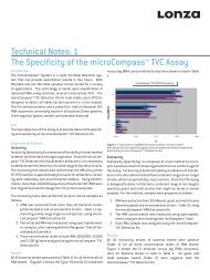
Key benefits › Up to 59% transfection efficiency<br />
100<br />
90<br />
80<br />
70<br />
60<br />
50<br />
40<br />
30<br />
20<br />
10<br />
0<br />
%<br />
96-well Shuttle ®<br />
› More than 90% cell viability<br />
› Excellent preservation of functionality (see also page 10 + 11)<br />
› Low cell numbers required<br />
(2.5 - 5 x 10⁴ cells for the 96-well Shuttle ® , 2.5 x 10⁵ for the standard Nucleofector ® )<br />
Efficiency<br />
Nucleofector ®<br />
Transfection performance.<br />
Mouse DC (Balb/C) were transfected according to the<br />
appropriate Optimized Protocol for nucleofection ® using<br />
pmaxGFP. Cells were analyzed 24 hours post nucleofection ®<br />
by flow cytometry for maxGFP ® expression and viability. Cell<br />
viability is given in percent compared to non-transfected<br />
control.<br />
The Nucleofector ® Kit for mouse DCs is only compatible with Nucleofector ® II Devices (carrying software version S3-7<br />
and higher for the Nucleofector ® II and version S4-4 and higher for Nucleofector ® II, serial version ”S“).<br />
› Page 4 › <strong>amaxa</strong> <strong>news</strong> # 10<br />
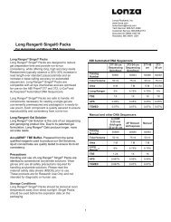
Functionality post nucleofection ® .<br />
The graph displays functionality of immature mouse DC<br />
(Balb/C mice) post nucleofection ® (Sample) on standard<br />
Nucleofector ® and 96-well Shuttle ® . Analyzed was their ability<br />
to secrete IL-6. Therefore, 2 hours post nucleofection ® , cells<br />
were stimulated by LPS. 22 hours later, functionality was<br />
analyzed by IL-6 specific ELISA and is given in percent compared<br />
to non-transfected control (Control).<br />
Ordering information Mouse Dendritic Cell Nucleofector ® Kit 25 reactions Cat. No.: VPA-1011<br />
Mouse Dendritic Cell (immature) 96-well Nucleofector ® Kit 96 reactions Cat. No.: VHPA-1011<br />
Mouse Dendritic Cell (mature) 96-well Nucleofector ® Kit 96 reactions Cat. No.: VHPA-1012<br />
Mouse Dendritic Cell (immature) 96-well Nucleofector ® Kit 960 reactions Cat. No.: VHPA-2011<br />
Mouse Dendritic Cell (mature) 96-well Nucleofector ® Kit 960 reactions Cat. No.: VHPA-2012<br />
<strong>amaxa</strong> web information For further information, please contact your local representative or visit www.<strong>amaxa</strong>.com/mouseDC<br />
Viability<br />
100<br />
90<br />
80<br />
70<br />
60<br />
50<br />
40<br />
30<br />
20<br />
10<br />
0<br />
Mouse DC functionality<br />
%<br />
96-well Shuttle ®<br />
Control Sample<br />
Nucleofector ®
new<br />
› Product News<br />
Basic Neuron SCN Nucleofector ® Kit:<br />
Transfect Only What You Need for One<br />
Coverslip!<br />
[k]<br />
<strong>amaxa</strong>’s new Basic Neuron Small Cell Number (SCN) Nucleofector ® Kit makes high efficiency transfection<br />
possible even for extremely low cell numbers. Now you can transfect just the number of cells needed<br />
for later culture and analysis (e.g., microscopic analyses of neuronal growth and morphology). Benefit<br />
from a drastic reduction in both number of donor animals and amount of preparation time. The new,<br />
optimized SCN Nucleofector ® Solution, SCN cuvettes, programs and optimized protocols make<br />
transfection efficiencies of up to 60% possible with as little as 15,000 cells per nucleofection ® .<br />
› Transfect as little as 15,000 cells per nucleofection ® –<br />
More experiments with fewer donor animals<br />
› Excellent non-viral transfection efficiencies and viabilities –<br />
50% and higher even for very difficult-to-transfect neurons like DRGs<br />
› Suitable for a broad range of neurons –<br />
Easy optimization for your neuron of interest<br />
Average transfection efficiencies and viabilities of<br />
embryonal rat hippocampal neurons (n = 3).<br />
20,000 and 50,000 freshly isolated rat hippocampal neurons<br />
(E17) were transfected, cultured and analyzed as described<br />
in <strong>amaxa</strong>’s Optimized Protocol for nucleofection ® . Viability<br />
was determined as number of living cells compared to<br />
non-transfected control.<br />
(Data courtesy of M. Kiebler, Department of Neuronal Cell Biology,<br />
Medical University of Vienna, Vienna, Austria).<br />
SCN nucleofection ® of freshly isolated mouse Dorsal Root<br />
Ganglion (DRG) cells.<br />
15,000 DRGs (E15.5) were transfected with Nucleofector ® SCN<br />
Basic Neuro Program 6 and 0.4 µg pmaxGFP ® (green, B) and<br />
seeded on a coated coverslip. 24 hours post nucleofection ® ,<br />
cells were fixed and immunostained for b -tubulin III<br />
(red, neuronal marker, A). In this experiment, transfection<br />
efficiency as determined by fluorescence microscopy is<br />
approximately 60%.<br />
(Data courtesy of B. Eickholt, MRC Centre for Developmental<br />
Neurobiology, King‘s College, London, United Kingdom).<br />
Ordering information Basic Neuron SCN Nucleofector ® Kit* Cat. No.: VSPI-1003<br />
Nucleofector ® II Device, Serial Version “S” Cat. No.: AAD-1001S<br />
*Due to the innovative pulse programs which are applied, SCN Kits are compatible with Nucleofector ® II Devices, serial version “S” only. Please make sure<br />
that you have this version of device available by checking the information at www.<strong>amaxa</strong>.com/program-update or contacting our Scientific Support. For<br />
customers with an older version of the Nucleofector ® Device who want to use SCN Kits, <strong>amaxa</strong> offers a trade-in program with very attractive conditions.<br />
80<br />
60<br />
40<br />
20<br />
0<br />
%<br />
Transfection efficiency<br />
A B<br />
<strong>amaxa</strong> web information www.<strong>amaxa</strong>.com/basic-neuron-scn.html<br />
› Page 5 › www.<strong>amaxa</strong>.com<br />
Viability<br />
5 x 10⁴ 2 x 10<br />
Cells<br />
4 Cells
› Hot Topic<br />
Introduction<br />
Nucleofection ® Enables Luciferase<br />
Reporter Assays in Primary Cells<br />
and Virtually Any Cell Line<br />
Promega – First-class Luciferase Reporter Gene Vectors and Assays<br />
› Reporter gene expression vectors for Firefly and Renilla luciferases<br />
› All common selectable markers available<br />
› Choice of different vectors containing no promoters, minimal promoters or promoters and response elements<br />
› Control of reporter protein stability using Rapid Response technology<br />
› Assay reagents for the detection of single or dual Firefly and Renilla luciferase activity<br />
› Highly sensitive, robust and homogeneous assays<br />
› Available as ‘flash’ or ‘glow’ type assay format<br />
› Ideally suited for high-throughput applications<br />
Reporter gene technologies are powerful tools<br />
to study gene expression, regulation and complex<br />
cellular processes such as gene networks.<br />
Successful reporter gene expression requires<br />
efficient delivery of state-of-the-art expression<br />
vectors into appropriate cells. In addition, fast,<br />
reliable and sensitive reporter assays are needed<br />
to obtain meaningful results. <strong>amaxa</strong>’s non-viral<br />
Nucleofector ® Technology is ideally suited for<br />
transfection of primary cells and difficult-totransfect<br />
cell lines. It provides easy and reproducibly<br />
efficient transfection of virtually any cell<br />
type in single reactions or up to 96-wells at a<br />
time. In combination with Promega’s luciferase<br />
reporter technology, it leads to rapid and<br />
high expression within a few hours. Promega’s<br />
luciferase reporter vectors and assays represent<br />
the gold standard for highly sensitive, simple<br />
and flexible reporter gene studies. In the past<br />
few years, new features were developed enabling<br />
the choice of selectable markers, different<br />
› Page 6 › <strong>amaxa</strong> <strong>news</strong> # 10<br />
promoters and essential control of protein stability. In addition,<br />
robust and homogeneous assays are available that allow for<br />
usage in high-throughput applications. Thus, these two technologies<br />
complement one another perfectly for such diverse uses<br />
as measurement of transcriptional activity, RNAi-mediated gene<br />
regulation and secondary screenings using difficult-to-transfect<br />
cell types such as primary cells.<br />
Nucleofection ® of Luciferase Reporter Vectors<br />
into Primary Cells and Suspension Cell Lines<br />
Influence of vector constructs on luciferase expression<br />
Successful gene expression experiments are influenced by<br />
numerous factors such as cell type, transfection method, the<br />
expression vector, and analysis method. Constructs containing<br />
the CMV promoter usually lead to stronger gene expression compared<br />
to vectors containing the SV40 promoter in mammalian<br />
cells, but there are exceptions such as BHK-21 cells. 1 In addition,<br />
DNA sequences like the polyadenylation signal, enhancer, Kozak<br />
sequence or introns may have an influence on gene expression<br />
even if two expression plasmids contain the same promoter.
› Hot Topic<br />
Kinetics of luciferase expression after nucleofection ® differ from other reporter genes<br />
Unlike conventional transfection methods, such as lipofection, nucleofection ® delivers substrates<br />
not only into the cytoplasm but also into the nucleus of the cell, resulting in a reduced time to expression.<br />
It is highly recommended to perform luciferase analysis 6 - 16 hours post nucleofection ® , whereas<br />
the optimal analysis time point for b –gal or GFP expression is 10 - 48 hours post nucleofection ® .<br />
Nucleofection ® and Luciferase Assays Offer a Wide Range of Applications<br />
Promoter studies by co-transfection of 4 expression vectors using nucleofection ®<br />
Overexpression of HMGA1 proteins is a constant feature in human carcinomas and plays a critical<br />
role in controlling growth of NK/T cell lymphocytes. By co-transfection of 4 expression constructs,<br />
Fedele et al. have shown that HMGA1 proteins and transcription factor NF- B exert a synergistic<br />
k<br />
effect on the activity of the IL-15 promoter in NK-T leukemic cell line DERL-7. 2 They investigated the<br />
functional consequences of HMGA1-binding on IL-15 promotor activity by transfecting a reporter<br />
vector in which luciferase expression is driven by the IL-15 promoter (pGL3 IL-15) with cDNAs expressing<br />
HMGA1 and NF- B p65 into the DERL-7 NK-T leukemic cell line (Figure 1).<br />
k<br />
Figure 1: HMGA1 and NF- k B exert a<br />
synergistic effect on the promoter<br />
activity of IL-15.<br />
Murine NK-T leukemic cell line DERL-7<br />
was transfected with the IL-15 promoter<br />
reporter vector alone, or co-transfected<br />
with 10 µg of NF- k B p65, or with 10 µg of<br />
both NF- k B p65 and HMGA1 expression<br />
vectors using Nucleofector ® Cell Line<br />
Kit V and Nucleofector ® Program O-017.<br />
Furthermore, a b -gal expression plasmid<br />
was used for normalization of the transfection<br />
efficiency. Luciferase activity<br />
was measured 4 hours post nucleofection ® .<br />
(Data reproduced from Fedele et al.<br />
(2005) Oncogene 24:3427-3435).<br />
Conclusions and Outlook<br />
The combination of Promega’s luciferase vectors and assays and the <strong>amaxa</strong> Nucleofector ® Technology<br />
provide unique tools to examine gene expression, RNAi-mediated gene regulation and many further<br />
applications in cell biology. These technologies allow studies in the scientifically most relevant<br />
primary cells and lead to more meaningful results with reliable and sensitive assays.<br />
References<br />
1. Liu et al. (1997) Anal Biochem 246(1): 150-152.<br />
2. Fedele et al. (2005) Oncogene 24:3427-3435.<br />
Promega and Rapid Response are trademarks of Promega Corporation.<br />
› Page 7 › www.<strong>amaxa</strong>.com<br />
12<br />
10<br />
8<br />
6<br />
4<br />
2<br />
0<br />
Luciferase activity (arbitrary units)<br />
_<br />
+ +<br />
_ _<br />
pGL3<br />
+<br />
pCMV- b -gal<br />
_<br />
+ +<br />
_ _<br />
pGL3 IL-15<br />
+<br />
NF B p65<br />
k<br />
HMGA1
› Application Note<br />
Down-regulation of a Key Component<br />
of the RNA Localization Machinery<br />
in Hippocampal Neurons<br />
Manuel Zeitelhofer and Ralf Dahm, Division of Neuronal Cell Biology, Center for Brain Research,<br />
Medical University of Vienna, Vienna, Austria<br />
The localization of mRNAs is an efficient way to target proteins to specific regions in a cell. In mammalian neurons, RNA localization is involved in<br />
compartmentalizing the cell during differentiation and is assumed to play an important role in synaptic plasticity. RNAs are packaged together with<br />
RNA-binding proteins into transport-competent ribonucleoprotein particles (RNPs) and transported into dendrites. The RNA-binding protein Staufen2<br />
(Stau2) is a key component of RNPs and implicated in dendritic RNA transport. In this study, Stau2 was successfully down-regulated by RNA interference<br />
(RNAi). The knockdown efficiency of Stau2 expression was assessed after nucleofection ® of shRNA constructs targeting Stau2 mRNA. In contrast to<br />
other transfection methods, the high transfection efficiencies attainable with the nucleofection ® technique allowed the determination of the extent of<br />
Stau2 protein down-regulation by quantitative western blot analysis.<br />
Introduction<br />
The localization of mRNAs is an efficient<br />
way to target gene products to specific<br />
regions in a cell. It occurs in a wide range<br />
of organisms and cell types 1, 2 . During<br />
Drosophila development, for example,<br />
localized mRNAs act as cell fate determinants<br />
and thereby specify the body<br />
axes 3 . In addition to its role in development,<br />
RNA localization also occurs in<br />
differentiated cells. In migrating cells, for<br />
instance, mRNAs are localized to the<br />
leading edges, thereby enabling directed<br />
movement. This plays a role in wound<br />
closure when fibroblasts migrate into the<br />
lesion or when cancer cells metastasize.<br />
Polarized cells, such as neurons, display<br />
a functional compartmentalization of the<br />
cytoplasm. The cell body is molecularly<br />
distinct from the axon and the dendrites.<br />
RNA localization is involved in compartmentalizing<br />
neurons during differentiation<br />
and it is assumed to also play<br />
an important role in synaptic plasticity,<br />
the experience-dependent remodeling<br />
of synapses that forms the basis of<br />
learning and memory 4 .<br />
How is RNA localization achieved?<br />
Localized RNAs contain cis-acting<br />
sequence elements, often within the<br />
3’-untranslated region (3’-UTR), which<br />
are recognized by proteins (trans-acting<br />
factors). The mRNAs and trans-acting<br />
factors are packaged into ribonucleoprotein<br />
particles (RNPs). These RNPs<br />
are then transported to and retained<br />
by sites of local synthesis 5 . During transport,<br />
the RNAs must be kept translationally<br />
silenced to prevent the ectopic<br />
expression of the corresponding protein.<br />
At their destinations, this translational<br />
block can be lifted by signals inducing<br />
translation 6 . A central hypothesis assu-<br />
mes that translation is only activated at<br />
synapses where synaptic plasticity has<br />
been induced.<br />
In this study, the nucleofection ® technique<br />
was used to assess the efficiency of<br />
down-regulation of Staufen2 (Stau2) in<br />
hippocampal neurons. Stau2 is one<br />
of the key components implicated in<br />
dendritic RNA localization. In Drosophila,<br />
Staufen is required for mRNA localization<br />
in the oocyte 7 and in neuroblasts 7 .<br />
In mammals, it is hypothesized that<br />
Stau2 is important in the transport of<br />
RNAs into dendrites of mature hippocampal<br />
neurons. To assess the consequences<br />
of a loss of Stau2 on dendritic<br />
RNA localization as well as the morphology<br />
of dendrites and dendritic spines,<br />
Stau2 was down-regulated by RNAi<br />
in primary cultures of hippocampal<br />
neurons 8 . In order to obtain quantitative<br />
information on the extent of down-regulation<br />
by western blot analysis, it was<br />
crucial that a large proportion of hippocampal<br />
neurons was transfected by<br />
nucleofection ® .<br />
Material and Methods<br />
Primary hippocampal neuron culture<br />
Neurons were isolated from the hippocampi<br />
of 17 day-old embryonic (E17)<br />
rat brains and cultured as described 9 .<br />
For nucleofection ® experiments, neurons<br />
were immediately used after dissociation.<br />
For the assessment of dendritic<br />
spine phenotypes and for fluorescence<br />
in situ hybridization (FISH) experiments,<br />
cells were used after 15 days in vitro (DIV).<br />
Nucleofection ® and western blot<br />
analysis<br />
After dissociation, neurons were cotransfected<br />
with an expression vector<br />
› Page 8 › <strong>amaxa</strong> <strong>news</strong> # 10<br />
encoding citrine and shRNA constructs<br />
(pSUPER, OligoEngine 10 ) against<br />
Stau2, mismatch Staufen2 (misStau2),<br />
and Septin7 (Sept7; CDC10). Cells were<br />
plated onto 6 cm cell culture dishes<br />
and lysed after 3 days of expression.<br />
The knockdown efficiency was assessed<br />
by western blot analysis.<br />
Immunostaining<br />
The following antibodies were used:<br />
an affinity-purified rabbit anti-Stau1<br />
antibody (1 µg/ml) and an affinity-purified<br />
rabbit anti-Stau2 antibody (1 µg/ml).<br />
Fluorophore-coupled phalloidin was used<br />
to label F-actin. Immunostaining experiments<br />
were performed as described 11 .<br />
Results<br />
Stau2 was down-regulated in hippocampal<br />
neurons by nucleofection ® of shRNA<br />
constructs to assess its function during<br />
neuronal differentiation and in mature<br />
neurons. To evaluate the success of the<br />
down-regulation, western blot analysis<br />
of the transfected neurons was performed.<br />
As neurons are post-mitotic cells,<br />
they are difficult to transfect. Conventional<br />
transfection methods like the<br />
calcium phosphate (CaPi) method have<br />
very poor transfection efficiencies for<br />
subsequent biochemical analyses and<br />
virus-based methods are too time-consuming<br />
and costly 12 . The nucleofection ®<br />
technique overcomes these limitations.<br />
Primary hippocampal neurons were<br />
co-transfected with an expression vector<br />
encoding citrine and shRNA constructs<br />
(pSUPER) against Stau2, mis-Stau2,<br />
and Septin7. First, the transfection<br />
efficiency was evaluated. To this aim,<br />
transfected neurons were fixed 3 days<br />
post nucleofection ® and the percentage
› Application Note<br />
A<br />
of transfected cells quantified (Figure<br />
1A, B). We reached transfection efficiencies<br />
of approximately 60% that allowed<br />
to perform biochemical experiments.<br />
Importantly the transfected neurons<br />
developed normally (Figure 1C, D). This<br />
allowed us to quantitatively downregulate<br />
Stau2 in neurons via RNAi. The<br />
levels and the specificity of Stau2 downregulation<br />
were assessed via western<br />
blot analysis (Figure 2A). The protein<br />
levels of Stau2 were significantly downregulated<br />
in shRNA transfected cells<br />
whereas the levels of the Stau2 paralogue<br />
Stau1 as well as that of the unrelated<br />
protein Septin7 remained unchanged.<br />
Moreover, nucleofection ® with mis-Stau2<br />
did not affect Stau2 protein levels,<br />
indicating that the down-regulation of<br />
Stau2 is specific.<br />
The extent of down-regulation in mature<br />
neurons was further controlled by<br />
immunocytochemistry (Figure 2B). 15 DIV<br />
neurons were co-transfected using the<br />
CaPi method with an expression vector<br />
encoding ECFP together with pSUPER<br />
vectors against Stau2, misStau2 and<br />
B<br />
C D<br />
o<br />
o<br />
A B<br />
o<br />
¬<br />
Figure 1: Hippocampal neurons are transfected efficiently via nucleofection ® .<br />
Hippocampal neurons were transfected with an shRNA construct against Stau2 and<br />
cultured in 6 cm cell culture dishes. After 3 DIV, neurons were fixed and analyzed to<br />
examine transfection efficiencies (B). The transfection efficiency reached up to<br />
60%. Neurons displayed normal morphology upon nucleofection ® (A). Higher<br />
magnification phase contrast (C) and fluorescence images (D) show the integrity of<br />
the transfected neurons. The arrow shows an outgrowing axon and the arrowheads<br />
indicate extending neurites with the typical growth cones at their tips. The inset in<br />
C shows the corresponding DAPI stained nucleus of the transfected neuron.<br />
RFP, respectively, and immunostained<br />
with Stau2 and Stau1 antibodies. Stau2<br />
staining was substantially reduced in<br />
neurons transfected with the shStau2<br />
plasmid but was abundant in neurons<br />
transfected with mis-Stau2 or RFP plasmid.<br />
The down-regulation of Stau2 did<br />
not affect Stau1 staining.<br />
Interestingly, down-regulation of Stau2<br />
caused a rearrangement of the actin<br />
cytoskeleton, which plays an important<br />
role in the maintenance and plasticity of<br />
dendritic spines. This effect was mirrored<br />
by changes in the morphology of dendritic<br />
spines from their characteristic mushroom-like<br />
shape to filopodia. In addition,<br />
it could be observed that the levels of<br />
PSD95, a key component of the postsynaptic<br />
density, were reduced in Stau2<br />
down-regulated neurons, indicating that<br />
there are fewer functional synapses<br />
than in normal cells. This hypothesis<br />
was borne out by electrophysiological<br />
recordings that indicate a reduction in<br />
synaptic transmission in Stau2 knockdown<br />
neurons 8 .<br />
As Stau2 has been implicated in dendritic<br />
› Page 9 › www.<strong>amaxa</strong>.com<br />
mRNA transport and since the actin<br />
cytoskeleton is rearranged upon Stau2<br />
down-regulation, it was further tested<br />
whether the levels of -actin mRNA<br />
b<br />
are changed in Stau2 down-regulated<br />
neurons. It was indeed observed that the<br />
-actin mRNA levels were reduced by<br />
b<br />
approximately 37%, indicating that<br />
Stau2 is important for the transport of<br />
this mRNA into dendrites 8 .<br />
Discussion<br />
The Nucleofector ® Technology is ideally<br />
suited to perform RNAi studies in postmitotic<br />
cells, such as hippocampal neurons.<br />
In contrast to other transfection<br />
methods, the high transfection efficiencies<br />
attainable with the nucleofection ®<br />
technique allow a proper analysis of the<br />
levels of Stau2 protein down-regulation<br />
by quantitative western blot analysis.<br />
In summary, this study showed that<br />
the down-regulation of Stau2 causes<br />
changes in dendrite morphology and<br />
suggests a role of Stau2 in the transport<br />
of b -actin mRNA into dendrites of hippocampal<br />
neurons.<br />
mock unr. mis Stau2 +siStau2<br />
+si-RFP +mis +siStau2<br />
Stau2<br />
Calnexin<br />
Stau1<br />
Calnexin<br />
*<br />
ECFP ECFP ECFP ECFP<br />
*<br />
Stau2 Stau2 Stau2 Stau1<br />
Figure 2: Assessment of the extent of down-regulation of Stau2.<br />
(A) Western blot analysis of hippocampal neurons transfected using the Nucleofector ® Technology. Neurons were co-transfected<br />
with a plasmid expressing citrine fluorescent protein (lane 1, mock) and shRNA-expressing plasmids against Septin7 (lane 2, labelled<br />
unr.), mismatch Stau2 (lane 3, mis) and Stau2 (lane 4, siStau2). Cells were lysed after 3 days of expression and processed for<br />
western blot analysis. The levels of the three Stau2 isoforms (lines indicate 62, 59 and 52 kD) were significantly down-regulated only<br />
in cells transfected with the shRNA plasmid siStau2 (lane 4). Calnexin served as internal loading control. The levels of Stau1 did not<br />
change upon down-regulation of Stau2. (B) Down-regulation of Stau2 in mature neurons. 15 DIV neurons were co-transfected with<br />
the following constructs: siStaufen2-2 (siStau2), si-RFP or mismatch Staufen2 (mis) pSUPER vectors together with ECFP (green).<br />
Neurons were stained 3 days after transfection with anti-Stau2 or anti-Stau1 antibodies (red). The Stau2 signal was strongly reduced<br />
in both the cell body (asterisk) and dendrites of transfected neurons (green) compared with untransfected neurons. By contrast,<br />
expression of misStau2 or si-RFP did not alter the Stau2 levels. Down-regulation of Stau2 also did not affect Stau1 expression. Scale<br />
bar: 10 µm. (Figure 2a reproduced from (2006) Journal of Cell Biology 172:221-231. Copyright 2006 Rockefeller University Press.)<br />
**<br />
**<br />
¬<br />
¬<br />
References<br />
1. Kloc, Zearfoss<br />
et al. (2002)<br />
Cell<br />
108(4): 533-44.<br />
2. Jansen (2001)<br />
Nat Rev Mol Cell Biol<br />
2(4): 247-56.<br />
3. St Johnston (2005)<br />
Nat Rev Mol Cell Biol<br />
6(5): 363-75.<br />
4. Sutton and<br />
Schuman (2006)<br />
Cell<br />
127(1): 49-58.<br />
5. Martin and Zukin<br />
(2006)<br />
J Neurosci<br />
26(27): 7131-4.<br />
6. Hüttelmaier<br />
et al. (2005)<br />
Nature<br />
438(7067):512-5.<br />
7. Broadus,<br />
Fuerstenberg<br />
et al. (1998)<br />
Nature 391(6669):<br />
792-5.<br />
8. Goetze, Tuebing<br />
et al. (2006)<br />
J Cell Biol<br />
172(2): 221-31.<br />
9. Zeitelhofer, Vessey<br />
et al. (2007)<br />
Nat Protoc<br />
2(7): 1692-704.<br />
10. Brummelkamp,<br />
Bernards<br />
et al. (2002)<br />
Science<br />
296(5567): 550-3.<br />
11. Goetze, Grunewald<br />
et al. (2004)<br />
J Neurobiol<br />
60(4): 517-25.<br />
12. Dahm, Zeitelhofer,<br />
Götze, Kiebler and<br />
Macchi<br />
(2008)<br />
Methods<br />
in Cell Biology<br />
(in press)
› Application Note<br />
Nucleofection ® –<br />
Combining High Transfection<br />
Performance with Superior<br />
Preservation of Functionality<br />
Nucleofection ® has become the method of choice whenever transfection of primary cells or hard-totransfect<br />
cell lines is required. Here we show that nucleofection ® of frequently used primary cells<br />
(mouse dendritic cells, human macrophages and human T cells) results in highly efficient transfer of<br />
DNA and other substrates while at the same time maintaining excellent cell viability and post<br />
transfection functionality. This combination of benefits makes nucleofection ® superior to other<br />
transfection methods.<br />
Introduction<br />
For some primary cells, transient transfection<br />
can be achieved by using<br />
classical transfection methods, like<br />
lipofection. However, none of the classical<br />
transfection methods gain high<br />
transfection rates combined with low<br />
post transfection mortality and a good<br />
preservation of cell-specific functionality.<br />
With the Nucleofector ® Technology,<br />
<strong>amaxa</strong> offers the first non-viral easyto-use<br />
transfection method unifying<br />
these three aspects. Great care is taken<br />
during <strong>amaxa</strong>’s optimization of the<br />
nucleofection ® parameters to further<br />
minimize the effects of the transfection<br />
process on cell functionality. This<br />
includes the verification of cellular<br />
functionality utilizing frequently used<br />
cell-specific assays early in the developmental<br />
process.<br />
This short report focuses on three frequently<br />
used, hard-to-transfect primary<br />
Table 1<br />
cells (primary mouse dendritic cells,<br />
primary human macrophages and<br />
primary human T cells) proving the high<br />
degree of post nucleofection ® functionality<br />
that can only be reached using<br />
nucleofection ® .<br />
Results and Discussion<br />
The experiments presented in this<br />
article have either been performed<br />
on the standard Nucleofector ® (mouse<br />
dendritic cells) or the high throughput<br />
96-well Shuttle ® Device (human<br />
macrophages and human T cells) using<br />
the individual primary cell-specific<br />
Nucleofector ® Kit or 96-well Nucleofector ®<br />
Kits. Each primary cell-specific kit has<br />
been optimized to perfection by <strong>amaxa</strong>’s<br />
team of R&D scientists. This resulted<br />
in excellent transfection efficiencies<br />
while maintaining cell functionality.<br />
Cells were transfected with pmaxGFP<br />
› Page 10 › <strong>amaxa</strong> <strong>news</strong> # 10<br />
using the parameters carefully optimized<br />
for the primary cell and analyzed<br />
24 to 48 hours post nucleofection ® by<br />
flow cytometry for maxGFP ® expression.<br />
Viability was either measured by<br />
propidium iodide (PI) staining or using<br />
microtiter plate based cell viability<br />
assays (CellTiter-Glo ® , Promega). Viability<br />
values are given in percent<br />
compared to the non-transfected<br />
control. Functionality of the cells post<br />
nucleofection ® was tested by frequently<br />
used cell-specific assays as described<br />
below.<br />
Mouse dendritic cells<br />
Nucleofection ® of mouse dendritic cells<br />
resulted in high transfection efficiency<br />
(59%) and high cell viability (93%)<br />
(Table 1, immature mouse dendritic<br />
cells, strain BALB/c). In parallel, the<br />
optimized parameters enabled an<br />
Cell Nucleofector ® Kit used Program Transfection Cell<br />
efficiency viability<br />
Mouse Dendritic Cells Mouse Dendritic Cell Nucleofector ® Kit Y-001 59% (±3%) 93% (±7%)<br />
Human Macrophages Human Macrophage 96-well Nucleofector ® Kit DP-148 42% (±2%) 60% (±6%)<br />
Human T Cells, resting Human T Cell 96-well Nucleofector ® Kit FI-115 80% (±6%) 53% (±7%)<br />
Human T Cells, resting Human T Cell 96-well Nucleofector ® Kit EO-115* 68% (±5%) 79% (±8%)<br />
Human T Cells, stimulated Human T Cell 96-well Nucleofector ® Kit EO-115 70 % (±3%) 60% (±6%)<br />
Transfection efficiency and viability 24 hours post nucleofection ® .<br />
Cells were transfected according to the appropriate Optimized Protocols for nucleofection ® using pmaxGFP. Cells were analyzed<br />
24 hours post nucleofection ® by flow cytometry. Cell viability is given in percent compared to non-transfected control (cells<br />
handled as described in the Optimized Protocol but not treated with DNA or nucleofection ® program). For human T cells, two<br />
programs were optimized giving either superior transfection efficiency (FI-115) or optimal post nucleofection ® functionality (EO-115).<br />
Data are representative of 48 - 96 experiments. *Functionality data for resting human T cells are only shown for the program EO-115.
› Application Note<br />
Percent functionality<br />
100<br />
90<br />
80<br />
70<br />
60<br />
50<br />
40<br />
30<br />
20<br />
10<br />
0<br />
Functionality post nucleofection ®<br />
Nucleofector ®<br />
Mouse Dendritic Cells /<br />
IL-6 secretion<br />
Figure 1: Cell functionality post nucleofection ® .<br />
The bar graph displays the relative functionality of mouse dendritic cells, human macrophages and human T cells post<br />
nucleofection ® . Functionality is given in percent related to non-transfected control. Functionality was analyzed by IL-6 specific<br />
ELISA for mouse dendritic cells, TNF- a specific ELISA for human macrophages, IFN- g specific ELISA for stimulated human T cells,<br />
and by flow cytometry using a CD25 specific antibody for both human T cell states (resting and stimulated). Experiments were<br />
performed on a standard Nucleofector ® (mouse dendritic cells) or the 96-well Shuttle ® (human macrophages and human T cells).<br />
excellent preservation of functionality,<br />
tested by the ability of the transfected<br />
cells to secret IL-6 (Figure 1) after<br />
induction with LPS. For this purpose<br />
immature mouse dendritic cells were incubated<br />
with 0.1 µg/ml LPS two hours<br />
post nucleofection ® . IL-6 secretion was<br />
analyzed by a standard sandwich ELISA<br />
(IL-6 ELISA MS, BioSource) and is<br />
given in percent compared to the nontransfected<br />
control. In addition, the<br />
transfected cells were able to form<br />
dendrites as shown in Figure 2.<br />
Human macrophages<br />
Nucleofection ® of human macrophages<br />
revealed high efficiency (42%) combined<br />
with low mortality as shown by<br />
the data presented in Table 1. Functionality<br />
of human macrophages has<br />
been tested by measuring the secretion<br />
of TNF- a triggered by stimulation of<br />
the cells with 1 µg/ml LPS 24 hours<br />
post nucleofection ® . Data shown in<br />
Figure 1 prove the excellent conservation<br />
of macrophage functionality post<br />
nucleofection ® . TNF- a secretion was<br />
analyzed by sandwich ELISA (TNF- a<br />
96-well Shuttle ® 96-well Shuttle ®<br />
96-well Shuttle ®<br />
Human Macrophages /<br />
TNF- a secretion<br />
Resting Human T Cells /<br />
CD 25 expression<br />
EASIA, BioSource) and is shown in<br />
comparison to non-transfected control<br />
(macrophages handled as described in<br />
the Optimized nucleofection ® Protocol<br />
but not treated with DNA).<br />
Human T cells<br />
Unstimulated human T cells were<br />
transfected according to the specific<br />
Optimized Protocol either by using a<br />
program suited for highest efficiency<br />
(FI-115) or a program optimized for<br />
highest functionality (EO-115). Stimulated<br />
human T cells were transfected according<br />
to the Optimized nucleofection ®<br />
Protocol for stimulated human T cells<br />
(transfection performance data, see<br />
Table 1). Preservation of the biochemical<br />
functionality of resting T cells after<br />
nucleofection ® was analyzed by the<br />
detection of interleukin receptor 2<br />
(IL-2R alpha chain or CD25). Therefore,<br />
resting human T cells were stimulated<br />
with anti-CD3 (1 ng/µl; eBioscience) and<br />
anti-CD28 (2 ng/µl; Research Diagnostics<br />
Inc.) antibodies 5 hours post<br />
nucleofection ® . For stimulated cells,<br />
maintenance of the stimulated state<br />
› Page 11 › www.<strong>amaxa</strong>.com<br />
Stimulated Human T cells /<br />
CD 25<br />
Control Sample<br />
96-well Shuttle ®<br />
Stimulated Human T cells /<br />
INF- g<br />
was analyzed by the expression of<br />
CD25, as well as the secretion of Interferon<br />
gamma (IFN- g ). Human T cells<br />
expressing the surface bound receptor<br />
were detected with labeled anti-CD25<br />
antibodies (PE Mouse Anti-Human<br />
CD25; BD Pharmingen) by flow cytometry.<br />
Secretion of IFN- g was analyzed<br />
by a sandwich ELISA (IFN- g EASIA,<br />
BioSource). Data (Figure 1) are given in<br />
percent compared to non-transfected<br />
control.<br />
Summary<br />
<strong>amaxa</strong>’s Nucleofector ® Technology addresses<br />
the spectrum of cell types,<br />
substrates and cell numbers you<br />
encounter in your research. With more<br />
than 50 years of combined laboratory<br />
experience, <strong>amaxa</strong>’s team of scientists<br />
optimizes Nucleofector ® Kits to reach<br />
excellent transfection performance<br />
combined with superior preservation of<br />
cell characteristics, as proven by the<br />
examples in this short application note<br />
and more than 160 publications about<br />
T cells, macrophages and dendritic cells<br />
cited in <strong>amaxa</strong>’s citation database.<br />
Figure 2: Mature mouse dendritic cells form dendrites<br />
post nucleofection ® .<br />
Immature mouse dendritic cells were transfected with<br />
pmaxGFP using the Mouse Dendritic Cell Nucleofector ® Kit.<br />
Two hours post nucleofection ® , cells were stimulated with LPS<br />
to mature. Cells were analyzed 24 hours post nucleofection ®<br />
by fluorescence microscopy for maxGFP ® expression and their<br />
ability to form dendrites (red circle).
› Product News<br />
Improved<br />
Jurkat<br />
Cells<br />
% Transfection efficiency<br />
Higher Efficiencies for<br />
Jurkat Cells Than Ever Before<br />
<strong>amaxa</strong>’s R+D Team is not only working on the development of Nucleofector ® Protocols for new cell<br />
types, but on improving existing protocols as well. Without a doubt, Jurkat cells are among the most<br />
frequently used blood cancer cell lines within the immunology/hematology research field. In order to<br />
always provide products delivering the best possible results, <strong>amaxa</strong>’s R+D Team re-optimized the<br />
existing Nucleofector ® Protocols for Jurkat E6-1 (ATCC ® TIB-152) and Jurkat ACC282 (DSMZ). These<br />
efforts led to optimal performance for both the ATCC ® and DSMZ derived Jurkat cell lines making<br />
these cells more transfectable than ever before.<br />
Improved performance › Up to 35% better efficiency than before<br />
High efficiency › Up to 88% efficiency<br />
High viability › Up to 90% viability<br />
High expression level › For virtually any construct<br />
A B<br />
100 Jurkat E6-1 (ATCC<br />
90<br />
80<br />
70<br />
60<br />
50<br />
40<br />
30<br />
20<br />
10<br />
0<br />
® New protocol<br />
TIB-152)<br />
Former protocol 100<br />
90<br />
80<br />
70<br />
60<br />
50<br />
40<br />
30<br />
20<br />
10<br />
0<br />
87% 84% 54%<br />
X-001 X-005 C-016<br />
High transfection efficiencies in Jurkat E6-1 (ATCC ® TIB-152) and Jurkat ACC282 (DSMZ) cell line clones.<br />
Using pmaxGFP Jurkat E6-1 (ATCC ® TIB-152) cells were transfected with Nucleofector ® Solution V either with Nucleofector ®<br />
Program X-01/X-001 or X-05/X-005 in comparison to the formerly recommended program C-16/C-016 (A). Nucleofector ® Program<br />
X-01/X-001 is recommended for higher viability in Jurkat E6-1 (ATCC ® TIB-152) cells and program X-05/X-005 is recommended for<br />
a higher expression level. Jurkat ACC282 (DSMZ) cells were transfected with Nucleofector ® Program X-01/X-001 using pmaxGFP<br />
(B). 24 hours post nucleofection ® , cells were analyzed on a Becton Dickinson FACSCalibur for maxGFP ® expression. Cell viability<br />
was determined as PI negative cells and ranged from 74% - 90%.<br />
Expression level is what counts<br />
It is widely known that different constructs are expressed differently after transfection. Vector<br />
backbones and inserts are both responsible for this phenomenon. To make your daily work easier,<br />
we focus not only on the transfection efficiency but also on the expression level. Therefore, the<br />
new Nucleofector ® Protocol for the Jurkat E6-1 (ATCC ® TIB-152) recommends two different<br />
Nucleofector ® Programs, X-01/X-001 for better viability and X-05/X-005 for a higher expression level.<br />
The latter program guarantees the expression of virtually any construct of your interest.<br />
› Page 12 › <strong>amaxa</strong> <strong>news</strong> # 10<br />
% Transfection efficiency<br />
Jurkat ACC282 (DSMZ)<br />
74% 68%<br />
X-001 A-017<br />
New protocol<br />
Former protocol
[k]<br />
[k]<br />
› Product News<br />
Ordering information<br />
Cell Line Nucleofector ® Kit V Cat. No.: VCA-1003<br />
<strong>amaxa</strong> web information www.<strong>amaxa</strong>.com/jurkat-standard.html www.<strong>amaxa</strong>.com/improved-jurkat<br />
› Product News<br />
new<br />
Expression level of maxGFP ® in Jurkat<br />
E6-1 (ATCC ® TIB-152) cells.<br />
Jurkat E6-1 (ATCC ® ) cells were transfected<br />
with Nucleofector ® Solution V either with<br />
Nucleofector ® Program X-01/X-001 or<br />
X-05/X-005 using 2 µg pmaxGFP. 24 hours<br />
post nucleofection ® , cells were analyzed<br />
on a Becton Dickinson FACSCalibur for<br />
determining maxGFP ® expression level<br />
(X-Mean).<br />
96-well Shuttle ® Protocols and Kits –<br />
Human T Cells, HepG2, SH-SY5Y<br />
› Page 13 › www.<strong>amaxa</strong>.com<br />
4000<br />
3500<br />
3000<br />
2500<br />
2000<br />
1500<br />
1000<br />
500<br />
0<br />
X-Mean<br />
m › Up to 95% efficiency and excellent viability<br />
› Low well-to-well variations<br />
› Preserved functionality (e.g., human T cells)<br />
› Suitable for a variety of substrates, e.g., DNA and siRNA<br />
› Fewer cells required compared to the single cuvette Nucleofector ®<br />
High transfection efficiency and viability in SH-SY5Y<br />
(ATCC ® CRL-2266), HepG2 (ATCC ® HB-8065) and<br />
stimulated human T cells with the 96-well Shuttle ® .<br />
SH-SY5Y, HepG2 cell lines and primary stimulated human<br />
T cells were transfected with the 96-well Shuttle ® System using<br />
the recommended Nucleofector ® Programs and pmaxGFP.<br />
24 hours post nucleofection ® , cells were analyzed on a Becton<br />
Dickinson FACSCalibur with HTS options for maxGFP ®<br />
expression. Cell viability was determined by using the<br />
CellTiter-Glo ® Luminescent Viability Assay (Promega) or<br />
CellTiter ® -Blue Cell Viability Assay (Promega).<br />
100<br />
90<br />
80<br />
70<br />
60<br />
50<br />
40<br />
30<br />
20<br />
10<br />
0<br />
%<br />
X-001 X-005<br />
Jurkat E6-1 Jurkat E6-1<br />
(ATCC ® TIB-152) (ATCC ® TIB-152)<br />
Other re-optimized protocols available<br />
› COS-7 › U-937 › PC-12<br />
Transfection efficiency<br />
For the latest addition of 96-well Shuttle ® and regular Nucleofector ® Kits, please check our website: www.<strong>amaxa</strong>.com<br />
Viability<br />
HepG2 SH-SY5Y Stimulated<br />
(ATCC ® HB-8065) (ATCC ® CRL-2266) human T cells<br />
Ordering information 96-well Nucleofector ® Kit 96 reactions 960 reactions<br />
Primary cells T cells, human, stimulated Human T Cell Cat. No.: VHPA-1002 Cat. No.: VHPA-2002<br />
Cell lines SH-SY5Y Cell Line Kit SF Cat. No.: VHCA-1002 Cat. No.: VHCA-2002<br />
HepG2 Cell Line Kit SF Cat. No.: VHCA-1002 Cat. No.: VHCA-2002
[k]<br />
› Product News<br />
new<br />
Mouse and human stem cells were<br />
transfected according to the dedicated<br />
Optimized Protocol for nucleofection ®<br />
using pmaxGFP.<br />
maxGFP ® expression was analyzed 24 hours<br />
post nucleofection ® by flow cytometry.<br />
Transfection efficiencies given for human<br />
stem cells are average values derived<br />
from customer data.<br />
Ordering information<br />
<strong>amaxa</strong> web information<br />
From Mouse to Human –<br />
The Latest Nucleofector ® Kits<br />
for Stem Cells<br />
Adult and embryonic stem cells are perfectly suited to study cell differentiation and tissue/organ<br />
development. The Nucleofector ® Technology presents a powerful tool to transfect these delicate cells<br />
with high performance and preservation of functional properties. The latest additions to our stem cell<br />
portfolio are Nucleofector ® Kits for human stem cells and a 96-well Nucleofector ® Kit for mouse<br />
embryonic stem cells.<br />
› Up to 90% transfection efficiency and 80% cell viability<br />
› Excellent preservation of differentiation ability<br />
› Suitable for a variety of substrates, e.g., DNA and siRNA<br />
100<br />
90<br />
80<br />
70<br />
60<br />
50<br />
40<br />
30<br />
20<br />
10<br />
0<br />
Transfection efficiency %<br />
96-well 96-well<br />
Mouse ES (E14) Mouse ES (D3) Mouse ES H9.2 H9<br />
Mouse ES Nucleofector ® Kit 25 reactions Cat. No.: VPH-1001<br />
96-well new Mouse ES 96-well Nucleofector ® Kit 96 reactions Cat. No.: VHPH-1001<br />
Human CD34 + Cell Nucleofector ® Kit 25 reactions Cat. No.: VPA-1003<br />
Human MSC (Mesenchymal Stem Cell) Nucleofector ® Kit 25 reactions Cat. No.: VPE-1001<br />
Mouse NSC (Neural Stem Cell) Nucleofector ® Kit 25 reactions Cat. No.: VPG-1004<br />
Rat NSC (Neural Stem Cell) Nucleofector ® Kit 25 reactions Cat. No.: VPG-1005<br />
new Human Stem Cell Nucleofector ® Starter Kit 18 reactions Cat. No.: VPH-5002<br />
new Human Stem Cell Nucleofector ® Kit 1 25 reactions Cat. No.: VPH-5012<br />
new Human Stem Cell Nucleofector ® Kit 2 25 reactions Cat. No.: VPH-5022<br />
› Page 14 › <strong>amaxa</strong> <strong>news</strong> # 10<br />
For further information, please contact your local representative or visit www.<strong>amaxa</strong>.com
[k]<br />
[k]<br />
› Product News<br />
new<br />
Ordering information<br />
Further expression vectors<br />
pmaxCloning Vector –<br />
Strong Expression of Your<br />
Individual cDNA<br />
Do you have difficulties in expressing your specific gene-of-interest?<br />
One crucial aspect for obtaining high expression levels is the selection of an appropriate expression<br />
vector. <strong>amaxa</strong>’s novel pmaxCloning vector promotes strong, constitutive expression of cloned DNA<br />
inserts in mammalian cells. It provides gene expression under the control of a CMV promoter* and<br />
contains a multiple cloning site for cloning purposes. With <strong>amaxa</strong>’s pmaxCloning and pmaxFP ®<br />
vectors for fluorescent protein expression you can choose from a broad variety of expression<br />
plasmids for your specific experimental setup.<br />
*The CMV promoter is covered under the U.S. patents 5,168,062 and 5,385,839 and its use is permitted for research purposes only. Any other use of the<br />
CMV promoter requires a license from the University of Iowa Research Foundation, 214 Technology Innovation Center, Iowa City, IA.<br />
Benefit from:<br />
› High expression rates in mammalian cells<br />
› Multiple cloning site for convenient insertion of your gene-of-interest<br />
› Proven performance (same backbone as positive control vector pmaxGFP ® )<br />
Cat. No.: VDC-1040 pmaxCloning<br />
Kan r<br />
pmaxCloning,<br />
2.9 kb<br />
pUC ori<br />
MCS Kpnl Pme I EcoR I EcoR V BamH I Nhe I Pme I Sac I<br />
947<br />
pmaxFP ® -Green-C Cat. No.: VDF-1011<br />
pmaxFP ® -Green-N Cat. No.: VDF-1012<br />
pmaxFP ® -Green-PRL Cat. No.: VDF-1013<br />
P<br />
PCMV<br />
IE<br />
SV40<br />
poly A<br />
chimeric<br />
intron<br />
GG.TAC.CGC.CAT.CAT.GAA.GTT.TAA.ACA.<strong>AG</strong>C.TTG.AAT.TCT.CTA.G<strong>AG</strong>.ATA.TCC.TGC.<strong>AG</strong>A.GAT.CTG.GAT.CCC.TCG.<strong>AG</strong>G.CTA.GCG.CGG.CCG.CGT.TTA.AAC.<strong>AG</strong>A.GCT.C<br />
Hind III Xbal Pst I Xho I Not I<br />
pmaxFP ® -Yellow- C Cat. No.: VDF-1021<br />
pmaxFP ® -Yellow-N Cat. No.: VDF-1022<br />
pmaxFP ® -Yellow-PRL Cat. No.: VDF-1023<br />
<strong>amaxa</strong> web information www.<strong>amaxa</strong>.com/expression-vectors.html<br />
MCS<br />
› Page 15 › www.<strong>amaxa</strong>.com<br />
pmaxFP ® -Red-C Cat. No.: VDF-1031<br />
pmaxFP ® -Red-N Cat. No.: VDF-1032<br />
pmaxFP ® -Red-PRL Cat. No.: VDF-1033
Morpholino<br />
› Application Note<br />
Achieving Efficient<br />
Delivery of Morpholino Oligos<br />
with Nucleofection ®<br />
Shannon T. Knuth, Jon D. Moulton and Paul A. Morcos, Biology and Customer Support, Gene Tools,<br />
LLC, Philomath, Oregon, USA<br />
Morpholino oligos provide a stable, specific and effective antisense activity. Developmental biologists have long since<br />
used these properties by microinjecting or electroporating Morpholinos into embryos to allow for splice-blocking or<br />
gene knockdown. However, efficient delivery of Morpholinos in mammalian cell cultures can be a challenge. While<br />
complex formation with lipid-based reagents is ineffective due to the non-ionic Morpholino backbone, we show here<br />
that nucleofection ® can be used as an efficient delivery method. As such, nucleofection ® of Morpholinos offers a<br />
straightforward alternative to siRNA-mediated gene knockdown and miRNA maturation inhibition.<br />
RNA<br />
DNA<br />
Figure 1: Comparison of the chemical structure of<br />
two-mers of Morpholino, RNA and DNA.<br />
Two significant differences in the Morpholino backbone<br />
compared to RNA and DNA are the non-charged phosphodiamiate<br />
linkage between the subunits and replacement<br />
of the five-membered sugar ring with the six-membered<br />
Morpholine ring.<br />
Introduction<br />
Morpholino oligos (MOs), also known as<br />
phosphodiamidate Morpholino oligos,<br />
are designed to bind to complementary<br />
RNA sequence and sterically inhibit<br />
proteins from binding the RNA.<br />
Morpholinos are commonly used to<br />
knock down gene expression and alter<br />
mRNA splicing in a variety of organisms,<br />
tissues or cells (for a searchable database<br />
of references see http://www.genetools.<br />
com/Publications/). For gene<br />
knockdowns the MOs are designed to<br />
complement mRNA sequence in the 5’<br />
untranslated region and/or the first<br />
25 bases of mRNA coding sequence,<br />
sterically inhibiting the movement<br />
of the translation initiation complex<br />
toward the start codon. This prevents<br />
the large subunit of the ribosome<br />
from assembling and, as a consequence,<br />
causing reduced gene expression.<br />
Morpholinos are also used to modify<br />
pre-mRNA splicing by targeting the<br />
oligo to a splice junction or regulatory<br />
Advantages of Morpholinos over DNA and RNA-based gene knockdown reagents:<br />
› Very robust and stable molecules 10, 11 due to the non-biological, chemically stable backbone structure (Figure 1)<br />
› Exquisite specificity: at low concentrations a Morpholino with just five mispairs along its length will have<br />
no antisense activity 12<br />
› Largely free of non-antisense effects as evidenced by rescue of the Morpholino phenotype via injection of the<br />
corresponding mRNA 13<br />
› Backbone is non-toxic<br />
› Predictable targeting<br />
› Steric block mechanism allows for additional applications such as altering mRNA splicing and blocking microRNA maturation<br />
› Page 16 › <strong>amaxa</strong> <strong>news</strong> # 10<br />
site, sterically blocking binding of<br />
snRNPs or other splice factors 1 . Spliceblocking<br />
Morpholinos have great promise<br />
for therapeutic use correcting and/or<br />
reducing the impact of the mutations<br />
that lead to b -Thalessemia 2 , Duchenne<br />
muscular dystrophy 3 and other diseases<br />
caused by mis-splicing 4,5 . Morpholinos<br />
have also been used to block microRNA<br />
maturation 6 and microRNA targets 7 ,<br />
block ribozyme activity 8 and induce<br />
frameshifts 9 .<br />
However, even more so than RNA and<br />
DNA based oligos, Morpholinos are<br />
challenging to deliver into mammalian<br />
cells in culture. Standard lipid-based<br />
nucleic acid transfection reagents rely<br />
on an ionic interaction with a charged<br />
backbone and are not effective with<br />
the uncharged MO backbone. While<br />
microinjection is suitable for delivery<br />
into organs (embryos or brains), most<br />
commonly used transfection methods<br />
for cultured cells are scrape loading 16 ,
› Application Note<br />
Figure 2: Dose-dependent delivery of<br />
Morpholinos into ON 705 HeLa cells.<br />
Cells were transfected with indicated<br />
concentrations of splice-correcting MO.<br />
In the ON 705 HeLa cell line, the<br />
Morpholino corrects a splicing mutation<br />
splicing out a stop codon and putting<br />
luciferase in frame. After 24 hours, the<br />
cells were lysed and luciferase activity<br />
and protein amount were determined.<br />
Delivery success is proportional to the<br />
light output (normalized to total protein<br />
amount).<br />
special delivery 17 , conjugated cell-penetrating<br />
peptides 19 , Endo-Porter delivery 18<br />
or electroporation 14 . Here, we present,<br />
the results of our study using the<br />
Nucleofector ® Technology for delivering<br />
Morpholinos.<br />
Materials and Methods<br />
The ON 705 HeLa cell line was grown to<br />
75-80% confluency in DMEM/F-12 media<br />
(Invitrogen) with 10% FBS (Invitrogen).<br />
On the day of the experiment the cells<br />
were treated with 0.5% trypsin (Sigma),<br />
removed from the plate and spun down.<br />
The supernatant was removed and the<br />
cells resuspended in 100 µl Nucleofector ®<br />
Solution R per manufacturer’s, direction.<br />
The indicated amount of MO was added<br />
to each sample and mixed by vortexing.<br />
The sample was transferred to a<br />
nucleofection ® cuvette and transfected<br />
using Nucleofector ® Program I-013. After<br />
nucleofection ® , 500 µL of serum free<br />
RPMI medium was added to each<br />
cuvette followed by transfer of contents<br />
to a 6-well plate. In the ON 705 HeLa<br />
cell line, the delivery of the Morpholino<br />
corrects a splicing mutation, splicing<br />
out a stop codon and putting luciferase<br />
in frame 15 . After 24 hours, the cells were<br />
lysed and luciferase (Promega) and pro-<br />
Figure 3: Modifications of 3' end do<br />
not alter delivery efficiency.<br />
Cells were transfected with 0.2 nmol<br />
(2 µM) of a standard MO, a carboxyfluorescein<br />
(Fl) or a lissamine (Li) labeled<br />
MO. 24 hours post nucleofection ® , delivery<br />
efficiency was analyzed by activity<br />
of splice-corrected luciferase (normalized<br />
to total protein amount).<br />
tein (Bio-Rad) assays were carried out in<br />
duplicate for each sample. Delivery success<br />
is proportional to the light output<br />
and the data is normalized per protein.<br />
Results/Discussion<br />
Morpholinos were delivered uniformly<br />
in nearly 100% of the cells when imaged<br />
by fluorescence microscopy (data not<br />
shown). To further analyze delivery<br />
efficiency we used our established<br />
splice correction assay with a luciferase<br />
reporter stably expressed in ON 705<br />
HeLa cells. The luciferase activity<br />
produced at the 1-8 µM range of MO<br />
reveals a consistent increase in MO<br />
activity (Figure 2). Generally, as shown<br />
by the efficacy of cytosolic microinjection<br />
of splice-modifying Morpholinos<br />
into embryos, delivery to the cytosol is<br />
sufficient to give nuclear splice-correcting<br />
activity of Morpholinos. However,<br />
average luciferase activities (Figure 2)<br />
seem to be slightly higher with<br />
nucleofection ® compared to activities<br />
commonly achieved with Endo-Porter.<br />
Effective MO amounts needed for<br />
nucleofection ® seem to be lower than<br />
those published for classical electroporation<br />
14 . This suggests that nucleofection ®<br />
could be advantageous in terms of<br />
› Page 17 › www.<strong>amaxa</strong>.com<br />
1600<br />
1400<br />
1200<br />
1000<br />
800<br />
600<br />
400<br />
200<br />
0<br />
700<br />
600<br />
500<br />
400<br />
300<br />
200<br />
100<br />
0<br />
Luciferase activity<br />
0 1 2 4 8<br />
MO concentration (µM)<br />
Luciferase activity<br />
effective amounts due to its direct<br />
nuclear delivery or to smaller reaction<br />
volumes (higher concentrated cell<br />
suspension).<br />
In an additional experiment (Figure 3),<br />
the MO concentration was fixed at 2 µM<br />
and we compared delivery of Morpholinos<br />
with different 3’ end modifications. The<br />
carboxyfluorescein (Fl) has two negative<br />
charges at physiological pH while the<br />
lissamine (Li) (sulforhodamine B) is a<br />
zwitterion with no net charge. We found<br />
that there were no significant differences<br />
in delivery of the MOs with 3’ end groups<br />
compared to MOs with unmodified ends.<br />
In our lab, nucleofection ® has also been<br />
used effectively in C2C12 muscle cells<br />
to produce a quantitative shift in the<br />
splicing of dystrophin.<br />
In conclusion, nucleofection ® has been<br />
shown to allow efficient delivery of<br />
Morpholino oligos in cultured mammalian<br />
cells. Thus, by using nucleofection ®<br />
for delivery, Morpholinos can now be<br />
explored in difficult-to-transfect cell<br />
types and primary cells as potent alternative<br />
for other DNA- or RNA-based<br />
gene knockdown substrates.<br />
Std. control Fl. std. control Li. std. control<br />
MO concentration (µM)<br />
References<br />
1. Morcos (2007)<br />
Biochem Biophys Res<br />
Commun 358(2): 521-7.<br />
2. Kole et al. (2004)<br />
Oligonucleotides<br />
14(1): 65-74.<br />
3. Yokota et al. (2007)<br />
Expert Opin Biol Ther<br />
7(6): 831-42.<br />
4. Scaffidi and Misteli<br />
(2006) Science<br />
312(5776): 1059-63.<br />
5. Ugarte et al. (2007)<br />
Am J Hum Genet 81(6).<br />
6. Kloosterman et al.<br />
(2007) PLoS Biol<br />
5(8): e203.<br />
7. Choi et al. (2007)<br />
Science 318: 271-4.<br />
8. Yen et al. (2004)<br />
Nature 431(7007): 471-6.<br />
9. Howard et al. (2004)<br />
RNA 10(10): 1653-61.<br />
10. Hudziak et al. (1996)<br />
Antisense Nucleic Acid<br />
Drug Dev 6(4): 267-72.<br />
11. Youngblood et al.<br />
(2007) Bioconjug Chem<br />
18(1): 50-60.<br />
12. Summerton (1999)<br />
Biochim Biophys Acta<br />
1489(1): 141-58.<br />
13. Heasman et al. (2000)<br />
Dev Biol 222(1): 124-34.<br />
14. Matter and Konig<br />
(2005) Nucleic Acids<br />
Res 33(4): e41.<br />
15. Schmajuk et al. (1999)<br />
J Biol Chem<br />
274(31): 21783-9.<br />
16. Partridge et al. (1996)<br />
Antisense Nucleic Acid<br />
Drug Dev 6(3): 169-75.<br />
17. Morcos (2001) Genesis<br />
30(3): 94-102.<br />
18. Summerton (2005)<br />
Ann N Y Acad Sci<br />
1058: 62-75.<br />
19. Wu et al. (2007)<br />
Nucleic Acids Res<br />
35(15): 5182-91.
› <strong>amaxa</strong> Insights<br />
<strong>amaxa</strong>’s QA<br />
and Production Team<br />
As a technology company, <strong>amaxa</strong> has a duty to ensure its products<br />
are manufactured and tested to the highest possible specifications.<br />
The <strong>amaxa</strong> culture of continuous improvement makes this<br />
a yet more demanding task. Each new technology must be<br />
thoroughly evaluated and then seamlessly integrated with existing<br />
systems in order to ensure our customers receive high quality<br />
products that meet both their current and future needs.<br />
Production and Quality Assurance (QA) departments are responsible<br />
for this overall process within <strong>amaxa</strong> and project leaders<br />
from both departments work closely with colleagues in Sales and<br />
Marketing, Finance, Purchasing, and Scientific Support, as well as<br />
with external partners such as suppliers of raw materials. The<br />
Production and QA departments support Sales Managers by<br />
reserving requested production lots for key customers or providing<br />
lot-specific Certificates of Analysis to customers with stringent<br />
internal validation and audit procedures.<br />
Both the Production and QA departments at <strong>amaxa</strong> have been<br />
undergoing some exciting changes in the last 12 months. Our<br />
Production department has fully automated Nucleofector ®<br />
Solutions filling due to the increasing demand for Nucleofector ®<br />
and 96-well Shuttle ® Kits and to proactively improve both the<br />
› Page 18 › <strong>amaxa</strong> <strong>news</strong> # 10<br />
Dr. Christoph Zander<br />
Head of Production<br />
consistency and quality of <strong>amaxa</strong> Nucleofector ®<br />
Solutions. A recently installed automation line<br />
consists of 11 fully integrated instruments controlled<br />
centrally by computer. The system includes<br />
a filling carousel, in-line sealing and labeling, and<br />
an instrument to QA test the amount of solution<br />
filled into each bottle gravimetrically.<br />
Dr. Christoph Zander, Head of Production, describes<br />
the integration project as a “very challenging,<br />
but rewarding process”. The entire project – “from<br />
concept through to daily operation of the device” –<br />
has taken 12 months. But Christoph is confident<br />
that the project is not only necessary, but will also<br />
deliver sustained improvements to customers.<br />
“The cost of such a project is significant, but this<br />
is a long-term investment in higher quality. Right<br />
now, we are making new expiry-date testing<br />
for Nucleofector ® Solutions filled with the automated<br />
process that shows a longer shelf-life<br />
due to more reproducible sealing of bottles. In<br />
addition, the people who were working on the<br />
Nucleofector ® Solution filling line now have more<br />
time for further projects. So we are delivering<br />
continuous improvement to customers and also<br />
to our employees!”
› <strong>amaxa</strong> Insights<br />
Dr. Rainer Hammermann<br />
Head of QA<br />
The <strong>amaxa</strong> QA<br />
and Production Team<br />
The Scientific Support department will be involved in monitoring<br />
and following up with statistics to show improvements made by<br />
the new production process and Dr. Rainer Hammermann, Head of<br />
QA, ensures that each piece of customer feedback is recorded and<br />
that each individual complaint is followed up to the customer’s<br />
satisfaction. “Scientific Support is a very important channel for<br />
monitoring both emerging customer needs and also customers’<br />
perception and acceptance of <strong>amaxa</strong> quality standards. We are not<br />
standing still – instead we are anticipating our customers needs for<br />
the next five years and beginning projects that will help to further<br />
associate the name <strong>amaxa</strong> with high quality and dependable<br />
products.”<br />
<strong>amaxa</strong> cGMP Kits (current Good Manufacturing Practice) and their<br />
components are manufactured and filled under Grade A clean<br />
room conditions (EU Guide to Good Manufacturing Practice-<br />
Manufacture of Sterile Medicinal Products, 1997), meaning that<br />
air in the manufacturing facility is monitored and that each<br />
cubic meter of air contains not more than 3500 particles of<br />
0.5 µm diameter or larger. Manufacturing each kit component<br />
in full compliance with cGMP regulations means that these kits are<br />
suitable for use in disease research and development of<br />
pharmaceuticals. Scientists working in heavily regulated areas<br />
such as protein production and drug discovery can now use <strong>amaxa</strong><br />
› Page 19 › www.<strong>amaxa</strong>.com<br />
cGMP Kits for their work without first validating<br />
kit performance and other features. In contrast<br />
to other manufacturers, <strong>amaxa</strong> includes a<br />
more detailed and batch-specific Certificate of<br />
Analysis (CoA) into each cGMP Nucleofector ® Kit,<br />
providing an ‘audit trail’ of performance and<br />
QA test data for each batch of kits. Rainer believes<br />
that this demonstrates “full disclosure of<br />
data in order to ensure reliable research results,<br />
thus protecting the safety of researchers in<br />
development departments of the pharmaceutical<br />
industry.”<br />
As the needs of Nucleofector ® users evolve and<br />
expand into new application areas such as stem<br />
cell research and drug discovery, <strong>amaxa</strong> Production<br />
and Quality Assurance will continue to set<br />
industry standards and enable researchers to be<br />
increasingly confident in their research data.
› Hot Topic<br />
Achieve Your Research Objectives<br />
with <strong>amaxa</strong> Transfection Services<br />
All <strong>amaxa</strong> services are designed to meet your needs and to assist you in successfully achieving<br />
your transfection goals. <strong>amaxa</strong>’s team of scientists have years of transfection experience and will<br />
provide you with fast, reliable results along with expert scientific customer support.<br />
Numerous pharmaceutical and biotech companies, in both the US and Europe, have already worked<br />
with <strong>amaxa</strong> to generate stable clones and optimize transfection conditions for their<br />
Nucleofector ® II or 96-well Shuttle ® Devices.<br />
These projects include the generation of stable cell clones expressing drug targets and the<br />
generation of reporter cells for several signal transduction pathways. Moreover <strong>amaxa</strong> has<br />
extensive experience in working with more than 100 different cell lines and primary cells and has<br />
successfully created stably transfected clones from cells that are very difficult-to-transfect, like<br />
J774, Jurkat, HL-60, BJAB, MCF-7, and suspension CHOs.<br />
<strong>amaxa</strong> scientists will optimize transfection protocols using our unique non-viral technology for<br />
any cell type and substrate, allowing you the freedom to focus your expertise on analysis and<br />
experimentation, not the creation of the clone or protocol.<br />
<strong>amaxa</strong>'s results and expertise are proven: Nucleofection ® is used in expression studies, RNAi<br />
knockdown experiments, transient protein expression, target screening, target validation, and<br />
stable clone generation. It is deployed on the lab bench and on high-throughput automated<br />
platforms. More than 4000 industrial-based scientists are now using nucleofection ® to help<br />
achieve their business objectives.<br />
<strong>amaxa</strong> web information www.<strong>amaxa</strong>/salesreps<br />
› Page 20 › <strong>amaxa</strong> <strong>news</strong> # 10<br />
”Thanks again for your excellent support,<br />
it has been a very pleasant experience<br />
to work together with you and<br />
I will be glad to further recommend<br />
your service for assay development!“<br />
Dr. Helmut Glantschnig<br />
Senior Research Biochemist, US Pharmaceutical Company<br />
Tell us how we can help you achieve your objectives,<br />
or find out more about our capabilities by contacting<br />
our dedicated sales team.
› FAQ<br />
FAQs on Nucleofection ® and<br />
Preservation of Functionality<br />
› Are the rat and mouse hepatocytes still<br />
functional after nucleofection ® ?<br />
›› Yes, transfected mouse hepatocytes show<br />
albumin synthesis rates comparable to those<br />
of untransfected cells. Moreover, the morphology<br />
and polarization of rat hepatocytes is not<br />
affected by nucleofection ® .<br />
› Are there any effects on the differentiation<br />
of neural stem cells following nucleofection ® ?<br />
›› The ability of transfected rat or mouse neural<br />
stem cells to be subsequently differentiated<br />
into a variety of cell types is well documented<br />
in scientific literature, for example:<br />
› Nucleofection ® of neural stem cells (NSC)<br />
does not affect their functionality or impinge<br />
on their ability to be subsequently differentiated<br />
into oligodendrocytes [Copray et al.<br />
(2006) Stem Cells 24(4): 1001-1010].<br />
› Have you analyzed whether nucleofection ®<br />
influences differentiation in mouse ES cells?<br />
›› During the optimization process of a<br />
Nucleofector ® Protocol, great care is taken<br />
about preservation of functionality of the<br />
cells post transfection. Internal experiments<br />
showed that nucleofection ® does not alter the<br />
differentiation ability of mouse ES. This was<br />
investigated by analyzing the expression level<br />
of the transcription factor GATA-6. Results<br />
have also shown that the differentiation process<br />
itself is not triggered by nucleofection ® .<br />
This is well documented in literature showing<br />
unaltered differentiation potential of mouse<br />
ES cells, e.g. (Lakshmipathy et al., Stem Cells<br />
22:531-543).<br />
› Page 21 › www.<strong>amaxa</strong>.com<br />
› Have you tested whether transfected macrophages<br />
maintain functionality?<br />
›› Yes, we analyzed the secretion of TNF-a triggered by stimulation<br />
of the cells with LPS 24 hours post nucleofection ® . In comparison<br />
to the untransfected control, the functionality is 70%.<br />
› Is it possible to stimulate transfected T cells?<br />
›› Yes, transfected resting T cells can be stimulated post<br />
nucleofection ® . It is important to wait at least 4 hours before<br />
stimulation (e.g., incubation in Human T Cell Medium provided<br />
by <strong>amaxa</strong>). Stimulating immediately after nucleofection ® may<br />
lead to increased cell mortality and poor activation of cells.<br />
For optimal stimulation of the transfected cells, we recommend<br />
coating 96-well plates for stimulation with a solution of<br />
anti-CD3 antibody [OKt 3; eBioscience, Cat. No. 14-0037-82]<br />
and anti-CD28 antibody [15E8; Research Diagnostics Inc.,<br />
Cat. No. RD1-111650clb]. For stimulated cells, maintenance of the<br />
stimulated state was analyzed by expression of CD25 as well as<br />
the secretion of interferon gamma.<br />
› Is it possible to transfect mRNA of tumor cells into<br />
antigen-presenting cells like B cells or dendritic cells<br />
with the Nucleofector ® Technology?<br />
›› Yes. Recently Coughlin and co-workers showed that RNA<br />
transfected CD40-activated B cells or DCs induce functional<br />
T cell responses against viral and tumor antigen targets<br />
(Coughlin et al. (2004) Blood 103:2046-2054).<br />
In short, the authors found:<br />
› mRNA uptake in B cells and dendritic cells up to 80%.<br />
› CD40-activated B cells or dendritic cells induce functional<br />
T cell responses against viral and tumor antigen targets.<br />
› mRNA transfection is less toxic and gave higher efficiencies.<br />
<strong>amaxa</strong> web information www.<strong>amaxa</strong>.com/faq
› <strong>amaxa</strong> Worldwide<br />
Distributor Overview<br />
Australia I Quantum Scientific Pty Ltd I 1/31 Archimedes Place I Murarrie, QL 4172, Australia<br />
New Zealand Phone +61-1800-777 168 I Fax +61-1800-625 547 I rods@quantum-scientific.com.au I www.quantum-scientific.com.au<br />
Canada ESBE Scientific I 80 McPherson St. I Markham ON L3R 3V6, Canada<br />
Phone +1-905-475-8232 +1-800-268-3477 I Fax +1-905-475-5688 I custserv@esbe.com I www.esbe.com<br />
China/Hongkong Jingmei Biotech co. Ltd. I 14 FI, Kaiyuan Plaza I No. 7001 Beihuan Ave. I Shenzhen 518034, China<br />
Phone +86-7558-3546 194 I Fax +86-7558-3546 196 I shenzhen@jingmei.com I www.jingmei.com<br />
Croatia INEL-medical technics I Avenija Dubrovnik 10A I 10 000 Zagreb, Croatia<br />
Phone +385-1-6520 546/556 I Fax +385-1-6520 966 I info@inel-mt.hr I www.inel-mt.hr<br />
Czech Republic Central European Biosystems Ltd. I U Habrovky 247/11 I 140 00 Prague 4, Czech Republic<br />
Phone +420-603-533-444 I Fax +420-257-013-411 I cebiosys@cebiosys.com I www.cebiosys.com<br />
Denmark I REACTIONLAB AS I Vassingerödvej 91C I 3540 Lynge, Denmark<br />
Iceland Phone +45-7027 9595 I Fax +45-7027 9596 I info.denmark@reaction-lab.com I www.reaction-lab.com<br />
Finland REACTIONLAB OY I Tykistökatu 4 D I FIN-20520 Turku, Finland<br />
Phone +358-2-410 11 46 I Fax +358-2-410 11 23 I info.finland@reaction-lab.com I www.reaction-lab.com<br />
Greece BioAnalytica S.A. I 3A-5 Ilission Street I 11528 Athens, Greece<br />
Phone +30-210-640 03 18 I Fax +30-210-646 27 48 I bioanalyt@bioanalytica.gr I www.bioanalytica.gr<br />
Hungary Central European Biosystems Kft. I 2040 Budaörs I Gyár u.2 Innovációs Központ, Hungary<br />
Phone +36-23 502 195 I Fax +36-23 502 196 I cebiosys@cebiosys.com I www.cebiosys.com<br />
India Genetix Biotech Asia Pvt. Ltd. I 71/1, Najafgarh Road I Near Moti Nagar I New Dehli - 110015, India<br />
Phone +91-11-4142-7031 I Fax +91-11-25419631 I genetix@genetixbiotech.com<br />
Israel Rhenium Co. LTD I P.O.B. 3580 I Jerusalem 91035, Israel<br />
Phone +972-2-5335599 I Fax +972-2-533559 I dana@rhenium.co.il I www.rhenium.co.il<br />
Italy Instrumentation Laboratory spa I Viale Monza 338 I 20128 Milano, Italy<br />
Phone +39-0225-22 766 I Fax +39-0225-22 736 I beretta@itmil.ilww.com I www.ilww.com<br />
Japan Wako Pure Chemical I Industries, Ltd. I 1-2, 3 Doshomach, Chuo-ku I Osaka 540-8605, Japan<br />
Phone +81-6-6203-2756 I Fax +81-6-6201-5965 I kushida.katsutoshi@wako-chem.co.jp I www.wako-chem.co.jp<br />
Korea Young-Wha Scientific Co., Ltd. I Gujung Bldg. 577-5 I Shinsa-dong, Gangnam-gu I Seoul 135-891, Korea<br />
Phone +82-2-2140-5484 I Fax +82-2-2140-5405 I ycjoo@youngwha.com I www.youngwha.com<br />
Malaysia I Research Biolabs I 73 Ayer Rajah Crescent #04-05 I Ayer Rajah Industrial Estate I Singapore 139952<br />
Indonesia Phone +65 6777 5366 I Fax +65 6778 5177 I sales@researchbiolabs.com I www.researchbiolabs.com<br />
Mexico Quimica Valaner I Jalapa 77 Col. Roma I Mexico D.F. 06700<br />
Phone +52-5525 5725 I Fax +52-5525 5625 I servicioaclientes@valaner.com I www.valaner.com<br />
Singapore Research Biolabs Pte Ld I 73 Ayer Rajah Crescent #04-05 I Ayer Rajah Industrial Estate I Singapore 139952<br />
Phone +65 6777 5366 I Fax +65 6778 5177 I sales@researchbiolabs.com I www.researchbiolabs.com<br />
Slovakia Central European Biosystems s.r.o. I Kazanská 12 I 84102 Bratislava, Slovakia<br />
Phone +421-905 534 365 I Fax +421-245 640 437 I cebiosys@cebiosys.com I www.cebiosys.com<br />
Slovenia MEDILINE d.o.o. I Perovo 30, P.O. Box 5 I SI-1241 Kamnik, Slovenia<br />
Phone +386-01-830 80 40 I Fax +386-01-830 80 70 I info@mediline.si I www.mediline.si<br />
South Africa Celtic Diagnostics I PO Box 13674 I Mowbray 7705 I South Africa<br />
Phone +27 21 685 1112 I Fax +27 21 685 1117 I Dermot@celticdiagnostics.com I www.celticdiagnostics.com<br />
Spain I Portugal IZASA, S.A. I Aragón 90 I 08015 Barcelona, Spain<br />
Phone +34-902-20 30 90 I Fax +34-902-22 33 66 I CLC@izasa.es I www.izasa.es<br />
Sweden I Norway REACTIONLAB SVERIGE AB I Box 16023 I 750 16 Uppsala, Sweden<br />
Phone +46-18-14 90 00 I Fax +46-18-14 90 10 I info.sweden@reaction-lab.com<br />
Latvia I Lithuania<br />
Estonia<br />
Taiwan Taigen Bioscience Corporation I 3F, No. 306, Section 4 I Chen-Der Road I Taipei, Taiwan<br />
Phone +886-2-2880 2913 I Fax +886-2-2880 2916 I tech@taigen.com I www.taigen.com<br />
Turkey ATC Saglik Ltd. I 7. Cadde, 73/A I 06610-Birlik Mahallesi I Cankaya, Ankara, Turkey<br />
Phone +90-312-496-43-19/20 I Fax +90-312-496-43-18 I erkurt@atcmed.com I www.atcmed.com<br />
› Page 22 › <strong>amaxa</strong> <strong>news</strong> # 10
› <strong>amaxa</strong> Insights<br />
We look forward to welcoming<br />
you at our booth at any of<br />
these meetings and shows!<br />
Meet <strong>amaxa</strong> in 2008:<br />
Feb. 10 - 13 ARABLAB the Expo 2008<br />
Dubai, United Arab Emirates, booth #468, www.arablab.com<br />
Mar. 13 - 14 RNAi2008<br />
Oxford, England<br />
www.libpubmedia.co.uk/Conferences/RNAi2008/Home.htm<br />
Apr. 1 - 4 Analytica<br />
Munich, Germany, booth #211, www.analytica-world.com<br />
Apr. 5 - 9 Experimental Biology<br />
San Diego, California, USA, booth #312, www.eb2008.org<br />
Apr. 12 - 16 AACR – Annual Meeting of the American Association<br />
for Cancer Research<br />
San Diego, California, USA, www.aacr.org<br />
Jun. 11 - 14 6th ISSCR Annual Meeting<br />
Philadelphia, Pennsylvania, USA, www.isscr.org<br />
Jul. 5 - 8 EACR20<br />
Lyon, France, booth #D3, www.fecs.be<br />
› Page 23 › www.<strong>amaxa</strong>.com<br />
Nov. 15 - 19 SFN – Annual Meeting of the Society for Neuroscience<br />
Washington DC, USA, www.sfn.org<br />
Nov. 18 - 20 BSI – Annual Meeting of the British Society for Immunology<br />
Glasgow, Scotland, www.immunology.org/congress/2008/index.php<br />
Dec. 13 - 17 ASCB – Annual Meeting of the American Society<br />
for Cell biology<br />
San Francisco, California, USA www.ascb.org<br />
www.<strong>amaxa</strong>.com/meetings
WAA-1001_10<br />
<strong>amaxa</strong> <strong>AG</strong><br />
Nattermannallee 1<br />
50829 Koeln<br />
Germany<br />
<strong>amaxa</strong> Inc.<br />
205 Perry Parkway<br />
Suite 7<br />
Gaithersburg, MD 20877<br />
USA<br />
webinars<br />
2008<br />
We hope<br />
you’ll join us!<br />
WEBINAR 5<br />
WEBINAR 6<br />
WEBINAR 7<br />
Scientific Support<br />
Europe/World<br />
Phone +49(0)221-99199-400<br />
Fax +49(0)221-99199-499<br />
e-mail scientific-support@<strong>amaxa</strong>.com<br />
Scientific Support<br />
USA<br />
Phone (888) 632-9110<br />
Fax (888) 632-9112<br />
e-mail scientific-support.US@<strong>amaxa</strong>.com<br />
Ordering<br />
Europe/World<br />
Phone +49(0)221-99199-500<br />
Fax +49(0)221-99199-599<br />
e-mail order@<strong>amaxa</strong>.com<br />
Ordering<br />
USA<br />
Phone (240) 632-9110<br />
Fax (888) 632-9112<br />
e-mail order.US@<strong>amaxa</strong>.com<br />
We are pleased to announce<br />
the schedule for the 2008 webinar series:<br />
Cell Line Optimization and Basic Nucleofector ® Kits<br />
› Tuesday, March 18, 2008<br />
9:30 A.M. CET (Europe) / 5:30 P.M. JST (Japan)<br />
› Wednesday, March 19, 2008<br />
2:00 P.M. EST (US) / 11:00 A.M. PST (US)<br />
Nucleofection ® of Stem Cells<br />
› Tuesday, June 3, 2008<br />
10:00 A.M. CET (Europe) / 5:00 P.M. JST (Japan)<br />
› Wednesday, June 4, 2008<br />
2:00 P.M. EST (US) / 11:00 A.M. PST (US)<br />
Improving Nucleofection ® Experiments<br />
› Tuesday, September 16, 2008<br />
10:00 A.M. CET (Europe) / 5:00 P.M. JST (Japan)<br />
› Wednesday, September 17, 2008<br />
2:00 P.M. EST (US) / 11:00 A.M. PST (US)<br />
Registration now available at: www.<strong>amaxa</strong>.com/webinars