Muscle strength measurements of the Hand - Handen Team Zeeland
Muscle strength measurements of the Hand - Handen Team Zeeland
Muscle strength measurements of the Hand - Handen Team Zeeland

You also want an ePaper? Increase the reach of your titles
YUMPU automatically turns print PDFs into web optimized ePapers that Google loves.
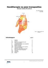
<strong>Muscle</strong> <strong>strength</strong> <strong>measurements</strong> <strong>of</strong> <strong>the</strong> <strong>Hand</strong><br />
Ton AR Schreuders, JW Brandsma, HJ Stam<br />
“Is it nothing to have <strong>the</strong> mind awakened to <strong>the</strong><br />
perception <strong>of</strong> <strong>the</strong> numerous pro<strong>of</strong>s <strong>of</strong> design<br />
which present <strong>the</strong>mselves in <strong>the</strong> study <strong>of</strong> <strong>the</strong><br />
<strong>Hand</strong>- to be brought to <strong>the</strong> conviction that<br />
everything in its structure is orderly and<br />
systematic, and that <strong>the</strong> most perfect<br />
mechanism, <strong>the</strong> most minute and curious<br />
apparatus, and sensibilities <strong>the</strong> most delicate<br />
and appropriate, are all combined in operation<br />
that we may move <strong>the</strong> hand?”<br />
The <strong>Hand</strong> - Its Mechanism and Vital Endowments<br />
as Evincing Design. Sir Charles Bell (1833).<br />
Introduction<br />
Toge<strong>the</strong>r with <strong>the</strong> brain, <strong>the</strong> hand is<br />
<strong>the</strong> most important organ for<br />
accomplishing tasks <strong>of</strong> adaptation,<br />
exploration, prehension, perception and<br />
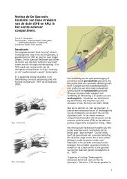
Ulnar<br />
manipulation, unique to humans. 1 To study<br />
<strong>the</strong> anatomy and kinetic chains <strong>of</strong> <strong>the</strong><br />
hand and <strong>the</strong> complex interplay <strong>of</strong> more<br />
than 40 muscles that control its<br />
movements requires an appreciation <strong>of</strong> <strong>the</strong><br />
biomechanics <strong>of</strong> <strong>the</strong> hand and its<br />
dexterity. 2<br />
The muscles <strong>of</strong> <strong>the</strong> lower arm and<br />
hand can be conveniently arranged<br />
according to innervation and localisation.<br />
Usually <strong>the</strong> muscles are divided into<br />
extrinsic ones, where muscles have <strong>the</strong>ir<br />
origin proximal to <strong>the</strong> hand and intrinsic<br />
muscles which have <strong>the</strong>ir origin and<br />
insertion within <strong>the</strong> hand (Table 1).<br />
Sterling Bunnell 3 wrote that “<strong>the</strong> intrinsic<br />
muscles <strong>of</strong> <strong>the</strong> hand, though tiny, are<br />
important because, with <strong>the</strong> long<br />
extensors and long flexors, <strong>the</strong>y complete<br />
<strong>the</strong> muscle balance in <strong>the</strong> hand”.<br />
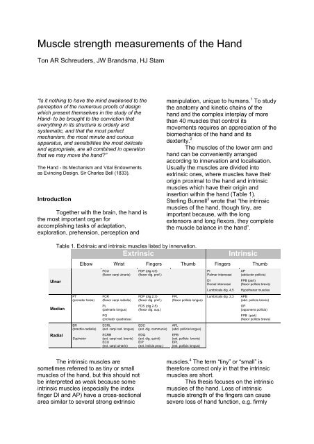
Table 1. Extrinsic and intrinsic muscles listed by innervation.<br />
Extrinsic Intrinsic<br />
Median<br />
Radial<br />
Elbow Wrist Fingers Thumb Fingers Thumb<br />
PT<br />
(pronator teres)<br />
BR<br />
(brachio-radialis)<br />
Supinator<br />
FCU<br />
(flexor carpi ulnaris)<br />
FCR<br />
(flexor carpi radialis)<br />
PL<br />
(palmaris longus)<br />
PQ<br />
(pronator quadratus)<br />
ECRL<br />
(ext. carpi rad. longus)<br />
ECRB<br />
(ext. carpi rad. brevis)<br />
ECU<br />
(ext. carpi ulnaris)<br />
The intrinsic muscles are<br />
sometimes referred to as tiny or small<br />
muscles <strong>of</strong> <strong>the</strong> hand, but this should not<br />
be interpreted as weak because some<br />
intrinsic muscles (especially <strong>the</strong> index<br />
finger DI and AP) have a cross-sectional<br />
area similar to several strong extrinsic<br />
FDP (dig 4,5)<br />
(flexor dig. pr<strong>of</strong>.)<br />
FDP (dig 2,3)<br />
(flexor dig. pr<strong>of</strong>.)<br />
FDS (dig 2-5)<br />
(flexor dig. sup.)<br />
EDC<br />
(ext. dig. communis)<br />
EDQ<br />
(ext. dig. quinti)<br />
EIP<br />
(ext. indicis prop.)<br />
FPL<br />
(flexor pollicis longus)<br />
APL<br />
(abd. pollicis longus)<br />
EPB<br />
(ext. pollicis brevis)<br />
EPL<br />
(ext. pollicis longus)<br />
PI<br />
Palmar interossei<br />
DI<br />
Dorsal interossei<br />
AP<br />
(adductor pollicis)<br />
FPB (part)<br />
(flexor pollicis brevis)<br />
Lumbricals dig. 4,5 Hypo<strong>the</strong>nar muscles<br />
Lumbricals dig. 2,3 APB<br />
(abd. pollicis brevis)<br />
OP<br />
(opponens pollicis)<br />
FPB (part)<br />
(flexor pollicis brevis)<br />
muscles. 4 The term “tiny” or “small” is<br />
<strong>the</strong>refore correct only in that <strong>the</strong> intrinsic<br />
muscles are short.<br />
This <strong>the</strong>sis focuses on <strong>the</strong> intrinsic<br />
muscles <strong>of</strong> <strong>the</strong> hand. Loss <strong>of</strong> intrinsic<br />
muscle <strong>strength</strong> <strong>of</strong> <strong>the</strong> fingers can cause<br />
severe loss <strong>of</strong> hand function, e.g. firmly
Ton A.R. Schreuders, JW Brandsma, HJ Stam<br />
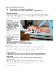
holding a key with paralysis <strong>of</strong> intrinsic<br />
muscles <strong>of</strong> <strong>the</strong> hand can become<br />
impossible (Figure 1).<br />
Figure 1. A patient with loss <strong>of</strong> all intrinsic<br />
muscles holding a key.<br />
<strong>Muscle</strong> <strong>strength</strong> testing<br />
For many centuries measuring<br />
muscle <strong>strength</strong> has been an area <strong>of</strong><br />
interest for those who have been studying<br />
and are responsible for diagnosing many<br />
diseases that are accompanied by loss <strong>of</strong><br />
<strong>strength</strong>. 5 Numerous neurological<br />
diseases are accompanied by atrophy <strong>of</strong><br />
<strong>the</strong> intrinsic muscles <strong>of</strong> <strong>the</strong> hand.<br />
Therefore, muscle function <strong>strength</strong> testing<br />
is frequently used for clinical decision<br />
making in rehabilitation medicine,<br />
neurology, hand surgery and physical<br />
<strong>the</strong>rapy. The purpose <strong>of</strong> this muscle<br />
<strong>strength</strong> testing is, besides diagnosis, to<br />
evaluate and compare treatments, to<br />
document progression or regression <strong>of</strong><br />
e.g. muscle <strong>strength</strong> during rehabilitation,<br />
to provide feedback during <strong>the</strong><br />
rehabilitation process, and to evaluate<br />
handicaps/restrictions <strong>of</strong> participation after<br />
injury. 6<br />
In an historical outline <strong>of</strong> manual<br />
muscle <strong>strength</strong> testing 7 (MMST), <strong>the</strong> first<br />
person to design a numerical system <strong>of</strong><br />
grading muscle action was Lowman in<br />
1911, followed closely by Lovett who<br />
introduced <strong>the</strong> testing grades based on<br />
gravity. 8 The British Medical Research<br />
Council (MRC) specified a similar 0 to 5<br />
scale where complete paralysis is graded<br />
as 0, grade 3 is when <strong>the</strong> limb segment<br />
can be moved actively against gravity, and<br />
grade 5 is normal <strong>strength</strong>. 9 The<br />
procedure for MMST is simple in that no<br />
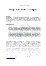
equipment is needed. The hand <strong>of</strong> <strong>the</strong><br />
examiner is used to feel <strong>the</strong> muscle<br />
activity and to give resistance to determine<br />
which grade <strong>the</strong> muscle can be given<br />
(Figure 2).<br />
Figure 2. Testing <strong>the</strong> abduction <strong>strength</strong> <strong>of</strong> <strong>the</strong><br />
little finger (ulnar innervated hypo<strong>the</strong>nar<br />
muscles); <strong>the</strong> right hand <strong>of</strong> <strong>the</strong> examiner gives<br />
resistance to determine which grade <strong>the</strong><br />
muscle can be given.<br />
The most frequently used<br />
textbooks on MMST are still based on this<br />
early system <strong>of</strong> muscle grading, e.g.<br />
Kendall and Kendall 10 and Daniels L.<br />
Worthingham. 11 Some modification for<br />
MMST <strong>of</strong> <strong>the</strong> hand has been proposed by<br />
Brandsma et al. 12 In MMST <strong>of</strong> <strong>the</strong> hand,<br />
gravity is not taken into consideration,<br />
<strong>the</strong>refore grade 3 is considered as <strong>the</strong><br />
ability <strong>of</strong> <strong>the</strong> muscle (group) to perform a<br />
full range <strong>of</strong> motion (ROM). When <strong>the</strong><br />
interossei and lumbricals are tested as a<br />
group in <strong>the</strong> intrinsic plus position (MCP<br />
flexion and IP extension), grade 2 is given<br />
when <strong>the</strong> proximal interphalangeal (PIP)<br />
joint extension is less than 30° short <strong>of</strong> full<br />
extension.<br />
Brooke modified <strong>the</strong> 0-5 scale into<br />
an 11-point scale, adding “+” and “-“. 13 A<br />
9-point scale has been investigated by<br />
Brandsma et al. for reliability in patients<br />
with neuritis due to leprosy. 14 Strength was<br />
graded on a modified MRC scale with 9<br />
grades: 5, 4+, 4, 3+, 3, 2+, 2, 1 and 0.<br />
Overall agreement appeared to be good or<br />
very good (Kappa; 0.61-1.00). However,<br />
when data for hands with normal <strong>strength</strong><br />
(grade 5) or complete paralysis (grade 0)<br />
were excluded from <strong>the</strong> analysis, <strong>the</strong><br />
2
<strong>Muscle</strong> <strong>strength</strong> <strong>measurements</strong> <strong>of</strong> <strong>the</strong> <strong>Hand</strong><br />
reliability <strong>of</strong> <strong>the</strong> remaining mid-range scale<br />
was not acceptable.<br />
Limitations <strong>of</strong> MMST<br />
a) Although <strong>the</strong> textbooks usually present<br />
<strong>the</strong> muscle tests as if muscles can be<br />
tested in isolation, clinicians should be<br />
aware that usually a muscle group is<br />
tested ra<strong>the</strong>r than just one muscle.<br />
Some have suggested labelling <strong>the</strong><br />
movement ra<strong>the</strong>r than <strong>the</strong> muscle, e.g.<br />
grading <strong>the</strong> palmar abduction<br />
movement <strong>of</strong> <strong>the</strong> thumb instead <strong>of</strong><br />
abductor pollicis brevis (APB),<br />
because several muscles are active<br />
when testing <strong>the</strong> palmar abduction <strong>of</strong><br />
<strong>the</strong> thumb. Only a few muscles can be<br />
graded in isolation, e.g. flexor pollicis<br />
longus, flexor digitorum pr<strong>of</strong>undus and<br />
first dorsal interosseous (1DI).<br />
b) The MRC uses a 6-point numeric scale<br />
(grades 0-5) and seems to indicate a<br />
constant distance between points.<br />
However, it is an ordinal scale with<br />
disproportional distances between<br />
grades; e.g. grade 4 is not twice as<br />
strong as grade 2. It might have been<br />
more appropriate to use terms such as<br />
normal, good, fair, trace and<br />
paralysed.<br />
c) Ano<strong>the</strong>r important comment<br />
concerning MMST was made in <strong>the</strong><br />
American Society <strong>of</strong> <strong>Hand</strong><strong>the</strong>rapists<br />
(ASHT) recommendations, 7 that its<br />
most appropriate use is in cases <strong>of</strong><br />
extreme muscle deterioration. MMST<br />
is not appropriate for higher-level<br />
muscle function due to lack <strong>of</strong><br />
sensitivity and precision, and should<br />
be used in conjunction with o<strong>the</strong>r<br />
evaluation tools. We contend that<br />
MMST is most useful for weak muscles<br />
with grades <strong>of</strong> 1, 2 and 3, but not for<br />
<strong>the</strong> higher grades.<br />
d) MMST is dependent on <strong>the</strong> examiner’s<br />
ability to assess <strong>the</strong> pressure as a<br />
parameter for <strong>strength</strong>. Experience <strong>of</strong><br />
<strong>the</strong> examiner is important for reliable<br />
<strong>measurements</strong>.<br />
History <strong>of</strong> dynamometers for <strong>the</strong> hand<br />
One <strong>of</strong> <strong>the</strong> first dynamometers for<br />
measuring hand <strong>strength</strong> was <strong>the</strong><br />
Graham-Desaguliers dynamometer, which<br />
was developed in London in 1763. The<br />
Regnier dynamometer was invented in<br />
Paris in 1798 to measure <strong>the</strong> traction<br />
properties <strong>of</strong> artillery horses, but was<br />
designed as an all-purpose instrument to<br />
measure specific human muscle groups as<br />
well. 8<br />
In <strong>the</strong> past decades many different<br />
dynamometers have been introduced, e.g.<br />
cable tensiometers, sphygmomanometers,<br />
vigorimeters, 15 isokinetic dynamometers<br />
and strain gauge dynamometers.<br />
In response to <strong>the</strong> confusion generated by<br />
<strong>the</strong> many commercial and experimental<br />
grip <strong>strength</strong> instruments, <strong>the</strong> California<br />
Medical Association in 1956 evaluated <strong>the</strong><br />
most commonly used instruments. 16 They<br />
found <strong>the</strong> Jamar, first introduced by<br />
Bechtol in 1954, 17 to be <strong>the</strong> most accurate.<br />
In 1978 <strong>the</strong> American Society for Surgery<br />
<strong>of</strong> <strong>the</strong> <strong>Hand</strong> recommended that <strong>the</strong> second<br />
position <strong>of</strong> <strong>the</strong> Jamar should be used and<br />
in 1981 <strong>the</strong> ASHT made additional<br />
recommendations, e.g. concerning posture<br />
and verbal instructions during<br />
<strong>measurements</strong>. 7<br />
Grip <strong>strength</strong> <strong>measurements</strong> with a<br />
dynamometer have become popular and<br />
have been studied extensively. Less<br />
studies have been conducted to<br />
investigate pinch <strong>strength</strong> <strong>measurements</strong>.<br />
Van der Ploeg et al. 18 noticed <strong>the</strong><br />
shortcoming <strong>of</strong> <strong>the</strong> MMST method by<br />
giving an example <strong>of</strong> <strong>strength</strong><br />
<strong>measurements</strong> <strong>of</strong> <strong>the</strong> biceps muscle as an<br />
elbow flexor. The biceps needed 5 N <strong>of</strong> its<br />
normal <strong>strength</strong> (250 N) to overcome<br />
gravity; thus grade 3 corresponds with<br />
only 2% <strong>of</strong> <strong>the</strong> full <strong>strength</strong> <strong>of</strong> <strong>the</strong> biceps<br />
muscle.<br />
Dynamometers for intrinsic muscle<br />
<strong>strength</strong> <strong>measurements</strong><br />
In his <strong>the</strong>sis van der Ploeg noted<br />
that most dynamometers have a scale far<br />
too crude for measuring forces in very<br />
small muscles like <strong>the</strong> abductor digiti<br />
quinti. However, assessing <strong>the</strong> <strong>strength</strong> <strong>of</strong><br />
<strong>the</strong>se muscles is <strong>of</strong> great importance in<br />
clinical neurology in <strong>the</strong> evaluation <strong>of</strong><br />
mono- and poly-neuropathies. He noted<br />
that <strong>the</strong>re is a need for an accurate device<br />
for <strong>the</strong>se muscles. 19<br />
One <strong>of</strong> <strong>the</strong> first to develop a<br />
dynamometer for <strong>the</strong> intrinsic muscle<br />
<strong>strength</strong> was Mannerfelt, who later<br />
3
Ton A.R. Schreuders, JW Brandsma, HJ Stam<br />
manufactured a new device called <strong>the</strong><br />
Intrins-o-meter. 20 In 1997 he reported a<br />
study in 48 patients with ulnar nerve<br />
compression. 21 Rosen et al. noted that<br />
assessing muscle function using <strong>the</strong><br />
Intrinsi-o-meter was difficult due to <strong>the</strong><br />
extremely small forces, and <strong>the</strong> instrument<br />
was difficult to handle and read. They<br />
suggested using MMST and grip <strong>strength</strong><br />
<strong>measurements</strong> to evaluate nerve function.<br />
Interestingly <strong>the</strong>y found a poor recovery <strong>of</strong><br />
<strong>the</strong> intrinsic muscle <strong>strength</strong> with <strong>the</strong><br />
Mannerfelt instrument and good grip<br />
<strong>strength</strong> recovery.<br />
22 23<br />
Several o<strong>the</strong>rs have developed<br />
instruments mainly to assess <strong>the</strong><br />
abduction <strong>of</strong> <strong>the</strong> thumb. 24-26 Some needed<br />
a specially constructed jig, e.g. to measure<br />
wrist, finger (metacarpo-phalangeal joints)<br />
and thumb extension <strong>strength</strong>. 27<br />
Rotterdam Intrinsic <strong>Hand</strong> Myometer<br />
(RIHM) (Figure 3)<br />
In 1995 inventories were made at<br />
our department to establish which clinical<br />
evaluation instruments were available to<br />
assess <strong>the</strong> outcome after peripheral nerve<br />
surgery. Three methods were <strong>of</strong>ten used<br />
to assess <strong>the</strong> recovery <strong>of</strong> muscle <strong>strength</strong>:<br />
MMST and grip and pinch <strong>strength</strong><br />
dynamometers.<br />
Having encountered several<br />
patients with good grip <strong>strength</strong> but poor<br />
recovery <strong>of</strong> <strong>the</strong> intrinsic muscles <strong>strength</strong>,<br />
we questioned whe<strong>the</strong>r grip <strong>strength</strong><br />
<strong>measurements</strong> were appropriate. We<br />
acknowledged <strong>the</strong> need for a<br />
dynamometer to measure <strong>the</strong> intrinsic<br />
muscles in isolation. Such a dynamometer<br />
should be easy to handle, e.g. portable<br />
and with an ergonomical design. It should<br />
also have <strong>the</strong> possibility to measure <strong>the</strong><br />
opposition force <strong>of</strong> <strong>the</strong> thumb. Reliability<br />
should be good with acceptable<br />
measurement error making it possible to<br />
detect reasonably small changes in<br />
muscle <strong>strength</strong>.<br />
Figure 3 RIHM dynamometer<br />
The Intrinsic <strong>Muscle</strong>s<br />
<strong>of</strong> <strong>the</strong> <strong>Hand</strong>:<br />
Function, Assessment and<br />
Therapy principles<br />
Introduction<br />
There have been many valuable<br />
studies concerning <strong>the</strong> anatomy 28-30 ,<br />
mechanics, 2 31 32 and architectural<br />
design, 33 <strong>of</strong> <strong>the</strong> intrinsic muscles <strong>of</strong> <strong>the</strong><br />
hand. Understanding <strong>the</strong> mechanics <strong>of</strong><br />
human dexterity requires an appreciation<br />
<strong>of</strong> <strong>the</strong> kinetic chains that comprise <strong>the</strong><br />
hand, and <strong>the</strong> intricate interplay <strong>of</strong><br />
muscles and ligaments that control its<br />
movements. 2 In <strong>the</strong>se chains, <strong>the</strong> intrinsic<br />
muscles <strong>of</strong> <strong>the</strong> hand are <strong>of</strong> paramount<br />
importance for efficient hand function. 33<br />
There is a considerable decrease<br />
in functional efficiency in hands with loss<br />
<strong>of</strong> <strong>the</strong> intrinsic muscles function, <strong>of</strong>ten<br />
referred to as <strong>the</strong> clawhand or intrinsic<br />
minus hand (Figure 1). 34-36<br />
A comprehensive analysis <strong>of</strong> hand<br />
function should include assessment <strong>of</strong> <strong>the</strong><br />
<strong>strength</strong> and length <strong>of</strong> <strong>the</strong> intrinsic<br />
muscles. This will provide important<br />
information and assist <strong>the</strong> assessor in e.g.<br />
determining nerve function, deciding which<br />
muscles need to be <strong>strength</strong>ened, what<br />
splint is needed, what surgery needs to be<br />
considered (tendon transfer), etc.<br />
Although assessment <strong>of</strong> muscle<br />
<strong>strength</strong> and length are important<br />
elements <strong>of</strong> hand function o<strong>the</strong>r functions,<br />
e.g. mobility, sensibility and central<br />
properties <strong>of</strong> <strong>the</strong> brain, are equally or more<br />
important for hand function. The latter<br />
4
<strong>Muscle</strong> <strong>strength</strong> <strong>measurements</strong> <strong>of</strong> <strong>the</strong> <strong>Hand</strong><br />
controls e.g. tonus, co-ordination and<br />
speed <strong>of</strong> hand movements.<br />
<strong>Hand</strong> tests to assess <strong>the</strong> ability <strong>of</strong><br />
<strong>the</strong> patient to perform certain tasks have<br />
been developed by e.g. Moberg, 37<br />
Bendz, 38 Sollerman et al, 39 and Light et<br />
al. 40 Most <strong>of</strong> such tests record how long it<br />
takes to finish a particular task. Clinicians<br />
<strong>of</strong>ten see that patients with impairments <strong>of</strong><br />
<strong>the</strong> hand have quickly learned<br />
compensatory mechanisms to<br />
compensate for <strong>the</strong> lost functions.<br />
Therefore, tests at this activity or skills<br />
level may only assess <strong>the</strong> ability <strong>of</strong> <strong>the</strong><br />
patient to compensate for lost function.<br />
While <strong>the</strong>re is little consensus<br />
about classifications <strong>of</strong> prehensile patterns<br />
<strong>of</strong> hand function, <strong>the</strong>re seems to be<br />
consensus about <strong>the</strong> more general<br />
classification, with <strong>the</strong> following<br />
categories: three pinch grips (tip pinch,<br />
lateral or key pinch, and tripod or chuck<br />
pinch) and three modes <strong>of</strong> gripping:<br />
(power grip, spherical or flexion grip, and<br />
extension grip in intrinsic plus position). 40<br />
It is estimated that for a full range <strong>of</strong><br />
natural common grips, a spherical grip is<br />
required for 10%, a tripod grip for 10%, a<br />
power grip for 25%, a lateral grip for 20%,<br />
a tip grip for 20%, and an extension grip<br />
for 10% <strong>of</strong> tasks <strong>of</strong> activities <strong>of</strong> daily living.<br />
Without intrinsic muscles a power<br />
grip is somewhat weaker but still possible,<br />
but for all o<strong>the</strong>r grips <strong>the</strong> intrinsic muscles<br />
play an important role.<br />
Long-term loss <strong>of</strong> intrinsic muscle function<br />
may result in irreversible joint<br />
contractures. An appropriate <strong>the</strong>rapy plan<br />
(e.g. which exercises are needed, what<br />
splint may enhance hand function and<br />
prevent contractures) is needed.<br />
The aim <strong>of</strong> this paper is fourfold:<br />
-to review <strong>the</strong> functional anatomy <strong>of</strong> <strong>the</strong><br />
intrinsic muscles <strong>of</strong> <strong>the</strong> hand.<br />
-to discuss <strong>the</strong> pathokinesiology <strong>of</strong> <strong>the</strong><br />
hand with intrinsic muscle paralysis, and<br />
its consequences for Activities for Daily<br />
Living (ADL)/dexterity and muscle<br />
shortening.<br />
-to present possibilities for <strong>the</strong> assessment<br />
<strong>of</strong> muscle <strong>strength</strong> (manual and<br />
instrumental).<br />
-to discuss <strong>the</strong>rapy principles (prevention<br />
<strong>of</strong> complications, exercises for<br />
<strong>strength</strong>ening).<br />
All intrinsic muscles will be<br />
discussed separately, except for <strong>the</strong><br />
hypo<strong>the</strong>nar muscles which are discussed<br />
as a group.<br />
1. Intrinsic muscles <strong>of</strong> <strong>the</strong><br />
fingers<br />
In general, every finger has six<br />
muscles controlling all <strong>the</strong> movements <strong>of</strong><br />
<strong>the</strong> fingers: three extrinsic tendons (two<br />
long flexors and one long extensor) and<br />
three intrinsic muscles (dorsal and palmar<br />
interosseous and lumbrical muscles).<br />
1.1. Dorsal and palmar interossei<br />
1.1.1 Functional anatomy<br />
Literature usually describes four<br />
dorsal (DI) and three palmar (PI)<br />
interosseous muscles. Stack et al. 41<br />
suggested that it might be more correct to<br />
divide <strong>the</strong> interossei into <strong>the</strong> proximal and<br />
distal, as it is <strong>the</strong>ir insertion ra<strong>the</strong>r than<br />
origin, which dictates <strong>the</strong>ir action. Most<br />
dorsal interossei muscles have a more<br />
proximal attachment while <strong>the</strong> palmar<br />
interossei have a more distal attachment<br />
similar to <strong>the</strong> lumbricals. 41 According to<br />
Zancolli 42 <strong>the</strong>re are three types <strong>of</strong><br />
insertions <strong>of</strong> <strong>the</strong> interossei:<br />
Type I most proximal, attached to:<br />
a. tubercle <strong>of</strong> proximal phalanx,<br />
b. transverse and oblique fibers <strong>of</strong><br />
extensor apparatus<br />
c. <strong>the</strong> volar plate.<br />
Type II is like type 1 except that <strong>the</strong>re is no attachment<br />
to <strong>the</strong> bone (a.) and part <strong>of</strong> <strong>the</strong> insertion is into:<br />
d. <strong>the</strong> lateral band<br />
Type III has all four attachments a, b, c and d.<br />
The first dorsal interosseous (1 DI)<br />
insertion is <strong>of</strong> type I, all o<strong>the</strong>r DI are <strong>of</strong><br />
type III. All PI have <strong>the</strong> type II insertion,<br />
but variations are possible. 28 30 Because <strong>of</strong><br />
this insertion <strong>the</strong> strongest activity <strong>of</strong> <strong>the</strong> 1<br />
DI is in key pinch when <strong>the</strong> thumb is<br />
pressed against <strong>the</strong> mid-phalanx <strong>of</strong> <strong>the</strong><br />
index finger. The 1 DI is also active in tip<br />
pinch, when <strong>the</strong> tip <strong>of</strong> <strong>the</strong> thumb is<br />
pressed against <strong>the</strong> tip <strong>of</strong> <strong>the</strong> index finger,<br />
but <strong>the</strong>n <strong>the</strong> main action is as a flexor at<br />
5
Ton A.R. Schreuders, JW Brandsma, HJ Stam<br />
<strong>the</strong> metacarpo-phalangeal (MCP) joint.<br />
The first palmar interosseous (1 PI)<br />
muscle is more active in tip pinch<br />
activities.<br />
In Type II, <strong>the</strong> insertion into <strong>the</strong><br />
lateral band <strong>of</strong> <strong>the</strong> extensor apparatus (d.)<br />
is responsible for an important extension<br />
force at <strong>the</strong> proximal interphalangeal (PIP)<br />
joints. The first palmar interosseous also<br />
produces some supination <strong>of</strong> <strong>the</strong> index<br />
finger to get good approximation <strong>of</strong> <strong>the</strong><br />
pulps. In this respect we might consider<br />
<strong>the</strong> 1 PI as an “opponens indices” muscle<br />
in tip pinch activities.<br />
Without interossei <strong>the</strong> finger is<br />
unstable and will collapse into <strong>the</strong> “claw”<br />
(i.e. intrinsic minus) position <strong>of</strong> flexed IP<br />
joints and (hyper-) extension <strong>of</strong> <strong>the</strong> MCP<br />
joint: <strong>the</strong>refore <strong>the</strong> interosseous are<br />
sometimes referred to as <strong>the</strong> “anti-claw”<br />
muscles. 35 The primary function <strong>of</strong> <strong>the</strong><br />
interossei is MCP flexion/stabilisation with<br />
extension <strong>of</strong> <strong>the</strong> interphalangeal (IP) joints.<br />
This is especially evident during pinch in<br />
which <strong>the</strong> collapse <strong>of</strong> <strong>the</strong> index PIP joint is<br />
apparent in, <strong>of</strong>ten > 90°, flexion. This is a<br />
sign <strong>of</strong> interosseous muscle weakness<br />
and sometimes referred to as <strong>the</strong><br />
Mannerfelt sign. 20 (Figure 2).<br />
FIGURE 2. Mannerfelt and Froment sign on<br />
left hand <strong>of</strong> patient with ulnar nerve paralysis.<br />
Recording <strong>the</strong> moments <strong>of</strong> <strong>the</strong><br />
intrinsic muscles that were generated after<br />
electrical stimulation Lauer et al. 43 found<br />
that <strong>the</strong> dorsal interossei muscles were<br />
strong abductors <strong>of</strong> <strong>the</strong> fingers and<br />
generated a significant moment in MCP<br />
joint flexion and IP joint extension.<br />
Similarly, Ketchum et al. 34 found that <strong>the</strong><br />
interossei muscles <strong>of</strong> <strong>the</strong> index finger<br />
contribute 73% to <strong>the</strong> overall moment for<br />
flexion <strong>of</strong> <strong>the</strong> MCP joint. 34 Li et al. 44<br />
investigated <strong>the</strong> role <strong>of</strong> <strong>the</strong> intrinsic finger<br />
flexor muscles during finger flexion tasks.<br />
When an external force is applied<br />
proximally to <strong>the</strong> PIP joint, <strong>the</strong> extensor<br />
mechanism (intrinsic muscle group) is <strong>the</strong><br />
largest component (70%) on force<br />
production <strong>of</strong> all flexors.<br />
Thus <strong>the</strong> interossei muscles are<br />
important flexors <strong>of</strong> <strong>the</strong> MCP joint toge<strong>the</strong>r<br />
with <strong>the</strong> long flexors: flexor digitorum<br />
pr<strong>of</strong>undus (FDP) and flexor digitorum<br />
superficiales (FDS). However, at <strong>the</strong> PIP<br />
joint level <strong>the</strong> long flexors, primarily <strong>the</strong><br />
FDS, and <strong>the</strong> interossei are antagonists.<br />
When <strong>the</strong> PIP joint is flexed, some<br />
reduction <strong>of</strong> <strong>the</strong> extension moment <strong>of</strong> <strong>the</strong><br />
interossei takes place, due to <strong>the</strong> volar<br />
displacement <strong>of</strong> <strong>the</strong> lateral bands (<strong>of</strong> about<br />
4-mm) at <strong>the</strong> PIP joint level. At full flexion<br />
<strong>of</strong> both <strong>the</strong> PIP and distal interphalangeal<br />
(DIP) joints, <strong>the</strong> lateral bands approach<br />
<strong>the</strong> flexion-extension axis <strong>of</strong> <strong>the</strong> PIP joint,<br />
<strong>the</strong>reby minimising <strong>the</strong> extensor moment. 2<br />
The action <strong>of</strong> <strong>the</strong> interossei<br />
muscles can be studied separately from<br />
<strong>the</strong> extensor digitorum communis (EDC)<br />
muscles in patients with a radial nerve<br />
paralysis. If <strong>the</strong> patient is asked to extend<br />
<strong>the</strong> fingers, no extension <strong>of</strong> <strong>the</strong> MCP will<br />
occur, but <strong>the</strong> finger IP joints will extend<br />
because <strong>of</strong> <strong>the</strong> ulnar and median nerve<br />
innervated interosseous and lumbrical<br />
muscles. However, in some patients <strong>the</strong><br />
fingers, especially <strong>the</strong> index, can be<br />
extended in <strong>the</strong> MCP joint to some degree,<br />
especially with <strong>the</strong> wrist flexed; this is<br />
probably because <strong>the</strong> angle <strong>of</strong> attachment<br />
<strong>of</strong> <strong>the</strong> dorsal interossei muscles is only 0°-<br />
5°. When <strong>the</strong> tension on <strong>the</strong> extensor<br />
tendon is increased due to <strong>the</strong> flexed wrist,<br />
this angle will go beyond <strong>the</strong> 0° and thus<br />
<strong>the</strong> interosseous muscle becomes an<br />
extensor <strong>of</strong> <strong>the</strong> MCP joint. The angle <strong>of</strong><br />
approach to <strong>the</strong> extensor mechanism for<br />
<strong>the</strong> palmar interossei muscles is 20°-25°<br />
and for <strong>the</strong> lumbrical muscles 35°. 28<br />
The index finger is deprived <strong>of</strong> all<br />
long flexors and <strong>the</strong> lumbrical muscles in<br />
patients with a high median nerve<br />
paralysis. The only active muscles are <strong>the</strong><br />
long extensors and <strong>the</strong> two interossei. In<br />
6
<strong>Muscle</strong> <strong>strength</strong> <strong>measurements</strong> <strong>of</strong> <strong>the</strong> <strong>Hand</strong><br />
an attempt to flex <strong>the</strong> fingers <strong>the</strong> index<br />
finger will remain extended, and is<br />
accurately described as <strong>the</strong> “pointing<br />
finger” where <strong>the</strong> IP joints are fully<br />
extended and <strong>the</strong> MCP joint flexed. In high<br />
median nerve paralysis <strong>the</strong> long finger is<br />
also deprived <strong>of</strong> <strong>the</strong> long flexor, but will<br />
usually flex in grip. This is due to <strong>the</strong><br />
attachments between <strong>the</strong> FDP tendons <strong>of</strong><br />
<strong>the</strong> long finger and <strong>the</strong> ring finger and is<br />
called <strong>the</strong> Quadriga phenomena. 45 46 This<br />
term suggests a symmetrical organisation<br />
<strong>of</strong> <strong>the</strong> four tendons; however, <strong>the</strong><br />
connections between <strong>the</strong> FDP <strong>of</strong> <strong>the</strong> index<br />
and middle finger are usually absent, and<br />
a strong connection between <strong>the</strong> flexor<br />
pollicis longus (FPL) and <strong>the</strong> FDP <strong>of</strong> <strong>the</strong><br />
index finger can exist.<br />
1.1.2 Pathokinesiology (paralyses,<br />
consequences for prehension/ADL,<br />
shortening)<br />
The deficiency mentioned in<br />
textbooks due to loss <strong>of</strong> interossei is<br />
usually that <strong>of</strong> controlled abduction and<br />
adduction <strong>of</strong> <strong>the</strong> fingers. The loss <strong>of</strong> this<br />
action is important for those who play<br />
musical instruments or operate keyboards,<br />
but in most patients this is not recognised<br />
as a severe deficit. The loss <strong>of</strong> <strong>the</strong><br />
interossei function <strong>of</strong> MCP flexion and PIP<br />
extension is a much more significant loss.<br />
In acute paralysis <strong>of</strong> <strong>the</strong> interossei<br />
muscles it will sometimes only be visible<br />
as a mild hyperextension at <strong>the</strong> MCP joints<br />
and slight flexed position <strong>of</strong> <strong>the</strong> PIP joints<br />
when <strong>the</strong> patient is asked to extend <strong>the</strong><br />
fingers. However, this deformity usually<br />
progresses depending on <strong>the</strong> natural joint<br />
laxity <strong>of</strong> <strong>the</strong> hands, i.e. long, thin and<br />
hypermobile fingers will develop a socalled<br />
claw hand much quicker than thick,<br />
stiff fingers. In <strong>the</strong> mobile hands <strong>the</strong> volar<br />
ligamentous structures stretch more<br />
easily, causing increased hyperextension<br />
and subsequently less PIP extension.<br />
Extension <strong>of</strong> <strong>the</strong> PIP joints is only possible<br />
by contraction <strong>of</strong> <strong>the</strong> EDC when <strong>the</strong> MCPs<br />
are 'blocked', ei<strong>the</strong>r actively (internally)<br />
through muscle contraction or passively<br />
(externally) through <strong>the</strong> examiners hand or<br />
a splint, preventing hyperextension <strong>of</strong> <strong>the</strong><br />
MCP joints. In <strong>the</strong> latter situation this is<br />
called assisted extension, which is used<br />
as a test to assess <strong>the</strong> integrity <strong>of</strong> <strong>the</strong><br />
extensor apparatus.<br />
O<strong>the</strong>r factors that may contribute to<br />
<strong>the</strong> development <strong>of</strong> a claw hand are: hand<br />
dominance, continued use <strong>of</strong> <strong>the</strong> hand (or<br />
<strong>the</strong> lack <strong>of</strong>), compliance <strong>of</strong> <strong>the</strong> patient to<br />
perform <strong>the</strong> routine exercises to prevent<br />
joint stiffness, and use <strong>of</strong> (night) splints.<br />
Without interosseous <strong>the</strong> hand can<br />
still make a full fist, but <strong>the</strong> pattern <strong>of</strong><br />
movement is changed (<strong>the</strong> MCP flexion<br />
takes place later than usual). The<br />
contribution <strong>of</strong> <strong>the</strong> interossei muscles in<br />
grip <strong>strength</strong> <strong>measurements</strong> will be<br />
discussed later.<br />
In patients with a “high” ulnar lesion, <strong>the</strong><br />
FDP muscles <strong>of</strong> <strong>the</strong> 4th and 5th fingers<br />
are paralysed and less clawing is <strong>of</strong>ten<br />
visible because <strong>the</strong> flexion moment at <strong>the</strong><br />
PIP joints is decreased compared to a<br />
“low” ulnar palsy. We might conclude from<br />
this observation that clawing is also <strong>the</strong><br />
result <strong>of</strong> visco-elastic tension <strong>of</strong> FDP.<br />
When <strong>the</strong> ulnar nerve recovers, <strong>the</strong> 4th<br />
and 5th finger will show an increasing<br />
clawing. Maintaining <strong>the</strong> length <strong>of</strong> <strong>the</strong><br />
FDPs, i.e. preventing flexor tightness,<br />
<strong>the</strong>refore also helps to prevent clawing.<br />
Ano<strong>the</strong>r sign <strong>of</strong> interossei muscle<br />
weakness was first described by Andre-<br />
Thomas in 1917 and is called <strong>the</strong> Thomas<br />
sign. 47 (p. 518) (Figure 3). This is <strong>the</strong><br />
tendency (compensation) <strong>of</strong> a patient with<br />
weak interossei muscles <strong>of</strong> <strong>the</strong> fingers to<br />
automatically flex <strong>the</strong> wrist in an attempt to<br />
gain a better opening <strong>of</strong> <strong>the</strong> hand, i.e.<br />
MCP extension, by means <strong>of</strong> increasing<br />
<strong>the</strong> pull on <strong>the</strong> EDC. This in fact increases<br />
<strong>the</strong> hyperextension <strong>of</strong> <strong>the</strong> MCP joints and<br />
adds to <strong>the</strong> progress <strong>of</strong> <strong>the</strong> development<br />
<strong>of</strong> <strong>the</strong> claw hand. This “trick” movement is<br />
adopted very quickly and even when <strong>the</strong><br />
interossei have regained <strong>the</strong>ir <strong>strength</strong>, will<br />
<strong>of</strong>ten take a long time to “un-learn”.<br />
In longstanding paralyses <strong>of</strong> <strong>the</strong><br />
interossei, e.g. when <strong>the</strong> ulnar nerve could<br />
not be repaired or did not recover, <strong>the</strong> PIP<br />
joints are continuously in a flexed position.<br />
This will cause a gradual stretching <strong>of</strong> <strong>the</strong><br />
dorsal expansion (sometimes called <strong>the</strong><br />
tri-angular ligament) over <strong>the</strong> PIP joint,<br />
which secures <strong>the</strong> lateral bands <strong>of</strong> <strong>the</strong><br />
extensor tendon in <strong>the</strong>ir dorsal position. In<br />
7
Ton A.R. Schreuders, JW Brandsma, HJ Stam<br />
<strong>the</strong> normal finger, <strong>the</strong> lateral bands shift<br />
dorsally and towards <strong>the</strong> central position <strong>of</strong><br />
<strong>the</strong> finger when <strong>the</strong> PIP joint is extended.<br />
Whereas when flexing <strong>the</strong> PIP joint <strong>the</strong><br />
dorsal expansion needs to allow <strong>the</strong> lateral<br />
bands to move volarly towards <strong>the</strong> flexionextension<br />
axis <strong>of</strong> movement. When this<br />
dorsal expansion is elongated, <strong>the</strong> lateral<br />
bands are too much volarly, resulting in a<br />
loss <strong>of</strong> PIP joint extension. Consequently,<br />
<strong>the</strong> oblique retinacular ligament (ORL) or<br />
Landsmeers ligament is slack most <strong>of</strong> <strong>the</strong><br />
time and will adjust to this new situation by<br />
shortening and this may result in<br />
hyperextension <strong>of</strong> <strong>the</strong> DIP joint. This is a<br />
similar progression <strong>of</strong> changes as in an<br />
extensor tendon central slip injury, causing<br />
a Boutonnière deformity. 4<br />
FIGURE 3. Thomas sign: compensation<br />
movement when interossei muscles are weak;<br />
flexion <strong>of</strong> <strong>the</strong> wrist in an attempt to gain a<br />
better opening <strong>of</strong> <strong>the</strong> hand, i.e. by means <strong>of</strong><br />
increasing <strong>the</strong> pull on <strong>the</strong> EDC.<br />
Ano<strong>the</strong>r impairment in longstanding<br />
paralyses <strong>of</strong> <strong>the</strong> interossei muscles is<br />
related to hyperextension <strong>of</strong> <strong>the</strong> MCP joint.<br />
This causes an upward pull <strong>of</strong> <strong>the</strong> EDC<br />
tendons (bow stringing), stretching <strong>the</strong><br />
sagittal bands. The laxity <strong>of</strong> <strong>the</strong> sagittal<br />
bands will result in an inability to maintain<br />
<strong>the</strong> EDC tendon on top <strong>of</strong> <strong>the</strong> MCP joint.<br />
The drop <strong>of</strong> <strong>the</strong> luxating tendon into <strong>the</strong><br />
groove between <strong>the</strong> MCPs is especially<br />
observable when flexing <strong>the</strong> MCP joint.<br />
This is sometimes called “guttering”<br />
because <strong>the</strong> tendon drops into <strong>the</strong> “gutter”<br />
between <strong>the</strong> MCP joints.<br />
The longstanding flexed position <strong>of</strong><br />
<strong>the</strong> IP joints will result in a physiological<br />
shortening <strong>of</strong> <strong>the</strong> long flexors. This flexor<br />
tightness will increase <strong>the</strong> PIP flexion<br />
position and can cause a deterioration <strong>of</strong><br />
<strong>the</strong> PIP flexion contractures. This is an<br />
additional argument to maintain <strong>the</strong> length<br />
<strong>of</strong> <strong>the</strong> extrinsic flexors in patients with<br />
intrinsic muscle weakness.<br />
Shortening <strong>of</strong> <strong>the</strong> interossei<br />
muscles is called intrinsic tightness (IT),<br />
which can be caused by a trauma <strong>of</strong> <strong>the</strong><br />
hand which can precipitate a cascade <strong>of</strong><br />
events. The interossei are situated in<br />
ra<strong>the</strong>r tight compartments, <strong>the</strong>refore<br />
oedema/swelling will cause an increase <strong>of</strong><br />
pressure in <strong>the</strong>se compartments.<br />
As a result blood circulation will be<br />
hampered causing anoxia and muscle<br />
fiber death, which results in fibrosis <strong>of</strong> <strong>the</strong><br />
muscle and shortening. This is identical to<br />
<strong>the</strong> process which causes Volkmann's<br />
ischemic contracture in <strong>the</strong> forearm. 48<br />
The IT test consists <strong>of</strong> two parts.<br />
First, passive PIP flexion is tested with <strong>the</strong><br />
MCP joint extended and, secondly,<br />
passive PIP flexion is tested again but now<br />
with <strong>the</strong> MCP joint flexed. If <strong>the</strong>re is a<br />
large difference in PIP flexion between <strong>the</strong><br />
two MCP positions, intrinsic tightness is<br />
present. The long-term complications <strong>of</strong> IT<br />
can result in decreased MCP extension<br />
and a swan neck finger, i.e.<br />
hyperextension <strong>of</strong> <strong>the</strong> PIP joint. A<br />
longstanding swan neck deformity might<br />
result in a painful snapping <strong>of</strong> <strong>the</strong> lateral<br />
bands at PIP level when <strong>the</strong> finger is<br />
flexed.<br />
In rheumatoid arthritis a different<br />
process can also lead to IT. The role <strong>of</strong> <strong>the</strong><br />
intrinsic muscles in producing MCP<br />
subluxation in <strong>the</strong> rheumatoid hand has<br />
49 50<br />
been documented.<br />
Ano<strong>the</strong>r intricacy sometimes<br />
observed is what we call interosseous<br />
plus, which is a paradoxal extension: <strong>the</strong><br />
harder <strong>the</strong> patient tries to bend <strong>the</strong> finger,<br />
<strong>the</strong> more <strong>the</strong> finger will extend in <strong>the</strong> PIP<br />
joint. This phenomena is sometimes seen<br />
in patients in which <strong>the</strong> interossei have<br />
been <strong>the</strong> only flexor <strong>of</strong> <strong>the</strong> finger for some<br />
time e.g. in high median nerve palsy, or in<br />
case <strong>of</strong> adhered flexor tendons. Although<br />
<strong>the</strong> flexors are active and can bend <strong>the</strong><br />
finger, when a stronger grip is required <strong>the</strong><br />
finger will extend.<br />
8
<strong>Muscle</strong> <strong>strength</strong> <strong>measurements</strong> <strong>of</strong> <strong>the</strong> <strong>Hand</strong><br />
The explanation is that in a nonresistant<br />
grip <strong>of</strong> a normal functioning hand<br />
<strong>the</strong>re is only minor activity <strong>of</strong> <strong>the</strong> interossei<br />
muscles. However, in a strong grip, <strong>the</strong><br />
stronger <strong>the</strong> flexors are pulling, <strong>the</strong> more<br />
<strong>the</strong> intrinsic muscles become active to<br />
stabilise <strong>the</strong> PIP joints and prevent<br />
luxation. If <strong>the</strong> long flexor is weak or poorly<br />
activated, <strong>the</strong> interossei will overpower <strong>the</strong><br />
flexor causing PIP extension. This<br />
paradoxal extension appears to be similar<br />
to <strong>the</strong> lumbrical plus phenomena (see<br />
lumbricals) and might be called<br />
interosseous plus. The patient has to be<br />
taught to, gently, contract <strong>the</strong> long flexors<br />
without <strong>the</strong> action <strong>of</strong> <strong>the</strong> interossei<br />
muscles.<br />
1.1.3 Assessment possibilities (manual<br />
and instrumental)<br />
Although <strong>the</strong> interosseous muscles<br />
have short fiber length, some are strong<br />
and have physiological cross-sectional<br />
areas comparable to <strong>the</strong> FDS muscles. 4 In<br />
standard textbooks on muscle testing, <strong>the</strong><br />
tests suggested are usually: abduction for<br />
dorsal and adduction for palmar interossei<br />
muscles. 10 51 These tests are useful for<br />
isolated (specific) testing <strong>of</strong> <strong>the</strong> interossei.<br />
For example, patients with an ulnar nerve<br />
paralysis can not move <strong>the</strong>ir middle finger<br />
sideways, which has been called <strong>the</strong><br />
Egawa sign. 47 Functionally, it is much<br />
more meaningful to test <strong>the</strong> interossei<br />
muscles in <strong>the</strong> intrinsic plus position, by<br />
giving resistance to flexion <strong>of</strong> <strong>the</strong> MCP<br />
52 53<br />
joints and extension <strong>of</strong> <strong>the</strong> PIP joint.<br />
(Figure 4).<br />
FIGURE 4.<br />
Testing <strong>the</strong><br />
<strong>strength</strong> <strong>of</strong><br />
<strong>the</strong> intrinsic<br />
muscles in<br />
<strong>the</strong> fingers.<br />
Some have suggested that<br />
dynamometry <strong>of</strong> <strong>the</strong> interossei muscles is<br />
possible, indirectly, by measuring <strong>the</strong> grip<br />
<strong>strength</strong> <strong>of</strong> <strong>the</strong> hand with e.g. a Jamar<br />
dynamometer. Janda et al. advocated to<br />
use <strong>the</strong> smallest handle position <strong>of</strong> <strong>the</strong><br />
Jamar because <strong>the</strong> intrinsic muscles were<br />
most active in that position. 54 Kozin et al. 36<br />
tested 21 healthy persons who underwent<br />
median and ulnar nerve blocks at <strong>the</strong> wrist<br />
level; <strong>the</strong> average decrease in grip<br />
<strong>strength</strong> was 38% after ulnar nerve block.<br />
Pinch data in <strong>the</strong> study by Kozin et<br />
al. revealed a significant decrease in key<br />
pinch <strong>of</strong> 77% after ulnar block and 60%<br />
after median block. 36 For evaluating and<br />
monitoring <strong>the</strong> motor function <strong>of</strong> <strong>the</strong> ulnar<br />
nerve, pinch <strong>strength</strong> <strong>measurements</strong> seem<br />
more meaningful than grip <strong>strength</strong><br />
<strong>measurements</strong>.<br />
Specific <strong>measurements</strong> <strong>of</strong> <strong>the</strong> first<br />
dorsal interosseous muscle can be done<br />
with dynamometers such as <strong>the</strong> RIHM, 55<br />
<strong>the</strong> Intrins-o-meter <strong>of</strong> Mannerfelt, 21 or <strong>the</strong><br />
Preston pinch gauge device. 56<br />
1.1.4 Therapy principles (prevention<br />
complications, <strong>strength</strong>ening, ADL)<br />
Prevention <strong>of</strong> contractures is<br />
directed at <strong>the</strong> PIP joints. A (night) splint<br />
with <strong>the</strong> MCP joints in flexion and IPs in<br />
extension is advisable. These splints can<br />
help to prevent PIP flexion contractures<br />
and are especially important in<br />
longstanding problems. Increased<br />
hyperextension <strong>of</strong> <strong>the</strong> MCP joints, due to<br />
stretching <strong>of</strong> <strong>the</strong> volar plate, is also<br />
prevented in this position.<br />
During <strong>the</strong> day <strong>the</strong> so-called<br />
“knuckle-bender” can assist <strong>the</strong> patient in<br />
some ADL functions. This splint will also<br />
help to move <strong>the</strong> PIP joint into full<br />
extension during <strong>the</strong> day and will maintain<br />
<strong>the</strong> integrity <strong>of</strong> <strong>the</strong> extensor mechanism.<br />
When patients choose not to wear a splint,<br />
<strong>the</strong>y need to be taught how to do assisted<br />
extension exercises by blocking <strong>the</strong> MCP<br />
joints with <strong>the</strong>ir hands and routinely<br />
massaging to extend <strong>the</strong> IP joints (Figure<br />
5).<br />
9
Ton A.R. Schreuders, JW Brandsma, HJ Stam<br />
FIGURE 5. Prevention <strong>of</strong> IP flexion<br />
contractures by massaging <strong>the</strong> fingers.<br />
Exercises to <strong>strength</strong>en <strong>the</strong><br />
interossei muscles are all aimed at<br />
movements which flex <strong>the</strong> MCP and<br />
extend <strong>the</strong> IP joints. Therefore, exercises<br />
for <strong>the</strong> interossei muscles are all activities<br />
in intrinsic plus position: e.g. grasping a<br />
book, plate or a cylindrical object like a<br />
large bottle. Specific training <strong>of</strong> <strong>the</strong> first<br />
dorsal and palmar interossei are activities<br />
for which key and tip pinch activities are<br />
required.<br />
To correct <strong>the</strong> long flexor tightness,<br />
<strong>the</strong> patient is taught to stretch <strong>the</strong> flexors<br />
by holding <strong>the</strong> hand flat on e.g. a table and<br />
by moving <strong>the</strong> forearm towards an angle<br />
perpendicular to <strong>the</strong> table. In a similar<br />
fashion, <strong>the</strong> hand can be placed on <strong>the</strong><br />
seat <strong>of</strong> <strong>the</strong> chair while <strong>the</strong> patient sits on<br />
<strong>the</strong> hand and pulls <strong>the</strong> forearm towards<br />
<strong>the</strong> body. A night splint with <strong>the</strong> fingers<br />
and hand in extension might be necessary<br />
in severe cases.<br />
1.2 Lumbricals<br />
1.2.1 Functional anatomy<br />
The lumbrical muscles are unique<br />
muscles in several aspects. They connect<br />
two extrinsic antagonistic muscles.<br />
Proximally <strong>the</strong> lumbricals are attached to<br />
<strong>the</strong> FDP and distally <strong>the</strong>y are inserted into<br />
<strong>the</strong> lateral band <strong>of</strong> <strong>the</strong> extensor tendon.<br />
The third and fourth lumbricals also<br />
connect, by <strong>the</strong>ir bi-penal origin, two<br />
adjacent FDP tendons.<br />
The function <strong>of</strong> <strong>the</strong> lumbrical<br />
muscles is much debated and some even<br />
considered <strong>the</strong>se muscles to be<br />
redundant. Brand suggested that <strong>the</strong><br />
lumbrical muscles are not relevant for<br />
MCP flexion. He explained this with an<br />
illustration <strong>of</strong> a fa<strong>the</strong>r carrying a child; it<br />
does not matter what <strong>the</strong> child (i.e.<br />
lumbrical) is carrying, <strong>the</strong> fa<strong>the</strong>r (i.e. FDP)<br />
has to carry it anyway. 4 Therefore, <strong>the</strong><br />
lumbrical muscles have a unique ability to<br />
contract without adding flexion torque at<br />
<strong>the</strong> MCP joint, in contrast with <strong>the</strong><br />
interosseous muscles which, when<br />
extending <strong>the</strong> IPs, need a stronger<br />
contraction <strong>of</strong> <strong>the</strong> EDC to counteract <strong>the</strong><br />
flexion moment at <strong>the</strong> MCP joint level. The<br />
lumbricals provide a more efficient source<br />
for IP extension than <strong>the</strong> interossei.<br />
Any contraction <strong>of</strong> <strong>the</strong> lumbrical<br />
muscle for IP extension simultaneously,<br />
reduces <strong>the</strong> visco-elastic force <strong>of</strong> <strong>the</strong> FDP<br />
tending to flex <strong>the</strong> IP joints. Accordingly<br />
<strong>the</strong> lumbrical can be regarded as a<br />
deflexor <strong>of</strong> <strong>the</strong> PIP joint. 57 Its direct<br />
contribution to MCP flexion is small and in<br />
<strong>the</strong> flexed finger may be non-existent, but<br />
its indirect contribution to IP joint extension<br />
by decreasing <strong>the</strong> flexion torque is quite<br />
substantial. 58<br />
With <strong>the</strong> smallest physiological<br />
cross-sectional area, it is certainly not a<br />
strong muscle. The lumbricals have a very<br />
long fiber length (40-48 mm) which<br />
indicates that <strong>the</strong>y are designed for long<br />
excursions. If <strong>the</strong> lumbrical fiber length<br />
was short, FDP excursion could stretch<br />
<strong>the</strong> lumbrical sarcomeres to a point that<br />
<strong>the</strong>y were unable to generate active<br />
force. 33<br />
The lumbrical muscles are richly<br />
endowed with muscle spindles, <strong>the</strong>ir<br />
passive stretch by contraction <strong>of</strong> <strong>the</strong> FDP<br />
might both inhibit finger extensors and<br />
facilitate wrist extensors. 58-61 For this<br />
reason <strong>the</strong> lumbrical muscles have been<br />
called “tensiometers” between long flexors<br />
and extensors. 62 Leijnse and Kalker 46<br />
concluded that <strong>the</strong> lumbricals are in an<br />
optimal position for proprioceptic feedback<br />
concerning <strong>the</strong> PIP-DIP joint mechanism.<br />
These unique properties <strong>of</strong> <strong>the</strong><br />
lumbricals indicate that <strong>the</strong>y are probably<br />
important in fast, alternating movements,<br />
e.g. in typing and playing musical<br />
instruments. 63<br />
10
<strong>Muscle</strong> <strong>strength</strong> <strong>measurements</strong> <strong>of</strong> <strong>the</strong> <strong>Hand</strong><br />
1.2.2 Pathokinesiology (paralyses,<br />
consequences for prehension/ADL,<br />
shortening)<br />
In low median nerve injuries <strong>the</strong><br />
lumbrical muscles <strong>of</strong> <strong>the</strong> index and middle<br />
finger are paralysed. In <strong>the</strong>se hands it is<br />
difficult to discover any problems in <strong>the</strong><br />
motion <strong>of</strong> <strong>the</strong>se fingers. 64 A mildly<br />
diminished extension <strong>of</strong> <strong>the</strong> DIP joint has<br />
been noticed in a few patients, which<br />
might be explained by <strong>the</strong> decreased<br />
extension force on <strong>the</strong> extensor apparatus.<br />
The “lumbrical plus” is a situation in<br />
which <strong>the</strong>re is a FDP tendon rupture distal<br />
<strong>of</strong> <strong>the</strong> lumbrical origin, or in <strong>the</strong> situation<br />
where a too long graft has been used. The<br />
FDP now pulls through <strong>the</strong> lumbrical<br />
muscle ra<strong>the</strong>r than through its tendon,<br />
causing PIP extension. 65<br />
1.2.3 Assessment possibilities (manual<br />
and instrumental)<br />
Manual muscle <strong>strength</strong> testing<br />
(MMST) <strong>of</strong> <strong>the</strong> lumbricals is practically<br />
impossible because <strong>of</strong> <strong>the</strong> synergistic<br />
action <strong>of</strong> <strong>the</strong> interossei muscles. Strength<br />
testing is most likely less relevant than<br />
evaluating <strong>the</strong> co-ordination and dexterity<br />
in e.g. a tapping test when <strong>the</strong> lumbrical is<br />
paralysed and normal function. However,<br />
no studies have been found in which this<br />
has been utilised.<br />
The <strong>strength</strong> can be measured in<br />
isolation <strong>of</strong> <strong>the</strong> interossei muscles in<br />
patients with an ulnar nerve lesion where<br />
<strong>the</strong> index and long fingers have only <strong>the</strong><br />
lumbrical to maintain <strong>the</strong> intrinsic plus<br />
position. One study measured a mean<br />
MCP joint flexion <strong>strength</strong> in <strong>the</strong> index and<br />
long finger <strong>of</strong> 0.8 kg (range 0.3-1.5)<br />
compared with 6.4 kg (range 4.6-7.9) in<br />
<strong>the</strong> non-involved hand. Thus, <strong>the</strong> affected<br />
fingers have only about 12% <strong>of</strong> <strong>the</strong><br />
<strong>strength</strong> <strong>of</strong> those <strong>of</strong> <strong>the</strong> non-involved<br />
hand. 64<br />
1.2.4. Therapy principles (prevention<br />
complications, <strong>strength</strong>ening, ADL)<br />
No specific training/splinting<br />
program for lumbrical paralysis has been<br />
advocated. Strength training is similar to<br />
interossei muscle training, with perhaps<br />
more focus on speed and co-ordination.<br />
2. Thenar muscles<br />
The median nerve innervates <strong>the</strong><br />
intrinsic thumb muscles that make <strong>the</strong><br />
hand a “human” hand. These muscles<br />
oppose <strong>the</strong> thumb to <strong>the</strong> fingers: abductor<br />
pollicis brevis (APB), opponens pollicis<br />
(OP) and <strong>the</strong> flexor pollicis brevis (FPB).<br />
All <strong>the</strong>se muscles originate from <strong>the</strong> flexor<br />
retinaculum and <strong>the</strong> trapezium carpal<br />
bone. Located in <strong>the</strong> thumb web <strong>the</strong><br />
adductor pollicis (AP) is sometimes also<br />
considered a <strong>the</strong>nar muscle and will be<br />
discussed separately.<br />
2.1 Abductor Pollicis Brevis (APB)<br />
2.1.1 Functional anatomy<br />
The insertion <strong>of</strong> <strong>the</strong> APB is at <strong>the</strong><br />
radial side <strong>of</strong> <strong>the</strong> proximal phalanx <strong>of</strong> <strong>the</strong><br />
thumb. Even though <strong>the</strong> APB is <strong>the</strong><br />
smallest intrinsic <strong>the</strong>nar muscle, atrophy is<br />
quickly noticed as it is <strong>the</strong> most superficial<br />
muscle. The main function <strong>of</strong> <strong>the</strong> APB is<br />
moving <strong>the</strong> thumb away from <strong>the</strong> palm, in<br />
a perpendicular direction to <strong>the</strong> palm <strong>of</strong> <strong>the</strong><br />
hand, when grasping objects. This is<br />
usually called palmar abduction <strong>of</strong> thumb,<br />
but in more recent terminology<br />
anteposition <strong>of</strong> <strong>the</strong> thumb. 66 The muscle is<br />
relatively weak as <strong>the</strong> action <strong>of</strong> abduction<br />
<strong>of</strong> <strong>the</strong> thumb is not one that is usually<br />
done against resistance; <strong>the</strong> APB “only”<br />
positions <strong>the</strong> thumb for action. This<br />
movement in <strong>the</strong> carpometacarpophalangeal<br />
(CMC) joint <strong>of</strong> <strong>the</strong><br />
thumb is a synergistic action with <strong>the</strong> OP<br />
and, especially, <strong>the</strong> FPB. 67<br />
An extrinsic synergist <strong>of</strong> <strong>the</strong> APB is<br />
<strong>the</strong> abductor pollicis longus (APL). The<br />
pull <strong>of</strong> <strong>the</strong> APL on <strong>the</strong> CMC joint <strong>of</strong> <strong>the</strong><br />
thumb causes palmar abduction,<br />
especially when <strong>the</strong> wrist is in flexion.<br />
Flexion <strong>of</strong> <strong>the</strong> wrist increases <strong>the</strong> moment<br />
arm <strong>of</strong> <strong>the</strong> APL due to bow stringing. 4<br />
Since <strong>the</strong> APB has a dual insertion<br />
into <strong>the</strong> base <strong>of</strong> <strong>the</strong> proximal phalanx and<br />
into <strong>the</strong> extensor tendon expansion, it also<br />
has an extension moment on <strong>the</strong> IP joint.<br />
After loss <strong>of</strong> <strong>the</strong> extensor pollicis longus<br />
(EPL) <strong>the</strong> APB toge<strong>the</strong>r with <strong>the</strong> oblique<br />
head <strong>of</strong> <strong>the</strong> AP may provide complete<br />
extension <strong>of</strong> <strong>the</strong> IP joint <strong>of</strong> <strong>the</strong> thumb. This<br />
is clinically important when evaluating<br />
11
Ton A.R. Schreuders, JW Brandsma, HJ Stam<br />
extension <strong>of</strong> <strong>the</strong> thumb and <strong>the</strong> EPL<br />
function. 68<br />
2.1.2. Pathokinesiology (paralyses,<br />
consequences for prehension/ADL,<br />
shortening)<br />
Loss <strong>of</strong> <strong>the</strong> APB sometimes has<br />
little effect because <strong>the</strong> ulnar nerve<br />
innervated part <strong>of</strong> <strong>the</strong> FPB, <strong>the</strong> superficial<br />
head, can <strong>of</strong>ten move <strong>the</strong> thumb in palmar<br />
abduction by itself. 69 Frykman et al. 70<br />
estimated that 50% <strong>of</strong> patients with a<br />
median nerve lesion would have<br />
satisfactory thumb opposition if no median<br />
nerve re-innervation occurred.<br />
When <strong>the</strong> thumb can not be palmar<br />
abducted (anteposition) <strong>the</strong>re will be<br />
problems with manipulating small objects<br />
and tip-tip pinch activities, but also in<br />
positioning <strong>the</strong> thumb to grasp larger<br />
objects. Rosen 71 found in 15 median nerve<br />
injured patients that <strong>of</strong> <strong>the</strong> 20 tasks <strong>of</strong> <strong>the</strong><br />
Sollerman test, only a few tasks were<br />
particularly difficult: picking up coins from<br />
a purse, picking up nuts, putting on bolts,<br />
and fastening <strong>of</strong> buttons.<br />
2.1.3 Assessment possibilities (manual<br />
and instrumental)<br />
Because it is one <strong>of</strong> <strong>the</strong> few<br />
intrinsic muscles innervated by <strong>the</strong> median<br />
nerve, it is an important muscle in clinical<br />
practice e.g. to evaluate motor function<br />
impairment in median nerve compression<br />
in carpal tunnel syndrome. Unfortunately<br />
manual muscle <strong>strength</strong> testing in isolation<br />
is <strong>of</strong>ten hindered by <strong>the</strong> many synergists.<br />
Especially in <strong>the</strong> thumb, most movements<br />
11 30<br />
are <strong>the</strong> product <strong>of</strong> multiple synergists.<br />
72<br />
When testing <strong>the</strong> palmar abduction<br />
<strong>of</strong> <strong>the</strong> thumb, i.e. APB muscle <strong>strength</strong>,<br />
APL substitution can be diminished to a<br />
certain level by holding <strong>the</strong> wrist in<br />
extension. For <strong>strength</strong> testing, pressure is<br />
applied perpendicular to <strong>the</strong> palm <strong>of</strong> <strong>the</strong><br />
hand at <strong>the</strong> MCP joint <strong>of</strong> <strong>the</strong> thumb 52<br />
(Figure 6).<br />
FIGURE 6. Strength test <strong>of</strong> <strong>the</strong> abductor<br />
pollicis brevis (APB).<br />
Several dynamometers that<br />
specifically measure <strong>the</strong> abduction<br />
<strong>strength</strong> <strong>of</strong> <strong>the</strong> thumb have been<br />
developed. 24-26 73 If <strong>the</strong>se are not available<br />
<strong>the</strong> <strong>strength</strong> <strong>of</strong> <strong>the</strong> APB can be evaluated<br />
indirectly with a pinch dynamometer. We<br />
found a strong correlation between <strong>the</strong><br />
pinch <strong>strength</strong> and <strong>the</strong> <strong>strength</strong> <strong>of</strong> <strong>the</strong><br />
APB. 74 Weakness <strong>of</strong> <strong>the</strong> APB can cause a<br />
diminished pinch <strong>strength</strong>, mainly because<br />
<strong>the</strong> strong thumb muscles cannot be put<br />
into action to exert <strong>the</strong>ir full <strong>strength</strong> when<br />
<strong>the</strong> APB is not able to position <strong>the</strong><br />
thumb. 55<br />
2.1.4 Therapy principles (prevention <strong>of</strong><br />
complications, exercises for <strong>strength</strong>ening)<br />
If <strong>the</strong> abduction <strong>of</strong> <strong>the</strong> thumb is weak<br />
(MRC < grade 3), <strong>the</strong>re is a danger for<br />
thumb web (adduction) contractures,<br />
especially when ADL activities and/or work<br />
do not require opposition/abduction <strong>of</strong> <strong>the</strong><br />
thumb. A night splint with thumb in<br />
maximum abduction is usually advised<br />
toge<strong>the</strong>r with instructions how to maintain<br />
<strong>the</strong> mobility <strong>of</strong> <strong>the</strong> thumb web by pushing<br />
<strong>the</strong> thumb metacarpal away from <strong>the</strong><br />
index, taking care not to push distal <strong>of</strong> <strong>the</strong><br />
MCP joint (Figure 7).<br />
To assist in ADL several aids have<br />
been suggested: a weight to pull <strong>the</strong><br />
thumb away from <strong>the</strong> index when <strong>the</strong> arm<br />
is in supination has been suggested by<br />
Wynn Parry. 75 An elastic sling, pulling <strong>the</strong><br />
thumb into palmar abduction and a static<br />
or semi-rigid (neoprene) splint, which<br />
maintains <strong>the</strong> thumb web in a wide<br />
position can be tried out. These splints are<br />
usually not well accepted by <strong>the</strong> patient,<br />
12
<strong>Muscle</strong> <strong>strength</strong> <strong>measurements</strong> <strong>of</strong> <strong>the</strong> <strong>Hand</strong><br />
due to <strong>the</strong> restrictions <strong>the</strong>y cause and <strong>the</strong><br />
cosmetic aspects.<br />
FIGURE 7 Prevention <strong>of</strong> thumb web<br />
contractures by pulling on <strong>the</strong> thumb proximal<br />
to <strong>the</strong> MCP joint <strong>of</strong> <strong>the</strong> thumb.<br />
Streng<strong>the</strong>ning exercises for weak<br />
APB (< MRC 3) are to move <strong>the</strong> thumb<br />
towards <strong>the</strong> tip <strong>of</strong> index finger, including<br />
grasping large objects. Trick movements,<br />
e.g. <strong>of</strong> wrist flexion to activate <strong>the</strong> APL, are<br />
<strong>of</strong>ten difficult to un-learn. When MRC 3 is<br />
reached, all kinds <strong>of</strong> pinch exercises are<br />
useful besides handling large and small<br />
objects.<br />
2.2 Opponens Pollicis (OP)<br />
2.2.1 Functional anatomy<br />
The OP originates from <strong>the</strong><br />
trapezium and flexor retinaculum and,<br />
spiralling around <strong>the</strong> thumb, inserts into<br />
<strong>the</strong> radial border <strong>of</strong> <strong>the</strong> metacarpal bone <strong>of</strong><br />
<strong>the</strong> thumb. The main function is to rotate<br />
<strong>the</strong> thumb and position <strong>the</strong> thumb towards<br />
<strong>the</strong> fingers, which is sometimes referred to<br />
as pronation. Moving <strong>the</strong> thumb towards<br />
<strong>the</strong> index or middle finger in an effort to<br />
pick up a coin from a smooth table, using<br />
<strong>the</strong> nails <strong>of</strong> both <strong>the</strong> thumb and finger and<br />
<strong>the</strong> need for <strong>the</strong> rotation <strong>of</strong> <strong>the</strong> thumb can<br />
be observed.<br />
Ano<strong>the</strong>r important function <strong>of</strong> <strong>the</strong><br />
OP is its ability to stabilise <strong>the</strong> CMC joint<br />
with regard to <strong>the</strong> torsion moment in pinch<br />
movements. 4<br />
The moment arm <strong>of</strong> <strong>the</strong> ABP for<br />
abduction is increased by <strong>the</strong> OP because<br />
<strong>the</strong> OP pushes <strong>the</strong> APB up during its<br />
contraction. 4<br />
2.2.2 Pathokinesiology (paralyses,<br />
consequences for ADL / prehension)<br />
OP muscle loss is <strong>of</strong>ten combined<br />
with o<strong>the</strong>r intrinsic thumb muscles, loss in<br />
isolation is very rare. Consequences for<br />
ADL are similar to APB loss.<br />
2.2.3 Assessment possibilities (manual<br />
and instrumental)<br />
In MMST <strong>the</strong> pressure is applied<br />
parallel to <strong>the</strong> palm <strong>of</strong> <strong>the</strong> hand at <strong>the</strong><br />
CMC joint <strong>of</strong> <strong>the</strong> thumb, while no IP flexion<br />
<strong>of</strong> <strong>the</strong> thumb is allowed. 52<br />
Not a <strong>strength</strong> testing method but<br />
useful to evaluate <strong>the</strong> ability to position <strong>the</strong><br />
thumb, <strong>the</strong> Kapandji 0-10 opposition<br />
thumb test is one in which <strong>the</strong> thumb is<br />
moved from <strong>the</strong> proximal phalanx <strong>of</strong> <strong>the</strong><br />
index (“1”), to <strong>the</strong> tip <strong>of</strong> <strong>the</strong> fingers (“3, 4, 5<br />
and 6”) and flexed along <strong>the</strong> little finger<br />
towards <strong>the</strong> distal palmar crease (“10”). 76<br />
In a similar approach <strong>the</strong> opposition can<br />
be tested by having <strong>the</strong> patient touch <strong>the</strong><br />
tip <strong>of</strong> <strong>the</strong> thumb to <strong>the</strong> tip <strong>of</strong> <strong>the</strong> little finger.<br />
At <strong>the</strong> end <strong>of</strong> <strong>the</strong> opposition, <strong>the</strong><br />
thumbnails <strong>of</strong> <strong>the</strong> little finger and thumb,<br />
i.e. <strong>the</strong> distal phalanges, should be in one<br />
line.<br />
Assessment <strong>of</strong> <strong>the</strong> OP <strong>strength</strong><br />
with pinch meters is only indirectly<br />
possible. The RIHM is <strong>the</strong> only<br />
dynamometer which can test <strong>the</strong> OP more<br />
or less in isolation. 55<br />
2.2.4 Therapy principles (prevention <strong>of</strong><br />
complications, exercises for <strong>strength</strong>ening)<br />
The OP can be exercised by<br />
moving from <strong>the</strong> tip <strong>of</strong> <strong>the</strong> index finger<br />
towards <strong>the</strong> little finger, focusing on <strong>the</strong><br />
pronation action <strong>of</strong> <strong>the</strong> thumb.<br />
Manipulating round objects, e.g. rolling a<br />
marble from <strong>the</strong> tip <strong>of</strong> <strong>the</strong> index finger<br />
towards <strong>the</strong> middle finger, can also be a<br />
helpful exercise. Rotation/supination <strong>of</strong> <strong>the</strong><br />
thumb can also be trained with, e.g. for <strong>the</strong><br />
right hand, unscrewing a nut and bolt.<br />
2.3 Flexor Pollicis Brevis (FPB)<br />
2.3.1 Functional anatomy<br />
Similar to <strong>the</strong> AP, <strong>the</strong> FPB has two<br />
heads, but both have different innervation:<br />
<strong>the</strong> superficial part is usually median<br />
innervated and <strong>the</strong> deep head is ulnar<br />
13
Ton A.R. Schreuders, JW Brandsma, HJ Stam<br />
innervated. The origin for <strong>the</strong> FPB is <strong>the</strong><br />
flexor retinaculum and <strong>the</strong> trapezium,<br />
respectively. Comparable to <strong>the</strong> APB, <strong>the</strong><br />
FPB inserts into <strong>the</strong> extensor tendon and<br />
<strong>the</strong> lateral sesamoid bone, and assists in<br />
extension <strong>of</strong> <strong>the</strong> IP joint <strong>of</strong> <strong>the</strong> thumb.<br />
The proximal fibers <strong>of</strong> <strong>the</strong> FPB are<br />
continuous with <strong>the</strong> OP, <strong>the</strong>refore, both act<br />
on <strong>the</strong> CMC joint <strong>of</strong> <strong>the</strong> thumb and flex <strong>the</strong><br />
metacarpal. The major effect <strong>of</strong> <strong>the</strong> FPB is<br />
in sequence with <strong>the</strong> AP, in that both flex<br />
<strong>the</strong> MCP <strong>of</strong> <strong>the</strong> thumb, although <strong>the</strong> FPB<br />
pronates and <strong>the</strong> AP supinates <strong>the</strong><br />
thumb. 4<br />
2.3.2 Pathokinesiology (paralyses,<br />
consequences for ADL/ prehension)<br />
Isolated weakness <strong>of</strong> <strong>the</strong> FPB is<br />
difficult to assess, not only because <strong>of</strong> <strong>the</strong><br />
variations in innervation, but mainly<br />
because <strong>of</strong> all <strong>the</strong> synergists in flexion <strong>of</strong><br />
<strong>the</strong> MCP joint and sometimes <strong>the</strong> lack <strong>of</strong><br />
mobility in <strong>the</strong> MCP joint <strong>of</strong> <strong>the</strong> thumb.<br />
Isolated loss <strong>of</strong> <strong>the</strong> FPB might go<br />
unnoticed, but loss in combination with<br />
loss <strong>of</strong> <strong>the</strong> AP, e.g. in ulnar nerve palsy,<br />
will cause significant loss <strong>of</strong> pinch <strong>strength</strong><br />
(see AP).<br />
Positioning <strong>of</strong> <strong>the</strong> thumb in pinch<br />
activities is a median nerve muscle<br />
function, but <strong>the</strong> <strong>strength</strong> <strong>of</strong> <strong>the</strong> pinch grip<br />
is derived from <strong>the</strong> ulnar nerve innervated<br />
muscles.<br />
2.3.3. Assessment possibilities (manual<br />
and instrumental)<br />
In MMST <strong>the</strong> <strong>strength</strong> <strong>of</strong> <strong>the</strong> FPB is<br />
evaluated by <strong>the</strong> assessment <strong>of</strong> flexion at<br />
<strong>the</strong> MCP joint <strong>of</strong> <strong>the</strong> thumb without flexion<br />
<strong>of</strong> <strong>the</strong> IP joint, which is <strong>the</strong> FPL action. A<br />
<strong>strength</strong> test aiming to diminish <strong>the</strong> FPL<br />
action has been studied but without<br />
convincing results. 77<br />
Measurements <strong>of</strong> <strong>the</strong> <strong>strength</strong> <strong>of</strong><br />
<strong>the</strong> FPB with pinch dynamometers in<br />
isolation is not possible. In <strong>the</strong><br />
dynamometry <strong>of</strong> <strong>the</strong> pinch grip, <strong>the</strong> FPB<br />
toge<strong>the</strong>r with <strong>the</strong> AP contribute<br />
significantly to pinch <strong>strength</strong>.<br />
2.3.4. Therapy principles (prevention <strong>of</strong><br />
complications, exercises for <strong>strength</strong>ening)<br />
All pinch activities can be<br />
exercised, in which <strong>the</strong> tendency to<br />
(hyper-) extend <strong>the</strong> MCP joint is a sign <strong>of</strong><br />
improper activation <strong>of</strong> <strong>the</strong> FPB and AP and<br />
needs to be corrected. A pinch whereby<br />
<strong>the</strong> MCP and IP joint is slightly flexed is<br />
also advantageous regarding <strong>the</strong> optimum<br />
(mid) position <strong>of</strong> <strong>the</strong> sarcomers <strong>of</strong> <strong>the</strong><br />
intrinsic muscles <strong>of</strong> <strong>the</strong> thumb and for least<br />
tension on <strong>the</strong> s<strong>of</strong>t tissues (ligaments,<br />
volar plate) <strong>of</strong> <strong>the</strong> thumb joints.<br />
2.4 Adductor Pollicis (AP)<br />
2.4.1 Functional anatomy<br />
The ulnar innervated AP is a<br />
fanshaped muscle with two heads: an<br />
oblique part with its origin at <strong>the</strong> 2 nd and 3 rd<br />
metacarpals and <strong>the</strong> transverse part with<br />
its origin at <strong>the</strong> anterior surface <strong>of</strong> <strong>the</strong> 3 rd<br />
metacarpal. The insertion <strong>of</strong> both heads is<br />
into proximal phalanx <strong>of</strong> <strong>the</strong> thumb and <strong>the</strong><br />
sesamoid bone.<br />
It is <strong>the</strong> most volar muscle in <strong>the</strong><br />
thumb web, making atrophy visible in <strong>the</strong><br />
palm <strong>of</strong> <strong>the</strong> hand. Of all <strong>the</strong> intrinsic<br />
muscles working on <strong>the</strong> thumb, <strong>the</strong> AP,<br />
working toge<strong>the</strong>r with <strong>the</strong> FPB, has <strong>the</strong><br />
largest flexion moment arm at <strong>the</strong> CMC<br />
joint. Therefore, <strong>the</strong> AP, toge<strong>the</strong>r with <strong>the</strong><br />
1 DI, are <strong>the</strong> most important pinching<br />
muscle <strong>of</strong> <strong>the</strong> thumb, while o<strong>the</strong>r thumb<br />
muscles are just positioners and<br />
synergists. 4 p 229 The synergists for<br />
adduction <strong>of</strong> <strong>the</strong> thumb are EPL, FPL and<br />
<strong>the</strong> first dorsal interosseus. 78<br />
2.4.2 Pathokinesiology (paralyses,<br />
consequences for prehension/ADL,<br />
shortening)<br />
Direct lesions <strong>of</strong> <strong>the</strong> AP sometimes<br />
occur after injuries into <strong>the</strong> thumb web,<br />
e.g. knife wounds. Paralyses <strong>of</strong> <strong>the</strong> AP<br />
usually occur after ulnar nerve lesion and<br />
<strong>the</strong>refore <strong>the</strong>re is also weakness <strong>of</strong> <strong>the</strong><br />
o<strong>the</strong>r important muscle for pinch; <strong>the</strong> 1 DI.<br />
In ulnar palsy <strong>the</strong>re may be enough<br />
median nerve innervated FPB to position<br />
<strong>the</strong> thumb for pinch, but when power is<br />
needed, <strong>the</strong> diminished AP <strong>strength</strong><br />
usually results in hyperflexion <strong>of</strong> <strong>the</strong> IP<br />
joint <strong>of</strong> <strong>the</strong> thumb (Froment’s sign) and<br />
sometimes in hyperextension <strong>of</strong> <strong>the</strong> MCP<br />
joint (Jeanne’s sign).<br />
4 p 54<br />
A patient who is bedridden for a<br />
prolonged time with little activity <strong>of</strong> <strong>the</strong><br />
hand (coma etc.) can develop a thumb<br />
web contracture due to shortening <strong>of</strong> <strong>the</strong><br />
14
<strong>Muscle</strong> <strong>strength</strong> <strong>measurements</strong> <strong>of</strong> <strong>the</strong> <strong>Hand</strong><br />
AP. After a trauma <strong>of</strong> <strong>the</strong> hand, similar to<br />
<strong>the</strong> IT, shortening <strong>of</strong> <strong>the</strong> AP can take<br />
place. The AP rests in a compartment and<br />
<strong>the</strong>refore muscle tightness can cause<br />
severe thumb web contractures.<br />
Spasticity, causing a strong pull <strong>of</strong> <strong>the</strong><br />
thumb into <strong>the</strong> palm can be seen in<br />
patients with cerebral palsy and who have<br />
suffered a stroke. The CMC joint is<br />
adducted, making it difficult to grasp or<br />
hold but also to release larger objects. In a<br />
surgical procedure releasing a<br />
considerable part <strong>of</strong> <strong>the</strong> AP is <strong>of</strong>ten<br />
necessary.<br />
2.4.3 Assessment possibilities (manual<br />
and instrumental)<br />
Tests are <strong>of</strong>ten described in which<br />
plain adduction <strong>of</strong> <strong>the</strong> thumb is tested. Due<br />
to <strong>the</strong> many synergists, this usually does<br />
not provide any useful information.<br />
Weakness or paralyses will result in loss<br />
<strong>of</strong> pinch <strong>strength</strong> and show <strong>the</strong> so-called<br />
Froment’s sign. (Figure 2). This was first<br />
described by Jules Froment, who was<br />
watching a train commuter reading his<br />
newspaper with one thumb flexed and <strong>the</strong><br />
o<strong>the</strong>r straight. 79<br />
In examining <strong>the</strong> pinch <strong>strength</strong> <strong>of</strong><br />
both hands, e.g. pulling on a piece <strong>of</strong><br />
paper, <strong>the</strong> IP joint angle is observed: if<br />
more flexion occurs at <strong>the</strong> involved hand,<br />
<strong>the</strong> Froment sign is positive. When <strong>the</strong><br />
MCP joint <strong>of</strong> <strong>the</strong> thumb has some laxity<br />
(hypermobile) into extension,<br />
hyperextension <strong>of</strong> <strong>the</strong> MCP joint <strong>of</strong> <strong>the</strong><br />
thumb will take place in pinch, which is<br />
called Jeanne’s sign. 20 Both are signs <strong>of</strong> a<br />
reduced flexion moment at <strong>the</strong> MCP joint<br />
<strong>of</strong> <strong>the</strong> thumb, i.e. weak or paralysed AP.<br />
Grading is not possible, besides a<br />
classification <strong>of</strong> a positive or negative sign.<br />
Instrumental assessment <strong>of</strong> <strong>the</strong> <strong>strength</strong> <strong>of</strong><br />
<strong>the</strong> AP can be done, indirectly, with pinch<br />
dynamometers.<br />
2.4.4 Therapy principles (prevention <strong>of</strong><br />
complications, exercises for <strong>strength</strong>ening)<br />
<strong>Muscle</strong> <strong>strength</strong>ening exercises are<br />
all movements/activities where a pinch is<br />
needed, especially <strong>the</strong> key pinch or lateral<br />
pinch. If possible, pinch <strong>strength</strong> is trained<br />
with <strong>the</strong> thumb IP and MCP joints in slight<br />
flexion.<br />
3. Hypo<strong>the</strong>nar muscles<br />
The muscles <strong>of</strong> <strong>the</strong> hypo<strong>the</strong>nar are<br />
from ulnar to radial: abductor digiti minimi<br />
(ADM), flexor digiti minimi (brevis) (FDM),<br />
opponens digiti minimi (ODM). The<br />
palmaris brevis (PB) is <strong>the</strong> most superficial<br />
muscle overlying <strong>the</strong>se muscles<br />
transversely and originates from <strong>the</strong><br />
aponeurosis palmaris. All <strong>the</strong> hypo<strong>the</strong>nar<br />
muscles are ulnar innervated. Isolated<br />
paralysis is rare and because <strong>the</strong>re are<br />
functionally only minor differences<br />
between <strong>the</strong>se muscles, <strong>the</strong>y are<br />
discussed here as a group.<br />
3.1 Functional anatomy<br />
The most ulnar situated muscles<br />
(especially ADM) have <strong>the</strong> strongest ulnar<br />
abduction action <strong>of</strong> <strong>the</strong> little finger. The<br />
ODM, attached to <strong>the</strong> 5 th metacarpal bone,<br />
has an important role in <strong>the</strong> opposition <strong>of</strong><br />
4 th and 5 th rays. All <strong>the</strong> hypo<strong>the</strong>nar<br />
muscles are active in <strong>the</strong> intrinsic plus<br />
position <strong>of</strong> <strong>the</strong> fingers, except for <strong>the</strong> ODM<br />
and <strong>the</strong> PB. In this position, similarly to <strong>the</strong><br />
interossei <strong>of</strong> <strong>the</strong> fingers, <strong>the</strong>y flex <strong>the</strong> MCP<br />
joint <strong>of</strong> <strong>the</strong> little finger and extend <strong>the</strong> IP<br />
joints.<br />
3.2 Pathokinesiology (paralyses,<br />
consequences for prehension/ADL,<br />
shortening)<br />
The 4 th and 5 th metacarpals are<br />
much more mobile in <strong>the</strong> CMC joints as<br />
compared to <strong>the</strong> 2 nd and 3 rd . This makes it<br />
possible to adjust <strong>the</strong> hand around a round<br />
object, but also e.g. <strong>the</strong> handle <strong>of</strong> a<br />
hammer. Flattening <strong>of</strong> <strong>the</strong> palmar arch <strong>of</strong><br />
<strong>the</strong> hand (MCP joints) is ano<strong>the</strong>r sign <strong>of</strong><br />
weakness <strong>of</strong> <strong>the</strong> interossei and<br />
hypo<strong>the</strong>nar muscles. The flexion <strong>of</strong> <strong>the</strong> 4 th<br />
and 5 th metacarpal bones at <strong>the</strong> CMC<br />
joints is diminished. This results in a<br />
flattening <strong>of</strong> <strong>the</strong> arch <strong>of</strong> <strong>the</strong> hand and in a<br />
weaker and less secure grip <strong>of</strong> <strong>the</strong> hand.<br />
(Figure 8). The loss <strong>of</strong> cupping function <strong>of</strong><br />
<strong>the</strong> hand can go unnoticed in many<br />
patients but, e.g. for people accustomed to<br />
eating with <strong>the</strong>ir hands, can cause some<br />
trouble.<br />
15
Ton A.R. Schreuders, JW Brandsma, HJ Stam<br />
In patients in whom <strong>the</strong> ulnar nerve<br />
is paralysed a Wartenberg sign can be<br />
seen, which is usually described as a sign<br />
FIGURE 8. Loss <strong>of</strong> <strong>the</strong> metacarpal arch in a<br />
patient three months after ulnar nerve injury in<br />
right hand.<br />
<strong>of</strong> activity <strong>of</strong> long extensor tendons<br />
<strong>of</strong> <strong>the</strong> little finger running radially <strong>of</strong> <strong>the</strong><br />
MCP joint. The extensors produce an<br />
abduction force on <strong>the</strong> little finger which is<br />
not opposed by <strong>the</strong> paralysed intrinsic<br />
muscles. 80 In hands in which <strong>the</strong> ulnar<br />
nerve innervated muscles are recovering,<br />
<strong>the</strong> abducted position <strong>of</strong> <strong>the</strong> little finger<br />
<strong>of</strong>ten increases. In this situation we think it<br />
is also a sign <strong>of</strong> poor recovery <strong>of</strong> <strong>the</strong> third<br />
palmar interosseous muscle, in which <strong>the</strong><br />
disbalance between <strong>the</strong> intrinsic abductors<br />
and adductors may cause an abducted<br />
little finger.<br />
3.3 Assessment possibilities (manual and<br />
instrumental)<br />
Due to <strong>the</strong> superficial location, <strong>the</strong><br />
hypo<strong>the</strong>nar muscles can be well observed<br />
and palpated during MMST. 53 This test is<br />
very useful to assess <strong>the</strong> recovery <strong>of</strong> <strong>the</strong><br />
ulnar nerve function. 53 When testing <strong>the</strong><br />
abduction <strong>of</strong> <strong>the</strong> little finger <strong>the</strong> ADM and<br />
FDM are tested. In <strong>the</strong> intrinsic plus<br />
position, pressing against <strong>the</strong> volar side <strong>of</strong><br />
<strong>the</strong> PIP <strong>of</strong> <strong>the</strong> 5 th finger, <strong>the</strong> same muscles<br />
can be tested as a group. ODM is tested in<br />
<strong>the</strong> cupping <strong>of</strong> <strong>the</strong> hand, when flexion <strong>of</strong><br />
<strong>the</strong> CMC <strong>of</strong> <strong>the</strong> 5 th ray is tested.<br />
Instrumented <strong>measurements</strong> <strong>of</strong> <strong>the</strong><br />
abduction <strong>strength</strong> <strong>of</strong> <strong>the</strong> 5 th finger are<br />
possible with dynamometers such as <strong>the</strong><br />
RIHM and Mannerfelt’s Intrins-o-meter.<br />
3.4 Therapy principles (prevention<br />
complications, <strong>strength</strong>ening)<br />
Similar training activities as<br />
described for <strong>the</strong> interossei can be<br />
recommended. Grasping round objects<br />
emphasising <strong>the</strong> cupping <strong>of</strong> <strong>the</strong> hand is<br />
specifically trained, and specific attention<br />
is given to <strong>the</strong> shaping <strong>of</strong> <strong>the</strong> arch <strong>of</strong> <strong>the</strong><br />
hand in grasping large objects and<br />
handles.<br />
References<br />
1. Chao EY, An K-N, Conney WP, Linscheid RL.<br />
Biomechanics <strong>of</strong> <strong>the</strong> <strong>Hand</strong>: A Basic<br />
Research Study. Singapore: World<br />
Scientific Pub Co, 1989.<br />
2. Linscheid RL. Historical perspective <strong>of</strong> finger joint<br />
motion: <strong>the</strong> hand-me-downs <strong>of</strong> our<br />
predecessors. The Richard J. Smith<br />
Memorial Lecture. J <strong>Hand</strong> Surg [Am]<br />
2002;27(1):1-25.<br />
3. Bunnell S. Surgery <strong>of</strong> <strong>the</strong> <strong>Hand</strong>. 2nd ed.<br />
Philadelphia: J.B. Lippincott Company,<br />
1948.<br />
4. Brand PW. Clinical Mechanics <strong>of</strong> <strong>the</strong> <strong>Hand</strong>. St.<br />
Louis: CV Mosby, 1999.<br />
5. Op de Coul AAW. De atr<strong>of</strong>ie van de kleine<br />
handspieren. (Thesis). Amsterdam, 1970.<br />
6. Rosen B. The sensational hand. Clinical<br />
assessment after nerve repair. (Thesis).<br />
Lund University, 2000.<br />
7. ASHT, editor. Clinical Assessment<br />
Recommendations. Garner, NC: American<br />
Society <strong>of</strong> <strong>Hand</strong> Therapists, 1981.<br />
8. Lovett RW, Martin EG. Certain aspects <strong>of</strong> infantile<br />
paralysis with a description <strong>of</strong> a method <strong>of</strong><br />
muscle testing. JAMA 1916;66:729-733.<br />
9. Medical Research Council. Special Report series<br />
no 282. London: Her Majesty's Stationary<br />
Office, 1954.<br />
10. Kendall FP, McCreary EK. <strong>Muscle</strong>s: Testing and<br />
Function. 3rd edition ed. Baltimore:<br />
Williams & Wilkins, 1983.<br />
11. Daniels C, Worhtingham L. <strong>Muscle</strong> Testing:<br />
Techniques <strong>of</strong> Manual Examination. 4th<br />
ed. Philadelphia: WB Saunders, 1980.<br />
12. Brandsma JW, Schreuders TA, Birke JA, Piefer<br />
A, Oostendorp R. Manual muscle <strong>strength</strong><br />
testing: intraobserver and interobserver<br />
reliabilities for <strong>the</strong> intrinsic muscles <strong>of</strong> <strong>the</strong><br />
hand. J <strong>Hand</strong> Ther 1995;8(3):185-90.<br />
13. Brooke MH, Griggs RC, Mendell JR, Fenichel<br />
GM, Shumate JB, Pellegrino RJ. Clinical<br />
trial in Duchenne dystrophy. I. The design<br />
<strong>of</strong> <strong>the</strong> protocol. <strong>Muscle</strong> Nerve 1981;4:186-<br />
197.<br />
14. Brandsma JW, Van Brakel WH, Anderson AM,<br />
Kortendijk AJ, Gurung KS, Sunwar SK.<br />
Intertester reliability <strong>of</strong> manual muscle<br />
<strong>strength</strong> testing in leprosy patients. Lepr<br />
Rev 1998;69(3):257-66.<br />
15. Merkies IS, Schmitz PI, Samijn JP, Meche FG,<br />
Toyka KV, van Doorn PA. Assessing grip<br />
16
<strong>Muscle</strong> <strong>strength</strong> <strong>measurements</strong> <strong>of</strong> <strong>the</strong> <strong>Hand</strong><br />
<strong>strength</strong> in healthy individuals and patients<br />
with immune-mediated polyneuropathies.<br />
<strong>Muscle</strong> Nerve 2000;23(9):1393-1401.<br />
16. Kirkpatrick JE. Evaluation <strong>of</strong> grip loss; a factor <strong>of</strong><br />
permanent partial disability in California.<br />
Ind Med Surg 1957;26(6):285-289.<br />
17. Bechtol C. Grips test: <strong>the</strong> use <strong>of</strong> a dynamometer<br />
with adjustable handle spacing. J Bone<br />
Joint Surg 1954;36:820.<br />
18. van der Ploeg RJ, Oosterhuis HJ, Reuvekamp J.<br />
Measuring muscle <strong>strength</strong>. J Neurol<br />
1984;231(4):200-3.<br />
19. van der Ploeg RJO. <strong>Hand</strong>-held dynamometry.<br />
(Thesis), 1992.<br />
20. Mannerfelt L. Studies on <strong>the</strong> hand in ulnar nerve<br />
paralysis. A clinical-experimental<br />
investigation in normal and anomalous<br />
innervation. Acta Orthop Scand<br />
1966(87):61-86.<br />
21. Mannerfelt LG. Studies on ulnar nerve<br />
compression neuropathies with a new<br />
computerised instrument--<strong>the</strong> intrins-ometer.<br />
Scand J Plast Reconstr Surg <strong>Hand</strong><br />
Surg 1997;31(3):251-60.<br />
22. Rosen B, Lundborg G. A model instrument for<br />
<strong>the</strong> documentation <strong>of</strong> outcome after nerve<br />
repair. J <strong>Hand</strong> Surg [Am] 2000;25(3):535-<br />
43.<br />
23. Rosen B, Dahlin LB, Lundborg G. Assessment<br />
<strong>of</strong> functional outcome after nerve repair in<br />
a longitudinal cohort. Scand J Plast<br />
Reconstr Surg <strong>Hand</strong> Surg 2000;34(1):71-8.<br />
24. Trumble TE, Kahn U, Vanderho<strong>of</strong>t E, Bach AW.<br />
A technique to quantitate motor recovery<br />
following nerve grafting. J <strong>Hand</strong> Surg [Am]<br />
1995;20(3):367-72.<br />
25. Boatright JR, Kiebzak GM, O'Neil DM, Peindl<br />
RD. Measurement <strong>of</strong> thumb abduction<br />
<strong>strength</strong>: normative data and a comparison<br />
with grip and pinch <strong>strength</strong>. J <strong>Hand</strong> Surg<br />
[Am] 1997;22(5):843-8.<br />
26. Liu F, Carlson L, Watson HK. Quantitative<br />
abductor pollicis brevis <strong>strength</strong> testing:<br />
reliability and normative values. J <strong>Hand</strong><br />
Surg [Am] 2000;25(4):752-9.<br />
27. Richards RR, Gordon R, Beaton D.<br />
Measurement <strong>of</strong> wrist,<br />
metacarpophalangeal joint, and thumb<br />
extension <strong>strength</strong> in a normal population.<br />
J <strong>Hand</strong> Surg [Am] 1993;18(2):253-61.<br />
28. Eyler DL, Markee JE. The anatomy and function<br />
<strong>of</strong> <strong>the</strong> intrinsic musculature <strong>of</strong> <strong>the</strong> fingers. J<br />
Bone Joint Surg 1954;36-A:1-10.<br />
29. Eladoumikdachi F, Valkov PL, Thomas J,<br />
Netscher DT. Anatomy <strong>of</strong> <strong>the</strong> intrinsic hand<br />
muscles revisited: part II. Lumbricals. Plast<br />
Reconstr Surg 2002;110(5):1225-31.<br />
30. Eladoumikdachi F, Valkov PL, Thomas J,<br />
Netscher DT. Anatomy <strong>of</strong> <strong>the</strong> intrinsic hand<br />
muscles revisited: part I. Interossei. Plast<br />
Reconstr Surg 2002;110(5):1211-24.<br />
31. Landsmeer JM, Long C. The mechanism <strong>of</strong><br />
finger control, based on electromyograms<br />
and location analysis. Acta Anat<br />
1965;60(3):330-47.<br />
32. Srinivasan H. Patterns <strong>of</strong> movement <strong>of</strong> totally<br />
intrinsic-minus fingers based on a study <strong>of</strong><br />
one hundred and forty-one fingers. J Bone<br />
Joint Surg Am 1976;58(6):777-85.<br />
33. Jacobson MD, Raab R, Fazeli BM, Abrams RA,<br />
Botte MJ, Lieber RL. Architectural design<br />
<strong>of</strong> <strong>the</strong> human intrinsic hand muscles. J<br />
<strong>Hand</strong> Surg [Am] 1992;17(5):804-9.<br />
34. Ketchum LD, Thompson D, Pocock G,<br />
Wallingford D. A clinical study <strong>of</strong> forces<br />
generated by <strong>the</strong> intrinsic muscles <strong>of</strong> <strong>the</strong><br />
index finger and <strong>the</strong> extrinsic flexor and<br />
extensor muscles <strong>of</strong> <strong>the</strong> hand. J <strong>Hand</strong> Surg<br />
[Am] 1978;3(6):571-8.<br />
35. Brandsma JW. Intrinsic minus hand: (patho)<br />
kinesiology, rehabilitation and<br />
reconstruction. (Thesis) [Thesis]. University<br />
<strong>of</strong> Utrecht, 1993.<br />
36. Kozin SH, Porter S, Clark P, Thoder JJ. The<br />
contribution <strong>of</strong> <strong>the</strong> intrinsic muscles to grip<br />
and pinch <strong>strength</strong>. J <strong>Hand</strong> Surg [Am]<br />
1999;24(1):64-72.<br />
37. ASHT, editor. Clinical Assessment<br />
Recommendations. 2nd ed. Garner, NC:<br />
American Society <strong>of</strong> <strong>Hand</strong> Therapists,<br />
1992.<br />
38. Bendz P. Systematization <strong>of</strong> <strong>the</strong> grip <strong>of</strong> <strong>the</strong> hand<br />
in relation to finger motor systems. A<br />
kinesiologic study using a new method for<br />
recording finger joint motions. Scand J<br />
Rehabil Med 1974;6(4):158-65.<br />
39. Sollerman C, Sperling L. Evaluation <strong>of</strong> ADLfunction--especially<br />
hand function. Scand J<br />
Rehabil Med Suppl 1978;6:139-43.<br />
40. Light CM, Chappell PH, Kyberd PJ. Establishing<br />
a standardized clinical assessment tool <strong>of</strong><br />
pathologic and pros<strong>the</strong>tic hand function:<br />
normative data, reliability, and validity.<br />
Arch Phys Med Rehabil 2002;83(6):776-<br />
83.<br />
41. Stack HG. <strong>Muscle</strong> function in <strong>the</strong> fingers. J Bone<br />
and J Surg 1962;44B:899-909.<br />
42. Zancolli EA. Structural and Dynamic Bases <strong>of</strong><br />
<strong>Hand</strong> Surgery. 2nd ed. Philadelphia:<br />
Lippincott, 1979.<br />
43. Lauer RT, Kilgore KL, Peckham PH, Bhadra N,<br />
Keith MW. The function <strong>of</strong> <strong>the</strong> finger<br />
intrinsic muscles in response to electrical<br />
stimulation. IEEE Trans Rehabil Eng<br />
1999;7(1):19-26.<br />
44. Li ZM, Zatsiorsky VM, Latash ML. Contribution<br />
<strong>of</strong> <strong>the</strong> extrinsic and intrinsic hand muscles<br />
to <strong>the</strong> moments in finger joints. Clin<br />
Biomech (Bristol, Avon) 2000;15(3):203-<br />
11.<br />
45. Neu BR, Murray JF, MacKenzie JK. Pr<strong>of</strong>undus<br />
tendon blockage: quadriga in finger<br />
amputations. J <strong>Hand</strong> Surg [Am]<br />
1985;10:878-83.<br />
46. Leijnse JN, Kalker JJ. A two-dimensional<br />
kinematic model <strong>of</strong> <strong>the</strong> lumbrical in <strong>the</strong><br />
human finger. J Biomech 1995;28(3):237-<br />
49.<br />
47. Tubiana R. The <strong>Hand</strong> Vol II. Philadelphia: WB<br />
Saunders, 1985:518.<br />
48. Del Pinal F, Herrero F, Jado E, Garcia-Bernal<br />
FJ, Cerezal L. Acute hand compartment<br />
syndromes after closed crush: a<br />
17
Ton A.R. Schreuders, JW Brandsma, HJ Stam<br />
reappraisal. Plast Reconstr Surg<br />
2002;110(5):1232-9.<br />
49. Hueston JT, Wilson WF. The role <strong>of</strong> <strong>the</strong> intrinsic<br />
muscles in <strong>the</strong> production <strong>of</strong><br />
metacarpophalangeal subluxation in <strong>the</strong><br />
rheumatoid hand. Plast Reconstr Surg<br />
1973;52(4):342-5.<br />
50. Smith RJ. Non-ischemic contractures <strong>of</strong> <strong>the</strong><br />
intrinsic muscles <strong>of</strong> <strong>the</strong> hand. J Bone Joint<br />
Surg Am 1971;53(7):1313-31.<br />
51. Daniels L, Worthingham C. <strong>Muscle</strong> testing<br />
Techniques <strong>of</strong> Manual Examination.<br />
Philadelphia: W.B. Saunders Co, 1986.<br />
52. Brandsma JW, Schreuders TAR, Birke JA,<br />
Piefer A, Oostendorp R. Manual muscle<br />
<strong>strength</strong> testing: intraobserver and<br />
interobserver reliabilities for <strong>the</strong> intrinsic<br />
muscles <strong>of</strong> <strong>the</strong> hand. J <strong>Hand</strong> Ther<br />
1995;8(3):185-90.<br />
53. Brandsma JW, Schreuders TA. Sensible manual<br />
muscle <strong>strength</strong> testing to evaluate and<br />
monitor <strong>strength</strong> <strong>of</strong> <strong>the</strong> intrinsic muscles <strong>of</strong><br />
<strong>the</strong> hand: a commentary. J <strong>Hand</strong> Ther<br />
2001;14(4):273-8.<br />
54. Janda DH, Geiringer SR, Hankin FM, Barry DT.<br />
Objective evaluation <strong>of</strong> grip <strong>strength</strong>. J<br />
Occup Med 1987;29(7):569-71.<br />
55. Schreuders TAR, Roebroeck ME, Goumans J,<br />
van Nieuwenhuijzen JF, Stijnen TH, Stam<br />
HJ. Measurement error in grip and pinch<br />
force <strong>measurements</strong> in patients with hand<br />
injuries. Phys Ther 2003;83(9):806-15.<br />
56. Fischer T, Nagy L, Buechler U. Restoration <strong>of</strong><br />
pinch grip in ulnar nerve paralysis:<br />
extensor carpi radialis longus to adductor<br />
pollicis and abductor pollicis longus to first<br />
dorsal interosseus tendon transfers. J<br />
<strong>Hand</strong> Surg [Br] 2003;28(1):28-32.<br />
57. Thomas DH, Long C, Landsmeer JMF.<br />
Biomechanical Considerations <strong>of</strong><br />
Lumbricalis Behavior in <strong>the</strong> Human Finger.<br />
J Biomech 1968:107-115.<br />
58. Ranney D, Wells R. Lumbrical muscle function<br />
as revealed by a new and physiological<br />
approach. Anat Rec 1988;222(1):110-4.<br />
59. Ranney DA, Wells RP, Dowling J. Lumbrical<br />
function: interaction <strong>of</strong> lumbrical<br />
contraction with <strong>the</strong> elasticity <strong>of</strong> <strong>the</strong><br />
extrinsic finger muscles and its effect on<br />
metacarpophalangeal equilibrium. j <strong>Hand</strong><br />
Surg (Am) 1987;12(4):110-114.<br />
60. Backhouse KM, Catton WT. An experimental<br />
study <strong>of</strong> <strong>the</strong> function <strong>of</strong> <strong>the</strong> lumbrical<br />
muscles in <strong>the</strong> human hand. J Anat<br />
1954;88(5):133-141.<br />
61. Devanandan MS, Ghosh S, John KLT. A<br />
quantitative study <strong>of</strong> <strong>the</strong> muscle spindles<br />
and tendon organs in some intrinsic<br />
muscles <strong>of</strong> <strong>the</strong> hand. Anat Rec<br />
1983;207:263-266.<br />
62. Rabischong P. [Basic problems in <strong>the</strong><br />
restoration <strong>of</strong> prehension]. Ann Chir<br />
1971;25(19):927-33.<br />
63. Leijnse JN. Why <strong>the</strong> lumbrical muscle should not<br />
be bigger--a force model <strong>of</strong> <strong>the</strong> lumbrical in<br />
<strong>the</strong> unloaded human finger. J Biomech<br />
1997;30(11-12):1107-14.<br />
64. Schreuders TAR, Stam HJ. Strength<br />
<strong>measurements</strong> <strong>of</strong> <strong>the</strong> lumbrical muscles. J<br />
<strong>Hand</strong> Ther 1996;9(4):303-5.<br />
65. Parkes A. The 'lumbrical plus' finger. J Bone<br />
Joint Surg Br 1971;53(2):236-9.<br />
66. (IFSSH) IFoSfSotH. Terminology for <strong>Hand</strong><br />
Surgery: Harcourth Health Sciences, 2001.<br />
67. Basmajian JV, DeLuca CJ. <strong>Muscle</strong>s Alive: Their<br />
function Revealed by Electromyography.<br />
5th ed. Baltimore: Williams and Wilkins,<br />
1985.<br />
68. Lemmen MH, Schreuders TA, Stam HJ, Hovius<br />
SE. Evaluation <strong>of</strong> restoration <strong>of</strong> extensor<br />
pollicis function by transfer <strong>of</strong> <strong>the</strong> extensor<br />
indicis. J <strong>Hand</strong> Surg [Br] 1999;24(1):46-9.<br />
69. Vanderho<strong>of</strong>t E. Functional outcomes <strong>of</strong> nerve<br />
grafts for <strong>the</strong> upper and lower extremities.<br />
<strong>Hand</strong> Clin 2000;16(1):93-104, ix.<br />
70. Strickland JW, Idler, R.S., DelSignore, J.L. Ulnar<br />
Nerve Repair. In: Gelberman RH, editor.<br />
Operative Nerve Repair and<br />
Reconstruction. Philadelphia: J.B.<br />
Lippincott Comp, 1991:425-436.<br />
71. Rosen B. Recovery <strong>of</strong> sensory and motor<br />
function after nerve repair. A rationale for<br />
evaluation. J <strong>Hand</strong> Ther 1996;9(4):315-27.<br />
72. Sunderland S. The actions <strong>of</strong> <strong>the</strong> extensor<br />
digitorum communis, interosseus and<br />
lumbrical muscles. J Anat 1945;77:189-<br />
209.<br />
73. An KN, Chao EY, Askew LJ. <strong>Hand</strong> <strong>strength</strong><br />
measurement instruments. Arch Phys Med<br />
Rehabil 1980;61(8):366-8.<br />
74. Schreuders TAR, Roebroeck ME, Jaquet J-B,<br />
Hovius SER, Stam HJ. Longterm outcome<br />
<strong>of</strong> muscle <strong>strength</strong> in patients with ulnar<br />
and median nerve injury: Comparing<br />
manual muscle <strong>strength</strong> testing, grip and<br />
pinch <strong>strength</strong> dynamometers and a new<br />
intrinsic muscle <strong>strength</strong> dynamometer. J<br />
Rehabil Med 2004;36.<br />
75. Wynn Parr CB. Rehabilitation <strong>of</strong> <strong>the</strong> hand. 4th<br />
ed. London: Butterworth, 1981.<br />
76. Kapandji AI. Clinical evaluation <strong>of</strong> <strong>the</strong> thumb's<br />
opposition. J <strong>Hand</strong> Ther 1992;5(2):102-6.<br />
77. Howell JW, Rothstein JM, Lamb RL, Merritt WH.<br />
An experimental investigation for <strong>the</strong><br />
extensor pollicis longus and flexor pollicis<br />
brevis muscles. J <strong>Hand</strong> Ther 1989;2(2):20-<br />
28.<br />
78. Kaufman KR, An KN, Litchy WJ, Cooney WP,<br />
3rd, Chao EY. In-vivo function <strong>of</strong> <strong>the</strong> thumb<br />
muscles. Clin Biomech (Bristol, Avon)<br />
1999;14(2):141-50.<br />
79. Froment, J. La prehension dans les paralysies<br />
du nerf cubital et le signe du pouce. La<br />
presse medicale, Paris. 1915;23:409.<br />
80. Blacker GJ, Lister GD, Kleinert MD. The<br />
abducted little finger in low ulnar nerve<br />
palsy. J <strong>Hand</strong> Surg [Am] 1976;1(3):190-6.<br />
18