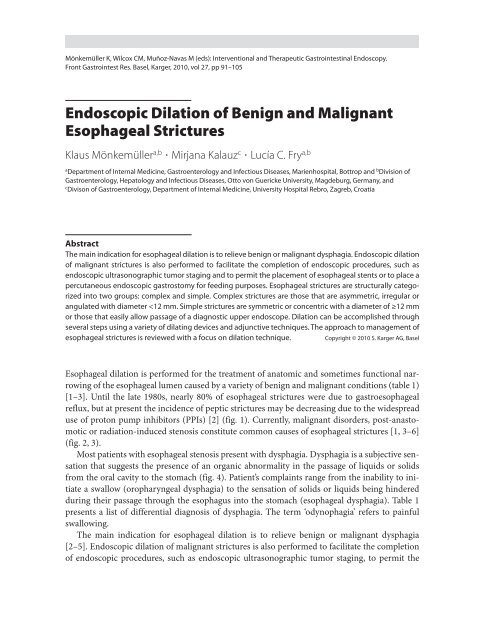
Endoscopic Dilation of Benign and Malignant Esophageal ... - Karger
Endoscopic Dilation of Benign and Malignant Esophageal ... - Karger
Endoscopic Dilation of Benign and Malignant Esophageal ... - Karger

You also want an ePaper? Increase the reach of your titles
YUMPU automatically turns print PDFs into web optimized ePapers that Google loves.
Mönkemüller K, Wilcox CM, Muñoz-Navas M (eds): Interventional <strong>and</strong> Therapeutic Gastrointestinal Endoscopy.<br />
Front Gastrointest Res. Basel, <strong>Karger</strong>, 2010, vol 27, pp 91–105<br />
<strong>Endoscopic</strong> <strong>Dilation</strong> <strong>of</strong> <strong>Benign</strong> <strong>and</strong> <strong>Malignant</strong><br />
<strong>Esophageal</strong> Strictures<br />
Klaus Mönkemüller a,b � Mirjana Kalauz c � Lucía C. Fry a,b<br />
a Department <strong>of</strong> Internal Medicine, Gastroenterology <strong>and</strong> Infectious Diseases, Marienhospital, Bottrop <strong>and</strong> b Division <strong>of</strong><br />
Gastroenterology, Hepatology <strong>and</strong> Infectious Diseases, Otto von Guericke University, Magdeburg, Germany, <strong>and</strong><br />
c Divison <strong>of</strong> Gastroenterology, Department <strong>of</strong> Internal Medicine, University Hospital Rebro, Zagreb, Croatia<br />
Abstract<br />
The main indication for esophageal dilation is to relieve benign or malignant dysphagia. <strong>Endoscopic</strong> dilation<br />
<strong>of</strong> malignant strictures is also performed to facilitate the completion <strong>of</strong> endoscopic procedures, such as<br />
endoscopic ultrasonographic tumor staging <strong>and</strong> to permit the placement <strong>of</strong> esophageal stents or to place a<br />
percutaneous endoscopic gastrostomy for feeding purposes. <strong>Esophageal</strong> strictures are structurally categorized<br />
into two groups: complex <strong>and</strong> simple. Complex strictures are those that are asymmetric, irregular or<br />
angulated with diameter
Table 1. Differential diagnosis <strong>of</strong> dysphagia<br />
Oropharyngeal <strong>Esophageal</strong><br />
Neuromuscular disorders Motility disorders<br />
Cerebrovascular accident, dementia Primary<br />
Brainstem tumors Achalasia<br />
Head trauma Diffuse esophageal spasm<br />
Parkinson’s disease Hypertensive LES<br />
Multiple sclerosis Non-specific esophageal motility disorder<br />
Amyotrophic lateral sclerosis Nutcracker esophagus<br />
Idiopathic UES dysfunction Secondary<br />
Manometric dysfunction <strong>of</strong> the UES or<br />
pharynx<br />
Reflux-related dysmotility<br />
Metabolic encephalopathies Scleroderma <strong>and</strong> other connective tissue<br />
Wilson’s disease Disorders<br />
Guillain-Barré syndrome Chagas’ disease<br />
Myopathic disorders Structural disorders<br />
Connective tissue disease Intrinsic<br />
Polymyositis <strong>Benign</strong> stricture (peptic, radiation, post-surgery,<br />
PDT, corrosive)<br />
Dermatomyositis Schatzki ring<br />
Myasthenia gravis <strong>Benign</strong> tumors<br />
Myotonic dystrophy Malignancy<br />
Oculopharyngeal dystrophy Eosinophilic esophagitis<br />
Metabolic myopathy Pill esophagitis<br />
Sarcoidosis <strong>Esophageal</strong> diverticula<br />
Paraneoplastic syndrome Crohn’s disease<br />
Extrinsic<br />
Infectious diseases Vascular compression (enlarged aorta, left<br />
Mucositis (herpes, cytomegalovirus, C<strong>and</strong>ida) atrium or aberrant subclavian artery)<br />
Lymes disease Mediastinal mass (lymphadenopathy,<br />
Diphtheria abscess, lung cancer)<br />
Cervical osteophytes<br />
Structural lesions Iatrogenic causes<br />
Tumors Pill injury<br />
92 Mönkemüller · Kalauz · Fry
Table 1. Continued<br />
Oropharyngeal <strong>Esophageal</strong><br />
Zenker diverticulum Medication side effects (chemotherapy,<br />
neuroleptics, etc.)<br />
Cricopharyngeal bar Postsurgical muscular or neurogenic<br />
Cervical webs Radiation<br />
Congenital abnormalities<br />
Miscellaneous<br />
Depression, Alzheimer’s disease Functional<br />
Decreased saliva (Sy-Sjögren) Functional dysphagia<br />
a b<br />
Fig. 1. Peptic strictures may be decreasing due to the widespread use <strong>of</strong> PPIs. a A Schatzki ring. b Severe<br />
esophagitis with stricture. Despite having a lesser degree <strong>of</strong> esophagitis, this patient also has a peptic stricture.<br />
This example shows that the Los Angeles classification is still an imperfect classification for gastroesophageal<br />
reflux disease.<br />
placement <strong>of</strong> esophageal stents or to place a percutaneous endoscopic gastrostomy for feeding<br />
purposes [6–8]. Figure 4 provides a useful algorithm for the endoscopic approach <strong>of</strong> patients<br />
with esophageal stenosis. However, not all patients with dysphagia will require an endoscopic<br />
intervention. Note that most patients with dysphagia or odynophagia have conditions that can<br />
be managed medically, such as gastroesophageal reflux disease, infectious ulcers <strong>and</strong> eosinophilic<br />
esophagitis [9] (fig. 5).<br />
<strong>Esophageal</strong> strictures can be structurally categorized into two groups: complex <strong>and</strong> simple.<br />
Complex strictures are those that are asymmetric, irregular or angulated with diameter
a<br />
Fig. 2. a An adenocarcinoma <strong>of</strong> the distal esophagus presenting as a partially obstructing mass. b <strong>Endoscopic</strong><br />
ultrasound. The tumor is very large <strong>and</strong> has spread into the mediastinum (T4).<br />
a b<br />
Fig. 3. a Radiation-induced stricture. b Using magnification endoscopy it is easy to see the proliferation <strong>of</strong><br />
submucosal vessels that is characteristically seen in radiation esophagitis.<br />
dilation therapy when patients develop symptoms <strong>of</strong> dysphagia. <strong>Dilation</strong> can be accomplished<br />
through several steps using a variety <strong>of</strong> dilating devices <strong>and</strong> adjunctive techniques (table 2). The<br />
approach to management <strong>of</strong> esophageal strictures is reviewed with a focus on dilation technique<br />
<strong>and</strong> special consideration for various stricture types <strong>and</strong> complications <strong>of</strong> the method.<br />
Procedural Aspects<br />
Patient Preparation<br />
Patient preparation will depend upon the main cause <strong>of</strong> dysphagia. Patients with achalasia may<br />
require prolonged fasting <strong>and</strong> removal <strong>of</strong> food rests using a nasoesophageal tube (see also chapter<br />
on therapy <strong>of</strong> achalasia). Besides remaining NPO, laboratory tests may be warranted in patients<br />
94 Mönkemüller · Kalauz · Fry
Tumor<br />
Solids<br />
Dysphagia<br />
Solids or liquids<br />
Progressive Intermittent<br />
Regurgitation<br />
Heartburn<br />
Heartburn Weight loss Schatzki Eosinophilic<br />
esophagitis<br />
Zenker Achalasia Sclerodermia<br />
GERD<br />
*Esophagotracheal fistula<br />
**Paralysis <strong>of</strong> the recurrent laryngeal nerve<br />
**Hoarseness<br />
Vocal cord irritation<br />
*Cough upon swallowing<br />
Fig. 4. Diagnostic algorithm for the assessment <strong>of</strong> a patient with dysphagia.<br />
Odynophagia<br />
*Infections<br />
Pill injury<br />
Foreign body<br />
with blood dyscrasias or those taking anticoagulant therapy. Prior to endoscopy all patients have<br />
to provide written informed consent, also with written information about the risk <strong>of</strong> perforation<br />
(i.e. 1%) <strong>and</strong> the possible need for surgery. Endoscopies <strong>and</strong> dilations are carried out in the<br />
morning after an overnight fast <strong>and</strong> as an outpatient procedure, with the exception <strong>of</strong> patients<br />
with complex strictures, who should be observed for 24 h after the procedure. Radiographic contrast<br />
examination is not performed routinely before dilation, but it is performed after dilation <strong>of</strong><br />
achalasia or complex strictures to exclude perforation.<br />
As dilation is an invasive <strong>and</strong> uncomfortable procedure, conscious sedation is generally used.<br />
<strong>Esophageal</strong> dilations should always be performed or closely supervised by experienced endoscopists.<br />
Antibiotics are not used routinely before dilation; endocarditis prophylaxis guidelines<br />
should be followed [11]. Anticoagulants should be discontinued [12].<br />
Accessories<br />
Most esophageal dilations can be performed without the use <strong>of</strong> fluoroscopy [13]. However, some<br />
endoscopists working in open-access or fast-track endoscopy units prefer to have the patients<br />
placed in the fluoroscopy room (fig. 8). This is to avoid the unpleasant, <strong>and</strong> not infrequent situation<br />
in which a contrast imaging <strong>of</strong> the esophagus is needed either before or after the procedure<br />
<strong>and</strong> when a stent placement is planned or anticipated. The worst case scenario is to have a perforation<br />
<strong>and</strong> suddenly have to move with the patient to an appropriate room with x-ray capabilities<br />
in order to be able to place an emergent covered metal stent. In addition, currently, most esophageal<br />
strictures treated at some tertiary centers are complex, <strong>and</strong> their dilation may be facilitated<br />
<strong>Endoscopic</strong> <strong>Dilation</strong> <strong>of</strong> <strong>Benign</strong> <strong>and</strong> <strong>Malignant</strong> <strong>Esophageal</strong> Strictures 95
a b<br />
c d<br />
Fig. 5. Most patients with dysphagia or odynophagia have conditions that can be managed medically,<br />
C<strong>and</strong>ida esophagitis (a), such as gastroesophageal reflux (b) disease, eosinophilic esophagitis (c) <strong>and</strong> pemphigoid<br />
(d).<br />
by the use <strong>of</strong> fluoroscopy [14]. Furthermore, one study showed that fluoroscopy may lead to better<br />
functional results <strong>and</strong> fewer adverse events [15]. Regardless <strong>of</strong> the existing <strong>and</strong> controversial<br />
data, we believe that all patients with complex strictures should be dilated under radiographic<br />
control. Figure 9 is a practical algorithm for the management <strong>of</strong> esophageal strictures.<br />
Technique<br />
<strong>Esophageal</strong> dilation is currently performed using either bougies or balloons (table 3) (fig. 10–12)<br />
[1–4, 16, 17]. The word bougie comes from the French <strong>and</strong> means ‘c<strong>and</strong>le’. Formerly, esophageal<br />
dilation was performed using large c<strong>and</strong>les. The words ‘bougienage’ <strong>and</strong> dilation mean the same,<br />
but we prefer to use the word ‘dilation’ <strong>and</strong> specify whether this was performed using a bougie<br />
or a balloon.<br />
Historically, there are a large variety <strong>of</strong> bougies that have been used to perform esophageal<br />
dilation. However, currently two main types <strong>of</strong> bougies are used: mercury or tungsten-filled<br />
bougies (Maloney or Hurst) <strong>and</strong> over-the-wire (OTW) polyvinyl bougies (Savary-Gilliard® or<br />
American, Wilson-Cook Medical, Inc., Winston-Salem, N.C., USA) [4] (fig. 10). The Maloney<br />
type bougies have a tapered tip <strong>and</strong> are passed either blindly or under fluoroscopic control [18].<br />
96 Mönkemüller · Kalauz · Fry
6<br />
a b<br />
c d<br />
Fig. 6. Complex esophageal strictures are those<br />
that are (a) asymmetric or ulcerated, (b) irregular or<br />
angulated or (c, d) associated with a fistula.<br />
Fig. 7. Simple esophageal strictures are symmetric<br />
or concentric with a diameter <strong>of</strong> ≥12 mm or those<br />
that easily allow passage <strong>of</strong> a diagnostic upper<br />
endoscope.<br />
This type <strong>of</strong> dilator is used for simple strictures with a diameter <strong>of</strong> 12–14 mm. The risk <strong>of</strong> esophageal<br />
perforation may be higher with blind passage <strong>of</strong> the Maloney dilators than with OTW<br />
Savary or TTS (through-the-scope) balloons, particularly in patients with a large hiatal hernia, a<br />
<strong>Endoscopic</strong> <strong>Dilation</strong> <strong>of</strong> <strong>Benign</strong> <strong>and</strong> <strong>Malignant</strong> <strong>Esophageal</strong> Strictures 97<br />
7
Table 2. Steps in esophageal dilation<br />
Patient preparation<br />
Informed consent, fasting overnight, anticoagulants discontinued, sedation<br />
Evaluate diameter <strong>and</strong> length <strong>of</strong> stenosis<br />
Choose method<br />
Bougie vs. balloon<br />
If complex stricture or achalasia<br />
Use fluoroscopy<br />
When using balloon use wire (Jagwire) Observe for 24 h<br />
Fluoroscopy: not needed for most stenosis<br />
Table 3. Type <strong>of</strong> dilators<br />
Mercury or tungsten-filled bougies<br />
Maloney<br />
Hurst<br />
Wire-guided polyvinyl dilators (OTW)<br />
Savary-Gilliard<br />
American<br />
Balloons<br />
TTS dilation balloons<br />
Without wire guidance<br />
With wire guidance (Jagwire)<br />
a b c<br />
Fig. 8. Some endoscopists working in open-access or fast-track endoscopy units prefer to dilate esophageal<br />
strictures under fluoroscopy. This allows for a precise advancement <strong>and</strong> placement <strong>of</strong> a guidewire (a, b),<br />
especially when the stricture is very tight or irregular. In addition, contrast can be applied after the dilation,<br />
<strong>and</strong> thus rule out (c) or discover a perforation.<br />
98 Mönkemüller · Kalauz · Fry
Schatzki<br />
<strong>Dilation</strong> with<br />
Savary bougies<br />
15–20 mm<br />
Luminal changes<br />
Eosinophilic<br />
esophagitis<br />
Biopsies, targeted<br />
<strong>and</strong> r<strong>and</strong>om<br />
Balloon dilation<br />
Peptic<br />
stenosis<br />
Dysphagia<br />
Motility problem<br />
Manometry<br />
Pneumatic balloon<br />
Rigiflex 30, 35, 40 mm<br />
<strong>Endoscopic</strong> <strong>Dilation</strong> <strong>of</strong> <strong>Benign</strong> <strong>and</strong> <strong>Malignant</strong> <strong>Esophageal</strong> Strictures 99<br />
EGD<br />
Fluoroscopy indicated: Achalasia, complex stenosis<br />
Complex stenosis: Tumor, radiation, long, tracheoesophageal fistula<br />
Use wire-guided technique for complex stenosis<br />
Graded dilation:<br />
balloon or Savary<br />
bougie<br />
<strong>Malignant</strong><br />
stenosis<br />
Fig. 9. Practical algorithm for the management <strong>of</strong> esophageal strictures.<br />
Fig. 10. Classic Savary or American dilators.<br />
Dilators are also called bougies, which originates<br />
from the French word ‘c<strong>and</strong>le’. Formerly, esophageal<br />
dilations were performed with wax c<strong>and</strong>les.<br />
American dilators are advanced OTW, usually the<br />
spring-tipped hard Savary wire. However, any s<strong>of</strong>t<br />
wire, such as a ‘Jagwire’, can be sued to advance the<br />
Savary dilator.<br />
EUS<br />
Metal stent<br />
Neoadjuvant TX<br />
Radiation<br />
Achalasia
a<br />
b<br />
c<br />
d<br />
Fig. 11. Balloons used for esophageal dilation are advanced TTS <strong>and</strong> can be st<strong>and</strong>ard or OTW. If the stricture<br />
is tight, irregular or high risk it is always better to advance OTW. a St<strong>and</strong>ard TTS balloon. b The Hercules® 3<br />
Stage Balloon Dilator (Cook, USA) is available in various sizes, the smallest being 8 mm. The advantage <strong>of</strong> this<br />
balloon is its three-stage capability, which means that it can dilate a stricture in three different inflation<br />
diameters, thus following the classic ‘rule-<strong>of</strong>-three’. The smallest balloon can dilate 8–9–10 mm (24–27–30<br />
Fr), whereas the largest balloon goes from 18 to 20 (54–57–60 Fr). c If the stricture is irregular, tight or high<br />
risk, it is always better to perform the dilation with OTW balloons. d Titan® biliary balloon (Cook). Although<br />
this balloon is used to dilate biliary strictures, we have also found it very useful to dilate very tight esophageal<br />
stenosis (permission for image reproduction granted by Cook Medical Inc., Bloomington, Ind., USA).<br />
tortuous esophagus, or those with complex strictures [1, 3, 4, 10] (fig. 11). Savary <strong>and</strong> American<br />
dilators are passed over a guidewire that has been positioned with a tip in the gastric antrum,<br />
with or without fluoroscopic guidance [19].<br />
Various types <strong>of</strong> balloons are available to dilate the esophagus. We have accumulated a large<br />
experience using two main types <strong>of</strong> balloons: (CRE TM , Controlled Radial Expansion, Boston<br />
Scientific Cork Ltd, Cork, Irel<strong>and</strong>, <strong>and</strong> Eclipse® Wire Guided Balloon Dilators Cook Irel<strong>and</strong> Ltd,<br />
100 Mönkemüller · Kalauz · Fry
a b<br />
c d<br />
Fig. 12. <strong>Dilation</strong> <strong>of</strong> an esophageal stricture<br />
(a) with a balloon. The balloon-catheter is<br />
advanced through the stricture (b). Once the<br />
balloon is in an adequate position, i.e. placed<br />
about 50% inside the stricture (c), the balloon is<br />
inflated (d). (e) Legend shows the status <strong>of</strong> the<br />
stricture after dilation.<br />
e<br />
Limerick, Irel<strong>and</strong>). Balloons for esophageal stricture dilation come in various shapes <strong>and</strong> sizes.<br />
Because these balloons can be advanced through the accessory channel <strong>of</strong> the endoscope they are<br />
also referred to as ‘through-the-scope’ or TTS balloons [20] (fig. 11). One <strong>of</strong> the most important<br />
aspects to consider when choosing these balloons is the ability to further advance them OTW.<br />
These wires generally refer to any wire ≤0.035 inch (e.g. Jagwire, Microvasive, Boston Scientific).<br />
These TTS OTW balloons are particularly useful for complex, large, angulated, irregular, very tight<br />
strictures or if the luminal diameter <strong>of</strong> the stricture is
TTS balloon without wire guidance in such strictures can lead to perforation, as the tip, even when<br />
it is floppy, can take a bend through the fibrosed or ulcerated stenosis <strong>and</strong> result in direct perforation<br />
or create a false passage, which after inflating the balloon, results in a large perforation.<br />
It is important to clearly differentiate these balloons from the pneumatic balloons used to<br />
treat achalasia (Rigiflex® II Achalasia Balloon Dilators, Boston Scientific Corp., Natick, Mass.,<br />
USA; Witzel Pneumatic Dilator, M-T-W-W Buderich, Germany, see achalasia chapter). The<br />
achalasia balloons can reach large diameters (30–40 mm).<br />
Although the choice <strong>of</strong> dilatation device is left to the individual endoscopist, dilations in<br />
patients with tumors are mainly performed with Savary dilators following the conventional technique,<br />
using incremental diameters <strong>of</strong> the bougies, but no more than three per session (‘rule<strong>of</strong>-three’),<br />
<strong>and</strong> always under fluoroscopic monitoring. The ‘rule <strong>of</strong> three’ has been the st<strong>and</strong>ard<br />
for bougie dilation [21]. The initial dilator chosen should be based on the known or estimated<br />
stricture diameter. After moderate resistance is encountered with the bougie dilator, no greater<br />
than three consecutive dilations in increments <strong>of</strong> 1 mm should be performed in a single session<br />
[21, 22]. Although this rule does not apply to balloon dilators, a recent study suggested that<br />
inflation <strong>of</strong> a single large-diameter dilator (>15 mm) or incremental dilation <strong>of</strong> >3 mm may be<br />
safe in simple esophageal strictures [23]. Interestingly, most balloons allow a three-step inflation<br />
process, each <strong>of</strong> 1 mm, practically paralleling the ‘rule-<strong>of</strong>-three’. Current AGA recommendations<br />
for management <strong>of</strong> peptic esophageal stricture include consideration that steroid injection into<br />
benign strictures immediately before or after dilation has been advocated to improve outcome<br />
by decreasing the need for repeat dilations [4]. A recent r<strong>and</strong>omized trial <strong>of</strong> intralesional steroid<br />
injection with PPI therapy versus sham injection with PPI therapy in patients with recalcitrant<br />
peptic esophageal strictures showed that the need for repeat dilation was significantly diminished<br />
in the steroid group [24].<br />
Limitations <strong>and</strong> Complications<br />
Regardless <strong>of</strong> the specific method <strong>of</strong> dilation, early improvement in the ability to swallow is<br />
achieved in virtually all patients. Longer-term outcomes are influenced by the underlying pathology.<br />
For peptic strictures, smaller lumen diameter, presence <strong>of</strong> a hiatal hernia >5 cm, persistence<br />
<strong>of</strong> heartburn after dilation, <strong>and</strong> number <strong>of</strong> dilations needed for initial dysphagia relief were significant<br />
predictors <strong>of</strong> early symptomatic recurrence [25]. A multivariate analysis revealed that<br />
a non-peptic etiology <strong>of</strong> strictures was a significant predictor <strong>of</strong> early symptomatic recurrence<br />
within 1 year <strong>of</strong> initial dilation [26]. Patients with peptic strictures should be treated with PPI<br />
therapy. Compared with histamine receptor antagonist therapy, PPI use decreases stricture<br />
recurrence <strong>and</strong> the need for repeat stricture dilation [27, 28].<br />
<strong>Esophageal</strong> perforation is the major complication associated with endoscopic dilation [4,<br />
29–32]. The perforation rate for esophageal strictures after dilation has been reported to be 0.1–<br />
1% [11]. A United Kingdom regional audit reported an overall perforation rate <strong>of</strong> 2.6% with a<br />
mortality <strong>of</strong> 1% [32]. In that study, perforation was less common following dilatation <strong>of</strong> benign<br />
strictures (1.1%) than following dilatation <strong>of</strong> malignant strictures (6.4%) [31]. In older studies,<br />
perforation was most commonly associated with the blind passage <strong>of</strong> Maloney or non-wireguided<br />
bougies into complex strictures [10, 31]. In another study from Engl<strong>and</strong> the incidence <strong>of</strong><br />
iatrogenic perforation for endoscopic treatment <strong>of</strong> tumors <strong>of</strong> the esophagus <strong>and</strong> cardia was 3.3%<br />
[32]. A common practice in Engl<strong>and</strong> at that time was the use <strong>of</strong> single-sized bougies. Whilst data<br />
102 Mönkemüller · Kalauz · Fry
were not available as to the caliber <strong>of</strong> dilators routinely used in that region, it was commonplace<br />
in the early 1990s to use large dilators (18–20 mm). Therefore, it appears that perforations are<br />
common when using a single bougie size dilator, irrespective <strong>of</strong> stricture diameter [32].<br />
Perforation after esophageal dilation usually occurs at the site <strong>of</strong> the stricture, either intraabdominally<br />
or intrathoracically. Some experts recommend endoscopic reinspection immediately<br />
upon completion <strong>of</strong> the dilatation procedure as the appearances may raise the possibility <strong>of</strong><br />
perforation <strong>and</strong> prompt early treatment. Perforation should be suspected if severe or persistent<br />
pain, dyspnea, tachycardia, or fever develops. Physical examination may reveal subcutaneous<br />
crepitus <strong>of</strong> the chest or cervical region. Although a chest radiograph may indicate a perforation,<br />
a normal study result does not exclude this diagnosis <strong>and</strong> a water-soluble contrast esophagram<br />
or contrast computed tomogram <strong>of</strong> the chest may be necessary to disclose a perforation [33].<br />
When using bougies we prefer to use OTW Savary dilators. Bougie-type dilators exert not<br />
only radial forces as they are passed but also longitudinal forces as the result <strong>of</strong> a shearing effect<br />
[34]. Therefore, it is possible that the risk <strong>of</strong> perforation is less when using balloon dilators. In<br />
contrast, longitudinal forces are not transmitted with balloon dilators because the entire dilating<br />
force is delivered radially <strong>and</strong> simultaneously over the entire length <strong>of</strong> the stenosis rather than<br />
progressively from its proximal to distal extent [34]. However, this has not been shown yet by<br />
clinical studies <strong>and</strong> no clear advantage has been demonstrated between the two dilator types<br />
[29, 35, 36].<br />
Data from the literature also confirm that the risk <strong>of</strong> perforation is higher in complex strictures<br />
<strong>and</strong> lower in simple strictures [10]. In a retrospective study performed at our endoscopy<br />
unit, we found that the perforation rate for malignant strictures was higher than for peptic strictures<br />
[14]. Some authors also suggest that perforation may be more common <strong>and</strong> severe with<br />
radiation-induced strictures [37]. Nevertheless, this was not observed in our study. Although we<br />
did not observe any overt perforations associated with radiation-induced strictures, we had two<br />
possible microperforations [14]. The perforation rate may also be influenced by the endoscopist’s<br />
level <strong>of</strong> experience; one study indicated that the perforation rate was four times greater when<br />
the operator had performed fewer than 500 previous diagnostic upper endoscopic examinations<br />
[31]. Therefore, in small hospitals where thoracic surgeons are not routinely available, arrangements<br />
should be made in order to have available a surgeon capable <strong>of</strong> repairing an esophageal<br />
perforation.<br />
The use <strong>of</strong> large-diameter covered metal stents <strong>and</strong> the use <strong>of</strong> exp<strong>and</strong>able, removable plastic<br />
stents have been shown to be effective in the management <strong>of</strong> perforations after dilation <strong>of</strong><br />
benign <strong>and</strong> malignant esophageal strictures [38, 39].<br />
To summarize, endoscopic esophageal dilation is a safe procedure for the management <strong>of</strong><br />
benign strictures, for the palliation <strong>of</strong> malignant strictures, <strong>and</strong> to exp<strong>and</strong> malignant strictures<br />
previous to the placement <strong>of</strong> a self-exp<strong>and</strong>ing metal stent. A thorough clinical knowledge <strong>of</strong> dysphagia<br />
<strong>and</strong> its management is paramount for the interventional endoscopist, as only a minority<br />
<strong>of</strong> patients will require endoscopic therapy. The endoscopist should be well versed <strong>and</strong> trained<br />
with all the available accessories used for esophageal dilation, including wires, bougies <strong>and</strong> balloons.<br />
As perforation occurs in about 0.5–1.5% <strong>of</strong> esophageal dilations, identification <strong>of</strong> patients<br />
at higher risk <strong>and</strong> with complex strictures is crucial to prevent iatrogenic perforation. In addition,<br />
knowledge <strong>and</strong> skills on placement <strong>of</strong> self-exp<strong>and</strong>ing covered metal stents <strong>and</strong> close collaboration<br />
with the surgeon is m<strong>and</strong>atory. As with any other therapeutic endoscopic procedure,<br />
esophageal dilation should only be performed by endoscopists well trained in interventional<br />
endoscopy.<br />
<strong>Endoscopic</strong> <strong>Dilation</strong> <strong>of</strong> <strong>Benign</strong> <strong>and</strong> <strong>Malignant</strong> <strong>Esophageal</strong> Strictures 103
References<br />
1 Lew RJ, Kochman ML: A review <strong>of</strong> endoscopic methods<br />
<strong>of</strong> esophageal dilation. J Clin Gastroenterol 2002;35:117–<br />
126.<br />
2 Richter JE: Peptic strictures <strong>of</strong> the esophagus. Gastroenterol<br />
Clin North Am 1999;28:875–891.<br />
3 Riley SA, Attwood SE: Guidelines on the use <strong>of</strong> oesophageal<br />
dilatation in clinical practice. Gut 2004; 53:i1–i6.<br />
4 Egan JV, Baron TH, Adler DG, Davila R, Faigel DO, Gan<br />
SL, Hirota WK, Leighton JA, Lichtenstein D, Qureshi WA,<br />
Rajan E, Shen B, Zuckerman MJ, VanGuilder T, Fanelli<br />
RD, St<strong>and</strong>ards <strong>of</strong> Practice Committee: <strong>Esophageal</strong> dilation.<br />
Gastrointest Endosc 2006;63:755–760.<br />
5 Lavu K, Mathew TP, Minocha A: Effectiveness <strong>of</strong> esophageal<br />
dilation in relieving nonobstructive esophageal<br />
dysphagia <strong>and</strong> improving quality <strong>of</strong> life. South Med J<br />
2004;97:137–140.<br />
6 Adler DG, Baron TH: <strong>Endoscopic</strong> palliation <strong>of</strong> malignant<br />
dysphagia. Mayo Clin Proc 2001;76:731–738.<br />
7 Pfau PR, Ginsberg GG, Lew RJ, et al: <strong>Esophageal</strong> dilation<br />
for endosonographic evaluation <strong>of</strong> malignant esophageal<br />
strictures is safe <strong>and</strong> effective. Am J Gastroenterol<br />
2000;95:2813–2815.<br />
8 Adler DG, Baron TH, Geels W, et al: Placement <strong>of</strong> PEG<br />
tubes through previously placed self-exp<strong>and</strong>ing esophageal<br />
metal stents. Gastrointest Endosc 2001;54:237–241.<br />
9 Mönkemüller KE, Wilcox CM: Diagnosis <strong>of</strong> esophageal<br />
ulcers in acquired immunodeficiency syndrome. Semin<br />
Gastrointest Dis 1999;10:85–92.<br />
10 Hern<strong>and</strong>ez LV, Jacobson JW, Harris MS: Comparison<br />
among the perforation rates <strong>of</strong> Maloney, balloon, <strong>and</strong><br />
Savary dilation <strong>of</strong> esophageal strictures. Gastrointest<br />
Endosc 2000;51:460–462.<br />
11 Hirota K, Petersen K, Baron TH, et al: Antibiotic prophylaxis<br />
for GI endoscopy. Gastointest Endosc 2003;<br />
58:475–482.<br />
12 Eisen GM, Baron TH, Dominitz JA, et al: Guideline on<br />
the management <strong>of</strong> anticoagulation <strong>and</strong> antiplatelet<br />
therapy for endoscopic procedures. Gastro intest Endosc<br />
2002;55:775–779.<br />
13 Marshall JB, Afridi SA, King PD, Barthel JS, Butt JH:<br />
<strong>Esophageal</strong> dilation with polyvinyl (American) dilators<br />
over a marked guidewire: practice <strong>and</strong> safety at one center<br />
over a 5-year period. Am J Gastroenterol 1996;91:<br />
1503–1506<br />
14 Fry LC, Mönkemüller K, Neumann H, Schulz HU,<br />
Malfertheiner P: Incidence, clinical management <strong>and</strong><br />
outcomes <strong>of</strong> esophageal perforations after endoscopic<br />
dilatation. Z Gastroenterol 2007;45:1180–1184.<br />
15 McClave SA, Brady PG, Wright RA, et al: Does fluoroscopic<br />
guidance for Maloney esophageal dilation impact<br />
on the clinical endpoint <strong>of</strong> therapy: relief <strong>of</strong> dysphagia<br />
<strong>and</strong> achievement <strong>of</strong> luminal patency. Gastrointest<br />
Endosc 1996;43:93–97.<br />
16 Vaezi MF, Richter JE: Current therapies for achalasia:<br />
comparison <strong>and</strong> efficacy. J Clin Gastroenterol 1998;27:<br />
21–35.<br />
17 Mikaeli J, Bishehsari F, Montazeri G, et al: Pneumatic<br />
balloon dilation in achalasia: a prospective comparison<br />
<strong>of</strong> safety <strong>and</strong> efficacy with different balloon diameters.<br />
Aliment Pharmacol Ther 2004;15:431–436.<br />
18 Ho SB, Cass O, Katsman RJ, et al: Fluoroscopy is not<br />
necessary for Maloney dilation <strong>of</strong> chronic esophageal<br />
strictures. Gastrointest Endosc 1995;42:11–14.<br />
19 Wang YG, Tio TL, Soehendra N: <strong>Endoscopic</strong> dilation <strong>of</strong><br />
esophageal stricture without fluoroscopy is safe <strong>and</strong><br />
effective. World J Gastroenterol 2002;8:766–768.<br />
20 Goldstein JA, Barkin JS: Comparison <strong>of</strong> the diameter<br />
consistency <strong>and</strong> dilating force <strong>of</strong> the controlled radial<br />
expansion balloon catheter to the conventional balloon<br />
dilators. Am J Gastroenterol 2000;95: 3423–3427.<br />
21 Langdon DF: The rule <strong>of</strong> three in esophageal dilation.<br />
Gastrointest Endosc 1997;45:111.<br />
22 Vaezi MF, Richter JE: Diagnosis <strong>and</strong> management <strong>of</strong><br />
achalasia. American College <strong>of</strong> Gastroenterology Practice<br />
Parameter Committee. Am J Gastroenterol 1999;94:<br />
3406–3412.<br />
23 Kozarek RA, Patterson DJ, Ball TJ, et al: <strong>Esophageal</strong> dilation<br />
can be done safely using selective fluoroscopy <strong>and</strong><br />
single dilating sessions. J Clin Gastroenterol 1995;20:<br />
184–188.<br />
24 Ramage JI Jr, Rumalla A, Baron TH, et al: A prospective,<br />
r<strong>and</strong>omized, double-blind, placebo-controlled trial <strong>of</strong><br />
endoscopic steroid injection therapy for recalcitrant<br />
esophageal strictures. Am J Gas troenterol 2005;100:2419–<br />
2425.<br />
25 Said A, Brust DJ, Gaumnitz EA, et al: Predictors <strong>of</strong> early<br />
recurrence <strong>of</strong> benign esophageal strictures. Am J<br />
Gastroenterol 2003;98:1252–1256.<br />
26 Chiu YC, Hsu CC, Chiu KW, et al: Factors influencing<br />
clinical applications <strong>of</strong> endoscopic balloon dilation for<br />
benign esophageal strictures. Endoscopy 2004;36:595–<br />
600.<br />
27 Barbezat GO, Schlup M, Lubcke R: Omeprazole therapy<br />
decreases the need for dilatation <strong>of</strong> peptic esophageal<br />
strictures. Aliment Pharmacol Ther 1999;13:1041–1045.<br />
28 Marks RD, Richter JE, Rizzo J, et al: Omeprazole versus<br />
H 2-receptor antagonists in treating patients with peptic<br />
stricture <strong>and</strong> esophagitis. Gastroent erology 1994;106:<br />
907–915.<br />
29 Portale G, Costantini M, Rizzetto C, Guirroli E, Ceolin<br />
M, Salvador R, Ancona E, Zaninotto G: Long-term outcome<br />
<strong>of</strong> laparoscopic Heller-Dor surgery for esophageal<br />
achalasia: possible detrimental role <strong>of</strong> previous endoscopic<br />
treatment. J Gastrointest Surg 2005;9:1332–1339.<br />
30 Metman EH, Lagasse JP, d’Alteroche L, et al: Risk factors<br />
for immediate complications after progressive pneumatic<br />
dilation for achalasia. Am J Gas troenterol 1999;94: 1179–<br />
1185.<br />
104 Mönkemüller · Kalauz · Fry
31 Quine MA, Bell GD, McCloy RF, et al: Prospective audit<br />
<strong>of</strong> perforation rates following upper gastrointestinal<br />
endoscopy in two regions <strong>of</strong> Engl<strong>and</strong>. Br J Surg 1995;<br />
82:530–533.<br />
32 Jethwa P, Lala A, Powell J, et al: A regional audit <strong>of</strong> iatrogenic<br />
perforation <strong>of</strong> tumours <strong>of</strong> the oesophagus <strong>and</strong> cardia.<br />
Aliment Pharmacol Ther 2005;21: 479–484.<br />
33 Fadoo F, Ruiz DE, Dawn SK, et al: Helical CT esophagography<br />
for the evaluation <strong>of</strong> suspected esophageal perforation<br />
or rupture: Am J Roentgenol 2004;182: 1177–1179<br />
34 McLean GK, LeVeen RF: Shear stress in the performance<br />
<strong>of</strong> esophageal dilation: comparison <strong>of</strong> balloon dilation<br />
<strong>and</strong> bougienage. Radiology 1989;172: 983–986.<br />
35 Saeed ZA, Winchester CB, Ferro PS, et al: Prospective<br />
r<strong>and</strong>omized comparison <strong>of</strong> polyvinyl bougies <strong>and</strong><br />
through-the-scope balloons for dilation <strong>of</strong> peptic strictures<br />
<strong>of</strong> the esophagus. Gastro intest Endosc 1995;41:189–<br />
195.<br />
Klaus Mönkemüller, MD, PhD, FASGE<br />
Marienhospital, Bottrop, Josef-Albers-Strasse 70<br />
DE–46236 Bottrop (Germany)<br />
Tel. +49 2041 106 1000, Fax +49 2041 106 1009<br />
E-Mail klaus.moenkemueller@mhb-bottrop.de<br />
36 Scolapio JS, Pasha TM, Gostout CJ, et al: A r<strong>and</strong>omized<br />
prospective study comparing rigid to balloon dilators for<br />
benign esophageal strictures <strong>and</strong> rings. Gastrointest<br />
Endosc 1999;50:13–17.<br />
37 Clouse RE: Complications <strong>of</strong> endoscopic gastrointestinal<br />
dilation techniques. Gastrointest Endosc Clin North<br />
Am 1996;6:323–341.<br />
38 Siersema PD, Homs MY, Haringsma J, et al: Use <strong>of</strong> largediameter<br />
metallic stents to seal traumatic non-malignant<br />
perforations <strong>of</strong> the esophagus. Gastro intest Endosc<br />
2003;58:356–361.<br />
39 Gelbmann CM, Ratiu NL, Rath HC, et al: Use <strong>of</strong> selfexp<strong>and</strong>able<br />
plastic stents for the treatment <strong>of</strong> esophageal<br />
perforations <strong>and</strong> symptomatic anastomotic leaks.<br />
Endoscopy 2004;36:695–699.<br />
<strong>Endoscopic</strong> <strong>Dilation</strong> <strong>of</strong> <strong>Benign</strong> <strong>and</strong> <strong>Malignant</strong> <strong>Esophageal</strong> Strictures 105