Andor Technology - iQ Software Brochure
Andor Technology - iQ Software Brochure
Andor Technology - iQ Software Brochure

You also want an ePaper? Increase the reach of your titles
YUMPU automatically turns print PDFs into web optimized ePapers that Google loves.
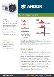
io-imaging<br />
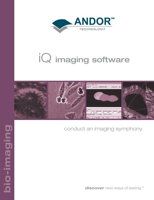
<strong>iQ</strong> imaging software<br />
conduct an imaging symphony
core software and<br />
modules:<br />
Fast λ/Z<br />
• Full camera frame rate<br />
synched with fast<br />
wavelength switching<br />
• z-sections at ~ 30/sec<br />
• Ratio analysis, including ‘Dual<br />
View’<br />
• Applications include: Fast<br />
4/5D Microscopy; Ca 2+ ratio;<br />
FRET...<br />
<strong>Andor</strong> <strong>iQ</strong> Core<br />
• Extensive hardware control – filter wheels, motorized microscopes, confocal<br />
spinning disk..<br />
• Optimized for EMCCD technology<br />
• Extensive and flexible multi-dimensional acquisition, analysis and visualization<br />
• Accessible wizards for protocol set-up<br />
• Applications include:<br />
Time-lapse fluorescence microscopy; FRAP/FLIP; Ion signalling;<br />
Single molecule microscopy; GFP; Gene expression; luminescence;<br />
Immunofluorescence microscopy…….<br />
Tracker<br />
• Fully automatic and interactive<br />
motion analysis<br />
• Multi-channel image<br />
management<br />
• Comprehensive data analysis<br />
options for display of<br />
displacement speed and<br />
direction<br />
• Overlay track presentation<br />
• Applications include all motility<br />
and single molecule tracking<br />
<strong>Andor</strong> <strong>iQ</strong> Core<br />
Multi-field<br />
• Capture and stitch multiple<br />
fields<br />
• Control of motorized stages<br />
• Autofocus to enhance<br />
performance over long-term<br />
studies<br />
ClearView<br />
• Image blurring and haze<br />
reduction<br />
• Deconvolution of 3D, 4D and<br />
5D data<br />
• Handle huge data sets<br />
<strong>Andor</strong> is available as core software and one of five modules<br />
depending on your application needs<br />
Multi-well<br />
• Capture and analyze data<br />
from wells of microtitre plates<br />
• Control of motorized stages<br />
• High content screening<br />
• Drug dose response testing
<strong>iQ</strong><br />
imaging software:<br />
Introduction<br />
<strong>Andor</strong> <strong>iQ</strong> is our flagship<br />
live-cell imaging software,<br />
designed with flexibility<br />
and power in mind. <strong>iQ</strong> –<br />
image and quantify –<br />
occupies a central role in<br />
our Revolution product<br />
range and provides<br />
optimized control of<br />
<strong>Andor</strong>’s award winning<br />
iXon EMCCD cameras<br />
and automation hardware<br />
for a range of Bioimaging<br />
applications. Continuous<br />
development and<br />
improvement ensures that<br />
<strong>Andor</strong> <strong>iQ</strong> represents a<br />
powerful and flexible core<br />
for live-cell imaging<br />
systems.<br />
The software is in use in a<br />
wide range of<br />
applications, including<br />
confocal spinning disk<br />
imaging, TIRFM, intracellular<br />
ion imaging, geneexpression<br />
monitoring<br />
with GFP derivatives and<br />
Luciferase reporters, cell<br />
motility and proteinprotein<br />
interactions. New<br />
applications are evolving<br />
as scientific enquiry<br />
demands.<br />
Key Benefits<br />
• Power and flexibility -<br />
<strong>Andor</strong> <strong>iQ</strong> the choice for<br />
building or enhancing<br />
systems.<br />
• Multi-dimensional core –<br />
Time-lapse, 3D, 4D, 5D<br />
and 6D imaging.<br />
• Fast 4D imaging with<br />
hardware<br />
synchronization up to<br />
100 sections per second.<br />
• Z-series imaging -<br />
deconvolution and 3/4D<br />
rendering module.<br />
• Capture and visualization<br />
of multi-fluorescence or<br />
DIC/phase-fluorescence.<br />
• On-line data charting -<br />
for single, multi-channel,<br />
ratio and FRET imaging.<br />
• Multi-field support (6D) -<br />
enhanced experiment<br />
statistics and/or<br />
Montage.<br />
• Multi-well plate support -<br />
multi-treatment or dose<br />
response live-cell work.<br />
Figure 1. Courtesy Dr Paul Thomas, UEA, Norwich, UK.<br />
Ratio Imaging – A triplet of HEK cells show heterogeneous<br />
response to Ach stimulation.<br />
Figure 2. Courtesy Dr James E. Bear, Lineberger<br />
Comprehensive Cancer Center, NC.<br />
Rat2 fibroblast expressing Pod-GFP (green) stained with<br />
phalloidin (red).<br />
bio-imaging 1
2<br />
bio-imaging<br />
multi-dimensional memory<br />
management:<br />
Multi-dimensional at<br />
its Core<br />
<strong>Andor</strong> <strong>iQ</strong> is multidimensional<br />
at its Core.<br />
This means you can<br />
handle multi-channel,<br />
time-series, Z series and<br />
multi-field data with<br />
consummate ease. <strong>iQ</strong><br />
provides easy to use tools<br />
for analysis, processing,<br />
visualization and<br />
management of live cell<br />
experiment data and<br />
“understands”<br />
dimensionality, so<br />
navigating and<br />
manipulating complex<br />
data sets is instant and<br />
interactive.<br />
Handle huge data<br />
sets with ease<br />
<strong>Andor</strong> <strong>iQ</strong> is built to handle<br />
huge data sets seamlessly<br />
with ImageDisk <br />
technology. ImageDisk <br />
has been developed and<br />
proven over more than<br />
ten years and is at the<br />
core of <strong>iQ</strong>’s multidimensional<br />
memory<br />
management. ImageDisk <br />
extends your system<br />
memory to include fast<br />
hard drives and even RAID<br />
arrays so that you are<br />
never limited by RAM.<br />
Figure 3. Schematic Overview of <strong>Andor</strong> <strong>iQ</strong> – showing the Core and Interfaces to Windows, Hardware and<br />
<strong>Software</strong> components. <strong>iQ</strong> Hosts provide API’s (shown in light blue) to support 3 rd party hardware and software<br />
extensions. The Interface to Windows API is at many levels, but pre-emptive swapping is encapsulated in the<br />
M-DMM.<br />
A smart, pre-emptive disk<br />
swapping algorithm<br />
monitors memory levels<br />
and current frame<br />
requirements during all<br />
acquisition and processing<br />
operations. It avoids<br />
Windows file swapping<br />
and handles frame access<br />
seamlessly, making sure<br />
the data you need is<br />
always available. GB data<br />
sets are no longer a<br />
problem to acquire or<br />
manipulate and visualize.<br />
Figure 4. <strong>iQ</strong> organizes data in a<br />
rectilinear multi-dimensional space.<br />
This figure represents a 5D space,<br />
where each point on the illustrated 3D<br />
matrix is a frame or 2D image.<br />
Dimensions are x,y,z,λ,t in this case. <strong>iQ</strong><br />
can handle further dimensions,<br />
including Fields and Wells for multi-field<br />
or multi-well plate studies. <strong>iQ</strong> has<br />
navigation, visualization and<br />
processing tools to operate<br />
conveniently on these multidimensional<br />
data sets.
powerful wizards help with<br />
settings and configuration:<br />
Figure 5. Protocol tree shows acquisition sequence<br />
control and enables easy editing of parameters.<br />
• Channels Wizard lets you manage<br />
illumination and imaging for any<br />
illuminescent, fluorescent or brightfield<br />
imaging configuration.<br />
• Scan Wizard creates XY and Z Scans.<br />
• Protocols define control acquisition<br />
sequence, timing, triggering and event<br />
monitoring.<br />
• Rapid Setting and Protocol editing via<br />
right mouse click pop-up menus.<br />
Figure 6. Precision Controller Box (PCB)<br />
• Create and manage experiment<br />
acquisition protocols for easy re-use and<br />
sharing.<br />
• Z-offset between channels to<br />
compensate for optical chromatic errors.<br />
• Control and synchronization to external<br />
hardware via programmable Trigger<br />
and Event interface (PCB).<br />
• Integrate with Electrophysiology<br />
Perfusion, Flash Photolysis and FRAP<br />
Solutions.<br />
Figure 7. Courtesy Drs Dmitriy Fayuk and Jerry Yakel’s Neurobiology Laboratory, NIEHS, NC.<br />
<strong>Andor</strong>’s iXon 887-BV and <strong>iQ</strong> software is used to image calcium with Fura2. Dr Fayuk studies neuron receptors in brain sections and cultures\to elucidate<br />
processes of addiction. In this example a cultured neuron was patch-clamped and an iontophoretic electrode used to generate a 50ms Choline injection<br />
(200nA) at the spot shown. Synchronization to electrophysiology apparatus is achieved with <strong>Andor</strong>’s <strong>iQ</strong> software and Precision Controller Box (PCB).<br />
3<br />
bio-imaging
4<br />
bio-imaging<br />
powerful processing:<br />
Figure 8. Courtesy Dr Tom Blanpied,<br />
UMD.<br />
4D maximum intensity image with pseudocolor<br />
shows live neuron labelled with<br />
quantum dots.<br />
On-Line Analysis and<br />
Charting<br />
• On-line Data is<br />
continuously streamed<br />
to a disk file. Charting<br />
of multi-channel, multiregion,<br />
multi-field or<br />
multi-well studies is<br />
possible, with optional<br />
ratio mode.<br />
• Histogram analysis of<br />
multiple user-defined<br />
regions of interest.<br />
• Line profiling for single or<br />
multi-channel image<br />
data on Live or stored<br />
data.<br />
High Performance Offline<br />
Processing<br />
• Ratio analysis for dual<br />
channel analysis, timeseries<br />
pairing and self<br />
ratio (with standing level<br />
from first n frames).<br />
Background correction<br />
and Mask image can be<br />
used to reduce noise in<br />
low signal regions. Ion<br />
concentration calibration<br />
options using either<br />
Grynkiewicz or Almers-<br />
Neher methods for ratio<br />
dyes.<br />
• Kymograph tool allows<br />
oblique line and polyline<br />
probing of multidimensional<br />
data, with<br />
preview. Now with<br />
“Lamella” mode for<br />
protrusion/contraction<br />
rate analysis.<br />
• Dual field splitting and<br />
alignment tool to allow<br />
analysis of simultaneous<br />
captured two or four<br />
channel data. Now with<br />
live-field subtraction<br />
alignment tool.<br />
• Multi-dimensional Math<br />
– difference, average,<br />
encode and projection<br />
processing.<br />
• Ergodic averaging –<br />
improve SNR from<br />
repetitive signal<br />
acquisition and<br />
processing.<br />
• Photobleaching<br />
compensation -<br />
minimizes effect on<br />
analysis results or<br />
visualization for AVI<br />
(movie) creation.<br />
Figure 9. Courtesy Dr James E. Bear, Lineberger Comprehensive Cancer Center, NC.<br />
Still image from a time-lapse movie of a Rat2 fibroblast (left) with kymograph lines marked in red<br />
(1-6). Corresponding kymographs or time-space plots (right). Kymographs give detailed<br />
information about lamellipodial dynamics.
targeted analysis tools and<br />
flexible plug in architecture:<br />
• Plug-in architecture<br />
allows for in-field<br />
updating and access to<br />
new processing<br />
modules as they<br />
become available. This<br />
structure replaces<br />
our legacy product,<br />
Lucida, as all previous<br />
functionality is migrated<br />
to the newer <strong>iQ</strong><br />
software architecture.<br />
We now offer <strong>iQ</strong>-WS to<br />
function on satellite<br />
workstations. We are<br />
already planning a future<br />
with scripting to allow<br />
automation of many<br />
processing and analysis<br />
tasks.<br />
• <strong>Andor</strong>’s ClearView<br />
Deconvolution product<br />
is supplied as a plug-in<br />
option and provides<br />
powerful constrained<br />
iterative algorithms for<br />
image resolution<br />
enhancement, haze<br />
removal and preparation<br />
of wide-field<br />
microscopic<br />
images for 3D/4D<br />
visualization.<br />
• ROI Feature definitions<br />
enhance ROI data<br />
manipulation and<br />
interpretation.<br />
Figure 10. Deconvolution – mouse gut epithelium before and after treatment by ClearView.<br />
Two-channel data acquired in widefield on a Zeiss Axiovert 200, with a plan-apo 100X/1.4NA oil<br />
immersion objective. Standard specimen from Invitrogen/Molecular Probes.<br />
• Thru-series analysis –<br />
Region-based, time, Z<br />
or spectral dimensions.<br />
Analyze single or multichannel<br />
image data with<br />
optional background<br />
correction.<br />
• Text and Dimension<br />
stamp overlays for<br />
monitoring and export<br />
of data.<br />
• Extend analysis dynamic<br />
range - “greys per<br />
second” mode.<br />
• Co-localization analysis<br />
of multi-channel, RGB<br />
image data.<br />
• Tracking - automatic<br />
and interactive tools for<br />
monitoring motion.<br />
Figure 11. Segmentation constrains<br />
values for analysis.<br />
5<br />
bio-imaging
6<br />
bio-imaging<br />
multi dimensional views<br />
and imaging options:<br />
Multi-channel Views,<br />
Voxel Rendering,<br />
Orthogonal Viewing<br />
and Support for<br />
Confocal Instruments<br />
<strong>Andor</strong> <strong>iQ</strong> offers powerful<br />
visualization tools as<br />
standard, with 3D, 4D and<br />
5D capabilities. Raw data<br />
from 3D, 4D, 5D and 6D<br />
acquisition protocols, can<br />
generate image data fast<br />
and <strong>iQ</strong> provides powerful<br />
tools for rapid data<br />
visualization and analysis.<br />
• Navigator - Explore the<br />
data with a multidimensional<br />
movie<br />
player interface and lets<br />
you view multi-channel<br />
data in overlay mode,<br />
merging imaging modes<br />
required.<br />
Figure 12. Courtesy Dr Rachel Errington,<br />
Medical Biochemistry, UWCM, Cardiff, UK.<br />
Single section and voxel rendering of a 3<br />
channel zseries, acquired on a Biorad<br />
confocal instrument, viewed in multi-channel<br />
mode in <strong>iQ</strong> software.<br />
• MC View - View and<br />
manipulate multichannel<br />
data in an<br />
overlay mode.<br />
Channel contrast and<br />
balance are individually<br />
user-controlled.<br />
3D/4D Viewer<br />
Voxel renderer – methods<br />
- maximum intensity,<br />
minimum intensity, opacity<br />
blending and first pixel on<br />
ray trace. Options include:<br />
• Zoom<br />
• Intensity range<br />
• Multi-channel with<br />
scaling/mapping to<br />
enhance features and<br />
study spatial<br />
coincidence.<br />
• Wire-frame dragging<br />
adjusts angle of view.<br />
Figure 13. Kymograph of floating point<br />
ratio image.<br />
• Animation method<br />
interpolates between<br />
user-defined key frames<br />
(and Zoom and Time<br />
points for 4D data) to<br />
create a movie.<br />
• Export to AVI files<br />
through the movie<br />
editor.<br />
OrthoView and<br />
Kymograph<br />
Create orthogonal and<br />
oblique slices through<br />
multi-dimensional images<br />
and visualize as images.<br />
Kymograph allows<br />
Tracking of waves and<br />
features, and new Lamella<br />
mode enables<br />
extension/contraction rate<br />
analysis of e.g. filopodia<br />
and ruffling.<br />
Figure 14. Orthogonal Viewer - new
Figure 15. Courtesy Dr Iain Johnson, Invitrogen, Eugene, OR.<br />
Cultured neurons were loaded with DiSBAC 2 (3), a voltage sensitive dye and visualized with the Revolution 488. Exposure time of the iXon 887-BV was 100ms and<br />
EM Gain 200. The figure shows a film strip from <strong>Andor</strong> <strong>iQ</strong> of maximum intensity projection time series. Time point 4 is taken almost simultaneously with the addition<br />
of KCl, which depolarizes the cells and shows a near instantaneous rise in DiSBAC 2 (3) signal. ∆F of almost 45% is observed in this example. Imaged at The 3D<br />
Microscopy of Living Cells Course, Vancouver 2005.<br />
Strip View<br />
Film strip view of multidimensional<br />
images.<br />
Control frame size,<br />
number of columns,<br />
selected dimension, range<br />
and increment between<br />
frames, border color and<br />
width. Images can be<br />
shown with dimension<br />
stamps e.g. time of<br />
acquisition or Z-depth.<br />
Montage Imaging<br />
<strong>iQ</strong>- MF module includes a<br />
Montage scan mode.<br />
Strip View lets you<br />
visualize the montage of<br />
large specimens acquired<br />
at high power, as shown in<br />
the figures.<br />
<strong>iQ</strong>-MF imaging also allows<br />
time-lapse recoding from<br />
multiple fields of view, so<br />
that data can be gathered<br />
on a large number of cells,<br />
or multiple treatment<br />
groups, increasing ‘n’ for<br />
statistical analyses or<br />
speeding experimental<br />
throughput or both.<br />
Movie Editor<br />
A multi-dimensional filehandling<br />
interface for<br />
Windows AVI files. Choose<br />
start-end frames and<br />
animation dimension to<br />
create focus, spectral or<br />
time-series movies. Movie<br />
Editor provides ROI,<br />
overlay and multi-channel<br />
options to simplify<br />
production of presentation<br />
data.<br />
File-import-export<br />
capabilities<br />
• AVI - images from <strong>iQ</strong><br />
can be exported via<br />
Windows CODECS for<br />
Powerpoint etc. <strong>Andor</strong><br />
<strong>iQ</strong> will read AVI files.<br />
• BioRad - file format<br />
support for confocal and<br />
multi-photon<br />
microscope systems.<br />
• ICS – International<br />
Cytometry Standard files<br />
- multi-dimensional<br />
images.<br />
• TIFF - most single frame<br />
and multi-frame formats<br />
supported.<br />
• Zeiss LSM - multidimensional<br />
file support<br />
for the Zeiss LSM 510<br />
confocal instruments.<br />
Figure 16. Courtesy Dr Stephen Goodall, Leicester<br />
University, UK.<br />
Montage scanning is included in the <strong>iQ</strong>-MF module. This<br />
montage of a blood vessel section was created from 108<br />
(12x9) fields. Raw data was acquired with <strong>iQ</strong>-MF controlling<br />
an iXon DV885 EMCCD camera and Ludl Biopoint stage on<br />
an Olympus BX51 with 20X/0.5 NA objective. Specimen was<br />
immuno- fluorescence labelled for elastin and also shows<br />
auto-fluorescence of collagen, when acquired with an FITC<br />
filter set.<br />
Figure 17. Courtesy Dr Lance Prince’s lab in Pediatrics, UA<br />
Birmingham, AL.<br />
Transmission/relief contrast image of a portion of a live lung<br />
explant. The image was acquired with a 20X, 0.5 objective<br />
and Zeiss Axiovert 25 microscope. Dr Prince is studying lung<br />
development in live explants to better understand fetal lung<br />
development and pathology. <strong>Andor</strong> <strong>iQ</strong> hosts his live cell<br />
imaging system.<br />
7<br />
bio-imaging
8<br />
bio-imaging<br />
extensive<br />
hardware support:<br />
Reconfigure your existing<br />
hardware or build new<br />
according to budget and<br />
technical requirements:<br />
Support for Scientific<br />
Grade CCD Cameras<br />
Cameras can be supplied<br />
for bright field,<br />
fluorescence and<br />
luminescence.<br />
<strong>Andor</strong> EMCCD,<br />
Hamamatsu, Roper<br />
Scientific, PCO, Q-Imaging.<br />
Support for Leading<br />
Confocal Spinning<br />
Disk Adapters<br />
Yokogawa CSU 10 and 22<br />
(Revolution systems) and<br />
Olympus DSU.<br />
<strong>Andor</strong>’s Solid State<br />
Light Source<br />
<strong>Technology</strong> with AOTF<br />
or Direct Modulation<br />
Lasers and LED<br />
illumination systems - fast<br />
switching, minimal sample<br />
exposure.<br />
Figure 20. <strong>Andor</strong> <strong>Technology</strong> - iXon<br />
EMCCD<br />
Filters, Filter Wheels,<br />
Shutters and<br />
Emission Field<br />
Splitters<br />
Chroma and Semrock,<br />
Sutter Instruments, Ludl<br />
Electronics, Prior, Uniblitz,<br />
Optical Insights and Cairn<br />
Research.<br />
Monochromator<br />
Control for Fast<br />
Switching Excitation<br />
TILL Photonics, Sutter and<br />
Cairn Instruments.<br />
Motorized Stages<br />
Used for multi-field or<br />
multi-well plate imaging.<br />
Prior, Marzhauser, Ludl,<br />
ASI, Madcity Labs, Physike<br />
Instrumente.<br />
Automated/Motorized<br />
Microscopes<br />
Olympus - IX81, BX61;<br />
Zeiss - Axiovert, Axioplan;<br />
Leica - DMRE and DMIRE;<br />
Nikon - TE2000 and<br />
E1000.<br />
Figure 21. Yokogawa - Confocal Spinning<br />
Unit 22<br />
Figure 18. Dr Zhang Hui, researcher in the lab of Dr<br />
Peixuan Guo at Purdue University, IN, uses an <strong>Andor</strong><br />
system with prism TIRF imaging of single molecule RNA<br />
complexes. <strong>Andor</strong> <strong>iQ</strong> controls <strong>Andor</strong>'s ALC-102 dual<br />
line laser source and iXon 887-BV for ultimate sensitivity<br />
and performance. "The <strong>Andor</strong> system provides the<br />
highest sensitivity and is the best on the market for our<br />
work with biomotors" says Dr Guo who is a leading<br />
authority on bio-nanotechnology.<br />
Figure 19. Dr Ralph Albrecht and his research<br />
colleagues, Angki and Rainer acquired <strong>Andor</strong> <strong>iQ</strong> and<br />
iXon 887-BV which has boosted the performance and<br />
power of their Zeiss-based CARV system. There<br />
delight is obvious and "the results are outstanding .."<br />
according to Ralph.<br />
Figure 22. <strong>Andor</strong> <strong>Technology</strong> - Laser<br />
Combiner Unit
andor<br />
revolution:<br />
revolution revolution<br />
A Family of Products<br />
<strong>Andor</strong> Revolution provides<br />
a framework for our laser<br />
spinning disk, live cell<br />
confocal microscopy<br />
solutions, which combine<br />
our own award winning<br />
iXon EMCCD camera with<br />
the renowned Yokogawa<br />
CSU 10 and 22 units<br />
“inside”. <strong>Andor</strong> has a<br />
global distribution<br />
agreement with<br />
Yokogawa Electric<br />
Corporation, to integrate<br />
powerful confocal<br />
solutions. This partnership<br />
brings you unrivalled<br />
performance, product<br />
understanding and<br />
support.<br />
Revolution encompasses<br />
a range of components,<br />
both hardware and<br />
software that fit<br />
seamlessly together<br />
creating a complete<br />
confocal microscopy<br />
solution. A flexible<br />
component focus also<br />
allows us to provide key<br />
pieces of hardware standalone.<br />
We want to tailor<br />
solutions matched to your<br />
needs.<br />
At the core of the<br />
Revolution systems is<br />
<strong>Andor</strong> <strong>iQ</strong>, which<br />
synchronizes iXon<br />
EMCCD cameras with the<br />
CSU 10 or 22 confocal<br />
spinning disk and other<br />
key hardware<br />
components such as<br />
Piezo Z100 z-stage and<br />
our SS Laser Combiner<br />
with AOTF for rapid laser<br />
line selection.<br />
Our recommended<br />
microscope platform is<br />
the Olympus IX81,<br />
optimized for live-cell<br />
imaging, but customer<br />
supplied instruments from<br />
Olympus, Leica, Nikon or<br />
Zeiss can also be<br />
supported.<br />
To find out more visit<br />
www.andorbio.com<br />
or contact your local<br />
<strong>Andor</strong> sales engineer.
low light imaging<br />
bio-imaging<br />
spectroscopy<br />
time resolved<br />
x-ray<br />
oem market<br />
Head Office / Europe<br />
7 Millennium Way<br />
Springvale Business Park<br />
Belfast BT12 7AL<br />
Northern Ireland<br />
Tel: +44 (0)28 9023 7126<br />
Fax: +44 (0)28 9031 0792<br />
North America<br />
425 Sullivan Avenue - Suite 3<br />
South Windsor<br />
CT 06074<br />
USA<br />
Tel: (860) 290-9211<br />
Fax: (860) 290-9566<br />
www.andor.com<br />
Japan<br />
7F Ichibancho Central Building<br />
22-1 Ichiban-cho<br />
Chiyoda-ku Tokyo 102-0082<br />
Japan<br />
Tel: 81-3-3511 0659<br />
Fax: 81-3-3511 0662<br />
Other products and company names mentioned throughtout may be the trademarks of their respective owners November 2005