Revolution DSD Confocal - Andor Technology
Revolution DSD Confocal - Andor Technology
Revolution DSD Confocal - Andor Technology

Create successful ePaper yourself
Turn your PDF publications into a flip-book with our unique Google optimized e-Paper software.
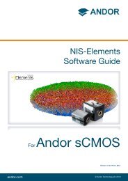
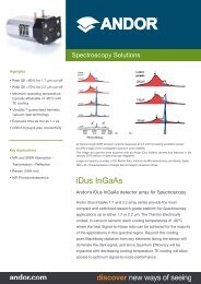
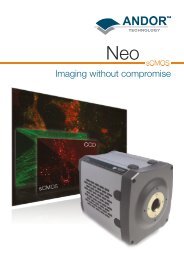
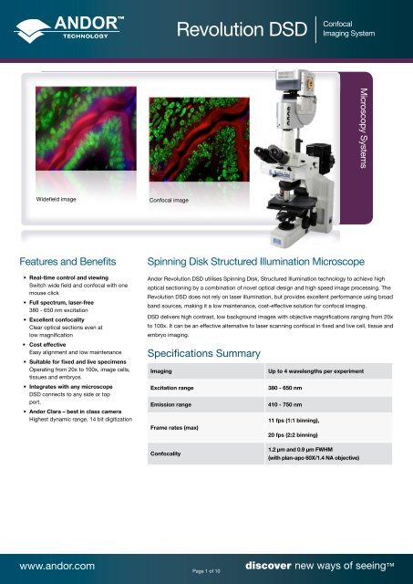
Widefield image <strong>Confocal</strong> image<br />
Features and Benefits<br />
• Real-time control and viewing<br />
Switch wide field and confocal with one<br />
mouse click<br />
• Full spectrum, laser-free<br />
380 - 650 nm excitation<br />
• Excellent confocality<br />
Clear optical sections even at<br />
low magnification<br />
• Cost effective<br />
Easy alignment and low maintenance<br />
• Suitable for fixed and live specimens<br />
Operating from 20x to 100x, image cells,<br />
tissues and embryos<br />
• Integrates with any microscope<br />
<strong>DSD</strong> connects to any side or top<br />
port.<br />
• <strong>Andor</strong> Clara – best in class camera<br />
Highest dynamic range, 14 bit digitization<br />
<strong>Revolution</strong> <strong>DSD</strong> <strong>Confocal</strong><br />
Imaging System<br />
Spinning Disk Structured Illumination Microscope<br />
<strong>Andor</strong> <strong>Revolution</strong> <strong>DSD</strong> utilises Spinning Disk, Structured Illumination technology to achieve high<br />
optical sectioning by a combination of novel optical design and high speed image processing. The<br />
<strong>Revolution</strong> <strong>DSD</strong> does not rely on laser illumination, but provides excellent performance using broad<br />
band sources, making it a low maintenance, cost-effective solution for confocal imaging.<br />
<strong>DSD</strong> delivers high contrast, low background images with objective magnifications ranging from 20x<br />
to 100x. It can be an effective alternative to laser scanning confocal in fixed and live cell, tissue and<br />
embryo imaging.<br />
Specifications Summary<br />
Imaging Up to 4 wavelengths per experiment<br />
Excitation range 380 - 650 nm<br />
Emission range 410 - 750 nm<br />
Frame rates (max)<br />
<strong>Confocal</strong>ity<br />
11 fps (1:1 binning),<br />
20 fps (2:2 binning)<br />
1.2 µm and 0.9 µm FWHM<br />
(with plan-apo 60X/1.4 NA objective)<br />
www.andor.com discover new ways of seeing<br />
Page 1 of 10<br />
Microscopy Systems
Hardware Specifications<br />
<strong>Andor</strong> Differential Spinning Disk Unit<br />
Filters<br />
<strong>Revolution</strong> <strong>DSD</strong> <strong>Confocal</strong><br />
Imaging System<br />
Spectral response 380 - 650 nm excitation<br />
410 - 750 nm emission<br />
<strong>Confocal</strong>ity 1.2 µm FWHM, High Signal mode<br />
0.9 µm FWHM, High Sectioning mode<br />
(Specified with 60x / 1.4 NA plan-apo oil immersion objective)<br />
Internal filter switching time 100 ms (N.B. 60 ms with quad band filter set)<br />
Disk rotation speed 6000 rpm<br />
Turret Name Lines<br />
Excitation & Emission<br />
Filter Pairs Centre/FWHM<br />
Pre Filters Fluorophores<br />
GFP/RFP 2 457/50 & 525/50, 556/20 & 617/73 457/50, 556/20 GFP, RFP<br />
CFP/YFP 2 427/10 & 475/50, 500/24 & 542/50 427/10, 500/24 CFP, YFP<br />
Cy3/Cy5 2 534/36 & 577/24, 635/31 & 690/50 534/36, 635/31 Cy3, Alexa 555, Cy5, Alexa 647<br />
D/F/T<br />
D/F/T/Cy5<br />
3 406/15 & 457/50, 494/20 & 536/40, 575/25 &<br />
628/40<br />
4 Ex (Quad): 387/11, 485/20, 560/25,650/13<br />
Em (Quad): 440/40, 520/24, 604/40, 700/56<br />
406/15, 494/20, 575/25 DAPI, FITC, Texas Red<br />
387/11, 485/20, 560/25, 650/13 DAPI, FITC, TRITC, Cy5<br />
Custom N/A ON SPECIAL REQUEST - -<br />
<strong>Andor</strong> Clara Interline CCD<br />
Active pixels (W x H) 1392 x 1040<br />
Pixel size (W x H) 6.45 x 6.45 μm<br />
Image area (W x H) 8.98 x 6.71 mm<br />
Read noise 2.4 e - @ 1MHz; 4.5 e - @ 10 MHz; 5 e - @ 20 MHz<br />
Maximum dynamic range > 6,500:1; 12,500 with binning<br />
Thermoelectric cooling -55ºC (-40°C fan off)<br />
Dark current 0.0003 e - /pixel/sec<br />
Maximum frame rate 11 fps full frame, 20 fps full frame @ 2 x 2 binning<br />
www.andor.com discover new ways of seeing<br />
Page 2 of 10
Hardware Specifications<br />
<strong>Andor</strong> Metal Halide Light Source<br />
<strong>Andor</strong> Piezo Z-Stage<br />
Motorized XY & Z Control<br />
Wavelength range 380 - 700 nm<br />
Switching speed ~ 60 ms filter wheel<br />
<strong>Revolution</strong> <strong>DSD</strong> <strong>Confocal</strong><br />
Imaging System<br />
Shutter Motorized intensity/shutter, 0% - 100%, 1% resolution, 40 ms Open/Close<br />
Optical coupling UV/Vis liquid light guide, NA 0.5, length 2 m (optional 3 m)<br />
Light source 200 W stabilized metal halide lamp<br />
Lamp lifetime 2200 hours<br />
Interface USB<br />
Dimensions 221 mm [8.7”] x 345 [13.58”] x 229 [9”]<br />
(Requires at least 200 mm free space at top, left, right and rear of unit. Mount on hard shelf)<br />
Weight 9.4 kg [20 lb 11 oz]<br />
Travel range 100 μm, 200 μm, 250 μm and 500 μm<br />
Accuracy / linearity 0.5% of travel<br />
Stage control Analog (0 - 10 VDC), USB and RS232<br />
Settling time > 10 ms (to 90%)<br />
Inserts Slide, Petri dishes (250, 500 µm ranges support microtitre plates)<br />
Output-Position Signal 0.0 - 10.0V<br />
Stages Closed loop with linear optical encoders<br />
Travel range Typically >100 x 75 mm, with 0.02 μm resolution<br />
Travel speed Up to 30 mm/sec<br />
Repeatability 0.3 μm rms<br />
Compatibility Accepts APZ-X00 Piezo stage insert for fast Z scanning<br />
www.andor.com discover new ways of seeing<br />
Page 3 of 10
Hardware Specifications<br />
Piezo Objective Control<br />
Stage Incubator<br />
iQ / Imaris Workstation<br />
Desktop PC<br />
Monitor<br />
<strong>Revolution</strong> <strong>DSD</strong> <strong>Confocal</strong><br />
Imaging System<br />
Travel range PI PIFOC ® P721 - 100 μm; PI PIFOC ® P725 - 400 μm<br />
Setting time Tuneable to < 10ms<br />
Resolution 1.25 nm<br />
Control Analogue or digital<br />
Objectives Oil and water<br />
Accuracy/Linearity 0.2% of travel<br />
Piezo compatibility Piezo inserts from <strong>Andor</strong>, Prior, Ludl, ASI supported<br />
Cooling options Electric, water and cryogenic<br />
Temperature range 3°C above T amb to 50°C<br />
Cryo control with water jacket Heating and cooling between 10 to 50 °C<br />
Temperature regulation ± 0.3°C (water) and ± 0.1°C (water and cryogenic)<br />
CO 2 range 0 - 100%<br />
Model Dell Precision T3500<br />
Weight 16.8 kg (37 lb)<br />
Dimensions 171 mm [6.73”] x 471 mm [18.54”] x 448 mm [17.63”]<br />
Model Dell 2709Wb<br />
Weight 10.95kg [24 lb 2 oz]<br />
Dimensions 632 mm [24.88”] x 200 mm [7.87”]- 230mm [9.05”] depending on monitor angle x 452 compressed (542 extended)<br />
www.andor.com discover new ways of seeing<br />
Page 4 of 10
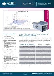
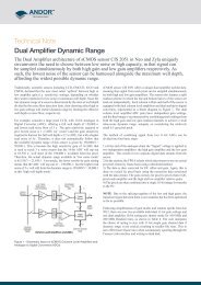
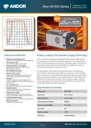
<strong>DSD</strong> Spectral Response Graph<br />
Emission sion TTransmitta<br />
nsmittance ce<br />
100%<br />
90%<br />
80%<br />
70%<br />
60%<br />
50%<br />
40%<br />
30%<br />
20%<br />
10%<br />
<strong>Revolution</strong> <strong>DSD</strong> <strong>Confocal</strong><br />
Imaging System<br />
<strong>DSD</strong> Emission Transmittance Spectrum<br />
0%<br />
400 450 500 550<br />
Wavelength (nm)<br />
600 650 700 750<br />
Clara Quantum Efficiency <strong>Andor</strong> MH Spectral Output<br />
QE (%)<br />
70<br />
60<br />
50<br />
40<br />
30<br />
20<br />
10<br />
0<br />
Normal mode<br />
Extended NIR mode<br />
300 400 500 600 700 800 900 1000<br />
Wavelength (nm)<br />
www.andor.com discover new ways of seeing<br />
Page 5 of 10<br />
Relative tive Int Intensity (Arb. units) its)<br />
1.0<br />
0.9<br />
0.8<br />
00.7 7<br />
0.6<br />
0.5<br />
0.4<br />
0.3<br />
0.2<br />
0.1<br />
0.0<br />
350 450 550 650 750<br />
Wavelength (nm)
Creating The Optimum<br />
Product for You<br />
How to customise <strong>Revolution</strong> <strong>DSD</strong> :<br />
Step 1.<br />
Choose <strong>DSD</strong> system mounting option.<br />
Step 2.<br />
Select filters required for your<br />
application.<br />
Step 3.<br />
Select feet to match <strong>DSD</strong> mounting<br />
option and inverted microscope<br />
models. Note: Feet not required for<br />
upright microscopes.<br />
Step 4.<br />
Select the appropriate objective<br />
scanner from the table.<br />
We provide both Piezo stages and<br />
objective scanners for Z or focus<br />
control. Please contact <strong>Andor</strong> for<br />
specific requests.<br />
Step 5.<br />
We also offer high performance<br />
solutions for photo-ablation,<br />
uncaging, switching and FRAP.<br />
Please contact us for a detailed<br />
specification and quotation.<br />
<strong>Revolution</strong> <strong>DSD</strong> <strong>Confocal</strong><br />
Imaging System<br />
Step 1.<br />
Choose mounting type<br />
RD-<strong>DSD</strong>- Uni<br />
example shown<br />
Uni: Universal mounting platform (for inverted and upright configurations) - no enclosure<br />
Core: Universal mounting platform (inverted configuration only) - with enclosure<br />
PLEASE NOTE - The <strong>Revolution</strong> <strong>DSD</strong> enclosure is not compatible with a Leica microscope<br />
Step 2.<br />
Select the appropriate filter from the table below:<br />
Step 3.<br />
Fluorophores Part Number<br />
GFP, RFP RD-<strong>DSD</strong>-FS1<br />
CFP, YFP RD-<strong>DSD</strong>-FS2<br />
DAPI, FITC, Texas Red RD-<strong>DSD</strong>-FS3<br />
DAPI, FITC, TRITC, Cy5 RD-<strong>DSD</strong>-FS5<br />
Cy3, Cy5 RD-<strong>DSD</strong>-FS6<br />
Identify inverted microscope model & feet from the table below:<br />
Microscope Feet Part Number<br />
Olympus IX71/81 TR-OLIX-MNT-XXX*<br />
Nikon TiE TR-NKTI-MNT-XXX*<br />
Nikon TE2000 TR-NK2K-MNT-XXX*<br />
Zeiss Axiovert 100/200 TR-ZSAV-MNT-XXX*<br />
Zeiss Axio Observer TR-ZAXO-MNT-XXX*<br />
Leica DMI4/5/6000 TR-LCDM-MNT-XXX*<br />
* XXX = 120 for Core enclosure, 110 for Uni mount<br />
Step 4.<br />
Select your required objective scanner from the table below:<br />
Travel Range Microscope Part Number<br />
100 µm Nikon, Leica MP-P100-OBJ-NKL-M25 *<br />
100 µm Nikon, Leica MP-P100-OBJ-NKL-M32 **<br />
100 µm Olympus, Zeiss MP-P100-OBJ-OLZ-RMS ***<br />
400 µm Nikon, Leica MP-P400-OBJ-NKL-M25 *<br />
400 µm Nikon, Leica MP-P400-OBJ-NKL-M32 **<br />
400 µm Olympus, Zeiss MP-P400-OBJ-OLZ-RMS ***<br />
* M25 objective thread, suitable for Nikon CFI60 and Leica Nosepiece B<br />
** M32 objective thread, suitable for Nikon M32 EPI CFI and Leica Nosepiece M<br />
*** RMS standard objectives (0.8”) suitable for all Olympus RMS and Zeiss RMS<br />
www.andor.com discover new ways of seeing<br />
Page 6 of 10
Software<br />
<strong>Andor</strong> iQ2<br />
Multi-dimensional imaging with Python IDE<br />
Features<br />
• User-controlled acquisition of confocal, wide field or both images from <strong>DSD</strong><br />
• Multidimensional at its core - from time-lapse to 4D multi-channel imaging<br />
• ImageDisk, virtual memory system, for smooth management of huge data sets<br />
• Accessible dashboard interface and flexible Protocol structure<br />
• Supports ratio imaging and analysis for functional imaging<br />
• Fast Live refresh even with long exposure times to ease specimen review<br />
• Multi-field & montage capability for large specimens or increased throughput<br />
<strong>Revolution</strong> <strong>DSD</strong> <strong>Confocal</strong><br />
Imaging System<br />
<strong>Andor</strong> iQ2 provides acquisition and control for the <strong>Revolution</strong> <strong>DSD</strong> system. iQ2 offers optimized control of <strong>Andor</strong>’s award<br />
winning iXon3 EMCCD and Clara CCD cameras with powerful multi-dimensional acquisition through flexible Protocols.<br />
Continuous development and improvement ensures that iQ2 represents a powerful and flexible core for a range of bio-imaging<br />
applications. iQ2 combined with Imaris provides a powerful and flexible platform.<br />
Imaris<br />
The Ultimate Tool for Visualisation and Analysis of Multi-Dimensional Images<br />
Imaris delivers all the necessary functionality for visualization, segmentation and interpretation of multidimensional datasets.<br />
By combining speed, precision and intuitive ease-of-use, Imaris provides a complete set of features for handling multi-channel<br />
image sets of any size up to 50 gigabytes.<br />
Imaris will read, visualize and analyze images acquired from almost any confocal and wide field microscope. Imaris and iQ have<br />
been co-designed to provide seamless ImageDisk access for Imaris 7.1 and above, avoiding the tedious save/open cycles<br />
required for third party data. Imaris has been specifically designed to target the critical data processing needs of the most<br />
demanding life-science imaging applications. Its intuitive workflow approach takes away the need to select and manage a<br />
range of imaging tools and frees the scientist to get on with their research.<br />
Features<br />
• Advanced Volume Rendering - Maximum Intensity Projection (MIP), Blend Projection and Real-Time Shadow Rendering<br />
• Surfaces, Segmentation and interactive Iso-Surfaces, Region Growing and Semi Automatic Surface Generation<br />
• Spots, Segmentation and Interaction - Identify and interact in 3D with hundreds of objects<br />
• Smart Handling of Huge Images > 50 GB<br />
• Multithreading & Advanced Computer Graphics - High-resolution, multiple light sources and 3D holographic rendering<br />
Core Features<br />
• Rotational drift correction<br />
• A free rotation image processing tool<br />
• <strong>Andor</strong> iQ ImageDisk reader<br />
• Additional support for ZVI files<br />
• IMOD data import functionality<br />
www.andor.com discover new ways of seeing<br />
Page 7 of 10
Product Drawings - <strong>DSD</strong> Core Platform<br />
Dimensions in mm [inches]<br />
�������������<br />
������������<br />
��������������<br />
�������������<br />
Weight:<br />
Enclosure with Clara & <strong>DSD</strong> = 15.3 kg [33 lb 12 oz]<br />
�������������<br />
Typical<br />
�������������������������������������������������������<br />
footprint of <strong>DSD</strong> fitted to microscope (Olympus)<br />
������������<br />
<strong>Revolution</strong> <strong>DSD</strong> <strong>Confocal</strong><br />
Imaging System<br />
Enclosure<br />
�����������<br />
Product Drawings - <strong>DSD</strong> Universal Mounting Platform<br />
Dimensions in mm [inches]<br />
Total wieght of system: 7Kg<br />
Clara = 2.35Kg<br />
<strong>DSD</strong> = 3.75Kg<br />
Bracket= 0.9Kg<br />
161.0 [6.34]<br />
261.7 [10.30]<br />
Connector rear panel<br />
���� ������� ���<br />
www.andor.com discover new ways of seeing<br />
Page 8 of 10<br />
�������<br />
�������������<br />
�<br />
�<br />
��������<br />
�������<br />
�����<br />
�������<br />
������<br />
�����<br />
������<br />
�����<br />
���<br />
��������������������<br />
259.0 [10.19]<br />
������������<br />
���<br />
��������<br />
�������<br />
74.9 [2.95]<br />
110.0 [4.33]
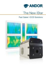
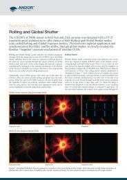
SD-SIM in more detail<br />
<strong>Revolution</strong> <strong>DSD</strong> <strong>Confocal</strong><br />
Imaging System<br />
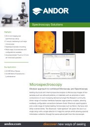
In a <strong>DSD</strong> system, the spinning element comprises a single synthetic quartz disk supporting a thin layer of coated aluminium in which the Structured<br />
Illumination Pattern (SIP) is created by photo-lithography. The aluminium SIP has a 1:1 mark to space ratio (half metal and half space), which means<br />
that approximately half of the light falling upon it is reflected (R) and half transmitted (T). This is true for light which is incident from either side of the<br />
disk and is a critical feature of the device. Figure 1. below illustrates the two patterns on the <strong>DSD</strong> disk: one with a pitch or period of 320 μm on the<br />
inner radius; the second with pitch 160 μm on the outer radius.<br />
Figure 1. The differential spinning disk is manufactured with two structured illumination patterns, radially disposed. In the <strong>Andor</strong> <strong>DSD</strong>, the inner and outer patterns are<br />
designed respectively with 320 and 160 μm pitch and 1:1 mark-space ratio. These patterns are referred to as the “high signal” and “high sectioning” SIPs and allow the user<br />
to adapt <strong>DSD</strong> sectioning and signal-to-noise ratio performance to their specimen thickness and magnification choice.<br />
Applications Guide<br />
• Cellular Biochemical Imaging, e.g. Ca2+ (not Fura2) & pH<br />
• Fluorescent Protein Dynamics e.g. Trafficking, Translocation<br />
• Development e.g. C. elegans, Zebrafish and Drosophila<br />
• Cytoskeleton and Membrane Dynamics and Motility<br />
• Membrane Trafficking, Endo and Exo-Cytosis<br />
• Nuclear Organization and Dynamics<br />
• Photo-Manipulation – e.g. Activation and Ablation<br />
• Viral Infection and Translocation<br />
• Motility and Chemotaxis Assays<br />
• Immunofluorescence<br />
Typical Configurations<br />
<strong>DSD</strong> Core with<br />
Nikon TIe<br />
<strong>DSD</strong> Core with<br />
Olympus IX 81<br />
<strong>DSD</strong> Uni with<br />
Nikon Eclipse<br />
<strong>DSD</strong> Core with<br />
Zeiss Axiovert 200<br />
<strong>DSD</strong> Uni with<br />
Leica DMI6000<br />
www.andor.com discover new ways of seeing<br />
Page 9 of 10
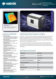
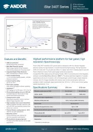
A two-colour image series of living MDCK cells acquired<br />
with <strong>Revolution</strong> <strong>DSD</strong>. Mito-tracker label shown in red<br />
and lysotracker in green. 200 images were acquired<br />
over a period of approximately 3 minutes of imaging and<br />
analyzed for bleaching as shown in the intensity graph<br />
to the right. <strong>DSD</strong> shows low levels of photo-bleaching<br />
with < 3% recorded over the 200 frame series (100 per<br />
channel).<br />
Operating & Storage Conditions<br />
Operating Temperature 15°C to 30°C ambient<br />
Relative Humidity < 70% (non-condensing)<br />
Storage Temperature -10°C to 50°C<br />
Power Requirements<br />
110 - 240 VAC, 50/60 Hz<br />
<strong>Revolution</strong> <strong>DSD</strong> <strong>Confocal</strong><br />
Imaging System<br />
Order Today<br />
Need more information? At <strong>Andor</strong> we are committed to finding<br />
the correct solution for you. With a dedicated team of technical<br />
advisors, we are able to offer you one-to-one guidance and<br />
technical support on all <strong>Andor</strong> products. For a full listing of our<br />
regional sales offices, please see: www.andor.com/contact<br />
Our regional headquarters are:<br />
Europe Japan<br />
Belfast, Northern Ireland Tokyo<br />
Phone +44 (28) 9023 7126 Phone +81 (3) 3518 6488<br />
Fax +44 (28) 9031 0792 Fax +81 (3) 3518 6489<br />
North America China<br />
Connecticut, USA Beijing<br />
Phone +1 (860) 290 9211 Phone +86 (10) 5129 4977<br />
Fax +1 (860) 290 9566 Fax +86 (10) 6445 5401<br />
Extended focus images of mouse kidney captured<br />
on <strong>Revolution</strong> <strong>DSD</strong> system - left is wide field and<br />
right confocal. Both images acquired simultaneously<br />
to illustrate contrast between modes. The <strong>DSD</strong> was<br />
attached to an Olympus IX81 with 60X/1.4 oil objective.<br />
Windows is a registered trademark of Microsoft Corporation.<br />
M<strong>DSD</strong>SS 0911 R1<br />
www.andor.com discover new ways of seeing<br />
Page 10 of 10