Canine Chronic Inflammatory Rhinitis
Canine Chronic Inflammatory Rhinitis
Canine Chronic Inflammatory Rhinitis

You also want an ePaper? Increase the reach of your titles
YUMPU automatically turns print PDFs into web optimized ePapers that Google loves.
<strong>Canine</strong> chronic inflammatory rhinitis 79<br />
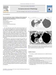
Figure 2 Sequential computed tomographic images through the<br />
frontal sinus of a dog with idiopathic LPR. The top panel shows<br />
soft-tissue or fluid opacification in the left frontal sinus. In the next<br />
7-mm slice, the frontal sinus has filled more completely and an<br />
air–fluid meniscus is present, suggesting that fluid is filling the<br />
frontal sinus.<br />
significant nasal mucosal pathology and visualization of the<br />
area sampled for histopathology.<br />
Other diagnostic techniques to perform after rhinoscopy<br />
are examination of the nasopharyngeal region using a<br />
dental mirror or retroflexed endoscope to look for nasopharyngeal<br />
polyps and dental probing to rule out oronasal<br />
fistula or dental disease as a cause for unilateral nasal<br />
discharge.<br />
Histopathology<br />
Nasal biopsy samples of dogs with chronic, idiopathic, inflammatory<br />
rhinitis are characterized by a primarily lymphoplasmacytic<br />
infiltrate, but concurrent neutrophilic or less<br />
commonly eosinophilic infiltrate may also be seen. Epithelial<br />
Table 2 Computed Tomography in 33 Dogs with Idiopathic<br />
<strong>Rhinitis</strong> 5<br />
Normal 4/33 (12%)<br />
Unilateral lesions 8/33 (24%)<br />
Bilateral lesions 21/33 (64%)<br />
Fluid accumulation 27/33 (82%)<br />
Soft-tissue opacification 25/33 (78%)<br />
Plaque-like lesions 24/33 (73%)<br />
Turbinate destruction 23/33 (70%)<br />
Gas pocketing 23/33 (70%)<br />
Frontal sinus opacification 14/33 (42%)<br />
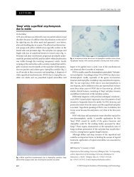
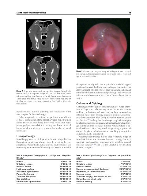
Figure 3 Rhinoscopic image of a dog with idiopathic LPR. Marked<br />
hyperemia and mucus accumulation are evident. (Color version of<br />
figure is available online.)<br />
changes are usually mild but may include epithelial hyperplasia<br />
and erosion. Turbinate remodeling or destruction can<br />
also be evident. The majority of dogs with unilateral clinical<br />
signs have bilateral nasal mucosal pathology, and severity of<br />
inflammation between the two sides of the nasal cavity often<br />
varies.<br />
Culture and Cytology<br />
Obtaining a positive culture of bacterial and/or fungal organisms<br />
in dogs with inflammatory rhinitis is not uncommon<br />
and likely reflects normal nasal mucosal flora or secondary<br />
infection rather than primary infectious rhinitis. Culture results<br />
from the rostral nasal cavity may differ from the caudal<br />
nasal cavity. 26 Similarly, brush or lavage samples from super -<br />
ficial epithelium may not adequately reflect bacterial and fungal<br />
infiltration into the nasal mucosa. When culture is desired,<br />
collection of a deep nasal lavage using a protected<br />
catheter brush or submission of a nasal biopsy sample for<br />
culture should be considered.<br />
Nasal mucosal cytology may be used to identify fungal or<br />
neoplastic lesions in some cases; however, cytology has poor<br />
sensitivity and specificity compared with histology in nasal<br />
mucosal samples27-29 and is often unreliable for detecting<br />
chronic changes. 29<br />
Table 3 Rhinoscopic Findings in 37 Dogs with Idiopathic <strong>Rhinitis</strong><br />
5<br />
Normal 1/37 (3%)<br />
Unilateral lesions 7/37 (19%)<br />
Bilateral lesions 21/37 (57%)<br />
Mucoid or mucopurulent discharge 29/37 (78%)<br />
Hyperemic, or inflamed mucosa 26/37 (70%)<br />
Mucosal edema 8/37 (22%)<br />
Turbinate atrophy, destruction, or loss 8/37 (22%)<br />
Hemorrhage or blood clots 5/37 (14%)<br />
Mucosal friability 4/37 (11%)