cytological outfit cytoset - MPW MED. INSTRUMENTS Spółdzielnia ...
cytological outfit cytoset - MPW MED. INSTRUMENTS Spółdzielnia ...
cytological outfit cytoset - MPW MED. INSTRUMENTS Spółdzielnia ...

You also want an ePaper? Increase the reach of your titles
YUMPU automatically turns print PDFs into web optimized ePapers that Google loves.
O P E R A T I N G I N S T R U C T I O N<br />
Catalogue Number 20452/ENG<br />
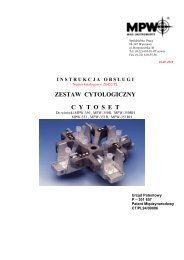
CYTOLOGICAL OUTFIT<br />
C Y T O S E T<br />
For the <strong>MPW</strong>-350, <strong>MPW</strong>-350R, <strong>MPW</strong>-350RH,<br />
<strong>MPW</strong>-351, <strong>MPW</strong>-351R, <strong>MPW</strong>-351RH centrifuges<br />
46 Boremlowska str.,<br />
04-347 Warsaw Poland<br />
Phone: (+48 22) 610 81 07 Service<br />
Fax: (+48 22) 610 55 36<br />
26-02-2010<br />
Patent Office<br />
P – 301 657<br />
International Patent<br />
CT/PL94/00006
Contents<br />
1. INTENDED USE. 3<br />
2. ADVANTAGES OF "CYTOSET". 3<br />
3. OPERATION PARAMETERS. 3<br />
4. SPECIFICATION OF EQUIPMENT. 4<br />
5. CYTOLOGICAL OUTFIT CONTENTS. 4<br />
6. WORKING PRINCIPLE. 5<br />
7. PREPARATION OF SAMPLES FOR CENTRIFUGATION. 5<br />
8. TABLE OF DILUTIONS IN HEMATOLOGY. 5<br />
9. PREPARATION OF THE CYTO INSERT WITH THE PASSING OF THE FILTRATE<br />
THROUGH BLOTTING PAPER (WITH A LOSS OF THE FILTRATE). 6<br />
10. PREPARATION OF THE CYTO INSERT WITH FILTRATE RETRIEVAL. 6<br />
DIAMETER OF OPENINGS IN FILTRATION PAPER 6<br />
11. CENTRIFUGATION PARAMETERS. 6<br />
12. STEPS IN THE USE OF CENTRIFUGE WITH THE "CYTOSET". 7<br />
13. PROCEDURE AFTER CENTRIFUGATION, WHEN THE FILTRATE IS LOST. 8<br />
14. PROCEDURE FOLLOWING CENTRIFUGATION, WITH THE FILTRATE RETRIEVED. 8<br />
15. MAIN VIEW. 9<br />
16. CLEANING, DISINFECTION, MAINTENANCE. 9<br />
16.1. CLEANING OF THE ACCESSORIES. 9<br />
16.2. CLEANING OF THE CYTO INSERT. 9<br />
16.3. LUBRICATION. 9<br />
16.4. STERILIZATION AND DISINFECTIONS OF THE ACCESSORIES. 10<br />
16.5. STERILIZATION AND DISINFECTIONS PROCESS VALIDATION. 10<br />
17. MANUFACTURER’S DATA. 10<br />
18. DISTRIBUTOR INFORMATION. 10<br />
2
1. Intended use.<br />
The “CYTOSET” <strong>cytological</strong> <strong>outfit</strong> catalogue number 12452c is as accessory equipment for these<br />
centrifuges: <strong>MPW</strong>-350, <strong>MPW</strong>-350R, <strong>MPW</strong>-350RH, <strong>MPW</strong>-351, <strong>MPW</strong>-351R, <strong>MPW</strong>-351RH, manufactured<br />
by <strong>MPW</strong> Med. instruments.<br />
The “CYTOSET” <strong>cytological</strong> <strong>outfit</strong> catalogue number 12271c possesses the <strong>MPW</strong>-223c specialized<br />
centrifuge.<br />
The “CYTOSET” <strong>cytological</strong> <strong>outfit</strong> is intended for in vitro diagnostics (IVD) equipped; used to<br />
centrifugal separation of body fluids into fine subconstituent elements from the filtrate. The fine elements<br />
are deposited directly on basic microscope slides, while the filtrate is accumulated in special tubes,<br />
following the settling of the particulate matter. The tested physiological fluids constitute a suspension of<br />
fine elements in the fluid fraction.<br />
Among them are the following:<br />
a) natural biological fluids including: cerebrospinal fluid, urines, effusions, serum leaks, joint fluid,<br />
leukorrhoea, pus and related others,<br />
b) suspensions in isotonic smear solutions, tissue punctuates, sputum, rinsings form the bronchi and<br />
respiratory pathways and related others.<br />
These suspensions may be tested in order to:<br />
1) obtain precipitates of fine matter and the filtrate from the same sample of tested biological suspension,<br />
2) obtain just a precipitate of the fine matter through passing of the filtrate into a blotting paper<br />
The <strong>cytological</strong> <strong>outfit</strong> is essential in medicine and veterinary science, and may also be used in industry as<br />
well as technology.<br />
2. Advantages of "CYTOSET".<br />
� fast deposition of cells on the microscope slide through centrifugation and the penetration of the filtrate<br />
into the blotting/ filter paper,<br />
� possibility of filtrate recovery after the cells have settled on the slide by its automatic collection into a<br />
test tube,<br />
� securing the fluid against its escaping into the centrifugation chamber (avoiding air-born<br />
contamination),<br />
� inserts used as disposable equipment will help prevent infections, contamination of lab personnel or the<br />
environment,<br />
3. Operation parameters.<br />
� Rotational speed 100 ÷ 2500 rpm<br />
� max. acceleration 750 x g<br />
� max. centrifugation capacity 2 ml.<br />
� area diameter of settling cells on the microscope slide � 8,7 mm<br />
3
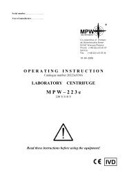
4. Specification of equipment.<br />
Depending on the type of the purchased centrifuge, the below-mentioned “CYTOSET” <strong>cytological</strong> <strong>outfit</strong><br />
may be ordered.<br />
Catalog No. Equipment name<br />
Max<br />
rpm<br />
RCF Number of piece<br />
in the set<br />
12452<br />
Cytology rotor for centrifuges<br />
<strong>MPW</strong>-350, <strong>MPW</strong>-350R, <strong>MPW</strong>-350RH,<br />
<strong>MPW</strong>-351, <strong>MPW</strong>-351R, <strong>MPW</strong>-351RH<br />
4 seats 2500 750 1<br />
13606 Hanger 4<br />
15123 Decantation test tube 2,2 ml with cap 100<br />
16610 Cyto insert 100<br />
16614 Basic microscope slide 100<br />
16616 Blotting/ filtration card � 9,5 mm 100<br />
or 16617 Blotting/ filtration card � 12,5 mm 100<br />
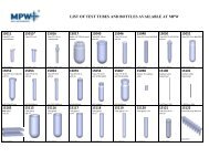
5. Cytological <strong>outfit</strong> contents.<br />
The <strong>outfit</strong> contains a swing-out four-seats rotor with hangers, into which the complete cyto insert is<br />
placed.<br />
6<br />
The complete insert contains the following:<br />
� base – collection chamber (1),<br />
� basic microscope slide (2),<br />
� blotting/ filter card (3),<br />
� cyto insert (4),<br />
� seal � 8,7 mm (60 mm2) (5),<br />
� plug (6),<br />
� plug (7),<br />
� decantation test tube (8),<br />
8<br />
7<br />
4<br />
5<br />
3<br />
2<br />
1<br />
Drawing No.1<br />
4
6. Working principle.<br />
During the centrifugation, the cyto insert turns into a horizontal position, which positioning is then<br />
blocked with snap fastening units located in the side arms of the rotor in case of the need to decant the<br />
filtrate.<br />
Under the centrifugal force the morphotic elements (cell precipitate) are separated from the suspension and<br />
settled on the microscope slide. The cell-free filtrate, depending on the method used, is either absorbed by<br />
the blotting paper or decanted into a test tube following centrifugation. After the decantation test tube<br />
containing the filtrate is removed in order to dry the precipitate on the microscope slide, the test tube is<br />
subjected to a second centrifugation.<br />
Drawing No. 2<br />
Cell precipitate<br />
CYTO<br />
Filtrate<br />
SUPERNATANT<br />
The CYTO insert, following the completion of the settling of the morphotic elements on the basic slide<br />
centrifugation with filtrate retrieval.<br />
7. Preparation of samples for centrifugation.<br />
The amount of fluid used in centrifugation depends on its contents of cells, as well as its<br />
availability. If the fluid is cell-dense in order to avoid layering of the cells on the slide or their settling too<br />
densely, the fluid should be diluted with 0.9% NaCl solution or PBS.<br />
8. Table of dilutions in hematology.<br />
Number of<br />
white cells<br />
101 - 300<br />
301 - 700<br />
701 - 1500<br />
1501 - 3000<br />
Dilution<br />
factor<br />
-<br />
1 : 2<br />
1 : 5<br />
1 : 10<br />
Sample<br />
drops<br />
Saline<br />
drops<br />
When a cell-poor fluid with a low protein content (about 0.2 mg/ml) is tested there exists a danger,<br />
that the centrifuged cells on the slide may be fragmented, as well as the quality of the preparation is often<br />
not satisfactory. For that reason, if the filtrate is not needed for further tests, it is recommended that a few<br />
drops of blood serum or albumin solutions be added. Samples destined for cyto-centrifugation should be<br />
fresh in order to maximize the possibility of getting intact cells.<br />
20<br />
10<br />
4<br />
2<br />
0<br />
10<br />
16<br />
18<br />
5
9. Preparation of the CYTO insert with the passing of the filtrate through blotting paper<br />
(with a loss of the filtrate).<br />
� Prepared and labeled basic microscope slide should be inserted into the base-collector. In order to obtain<br />
better cell adhesion to the slide it is recommended that the slides be covered with poly-L-lysine,<br />
manufactured by “Sigma” – Chemical<br />
� Place the blotting card with an opening of � 9,5.<br />
� Place the plugs on the cyto insert.<br />
� Place the complete cyto insert onto the base-collector and symmetrically close the snap-fastening catch.<br />
� Pour the prepared fluid sample into the central cylinder of the cyto insert and close with a plug.<br />
The maximum amount of fluid, which the cyto insert can hold is 2 ml.<br />
That volume is marked with a line.<br />
Drawing No. 3<br />
10. Preparation of the CYTO insert with filtrate retrieval.<br />
� Prepared and labeled basic microscope slide should be inserted into the base-collector.<br />
� Place the blotting/ filter card with an opening of � 12,5 over the slide.<br />
� Place the plug and the decantation tube over the CYTO insert.<br />
� Place the complete CYTO insert over the base-collector and symmetrically close the snap-fastening<br />
catch.<br />
� Pour in the tested biological fluid, suspension into the central cylinder of the cyto insert and close with a<br />
plug (max. 2 ml).<br />
DIAMETER OF OPENINGS IN FILTRATION PAPER<br />
Centrifugation<br />
With a loss of the filtrate into<br />
the filtration paper (card)<br />
With filtrate retrieval into<br />
decantation test tube<br />
Seal<br />
� 8,7<br />
� 9,5<br />
� 12,5<br />
Drawing No. 4<br />
11. Centrifugation parameters.<br />
The selection of parameters for the centrifugation is dependent on the type of the preparation tested<br />
and the resistance of the cells to the level of acceleration. Delicate cells require low acceleration (500 rpm),<br />
in order to maintain their microscopic structure.<br />
Sample centrifugation time is 3 –15 min., on the average.<br />
6
12. Steps in the use of centrifuge with the "CYTOSET".<br />
� place the centrifuge at the work station,<br />
� plug into the power supply (socket with grounding),<br />
� press the POWER switch,<br />
� open the lid by pressing the lid-opening key,<br />
� place the „ CYTOSET” rotor onto the sleeve and with a six-angle wrench turn the clamp until it will<br />
not turn any more<br />
� place the hungers (9) into the rotor in the suitable position for the method used:<br />
a) when losing the filtrate into the blotting paper put the hanger (9) with the peg (10) from the<br />
rotor arm side without the snap (on the drawing No. 5, rotor pocket 2 and 4),<br />
b) when recovering the filtrate from the decantation tube, place the hanger (9) with the peg (10) at<br />
the rotor arm side with the snap (11) (on the drawing No. 5, rotor pocket 1 and 3),<br />
� place the complete CYTO inserts filled with fluid into the hangers (minimum of two inserts in opposite<br />
pockets) in position like on the drawing No.8.,<br />
� close the lid of the centrifuge,<br />
� set the centrifugation parameters (time, speed) and start the centrifuge according to the instruction<br />
manual,<br />
� the timer will stop the centrifuge after the required time has elapsed,<br />
� an earlier stop may be done by pressing STOP key.<br />
10 11 10 9<br />
Drawing No. 5<br />
� When the centrifuge has signalled the end of centrifuging – open the lid.<br />
� Remove the insert in the position, in which it is located in the rotor, pay particular attention to the<br />
inserts with the decantation tube, which should be in the centrifugation position with the test tube facing<br />
down.<br />
7
13. Procedure after centrifugation, when the filtrate is lost.<br />
Take out the insert from the hanger of the rotor in the horizontal position.<br />
The CYTO insert should not be tipped, to avoid the possibility of the spillage of the filtrate from the basecollector,<br />
which has not been absorbed by the blotting paper.<br />
9 10<br />
Drawing No. 6<br />
Place the CYTO insert on a piece of blotting paper, which has one foiled side, and open it by lifting the<br />
snaps of the catch. Take the microscope slide out by lifting the insert at an angle of 45 o .<br />
With forceps, take off the blotting card. Dry the tested filtrate, permeabilize in 96% ethyl alcohol, and then<br />
stain with a method of choice. The used elements of the insert should be placed in a sanitary bag for<br />
plastics.<br />
14. Procedure following centrifugation, with the filtrate retrieved.<br />
Take the CYTO insert from the blocked hanger of the rotor in the centrifugation position, keeping<br />
the decantation test tube tilted down. With a gentle twisting motion we remove the decantation tube and<br />
close it with a plug. Place the CYTO insert on single-side foiled blotting paper and open the snaps of the<br />
catches, Remove just the CYTO insert by lifting it up and place another blotting paper with an opening of<br />
12.5 onto the slide, while trying not to move the remaining sample drop with the suspension. Put on the<br />
CYTO insert and close the snaps. Then, in order to dry the sample, spin again for 3 min. with the rpm set<br />
previously. Following the centrifugation remove the CYTO insert from the hanger, place onto blotting paper<br />
with one side foiled and open the the insert by lifting the snaps of the catches. Take out the microscope slide<br />
by tilting the insert at an angle of 45 o. . Remove the filtration paper using forceps. Dry the tested precipitant<br />
with 96% ethyl alcohol and then stain with a technique of choice. The used CYTO insert should be placed<br />
in a sanitary bag for plastics, in order to reduce the danger of personnel and environment contamination.<br />
Drawing No. 7<br />
8
15. Main view.<br />
Drawing No. 8 Main view of “CYTOSET” <strong>cytological</strong> <strong>outfit</strong>.<br />
16. Cleaning, disinfection, maintenance.<br />
CAUTION! It is necessary to use protective gloves during following work.<br />
16.1. Cleaning of the accessories.<br />
In order to ensure safe operation one shall carry out in regular way periodical maintenance of the<br />
accessories. Manufactured rotors, hangers and round carriers have to withstand steady high stresses<br />
originating. Chemical reactions as well as corrosion (combination of variable pressure and chemical<br />
reactions) can cause corrosion or destruction of metals. Hard to observe surface cracks increase gradually<br />
and weaken material without visible symptoms. In the case of observation of surface damage, crevice or<br />
other change, as well as the corrosion, the given part (rotor, hanger, etc.) shall be immediately replaced. In<br />
order to prevent corrosion one has to clean regularly the rotor with the complete clamp and hangers.<br />
Cleaning of the accessories shall be carried out outside of the centrifuge once every week or still better after<br />
each use. Then, those parts shall be dried using soft fabric or in the chamber drier at ca. 50� C. Especially<br />
prone to the corrosion are parts made of aluminum. For cleaning them one should use neutral agent of pH<br />
value from 6 to 8. It is forbidden to use alkaline agent of pH above 8. In this way, the useful service life of<br />
the device is substantially increased and susceptibility to corrosion is diminished. Accurate maintenance<br />
increases the service life as well and protects against premature rotor failures. Corrosion and damages<br />
resulting from insufficient maintenance could not be subject of claims lodged against the manufacturer.<br />
16.2. Cleaning of the CYTO insert.<br />
It is possible to depending on needs to use the insert many times till destroy. The used elements of<br />
the insert should be placed in a sanitary bag.<br />
It is necessary before every single usage of the insert to clean her and then dried using soft fabric or in the<br />
chamber drier at ca. 50 �C.<br />
16.3. Lubrication.<br />
The rotor pins shall be always lubricated with technical petroleum jelly. In this way, the uniform<br />
deflection of the buckets and quiet centrifuge operation is ensured.<br />
9
16.4. Sterilization and disinfections of the accessories.<br />
One can use all standard disinfectants. Accessories are constructed from various materials and one<br />
should to take into account possible variety of materials. During sterilization by means of steam one should<br />
to consider temperature resistance of individual materials.<br />
Rotors and hangers can be sterilized in autoclave with temperature 121 o – 124 o C and pressure 215 kPa<br />
during 20 min. In the centrifuge, disinfectants and cleaning agents generally used in medical care should be<br />
used (e.g. Aerodesina-2000, Lysoformin 3000, Melseptol, Melsept SF, Sanepidex, Cutasept F).<br />
CYTO inserts aren't able to be sterilized in autoclave!!!<br />
During the above mentioned works one must wear safety gloves.<br />
16.5. Sterilization and disinfections process validation.<br />
It is necessary to execute validation of inserts cleaning and disinfection process in accordance to<br />
domestic instructions binding in the given laboratory tests unit.<br />
17. Manufacturer’s data.<br />
<strong>MPW</strong> Med. instruments<br />
46 Boremlowska str., 04-347 Warsaw Poland<br />
Tel. (+ 48 22) 610 56 67 Selling<br />
(+ 48 22) 610 81 07 Service<br />
Fax. (+ 48 22) 610 55 36<br />
E-mail: mpw@mpw.pl<br />
www.mpw.pl<br />
18. Distributor information.<br />
YOUR DISTRIBUTOR:<br />
10