7 PULSED LASER DEPOSITION - A REVIEW - RRCAT
7 PULSED LASER DEPOSITION - A REVIEW - RRCAT
7 PULSED LASER DEPOSITION - A REVIEW - RRCAT
You also want an ePaper? Increase the reach of your titles
YUMPU automatically turns print PDFs into web optimized ePapers that Google loves.
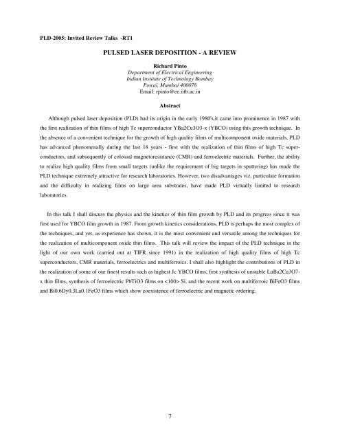
PLD-2005: Invited Review Talks -RT1<br />
<strong>PULSED</strong> <strong>LASER</strong> <strong>DEPOSITION</strong> - A <strong>REVIEW</strong><br />
Richard Pinto<br />
Department of Electrical Engineering<br />
Indian Institute of Technology Bombay<br />
Powai, Mumbai 400076<br />
Email: rpinto@ee.iitb.ac.in<br />
Abstract<br />
Although pulsed laser deposition (PLD) had its origin in the early 1980's,it came into prominence in 1987 with<br />
the first realization of thin films of high Tc superconductor YBa2Cu3O3-x (YBCO) using this growth technique. In<br />
the absence of a convenient technique for the growth of high quality films of multicomponent oxide materials, PLD<br />
has advanced phenomenally during the last 18 years - first with the realization of thin films of high Tc super-<br />
conductors, and subsequently of colossal magnetoresistance (CMR) and ferroelectric materials. Further, the ability<br />
to realize high quality films from small targets (unlike the requirement of big targets in sputtering) has made the<br />
PLD technique extremely attractive for research laboratories. However, two disadvantages viz, particulate formation<br />
and the difficulty in realizing films on large area substrates, have made PLD virtually limited to research<br />
laboratories.<br />
In this talk I shall discuss the physics and the kinetics of thin film growth by PLD and its progress since it was<br />
first used for YBCO film growth in 1987. From growth kinetics considerations, PLD is perhaps the most complex of<br />
the techniques, and yet, as experience has shown, it is the most convenient and versatile among the techniques for<br />
the realization of multicomponent oxide thin films. This talk will review the impact of the PLD technique in the<br />
light of our own work (carried out at TIFR since 1991) in the realization of high quality films of high Tc<br />
superconductors, CMR materials, ferroelectrics and multiferroics. I shall also highlight the contributions of PLD in<br />
the realization of some of our finest results such as highest Jc YBCO films, first synthesis of unstable LuBa2Cu3O7-<br />
x thin films, synthesis of ferroelectric PbTiO3 films on Si, and the recent work on multiferroic BiFeO3 films<br />
and Bi0.6Dy0.3La0.1FeO3 films which show coexistence of ferroelectric and magnetic ordering.<br />
7
PLD-2005: Invited Review Talks -RT2<br />
Pulsed Laser Deposition for MOS Gate Dielectric Films<br />
Nandita DasGupta*, RavneetSingh, Roy Paily, Amitava DasGupta, Pankaj Misra<br />
and Lalit M. Kukreja<br />
* Department of Electrical Engg., IIT Madras, Chennai 600 036<br />
*Email: nand@ee.iitm.ernet.in<br />
Abstract<br />
Downscaling of device dimensions is essential for the development of new generation Ultra Large Scale<br />
Integrated Circuits (ULSI) based on Complementary Metal Oxide Semiconductor Field Effect Transistors<br />
(CMOSFET). Silicon dioxide(SiO2) has been used for more than 35 years as the primary gate-dielectric material in<br />
MOSFETs because of its excellent properties. However, current technology requires that the thickness of the gate<br />
dielectric be reduced to only a few monolayers of SiO2. Further thinning of SiO2 poses a serious challenge because<br />
of large gate leakage current 1,2 . In order to overcome the large gate leakage current mainly due to direct tunneling,<br />
introduction of new gate dielectric materials with high dielectric constant (high-k) is being seriously investigated 3-6 .<br />
Using high-k dielectric, the physical thickness of the dielectric layer can be kept large, thereby reducing the gate<br />
leakage current, while maintaining the same value of capacitance. There are many materials systems under<br />
consideration which have potential to replace SiO2 as the gate dielectric material. Of the various high-k dielectric<br />
materials, TiO2, Ta2O5, ZrO2, and HfO2 have generated a lot of interest due to their high dielectric constant and<br />
adequate barrier height. Various deposition techniques have been employed to deposit these materials.<br />
We have recently reported for the first time, the use of Pulsed Laser Deposition (PLD) technique for the<br />
deposition of TiO2 as gate dielectric in Metal-TiO2-SiO2-Si (MTOS) capacitors with TiO2-SiO2 stacked gate<br />
dielectric 7, 8 . One interesting observation in our work is that by optimizing the conditions during PLD, one can<br />
actually achieve an increase in capacitance of the MTOS capacitor by introducing the additional TiO2 layer over<br />
SiO2. The reduction in the Effective Dielectric Thickness (EDT), defined as oxA/Cmax, where ox is the dielectric<br />
constant of SiO2, A is the device area and Cmax is the accumulation capacitance is due to an intermixing of the TiO2<br />
layer with the underlying SiO2.<br />
Previous reports indicate that it has been rarely possible to obtain an EDT < 2 nm using TiO2 thin films 3 .<br />
We have however, been able to combine the reduction in the EDT with a reduction in the gate leakage current by<br />
controlling the intermixing of the TiO2 and SiO2 layers during PLD. To achieve this, we have used a dual-<br />
temperature deposition process, where a buffer layer of TiO2 has been deposited at low temperature followed by<br />
deposition of TiO2 at higher temperature and annealing resulting in an EDT < 2 nm as well as low leakage 9 . As<br />
shown in Fig.1, the leakage current densities (J) of the MTOS devices are at least five orders of magnitude smaller<br />
than that in the simulated MOS devices with comparable EDT. It can also be seen that while for the simulated MOS<br />
devices, the leakage current changes by five orders of magnitude as the gate oxide thickness reduces from 2.5 to 1.5<br />
nm, for the MTOS devices, the leakage current changes only by one order of magnitude for a similar reduction in<br />
EDT from 2.4nm to 1.6 nm. This is because, the leakage current is determined by the physical thickness and the<br />
8
physical thickness of the gate dielectric for these MTOS devices is comparable, even though their EDT values are<br />
different.<br />
Even though the results of MTOS devices have been encouraging, for preserving the interface quality, a<br />
very thin layer of SiO2 has to be grown on silicon prior to PLD of TiO2. Conventional thermal oxidation has been<br />
used for growing this thin SiO2 layer. It would, however, be extremely useful if both PLD of TiO2 and the growth of<br />
SiO2 can be carried out using the same process. To this end, we have developed a novel technique of Laser Induced<br />
Oxidation (LIO) to grow ultrthin SiO2 ( 10 MV/cm<br />
for LIO3), signifying the excellent quality of the Laser-induced oxide.<br />
References<br />
1) S. -H. Lo, D. A. Buchanan, Y. Taur, and W. Wang, IEEE ED Lett. 18 (1997) 209.<br />
2) D.A.Buchanan and S.H.Lo, Microelectronic Engineering 36 (1997) 13.<br />
3) Masaru Kadoshima, Masahiko Hiratani, Yasuhiro Shimamoto, Hiroshi Miki, Shinichiro Kimura,<br />
Toshihide Nabatame, Thin Solid Films, 424 (2003) 224.<br />
4) Jing-Chi Yu, B.C. Lai, J.Y. Lee, IEEE ED Lett. 21 (2000) 537.<br />
5) W. K. Chim, T. H. Ng, B. H. Koh, W. K. Choi, J. X. Zheng, C. H. Tung, A.Y. Du, J. Appl. Phys. 93<br />
(2003) 4788.<br />
6) Q. Fang, J.-Y. Zhang, Z. M. Wang , J. X. Wu, B. J. O’Sullivan, P. K. Hurley, T. L. Leedham, H. Davies,<br />
M. A. Audier, C. Jimenez, J.-P. Senateur, Ion W. Boyed, Thin Solid Films, 428 (2003) 263.<br />
7) Roy Paily, Amitava Das Gupta, Nandita Das Gupta, Pijush Bhattacharya, Pankaj Mishra, Tapas<br />
Ganguli,Lalit M. Kukreja, A.K. Balamurugan, S.Rajgopalan, A.K.Tyagi, Applied Surface Science 187<br />
(2002) 297.<br />
8) Roy Paily, Amitava DasGupta, Nandita DasGupta, Pankaj Misra and Lalit M.Kukreja, Thin Solid Films,<br />
Vol. 462-463C pp. 57-62, 2004.<br />
9) Ravneet Singh, Roy Paily, Amitava DasGupta, Nandita DasGupta, Pankaj Misra and Lalit M. Kukreja,<br />
Semiconductor Sc. & Technol., Vol.20, No.1, Jan.2005<br />
10) Ravneet Singh, Roy Paily, Amitava DasGupta, Nandita DasGupta, Pankaj Misra and Lalit M. Kukreja,<br />
Electronics Letters, Vol. 40, No.25, pp.1606-08, Dec. 9, 2004<br />
9
Leakage Current Density (A/cm 2<br />
)<br />
100<br />
1<br />
0.01<br />
1E -4<br />
1E -6<br />
E D T = 15A 0 M T O S B1 (E D T =1.7 n m )<br />
M T O S B2 (E D T =1.6 n m )<br />
M T O S B3 (E D T =2.4 n m )<br />
,S im u lated for S iO 2<br />
E D T = 20A 0 ,S im u lated for S iO 2<br />
E D T = 25A<br />
0.0 0.5 1.0 1.5 2.0 2.5 3.0<br />
0 ,S im u lated for S iO 2<br />
Fig.1: Comparison of J-V characteristics of MTOS devices with those of simulated MOS devices with comparable<br />
Leakage Current density (A/cm 2 )<br />
10 0<br />
10 -2<br />
10 -4<br />
10 -6<br />
10 -8<br />
|G ate V o ltag e| (V )<br />
EDT<br />
LIO1, 30s, t =5.1 nm<br />
ox<br />
LIO2, 60s,t =5.5 nm<br />
ox<br />
LIO3, 90s,t =3.9 nm<br />
ox<br />
MOS, t =3.9 nm<br />
ox<br />
-0 -2 -4 -6 -8<br />
Gate Voltage (V)<br />
Fig. 2. J-V characteristics of LIO1, LIO2, LIO3 and MOS capacitors.<br />
10
PLD-2005: Invited Review Talks -RT3<br />
Some studies on oxide and nitride thin films grown by pulsed laser deposition<br />
K. P. Adhi<br />
Centre for Advanced Studies in Materials Science and Solid State Physics, Department of Physics, University of<br />
Pune, Pune - 411 007, India<br />
Email: kpa@physics.unipune.ernet.in<br />
Abstract<br />
Pulsed laser deposition (PLD) has emerged as a relatively simple and highly versatile technique for the<br />
growth of thin films of variety of materials 1 . Deposition of epitaxial, stoichiometric thin films of simple materials 2 or<br />
multi-element complex compounds on suitable substrates 3 , multilayers 4 , nano-particles 5 and nano-structures etc. are<br />
some of the achievements which reflect the versatility of this technique. We present a review of our recent research<br />
work on the growth, characterization and analysis of oxide and nitride thin films. The presentation is focused on the<br />
following oxide materials; a) Fe3O4 b) La0.7Ca0.3MnO3 and c) bi-layers of Fe3O4 / La0.7Ca0.3MnO3. Some of the<br />
issues which will be addressed are growth of highly oriented thin films of the above mentioned materials,<br />
modifications in their structural and electrical properties due to external processing like swift heavy ion irradiation,<br />
ionimplantation etc . In case of nitrides, the growth of highly oriented AlN thin films on sapphire, its<br />
characterization along with brief mention of InN and GaN thin films will be discussed. Generation of nanoparticles<br />
of Fe3O4 / FeO by PLD will also be discussed.<br />
References:<br />
1. “Pulsed Laser Deposition of Thin Films” edited by D. B. Chrisey and G. K. Hubler, A Wiley –<br />
interscience publication, New York (1994)<br />
2. S. M. Jejurikar, A. G. Banpurkar, A. V. Limaye, S. K. Date, S.I. Patil, K. P. Adhi, P. Misra, L. M.<br />
Kukreja, Ravi Bathe, communicated to J. Appl. Phys.<br />
3. Ravi Bathe, K. P. Adhi, S. I. Patil, G. Marest, B. Hannoyer, S. B. Ogale, Appl. Phys. Lett. 76, 2104<br />
(2000)<br />
4. S. N. Sadakale, R. J. Choudhary, M. S. Sahasrabudhe, A. G. Banpurkar, K. P. Adhi, S. I. Patil, S. K.<br />
Date, J. Mag. Mag. Mater. 286, 450 (2005)<br />
5. S. R. Shinde, A. G. Banpurkar, K. P. Adhi, A. V. Limaye, S. B. Ogale, S. K. Date, G. Marest,<br />
Mod. Phys. Lett. B 10, 1517 (1996)<br />
11
PLD-2005: Invited Review Talks -RT4<br />
Micro- nano patterning in a single step via selective laser ablation<br />
Alika Khare<br />
Department of Physics<br />
Indian Institute of Technology Guwahati, Guwahati 781039<br />
Email: alika@iitg.ernet.in<br />
Abstract:<br />
Optoelectronics devices viz; grating couplers, micro mirrors, tiny arrays of lasers and photonic band gap materials<br />
require ordered arrays of dimensions ranging from nanometers to tens of microns. These tiny arrays of materials can<br />
be produced by modifying the surface morphology of thin films by illuminating it with interference pattern formed<br />
by interference of multiple beams from a pulsed high power laser. The interference patterns are periodic, so the<br />
materials of the thin film exposed to the maximum intensity (bright fringe) gets ablated leaving the area of minimum<br />
intensity (dark fringe) unaffected. Width and periodicity of the ablated region depends on the intensity distribution<br />
with in the bright fringe and wavelength of laser respectively. For writing the grating like structure, a simple two<br />
beam Michelson interferometer can be used. For patterning in the format of arrays of dots of the material in square<br />
or rectangular geometry, two Michelson interferometers in tandem can be used. For hexagonal geometry,<br />
interference pattern from the eight beams coming out of system of three interferometers in tandem can be used. This<br />
is a direct lithographic technique without requiring any mask. The whole writing can be performed in a single step.<br />
The technique of selective laser ablation via high power interferometer can be applied to the thin film of any<br />
material. The material ablated from the region of bright fringe results into the formation of arrays of cold atomic<br />
beam having relatively low divergence. The application of these atomic beams for nano lithography via dipole force<br />
shall also be presented in the talk.<br />
12
PLD-2005: Invited Review Talks -RT5<br />
Electron doped rare-earth manganites: A current scenario<br />
Pratap Raychaudhuri<br />
Department of Condensed Matter Physics and Materials Science,Tata Institute of Fundamental Research,<br />
Homi Bhabha Rd., Colaba, Mumbai-400005.<br />
Email: pratap@tifr.res.in<br />
Abstract<br />
Electron doped rare-earth manganites of the form (R1-xAxMnO3, where R=rare-earth, A=tetravalent cation) fall the<br />
class of compounds which have so far been synthesized in single phase only through the pulsed laser deposition<br />
technique. The most well known member of this series La1-xCexMnO3 was first synthesized in TIFR in 1999 and<br />
showed the magnetic and transport properties to be very similar to its hole-doped counterpart [1]. Subsequently,<br />
studies using a variety of techniques such X-ray absorption spectroscopy[2], Tunneling conductance in artificial<br />
tunnel junctions with La0.7Ce0.3MnO3[3] established this material to be a minority spin carrier ferromagnet where the<br />
manganese is in a mixture of Mn 3+ and Mn 2+ valence states. These findings led to an active search for other electron<br />
doped manganites among many groups in recent years and several new candidates have been reported. This also led<br />
to the synthesis electron doped cobaltates [4] using Pulsed Laser deposition using the same principle as that used for<br />
the manganites.<br />
In this talk I will present an overview of the current status on the investigations on electron doped manganites and<br />
related systems.<br />
References:<br />
1. P. Raychaudhuri, S. Mukherjee, A. K. Nigam, J. John, U. D. Vaisnav, R. Pinto, and P. Mandal, J. Appl. Phys. 86,<br />
5718 (1999).<br />
2. C. Mitra, Z. Hu, P. Raychaudhuri, S. Wirth, S. I. Csiszar, H. H. Hsieh, H.-J. Lin, C. T. Chen, and L. H. Tjeng,<br />
Phys. Rev. B 67, 092404 (2003).<br />
3. C. Mitra, P. Raychaudhuri, K. Dörr, K.-H. Müller, L. Schultz, P. M. Oppeneer, and S. Wirth, Phys. Rev. Lett. 90,<br />
017202 (2003).<br />
4. D. Fuchs, P. Schweiss, P. Adelmann, T. Schwarz, and R. Schneider, Phys. Rev. B 72, 014466 (2005).<br />
13
PLD-2005: Invited Review Talks -RT6<br />
Exploring Novel Magneto-resistive and Transport Properties in Pulsed Laser Deposited<br />
Manganite Thin Films<br />
D. G. Kuberkar<br />
Department of Physics, Saurashtra University<br />
Rajkot 360 005<br />
Email: dgk@icenet.net<br />
Abstract<br />
For the past one decade, a significant upsurge in research on ABO3 type manganites is mainly attributed to<br />
the application potential of colossal magnetoresistance (CMR) property exhibited by these compounds. Though the<br />
realization of applications of these oxides still remains an open question, the compounds continue to attract the<br />
scientific community due to the rich physics evolving as a result of spin, charge and orbital degrees of freedom.<br />
During this talk , the results of our recent studies, both on tailoring these materials to obtain enhanced magneto-<br />
resistance (MR) for applications point of view and on some exotic transport properties at low temperatures, will be<br />
presented.<br />
The fabrication of manganite thin films is essential because films could find applications and also help in<br />
studying the clean physics in the absence of dominant grain boundary effects. Pulsed Laser Deposition (PLD) is an<br />
efficient tool to fabricate the high quality epitaxial thin films of manganite oxides and to grow multilayer structures,<br />
which could be evaluated for possible applications. We show that, by controlling the growth parameters, phase-<br />
separation may be induced to obtain unusually high MR in A-site disordered (La,Pr)0.7A0.3MnO3 (A=Sr, Ba)<br />
manganites. Swift Heavy Ion (SHI) irradiation is another technique employed to induce the controlled defects in the<br />
thin films. Our detailed investigations on the SHI irradiation induced modifications in the structural and transport<br />
properties of thin films of varying thicknesses reveal that, SHI effect on transport and MR properties gets more<br />
pronounced with increasing thickness of the films. In addition to MR properties, we also observed some low<br />
temperature transport anomalies arising due to structural disorder in Ba-based compounds. Present talk will<br />
highlight some interesting findings of our recent investigations on CMR manganites in the context of all the above-<br />
mentioned aspects.<br />
14
PLD-2005: Invited Review Talks -RT8<br />
Photonic and Spin-photonic Semiconductor Nanostructures Grown by Pulsed<br />
Laser Deposition<br />
Lalit. M. Kukreja<br />
Thin Film Laboratory<br />
Centre for Advanced Technology, Indore – 452 013, INDIA<br />
Email: kukreja@cat.ernet.in<br />
Abstract<br />
This paper reviews our recent research on structural and optical properties of quantum dots of Si, quantum wells and<br />
dots of ZnO and thin films of Mn and Co doped ZnO grown by Pulsed Laser Deposition (PLD). A particulate free<br />
multilayer structure of Al2O3 capped Si quantum dots of different mean sizes grown by an off-axis deposition<br />
scheme showed variable band-gap in photoabsorption spectra in line with the putative quantum confinement effects.<br />
Room temperature photoluminescence from Si quantum dots grown for different times showed features without any<br />
apparent size dependent spectral shift which, albeit has earlier been explained by others originating from the defect<br />
levels at the interface of Si and SiO2 shells surrounding the nanoparticles but still have certain mysteries attached.<br />
On the contrary ZnO quantum dots, also capped with Al2O3 in multilayer structure, showed size dependent band-gap<br />
shifting in photoabsorption spectra in the range from about 3.3 to 4.5 eV when the mean dot radii varied from about<br />
4 to 2 nm. High quality ZnO/MgZnO quantum wells grown by an in-house developed methodology of buffer<br />
assisted growth showed a monotonic blue shift of the band-gap from about 3.35 to 3.75 eV both in photoabsorption<br />
and photoluminescence when the well thickness decreased from about 5 to 1 nm. These quantum structures are<br />
expected to play vital role for the development of future photonic devices.<br />
An equally exciting area of spint-photonics is currently on the horizon. Diluted magnetic semiconductors (DMS) of<br />
Mn and Co doped ZnO are being studied extensively to explore if those could be potentially useful for spin-<br />
optoelectronic devices. We have synthesized bulk and thin films of these materials with different dopand<br />
concentrations. The PLD grown films of a few hundred nm thickness are found to have high crystalline quality and a<br />
homogenous wurtzite phase with monotonic increase in the band-gap of the resulting alloy with increasing<br />
concentration of Mn and Co in their corresponding films. We have also observed broad mid gap absorption in the<br />
photoabsorption spectra of both Mn and Co doped ZnO films. Low temperature photoluminescence of MnZnO alloy<br />
films with different concentrations of Mn, showed efficient band edge emission with additional features, which<br />
might originate from the clusters of MnO or MnO2 due to the crystal field transitions of Mn 2+ ions. Further<br />
investigations on the optical and magnetic characteristics of these spin-photonic semiconductor films are under way.<br />
15
PLD-2005: Invited Review Talks -RT9<br />
The Evolution of PLD: From High Tc Superconductors to Spintronics<br />
M.S. Ramachandra Rao<br />
Department of Physics and Materials Science Research Centre<br />
Indian Institute of Technology (IIT) Madras, Chennai – 600 036<br />
Email: msrrao@iitm.ac.in<br />
Abstract<br />
Pulsed laser deposition (PLD) has proved to be one of the most versatile techniques to realize high quality thin films<br />
of not only oxide materials but also a variety of solid state materials. With a modest beginning as a not-so-popular<br />
technique in the sixties [1], it has come to stay, with the advent of high Tc superconductivity [2], as the most<br />
profoundly used deposition technique in the past two decades. Applications of the technique include the fabrication<br />
of high current density superconducting films, high quality ultrathin gate-dielectric layers, biocompatibility for<br />
medical applications, hardware resistant coatings, diamond coatings, production of carbon nanotubes, epitaxial<br />
transparent conducting oxide (TCO) films, hydrogen and other gas sensors, films with nanostructured and self-<br />
assembled arrays, magnetic multilayers and heterostructures and GMR/CMR based magnetic tunnel junctions<br />
(MTJs) and diluted magnetic semiconductors (DMS) for spintronic applications. PLD by virtue of its simplicity<br />
scores over other techniques in terms of, i). stoichiometric production of films of multicomponent systems, ii).<br />
relatively high deposition rate (~ 100 Å/min @ moderate laser fulences), iii). use of laser as an external energy<br />
source to avoid contamination and iv). facilitation of multilayer film deposition without breaking vacuum. It is the<br />
fastest evaporation (occurring in time scales of few nanoseconds) technique in which the laser produced plasma<br />
(plume) expands rapidly away from the target surface with particle velocities typically in the range 10 6 cms -1 and<br />
kinetic energies of the emanating species ~ 80 eV as compared to 2-10 eV in the case of filament-based-thermal-<br />
evaporation. The fundamental aspect of plume generation during the laser-target interaction process is still a matter<br />
of intense research.<br />
In this review talk, I will give a brief glimpse of the technique, highlighting salient results pertaining to two research<br />
areas, HTSC and manganites, in which PLD was extensively used. I will also highlight the recent trends in PLD to<br />
realize nanostructured and self assembled arrays of some oxide systems. I will then talk about the emerging field of<br />
spintronics, in the context of oxide electronic materials, which is an emerging field for future spin electronic and<br />
quantum computational devices [3,4].<br />
References:<br />
[1]. H.M. Smith and A.F. Turner. Appl.Opt. 4 (1965) 147.<br />
[2]. D. Dijkamp et al. Appl. Phys. Lett. 81 (1987) 619.<br />
[3]. H. Ohno et al. Appl. Phys. Lett. 69 (1996) 363.<br />
[4]. D.D. Awschalom and J.M. Kikkawa. Physics Today, 52 (1999) 33.<br />
16
PLD-2005: Invited Review Talks -RT10<br />
Pulsed laser Deposition of Oxides on polymer substrates for Optoelectronic Applications<br />
M.K.Jayaraj*, R.Manoj, R.S.Ajimsha., R.Reshmi<br />
Optoelectronics Device laboratory, Department of Physics,<br />
Cochin University of Science and Technology, Cochin – 682 022.<br />
*Email: mkj@cusat.ac.in<br />
Abstract<br />
Wide band gap oxide films are important component in Optoelectronic devices. Thin films deposited on organic<br />
substrates can be used in plastic liquid crystal displays, Transparent electromagnetic shielding material, flexible<br />
electro optical devices, solar cells, thin film electro luminescent devices etc. Due to the poor thermal endurance of<br />
organic substrates films should be deposited at low substrate temperature. Wide variety of methods, such as<br />
sputtering, chemical deposition, and sol gel method are commonly used to deposited oxide films. A high<br />
temperature post deposition heat treatment is required to produce crystalline film which is not possible with these<br />
organic substrates. In this paper we review the work that we have carried out on ZnO and ZnGa2O4 thin films grown<br />
by pulsed laser deposition. Highly oriented ZnO films and polycrystalline ZnGa2O4 films were grown by PLD on<br />
various substrates like quartz, silicon and organic polymer substrates (kapton HPP-ST polyamide). By controlling<br />
the deposition parameters like substrate to target distance, oxygen partial pressure and laser fluence crystalline films<br />
were grown on organic substrates at a substrates temperature of 25 0 C. The films were characterized by studying the<br />
optical, electrical and structural properties. The Photoluminescent emission and excitation of the PLD grown oxide<br />
films on organic substrates were also discussed.<br />
17
PLD-2005: Invited Review Talks -RT11<br />
Synthesis of Epitaxial AlN thin films by pulsed laser deposition<br />
Ravi Bathe<br />
International Advanced Research Center for Powder Metallurgy and New Materials, Balapur PO, Hyderabad 500<br />
-005, India<br />
Email: ravi@arci.res.in<br />
Abstract<br />
AlN has a large potential to become important in a wide range of applications. Its wide (6.2 eV), direct, band gap<br />
combined with well matched structural and thermal properties to SiC makes it an ideal candidate for future<br />
MESFET and MISFET SiC-transistors as well as high temperature and high power electronics applications. Due to a<br />
small lattice mismatch to SiC (~ 1%), epitaxally grown AlN thin films seem to be promising candidates for<br />
dielectric applications and ion-implantation anneal cap. We have investigated the epitaxy, interfaces, surfaces and<br />
defects in epitaxial AlN thin films grown on SiC by pulsed laser deposition. The crystalline structure and surface<br />
morphology of the epitaxially grown AlN thin films on SiC (0001) substrates have been studied using x-ray<br />
diffraction ( and scans) and atomic force microscopy, respectively. The defect density and analysis have<br />
been studied by using Rutherford backscattering spectrometry, ion channeling technique and transmission electron<br />
microscopy. The films were grown at various substrate temperatures ranging from 500 to 1100 0 C. X-ray diffraction<br />
measurements show highly oriented AlN films above growth temperature of 750 0 C, and single crystalline nature<br />
above 800 0 C. The films grown in the temperature range of 950 0 C to 1000 0 C have been found to be highly strained,<br />
where as the films grown above 1000 0 C were found to be relaxed after crack propagation along the crystallographic<br />
axes. We found that during initial stages, growth of a 20 nm thick AlN low-temperature buffer layer is critical for<br />
obtaining crack free, smooth, high-quality epitaxial films. By controlling the initial stages of growth in a two-step<br />
deposition process, it is shown that high quality epitaxial layers on SiC can be obtained with low intrinsic stresses,<br />
good surface morphology, and higher electrical break-dawn strength. The significance of these results towards<br />
development of high temperature-high-power electronics is also discussed.<br />
18
PLD-2005: Invited Talks –IT1<br />
Nanocrystalline Films of Gadolinia Doped Ceria Prepared<br />
by Pulsed Laser Ablation<br />
P. Kuppusami<br />
Physical Metallurgy Section<br />
Indira Gandhi Centre for Atomic Research<br />
Kalpkkam-603 102, Tamilnadu<br />
Email : pk@igcar.ernet.in<br />
Abstract<br />
Nanocrystalline oxides are displaying electrical properties which appear to be unique and may<br />
lead to applications that are not attainable by conventional microcrystalline oxides. As a result, it is very<br />
important to understand the relationship between microstructure and electrical properties as well as to be<br />
able to control the microstructure in the nanocrystalline range. New phenomena observed in nanocrystalline<br />
oxides are related to the increasing grain boundary volume and in the change of stoichiometry, which may<br />
lead to enhance the reaction kinetics and electrical conductivity.<br />
Synthesis of nanocrystalline materials has been carried out most frequently by assembling pre-<br />
generated small clusters by means of consolidation and sintering. A variety of cluster generation methods<br />
such as sol-gel technique, laser ablation, sputter deposition and precursor spin coating technique have been<br />
reported. In comparison with methods such as consolidation and sintering of clusters, the latter techniques<br />
are the low temperature methods, which have significant advantage. Pulsed laser ablation is a unique<br />
technique where the deposition is not only carried out at low substrate temperatures, but also the<br />
stoichiometry of the target is retained in the ablated films. The technique is also capable of depositing<br />
metastable materials that are difficult to synthesize in bulk form by other deposition methods. These<br />
features enable to control the grain size and to obtain stable microstructure and make it possible to study<br />
microstructure-property relationships.<br />
In the present work, microstructure of thin films of gadolinia doped ceria (GDC) prepared by<br />
pulsed laser ablation is investigated. The growth characteristics of the films as a function of substrate<br />
temperature, oxygen partial pressure and laser energy are investigated using the techniques of x-ray<br />
diffraction and electron microscopy. The influence of growth induced defects on the ionic conductivity of<br />
the doped ceria will be highlighted.<br />
19
PLD-2005: Invited Talks –IT2<br />
Pulsed Laser Ablation grown Relaxor based bilayers, multilayers and heterostructures for<br />
multiferroic applications<br />
R. Ranjith ∗ and S. B. Krupanidhi<br />
Materials Research Centre, Indian Institute of Science, Bangalore-560 012<br />
*Email: ranjith@mrc.iisc.ernet.in<br />
Abstract<br />
Ferroelectric heterostructure like super lattices and multilayers have shown superior properties and have been the interest<br />
of study in the recent years 1 . The interfaces present and the size of the individual layers present play a crucial role in<br />
these heterostructure. In this work multilayer of PMN-PT with varying composition of PT across the film was fabricated<br />
using pulsed laser ablation technique. Samples with varying individual layer thickness were fabricated to study the size<br />
dependent behavior of these multi-layer thin films. Multilayer films with individual layer thickness of 10,20,30,50,70<br />
and 100nm were fabricated. Polarization studies were carried out on these films with a multilayer structure. A field<br />
driven antiferroelectric to ferroelectric transition was observed in the films of individual layer thickness greater than<br />
50nm. The dominance of the interaction between the adjacent layers via the interfacial coupling dominates at low fields<br />
to stabilize the antiferroelectric coupling and the dominance of the external field coupling with the individual layers<br />
stabilizes the ferroelectric behavior of these films. Figure 1. shows the field driven ferroelectric polarization. Films with<br />
low individual layer thickness exhibited ferroelectric behavior and on increase of individual layer thickness they<br />
exhibited an anti-ferroelectric behavior and on further increase of thickness they showed a weak anti-ferroelectric<br />
followed by a weak ferroelectric behavior on further increase of thickness. This phenomenon is attributed to the long-<br />
range coupling observed in these materials 2 , which gives an averaged property, and on increase of thickness they behave<br />
as individual materials put together. Figure 2. shows the size induced antiferroelectric polarization behavior.<br />
Artificially grown superlattice structures have been an interest of study due to their striking properties both in<br />
technological point of view and fundamental physics aspects. In this work superlattices based on Pb(Mg1/3Nb2/3)O3<br />
(PMN) – PbTiO3 (PT) were deposited through pulsed laser ablation deposition with different periodicities<br />
(10,20,30,40,50,60 and 70nm) for a constant total thickness of the film. The presence of superlattice reflections in the X-<br />
ray diffraction pattern clearly shows the superlattice behavior of the films. Polarization hysteresis and the Capacitance –<br />
Voltage characteristics of these films shows a clear size dependent Ferro and Antiferro characteristics. Presence of long<br />
range coupling in superlattices with lower periodicities (
Figure 1. Field driven Antiferroelectric to Ferroelectric transition.<br />
Figure 2. Size driven Antiferroelectric characteristics.<br />
Major References<br />
1. G.Rijnders and Dave.H.A.Blank, Nature, 433, 369 (2003).<br />
21<br />
Capacitance (nF)<br />
2.3<br />
2.2<br />
2.1<br />
2.0<br />
27 o C<br />
120 o C<br />
10kHz<br />
d=40nm<br />
-10 -5 0 5 10<br />
Voltage (Volts)<br />
Figure 3. CV characteristics of a PT-<br />
PMN superlattice<br />
2. H.M.Christen,E.D.Specht, D.P.Norton, M,F,Chisholm and L.A.Boatner, Appl.Phys.Lett., 72,2535,(1998).<br />
3. Jian Shen and Yu-qiang Ma, Phys. Rev.B, 61(21), 14279, (2000).
PLD-2005: Invited Talks –IT3<br />
Optical Quantum Confinement Effects in ZnO/MgZnO Multiple Quantum Wells Grown<br />
by Pulsed Laser Deposition<br />
P. Misra * , T. K. Sharma, S. Porwal and L. M. Kukreja<br />
Thin Film Laboratory, Centre for Advanced Technology, Indore 452 013<br />
*Email: pmisra@cat.ernet.in<br />
Abstract<br />
Current worldwide interest in ZnO as a semiconductor to evolve futuristic optoelectronic, spintronic and other<br />
devices has spurred rigorous research on its quantum structures [1]. We have grown ZnO Multiple Quantum Well<br />
(MQW) structures on (0001) Sapphire substrates by Pulsed Laser Deposition using a third harmonic of Q-switched<br />
Nd: YAG laser. A 10 layer MQW structure was grown with 8 nm thick ternary alloy Mg0.16Zn0.84O layer with a band<br />
gap of ~ 4.1 eV as a barrier and the active layer of ZnO had variable thickness in the range of 5 – 1 nm . Prior to the<br />
growth of MQWs a 50 nm thick ZnO buffer layer was grown at 750°C, which provided a highly crystalline, smooth<br />
and oxygen terminated template for subsequent growth of nanostructures at a lower temperature [2] of 600°C. This<br />
low temperature growth ensured chemically sharp interfaces while the high crystalline quality was facilitated by the<br />
high temperature grown buffer layer. Room temperature absorption spectra of MQW structures showed two<br />
prominent peaks due to excitonic transitions with in the well and barrier layers. The ZnO absorption edge shifted<br />
monotonically towards blue with decreasing well layer thickness up to 1 nm due to putative size dependent quantum<br />
confinement effects. Photoluminescence (PL) measurements carried out on all the quantum wells at 10K and room<br />
temperature using a He-Cd laser to further strengthen our observation. Room temperature PL in the UV spectral<br />
range was observed for the MQW samples up to 2 nm of well thickness bellow which the PL signals was too weak<br />
to be detected by our PL setup. It is worth mentioning here that the minimum thickness of ZnO QW grown on<br />
sapphire by us which showed quantum confinement effect is 1 nm, which is better than reported by Ohtomo et al<br />
which was 1.7 nm. Ohtomo et al also could not observe room temperature PL observed by us. All the samples<br />
showed strong PL at 10K due to excitonic recombination in ZnO QW. PL spectra of these samples showed a clear<br />
blue shift in the ZnO band edge from ~ 3.4 to ~ 3.7 eV with decreasing well layer thickness. The FWHM of PL<br />
peak was found to increase monotonically with decreasing well layer thickness probably due to fluctuation in the<br />
well layer thickness which is more pronounced at lower thickness of QW. The band gaps obtained from the<br />
experimental PL data at 10K were compared with the theoretically calculated values by using one dimensional<br />
square well potential approximation and a band offset ratio, Ec: Ev of 9:1. Both were found to be in good<br />
agreement. Further experiments are underway to investigate the interface quality and to measure the accurate<br />
thickness of the quantum wells and to include size dependent variation of the excitonic binding energy in theoretical<br />
calculations.<br />
References:<br />
1. A. Tsukazaki, A. Ohtomo, T. Onuma, M. Ohtani, T. Makino, M. Sumiya, K. Ohtani, S. F. Chichibu, S. Fuke,<br />
Y. Segawa, H. Ohno, H. Koinuma, M. Kawasaki, Nature Materials, 4, 42-46, (2005)<br />
2. P. Misra and L.M. Kukreja, Thin Solid Films, 485, Issues 1-2, 42-46 (2005)<br />
3. A. Ohtomo, M. Kawasaki, I. Ohkubo, H. Koinuma, T. Yasuda, Y. Segawa, Appl. Phys. Lett. 75, 980 (1999)<br />
22
PLD-2005: Invited Talks –IT4<br />
High-pulse energy excimer lasers for precise material ablation<br />
Burkhard Fechner and Ralph Delmdahl<br />
Coherent Lambda Physik GmbH, Hans-Boeckler-Str. 12, D-37079 Goettingen, Germany<br />
Email: bfechner@lambdaphysik.com<br />
Abstract<br />
Pulsed excimer lasers are the strongest and most efficient laser sources in the ultraviolet spectral<br />
region. Record short wavelengths from 351 nm down to 157 nm as well as record high 1200 mJ<br />
pulse energy as available for the 248 nm excimer lasers are commercially provided for numerous<br />
laser material ablation approaches. Virtually no material is able to withstand the high photon<br />
energies ranging from 3.5 to 7.9 eV emitted by excimer lasers. As a result of the irradiation of<br />
material with high energy excimer laser photons at sufficient fluence immediate bond breaking<br />
due to electronic excitation is induced. In combination with short-term laser material interaction<br />
of only 10 to 30 ns excimer pulse duration, material ablation proceeds via fast vaporization and<br />
consecutive ejection of material with only negligible dissipation of heat transfer to the<br />
surrounding zone. The effect is an inherently precise and clean ablation quality. Latest<br />
developments in excimer laser technology with particular respect to pulsed laser deposition as<br />
well as applications will be discussed.<br />
23
PLD-2005: Invited Talks –IT5<br />
Pulsed laser ablation at the liquid solid interface<br />
D. M. Phase<br />
UGC-DAE Consortium for Scientific Research, Khandwa Road, University Campus, Indore-452 017.<br />
Email: dmphase@csr.ernet.in<br />
Abstract<br />
Pulsed laser ablation (PLA) is a well-known method to produce thin films by ablating material from a solid<br />
target of known composition 1 . PLA usually occurs in vacuum or in a background of inert gas or reactive gas. Very<br />
recently, PLA at the liquid/solid interface, a new variation of PLA has been reported by Simakin et al 2 . Earlier the<br />
concept of pulsed laser induced liquid solid interfacial reaction was used to prepare surface alloys and compounds,<br />
which are in the metastable states 3 . Now this novel LP-PLA technique which is also based on same concept has been<br />
used to produce a variety of materials, including diamond like carbon films, nanocrystals of carbon nitride and nano<br />
meter size particles of Ti, Ag, Au, Si and TiC. This technique shows a great potential as route to novel nano-<br />
crystalline materials. However this technique is still in its infancy with much of the parameter space yet to be<br />
investigated. In this talk a basic concept of pulsed laser reaction at liquid/solid interface will be described along with<br />
some examples of synthesis of metastable compounds. This will be followed by a detailed description of LP-PLA<br />
technique with examples of synthesis of nano-structures and nano-particles.<br />
References:<br />
1. R.K.Singh and J.Narayan, Phys.Rev.B 41 (13) (1990) 8843<br />
2. A.V.Simakin, G.A.Shafeev, E.N.Loubnin, Appl. Surf. Sci. 154 (2000) 405.<br />
3. P.P.Patil, D.M.Phase, S.V.Ghaisas, S.K.Kulkarni, S.M.Kanetkar and S.B.Ogale,<br />
Phys.Rev.Lett 58(3) (1987) 238.<br />
24
PLD-2005: Invited Talks –IT6<br />
Pulsed laser deposition of ZnO and Silicon thin films<br />
V. Narayanan and R. K. Thareja<br />
Department of Physics and Centre for laser Technology, Indian Institute of Technology – Kanpur<br />
Email: vnara@iitk.ac.in<br />
Abstract<br />
The thin films of zinc oxide (ZnO) and silicon (Si) were deposited employing reactive and nonreactive<br />
pulsed laser deposition technique under various ambient gas conditions of oxygen and helium respectively. The thin<br />
films were characterized by atomic force microscopy (AFM). The deposited films were studied using<br />
photoluminescence (PL). The work on second harmonic generation (SHG) and third harmonic generation (THG) in<br />
ZnO thin films will be discussed. ZnO targets were ablated in ambient oxygen and vacuum using 355 nm third<br />
harmonic of Nd:YAG laser with the pulse width of 5 ns and repetition rate of 10 Hz. Laser was focused onto the<br />
rotating cylindrical target and plume is emitted normal to the surface and a thin film deposited on the quartz<br />
substrate which was kept at 4 cm from the target. The role of zinc and oxygen species on the reactive pulsed laser<br />
deposited ZnO films in the ambient oxygen and vacuum was investigated by studying the spatial and temporal<br />
evolution of ZnO plasma using optical emission spectroscopy and imaging techniques. Spatially resolved emission<br />
spectra showed the abundance of Zn I at 100 mTorr and Zn II at 900 mTorr ambient oxygen respectively. The<br />
temporally resolved 2D-images of the expanding ZnO plume were recorded in the ambient gas environment using<br />
intensified-CCD (ICCD) and the distance – time (R-t) plot from the recorded images followed shock model of the<br />
form R(t) = at n (n = 0.36 at 100 mTorr and n = 0.4 at 900 mTorr of oxygen), where a is a constant. ZnO thin films<br />
were deposited at ambient oxygen pressure of 100 and 900 mTorr and an attempt is made to correlate the<br />
spectroscopic observations with that of film properties. The films were deposited at room temperature. Surface<br />
morphology of the films were carried out using AFM and showed dependence on oxygen pressure. To investigate<br />
the non-linear properties of ZnO thin films, harmonic generation (SHG and THG) were performed in the deposited<br />
films. The second harmonic co-efficient (χeff (2) ) and third harmonic co-efficient (χ (3) ) were 3.2 pm/V and 0.9 x 10 -<br />
12 esu respectively for the films deposited at 100 mTorr oxygen. The third harmonic generated at varying input<br />
intensity of fundamental wavelength showed cubic dependence on intensity. A third harmonic (355 nm, pulse width<br />
5 ns FWHM) at pulsed repetition rate of 10 Hz of a Q-switched Nd:YAG laser was used for creating silicon plasma<br />
both in vacuum and ambient helium gas. For the thin film deposition of silicon, silicon and quartz substrates were<br />
kept close to the target and the helium gas was used for condensation of the nanoclusters in the gas phase. The<br />
particle size distribution in the deposited films was analyzed using AFM. The mean cluster size ranging from1.8 nm<br />
to 4.4 nm is observed that depended on the laser intensity. To investigate PL properties of the nc-Si films, the films<br />
were optically pumped by third harmonic (355 nm) of the Nd:YAG (1064nm) laser. The PL spectra of the silicon<br />
thin films showed three distinct emission bands at 2.7, 2.2, and 1.69 eV.<br />
25
PLD-2005: Invited Talks –IT7<br />
Pulsed laser deposited, highly c-axis oriented GaN thin films for field emitter applications<br />
S. M. Jejurikar*, A. V. Limaye and K. P. Adhi<br />
Advanced Laser Material Processing Laboratory, Centre for advanced studies in Materials Science and Solid State<br />
Physics, Department of Physics, University of Pune, Pune -411 007, India<br />
P. M. Koinkar, M. A. More, and D. S. Joag<br />
Field ion microscopy and Field emission spectroscopy Laboratory, Centre for advanced studies in Materials Science<br />
and Solid State Physics, Department of Physics, University of Pune, Pune -411 007, India<br />
L. M. Kukreja<br />
Thin Film Laboratory, Center for Advanced Technology, Indore – 452 013, India<br />
*Email: suhas@physics.unipune.ernet.in<br />
Abstract<br />
Realizing GaN in highly oriented / epitaxial thin film form is currently a subject of active research interest.<br />
This interest stems from the fact that GaN is potentially an important material for applications like UV-visible light<br />
emitting device (LED), laser diodes, detecting devices, high temperature / high power electronics etc 1-3 . Further, the<br />
lattice mismatch between GaN/ZnO and GaN/AlN is ~ 2 % and ~ 4 % respectively, suggesting that the thin films of<br />
GaN could be ideal buffer layers for the epitaxial / highly oriented growth of AlN wide band gap semiconductor<br />
films, for which no suitable low cost substrates are presently available. Here, we explore the possibility of using<br />
GaN thin films for applications based on cold emission. We also discuss the field enhancement factor, stability of<br />
emission etc. However in such applications, it is imperative to grow good quality thin films of GaN especially on<br />
substrates of the most used electronic material i.e. Silicon (Si) albeit it is totally lattice mismatched. GaN thin films<br />
were grow on Si/SiOx substrates by PLD. Excimer-laser (KrF gas; wavelength = 248 nm, pulse duration tp = 20<br />
nsec, repetition rate = 10Hz) was used for the ablation of the GaN target which was synthesized in-house using<br />
99.999% purity GaN powder (Aldrich Sigma). The laser fluence on the target surface was kept at 1.5 J/cm 2 . Base<br />
vacuum in the chamber was of the order of 1 x 10 -6 Torr. High purity (99.999%) nitrogen was introduced into the<br />
deposition chamber and the pressure was maintained at 5 x10 -5 Torr throughout the deposition. The depositions<br />
were carried out at substrate temperature of 800 °C for duration of 1200 sec.<br />
Inspite of large lattice mismatch (16 %), high thermal mismatch 4-6 (~ 54%) and the large difference in the<br />
crystal structure, highly c-axis oriented growth of GaN has been successfully obtained on Si / SiOx substrate. This is<br />
clearly evident from the presence of (0002) plane of GaN in the XRD pattern. The FWHM of the (0002) peak is<br />
estimated to be ~1.0 o suggesting a highly strained film which is obvious. The surface morphology, as seen by AFM,<br />
however does not show any cracks in the films, which is encouraging. The rms surface roughness of the films is ~<br />
3.5 Å.<br />
The field emission current-voltage (I-V) characteristics were recorded at a base pressure of 10 -6 Torr. Field<br />
emission current of ~ 30 nA was obtained at an applied voltage of 2.8 kV. Linear relationship in the corresponding<br />
26
Fowler-Nordheim (F-N) plot of log (I/V 2 ) versus 10 4 /V confirms that the current is due to field emission 7 . The field<br />
enhancement factor can be calculated using formula<br />
= [ 2.97 × 10 3 × 3/2 ] / m<br />
where is the work function of GaN (4.995 eV) and m is the slop of F-N plot. The factor in our case is estimated<br />
to be 28,931 cm -1 . High factor is desirable for devices using cold emission.<br />
Acknowledgement: The authors KPA, AVL, LMK and SMJ would like to thank DAE- BRNS for the financial<br />
support extended for carrying out this work under the project sanction No. (2002/34/21/BRNS).<br />
References:<br />
1. A. Castaldini, A. Cavallini, and L. Polenta, Appl. Phys. Lett. 87, 122105 (2005)<br />
2. M. A. Reshchikov and H. Morkoc, Appl. Phys. Lett. 97, 061301 (2005)<br />
3. S.Ito, J. Ohta, H. Fujioka, M. Oshima, Appl. Surf. Sci. 197 -198, 384 (2002)<br />
4. Srinivasan Raghavan, Xiaojun Weng, Elizabeth Dickey, and Joan M. Redwing, Appl. Phys. Lett. 87, 142101<br />
(2005)<br />
5. A. Krost, A. Dadgar, G. Strassburger, and R. Clos, Phys. Status Solidi A 200, 26 (2003)<br />
6. L. Macht,a_ P. R. Hageman, S. Haffouz, and P. K. Larsen, Appl. Phys. Lett. 87, 131904 (2005)<br />
7. V. N. Tondare, C. Balasubramanian, S. V. Shende, D. S. Joag, V. P. Godbole,, S. V. Bhoraskar, M. Bhadbhade,,<br />
Appl. Phys. Lett. 80, 4813 (2002)<br />
27
PLD-2005: Invited Talks –IT8<br />
Growth and characterization of excimer laser-ablated bismuth vanadate<br />
(Bi2VO5.5) thin films<br />
Neelam Kumari*, K.B.R. Varma, S.B. Krupanidhi<br />
Materials Research Centre, Indian Institute of Science, Bangalore-560 012<br />
*Email : neelam@mrc.iisc.ernet.in<br />
Abstract<br />
Ferroelectric thin films have become increasingly important as future materials for electronic devices. Ferroelectric<br />
random access memory (FeRAM) has been developed as an ultimate memory with both nonvolatility and a high-<br />
speed read /write operation cycle, which have been quite difficult to attain in conventional fast static (SRAM) or<br />
electrical erasable programmable read only memories (EEPROM)1. Bismuth based layered ferroelectric compounds<br />
are being considered as potential candidates for memory devices due to their better fatigue characteristics2. Bismuth<br />
vanadate Bi2VO5.5 (BVO) is a vanadium analogue of an n=1 member of Aurivillius family, [Bi2O2]2+[An-<br />
1BnO3n+1]2- of oxides3.Bismuth vanadate, Bi2VO5.5 (BVO) is one of the most promising ferroelectric materials<br />
owing to its low relative dielectric constant and requirement for low deposition temperature to grow an epitaxial thin<br />
film4. Pulsed laser ablation technique has been employed to deposit the polycrystalline thin films of layered -<br />
structure ferroelectric Bi2VO5.5 (BVO) on Pt coated Si substrates. The effect of oxygen pressure on the growth of<br />
BVO thin films has been studied by depositing the thin films at different pressures. The substrate temperature was<br />
optimized to be 6500C to obtain crystalline films. Figure 1.shows the x-ray diffraction pattern of BVO thin films at<br />
different oxygen pressures. The strong and sharp Bragg peaks indicate that the pulsed laser ablation-grown films<br />
were highly textured and possessed high degree of crystallinity. Scanning electron microscopy (SEM) was employed<br />
to study the microstructure and the cross-sectional SEM images revealed a densely packed grains across the film and<br />
the same was used to estimate the thickness of the film. Figure 2a and2b shows the surface and cross-sectional SEM<br />
micrograph respectively and the thickness of the film estimated was around 600 30nm. The electrical properties<br />
were studied in Metal-Insulator-Metal configuration. Ferroelectricity of the films was verified by examining the<br />
polarization with the applied electric field and was also confirmed from the capacitance voltage characteristics (C-<br />
V). Figure 3a and 3b shows the polarization hysteresis and the capacitance-voltage characteristics of the film<br />
deposited at 6500C. The film exhibited well-defined hysteresis loops, and the values of saturation (Ps) and remnant<br />
(Pr) polarization were 7.89 C/cm2 and 3.09 C/cm2, respectively. Figure 4 shows the dielectric constant and loss<br />
as a function of frequency at room temperature. The room temperature dielectric constant and dissipation factor<br />
were 88 and 0.7, respectively, at a frequency of 100kHz. The charge transport in terms of oxygen ion vacancy<br />
migration and dielectric relaxation phenomena are the most important characteristics for any oxide thin film device,<br />
for practical as well as scientific reasons. These phenomena will be discussed.<br />
28
PLD-2005: Invited Talks –IT9<br />
Pyramidal Nanostructures of ZnO<br />
S. Angappane,* Neena Susan John and G. U. Kulkarni<br />
Chemistry and Physics of Materials Unit, Jawaharlal Nehru Centre for Advanced Scientific Research, Jakkur P.O.,<br />
Bangalore - 560 064, INDIA.<br />
*Email: angappan@jncasr.ac.in<br />
Abstract<br />
Nanostructures of Zinc oxide have received considerable attention. 1,2 Pulsed laser deposition (PLD) is a versatile<br />
technique which has been used to obtain nanowires and nanorod arrays of ZnO. 3 We have sought to prepare ZnO<br />
nanostructures on silicon substrates using PLD under different deposition conditions and find their hardness and gas<br />
sensing characteristics. We report here an unusual growth of ZnO in the form of well-defined pyramidal<br />
nanostructures grown on a thin film of the same material.<br />
A frequency tripled pulsed Nd:YAG laser (Quanta-Ray GCR-170, Spectra-Physics, USA) with a pulse<br />
width of ~ 5 ns and repetition rate of 10 Hz was used for the ablation of ZnO target. A convex lens of 50 cm focal<br />
length was used to focus the laser beam on to the target, through a quartz window fastened to the deposition<br />
chamber, held at 10 -6 Torr. The substrate, Si(100) was placed directly opposite to the target at ~ 5 cm, fastened to a<br />
molybdenum boat whose temperature could be varied up to 1500 ºC. Prior to mounting, the silicon substrate was<br />
cleaned using the piranha solution (1:2 H2O2:H2SO4) (Caution: this mixture reacts violently with organic matter) and<br />
etched in HF (1:10 HF:H2O). The energy of the laser was optimized at 200 mJ per pulse to enable the desired<br />
growth of the nanostructures. The deposition was made at different substrate temperatures (600 ºC and 900 ºC) and<br />
for different deposition times (15, 30 and 45 minutes) under a pressure of 10 mTorr of oxygen.<br />
29<br />
Figure 1: AFM images of ZnO deposited for<br />
45 minutes on a Si(100) surface held at<br />
600ºC: (a) height image (b) friction image.<br />
(c) Profile analysis of the image in (a), (d)<br />
SEM image of the nanostructures collected<br />
with the substrate oriented at ~ 5º to the<br />
beam.
Atomic force microscope (AFM) and scanning electron microscope (SEM) images of the ZnO<br />
nanostructures obtained after 45 minutes of deposition on the Si(100) surface held at 600 ºC, are shown in Figure 1.<br />
The topography and friction images shown in Figures 1 a and b respectively reveal complimentary details of the<br />
nanostructures. While the presence of micron-sized structures is apparent from the topography image (Fig. 1a), their<br />
pyramidal morphology is revealed by the friction image in Figure 1b. The facets and the associated fine structures<br />
with sharp edges are clearly seen in the friction image. The line-profiles of two of the nanostructures in Figure 1c<br />
provide a base width of ~ 2 µm and a height of ~ 1 µm. The SEM image shown in Figure 1d, contains several such<br />
pyramidal structures. Imaging in larger areas has shown that the pyramidal structures vary in a narrow size range of<br />
1.5 to 2 µm. AFM images show a few small features after 15 minutes of deposition and a longer deposition for 30<br />
minutes clearly produces larger and more number of structures of pyramidal morphology. Though pyramid-like<br />
surface roughness has been reported, 4 the pyramids observed in this work are unique in that they exhibit well<br />
defined ordered growth of pyramidal nanostructures. By employing a higher substrate temperature of 900 ºC, we<br />
could obtain a higher density of ZnO structures in the form of hexagonal islands.<br />
Figure 2: (a) AFM image of a single pyramid (b) Growth habit of ZnO.<br />
The pyramidal morphology of ZnO nanostructures can be explained based on the growth habit of ZnO, as<br />
illustrated in Figure 2. The growth rates of different faces of ZnO bear the following relation:<br />
5<br />
V < 00<br />
01<br />
> > V > V > V > V . It may be noted that a crystal face whose growth is relatively fast would<br />
< 01<br />
1 1><br />
< 0 1 1 0><br />
< 01<br />
1 1><br />
< 00<br />
0 1><br />
eventually disappear giving space to a face that grows at a slower rate. Thus, the {0 1 1 1 } and {01 1 1 } faces having<br />
higher growth rates have almost disappeared resulting in a four faced pyramid structure (see Fig. 2). Such a structure<br />
perhaps belongs to an intermediate state in the growth of hexagonal nanorods reported by others. 3 X-ray diffraction<br />
from the sample containing pyramidal nanostructures showed a prominent peak corresponding to the (002) plane,<br />
thereby implying a highly oriented nature of the nanostructures. As can be seen from Figure 1, the edges along the<br />
30
ase of the pyramids are oriented along the axes of Si. The oriented pyramids of ZnO could be due to matching of<br />
domains of 5 unit cells of ZnO (a, b = 3.25 Å) with 3 unit cells of Si (a = 5.43 Å).<br />
The force-distance response following nanoindentation on a ZnO pyramid is shown in Figure 3 along with<br />
that from the film surface. The projected area of the indent was calculated from the AFM images. The projected area<br />
of the indent on the pyramid (770 nm 2 ) is much less than that on the plane surface (4330 nm 2 ). The hardness value<br />
comes out to be 70 ± 10 GPa for the pyramid, in contrast to 6 ± 0.5 GPa for ZnO film. 6 The increased hardness for<br />
ZnO nanorods could be due to the increased surface energy relative to bulk.<br />
Using conducting AFM measurements, 7 the gas sensing properties of the pyramidal structures were<br />
studied while controlling the flow of oxygen. In an oxygen atmosphere, the current decreases for positive bias<br />
voltages, due to depletion of electrons from the conduction band due to adsorbed oxygen ions. By holding the AFM<br />
tip engaged while leaking oxygen into the environmental hood, upto 70% variation in the resistance was obtained<br />
from a pyramid.<br />
References<br />
1. Z.L. Wang, Materials Today 7, 26 (2004).<br />
2. C. N. R. Rao, F. L. Deepak, G. Gundiah and A. Govindaraj, Progr. Solid State Chem. 31, 5 (2003).<br />
3. Y. Sun, G. M. Fuge and M. N. R. Ashfold, Chem. Phys. Lett. 396, 21 (2004) and references therein.<br />
4. E. Vasco, C. Zaldo and L. Vázquez, J. Phys.: Condens. Matter 13, L663(2001).<br />
5. W. –J. Li, E. –W. Shi, W. –Z. Zhong and Z. –W. Yin, J. Crystal Growth 203, 186 (1999).<br />
6. V. A. Coleman, J. E. Bradby, C. Jagadish, P. Munroe, Y. W. Heo, S. J. Pearton, D. P. Norton, M. Inoue<br />
and M. Yano, Appl. Phys. Lett. 86, 203105 (2005).<br />
7. N. S. John and G. U. Kulkarni, J. Nanosci. Nanotech. 5, 587 (2005).<br />
31<br />
Figure 3: Nanoindentation on<br />
(a) ZnO pyramid and (b)<br />
surface of the ZnO film on<br />
Si(100). Inset of (a) shows the<br />
phase image of the indented<br />
region. The corresponding<br />
force-distance curves are shown<br />
in (c) and (d). Hysteresis in F-D<br />
response is an indication of<br />
deformation.
PLD-2005: Invited Talks –IT10<br />
Synthesis of novel lithiated transition metal oxide thin films for microbattery application<br />
O.M.Hussain<br />
Thin Film Laboratory, Department of Physics, Sri Venkateswara University, Tirupati-517 50,<br />
Email: hussainom48@yahoo.co.in<br />
Abstract<br />
Introduction: Advances in microelectronic industry, in particular, the development of microelectromechanical<br />
systems (MEMS) technology, have reduced the current and power requirements to<br />
extremely low levels. This has prompted the development of all solid state thin film microbatteries as<br />
light weight, noise free and compact power sources. The realization of such thin film batteries originate<br />
from the identification of new thin film cathode materials with high energy density, high specific<br />
capacity and structural stability towards lithium insertion. The most recent candidates are a family of<br />
lithiated transition metal oxides (TMO) 1,2 . These compounds exhibit high potentials (>4V) with lithium<br />
anode, structurally stable in fully lithiated state and can show very good reversibility. The synthesis of<br />
these compounds in thin film form is of great interest as a result of their possible use as a binder free<br />
positive electrode in all solid state microbatteries to power microelectronics. In the fabrication of TMO<br />
films, the formation of open structure is found to be more crucial. The low temperature synthesis<br />
provides smaller grain size and high surface area that greatly improves the cell performance. Recently<br />
the pulsed laser deposition technique has been widely recognized as a very promising, versatile and<br />
efficient method in the growth of high quality films from a variety of materials even containing volatile<br />
components with complex stoichiometry 3 . For this reason, it is a well suited for the growth of<br />
transition metal oxide thin films compared to other conventional evaporation techniques where lithium<br />
loss occurs due to volatilization. Hence in the present investigation, thin films of lithiated transition metal<br />
oxides such as LiCoO2 and LiMnO2 were prepared by pulsed laser deposition technique. The structure<br />
and surface morphology of these films were studied as a function of deposition parameters. The<br />
electrochemical behavior of these films were studied by investigating the charge – discharge profiles for<br />
their effective utilization as cathode materials in microbattery applications.<br />
Experimental: Thin films of LiCoO2 and LiMnO2 were prepared by pulsed laser deposition technique on<br />
silicon substrates. The targets were prepared from high purity powders pressed at 5 tons/cm 2 to make<br />
pellets of 3 mm thickness and 13 mm diameter and sintered at 800 o C for 10 hrs. The target was rotated<br />
at 10 rotations per minute to avoid depletion of material at the same spot. A KrF excimer laser with a<br />
wavelength of 248 nm was used to ablate the target with an energy density of 300 mJ with a pulse<br />
repetition rate of 10 Hz. The distance between the target and the substrate was typically 4.0 cm. The<br />
films were deposited at various substrate temperatures (100 – 600 o C) and oxygen partial pressures (50 –<br />
200 mTorr).The structure of the films was studied by a Seifert X-ray diffractometer with a nickel filtered<br />
CuK radiation ( = 1.5406 Å). The surface morphology of the films was studied by atomic force<br />
microscopy (Digital instruments, 3100 series). The electrochemical measurements were carried out using<br />
galvanostatic mode of a Mac-pile system in the potential range 2.0 – 4.2 V.<br />
Results and discussion: Pulsed laser deposited films were found to pin hole free and well adherent to the<br />
substrate surface. The influence of oxygen partial pressure ( pO2) and substrate temperature (Ts) on the<br />
structure and surface morphology of the films was studied. The electrochemical properties of these films<br />
were studied.<br />
LiCoO2 thin films: The X- ray diffraction patterns of LiCoO2 thin films grown on silicon substrates<br />
maintained at a substrate temperature of 300 o C in an oxygen partial pressure of 100 mTorr from a target<br />
without Li2O additive displayed the presence of two additional small peaks at 2 = 45 and 59 o along<br />
with the peaks at 2 = 18.95 and 38.48 o which can be attributed to the presence of cobalt oxide<br />
impurities (Co3O4 Phase) due to lithium deficiency 4 .<br />
32
Fig. 1 XRD pattern of LiCoO2 thin film deposited at Ts = 300 o C in pO2 = 100 mTorr<br />
the LiCoO2 + 10% of Li2O target. The films exhibited only two peaks at 2 = 18.95 and 38.48 o which<br />
are indexed as the (003) and (006) reflections (Fig.1) respectively, of hexagonal LiCoO2. The other<br />
reflections such as (101), (012) and (104) which were usually observed for LiCoO2 powder samples were<br />
not observed in XRD pattern. This indicates that the film has a preferred c-axis (00l) orientation<br />
perpendicular to the substrate surface. In fact, this is the advantage of pulsed laser deposition for the<br />
growth of oriented films at low temperatures when compared to other physical deposition methods like<br />
electron beam evaporation. The AFM data demonstrated that the films deposited at 300 o C are<br />
homogeneous and uniform with regard to the surface topography and thickness over an area of 1 cm 2 .<br />
The surface topography reveals that the film is composed of roughly spherical grains of varying sizes and<br />
the estimated average grain size was found to be 80 nm with a root mean square surface roughness of<br />
about 6 nm. The individual grains are clearly visible and are seem to be in good contact with each other.<br />
The films exhibit characteristic open and porous structure with small grains when deposited at low<br />
substrate temperature (300 o C) and are highly useful as cathode materials.<br />
The electrochemical properties of LiCoO2 films were tested by fabricating an electrochemical cell<br />
with 1 M LiClO4 in propylene carbonate as an electrolyte and Lithium as an anode. The electrochemical<br />
measurements were carried out at a rate of C/100 in the potential range 2.0 - 4.2 V. Typical chargedischarge<br />
curves of Li//LiCoO2 cell is shown in Fig.2. The electrochemical process is seems to be a<br />
classical intercalation mechanism for lithium ions into LixCoO2 matrix. In the high voltage region, the<br />
cell delivers a specific capacity of 195 mC/cm 2 . m.<br />
Fig. 2 Charge Discharge profile of Li / LiCoO2 cell Fig. 3 Charge Discharge curves of a Li / LiMn2 O4Microbattery<br />
LiMn2O4 films: Thin films of LiMn2O4 were prepared by pulsed laser deposition technique onto well<br />
cleaned silicon wafers maintain at 300 o C in an oxygen partial pressure of 100 mTorr from a target of<br />
LiMn2O4 in which the Li/Mn ratio was 1.1. The X-ray diffraction pattern displays peaks at 2 = 16.1,<br />
35.9 and 47.2 o which are attributed to the (111), (311) and (400) Braggs lines of regular spinel<br />
33
structure 5 . The surface morphological data of these films demonstrated that the film consists of uniform<br />
spherical grain with an average grain size of 50 nm. The films were used as cathode materials and tested<br />
in lithium microbatteries with 1 M LiC1O4 in propylene carbonate as an electrolyte. The charge and<br />
discharge curves of Li//LiMn2O4 were tested in the potential region 3.0 – 4.2 V at a rate of C/100 (Fig.3).<br />
An initial voltage of about 3.4 V vs. Li/Li + was observed for the LiMn2O4 thin film cathode cells. The<br />
cell voltage profiles displayed several plateaus and the voltage of each plateau is a function of structural<br />
arrangement. In the high voltage, region the cell delivers a specific capacity of 120 mC/cm 2 m.<br />
Conclusions: Lithiated transition metal oxides such as LiCoO2 and LiMn2O4 thin films were deposited<br />
by pulsed laser deposition technique. The films deposited in an oxygen partial pressure of 100 mTorr and<br />
at a substrate temperature of 300 o C were found to be nearly stoichiometric with good crystalline<br />
structure. The surface morphology of these films exhibited uniformly distributed roughly spherical<br />
grains. The electrochemical properties of these were tested by fabricating electrochemical cells with the<br />
grown films as cathode materials and Lithium as an anode. The cells with LiCoO2 thin films as cathode<br />
delivered a specific capacity of 190 mC/cm 2 m where as the cells with LiMn2O4 thin films delivered<br />
only 120 mC/cm 2 m. The results suggest that the pulsed lased deposition is an excellent method for the<br />
growth of lithiated transition metal oxide thin films with a promising application in the fabrication of all<br />
solid state thin film microbatteries.<br />
References:<br />
1. J.B.Bates, N.J.Dudney, B.Neudecker, A.Ueda, C.D.Evans, Solid State Ionics, 135(2000)33<br />
2. C.Julien, H.E.Parriatovski, O.M.Hussain and C.V.Ramana, Ionics, 7(2001)165<br />
3. J.C.Miller and R.F.Haglmel , Laser Ablation and Deposition, Academic Press, New York,1998<br />
4. K.A.Striebel, C.Z.Deng, S.J.Wen and E.J.Cairns, J.Electrochem.Soc., 143(1996)1821<br />
5. D.Singh, W.S.Kim, V.Cracium, H.Hofmann, R.K.Singh, Applied Surface Science,<br />
197(2002)516.<br />
34
PLD-2005: Invited Talks –IT11<br />
Preparation of Pure and Al-; Ga-; In-Doped ZnO Thin Films by Pulsed Laser Deposition<br />
and Radio Frequency Sputtering and Their Characterization – An Overview<br />
V. N. Mani<br />
Centre for Materials for Electronics Technology (C-MET),<br />
Cherlapalli, Hyderabad 500 051<br />
Email: vnm_crystal272001@yahoo.com<br />
Abstract<br />
The results highlighted in this talk pertain to the pure and Al-, Ga- and In-doped ZnO thin films growth by pulsed<br />
laser deposition (PLD), radio frequency (RF) sputtering and their structural, optical, electrical and surface<br />
characterization. The focused research methodology, which was adopted during the study, is as follows: Initially a<br />
batch of pure and Al-, Ga-, In-doped ZnO samples have been prepared and their properties were studied. After<br />
ascertaining the property improvement with respect to the varied and modified experimental conditions and<br />
parameters, further optimized deposition cycles have been carried out. A series of pure and Al-, Ga-, In-doped ZnO<br />
thin films on glass and silicon substrates have been grown by the PLD and pure ZnO thin films were also deposited<br />
by RF sputtering. Gallium of 5N+ purity, aluminum and indium of 4N purity were used for depositing of doped ZnO<br />
films. The effect of various experimental conditions and parameters such as laser and r.f. power, substrate<br />
temperature, deposition time, partial pressure of gases on the structural, optical, electrical properties of the ZnO thin<br />
films have been studied. X-Ray diffraction (XRD), atomic force microscopy (AFM), UV-Vis-NIR spectroscopy,<br />
hall measurement system were used to characterize the ZnO films. To conclude, growth parameters and heat<br />
treatment influence the structural homogeneity and surface properties of the ZnO thin films. The results on the<br />
crystalline quality and surface morphology of the pure and doped ZnO films vis-à-vis deposition conditions and<br />
parameters are interpreted.<br />
35
PLD-2005: Posters – P 1<br />
Bandwidth control effects in electron doped manganite La 0.7-x Y x Ce 0.3 MnO3<br />
K. P. Bajaj # , P. Raychaudhuri * , John J * and V. Bagwe *<br />
# Dept.of physics,Mumbai University,Mumbai-400089.<br />
#Email: bajajkp@rediffmail.com<br />
*Department of Condensed Matter Physics and Materials Science, Tata Institute of Fundamental Research, Homi<br />
Bhabha Rd., Colaba, Mumbai 400005.<br />
Abstract<br />
We report on the effect of average A site cation radius on the structural, magnetic & electrical<br />
properties of electron doped manganite La0.7Ce0.3MnO3 thin films. A site cation radius rA is varied<br />
systematically by replacing La +3 ions by smaller Y +3 ions in the parent compound. The carrier doping,<br />
i.e. the fraction of tetravalent Ce atoms at the A-site was kept at 30%. A series of La0.7-xYxCe0.3MnO3<br />
(x=0,0.05,0.1,0.15,0.25) thin films were prepared under identical conditions by using pulsed laser<br />
deposition technique. Metal insulator transition temperature (Tp) & Ferromagnetic Curie temperature<br />
(Tc) are found to be decreasing significantly with increasing yttrium concentration i.e decreasing rA .<br />
Amplitude of resistivity increases by one order of magnitude, while Spontaneous magnetization<br />
decreases with decreasing rA . Magnetoresistance as measured under field of 1Tesla is significant near<br />
Tc. Structural analysis reveals the films are having single phase & c-axis lattice parameter decreasing<br />
linearly from 7.7895 A o to 7.7406 A o as rA decreases from 1.294 nm for parent compound to 1.25 nm<br />
for the highest doped sample. The decrease in rA (lattice distortion) results in decrease in Mn-O-Mn<br />
bond angle which in turn reduces the matrix element of electron hopping between Mn +2 and Mn +3 and<br />
reduces the carrier bandwidth of eg band. Thus we have studied the evolution of magnetotransport<br />
properties of electron doped manganite by controlling Bandwidth.<br />
36
PLD-2005: Posters – P 2<br />
Electrochemical Properties of<br />
Pulsed Laser Deposited TiO2 – Doped LiCoO2 Thin Films<br />
M. C. Rao and O.M.Hussain*<br />
Thin Film Laboratory, Department of Physics, Sri Venkateswara University,<br />
TIRUPATI – 517 502, India.<br />
*Email: hussainom48@yahoo.co.in<br />
Abstract<br />
LiCoO2, one among the transition metal oxides has received significant attention in the fabrication of<br />
rechargeable lithium ion batteries because of its high theoretical specific capacity, energy density and high cycling<br />
stability. The deposition of LiCoO2 in thin film form is of great interest because of their possible use as positive<br />
electrode in all solid state microbatteries to power microelectronics. Hence in the present investigation Ti-doped<br />
LiCoO2 thin films were grown by pulsed laser deposition technique. The influence of deposition parameters on the<br />
growth and electrochemical properties of Ti-doped LiCoO2 thin films were studied. Li//LiTiyCo1-yO2 cells were<br />
tested in the potential range 2.6-4.2 V. Specific capacity as high as 225 mC/cm 2 µm was measured. These results<br />
suggest that the Ti-doped LiCoO2 PLD films find potential applications as binder free electrode in the fabrication of<br />
all solid state microbatteries.<br />
37
LD-2005: Posters – P 3<br />
38
PLD-2005: Posters – P 4<br />
Structural and electrical behavior of Mg doped ZnO thin films grown by pulsed laser ablation<br />
Dhananjayand Nagaraju J.<br />
Department Of Instrumentation, Indian Institute Of Science, Bangalore 560012, India<br />
Palash Roy Choudhury and S.B.Krupanidhi<br />
Email: dhaya@isu.iisc.ernet.in<br />
Materials Research Center, Indian Institute Of Science, Bangalore, 560012, India<br />
Abstract<br />
Mg doped ZnO thin films were grown on various substrates like (100) oriented Si and corning glass by pulsed laser<br />
deposition (PLD) technique. Highly c-axis oriented films were grown at a substrate temperature of 500 0 C and<br />
100mTorr oxygen ambient. The films were highly resistive and possess a compact nodular surface morphology with<br />
a columnar structure in cross-section. Both dc and ac transport properties of the films were carried out in order to<br />
reveal the conduction mechanism in these films. The current-voltage characteristics of these films indicated an<br />
ohmic behavior in the low voltage region, while higher voltages induced bulk space charge. Dielectric response of<br />
these films deposited by PLD has been studied as a function of frequency over a wide range of temperature. The<br />
films exhibited frequency dispersion in both real and imaginary part of the dielectric constant and could be attributed<br />
to the space charge effect. It has been observed that the incorporation of Mg into the ZnO lattice enhances the<br />
dielectric constant. The average transmittance of the films was higher than 90% in the wavelength range 400-<br />
900nm. The band gap was enhanced to 3.7eV with 20%Mg doping into the ZnO lattice making the band gap<br />
engineering feasible.<br />
41
Intensity (arb.units)<br />
(c)<br />
(b)<br />
(a)<br />
20 30 40 50 60 70<br />
2θ<br />
(a) 300 0 C<br />
(b) 400 0 C<br />
(c) 500 0 C<br />
42
Current Density (Acm -2 )<br />
Dielectric Constant (ε)<br />
1E-4<br />
1E-5<br />
1E-6<br />
1E-7<br />
1E-8<br />
1E-9<br />
35<br />
30<br />
25<br />
20<br />
15<br />
10<br />
5<br />
References:<br />
0.1 1<br />
Voltage (V)<br />
100 1000 10000 100000<br />
Frequency (Hz)<br />
223 K<br />
233 K<br />
243 K<br />
273 K<br />
293 K<br />
303 K<br />
323 K<br />
Transmittance (T)<br />
100<br />
Imaginary part of<br />
dielectric constant (ε")<br />
90<br />
80<br />
70<br />
60<br />
50<br />
200 300 400 500 600 700 800 900<br />
43<br />
15<br />
10<br />
5<br />
0<br />
(αhνν) 2 (cm -2 ev 2 )<br />
1.4<br />
1.2<br />
1.0<br />
0.8<br />
0.6<br />
0.4<br />
0.2<br />
0.0<br />
-0.2<br />
1.0 1.5 2.0 2.5 3.0 3.5 4.0 4.5<br />
Energy (hν)<br />
Wavelength (nm)<br />
100 1000 10000 100000<br />
Frequency (Hz)<br />
1. K.Matsubara, H.Tampo, H.Shibata, A.Yamada, P.Fons, K.Iwata, and S.Niki, Appl.Phys.Lett. 85 (2004)<br />
1374<br />
2. S.Choopun, R.D.Vispute, W.Yang, R.P.Sharma, and T.Venkatesan, Appl.Phys.Lett. 80 (2002) 1529<br />
3. T.Minemoto, T.Negami, S.Nishiwaki, H.Takakura, and Y.Hamakawa, Thin Solid Films 372 (2000) 173<br />
223 K<br />
233 K<br />
243 K<br />
273 K<br />
293 K<br />
303 K<br />
323 K
PLD-2005: Posters – P 5<br />
Bright Luminescence from Gadolinium<br />
doped Silicon nanoparticles prepared by off axis Pulsed Laser Deposition<br />
J.R.Rani*, and V.P.Mahadevan Pillai<br />
Department of Optoelectronics, University of Kerala, Thiruvananthapuram,Kerala, India - 695581<br />
*Email: ranijnair @rediffmail.com<br />
44<br />
Abstract<br />
Silicon , which is the backbone of microelectronic industry is not widely used for optoelectronic<br />
industry because of its indirect band gap . But silicon nanostructures having a quantum confinement effect have<br />
provided a breakthrough to optoelectronic applications because the quantum confinement effect enhances the<br />
electron-hole radiative recombination rate 1 .Rare earth doping of silicon based compounds has been the subject of<br />
intensive research because of its potential to combine sharp , temperature stable rare earth luminescence with the<br />
convenience of electrical excitation . The approach of introducing Gd ions in to Silicon networks is a very promising<br />
alternative for using Silicon in Optoelectronic industry . The distinctive energy level diagram of Gd3+ ions is<br />
motivating the perspectives of a new compound for photonic applications . As a Light-emitting devices made of<br />
silicon-based materials can be integrated into the existing microelectronic and optoelectronic technologies in a<br />
highly economic way; therefore enormous efforts have been devoted to the development of silicon-based structures<br />
that promise efficient light emission in the past decade 2 . From the point of view of optoelectronic applications ,<br />
such devices should offer tunable light emission with utilizable efficiency in the whole visible light range or at even<br />
shorter wavelengths.<br />
In this paper we report the pulsed laser deposition of Gadolinium doped Si nanoparticles at room<br />
temperature . The deposition was carried out by keeping the substrate in the off axis configuration . Gadolinium<br />
doped Si pellets were used as the target material and fused quartz as the substrate. A Q - switched frequency<br />
doubled Nd: YAG laser ( fluence of 4x 10 -6 J/m 2 at 532 nm, 9 ns pulse width, 10Hz repetition frequency ) was used<br />
to ablate the target . The Gadolinium concentration used as 1at%.The Target was rotated with constant speed to<br />
ensure uniform ablation .The substrates were kept at target to substrate distance 5mm and 3cm off axis with respect<br />
to laser plume<br />
Deposition chamber was initially evacuated to a base pressure of 5x10 -6 mbar and deposition was done at<br />
room temperature. Optical absorption spectra were recorded using a UV-VIS-NIR spectrophotometer (Hitachi U<br />
3410) in the spectral range of 200 – 800 nm. The band gaps were determined from the plot ( h ) m verses h and<br />
by extrapolating the linear position near the onset of absorption to the energy axis 3-4 . Photoluminescence spectra of<br />
erbium doped Silicon nanoparticles specimens have been measured and analyzed to extract spectral contributions<br />
due to quantum confinement effects . The PL measurements were recorded by JobinYvon Spectro flurometer<br />
(Flurolog III) . PL emission wavelength varies between 375 and 550nm depending upon the excitation wavelength .<br />
PL results shows that luminescence does not originate from localized states in gap but from extended states.<br />
The nano structure of films was examined by a HITACHI H – 600 TEM operated at 75 KV.. The<br />
transmission electron microscope image clearly shows that Si quantum dots are well organized in the silicon matrix<br />
and the average grains size is around 1.5 nm.<br />
[1] Takagi H, Ogawa H, Yamazaki Y, Ishizaki A and Nakagiri T 1990 Appl. Phys. Lett. 56 2379<br />
[3] Baru V G, Chernushich A P, Lauzanov V A, Stepanov G V,Zakharov L Yu, O’Donnell K P, Bradley I V and<br />
Melnik N N 1996 Appl. Phys. Lett. 69 4143<br />
[4]A.Goswami, Thin film Fundamentals, New Age International (p) Limited (1996)<br />
[5] Pankove J I, Optical processes in semiconductors, New Jersey, USA, 1971, p. 34
PLD-2005: Posters – P 6<br />
Characterization of Pulsed Laser Deposited Tungsten Trioxide (WO3) Thin films<br />
K.J. Lethy*, J.R.Rani, D.Beena, R.Vinodkumar, K.G. Gopchandran &V.P.Mahadevan Pillai<br />
Department of Optoelectronics,University of Kerala, Thiruvananthapuram,Kerala, India -695 581<br />
*Email: lethykj@yahoo.co.in<br />
Abstract<br />
Tungsten trioxide (WO3) thin films are of great technological interest as transparent conducting<br />
electrodes and hold a central role in the emerging field of optical switching devices 1, 2 . WO3 is an n-type transition<br />
metal oxide semiconductor which is a representative of a group of materials known as chromogenics. It displays<br />
both electrochromism -change of colour with an applied electric field, and photochromism –the change in colour<br />
under illumination. Moreover WO3 is a widely used gas detector to detect toxic gases like CO, H2S, and NOx in<br />
domestic, commercial and industrial applications 3 .<br />
Thin films of tungsten oxide were deposited on fused quartz (silica) substrates using pulsed laser deposition<br />
technique. A Q-switched Nd: YAG laser (Quanta-Ray INDI – series, Spectra Physics) with a wavelength of 532 nm,<br />
pulse width 8 ns, repetition rate of 10 Hz, and maximum output energy 250 mJ was used to ablate the WO3 target.<br />
Commercial WO3 powder of 99.995% purity was used to make pressed target (11 mm in diameter and 4 mm<br />
thickness). The target was rotated uniformly during deposition to avoid depletion of material at any given spot and<br />
to obtain uniform thin films. The deposition chamber was evacuated to a base pressure of 4x10 -6 mbar using a<br />
diffusion pump and a rotary pump. Thin films were grown in a non-reactive atmosphere at room temperature. Thin<br />
films were deposited by both on-axis (substrate to target distance 7.5 cm) and off-axis (substrate to target distance 3<br />
cm) laser deposition method. The deposition time was 15 minutes and the energy of the laser beam was maintained<br />
at 93 mJ during deposition. The as deposited films (both on-axis and off-axis ) were metallic in appearance. It has<br />
been reported by J.G Zhuang et.al that WO3-y films have a metallic aspect for y>0.5 and are conductors 4 . Films<br />
were annealed at two different temperatures 623K & 773K for 3 hours in air.<br />
X-ray diffraction (XRD) measurements were carried out to study the crystalline properties of the prepared (as-<br />
deposited and annealed at 623K &773K) WO3 films. The XRD pattern was recorded using CuK - radiation of<br />
wavelength (1.54056 A o ). Study of x-ray diffraction pattern of films reveals that as-deposited films are amorphous<br />
while the films heat-treated at 773K for 3 hours in air crystallize to WO3. The average grain size of the crystallites<br />
were estimated to be about 30 nm by using Scherrer’s formula 5 . Effect of post -annealing on crystallization and<br />
grain size was also studied.<br />
Optical transmittance (T) and reflectance (R ) were measured by spectrophotometry in the wavelength range<br />
200-800 nm. The quartz substrates used are transparent in this range. Measurements were carried out using UV-<br />
VIS-NIR Spectrophotometer (Hitachi U 3410). The transmittance of the as - deposited films were nearly 50%.<br />
From the absorbance spectra band gap energy of the as deposited films were estimated to be about 3.8eV and it<br />
shows a blue shift in band gap energy compared with the band gap energy of bulk sample which is about 3.25 eV.<br />
The impact of heat-treatment on percentage transmittance and band gap energy were also examined.<br />
45
6, 7.<br />
Only a few reports were available on the photoluminescence properties of tungsten trioxide thin films<br />
Photoluminescence spectra of the films were recorded using Jobin Yvon Fluorolog-III (450 W Xe arc-<br />
lamp,excitation at 260 nm) Spectroflurometer. These laser ablated films exhibit strong PL emission at 404 nm.<br />
Variation of photoluminescence emission with annealing was also studied. The surface morphology and the<br />
crystalline grain size of the grown films were investigated using Transmission Electron Microscopy (HITACHI H-<br />
600 TEM) operating at 75 kV.<br />
References<br />
[1] Hiroharu Hawasaki, Jun Namba, Keitarou Iwatsuji , Appl.Surf.Sci. 197 -198 (2002) 547-551<br />
[2] Robert G.Palgrave, Ivan P Parkin, J.Mater.Chem. 2004, 14, 2864-2867<br />
[3 ]W.Gopel, K.D Schierbaum , Sens. Actuators B 26-27 (1995) 1<br />
[4] J.G Zhuang, D.K benson, C.E Tracy, S.K Deb, A.W Czanderna, C.Bechinger, J.Electrochem.Soc. 144 (6) (1997)<br />
2022<br />
[5] B.D Cullity, Elements of X-ray Diffraction (Addison-Wesley, Reading, MA,1959)<br />
[6] Kwangyeol Lee, Won Seok Seo, and Joon T. Park, J.AM.CHEM.SOC.2003, 125,3408-3409<br />
[7] Feng. M , Pan A.L, Zhang H.R, Li . Z .A, Appl.Phys.Lett. 86 (14) 2005<br />
46
PLD-2005: Posters – P 7<br />
AC conduction studies of pulsed laser ablated multiferroic BiFeO3 thin film<br />
Somenath Bose ♣ and S.B.Krupanidhi<br />
Materials Research Centre, Indian Institute of Science, Bangalore-560 012<br />
*Email: bose@mrc.iisc.ernet.in<br />
Abstract<br />
Magnetoelectric materials, in which both magnetic and electric ordering exists, has generated increasing interest in<br />
recent times due to their application potential in different devices, e.g. sensors, memories, actuators etc [1]. Bismuth<br />
ferrite (BFO) is a magnetoelectric multiferroic material in which both ferroelectricity and anti-ferromagnetism exists<br />
at room temperature. In the present work BiFeO3 (BFO) thin films were deposited from sintered target of BiFeO3 by<br />
pulsed laser deposition technique. BFO films were deposited at 675°C at 50mTorr oxygen pressure. Laser pulse<br />
frequency was 5 Hz and fluence 4 J/cm 2 (approx.) during deposition. Polycrystalline nature of as-deposited films<br />
was verified by x-ray diffraction pattern in a scintag xrd-instrument. BFO films obtained show a preferential<br />
orientation along (110) direction with low intensity (012) and (024) peaks. Gold dots were deposited on top of asdeposited<br />
films by thermal evaporation for electrical characterization. Ferroelectric hysteresis (fig.1) measured in a<br />
RT-66A loop tracer confirms the ferroelectric nature of BFO films. A maximum polarization of 4.2 µC/cm 2 was<br />
obtained at a field of 81.7 kV/cm, which is comparable to other studies on polycrystalline BFO films [2]. Saturated<br />
hysteresis loop could not be obtained due to leaky nature of the sample. Magnetic hysteresis was measured in a<br />
lakeshore vibrating sample magnetometer and shows the ferromagnetic nature (fig.2) of the sample. Saturation<br />
magnetization attained (1.75 emu/cm 3 ) is very small as compared to magnetic ferrite thin films. This unexpected<br />
ferromagnetic nature in thin film form is explained by the canting of spins of Fe atoms. DC and AC transport studies<br />
were performed on BFO thin films to find out the exact nature of electrical conduction and dielectric relaxation<br />
mechanism respectively. Leakage current density increases very fast with increase in temperature. AC impedance<br />
analysis shows that the material response is non-Debye type with distribution of relaxation times. Only one<br />
semicircle (fig.3) was obtained in the complex impedance plane plot (Z'-Z"). This is believed to arise from the grain;<br />
grain boundary or electrode response was not observed in the frequency (100Hz-100kHz) window of the<br />
experiment. AC conductivity of the material increases with frequency (fig.4) at low temperatures and obeys<br />
Jonscher’s power law [3] relationship. A frequency independent plateau in ac conductivity was observed at high<br />
temperatures, which shifts towards high frequency side with increase in temperature. At temperatures higher than<br />
200 º C ac conductivity becomes almost frequency independent, this was due to dc conduction, which is frequency<br />
independent. AC conductivity shows Arrhenius type behavior with temperature (fig.5) with two distinct activation<br />
energies, which can be attributed to two different conduction mechanisms. At low temperatures activation energy<br />
varies between (0.07 to 0.13eV) for different frequencies and is expected to arise from hopping conduction between<br />
defect states. At high temperatures the activation energy increases to 0.9 to 1.1eV, which is very common in<br />
ferroelectric oxide thin films [4] and arises due to oxygen vacancy conduction. A further confirmation of the oxygen<br />
vacancy transport was obtained from DC studies, where the dc conductivity v/s reciprocal temperature plot also<br />
gives activation energy in the same range (0.85-1.15eV).<br />
♣ Contact author email id: bose@mrc.iisc.ernet.in<br />
47
6 v<br />
8 v<br />
10 v<br />
Polarization<br />
µC/cm 2<br />
5<br />
4<br />
3<br />
2<br />
1<br />
-160 -120 -80 -40<br />
0<br />
0<br />
-1<br />
40 80 120<br />
Electric field<br />
160<br />
-2<br />
kV/cm<br />
-3<br />
-4<br />
-5<br />
Fig1. Ferroelectric hysteresis loop of BFO thin film at room temperature.<br />
Magnetization (emu/cm 3 )<br />
2.0<br />
1.5<br />
1.0<br />
0.5<br />
0.0<br />
-1000 -500 0 500 1000<br />
-0.5 Applied magnetic field (G)<br />
-1.0<br />
-1.5<br />
-2.0<br />
Fig2. Ferromagnetic hysteresis loop of BFO thin film at room temperature.<br />
Z" (Ω)<br />
80000<br />
60000<br />
40000<br />
20000<br />
(205 o C)<br />
(215 o C)<br />
(225 o C)<br />
0<br />
0 20000 40000<br />
Z' (Ω)<br />
60000 80000<br />
Fig.3. Complex impedance plane plots at different temperatures.<br />
48
Major References:<br />
AC conductivity (ohm -1 m -1 )<br />
AC conductivity (ohm -1 m -1 )<br />
1E-3<br />
1E-4<br />
1E-5<br />
1E-6<br />
1E-7<br />
100 1000 10000 100000<br />
Frequency (Hz)<br />
Fig.4. AC conductivity with frequency at different temperatures.<br />
1E-3<br />
1E-4<br />
1E-5<br />
1E-6<br />
ac conductivity(1khz)<br />
ac conductivity (10khz)<br />
ac conductivity (100 khz)<br />
1.8 2.0 2.2 2.4 2.6 2.8 3.0 3.2<br />
1000/T (K -1 )<br />
Fig.5. Arrhenius type behaviour of ac conductivity.<br />
1. J. Wang, J. B. Neaton, H. Zheng, V. Nagarajan, S. B. Ogale, B. Liu, D. Viehland, V. Vaithylyanathan, D.<br />
G. Schlom, U. V. Waghmare, N. A. Spal-din, K. M. Rabe, M. Wuttig, and R. Ramesh, Science 299,1719<br />
(2003).<br />
2. V. R. Palkar, J. John, and R. Pinto, Appl. Phys. Lett. 80, 1628 (2002).<br />
3. A. K. Jonscher, Dielectric Relaxation in Solids (Chelsea Dielectrics, London, 1983).<br />
4. S. Saha and S. B. Krupanidhi, J. Appl. Phys. 87,849 (2000).<br />
49<br />
40<br />
100<br />
130<br />
150<br />
175<br />
195<br />
215<br />
235<br />
255
PLD-2005: Posters – P 8<br />
INTRODUCTION<br />
Pulsed Laser Deposition of Magnetite thin films<br />
Murtaza Bohra 1 , Naresh Kumar 1 , D. S. Misra 1 , N.Venkataramani 2 and Shiva Prasad 1<br />
1 Physics Department, 2 Department of Metallurgical Engineering and Material Science 2 ,<br />
Indian Institute of Technology Bombay Powai, Mumbai 400076,<br />
Abstract<br />
Pulsed laser deposition (PLD) has been shown to be very successful method for growth of materials in thin film<br />
from both as epitaxial layers and as amorphous films. Few of the characteristics feature of PLD are, stoichiometric<br />
transfer, and growth from an energetic beam, reactive deposition, and simplicity of operation. 1 Recently Fe3O4 films<br />
have received a lot of attention due to combination of several interesting properties. They are half metallic as per<br />
band-structure calculations 2 . They also have high Curie temperature (Tc) of 858K and a weak magneto-crystalline<br />
anisotropy. Hence they are being looked as future spintronic materials.<br />
We have deposited Fe3O4 thin film by PLD from Fe3O4 and α-Fe2O3 targets. In this brief report, we will<br />
discuss the magnetic, electrical, and crystalline properties of Fe3O4 film deposited by PLD from α-Fe2O3 target on to<br />
fused quartz substrate.<br />
EXPERIMENTAL DETAILS<br />
The Fe3O4 thin films were grown on fused quartz substrates by PLD using Q switched Nd:YAG laser ( =355 nm,<br />
pulsed width 5 ns and 10 Hz repetition rate) from α-Fe2O3 target. The typical fluence of the focused laser beam on<br />
the target was 2.5 J/cm 2 . The substrates were kept at a distance 3.5 cm from the target and heated in situ to 350°C<br />
during deposition. The chamber was evacuated to a base vacuum of 5.4 × 10 -6 mbar and during the deposition<br />
vacuum of 1 × 10 -5 mbar was maintained. The as deposited film was also annealed in wet H2 atmosphere at 450°C<br />
for 15 min. The crystal structures of the films were studied by x-ray diffraction (XRD). The MS was measured at RT<br />
using a vibrating sample magnetometer (VSM). Resistivity (ρ) of the films was measured by four-probe method in<br />
range of 50 K to RT and the magneto -resistance (MR) at RT in a field of 2.4T.<br />
RESULTS AND DISSCUSTION<br />
Intensity (a.u.)<br />
(111)<br />
(220)<br />
(311)<br />
(222)<br />
(400)<br />
(333)<br />
(440)<br />
15 30 45 60 75 90<br />
50<br />
(444)<br />
2θθθθ(Degree)<br />
Figure1. XRD patterns for Fe3O4 film.
Figure 1 shows the X-ray diffraction pattern of the Fe3O4 film. The lattice constant a = 0.8392 nm is close .to the<br />
JCPDS (card no.19-0629) value of cubic Fe3O4 bulk powder.<br />
γ -Fe2O3 has a similar crystal structure to that of Fe3O4 with a lower magnetization value. Hence the film has<br />
been characterized using x-ray photoelectron spectroscopy (XPS). It is well established that the satellite peaks in the<br />
XPS spectroscopy can help to identify the chemical states of iron in its oxides. 3 One remarkable difference between<br />
the γ-Fe2O3 and the Fe3O4 is that the former has satellites in the Fe 2p core level spectra while the latter does not<br />
have this satellite feature. Figure 2 shows the Fe 2p core-level spectroscopy of the film, obtained with normal<br />
emission using Mg-K a radiation, which agrees well with the reported Fe3O4 spectra. 3 The broad Fe-2p peaks are<br />
attributed to the coexistence of Fe 3+ and Fe 2+ states, and at the same time, no satellites could be identified around the<br />
binding energy of 719 eV. This excludes the possible presence of γ-Fe2O3 in our film.<br />
Intensity (a.u.)<br />
Fe +3<br />
Fe +2<br />
Fe 2p1/2<br />
705 710 715 720 725 730<br />
B.E (eV)<br />
51<br />
Fe +3<br />
Fe +2<br />
Fe 2p 3/2<br />
Figure2. Fe 2p core-level XPS spectra for Fe3O4 film.<br />
The value of saturation magnetization (4 Ms) of the Fe3O4 film is 5370 G, which is 91% of the bulk value of<br />
5900G and the coercive field is about 320 Oe. The lower magnetization value for the film is in keeping with several<br />
reported observations in the case of thin film materials.<br />
The room temperature resistivity (ρ) values for Fe3O4 film was found to be ~90 mΩcm. In Fig.3, the four-probe<br />
resistance was recorded as a function of temperature. The Arrhenius plot (ln ρ vs 1/T in the inset) shows a linear<br />
relation, suggesting a thermally activated hopping transport mechanism. An activation energy of Ea ~ 76 meV was<br />
estimated by fitting the curve using ρ = ρ0 exp (Ea/kBT). Also noteworthy is the absence of Verwey transition in<br />
these films, which has also been observed in polycrystalline Fe3O4 films 4 . It was suggested that in a system with<br />
high resistivity and small grain size, the linear hopping chain lengths are shortened 5 and thus preventing the<br />
occurrence of Verwey transititon.<br />
The magnetoresistance, MR = 1− (RH/R0) for a resistance RH in a magnetic field H and the maximum value R0 for<br />
the Fe3O4 film measured by applying the magnetic field of 2.4 T.
R(Ω)<br />
2.0M<br />
1.5M<br />
1.0M<br />
ln( ρ)<br />
15<br />
12<br />
9<br />
6<br />
500.0k<br />
0.0<br />
3<br />
0.004 0.008 0.012<br />
1/T(K)<br />
0 50 100 150 200 250 300<br />
T(K)<br />
Figure3. Resistance (R) vs temperature (T) curve. (Inset) lnρ plotted as a function of 1/T.<br />
The MR was measured in transverse geometry, with the current perpendicular to the magnetic field. A negative MR<br />
of 2.1 % was observed for the films at room temperature in a magnetic field of 2.2 T. The negative MR in such thin<br />
films has been described to occur though a spin dependent tunneling in the network of contiguous grains 5 .<br />
Conclusion: We have deposited Fe3O4 thin films with magnetization value close to the bulk, from Fe2O3 target,<br />
on quartz substrates using PLD. XPS data correlates the presence of single phase Fe3O4 inferred from the XRD<br />
observation. A room-temperature MR of ~2.1 % was also observed.<br />
References<br />
1. W. M. K. P. Wijekoon and M. Y. M. Appl. Phys. Lett. 67, 1698 (1995).<br />
2. Yanase and K. Siratori, J. Phys. Soc. Jpn. 53, 312 (1984).<br />
3. Ruby, B. Humbert, and J. Fusy, Surf. Interface Anal. 29, 377 (2000).<br />
4. Hui Liu, E.Y.Jing and X.X.Zhang Appl. Phys. Lett. 83, 3531 (2005).<br />
5. W. Eerenstein, and S. Celotto, Phys. Rev. B 66, 20110(R) (2002).<br />
52
PLD-2005: Posters – P 9<br />
Physical properties of doped ZnO thin films grown by Pulsed Laser Deposition<br />
Shubra Singh, N Rama, M.S.Ramachandra Rao<br />
Department of Physics and Material Science Research Centre,<br />
Indian Institute of Technology (IIT) Madras, Chennai - 600 036.<br />
Abstract<br />
Pulsed laser Deposition (PLD) has been found to be a very viable technique for the deposition of diluted magnetic<br />
semiconductor (DMS) thin films due to its versatility, simplicity, and control of stoichiometry. Recent trends in this<br />
area have emphasized its unique properties and have made it a prime thin film growth tool for growing highly<br />
crystalline compound semiconductor epitaxial layers. The purpose of this paper is to evaluate the physical properties<br />
of rare-earth (RE) and transition metal (TM) ion doped ZnO thin films grown by PLD. The recent spur of activity<br />
that promoted ZnO as a promising DMS (diluted magnetic semiconductor) host, compared to Mn-doped GaAs, with<br />
metal ion doping [1] has prompted us to undertake the work reported in this abstract. ZnO can be grown into large-<br />
scale, high-quality single crystalline thin-films and ZnO is a potential host for rare-earth (RE) ion doping [2].<br />
In a search for new methods for growing diluted magnetic semiconductors (DMS), we have made an attempt to<br />
make Zn1-xDyxO and its structural and magnetic properties were studied. Role of ZnO as DMS host has also been<br />
explored by doping it with a transition metal ion like Ni and the electrical, optical as well as magnetic properties<br />
were studied. The bulk as well as thin film resistivity was found to decrease remarkably with small concentrations<br />
(0.0
PLD-2005: Posters – P 10<br />
Deposition of silicon nitride films by DC discharge aided pulsed laser deposition<br />
Ram Prakash * and D.M.Phase<br />
UGC-DAE Consortium for Scientific Research, Khandwa Road, University Campus, Indore-452 017.<br />
*Email : ramprakash@csr.ernet.in<br />
Abstract<br />
Silicon nitride is one of the most interesting thin film materials in the semiconductor device technology.<br />
The outstanding advantages of thin films in the silicon-nitrogen system are the tailorable electronic and optical<br />
properties, which are highly dependent on the chemical composition. There are some reports on the fabrication of<br />
silicon nitride films by Pulsed Laser Ablation (PLA) technique in ammonia gas. Preparation of the silicon nitride<br />
film from a Si target and nitrogen gas thought to be difficult since nitrogen gas is stable. In this paper we report a<br />
synthesis of silicon nitride films by DC discharge aided reactive pulsed laser deposition (PLD). The PLD was<br />
performed in a custom made high vacuum chamber. This PLD chamber is modified and two planer circular (7cm<br />
dia.) electrodes were fitted above and below the target assembly. DC supply of 500V was connected to generate the<br />
discharge. The ablation energy source was a KrF excimer laser of λ = 248 nm. The beam was focused down to a size<br />
of ~ 2 x 1 mm 2 , onto the surface of a target. A high purity single crystal silicon wafer was used for the target and<br />
substrate. The distance between target and substrate was 40 mm. The target was rotated at 5 rev/min.. Silicon nitride<br />
films were synthesized at room temperature by means of laser ablation of a silicon target with and without DC<br />
discharge in pure nitrogen gas. Deposited films were characterized by using Scanning electron microscopy with<br />
EDX analysis, Atomic force microscopy and x-ray photoelectron spectroscopy. The film deposited with and without<br />
DC discharge show drastically different behavior. It is found that DC discharge aided films show higher and<br />
uniform nitrogen content than that of film deposited without DC discharge. Our results indicate that presence of the<br />
DC discharge during the deposition lead to enhance nitridation of the ablated silicon.<br />
54
PLD-2005: Posters – P 11<br />
PLD grown nanostructured n-Carbon/p-Si thin film interfaces<br />
K. Mohan Kant 1,2,* , K.Sethupathi 2 , and M.S. Ramachandra Rao 1,2<br />
1 Materials Science Research Centre, Indian Institute of Technology Madras, Chennai.<br />
2 Department of Physics, Indian Institute of Technology Madras, Chennai.<br />
*Email: mohankant@physics.iitm.ac.in<br />
Abstract<br />
Nanostructured carbon thin films are of great interest due to their potential device applications. Carbon films<br />
interfaced with (n or p type) Si substrates which show non linear I-V characteristics have potential application in<br />
field-emission devices (FEDs) [1]. We have studied the effect of growth parameters on the physical properties of<br />
PLD grown carbon films using a graphite target at different Ar-partial pressures. Atomic force microscopy (AFM)<br />
studies showed that the grain size is about 80-90 nm. Substructures were seen in the thin films grown for higher<br />
deposition time (~ 30 min) corresponding to a thickness of 300 nm. With decrease in deposition time, grain size was<br />
found to decrease correspondingly. All the films showed semiconducting behaviour. The conduction mechanism<br />
was found to be 3D Variable Range Hopping (VRH) mechanism. Carbon films were found to be n-type (n-C). We<br />
have deposited n-C films on p-Si substrates to study the current-voltage (I-V) characteristics. It is interesting to note<br />
that the n-C/p-Si heterostructures showed non- linear current-voltage characteristics indicating the diode-like nature<br />
of the interface. Electroresistance (ER) measured on these junctions yielded 96% ER which is a very significant<br />
result. We are in the process of growing carbon films using metal catalyst to enhance the nanostructured growth<br />
suitable for field emission in conjunction with a top phosphor layer. All the above results will be presented and<br />
discussed in detailed.<br />
55
PLD-2005: Posters – P 12<br />
Pulsed laser deposited Ca3MgSi2O8:Ce phosphor thin films for near UV LED converted<br />
blue light emission<br />
P.Thiyagarajan 1,2 , M.Kottiasamy 2 , M.S.Ramachandra Rao 1,2,*<br />
1 Department of Physics, Indian Institute of Technology Madras, Chennai - 600 036.<br />
2 Material Science Research Centre, IIT-M, Chennai - 600 036.<br />
*Email: msrrao@iitm.ac.in<br />
Abstract<br />
Thin film of Ca3MgSi2O8:Ce phosphor have been deposited by Pulsed Laser deposition (PLD) thin film<br />
growh technique using Si as a substrate. Ce doping in Ca3MgSi2O8 produces a violet-blue emission on excitation by<br />
UV and near UV regions 1 . To optimize the photoluminescent (PL) emission intensity, the concentration of the Ce<br />
dopant was varied from 1 mol% to 5 mol%. Ce doped Ca3MgSi2O8 phosphor was prepared by carbothermal<br />
reduction method using the chemical ingredients viz., CaCO3, MgCO3.4Mg(OH)2.5H2O, SiO2 and CeO2 in a<br />
reducing atmosphere at an elevated temperature (1200º C). The powder was then pressed and sintered into a target<br />
for PLD growth. Thin films were grown on Si substrate at low substrate temperature ~ 700º C in an oxygen partial<br />
pressure of 0.32 mbar. The fluence of the laser power was kept at 2.2 Jcm -2 during the deposition. X- ray diffraction<br />
(XRD) studies confirmed the phase formation. SEM pictures were taken.<br />
Thin film photo-luminescent (PL) emission and<br />
excitation spectra of the Ce doped Ca3MgSi2O8 are<br />
shown in the Figure. Details of thin film growth and<br />
PL spectra will be presented and discussed.<br />
Reference<br />
[1]. Huang Lihui, Zhang Xiao, Liu Xingren,<br />
J.All.Com. 305 (2000) 14-16<br />
56<br />
Intensity (a.u)<br />
λ em = 390 nm Ca 3 MgSi 2 O 8 :Ce 0.02<br />
λ ex = 322 nm<br />
200 250 300 350 400 450 500<br />
Wavelength (nm)<br />
Fig. PL emission and excitation spectra of PLD grown<br />
Ca3MgSi2O8:Ce0.02 phosphor thin film
PLD-2005: Posters – P 13<br />
Effect of laser fluence on structure and properties of pulsed Nd/YAG laser<br />
deposited iron oxide thin films.<br />
Shailja Tiwari, Ram Prakash, Atul Tiwari, U.P.Deshpande, T.Shripathi, D.M.Phase *<br />
UGC-DAE Consortium for Scientific Research, Khandwa Road, University Campus, Indore-452 017.<br />
email : dmphase@csr.ernet.in<br />
57<br />
and<br />
Pankaj Misra and L.M.Kukreja<br />
Thin Film Lab, Centre for Advanced Technology, Indore-452 013<br />
Abstract<br />
Magnetite (Fe3O4) is perhaps one of the most studied iron compound of the past 50 years because of its rather<br />
unique and interesting set of transport and magnetic properties. It has a cubic inverse spinel structure with<br />
tetrahedral sites occupied by Fe 3+ ion and octahedral sites shared by Fe 2+ and Fe 3+ ions. The moments of the Fe 3+<br />
ion on octahedral sites are opposite to each other and the net moment arises only from the Fe 2+ ion. The arrangement<br />
being termed as Ferrimagnetic. The presence of Fe 2+ and Fe 3+ ion on octahedral sites leads to a Fairly low electrical<br />
resistivity in this compound at room temperature. Due to carrier hopping between the Fe 2+ and Fe 3+ ion, it undergoes<br />
the Verway transition at 120K, below which it becomes a nonmagnetic insulator.<br />
Pulsed laser deposition has been extensively used in obtaining thin films of magnetites from Fe3O4 or α-Fe2O3<br />
target. The previous research has concentrated on the dependence of the structural and magnetic properties with<br />
oxygen flow rate and the substrate temperature. With increasing oxygen flow rate, the following sequence of phases<br />
has been reported: Fe, Fe3O4, and Fe2O3. In addition, granular composite films of Fe/ Fe3O4, Fe/Fe1-xO and Fe3O4/<br />
Fe2O3 have been reported between the single-phase regions. The purpose of our present investigation is to consider<br />
the effect of laser fluence on the structural, compositional and magnetic behavior of Fe3O4 films. Magnetite thin<br />
films were prepared by pulsed laser ablation from α-Fe2O3 target on single crystal Strontium titanate (STO)<br />
substrate in a custom made high vacuum chamber. The ablation energy source was an Nd-YAG laser of λ = 355 nm.<br />
Laser fluence was varied from 1 J/cm 2 to 3 J/cm 2 . The films were grown at a temperature of 600 o C in vacuum (~10 -6<br />
torr). Deposited films were characterized using x-ray diffraction, scanning electron microscopy, x-ray photoelectron<br />
spectroscopy and magneto optical Kerr effect (MOKE) technique. From obtained results an attempt have been made<br />
to correlate the effect of laser fluence on structure and properties of deposited thin films.
PLD-2005: Posters – P 14<br />
Studies on La0.5Pr0.2Sr0.3MnO3 Epitaxial Thin Films: An Application Point of View<br />
J. H. Markna 1 , R. N. Parmar 1 , C. M. Thaker 1 , P. S. Vachhani, J. A. Bhalodia 1 , P. Misra 2 , L. M. Kukreja 2 , D.<br />
G. Kuberkar 1<br />
1 Department of Physics, Saurashtra University, Rajkot-360 005, India<br />
2 Thin Film Lab., Centre for Advances Technology, Indore- 452 013, India<br />
Abstract<br />
La1-XAXMnO3 ; A=Ca +2 , Sr +2 , Ba +2 etc. manganite having ABO3 type perovskite structure has recently<br />
attracted much interest due to their potential application using the large magnetoresistance effect exhibited by them<br />
[1]. In this communication we report the results of the studies on magnetotransport properties of La0.5Pr0.2Sr0.3MnO3<br />
(LPSMO) epitaxial thin films. Samples of LPSMO thin films with thickness 50 nm and 100 nm were grown by<br />
Pluses Laser Deposition (PLD) technique using the third harmonic (355 nm) of a Q-switched Nd: YAG laser having<br />
energy density of about 2.17 J/cm 2 at 10 Hz repetition rate. The films were deposited on chemically cleaned single<br />
crystal SrTiO3 (l00) substrates. The structural studies using XRD revealed the epitaxial, single phase nature of<br />
LPSMO films having (h 0 l) orientation on STO substrate.<br />
The magnetotransport measurement performed on the 50 nm and 100 nm LPSMO thin films at various<br />
temperatures under 0 to 9 Tesla applied magnetic field show that, both the films exhibits large magnetoresistance<br />
(MR % ~ 55 %) near the insulator to metal transition temperature (TP) which can be primarily attributed to the large<br />
size disorder at A-site in LPSMO system. At low temperature, the films exhibit negligible MR, probably due to no<br />
grain boundary effect (Fig.1). To explore the half metallic nature of the films, unconventional one magnon scattering<br />
law (T) = 0 + BT n was fitted on to – T data, in which 0 is residual resistivity and B is electron – magnon<br />
scattering coefficient( not shown). The half metallicity is useful in understanding the spin valve mechanism in the<br />
manganites, which is originate from low field magnetoresistance and spin polarized current [2].<br />
Field coefficient of resistance (FCR) defined as FCR = 1/R×dR/dT % Tesla -1 is an important parameter<br />
from application point of view. In the present studies, it is observed that in the 50 nm LPSMO thin film, FCR value<br />
is 13 % in the 0.5 Tesla magnetic filed which is useful in the bolometric sensors. [3, 4]<br />
58
MR %<br />
MR %<br />
50<br />
40<br />
30<br />
20<br />
10<br />
0<br />
50<br />
40<br />
30<br />
20<br />
10<br />
( La 0.5 Pr 0.2 )Sr 0.3 MnO 3<br />
Thin film on STO<br />
100 nm<br />
50 nm<br />
300 K<br />
250 K<br />
200 K<br />
100 K<br />
5 K<br />
250 K<br />
300 K<br />
200 K<br />
100 K<br />
0<br />
0 2 4 6 8<br />
5 K<br />
10<br />
H ( T )<br />
Figure 1 MR vs H(T) isotherms plots of LPSMO thin films(50 nm and 100 nm)<br />
FCR ( % T -1 )<br />
-12<br />
0 (La 0.5 Pr 0.2 )Sr 0.3 MnO 3<br />
Thin film on STO<br />
-4<br />
-8<br />
0 2 4 6 8 10<br />
H (T)<br />
50 nm - 250 K<br />
100 nm - 250 K<br />
Figure 2 FCR vs H (T) plots of LPSM thin films (50 nm and 100 nm)<br />
References:<br />
1. Colossal Magnetoresistance, Charge Ordering and Related Properties of Manganese Oxides, ed. by C. N.<br />
R. Rao, B. Reveau. (World Scientific Publishing Co. Pvt. Ltd. 1998).<br />
2. T. Akimoto, Y. Moritomo, A. Nakamura and N. Kurukawa Phys. Rev. Lett. 85, 39149 (2000)<br />
3. M. Rajeswari, A. Goyal, A. K. Raychaudhuri, M. C. Robson, G. C. Xiong, C. Kwon, R. Ramesh, R. L.<br />
Greene, T. Venkatesan, and S. Lakeou, Appl. Phys.Lett. 69 851(1996)<br />
4. Ravi Bathe, K. P. Adhi , S. I. Patil, G. Marest, B. Honneyer, and S. B. Ogle, Appl.Phys.Lett. 76<br />
2104 (2000)<br />
59
PLD-2005: Posters – P 15<br />
Pulsed Laser Deposited Iso-Epitaxial WO3 thin films for Gas Sensing Applications<br />
A.S.Swapnasmitha a , O.M.Hussain a* and R.Pinto b<br />
a Thin Film Laboratory, Department of Physics, Sri Venkateswara University, Tirupati-517 502<br />
b Condensed Matter Physics and Materials Science, Tata Institute of Fundamental Research,<br />
MUMBAI – 400 005, India<br />
*Email: hussainom48@yahoo.co.in<br />
Abstract<br />
The sensing of explosive, toxic and other pollutant gases has been the subject of research for more than two decades<br />
for monitoring the environmental pollution. Recently, transition metal oxide thin films are being used as gas sensors<br />
because of their suitable surface structure and good electrical properties. Tungsten oxide (WO3) thin films with<br />
many interesting physical and chemical properties have been widely considered as good candidates for their use as<br />
environmental gas sensors for detecting pollutant gases like NOx, NH3, COx etc. Among these pollutant gases the<br />
nitrogen oxide NOx (NO & NO2) released from combustion facilities and automobiles have been one of the main<br />
causes of acid rain and photochemical smog. Also this can cause diseases of respiratory system of human beings.<br />
Hence the detection of nitrogen oxides are highly demanded to reduce the noxious effects on environment and<br />
human beings. In the case of environmental monitoring, the threshold limit value (TLV) for NO2 is 3 ppm.<br />
Accordingly, an NO2 sensor is required to have a high sensitivity than can correspond to such low TVLs. Thin films<br />
of WO3 are considered to be one of the best candidates among NOx sensing materials 1 due to its high sensitivity and<br />
good selectivity to low concentrations. Several thin film deposition techniques have been employed to prepare WO3<br />
thin films for their effective utilization in gas sensor applications. However the sensitivity, stability and repeatability<br />
towards a particular gas are mainly dependent on the surface structure and electrical properties of WO3 thin films<br />
which inturn depend on the deposition technique and the process parameters. Recently, pulsed laser deposition<br />
technique has been widely recognized as a very promising, versatile and efficient method for the deposition of metal<br />
oxide thin films because of its reproducibility, controllability of stoichiometry and crystal structure. Another chief<br />
advantage is that pulsed laser deposited thin films crystallize at relatively lower deposition temperatures than the<br />
other physical vapor deposited films due to the high kinetic energy (>1eV) of the ionized species in the laser<br />
produced plasma 2 . Hence in the present investigation WO3 thin films were prepared by reactive pulsed laser<br />
deposition technique. The influence of process parameters like oxygen partial pressure and substrate temperature on<br />
WO3 thin films were studied. The NO2 gas sensing properties of WO3 thin films coated on SrTiO3 substrates were<br />
also examined.<br />
A KrF excimer laser (Luminics PM 882) with a wave length of 248 nm and a pulse duration of 30 ns<br />
delivered an energy of 300 mJ per pulse was used for ablation. The energy density of laser beam was 3 J/cm 2 . The<br />
pulse repetition rate was set at 10 Hz. The distance between the target and the substrate was 4 cm. A sintered WO3<br />
target at 1073 K for 20 h was used for laser ablation The target was rotated at the rate of 10 rotations/min to avoid<br />
depletion of the material at the same spot during the deposition. The chamber was evacuated to a base pressure of<br />
60
2x10 -6 Torr before the film deposition. During the deposition pure oxygen was introduced into the chamber and the<br />
desired pressure was maintained with a flow controller. The substrates were maintained in the temperature range 473<br />
– 873 K and the oxygen partial pressure was maintained in range 100 – 200 mTorr. The thickness of the laser<br />
ablated WO3 thin films was about 0.3 m. The substrates used were (100) SrTiO3 single crystal substrates.<br />
Single crystal (100) SrTiO3 substrate was chos<br />
Fig. 1: The XRD spectra of W03 thin films<br />
deposited at 873K and at various oxygen<br />
partial pressures on (100) SrTi03<br />
Fig. 2: Surface morphology of the W03 thin film<br />
deposited at a substrate temperature of 873K<br />
with an oxygen partial pressure of 150 mTorr.<br />
en as an ideal substrate because it is a good elelctrical insulator having high thermal and structural stability at higher<br />
temperatures. Also it is having close lattice matching with WO3 thin films. The reported 2 positions for the (100)<br />
peak of SrTiO3 and the (002) peak of WO3 in bulk are 22.782 0 and 23.118 0 respectively 3 . SrTiO3 possesses a cubic<br />
perovskite structure and its lattice constant is 3.905 Å. The laser ablated WO3 thin films deposited on SrTiO3<br />
substrates are found to be well adherent to the substrate surface. A minimum oxygen partial pressure of 100 mTorr<br />
was maintained to grow transparent and stoichiometric WO3 thin films. The WO3 thin films deposited in the<br />
temperature range 473-873 K in an oxygen partial of 100 mTorr exhibited three peaks in the 2 range 23-25 0 with<br />
(002), (200) and (020) orientations. However all the films exhibited (002) predominant orientation with monoclinic<br />
structure in consistant with the other reports 4 . The X-ray diffraction spectra of WO3 films deposited at 873 K and at<br />
various oxygen partial pressures on (100) SrTiO3 is shown in figure1. It is observed from the X-ray diffraction<br />
spectra that the (002) peak of the WO3 films overlaps with the (100) substrate peak because of very similar<br />
interplanar spacings 5 . The 2 positions for the (100) peak of SrTiO3 and the (002) peak of WO3 are observed to be<br />
at 22.78 0 and 23.10 0 respectively. A very low intensity of the (020) film peak is also observed in the films, whereas<br />
61
the (200) peak is not seen in the films. The intensity of the (020) peak decreased with the increase of oxygen partial<br />
pressure and was almost diminished at an oxygen partial pressure of 150 mTorr. The diminishing of the (020) peak<br />
implies the improved epitaxy. These observations reveal the (00l) plane epitaxy between WO3 films and (h00)<br />
SrTiO3, which suggests that the film planes are crystallographically aligned with the substrate planes. These results<br />
are comparable with the epitaxial WO3 films grown by Garg et al. 3 using dc magnetron sputtering. The surface<br />
morphology of the films deposited at 873 K in an oxygen partial pressure of 150 mTorr is shown in figure 2. Iso-<br />
epitaxial columnar growth has been observed in the topography of these films. These results indicate that the<br />
epitaxial WO3 thin films grown on SrTiO3 substrates are attractive for NOx gas sensing applications. These iso-<br />
epitaxial WO3 films for NO2 gas testing were prepared by evaporating two gold contacts in gap configuration 6 . The<br />
contacts were found to be ohmic for a wide range of voltages. The sample under test was placed on to a heated<br />
sample holder in a stainless steel cell and exposed to different gas concentrations. A constant flow rate of 100 sccm<br />
was maintained with a Tylan mass flow rate controller. The gas to be tested coming from a certified bottles was<br />
diluted with dry air to obtain the desired composition. The temperature of the sample was continuously monitored<br />
with a thermocouple. The gas sensitivity of WO3 thin films for 100 ppm NO2 at various temperatures was measured.<br />
The gas sensitivity defined as ∆R/Rair where ∆R is the resistance change of the films upon exposure of NO2 and Rair<br />
is the resistance in air. The sensitivity of WO3 thin films increases with the increasing temperature of the sensor. The<br />
maximum sensitivity of WO3 films deposited at 873 K in an oxygen partial pressure of 150 mTorr for 100 ppm NO2<br />
was about 150 at an operating temperature of 673 K. The response time to reach 90% of the maximum value of the<br />
signal was about 2 minutes.<br />
Pulsed laser deposited WO3 thin films were found to be highly influence by the substrate temperature and<br />
oxygen partial pressure. We have observed that control of the deposition parameters promotes the films<br />
stioichiometry, surface morphology and the crystal structure. WO3 thin films deposited on single crystal SrTiO3<br />
substrates at a temperature of 873 K and in an oxygen partial pressure of 150 mTorr were found to have (001) plane<br />
epitaxy between WO3 thin films and SrTiO3 substrate. The AFM data demonstrated the iso-epitaxial columnar<br />
growth in the films.The sensing properties of these films for NO2 gases were studied to see the applicability of these<br />
films for environmental monitoring. The iso-epitaxial WO3 thin films were found to be more sensitive to NO2 gas<br />
with a sensitivity of about 150 at an operating temperature of 673 K.<br />
1. T.Inoue, K.Ohtsuka, Y.Yoshida, Y.Matsuura and Y.Kajiyama,Sensors and Actuators B 24-25 (1995) 388<br />
2. D.B.Chrisey, G.K.Hubler, Pulsed Laser Deposition of Thin Films (Wiley, Newyork) 1994<br />
3. A.Garg, J.A.Leake, and Z.H.Barber, J.Phys.D.Appl.Phys. 33 (2000) 1048.<br />
4. P.Tagtstrom and U.Jansson, Thin Solid Films 352 (1999) 107.<br />
5. O.M.Hussain, A.S.Swapnasmitha, J.John and R.Pinto, Appl.Phys.A (2004),<br />
DOI:10.1007/s00339-004-3041-z<br />
6.O.M.Hussain and K.S.Rao, Materials Chemistry and Physics 80 (2003) 638<br />
62
PLD-2005: Posters – P 16<br />
PLD GROWN PALLADIUM COATED WO3 THIN FILMS<br />
FOR HYDROGEN SENSORS<br />
M. Krishna Kumar 1 M. S Ramachandra Rao 2 and S. Ramaprabhu 1<br />
1 Alternative Energy Technology, Department of Physics,<br />
2 Material Science Research Center and Department of Physics<br />
Indian Institute of Technology Madras, Chennai - 600 036, India.<br />
mkkumar@physics.iitm.ac.in , msrrao@iitm.ac.in and ramp@iitm.ac.in<br />
Abstract<br />
Hydrogen is essential in many fields of research and industry, and with the development of fuel cell technology the<br />
application prospects of hydrogen are increased. Hydrogen concentrations in air exceeding 4% are easily flammable<br />
and are highly explosive, hence detection and monitoring of hydrogen gas has received a great deal of importance.<br />
Therefore, a sensor that can detect H2 gas at ambient conditions is a necessity. This paper presents the fabrication of<br />
hydrogen sensors based on the changes in electrical and optical properties of Pd coated WO3 thin films grown using<br />
pulsed laser deposition (PLD) technique. Pd is well known for its catalytic nature towards breaking of molecular<br />
hydrogen into atomic hydrogen [1, 2]. WO3, a well known gasochromic material, when engineered in thin film form<br />
with island like growth of Pd will be a good hydrogen sensing material. Pd thin films with different thickness have<br />
been deposited on WO3 coated quartz substrates by PLD and the effect of film thickness on the performance of<br />
sensor has been studied. Optimization of the thin film growth condition has been carried out by systematic variation<br />
of growth parameters like substrate temperature, laser power density and ambient Ar gas pressure. The morphology<br />
and composition of the films have been analyzed by XRD, SEM, AFM and EDAX. In-situ electrical resistance using<br />
linear four-probe technique and in-situ optical properties have been measured and their dependence on different<br />
concentrations and flow rates of hydrogen gas have been studied and discussed.<br />
References<br />
[1].T. Xu, M. P. Zach, Z. L. Xiao, D. Rosenmann, U. Welp, W. K. Kwok, and G. W.<br />
Crabtree J. Appl. Phys. 86, 203104 (2005)<br />
[2]. A. Chtanov and M. Gal, Sens. Actuators, B. 79 (2001) 196–199.<br />
63
PLD-2005: Posters – P 17<br />
EPITAXIAL GROWTH OF ZINC OXIDE ON GALLIUM NITRIDE TEMPLATE BY <strong>PULSED</strong> <strong>LASER</strong><br />
DEPOSTION<br />
T. Premkumar, P. Manoravi*, M. Joseph* and K. Baskar<br />
Crystal Growth Centre, Anna University, Chennai 600 025, India<br />
* Radiochemistry Laboratory, IGCAR, Kalpakkam 603102, India<br />
email:prem_cgc@yahoo.co.in<br />
Abstract<br />
Lattice-matched epitaxy and good luminescence properties of ZnO/GaN heterostructures are promising for<br />
optical devices. The near perfect lattice alignment of the ZnO epilayers on GaN as compared to those grown<br />
directly on sapphire exhibits excellent properties for commercial applications [1]. ZnO epitaxial layer have been<br />
grown heteroepitaxially on GaN templates using Pulsed Laser Deposition (PLD) system using Nd:YAG (λ=532nm)<br />
laser as a excitation source to ablate ZnO. The power density of the laser was 1x10 8 W/cm 2 . The base pressure of the<br />
deposition chamber during the growth was maintained at 8×10 -6 torr.<br />
The surface morphology of the grown epilayers were studied using Scanning Electron Microscopy (SEM).<br />
To study the optical properties the ZnO layers Photoluminescence (PL) and Time Resolved Photoluminescence<br />
(TRPL) have been employed. The results of surface morphology of the layers, the full-width at half maximum of<br />
photoluminescence spectrum and lifetime of the minority carriers were discussed.<br />
References:<br />
1. R.D. Vispute, M.He, and Y. X. Li “Heteroepitaxy of ZnO on GaN and its implications for fabrication of<br />
hybrid optoelectronic devices” Applied Physics Letter, Vol.73, No. 3, (1998), pp.348-350.<br />
64
PLD-2005: Posters – P 18<br />
Pulsed laser deposited Y3Al5O12:Ce phosphor thin films for blue light converted white light<br />
emitting diodes<br />
M.Kottiasamy 1 , P.Thiyagarajan 1,2 M.S.Ramachandra Rao 1,2<br />
1 Material Science Research Centre, Indian Institute of Technology – Madras, Chennai-600 036.<br />
2 Department of Physics, Indian Institute of Technology Madras, Chennai-600 036.<br />
Abstract<br />
Y3Al5O12:Ce phosphor thin films were deposited on quartz substrate by pulsed laser deposition (PLD)<br />
technique. The as-deposited film showed an yellow colour emission with an emission maximum at 550 nm at the<br />
blue LED excitation wavelength (465 nm). For the PLD deposition, the required target was prepared from phosphor<br />
powder which was obtained by sol-gel method using stoichiometric starting chemicals viz., yttrium nitrate,<br />
aluminium nitrate and cerium nitrate and citric acid. This method ensures the homogeneous distribution of Ce and<br />
low temperature formation of the YAG. The as formed phosphor was then pressed and sintered at 1200 o C for 24<br />
hours to obtain a dense target for PLD growth. Thin films were grown on quartz substrates at low substrate<br />
temperature ~ 700º C in an oxygen partial pressure of 0.32 mbar. The flounce of the laser power was kept at 2.2<br />
Jcm -2 during the deposition. X- ray diffraction (XRD) studies confirmed the phase formation. SEM pictures were<br />
taken to analyze the surface morphology of the films. Fig.1 shows a luminescent emission of the as deposited<br />
YAG:Ce thin film phosphor along with a blue LED emission which results in white light. It is expected that further<br />
annealing enhances the crystallinity and PL emission properties of the thin film. Details of thin film growth and PL<br />
spectra will be presented and discussed.<br />
PL Intensity (a.u)<br />
YAG:Ce Thin film<br />
300 400 500 600 700 800<br />
Wavelength (nm)<br />
65<br />
YAG:Ce Thin film<br />
deposited by PLD<br />
Fig.1.White light emission from YAG:Ce thin film at the excitation of blue LED
PLD-2005: Posters – P 19<br />
Pulsed Laser Deposition of ZnO:Al thin films at room temperature<br />
Manoj R and M.K. Jayaraj*<br />
Optoelectronics Devices Laboratory, Department of Physics, Cochin University of Science & Technology, Kochi-22,<br />
India<br />
*Email: mkj@cusat.ac.in<br />
Abstract<br />
The excellent optoelectronic properties of zinc oxide have attracted considerable interest over the past few<br />
years. The growth of crystalline ZnO at room temperature would be highly interesting from the point of device<br />
development. It has been reported that ZnO can also be made p type by codoping third group elements (Ga, Al etc)<br />
and with nitrogen. The development of room temperature thin film growth techniques would be very useful to the<br />
optoelectronics industry.<br />
In this paper we report the deposition of highly oriented ZnO:Al thin films by PLD at room temperature.<br />
The ZnO films were deposited using Nd:YAG laser with pulse width 6-7 ns and repetition frequency of 10 Hz. The<br />
second harmonics (λ = 532nm) as well as the third harmonics (λ = 355nm) were used for depositing the films. Better<br />
film morphology and growth rates were obtained when the deposition was carried out using the third harmonics. The<br />
substrate to target distance was kept at 6 cm. Oxygen gas was fed into the chamber during the deposition through a<br />
mass flow controller (0.003 m bar to 0.008 m bar). The structural, optical and electrical properties of the thin films<br />
were studied. The crystal structure of the ZnO:Al thin film was analysed using X-ray diffraction(XRD) technique.<br />
The films were deposited at room temperature on glass, fused quartz and plastic substrates. All the films were<br />
crystalline and showed good transmittance >85% in the visible range.<br />
66
PLD-2005: Posters – P 20<br />
Transparent p-AgCoO2/n-ZnO p-n junction fabricated by Pulsed Laser Deposition<br />
R.S.Ajimsha, K.A.Vanaja, M.K.Jayaraj *<br />
Optoelectronics Devices Laboratory, Department of Physics,<br />
Cochin University of Science and Technology, Cochin-22.<br />
*Email: mkj@cusat.ac.in<br />
P Misra and L.M.Kukreja<br />
Thin Film Lab, Centre for Advanced Technology, Indore 452 013.<br />
Abstract<br />
Wide bandgap oxide semiconductors are now being extensively studied for their potential applications for<br />
transparent electronics and opto-electronics. Transparent electronic and optoelectronic devices may be realized if<br />
sufficiently high conductivity through electrons and holes doping could be achieved in such semiconductors 1 . The<br />
wide bandgap oxides of p-block heavy metallic cations with ns 0 electronic configuration (ZnO, In2O3 etc) show high<br />
conductivity and their mixed oxides can be changed to an n-type by an appropriate doping with donor elements. In<br />
practice it is difficult to obtain p-type Transparent Conducting Oxide (TCOs) because of the lower carrier mobility<br />
and densities associated with narrow valance bands. The materials that are currently being investigated for the<br />
application of p-type TCOs are ABO2 delafossites where A is the monovalent cation and B is the trivalent anion.<br />
p-type delafossite thin films are all so far based on copper delafossites 2 . Several strategies have been adopted to<br />
explore the possibilities in the delafossite materials and have been implemented in thin film form. These include<br />
varying the trivalent B cation and appropriate dopants and producing new films based on silver rather than copper.<br />
In the present study we report the fabrication of p-n heterojunction using n-ZnO and p-AgCoO2. p-AgCoO2 thin<br />
films were deposited by pulsed laser deposition of sintered target of AgCoO2 using third harmonic of a Q-switched<br />
Nd: YAG laser with a fluence of 1 J/cm 2 at 355nm, 7ns pulse width and10Hz repetition frequency. The bulk powder<br />
of AgCoO2 was synthesized by hydrothermal reaction of AgNO3, Co3O4 and KOH in a Parr bomb at 250 0 C. The p-n<br />
heterojunction diodes were grown with a structure of p-AgCoO2/n-ZnO/n-ITO/glass. Glass substrates coated with a<br />
200nm thick sputtered ITO film had a transparency > 85% in the visible region. The ZnO was deposited over the<br />
ITO coated glass by PLD under the conditions mentioned above. The ITO layer forms an ohmic contact with ZnO.<br />
The AgCoO2 layer of thickness 200nm had a transmission of about 60% in the visible region. The current-voltage<br />
(I-V) characteristics of the junction yielded an ideality factor which was much greater than 2. The I-V characteristics<br />
showed that the turn on voltage was 0.75V. The low turn on voltage may be due to the large number of defects and<br />
interfacial states. Rectification was observed with a ratio of forward to reverse current of 7 at 1.5V. Further work to<br />
improve the quality of the diodes is underway.<br />
References<br />
1. G.Thomas, Nature, 389,907(1997)<br />
2. N.Duan, A.W.Sleight, M.K.Jayaraj, J.Tate, Appl.Phys.Lett, 77,1325 (2000)<br />
67
PLD-2005: Posters – P 21<br />
Structural, morphological and electrical characterization of InN thin films grown by pulsed<br />
laser deposition<br />
S. S. Harchirkar*, A.G.Banpurkar and K. P. Adhi<br />
Advanced Laser Material Processing Laboratory, Centre for advanced studies in Materials Science and Solid State<br />
Physics, Department of Physics, University of Pune, Pune India-411 007<br />
P. M. Koinkar, M. A. More, and D.S.Joag<br />
Field ion microscopy and Field emission spectroscopy Laboratory, Centre for advanced studies in Materials<br />
Science and Solid State Physics, Department of Physics, University of Pune, Pune India-411 007<br />
L. M. Kukreja<br />
Thin Film Laboratory, Center for Advanced Technology, Indore – 452 013<br />
* Corresponding authors e-mail address: hsanjay@physics.unipune.ernet.in<br />
Abstract<br />
Indium nitride (InN), so far, is least studied among the III- nitride semiconductors. Recent studies on wurtzite<br />
InN have firmly established it to be a narrow band gap (0.6 to 0.7 eV) material 1,2 . This not only enhances the range of<br />
emission spectra from deep UV to near Infrared region of III-nitrides, but with emission around 1.55 m, also becomes a<br />
potential candidate for the telecommunication industry 3 . InN has also been demonstrated as a useful material for cost<br />
effective solar cells, optical coatings, sensor for chemical and biological applications etc. 4-5 . We have attempted to study<br />
thin films of wurtzite InN grown by pulsed laser deposition (PLD) technique on (0001) sapphire from the point of view<br />
of cold emission. Excimer-laser (KrF gas; wavelength = 248 nm, pulse duration tp = 20 nsec, repetition rate = 5Hz)<br />
was used for the ablation of the commercial Indium (In) target (Kurt J. Lesker – USA, purity 99.99%). The laser fluence<br />
on the target surface was kept at about 0.5 J/cm 2 . High purity (99.999%) nitrogen was introduced into the chamber and<br />
the pressure was maintained at 25mTorr through out the deposition. Discharge in the nitrogen ambient was initiated and<br />
maintained by application of 900 V dc across the target holder and a grid placed in between the substrate and target. The<br />
substrate temperature was maintained at 500 °C. After the deposition, samples were cooled to room temperature slowly<br />
under the same conditions of pressure and discharge.<br />
The thickness of the films, as estimated from Tallystep measurements was ~ 4000Å. The presence of<br />
(1011) and (0002) planes in X-ray diffraction (XRD) pattern indicates the polycrystalline growth of wurtzite InN.<br />
No peaks corresponding to Indium were observed within the detection limit. The surface morphology, RMS<br />
roughness and the crystallite sizes were recorded by AFM in the contact mode (Jeol- JSPM 5200). The AFM results<br />
show that the InN films are granular in nature and the rms surface roughness is approximately 35 Å<br />
68
The cold electron emission from the thin films was studied by the technique of Field Emission Microscopy<br />
(FEM). In this technique, the film is mounted on an insulating stand and placed in front of a ZnS screen at a distance<br />
of ~ 2 mm. The chamber is evacuated to low pressures of the order of 10 -9 Torr using a sputter ion pump in<br />
conjunction with liquid nitrogen trap. High voltage of the order of few kV is applied between the film and the screen<br />
in order to study the emission from the films. The field emission current–voltage characteristics were analyzed by<br />
using the Fowler–Nordheim (FN) equation;<br />
2<br />
-6<br />
E<br />
7 φ<br />
J = 1.54 × 10 exp − 6.<br />
83×<br />
10 f y<br />
2<br />
φ t y E<br />
( )<br />
where J is current density, E is applied field, is work function of InN respectively. The turn on field 6 defined as<br />
the field required to obtain current density of 10 A/cm 2 is around 3.4V/ m. The FN plot of log(J/E 2 ) vs 10/V 4 has<br />
a linear relation within the measurement range, which confirms that the current results from field emission.<br />
Acknowledgement: The authors, KPA, AVL and LMK thank DAE - BRNS for the financial support to carryout<br />
this work.<br />
References<br />
1. J. Wu, W. Walukiewicz, K. M. Yu, J. W. Ager III, E. E. Haller, H. Lu, W. J. Schaff, Y. Saito, and Y. Nanishi,<br />
Appl. Phys. Lett. 80, 3967 (2002)<br />
2. K. Sugita, H. Takatsuka, A. Hashimoto, and A. Yamamoto, Phys. Status Solidi B 240, 421 (2003).<br />
3. B. Arnaudov, T. Paskova, P. P. Paskov, B. Magnusson, E. Valcheva, B. Monemar, H. Lu, W. J. Schaff, H.<br />
Amoano, and I. Akasaki, Phys. Rev. B 69, 115216 (2004)<br />
4. Hai Lu, William J. Schaff, and Lester F. Eastman, J. Appl. Phys. 96, 3577 (2004)<br />
5. Z. G. Qian, W. Z. Shen, H. Ogawa, and Q. X. Guo, J. Appl. Phys. 92, 3683 (2002)<br />
6. Y. B. Tang, H. T. Cong,a! Z. G. Zhao, and H. M. Cheng, Appl. Phys. Lett. 86, 153104 (2005)<br />
69<br />
3<br />
2<br />
( )
PLD-2005: Posters – P 22<br />
Study of irradiation induced changes in the morphology and transport properties of<br />
La0.7Sr0.3MnO3 thin films<br />
M. S. Sahasrabudhe*, K. P. Adhi, and S. I. Patil<br />
Advanced Laser Material Processing Laboratory, Centre for Advanced Studies in Materials Science and Solid State<br />
Physics, Department of Physics, University of Pune, Pune - 411 007, India<br />
Ravi Kumar<br />
Nuclear Science Centre, Aruna Asaf Ali Marg, New Delhi - 110 067, India<br />
* Corresponding authors e-mail address: mandar@physics.unipune.ernet.in<br />
Abstract<br />
The doped perovskite manganites, with the chemical formula R1-xAxMnO3, where R and A are rare–earth<br />
(La, Nd, Pr, etc.) and alkaline earth (Ca, Sr, Ba etc.) ions respectively, have been the focus of immense study in the<br />
recent past 1-3 . With a high Curie temperature Tc of ~ 370 K, La0.7Sr0.3MnO3 (LSMO) appears to be an attractive<br />
material for magnetic field sensing and magnetic storage applications at or above room temperature 4 . Controlling /<br />
tailoring transport properties of this material is hence of importance. While the oxygen content of the film influences<br />
the transport properties drastically, the interfacial strain has little effect as the thickness of the film increases beyond<br />
~ 1000 Å 5 . In the present study, swift ion irradiation of LSMO thin films by 200 MeV Ag ions has been used to<br />
create defects and hence, related strain over the entire thickness, which lead to the modifications of structural,<br />
electrical and magneto resistance properties.<br />
Highly c-axis oriented magneto resistive films of La0.7Sr0.3MnO3 (LSMO) were deposited on LaAlO3 (LAO<br />
(100)) substrate by pulsed laser deposition (PLD) technique. During deposition the energy density of the incident<br />
radiation on the surface of target was maintained at 2 J/ cm 2 and the substrate temperature was 700 °C. Oxygen was<br />
then introduced into the chamber and maintained at a pressure of 400 mTorr during deposition. After deposition, the<br />
samples were slowly cooled at the rate of 5 °C/min. to room temperature in oxygen ambient maintained at<br />
atmospheric pressure. The films thus deposited were characterized and then subjected to post deposition annealing<br />
in air at 800 O C. The structural quality in terms of orientation and phase formation was studied using x-ray<br />
diffraction. The films were also characterized by four probe resistivity measurement technique from 400 K down to<br />
125 K. The morphology of the films was studied using Atomic Force Microscopy (AFM). The effect of swift heavy<br />
ion (SHI) irradiation on structural and electrical properties of these annealed films has been investigated. 200 MeV<br />
silver ions at different dose values ranging from 1×10 11 to 1×10 12 ions/cm 2 were used for the irradiation.<br />
Post deposition annealed films show metallic behavior over a wide studied temperature range, which is<br />
expected for LSMO films. Irradiated films show the metal-insulator transition. The peak transition temperature ‘Tp’<br />
of the irradiated films vary systematically, shifting towards room temperature with increasing dose values. The<br />
70
structural properties also change with the irradiation dose value. The changes in the morphology of these films were<br />
studied using AFM. The rms roughness of the film changes with the dose value. These variations were analyzed on<br />
the basis of swift ion irradiation induced defects and related strain rather than change in oxygen content of the films.<br />
Acknowledgement: One of the authors MSS would like to thank NSC-Delhi for providing the fellowship under the<br />
UFUP program.<br />
* A detailed paper on irradiation study has been communicated to NIMB.<br />
References:<br />
1. K. Chahara, T. Ohno, M. Kasai and Y. Kosono, Appl. Phys. Lett. 63, 1990 (1993).<br />
2. R. Von Helmolt, J. Weckerg, B. Holzapfel, L. Schultz, and K. Samwer, Phys. Rev. Lett. 71, 2331 (1993).<br />
3. S. Jin, T. H. Tiefel, M. McCromark, R. A. Fastnatch, R. Ramesh, and L. H. Chen, Science 264, 413 (1994).<br />
4. T. Venkatesan, M. Rajeswari,Z. W. Dong, S. B. Ogale and R. Ramesh, Philos. Trans. R. Soc. London, Ser.<br />
A 356, 1661 (1998)<br />
5. ″ Colossal Magnetoresistance, Charge Ordering and Related Properties of Manganese Oxides″ Edt. By C.<br />
N. R. Rao and B. Raveau, World Scientific Publication 1998, pg 155 -187<br />
71
PLD-2005: Posters – P 23<br />
Influence of oxygen variation on the chemical properties of La0.7Ca0.3MnO3 thin films<br />
M. S. Sahasrabudhe, S. K. Date, S. I. Patil and K. P. Adhi<br />
Advanced Laser Material Processing Laboratory, Centre for Advanced Studies in Materials Science and Solid State<br />
Physics, Department of Physics, University of Pune, Pune - 411 007, India<br />
Ravi Bathe<br />
International Advanced Research Center for Powder Metallurgy and New Materials, (ARCI)<br />
Hyderabad -500 005, India.<br />
S. Kharrazi, R. C. Purandare and S. K. Kulkarni<br />
Surface Physics Laboratory, Centre for Advanced Studies in Materials Science and Solid State Physics,Department<br />
of Physics, University of Pune, Pune - 411 007, India<br />
* Corresponding authors e-mail address: mandar@physics.unipune.ernet.in<br />
Abstract<br />
The manganites of the form La1-XAXMnO3 (A = Ca, Sr, Ba etc., a divalent element) show variety of<br />
interesting properties 1-3 amongst which Colossal Magneto Resistance (CMR) is perhaps the most appealing one for<br />
the applications such as memory devices, magnetic field sensors etc. The MnO6 octahedra or Mn – O – Mn network<br />
plays an important role in defining the properties in these materials. It has been shown previously that in bulk<br />
materials the oxygen stoichiometry plays a crucial role and with increase in oxygen deficiency the peak resistivity<br />
temperature decreases where as CMR and the resistivity increases 4, 5 . The present work is focused on the changes in<br />
oxygen content of the La0.7Ca0.3MnO3 thin films and its influence on the local chemical environment in these films.<br />
X-ray Photoelectron Spectroscopy has been used for this purpose.<br />
Thin films of La0.7Ca0.3MnO3 were deposited on single crystal LaAlO3 (001) substrates by pulsed laser<br />
deposition under different oxygen pressure conditions. The LCMO target was prepared by the standard solid-state<br />
reaction route, taking high purity (99.99 %) component oxides in their stoichiometric ratios. The single phase<br />
LCMO target, as conformed by XRD, was mounted in the chamber such that the polar angle between the incident<br />
radiation and the normal to the surface of the target was 45 o . The energy density at the target surface was adjusted to<br />
2 J/cm 2 . The substrate temperature was maintained at 650° C throughout the deposition for all the depositions. Films<br />
with varying content of oxygen were deposited by changing the oxygen ambient pressure in the chamber viz. 50<br />
mTorr, 100 mTorr, 200 mTorr, 300 mTorr and 400 mTorr. After the deposition, the films were slowly (
photoelectrons. Cleaning of the surface of the films was done by Ar + ion sputtering where in the ion energy was 4<br />
keV and the duration of etching was 30 sec.<br />
The XPS data was collected near La3d5/2, Ca2p, O1s and Mn2p3/2 levels. Studying these spectra illustrates<br />
the changes in Mn +3 /Mn +4 ratio caused due to oxygen variation. La3d5/2 peak after deconvolution shows two<br />
components; one of lanthanum on the lower binding energy side and a satellite peak on the higher binding energy<br />
side. Ca2p peak shows two components one of 2p1/2 and other of 2p3/2. As the oxygen pressure during deposition<br />
increases the oxygen peak shifts towards higher binding energy side. The de-convoluted peaks of O1s and Mn2p3/2<br />
levels further help in understanding the changes in MnO6 octahedra caused by oxygen content variation. The<br />
conversion of Mn +4 to Mn +3 is a result of reduction in oxygen content of the films has also been established in our<br />
case.<br />
Acknowledgement: One of the authors MSS would like to thank NSC-Delhi for providing the fellowship under the<br />
UFUP program.<br />
References<br />
1. R. von Helmolt, J. Wecker, B. Holzapfel, L. Schultz, and K. Samwer, Phys. Rev. Lett. 71, 2331 (1993)<br />
2. S. Jin, T. H. Tiefel, M. McCromack, R. A. Fastnacht, R. Ramesh, L. H. Chen, Science 264, 413 (1994)<br />
3. H. L. Ju, C. Kwon, Q. Li, R. L. Greene, and T. Venkatesan, Appl. Phys. Lett. 65, 2108 (1994)<br />
4. G. C. Xiong, Q. Li, H. L. Ju, R. L. Greene, and T. Venkatesan, Appl. Phys. Lett. 66, 1689 (1995)<br />
5. H. L. Ju, J. Gopalakrishnan, J. L. Peng, Q. Li, G. C. Xiong, T. Venkatesan, and R. L. Greene, Phys. Rev. B<br />
51, 6143 (1995)<br />
73
PLD-2005: Posters – P 24<br />
Optical and Electrical Characteristics of Lithium Doped Zinc Oxide Thin Films Grown<br />
By Pulsed Laser Deposition<br />
N. Bodas * , B. N. Singh # , Ravi Kumar,V. K. Dixit, P. Misra and L. M. Kukreja<br />
Thin Film Lab., Center for Advanced Technology, Indore 452 013<br />
* Department of Applied Physics, SGSITS, Indore 452003<br />
# Presenting and corresponding author, Email: bnsingh@cat.ernet.in<br />
Abstract<br />
In recent years, the current interest is to use wide-band gap semiconducting materials for optoelectronic devices<br />
applications like blue light emitting diodes, UV laser etc. The Zinc Oxide (ZnO) is a strong and potential candidate<br />
for such applications due to its higher band-gap of 3.37eV, high excitonic binding energy of 60 meV at room<br />
temperature and having property of a high radiation resistance. Naturally occurring ZnO is predominantly n-type<br />
and therefore p-type doping in ZnO is difficult to achieve. For this it is imperative to first suppress the n-type<br />
conductivity of ZnO, which is of great importance for various applications. In present report, we discuss structural,<br />
optical and electrical characteristics of highly transparent and crystalline lithium doped ZnO (Li:ZnO) thin films on<br />
(0001) Sapphire substrates using Pulsed Laser Deposition. The deposition was carried out by using third harmonic<br />
(355nm) of Q-Switched Nd-YAG laser, a pulse width of 6ns and 10Hz repetition rate and in an oxygen partial<br />
pressure of ~ 1x10 -4 Torr. The substrate temperature was kept at 600ºC and 300 nm thick films were grown at a laser<br />
fluence of ~ 2J/cm 2 .<br />
The X-ray Diffraction measurement and transmittance spectra of undoped and Li:ZnO thin films with different Li<br />
concentration are indicating high crystalline and optical quality of all the films. The behavior of resistivity for ZnO<br />
thin films with Li doping concentrations was studied. Resistivity of undoped ZnO film which was ~2 x10 -2 -cm<br />
increased up to ~2 -cm with ~1% of Li doping concentration and then started decreasing with further increase in Li<br />
doping. This behavior in resistivity has been attributed to highly mobile Li atoms, as majority of them occupy<br />
substitutional positions, thereby acting as acceptors up to concentration of ~1% and beyond this concentration, they<br />
occupy interstitial sites, thereby acting as donors. Temperature dependent conductivity ( ) measurements were<br />
performed for all the samples in the temperature range of 40-353K to deduce the activation energy of Li doped ZnO<br />
using ln vs. 1000/T plot. The calculated activation energy was ~15.4 meV for the un-doped film, 49.7 meV for the<br />
1% Li:ZnO film and ~10.5 eV for the 2% Li:ZnO film. For the undoped film, the carrier concentration was<br />
estimated ~ 1x10 19 /cm 3 at room temperature which dropped to 3x10 18 /cm 3 in the case of 0.5% Li doped ZnO thin<br />
films. A decrease in the free carrier concentration can be attributed to the fact that Li takes substitutional positions<br />
and acts as an acceptor. The variation of carrier concentration with temperature is fairly constant for undoped and<br />
0.5% Li doped ZnO thin films indicating that these films are degenerate throughout the temperature range of<br />
measurement. We have observed significant reduction in the conductivity of ZnO thin film with Li doping, which<br />
might be put to use in different piezo-electric and opto-electronic devices seeking resistive ZnO. Further studies are<br />
underway to explore and understand electrical and photo-luminescence characteristics of Li doped ZnO thin films.<br />
74
PLD-2005: Posters – P 25<br />
Structural and Optical Characterization Of CoxZn1-XO Thin Films grown by<br />
Pulsed Laser Deposition.<br />
Jabivul J. Sk*, Pankaj Misra, P. K. Sen* and L.M. Kukreja<br />
Thin Film Laboratory, Centre for Advanced Technology, Indore-452 013<br />
*Department of Applied Physics, SGSITS, Indore- 452 003<br />
*Email: jabivul@hotmail.com<br />
Recently there has been great interest in diluted magnetic semiconductors for their possible technological<br />
applications in optoelectronic, magneto opto-electronic and microwave devices. Such applications using III-V<br />
semoconductor materials have been demonstrated only at low temperatures because of their low Curie temperature<br />
(Tc~110k) [1]. ZnO, a II-VI oxide semiconductor with a direct wideband gap of ~ 3.3 eV at room temperature with<br />
the possibility of independent control on spin and charge carriers, is a suitable host material for such applications. In<br />
particular, Zinc oxide based thin films doped with transition metal elements like Mn, Co etc. have strengthened the<br />
hope of obtaining ferromagnetism at above room temperature [2]. We have studied structural and optical properties<br />
of CoxZn1-xO alloy films grown by Pulsed Laser Deposition. The single wurtzite phase CoxZn1-xO targets with Co<br />
concentrations ranging from 1 to 20 mole % were prepared by mixing CoO (99.997%) and ZnO (99.999%) powders<br />
using standard ceramic processing. Thin films were grown at a temperature of 600°C on (0001) sapphire substrates<br />
using third harmonic of a Q-switched Nd: YAG laser (355 nm, 10 Hz, and 6 ns) at a fluence of ~ 2 J/cm 2 . The films<br />
were characterized using X-ray diffraction studies and optical transmission spectroscopy.<br />
The High Resolution XRD of the grown thin films revealed the highly crystalline and c-axis oriented growth without<br />
changing wurtzite structure. There were no impurity peaks corresponding to CoO related phase segregation, which<br />
indicated the homogeneous distribution of Co in the PLD grown films. The c-axis length and FWHM of (002) ZnO<br />
peak increased monotonically with increasing Co composition up to ~ 7%. The optical transmittance spectra<br />
measured at room temperature in the spectral range of 200 - 900 nm revealed highly transparent ~ 80% Co-ZnO thin<br />
films with a conspicuous mid gap absorption at ~659, 617 and 568 nm respectively due to intra-band Co +2<br />
transitions. In order to determine the band gap (Eg) of the films, the absorption coefficient,<br />
75<br />
2 was plotted with<br />
respect to photon energy and linear portion of 2 was extrapolated to = 0. The band gap of Co doped ZnO blue<br />
shifted monotonically with increasing Co concentration. The similar trend of occurrence of mid-gap absorption due<br />
to Co doping was also reported by Tiwari et al. [3]. Further studies in this direction are underway.<br />
References<br />
1. H. Ohno, J. Magn. Magn. Matter. 200, 110 (1999)<br />
2. K. Ueda, H. Tabata and T. Kawai, Appl. Phys. Lett. , 79, 988 (2001)<br />
3. S. Ramachandran, A. Tiwari and J. Narayan, App. Phy. Lett. 84, 5255 (2004)
PLD-2005: Posters – P 26<br />
Growth of Nanostructured Al doped ZnO Thin Films by PLD<br />
K.C. Dubey, Atul Srivastava, Anchal Srivastava+, R.K. Shukla, P. Misra* and L. M. Kukreja*<br />
Department of Physics, Lucknow University, Lucknow-226 007, India<br />
*Thin Film Lab., Centre for Advanced Technology, Indore 452 013<br />
+ Presenting and corresponding author Email: vpsri@rediffmail.com<br />
Abstract<br />
Zinc Oxide, which is a transparent oxide semiconductor with naturally occuring n-type conductivity is emerging as<br />
an alternative potential material to Indium tin oxide.This work reports the structural, electrical and optical<br />
properties of ZnO and Aluminium doped ZnO (AZO) films deposited on glass substrates at a substrate temperature<br />
of 400 0 C by PLD using third harmonic Q-switched Nd:YAG laser (355 nm, 10Hz, 6 ns). The oxygen partial<br />
pressure was kept at ~ 10 -3 Torr. The AZO film have Al doping of 2, 3 and 5 atomic percent in ZnO.The -2 XRD<br />
patterns of these films show that the prominent peak occurs at 2θ ~ 34 0 and corresponds to (002) diffraction line<br />
indicating the presence of hexagonal wurtzite ZnO phase with strong c-axis orientation in all the cases. As the Al<br />
doping increased from 0% to 5% a) the nano grain size in the film decreases from ~ 38 nm to ~ 25 nm as determined<br />
by full width at half maxima of (002) ZnO peak using Debye-Scherer method, and b) the inter planar spacing of<br />
(002) planes of ZnO increases as determined by the XRD peak shift to lower values of θ. Such an effect is probably<br />
due to the strain produced by the Al doping. Electrical characteristics of these films were studied at room<br />
temperature by I-V and Hall measurements using Vander Paw four point probe method. The resistivity decreased<br />
from ~3x10 -2 Ω-cm for undoped ZnO to ~6x10 -4 Ω-cm for 2% Al doping. However with further increase in Al<br />
doping, the resistivity started increasing. The carrier concentration first increased from a value of ~7x10 18 cm -3<br />
(mobility ~24 cm 2 /V-sec) for undoped ZnO to the highest carrier concentration of ~8x10 20 cm -3 (mobility ~13<br />
cm 2 /V-sec) at 2% Al doping and then decreased. The electrical conductivity of the AZO film reported here<br />
compares favorably well with those reported earlier by others [1,2]. The transmission spectra of these films show an<br />
average transmission of ~ 80 % in the visible spectral region. A blue shift in the absorption edge of ZnO with<br />
increasing Al concentration in the films is noteworthy as it leads to increase in the width of the transmission<br />
window. The bandgap of ZnO and AZO films has been calculated by using 2 vs plot. It varies from 3.27eV to<br />
3.67 eV as the Al doping increases from 0% to 5% and the variation is attributed to Burstein-Moss shift. Thus Al<br />
doping is doubly beneficial as it increases the average transparency of ZnO film as well as the width of the<br />
transmission window.<br />
Acknowledgements AS and RKS thank the UGC New Delhi for financial assistance.<br />
References<br />
1. J. Mass, P. Bhattacharya and R.S. Katiyar; Mat. Sc. & Eng. B103 (2003) 9-15;<br />
2. F. Shan, G.X. Liu, W.J. Lee, G.H. Lee, I.S. Kim, B.C.Shin, Y.C. Kim; J. of Crystal<br />
Growth 277 (2005) 284-292<br />
76
PLD-2005: Posters – P 27<br />
Structural and Optical Characteristics of Zn1-xMnxO Thin Films Grown by<br />
Pulsed Laser Deposition<br />
U. K. Pandey*, Pankaj Misra & L. M. Kukreja<br />
Thin Film Lab, Centre for Advanced Technology, Indore-452013<br />
*Department of Applied Physics, SGSITS, Indore- 452003<br />
Abstract<br />
Recently there has been worldwide interest in wide bandgap diluted magnetic semiconductors (DMS) which exploit<br />
both the spin and charge of the carriers for the development of transparent spintronic and magneto-optical devices<br />
such as spin valve transistors, spin light emitting diodes, and non-volatile storage and logic devices. ZnO with a<br />
direct wideband gap of ~ 3.3 eV at room temperature, rugged wurtzite structure and controlled n-type doping is<br />
being explored as a host material for such applications [1]. We have deposited Zn1-xMnxO thin films with x in the<br />
range of 0.01 to 0.3 by Pulsed Laser Deposition technique and studied their optical and structural characteristics.<br />
Predetermined amount of ZnO (99.999%) and MnO (99.997%) powders were mixed, calcined at 800 0 C for 4 Hrs,<br />
pelletised and sintered at 1100 0 C for 2 Hrs for making ceramic targets. Thin films were grown on sapphire (0001)<br />
substrates at 600 0 C, in 1 × 10 -4 Torr of oxygen pressure using third harmonic of a Q-switched Nd:YAG (Quantel<br />
YQ980) laser pulses (335 nm, 10 Hz, 6 ns) at a fluence of about 2 J/cm 2 . The distance between the substrate and<br />
target was ~ 5.5 cm.<br />
The High Resolution XRD of these films showed only (0002) and (0004) peaks of wurtzite Zn1-xMnxO without any<br />
peaks corresponding to MnO related phase segregation indicating homogeneous distribution of Mn in the films. The<br />
c-axis length of ZnO lattice was found to expand monotonically with the increase of Mn content up to x=0.30. The<br />
optical transmittance spectra of these films measured at room temperature in the spectral range of 200 - 800 nm<br />
revealed high transparency ~ 80% in the visible spectral region for all the films. The band gap (Eg), of the films was<br />
found to increase monotonically with increasing Mn concentration in the film. A significant mid gap absorption,<br />
which increased with increasing Mn concentration in the films, was also observed. This dominant mid-gap<br />
absorption was assigned as 6 A1- 4 T2, d-d transitions due to high spin d 5 electron configuration of Mn +2 ions in the<br />
crystal field of ZnO. The Energy dispersive analysis of the films confirmed that the Mn content in the film was<br />
approximately the same as that in the targets. The photoluminescence measurements of Zn1-xMnxO thin films with<br />
different Mn compositions at 10K revealed a strong luminescence at 368 nm (~3.369 eV) corresponding to<br />
Zn1-xMnxO band gap which shifted slightly towards blue with increasing Mn concentration in ZnO from 3 - 20% .<br />
We also observed an efficient transition at ~ 3.320 eV in Zn1-xMnxO thin films which was not present in pure ZnO.<br />
This transition has been attributed in literature to the nano clustering of MnO or MnO2, which are anti-ferromagnetic<br />
at 10K [2] or due to an efficient donor-acceptor pair transition as reported by Zang et al in ZnO nanorods [3] . The<br />
nano segregations of MnO or MnO2 are generally difficult to be resolved by High resolution X-ray diffraction but<br />
may contribute significantly in luminescence measurements. Further studies are underway to understand the<br />
observed results.<br />
References<br />
1. T. Fukumura, Zhengwu Jin, A Ohtono, H Koinuma and M. Kawasaki, Appl. Phys. Lett., 75, 21,(1999)<br />
2. Mariyana Diaconu et. al., Thin Solid Films, 486, 117(2005)<br />
3. B.P. Zang et al., Appl.Phys.Lett., 83,1635, (2003)<br />
77
PLD-2005: Posters – P 28<br />
Nanostructure Formation of Si and SiO2 from Laser Ablation of Amorphous Silicon<br />
J Anto Pradeep, Kamlesh Alti, Siddananda Sarma, Pratima Agarwal and Alika Khare *<br />
Department of Physic<br />
Indian Institute of Technology Guwahati, Guwahti 781039<br />
* Corresponding author email: alika@iitg.ernet.in<br />
Abstract<br />
Pulsed laser induced nano structures of Si and SiO2 formed under vacuum as well as in air are reported in the<br />
present paper. High power Q switched Nd: YAG laser was focused on to the amorphous silicon wafer in the vacuum<br />
(10 -5 Torr). The ablated material was deposited on to the microscopic glass slide for the deposition of thin film of<br />
nano crystallites of silicon. XRD and AFM studies confirm the formation of particle sizes down to 20nm. The<br />
particle size shows the dependence on to the laser power as well as on to the exposure time. The amorphous Si<br />
wafers were also exposed to the high power laser directly (unfocused ) and show the drastic modification in the<br />
surface morphology. The XRD spectrum confirms the formation of nano crystallites of SiO2 on to the amorphous<br />
target. The detail studies of the dependence of the nano crystallite size of Si as well as SIO2 will be presented in the<br />
paper. These studies may find application in designing the waveguide for the optical integrated devices.<br />
78


