Color Atlas of the Anatomy and Pathology of the - Karger
Color Atlas of the Anatomy and Pathology of the - Karger
Color Atlas of the Anatomy and Pathology of the - Karger
Create successful ePaper yourself
Turn your PDF publications into a flip-book with our unique Google optimized e-Paper software.
<strong>Color</strong> <strong>Atlas</strong><br />
<strong>of</strong> <strong>the</strong> <strong>Anatomy</strong> <strong>and</strong><br />
<strong>Pathology</strong> <strong>of</strong> <strong>the</strong><br />
Epitympanum<br />
<strong>Color</strong> <strong>Atlas</strong><br />
<strong>of</strong> <strong>the</strong> <strong>Anatomy</strong> <strong>and</strong><br />
<strong>Pathology</strong> <strong>of</strong> <strong>the</strong><br />
Epitympanum<br />
T. Palva<br />
In collaboration with<br />
H. Ramsay<br />
C.Northrop<br />
Palva, T. (Helsinki); Ramsay, H. (Helsinki);<br />
Northrop, C. (Boston, Mass.)<br />
<strong>Color</strong> <strong>Atlas</strong> <strong>of</strong> <strong>the</strong> <strong>Anatomy</strong> <strong>and</strong><br />
<strong>Pathology</strong> <strong>of</strong> <strong>the</strong> Epitympanum<br />
X + 104 p., 171 fig., 166 in color,<br />
hard cover, 2001<br />
CHF 257.– / EUR 170.77 / USD 223.50<br />
ISBN 3–8055–7227–1<br />
Prices subject to change<br />
EUR price for Germany,<br />
USD price for USA only<br />
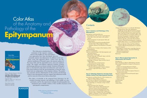
This <strong>Atlas</strong> gives a detailed documentation<br />
<strong>of</strong> <strong>the</strong> superior compartments<br />
<strong>of</strong> <strong>the</strong> middle ear. New microdissection<br />
approaches have been developed, most importantly<br />
anterior microdissection. In addition to <strong>the</strong> tympanic<br />
cavity, this approach allows a direct view into <strong>the</strong><br />
anterior membrane <strong>of</strong> Prussak’s space, <strong>the</strong> anterior surface <strong>of</strong><br />
<strong>the</strong> tensor fold, <strong>and</strong> <strong>the</strong> supratubal recess, areas not explored<br />
earlier. The regular <strong>and</strong> hi<strong>the</strong>rto unknown auxiliary aeration<br />
<strong>and</strong> drainage pathways are shown in detail. Aeration <strong>of</strong> Prussak’s<br />
space is documented using both microdissection <strong>and</strong> serial sections;<br />
<strong>the</strong>se pathways from <strong>the</strong> mesotympanum or from <strong>the</strong> lower<br />
lateral attic are independent <strong>of</strong> <strong>the</strong> tympanic isthmus. The pathology<br />
caused by chronic inflammation on <strong>the</strong> aeration <strong>and</strong> drainage<br />
routes is also documented, <strong>and</strong> new surgical microdissection methods<br />
for removal <strong>of</strong> <strong>the</strong> tensor fold are described.<br />
This <strong>Atlas</strong> is invaluable in <strong>the</strong> temporal bone laboratory for all<br />
residents learning anatomy <strong>and</strong> pathology <strong>of</strong> <strong>the</strong> middle ear compartments,<br />
<strong>and</strong> for <strong>the</strong> experienced otologist <strong>the</strong> photographic documentation<br />
gives reliable evidence <strong>of</strong> <strong>the</strong> variable structures in <strong>the</strong><br />
epitympanic compartments.<br />
Fields <strong>of</strong> Interest: Otorhinolaryngology,<br />
Audiology, Histology,<br />
<strong>Pathology</strong>, Pediatrics<br />
Contents<br />
Preface<br />
Part 1: <strong>Anatomy</strong> <strong>and</strong> <strong>Pathology</strong> <strong>of</strong> <strong>the</strong><br />
Epitympanum<br />
Introduction <strong>and</strong> General Review<br />
� Development <strong>of</strong> <strong>the</strong> concept <strong>of</strong> epitympanum<br />
� Early data <strong>of</strong> <strong>the</strong> s<strong>of</strong>t tissues in <strong>the</strong> epitympanum<br />
� Fetal development <strong>of</strong> epitympanic folds <strong>and</strong><br />
compartments<br />
Tensor Fold � Lateral Incudomalleal Fold �<br />
Chordal Fold � O<strong>the</strong>r Duplicate Folds �<br />
Tympanic Isthmus � Development <strong>of</strong> Prussak’s<br />
Space<br />
� Contemporary Concepts <strong>of</strong> <strong>the</strong> <strong>Anatomy</strong> <strong>of</strong> <strong>the</strong><br />
Epitympanum<br />
Material <strong>and</strong> Methods<br />
<strong>Anatomy</strong> <strong>and</strong> <strong>Pathology</strong> <strong>of</strong> <strong>the</strong> Epitympanum <strong>and</strong><br />
Supratubal Recess<br />
� Epitympanic Diaphragm<br />
� Normal <strong>Anatomy</strong> <strong>of</strong> Prussak’s Space (with <strong>the</strong><br />
Lateral Malleal Space)<br />
Microdissection � Serial Sections<br />
� <strong>Pathology</strong> <strong>of</strong> Prussak’s Space <strong>and</strong> <strong>the</strong> Lateral<br />
Malleal Space<br />
Microdissection � Serial Sections<br />
� Cholesteatoma in Prussak’s Space<br />
� Large Epitympanic Compartments<br />
Posterior Epitympanum � Anterior Epitympanum<br />
� Supratubal Recess (Space)<br />
References<br />
Part 2: <strong>Pathology</strong> Related to Amniotic Fluid<br />
Cellular Content <strong>and</strong> Superimposed Infection<br />
Introduction <strong>and</strong> Short Review <strong>of</strong> <strong>the</strong> Literature on<br />
Amniotic Fluid Cellular Content<br />
Short Review <strong>of</strong> Mastoid Pneumatization<br />
Amniotic Fluid Cellular Content-Related Middle<br />
Ear <strong>Pathology</strong> as a Function <strong>of</strong> Age in Serial<br />
Sections<br />
� Temporal Bones from Neonates<br />
Superior <strong>and</strong> Anterior Epitympanum <strong>and</strong><br />
Antrum � Tensor Fold <strong>and</strong> Supratubal<br />
Recess � Lateral Incudomalleal Fold <strong>and</strong><br />
Lateral Attics � Lateral Malleal Space <strong>and</strong><br />
Prussak’s Space � Medial Attic <strong>and</strong> Tympanic<br />
Isthmus � Tympanic Cavity � Eustachian Tube �<br />
Mastoid Pneumatization � Comment<br />
� Temporal Bones from 2- to 4-Month-Old Infants<br />
Compartments above <strong>the</strong> Epitympanic<br />
Diaphragm <strong>and</strong> <strong>the</strong> Mastoid Antrum � Tensor<br />
Fold <strong>and</strong> Supratubal Recess � Lateral Malleal<br />
Space <strong>and</strong> Lateral Attics � Prussak’s Space <strong>and</strong><br />
Its Aeration Pathways � Tympanic Isthmus <strong>and</strong><br />
Posterior Tympanum � Tympanic Sinus <strong>and</strong><br />
Round Window Niche � Eustachian Tube �<br />
Elements Specific to Amniotic Fluid Cellular<br />
Content � Mastoid Pneumatization � Comment<br />
� Temporal Bones from 5- to 23-Month-Old Infants<br />
Case 1 � Case 2 � Case 3 � Case 4 � Case 5 �<br />
Elements Specific to Amniotic Fluid Cellular<br />
Content � Mastoid Pneumatization � Comment<br />
General Comments<br />
� Histological Considerations regarding Amniotic<br />
Fluid Cellular Content- Associated <strong>Pathology</strong><br />
� Mastoid Pneumatization<br />
� Clinical Considerations<br />
References<br />
Part 3: Microsurgical Approaches to<br />
Inflammatory Ear Disease<br />
Introduction<br />
Early Attempts to Improve Epitympanic Aeration<br />
Microsurgical Methods in Surgery for Retraction<br />
Pockets<br />
� Surgery for Incipient Retraction Pocket<br />
� Surgery for Established Retraction Pockets<br />
Frontolateral Atticotomy<br />
� Extensive Attic <strong>and</strong> Mesotympanic Disease in<br />
Chronic Otitis media<br />
Spread <strong>of</strong> Cholesteatoma from Prussak’s Space<br />
� Posterior Route<br />
� Inferior Central Route<br />
� Superior Route<br />
� Anterior Route<br />
Final Remarks<br />
References<br />
Subject Index


