

World Journal of Radiology (World J Radiol
World Journal of Radiology (World J Radiol
World Journal of Radiology (World J Radiol

Create successful ePaper yourself
Turn your PDF publications into a flip-book with our unique Google optimized e-Paper software.
A<br />
Restrepo CS et al . Infectious chest tumors<br />
Figure 7 Multicentric Castleman’s disease in a 40-year-old male with acquired immunodeficiency syndrome. Contrast-enhanced chest computed tomography,<br />
axial images at two different levels (A, B) reveal numerous enhancing abnormally enlarged lymph nodes in the axillae and mediastinum.<br />
A B<br />
C<br />
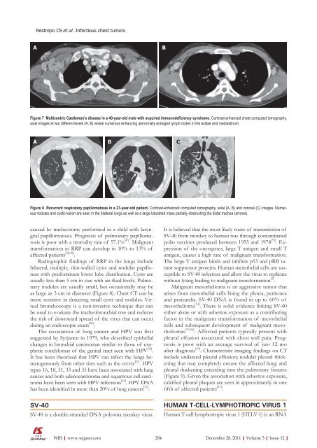
Figure 8 Recurrent respiratory papillomatosis in a 27-year-old patient. Contrast-enhanced computed tomography, axial (A, B) and coronal (C) images. Numerous<br />
nodules and cystic lesion are seen in the bilateral lungs as well as a large lobulated mass partially obstructing the distal trachea (arrows).<br />
caused by tracheotomy performed in a child with laryngeal<br />
papillomatosis. Prognosis <strong>of</strong> pulmonary papillomatosis<br />
is poor with a mortality rate <strong>of</strong> 57.1% [47] . Malignant<br />
transformation in RRP can develop in 10% to 13% <strong>of</strong><br />
affected patients [48,49] .<br />
Radiographic findings <strong>of</strong> RRP in the lungs include<br />
bilateral, multiple, thin-walled cysts and nodular papillomas<br />
with predominant lower lobe distribution. Cysts are<br />
usually less than 5 cm in size with air-fluid levels. Pulmonary<br />
nodules are usually small, but occasionally may be<br />
as large as 3 cm in diameter (Figure 8). Chest CT can be<br />
more sensitive in detecting small cysts and nodules. Virtual<br />
bronchoscopy is a non-invasive technique that can<br />
be used to evaluate the tracheobronchial tree and reduces<br />
the risk <strong>of</strong> downward spread <strong>of</strong> the virus that can occur<br />
during an endoscopic exam [46] .<br />
The association <strong>of</strong> lung cancer and HPV was first<br />
suggested by Syrjanen in 1979, who described epithelial<br />
changes in bronchial carcinomas similar to those <strong>of</strong> exophytic<br />
condylomas <strong>of</strong> the genital tract seen with HPV [50] .<br />
It has been theorized that HPV can infect the lungs hematogenously<br />
from other sites such as the cervix [51] . HPV<br />
types 16, 18, 31, 33 and 35 have been associated with lung<br />
cancer and both adenocarcinoma and squamous cell carcinoma<br />
have been seen with HPV infections [44] . HPV DNA<br />
has been identified in more than 20% <strong>of</strong> lung cancers [52] .<br />
SV-40<br />
SV-40 is a double-stranded DNA polyoma monkey virus.<br />
WJR|www.wjgnet.com<br />
B<br />
It is believed that the most likely route <strong>of</strong> transmission <strong>of</strong><br />
SV-40 from monkey to human was through contaminated<br />
polio vaccines produced between 1955 and 1978 [53] . Expression<br />
<strong>of</strong> the oncogenes, large T antigen and small T<br />
antigen, causes a high rate <strong>of</strong> malignant transformation.<br />
The large T antigen binds and inhibits p53 and pRB tumor<br />
suppressor proteins. Human mesothelial cells are susceptible<br />
to SV-40 infection and allow the virus to replicate<br />
without lysing leading to malignant transformation [2] .<br />
Malignant mesothelioma is an aggressive tumor that<br />
arises from mesothelial cells lining the pleura, peritonea<br />
and pericardia. SV-40 DNA is found in up to 60% <strong>of</strong><br />
mesothelioma [54] . There is solid evidence linking SV-40<br />
either alone or with asbestos exposure as a contributing<br />
factor in the malignant transformation <strong>of</strong> mesothelial<br />
cells and subsequent development <strong>of</strong> malignant mesotheliomas<br />
[55,56] . Affected patients typically present with<br />
pleural effusion associated with chest wall pain. Prognosis<br />
is poor with an average survival <strong>of</strong> just 12 mo<br />
after diagnosis [54] . Characteristic imaging findings on CT<br />
include unilateral pleural effusion; nodular pleural thickening<br />
that may completely encase the affected lung; and<br />
pleural thickening extending into the pulmonary fissures<br />
(Figure 9). Given the association with asbestos exposure,<br />
calcified pleural plaques are seen in approximately in one<br />
fifth <strong>of</strong> affected patients [57] .<br />
HUmaN T-CEll-lymPHOTROPIC VIRUS 1<br />
Human T-cell-lymphotropic virus 1 (HTLV-1) is an RNA<br />
284 December 28, 2011|Volume 3|Issue 12|


