

World Journal of Radiology (World J Radiol
World Journal of Radiology (World J Radiol
World Journal of Radiology (World J Radiol

Create successful ePaper yourself
Turn your PDF publications into a flip-book with our unique Google optimized e-Paper software.
A<br />
1 cm<br />
WJR|www.wjgnet.com<br />
B<br />
1 cm<br />
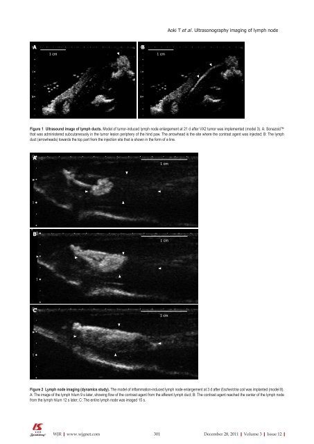
Figure 1 Ultrasound image <strong>of</strong> lymph ducts. Model <strong>of</strong> tumor-induced lymph node enlargement at 21 d after VX2 tumor was implemented (model 3). A: Sonazoid<br />
that was administered subcutaneously in the tumor lesion periphery <strong>of</strong> the hind paw. The arrowhead is the site where the contrast agent was injected; B: The lymph<br />
duct (arrowheads) towards the top part from the injection site that is shown in the form <strong>of</strong> a line.<br />
A<br />
B<br />
C<br />
1 cm<br />
1 cm<br />
1 cm<br />
Aoki T et al . Ultrasonography imaging <strong>of</strong> lymph node<br />
Figure 2 Lymph node imaging (dynamics study). The model <strong>of</strong> inflammation-induced lymph node enlargement at 3 d after Escherichia coli was implanted (model 8).<br />
A: The image <strong>of</strong> the lymph hilum 9 s later, showing flow <strong>of</strong> the contrast agent from the afferent lymph duct; B: The contrast agent reached the center <strong>of</strong> the lymph node<br />
from the lymph hilum 12 s later; C: The entire lymph node was imaged 15 s.<br />
301 December 28, 2011|Volume 3|Issue 12|


