

World Journal of Radiology (World J Radiol
World Journal of Radiology (World J Radiol
World Journal of Radiology (World J Radiol

You also want an ePaper? Increase the reach of your titles
YUMPU automatically turns print PDFs into web optimized ePapers that Google loves.
A<br />
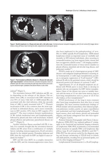
Figure 2 Burkitt lymphoma in a 38-year-old male with a left axilla mass. Contrast-enhanced computed tomography, axial (A) and coronal (B) images demonstrates<br />
a bulky s<strong>of</strong>t tissue mass involving the axilla, subpectoral and subscapular spaces.<br />
Figure 3 Post-transplant proliferative disease in a 50-year-old male postright<br />
lung transplant. Contrast-enhanced computed tomography shows an<br />
infiltrative right hilar mass surrounding the right lower lobe bronchus and involving<br />
the subcarinal region. Ipsilateral small pleural effusion is also noted.<br />
astinum [9] (Figure 1).<br />
The association between EBV infection and BL varies<br />
depending on the subtype <strong>of</strong> the disease. The endemic<br />
form (eBL) is associated with EBV infection in<br />
over 95% <strong>of</strong> cases. Only half <strong>of</strong> AIDS-related BLs are<br />
associated with this viral infection, while the sporadic<br />
form <strong>of</strong> (sBL) is rarely associated with it [11,12] . Thoracic<br />
involvement in BL is less common than the more typical<br />
facial or abdominal disease, and is more commonly<br />
seen in HIV positive (10%) than in HIV negative (2%)<br />
patients [13] . The most common thoracic manifestations<br />
<strong>of</strong> BL include mediastinal mass and lymphadenopathy<br />
followed by pleural and chest wall involvement. Isolated<br />
pleural effusion and pulmonary parenchymal disease are<br />
rare [14,15] (Figure 2).<br />
Another infectious disease associated with BL is malaria.<br />
It is currently thought that the association between<br />
malaria and BL arises from a combination <strong>of</strong> multiple<br />
factors including immunosuppression, B-cell activation<br />
directly by the malarial parasite Plasmodium falciparum and<br />
EBV, since the viral loads for EBV are higher in areas endemic<br />
for malaria [11] .<br />
EBV and KS virus (KSV) in a complex interaction<br />
with the HIV, and the immunosuppression status have<br />
WJR|www.wjgnet.com<br />
B<br />
Restrepo CS et al . Infectious chest tumors<br />
also been implicated in the pathophysiology <strong>of</strong> non-<br />
HLs in AIDS, typically B-cell lymphomas. AIDS-related<br />
lymphomas remain an important cause <strong>of</strong> morbidity and<br />
mortality in HIV-infected individuals, <strong>of</strong>ten occurring in<br />
extranodal locations (e.g. bone marrow, brain, viscera) and<br />
have an aggressive clinical course [16] . On imaging examinations<br />
the extranodal location <strong>of</strong> these tumors manifest as<br />
pleural effusion, interstitial and alveolar lung disease, and<br />
pulmonary nodules [17] .<br />
PTLD is made up <strong>of</strong> a heterogeneous group <strong>of</strong> EBV<br />
diseases with malignant lymphoproliferation occurring after<br />
hematopoietic or solid organ transplantation secondary<br />
to iatrogenic suppression <strong>of</strong> T-cell function [18] . EBVinfected<br />
B cells in the germinal centers, which should be<br />
destroyed, persist in the absence <strong>of</strong> cytotoxic T cells [6] .<br />
Approximately 2% <strong>of</strong> all allograft recipients are afflicted<br />
with PTLD, and it is more likely to develop in<br />
patients who are seronegative for EBV before transplantation.<br />
Onset <strong>of</strong> the disease can occur at variable intervals<br />
with the mean at 2-5 mo after bone marrow, lung or<br />
heart-lung transplantation and at a mean <strong>of</strong> 22-32 mo<br />
after kidney, or liver transplantation [19] .<br />
PTLD involvement <strong>of</strong> the lung is more common<br />
after heart-lung transplantation than after liver or renal<br />
transplant. The most common intrathoracic findings <strong>of</strong><br />
PTLD include well-circumscribed, <strong>of</strong>ten bilateral peripheral<br />
pulmonary nodules, patchy air space consolidation<br />
and mediastinal and hilar lymphadenopathy [19] . Pleuralbased<br />
masses, chest wall masses, pleural and pericardial<br />
effusion, and thymic enlargement have also been reported<br />
[20] (Figures 3 and 4).<br />
Positron Emission Tomography with Fluoro-2-deoxy-<br />
D-glucose (FDG-PET) has replaced gallium-67 scintigraphy<br />
in the functional and metabolic imaging evaluation<br />
<strong>of</strong> lymphomas. FDG-PET, in particular, is recommended<br />
before treatment in typically FDG avid lymphomas like<br />
diffuse large B cell lymphoma (DLBCL) and HL [21] .<br />
FDG-PET has also proven useful for the diagnosis, staging<br />
and therapy monitoring <strong>of</strong> PTLD both in infants<br />
and adult patients, revealing foci <strong>of</strong> increased uptake, not<br />
seen by other imaging techniques [22-24] .<br />
Lymphomatoid Granulomatosis is a B-cell lymphop-<br />
281 December 28, 2011|Volume 3|Issue 12|


