

2 - World Journal of Gastroenterology
2 - World Journal of Gastroenterology
2 - World Journal of Gastroenterology

You also want an ePaper? Increase the reach of your titles
YUMPU automatically turns print PDFs into web optimized ePapers that Google loves.
Freeman HJ et al . Intestinal lymphangiectasia<br />
<strong>of</strong> the total body albumin pool using a radio-labeled albumin<br />
method were reduced while fecal excretion was<br />
increased. In some, steatorrhea was also present. Biopsies<br />
<strong>of</strong> the small bowel are needed to confirm the diagnosis [2] .<br />
These show prominent dilated lymphatics in the lamina<br />
propria region <strong>of</strong> the villous structure, <strong>of</strong>ten with extension<br />
into submucosa (Figures 1 and 2). Others have commented<br />
that these changes may be difficult to detect since<br />
dilation <strong>of</strong> intestinal lacteals or lymph containing capillaries<br />
may be quite focal [3] . Familial presentations were also<br />
recorded [1] . Most <strong>of</strong>ten affected were children and young<br />
adults, although even historically, diagnosis was initially<br />
also occasionally defined in older, even elderly, adults. The<br />
precise cause <strong>of</strong> intestinal lymphangiectasia with enteric<br />
protein loss remains obscure but changes in regulatory<br />
molecules involved in lymphangiogenesis have been reported<br />
in the duodenal mucosa [4] .<br />
CLINICAL FEATURES<br />
Commonly reported clinical findings have traditionally<br />
described pitting edema, usually in a symmetrical distribution<br />
involving the lower limbs. If severe edema is evident,<br />
facial and scrotal (or vaginal) involvement may occur.<br />
Effusions may also develop in pleural, pericardial and<br />
peritoneal cavities with gross chylous ascites. Sonographic<br />
evidence <strong>of</strong> fetal ascites has also been reported [5] . Rarely,<br />
lymphedema (that may be difficult to differentiate from<br />
edema due to hypoalbuminemia and low oncotic pressure)<br />
and the so-called “Stemmer’s sign” (i.e. skin fibrosis on<br />
dorsum <strong>of</strong> the second toe due to chronic lymphedema) [6]<br />
have been described. It should be emphasized, however,<br />
that many <strong>of</strong> these clinical features likely reflect the severe<br />
end <strong>of</strong> the spectrum <strong>of</strong> clinical changes, especially<br />
in adults with intestinal lymphangiectasia. In recent years,<br />
directly due to the emergence <strong>of</strong> newer endoscopic methods,<br />
particularly video capsule endoscopy in adults, detection<br />
<strong>of</strong> mucosal changes suggestive <strong>of</strong> lymphangiectasia<br />
seem to be relatively common. For example, in a recent<br />
evaluation <strong>of</strong> 1866 consecutive endoscopic examinations,<br />
histological confirmation <strong>of</strong> duodenal lymphangiectasia<br />
was evident in 1.9% [7] . More significant, even if lymphangiectasia<br />
persisted on consecutive endoscopic studies over<br />
a year apart, there was no clinical evidence <strong>of</strong> malabsorption<br />
[7] . Therefore, it would seem that in most histologically-confirmed<br />
intestinal lymphangiectasia, clinical features<br />
may be very limited and the classical “textbook” recorded<br />
changes may be more reflective <strong>of</strong> the most severe end <strong>of</strong><br />
the clinicopathological spectrum in adults.<br />
In some, an abdominal mass may be noted [8] , either<br />
on clinical evaluation or with radiological imaging studies,<br />
particularly ultrasound (with or without endoscopy)<br />
or computerized tomography. These masses are usually<br />
cystic and <strong>of</strong>ten represent associated lymphangiomas. The<br />
latter are entirely benign lesions that may be associated<br />
with lymphangiectasia in either small or large bowel [9,10] .<br />
The most commonly reported intestinal site is the duodenum<br />
[11] , in part, perhaps, because these have been easier<br />
to detect with endoscopic evaluation showing a smooth,<br />
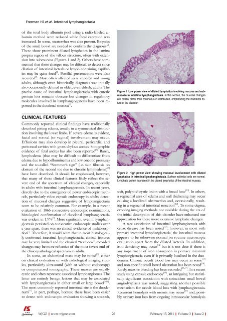
Figure 1 Low power view <strong>of</strong> dilated lymphatics involving mucosa and submucosa<br />
in intestinal lymphangiectasia. In this section, the mucosal changes<br />
are patchy rather than continuous in distribution, emphasizing the multifocal nature<br />
<strong>of</strong> the disorder.<br />
Figure 2 High power view showing mucosal involvement with dilated<br />
lymphatics in intestinal lymphangiectasia. Surface epithelial cells are normal.<br />
Lymphatic protein is present in the dilated lymphatics <strong>of</strong> the intestinal mucosa.<br />
s<strong>of</strong>t, polypoid cystic lesion with a broad base [10] . In others,<br />
a segmental area <strong>of</strong> edema and wall thickening may occur<br />
causing a localized obstruction and, occasionally, resulting<br />
in a segmental intestinal resection [12] . To some degree,<br />
evolving imaging methods not available during the era <strong>of</strong><br />
the initial description <strong>of</strong> this disorder have enhanced our<br />
appreciation for these more extensive lymphatic changes.<br />
A rare association <strong>of</strong> intestinal lymphangiectasia with<br />
celiac disease has been noted [13] ; however, in most with<br />
primary intestinal lymphangiectasia, the intestinal mucosa<br />
appears to be otherwise normal on routine microscopic<br />
evaluation apart from the dilated lacteals. In addition,<br />
iron deficiency may occur [14] but it is not clear if there is<br />
any impairment <strong>of</strong> iron absorption in primary intestinal<br />
lymphangiectasia even if it primarily localized in the duodenum.<br />
Chronic occult blood loss may occur in some [15]<br />
and non-specific small bowel ulceration has been noted [14] .<br />
Rarely, massive bleeding has been recorded [16,17] . In a recent<br />
study using capsule endoscopy [18] , an intriguing but statistically<br />
significant association with coincident small bowel<br />
angiodysplasia was noted, suggesting another possible<br />
mechanism for occult blood loss with lymphangiectasia.<br />
Recurrent hemolysis with uremia may occur [19] and, possibly,<br />
urinary iron loss from ongoing intravascular hemolysis<br />
WJGO|www.wjgnet.com 20<br />
February 15, 2011|Volume 3|Issue 2|


