RETRON Implants - tantum AG
RETRON Implants - tantum AG
RETRON Implants - tantum AG

You also want an ePaper? Increase the reach of your titles
YUMPU automatically turns print PDFs into web optimized ePapers that Google loves.
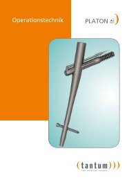
Operative Technique<br />
<strong>RETRON</strong> Humeral Head Nail<br />
<strong>RETRON</strong>)
)))<br />
<strong>tantum</strong> · Operative technique <strong>RETRON</strong><br />
<strong>RETRON</strong> Humeral Head Nail System<br />
The number and complexity of humeral head fractures<br />
increase with advancing age. A definite increase in incidents<br />
due to demographic changes can be expected.<br />
As a result of growing demands on physical activities,<br />
the tendency in favour of operative treatment for humeral<br />
head fractures will be observed.<br />
The <strong>RETRON</strong> nail is suited to this development, given<br />
that, alternative procedures of treatment due to long<br />
physical-therapeutic follow-up treatment with only<br />
gradual improvement of the mobility, the patient’s<br />
claim to untimely physical activity is partially unjust.<br />
An implant system for this indication should include<br />
the possibility of:<br />
• Prevention of the humeral head<br />
• Minor invasive Operative procedure<br />
• Protection of the subacromial region<br />
The goal of treatment with the <strong>RETRON</strong> system is the<br />
stabilisation and conservation of the bone structure.<br />
Entry of the <strong>RETRON</strong> nail occurs laterally transdermal,<br />
underneath the onset of the Delta muscle. The joint is<br />
not opened.<br />
This system is suitable for subcapital and pertubercullary<br />
humeral fractures.<br />
The <strong>RETRON</strong> system offers young patients with<br />
good bone quality splendid support of the fracture.<br />
Protection of the subacromial region through lateral<br />
entry underneath the onset of the Delta muscle is an<br />
important prerequisite for good rehabilitation results<br />
especially with young patients.<br />
For the treatment of older patients with increasing<br />
osteoporosis, the demand for anchoring the implant<br />
in the humeral head is escalated.<br />
The subchondral region of the bone is used for a reliable<br />
fixation of the <strong>RETRON</strong> nail. The nail is guided in<br />
just to the cartilage and generates a good condition<br />
for a stable osteosynthesis due to its proximal thread.<br />
An additional advantage of the <strong>RETRON</strong> system is the<br />
complete preservation with all types of retraction.<br />
All in all, the <strong>RETRON</strong> system complies with the accustomed<br />
<strong>tantum</strong> principle: The system is an innovative<br />
product, of the best quality, to economic conditions.<br />
The anchorage of the fragments is carried out with<br />
the cannulated screws, which are angle and slip stable<br />
though the implantation of the <strong>RETRON</strong> nail. This<br />
principle of intramedullary force conduction and cannulated<br />
screws creates good stabilisation of the fragments<br />
and reduces secondary dislocations.<br />
))<br />
( 2
)))<br />
)))<br />
)))<br />
)))<br />
)))<br />
)))<br />
)))<br />
)))<br />
Features and benefits at a glance<br />
)))<br />
The washers can be used for increased incursion<br />
safety due to severe osteoporosis<br />
Cannulated screw with TORX adaptor<br />
Optional interlocked-drilling in the<br />
shaft region<br />
Access area for the carbon-fiber targeting<br />
device and diaphysis anchor<br />
Diaphysis anchor in four different<br />
lengths<br />
Distal anchoring supplies the stability<br />
Diaphysis anchor with three locking<br />
holes<br />
Flat screw-heads for minimal soft-tissue<br />
irritations<br />
)))<br />
)))<br />
)))<br />
)))<br />
<strong>tantum</strong> · Operative technique <strong>RETRON</strong><br />
Proximal nail end with self-cutting<br />
thread for firm sit of the nail<br />
Self-cutting cannulated screws Ø 4 mm in<br />
2 mm increments. Implantation through<br />
the use of a 1.6 mm repositioning wire<br />
Thread appendage in the nail shaft assures<br />
angle and glide stability of the interlocked<br />
screw<br />
Nail, cannulated out of titanium alloy, Ø 8<br />
mm, in two lengths (76 mm, 86 mm) and<br />
right or left versions are available<br />
3 )
)))<br />
<strong>tantum</strong> · Operative technique <strong>RETRON</strong><br />
Fig. 1<br />
Fig. 2<br />
Fig. 3<br />
( 4<br />
Operative technique<br />
Type I<br />
Type II<br />
Type III<br />
1. Indication<br />
Subcapital and pertubercullary multiple fragment fractures<br />
of the humeral head up to Habermeyer type II, in individual<br />
cases up to type III.<br />
2. Positioning of the Patient<br />
Position the patient in a dorsal position on the shoulder<br />
table. The arm should be placed in a freely moveable position<br />
(Fig. 2). The C-arm should be positioned towards the<br />
head in the direct axis of the table.<br />
3. Reposition of the Fragments<br />
An x-ray in two levels (a-p and axis level) is required for<br />
reduction of the fragments. On the basis of the fracture<br />
type, the number of fragments, as well as their correlated<br />
position to each other can draw conclusions to the soft tissue<br />
anastomosen between the fragments (periosteum and<br />
sinews) and the required reduction technology.<br />
CT-3D-Reconstructions using the percutaneous method, at<br />
least initially are for a complete compilation of the fracture<br />
very helpful.<br />
The goal of the reduction is the restoration of the slide<br />
mechanism in the subacromial cavity. In other words, the<br />
tubercle and the articular fragment are brought into the<br />
anatomic position and temporarily fixated with K-wires. The<br />
fixation can remain until the introduction of the implant, as<br />
long as the wires are not in the way (Fig. 3). The reduction<br />
can alternatively be held by the assistant with an oval awl.
Fig. 4<br />
Fig. 5<br />
Fig. 6<br />
Inzision<br />
206-107<br />
Fig. 6a<br />
align<br />
Constant<br />
Diameter<br />
4. Entry and opening<br />
For localization of the entry-point angle the arm in a light<br />
outward rotational position, so that the greater tuberosity<br />
is prominently depicted.<br />
The guide wire (Cat no. 206-107) is then placed on the upper<br />
arm and is positioned in the designated direction and under<br />
fluoroscopy aligned in the intended direction of the nail.<br />
The planned incision can then be marked on the skin using<br />
a pen (Fig. 4).<br />
Particular attention should be placed on the axillary nerve<br />
while entering using the puncture incision and blunt preparation.<br />
5. Affixing the <strong>RETRON</strong> Positioning Guide<br />
The <strong>RETRON</strong> Positioning Guide (Cat no. 204-115) is assembled<br />
next.<br />
Screw the positioning guide (Cat no. 204-115A) and the base<br />
together, as well as the positioning guide sleeve.<br />
The <strong>RETRON</strong> Positioning Guide is directed percutaneously<br />
to the cortex.<br />
Attention: Note the location of the axillary nerve. Place<br />
the positioning guide severely lateral on the onset of the<br />
greater tubercle crest. The longitudinal alignment positions<br />
itself due to the concave contact surface. Position it so that<br />
the proximal end stands upright, where the humeral shaft<br />
begins to nearly show a constant cross-section (Fig. 5).<br />
6. Aligning the <strong>RETRON</strong> Positioning Guide<br />
<strong>tantum</strong> · Operative technique <strong>RETRON</strong><br />
The attachment for positioning guide (Cat no. 204-116) can<br />
optionally be used to identify the ideal point for the positioning<br />
guide. The short indicator (Cat no. 204-116A, yellow<br />
colour-coded) is then assembled to the attachment for this<br />
purpose. The attachment is screwed onto the positioning<br />
guide and inserted onto the bone. Align the unit in the a-p<br />
path of rays under X-ray control (Fig. 6a). The positioning<br />
guide is optimally in the path of rays, when the two pins of<br />
the attachment are aligned.<br />
The height of the positioning guide should be selected so<br />
that the end of the indicator is subchondral in the middle<br />
third of the humeral head. If necessary exchange indicator<br />
L76 for indicator L86 (Cat no. 204-116B).<br />
5 )
)))<br />
<strong>tantum</strong> · Operative technique <strong>RETRON</strong><br />
Fig. 7<br />
Fig. 8<br />
Fig. 9<br />
( 6<br />
Operative Technique<br />
!<br />
7. Fixation of <strong>RETRON</strong> Positioning Guide<br />
Fixation of the positioning guide with the fixing screw for<br />
positioning guide (Cat no. 204-142). For this purpose insert<br />
the guide sleeve (Cat no. 202-130 black ring marking) into<br />
the positioning guide and using the drill (Cat no. 203-142),<br />
drill bicortically (Fig. 7).<br />
The fixation screw is temporarily affixed and later exchanged<br />
with a cannulated screw, therefore only a 30mm length is<br />
required.<br />
Remove the guide sleeve and screw in the fixing screw (Cat<br />
no. 204-142) with the cannulated screwdriver (Cat no. 201-<br />
146). Screw the fixing screw into the bone until the positioning<br />
guide is set firmly onto the bone (Fig. 8).<br />
8. Bone preparation<br />
To prevent the wire guide from straying, the bone should<br />
be prepared with the spot facer Ø 8 (Cat no. 203-139). The<br />
spot facer should then be guided through the sleeve (Cat no.<br />
204-115c) of the positioning guide using a high rotational<br />
until completely inserted.<br />
Note: When opening the lateral cortex with the spot facer, it<br />
is important to lightly press the proximal portion of the positioning<br />
guide, so that the drilling is not cranially diverted<br />
(Fig. 9).
Fig. 10<br />
Fig. 10a Fig. 10b<br />
Fig. 11<br />
Reading the<br />
length<br />
9. Insertion of the Nail Guide Wire<br />
The guide sleeve Ø 8 / Ø 2.5 (Cat no. 202-129) is pushed<br />
through the positioning guide to the cortex. The nail guide<br />
wire Ø 2.5 / L 300 (Cat no. 206-107) is inserted into the<br />
humeral head through the guide sleeve using a drill. The<br />
wire ends at the middle third of the humeral head and in<br />
the subchondral region (Fig. 10).<br />
The tip of the tissue protection sleeve is bevelled on one<br />
side. It is imperative that attention is paid to the alignment<br />
of the sleeve on the bone (Fig. 10a and Fig. 10b).<br />
Depending on the adjustment of the image intensifier during<br />
X-ray, occasional projection errors may occur. A safety<br />
distance between the tip of the nail guide wire and the<br />
calotte must be kept in order to avoid drilling through the<br />
humeral head.<br />
Note: A soft pressing of the proximal part of the positioning<br />
guide is advisable when introducing the nail guide wire.<br />
10. Length measuring<br />
<strong>tantum</strong> · Operative technique <strong>RETRON</strong><br />
Calculate the drill depth with the <strong>RETRON</strong> length gauge<br />
initially (Cat no. 208-106).<br />
Note: To determine the length correctly, the nail guide wire<br />
is placed subchondrally, the guide sleeve is pushed forward<br />
as far as possible, and the <strong>RETRON</strong> length gauge is attached<br />
to the guide sleeve. The length is read from the side which<br />
is marked “NAIL GAUGE” (Fig. 11).<br />
7 )
)))<br />
<strong>tantum</strong> · Operative technique <strong>RETRON</strong><br />
Fig. 12<br />
Fig. 13<br />
Fig. 14<br />
( 8<br />
Operative Technique<br />
L76<br />
1<br />
Fig. 14a<br />
2<br />
3<br />
11. Drilling the bone<br />
After removing the guide sleeve, the bone is drilled over the<br />
lying wire using the step drill Ø 8 / Ø 2.5 (Cat no. 203-141).<br />
Insert the step drill carefully with slight feed and high rotation<br />
speed in order to avoid the drill getting stuck.<br />
Note: The required drill depth conforms exclusively to the<br />
positioning of the drill in the humeral head, which is to be<br />
verified with the help of the image intensifier. The end of<br />
the drill defines the future position of the nail. The marking<br />
on the drill shaft provides an orientation for the nail length<br />
which is to be used (Fig. 12). If the determined length is<br />
between the yellow and the blue marking, the short nail<br />
L 76 is to be chosen. By means of the diaphysis anchor, the<br />
missing length will be compensated in a later step.<br />
12. Removal of the <strong>RETRON</strong> Positioning Guide<br />
To remove the positioning guide, first remove the step drill<br />
and the nail guide wire.<br />
Unscrew the fixation screw by means of the cannulated<br />
screw driver (Cat no. 201-146) and remove the positioning<br />
guide (Fig. 13).<br />
13. Selection and Assembly of the Implant<br />
Selection of the nail in the correct version (right/left) with<br />
the previously defined length (selectively L=76 mm or L=86<br />
mm)<br />
Analogically to the defined nail length, preposition the<br />
according nail holding screw (Cat no. 204-136 or 204-138)<br />
and the suitable nail adaptor (Cat no. 204-135 or 204-137),<br />
then assemble as a unit (Fig. 14).<br />
The nail holding screw (1) is guided through the nail adaptor<br />
(2) and is screwed together with the nail (3) (Fig. 14 a).<br />
Note: Before insertion of the nail, the chosen implants<br />
should be verified by assembling with the targeting device.<br />
For this purpose have the targeting device (Cat no. 204-132)<br />
and the fastening screw (Cat no. 204-131) ready. Close the<br />
target drill holes that are not required (depending on the<br />
side R=right or L=left) with Silicone Plugs (Cat no. 204-124).<br />
Put the targeting device onto the nail adaptor and tighten<br />
the connection of the nail to the nail holding screw by<br />
means of the screw driver SW 4, until it is hand-tight. Attach<br />
the connecting screw and tighten with the screwdriver SW<br />
4. Introduce the guide sleeve Ø 7 / Ø 1.6 (Cat no. 202-122)<br />
into the tissue protection sleeve Ø 9 / Ø 7 (Cat no. 202-121)<br />
and affix it using one rotation. Push the sleeves little by little<br />
through the open target drill holes in the targeting device.<br />
Check the passage through the locking drill holes in the nail<br />
by pushing a guide wire Ø 1.6 / L 245 through.
Fig. 15<br />
Fig. 16<br />
Fig. 17<br />
3<br />
1<br />
2<br />
Fig. 15a<br />
15°-20°<br />
201-104<br />
14. Implantation of the Nail<br />
Direct the unit consisting of nail adaptor, nail holding screw<br />
and nail (see Fig. 14) to the bone and implant it with the<br />
insertion / removal driver (Cat no. 201-103), using light pressure.<br />
A feed rate of approx. 2 mm per rotation is achieved.<br />
The nail is to be screwed in until the proximal end of the<br />
nail is sitting subchondrally. The distal end of the nail should<br />
then be positioned 2-5 mm within the lateral cortex (Fig. 15).<br />
The correct position of the nail is to be verified under X-ray<br />
control.<br />
For a rough alignment, the lines on the nail adaptor and<br />
insertion / removal driver must point in the lateral direction<br />
(Fig. 15a).<br />
15. Attaching the targeting device<br />
<strong>tantum</strong> · Operative technique <strong>RETRON</strong><br />
The targeting device (1) (Cat no. 204-132) is seated on the<br />
nail adaptor, and the nail holding screw is tightened with<br />
the screwdriver SW 4 (Cat no. 201-104). Attach the connecting<br />
screw (2) (Cat no. 204-131) and tighten with the screwdriver<br />
SW 4 (Fig. 16).<br />
The targeting device is useable on both sides. For a better<br />
orientation, the target drill holes that are not required<br />
(R=right and L=left respectively) have been closed with silicone<br />
plugs (3) (Cat no. 204-124) previously (see paragraph<br />
13).<br />
The targeting device is adjusted so that the screws for the<br />
fixation of the minor tubercle and the major tubercle lie on<br />
both sides of the biceps tendon.<br />
The correct alignment of the targeting devices is given when<br />
the ventral locking screw is led centrally through the minor<br />
tubercle. To adjust this position, the lower arm is angled<br />
approx. 90°. From a cranial view, the ventral drilling is positioned<br />
by a 15-20° angle relative to the angled lower arm;<br />
rotated outwards (Fig. 17).<br />
Tip: The screwdriver SW 4, which is inserted into the ventral<br />
drilling, may serve as an indicator.<br />
9 )
)))<br />
<strong>tantum</strong> · Operative technique <strong>RETRON</strong><br />
Fig. 18<br />
Fig. 19<br />
( 10<br />
Operative Technique<br />
16. Introduction of the Cannulated Screws<br />
The cannulated screw is initially inserted for the fixation of<br />
the minor tubercle. It is followed by the screws for the fixation<br />
of the major tubercle and the shaft screw.<br />
Exemplarily, the procedure for the introduction of a shaft<br />
screw is described:<br />
The guide sleeve Ø 7 / Ø 1.6 (Cat no. 202-122) is introduced<br />
into the tissue protection sleeve Ø 9 / Ø 7 (Cat no. 202-<br />
121) and is fixed with one rotation. The sleeves are pushed<br />
through the target drillings lying outside (R=right and L=left<br />
respectively) into the targeting device, and are advanced to<br />
the bone following a puncture incision and blunt preparation.<br />
Before setting the guide wires Ø 1.6 / L 245 (Cat no. 206-<br />
108), the correct reduction is to be checked under X-ray<br />
control in two planes.<br />
Note: Before introducing the guide wire, make sure that the<br />
nail guide wire has been removed.<br />
The screws in the shaft area of the bone are locked bicortically;<br />
the screws in the head area are locked monocortically.<br />
For the setting of the screw in the firmer shaft area of the<br />
humerus, it is necessary to pre-drill bicortically with the cannulated<br />
dill Ø3 / Ø1.6 (Cat no. 203-140) over the lying guide<br />
wire. Generally, it is not necessary to pre-drill the cannulated<br />
screws that are to be placed in the humeral head area. A<br />
monocortical pre-drilling is recommended here if required<br />
in case of an especially firm bone substance.<br />
The required screw length is determined with the <strong>RETRON</strong><br />
length gauge (Cat no. 208-106) by attaching it to the guide<br />
sleeve and reading off the scale “SCREW GAUGE” (Fig. 18).<br />
After removing the guide sleeve, the cannulated screw is<br />
inserted over the wire by means of the cannulated screw<br />
driver T10 (Cat no. 201-146) (Fig. 19).<br />
The ring marking of the screw driver provides an indication<br />
for reaching the cortex with the screw head. If the marking<br />
reaches the end of the tissue protection sleeve, the screw<br />
head lies in front of or at the cortex (Fig. 19). The position<br />
of the screw head is to be verified subsequently with the<br />
image intensifier.
Fig. 20<br />
Fig. 21<br />
Note: If washers (Cat no. 105-130) are to be used, the cannulated<br />
screws should be inserted with the washer that<br />
was attached beforehand, without tissue protection sleeve.<br />
When screwing in the screws ensure that no soft tissue gets<br />
between the washer and bone (Fig. 20).<br />
17. Distal anchorage of the implant<br />
<strong>tantum</strong> · Operative technique <strong>RETRON</strong><br />
Four diaphysis anchors (Cat no. 105-151, 105-152, 105-153,<br />
105-154) with different lengths from 0 mm to 7.5 mm (2.5<br />
mm steps) are available. The required length is determined<br />
by means of the sizer (Cat no. 208-107). For this purpose,<br />
insert the sizer into the end of the nail and read off the<br />
necessary length at the shaft under X-ray control. The ring<br />
groove that ends in a-p alignment with the lateral cortex,<br />
describes the correct length (Fig. 21).<br />
11 )
)))<br />
<strong>tantum</strong> · Operative technique <strong>RETRON</strong><br />
Fig. 22<br />
Fig. 23<br />
Fig. 24<br />
( 12<br />
Operative Technique<br />
Direct the diaphysis anchor (Cat no. 105-151, 105-152, 105-<br />
153, 105-154) together with the connection screw (Cat<br />
no. 105-150) to the distal end of the nail, and screw the<br />
connecting screw in with the screw driver SW4 so far that<br />
the diaphysis anchor lies against the nail, but is not finally<br />
tightened. Pay attention to the correct alignment of the<br />
diaphysis anchor (Fig. 22).<br />
Tip: If the nail guide wire is inserted again in the distal end<br />
of the nail after the removal of the targeting device, the<br />
diaphysis anchor can be inserted together with the connection<br />
screw, via the wire.<br />
For the fixation of the diaphysis anchor at the bone, a cannulated<br />
screw is inserted in the screw hole, which had served<br />
the fixation of the positioning guide previously.<br />
The cannulated screw is screwed in with the cannulated<br />
screw driver T10 (Cat no. 201-146) (Fig. 23). Finally, the connection<br />
screw is ultimately tightened.<br />
Fig. of the locked <strong>RETRON</strong> Humeral Nail System (Fig. 23, Fig.<br />
24).<br />
18. Follow-up Care and Management<br />
1-3 weeks immobilisation, with cuff and collar sling or - if<br />
necessary - gilchrist sling. Afterwards guided physiotherapy,<br />
according to the individual situation. Intra-operative X-ray<br />
control, hereafter approx. 10 days post-operatively.<br />
19. Implant removal<br />
In case the implant is distracting, it should be removed after<br />
sufficient healing of the fracture. The implant removal is to<br />
be carried out in the following order:<br />
Firstly, release the diaphysis anchor. For this purpose, loosen<br />
the connection screw (Cat no. 105-150) to the nail by means<br />
of screw driver SW 4 (Cat no. 201-104). Then loosen the cannulated<br />
screw with the cannulated screw driver T10 (Cat no.<br />
201-146) and take off the diaphysis anchor.<br />
Determine the length of the implanted nail (L 76 or L 86<br />
mm), and attach the nail adaptor (Cat no. 204-135 or Cat<br />
no. 204-137) and the nail holding screw (Cat no. 204-136 or<br />
204-138) to the nail (see paragraph 13 on page 8). Attach<br />
the targeting device (Cat no. 204-132) to the nail adaptor<br />
and retighten the nail holding screw with the screw driver<br />
SW 4 (Cat no. 201-104). Attach the connecting screw (Cat no.<br />
204-131) and tighten it with the screwdriver SW 4.
Fig. 25<br />
Constant<br />
Diameter<br />
Tip: The thread in the nail can be found more easily, using<br />
the nail guide wire (Cat no. 206-107). For this purpose,<br />
introduce the guide wire into the end of the nail, guide the<br />
nail adaptor and the nail holding screw over the guide wire<br />
into the nail and tighten them by means of screw driver SW<br />
4.<br />
For a better orientation, the target drill holes which are not<br />
needed, (R = right and L = left respectively) are closed with<br />
silicone plugs (Cat no. 204-124) (Fig. 16).<br />
The guide sleeve Ø 7 / Ø 1.6 (Cat no. 202-122) is introduced<br />
into the tissue protection sleeve Ø 9 / Ø 7 (Cat no. 202-121)<br />
and is affixed using one rotation. The sleeves are pushed<br />
through the outside positioned drillings into the targeting<br />
device, and are advanced to the skin.<br />
Following a puncture incision and blunt preparation, the<br />
sleeves are advanced to the bone. Remove the guide sleeve<br />
and insert the cannulated screw driver T 10 (Cat no. 201-<br />
146). Then screw out the cannulated screw. Repeat this<br />
procedure for all remaining cannulated screws. Take off the<br />
targeting device and attach the <strong>RETRON</strong> insertion / removal<br />
driver (Cat no. 201-103) to the nail adaptor. Screw out the<br />
nail.<br />
Alternative procedure to locate the<br />
entrance point<br />
The <strong>RETRON</strong> wire guide may be used as an alternative to the<br />
positioning guide.<br />
A5. Affixment of the Wire Guide<br />
<strong>tantum</strong> · Operative technique <strong>RETRON</strong><br />
Following the reduction and opening (step 4 and 6), guide<br />
the correct side version (right or left) of <strong>RETRON</strong> wire guide<br />
(Cat no. 204-139 or 204-140) percutaneous to the cortex.<br />
Attention: Note the position of the axillary nerve.<br />
Attach the wire guide strictly lateral at the dorsal border of<br />
the greater tuberosity. Place it in a way that its proximal end<br />
lies at the same height where the humeral shaft begins to<br />
have a nearly constant diameter (Fig. 25).<br />
13 )
)))<br />
Fig. 26<br />
Fig. 27<br />
Fig. 28<br />
( 14<br />
Operative Technique<br />
!<br />
A6. Alignment of the Wire Guide<br />
The correct position of the wire guide is obtained, when the<br />
virtual extension of the distal sleeve comes to lie centrally in<br />
the humeral head (Fig. 25).<br />
The wire guide exhibits spikes on its underside, which press<br />
into the bone and avoid a shifting of the wire guide.<br />
Tip: Additionally the position of the wire guide may be fixed<br />
with two guide wires (Cat no. 206-108). The plate of the<br />
wire guide has one drilling on the dorsal and one on the<br />
ventral end through which the guide wires can be inserted<br />
into the bone (Fig. 26).<br />
A7. Bone preparation<br />
The bone is prepared with the spot facer Ø 2.5 (Cat no. 203-<br />
143) to prevent the nail guide wire from deviating. For this<br />
purpose set the spot facer with high rotation speed through<br />
the sleeve of the wire guide until the stopper is reached.<br />
Note: During opening of the lateral cortex with the spot<br />
facer, a light pressing of the proximal part of the wire guide<br />
is important to prevent the drilling from straying in the<br />
cranial direction.<br />
A8. Insertion of the nail guide wire<br />
The nail guide wire Ø 2.5 (Cat no. 206-107) is inserted into<br />
the humeral head through the sleeve of the wire guide with<br />
a drill. The wire lies in the middle third of the humeral head<br />
and ends in the subchondral area (Fig. 28).<br />
Depending on the alignment of the image intensifier during<br />
X-ray, occasional projection errors may occur. A safety<br />
distance between the tip of the nail guide wire and the<br />
calotte must be kept in order to avoid drilling through the<br />
humeral head.<br />
Note: During the insertion of the nail guide wire, a light<br />
pressing of the proximal part of the positioning guide is<br />
also recommended to prevent the wire from deviating in<br />
the cranial direction.
Fig. 29<br />
Fig. 30<br />
Fig. 31<br />
A9. Attaching of the tissue protection sleeve<br />
Take the wire guide off first, then advance the tissue protection<br />
sleeve Ø 8 (Cat no. 202-128) with inserted guide sleeve<br />
Ø 8 / Ø 2.5 (Cat no. 202-129) onto the cortex.<br />
Note: The tip of the tissue protection sleeve is bevelled.<br />
Therefore it is stringently necessary to attend to the alignment<br />
of the sleeve at the bone (Fig. 29).<br />
A10. Measuring the length<br />
The drill depth is first determined by means of the <strong>RETRON</strong><br />
length gauge (Cat no. 208-106).<br />
Note: To determine the length correctly, the nail guide<br />
wire is placed subchondrally, the guide sleeve has contact<br />
to the lateral cortex and the length gauge is laid alongside<br />
the guide sleeve. The length is read off at the side which is<br />
marked “NAIL GAUGE” (Fig. 30).<br />
A11. Prepare the bone<br />
<strong>tantum</strong> · Operative technique <strong>RETRON</strong><br />
After removing the guide sleeve, the bone is drilled with the<br />
step drill Ø 8 / Ø 2.5 (Cat no. 203-141) over the lying wire.<br />
Insert the step drill carefully with slight feed and high rotation<br />
speed in order to avoid the drill to get stuck.<br />
The drill depth is read off from the shaft marking on the<br />
drill (Fig. 31). Check the drill depth by means of fluoroscopy.<br />
Afterwards remove the drill and the nail guide wire.<br />
Note: The required drill depth conforms exclusively to the<br />
positioning of the drill in the humeral head, which is to be<br />
verified with the help of the image intensifier. The end of<br />
the drill defines the future position of the nail. The marking<br />
on the drill shaft provides an orientation for the nail length<br />
which is to be used (Fig. 31). If the determined length is<br />
between the yellow and the blue marking, the short nail L76<br />
is to be chosen. By means of the diaphysis anchor, the missing<br />
length will be compensated in a later step.<br />
For the further procedure refer to paragraph 13 on page 8.<br />
15 )
)))<br />
<strong>tantum</strong> · Operative technique <strong>RETRON</strong><br />
( 16<br />
<strong>RETRON</strong> <strong>Implants</strong><br />
Humeral Nail<br />
Material: Ti6Al4V (ISO 5832-3)<br />
Size Side<br />
Cat no.<br />
non sterile sterile<br />
Ø 8, L 76 left 105-141 105-161<br />
Ø 8, L 86 left 105-142 105-162<br />
Ø 8, L 76 right 105-145 105-165<br />
Ø 8, L 86 right 105-146 105-166<br />
Diaphysis Anchor<br />
Material: Ti6Al4V (ISO 5832-3)<br />
Cat no.<br />
Size non sterile sterile<br />
I (0 mm) 105-151 105-171<br />
II (2.5 mm) 105-152 105-172<br />
III (5.0 mm) 105-153 105-173<br />
IV (7.5 mm) 105-154 105-174<br />
Connection Screw<br />
Material: Ti6Al4V (ISO 5832-3)<br />
Cat no.<br />
Size non sterile sterile<br />
SW 4, L 16 105-150 105-170<br />
Washer, plain<br />
Material: Ti6Al4V (ISO 5832-3)<br />
Cat no.<br />
Size non sterile sterile<br />
SW 4, L 16 105-130 105-180<br />
Cannulated Screws<br />
Material: Ti6Al4V (ISO 5832-3)<br />
Connection: TORX<br />
Wire Diameter: 1.6 mm<br />
Material: Ti6Al4V (ISO 5832-3)<br />
Cat no.<br />
Size non sterile sterile<br />
Ø 4, L 20, T15 101-530 101-560<br />
Ø 4, L 22, T15 101-531 101-561<br />
Ø 4, L 24, T15 101-532 101-562<br />
Ø 4, L 26, T15 101-533 101-563<br />
Ø 4, L 28, T15 101-534 101-564<br />
Ø 4, L 30, T15 101-535 101-565<br />
Ø 4, L 32, T15 101-536 101-566<br />
Ø 4, L 34, T15 101-537 101-567<br />
Ø 4, L 36, T15 101-538 101-568<br />
Ø 4, L 38, T15 101-539 101-569<br />
Ø 4, L 40, T15 101-540 101-570<br />
Ø 4, L 42, T15 101-541 101-571<br />
Ø 4, L 44, T15 101-542 101-572<br />
Ø 4, L 46, T15 101-543 101-573<br />
Ø 4, L 48, T15 101-544 101-574<br />
Ø 4, L 50, T15 101-545 101-575<br />
Ø 4, L 52, T15 101-546 101-576<br />
Ø 4, L 54, T15 101-547 101-577<br />
Ø 4, L 56, T15 101-548 101-578<br />
Ø 4, L 58, T15 101-549 101-579<br />
Ø 4, L 60, T15 101-550 101-580<br />
The Fig.s are schematic and do not correspond with the original dimensions. Subject to technical changes. Revision Feb. 09
)))<br />
<strong>RETRON</strong> Instruments<br />
(204-115A Handle for Positioning Guide)01-120 201-103 Insertion / Removal Driver<br />
204-135 Nail Adaptor L 76<br />
(204-115C Sleeve for Positioning Guide)01-120<br />
204-136 Nail Holding Screw L 76<br />
(204-115B Base Body for Positioning Guide)01-120 204-137 Nail Adaptor L 86<br />
204-115 <strong>RETRON</strong> Positioning Guide<br />
204-116 Attachment for Positioning Guide<br />
204-116A Indicator L76 f. Positioning Guide01-120<br />
204-116B Indicator L86 f. Positioning Guide<br />
202-130 Guide Sleeve Ø 7, Ø 30<br />
203-142 Drill with AO Coupling Ø 3, L 235<br />
204-142 Fixing Screw for Position Guide Ø 4, L 30<br />
201-148 Screwdriver cannulated T 15<br />
203-139 Spot Facer Ø 8, L 180<br />
202-129 Guide Sleeve Ø 8, Ø 2.5<br />
206-107 Guide Wire Ø 2.5, L 300<br />
208-106 <strong>RETRON</strong> Length Gauge<br />
203-141 Step Drill Ø 8, Ø 2.5, L 280<br />
202-128 Tissue Protection Sleeve Ø 8<br />
204-138 Nail Holding Screw L 86<br />
204-132 Targeting Device<br />
201-104 Screwdriver cannulated SW 4<br />
202-121 Tissue Protection Sleeve Ø 9, Ø 7<br />
202-122 Guide Sleeve Ø 7, Ø 1.6<br />
203-140 Drill, cannulated AO Coupling Ø 3, Ø 1.6, L 235<br />
206-108 10x Guide Wire Ø 1.6, L 245<br />
208-107 Sizer<br />
204-124 4x Silicon Plug<br />
204-131 Fastening Screw<br />
M12, SW 4<br />
209-122 <strong>RETRON</strong> Tray<br />
209-123 <strong>Implants</strong> Case for <strong>RETRON</strong> Tray<br />
209-124 Silicon insert for Tray<br />
204-139 <strong>RETRON</strong> Wire Guide right<br />
204-140 <strong>RETRON</strong> Wire Guide left<br />
203-143 Spot Facer Ø 2.5, L 130<br />
<strong>tantum</strong> · Operative technique <strong>RETRON</strong><br />
The Fig.s are schematic and do not correspond with the original dimensions. Subject to technical changes. Revision Feb. 09
<strong>tantum</strong> · Operative technique <strong>RETRON</strong> B04_19.4_engl.pdf<br />
<strong>tantum</strong> <strong>AG</strong><br />
Memellandstraße 2<br />
D-24537 Neumünster<br />
Fon +49 43 21 200 59-0<br />
Fax +49 43 21 200 59-19<br />
info@<strong>tantum</strong>-ag.de<br />
www.<strong>tantum</strong>-ag.de