Pichia Expression Kit - Invitrogen
Pichia Expression Kit - Invitrogen
Pichia Expression Kit - Invitrogen

You also want an ePaper? Increase the reach of your titles
YUMPU automatically turns print PDFs into web optimized ePapers that Google loves.
<strong>Pichia</strong> <strong>Expression</strong> <strong>Kit</strong><br />
For <strong>Expression</strong> of Recombinant Proteins in<br />
<strong>Pichia</strong> pastoris<br />
Catalog no. K1710-01<br />
Revision date: 07 September 2010<br />
Manual part no. 25-0043<br />
MAN0000012<br />
User Manual
Table of Contents<br />
<strong>Kit</strong> Contents and Storage.................................................................................................................................. iv<br />
Required Equipment and Supplies................................................................................................................. vi<br />
Introduction ................................................................................................................... 1<br />
<strong>Pichia</strong> pastoris <strong>Expression</strong> System ......................................................................................................................1<br />
Experimental Outline..........................................................................................................................................4<br />
Methods ......................................................................................................................... 7<br />
<strong>Pichia</strong> Strains.........................................................................................................................................................7<br />
E. coli Strains.......................................................................................................................................................10<br />
Selecting a <strong>Pichia</strong> <strong>Expression</strong> Vector ...............................................................................................................11<br />
pHIL-D2..............................................................................................................................................................14<br />
pPIC3.5................................................................................................................................................................15<br />
pHIL-S1...............................................................................................................................................................16<br />
pPIC9...................................................................................................................................................................17<br />
Signal Sequence Processing .............................................................................................................................18<br />
Cloning into the <strong>Pichia</strong> <strong>Expression</strong> Vectors....................................................................................................19<br />
Transformation into E. coli ...............................................................................................................................25<br />
Preparing Transforming DNA.........................................................................................................................27<br />
Growing <strong>Pichia</strong> for Spheroplasting .................................................................................................................31<br />
Preparing Spheroplasts.....................................................................................................................................33<br />
Transforming <strong>Pichia</strong>...........................................................................................................................................35<br />
Screening for Mut + and Mut S Transformants ................................................................................................38<br />
PCR Analysis of <strong>Pichia</strong> Integrants ...................................................................................................................43<br />
<strong>Expression</strong> of Recombinant <strong>Pichia</strong> Strains......................................................................................................45<br />
Analyzing Samples by SDS-Polyacrylamide Gel Electrophoresis..............................................................49<br />
Optimizing <strong>Pichia</strong> Protein <strong>Expression</strong> ............................................................................................................52<br />
Scaling Up <strong>Expression</strong> ......................................................................................................................................54<br />
Protein Purification and Glycosylation ..........................................................................................................57<br />
Appendix...................................................................................................................... 59<br />
E. coli Media Recipes .........................................................................................................................................59<br />
<strong>Pichia</strong> Media Recipes .........................................................................................................................................60<br />
Proteins Expressed in <strong>Pichia</strong>.............................................................................................................................67<br />
Recombination and Integration in <strong>Pichia</strong> .......................................................................................................69<br />
Electroporation of <strong>Pichia</strong>...................................................................................................................................73<br />
PEG 1000 Transformation Method for <strong>Pichia</strong>.................................................................................................74<br />
Lithium Chloride Transformation Method....................................................................................................76<br />
Direct PCR Screening of <strong>Pichia</strong> Clones ...........................................................................................................78<br />
Isolating Total DNA from <strong>Pichia</strong> .....................................................................................................................79<br />
Detecting Multiple Integration Events ...........................................................................................................81<br />
Procedure for Total RNA Isolation from <strong>Pichia</strong>.............................................................................................84<br />
�-Galactosidase Assay ......................................................................................................................................86<br />
Accessory Products ...........................................................................................................................................88<br />
Technical Support..............................................................................................................................................89<br />
Purchaser Notification ......................................................................................................................................91<br />
References...........................................................................................................................................................94<br />
iii
<strong>Kit</strong> Contents and Storage<br />
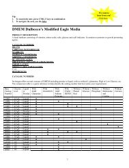
<strong>Kit</strong> Contents The <strong>Pichia</strong> <strong>Expression</strong> <strong>Kit</strong> is shipped at room temperature and contains the<br />
following components.<br />
iv<br />
Spheroplast Module (Box 1). Store at room temperature.<br />
Reagent Amount Components<br />
SOS medium 20 mL 1 M Sorbitol<br />
0.3X YPD<br />
10 mM CaCl2<br />
Sterile Water 2 � 125 mL Autoclaved, deionized water<br />
SE 2 � 125 mL 1 M Sorbitol<br />
25 mM EDTA, pH 8.0<br />
SCE 2 � 125 mL 1 M Sorbitol<br />
10 mM Sodium citrate buffer, pH 5.8<br />
1 mM EDTA<br />
1 M Sorbitol 2 � 125 mL --<br />
CaS 2 � 60 mL 1 M Sorbitol<br />
10 mM Tris-HCl, pH 7.5;<br />
10 mM CaCl2<br />
40% PEG 25 mL 40% (w/v) PEG 3350 (Reagent grade) in water<br />
CaT 25 mL 20 mM Tris-HCl, pH 7.5<br />
20 mM CaCl2<br />
Spheroplast Module (Box 2). Store at –20°C.<br />
Reagent Amount Components<br />
Zymolyase 10 � 20 μL 3 mg/mL Zymolyase in water<br />
(100,000 units/g lytic activity)<br />
1 M DTT 10 � 1 mL 1 M dithiothreitol in water<br />
Stab Vials: <strong>Pichia</strong> and E. coli stabs. Store at 4°C.<br />
Strain Amount Genotype Phenotype<br />
(<strong>Pichia</strong> only)<br />
GS115 1 stab his4 Mut +<br />
KM71 1 stab arg4 his4 aox1::ARG4 MutS , Arg +<br />
GS115 Albumin 1 stab HIS4 MutS GS115 �-Gal 1 stab HIS4 Mut +<br />
TOP10F´ 1 stab F´ {proAB, lacI q<br />
, lacZ�M15, Tn10 (TetR )} mcrA,<br />
�(mrr-hsdRMS-mcrBC), �80lacZ�M15, �lacX74,<br />
deoR, recA1, � – araD139, �(ara-leu)7697, galU,<br />
galK, rpsL(StrR ), endA1, nupG<br />
Continued on next page
<strong>Kit</strong> Contents and Storage, continued<br />
<strong>Kit</strong> Contents,<br />
continued<br />
Vectors. Store at –20°C.<br />
Reagent Description<br />
pHIL-D2<br />
Vector for intracellular expression in <strong>Pichia</strong>.<br />
10 μg, 20 μL at 0.5 μg/μL in<br />
TE buffer, pH 8.0*<br />
pPIC3.5<br />
Vector for intracellular expression in <strong>Pichia</strong>.<br />
10 μg, 20 μL at 0.5 μg/μL in<br />
TE buffer, pH 8.0<br />
pHIL-S1<br />
Vector for secreted expression in <strong>Pichia</strong>.<br />
10 μg, 20 μL at 0.5 μg/μL in Uses the PHO1 signal sequence.<br />
TE buffer, pH 8.0<br />
pPIC9<br />
Vector for secreted expression in <strong>Pichia</strong>.<br />
10 μg, 20 μL at 0.5 μg/μL in Uses the α-factor signal sequence.<br />
TE buffer, pH 8.0<br />
*TE buffer, pH 8.0: 10 mM Tris-HCl, 1 mM EDTA, pH 8.0<br />
Primers. Store at –20°C.<br />
5´ AOX1 sequencing primer<br />
2 μg (312 pmoles), lyophilized<br />
3´ AOX1 sequencing primer<br />
2 μg (314 pmoles), lyophilized<br />
�-Factor sequencing primer<br />
2 μg (315 pmoles), lyophilized<br />
5´-GACTGGTTCCAATTGACAAGC-3´<br />
5´-GCAAATGGCATTCTGACATCC-3´<br />
5´-TACTATTGCCAGCATTGCTGC-3´<br />
Media The following prepackaged media is included for your convenience. Instructions<br />
for use are provided on the package. Store at room temperature.<br />
Media Amount Yield<br />
YP Base Medium 2 pouches 2 liters of YP medium<br />
YP Base Agar Medium 2 pouches 2 liters of YP medium<br />
Yeast Nitrogen Base 1 pouch 500 mL of 10X YNB<br />
The <strong>Pichia</strong> Spheroplast Module for transforming <strong>Pichia</strong> by spheroplasting is<br />
available separately from <strong>Invitrogen</strong> (see page 88 for ordering information).<br />
Intended Use For research use only. Not intended for any animal or human therapeutic or<br />
diagnostic use.<br />
v
Required Equipment and Supplies<br />
Required Equipment<br />
and Supplies<br />
(not provided)<br />
vi<br />
• 30°C rotary shaking incubator<br />
• Water baths capable of 37°C, 45°C, and 100°C<br />
• Centrifuge suitable for 50 mL conical tubes (floor or table-top)<br />
• Baffled culture flasks with metal covers (50 mL, 250 mL, 500 mL, 1000 mL,<br />
and 3 L)<br />
• 50 mL sterile, conical tubes<br />
• 6 mL and 15 mL sterile snap-top tubes (Falcon 2059 or similar)<br />
• UV Spectrophotometer<br />
• Mini agarose gel apparatus and buffers<br />
• Agarose and low-melt agarose<br />
• Polyacrylamide gel electrophoresis apparatus and buffers<br />
• Media for transformation, growth, screening, and expression (see Recipes,<br />
pages 59–66)<br />
• 5% SDS solution (10 mL per transformation)<br />
• Sterile cheesecloth or gauze<br />
• Breaking Buffer (see Recipes, page 66)<br />
• Acid-washed glass beads (available from Sigma)<br />
• Replica-plating equipment (optional)<br />
• Bead Beater (optional, available from Biospec)
Introduction<br />
<strong>Pichia</strong> pastoris <strong>Expression</strong> System<br />
Review Articles The information presented here is designed to give you a concise overview of the<br />
<strong>Pichia</strong> pastoris expression system. It is by no means exhaustive. For further<br />
information, read the articles cited in the text along with the following review<br />
articles (Buckholz & Gleeson, 1991; Cregg & Higgins, 1995; Cregg et al., 1993;<br />
Nico-Farber et al., 1995; Romanos, 1995; Sreekrishna et al., 1988; Wegner, 1990). A<br />
general review of foreign gene expression in yeast is also available (Romanos et<br />
al., 1992).<br />
General<br />
Characteristics<br />
of <strong>Pichia</strong><br />
pastoris<br />
Similarity to<br />
Saccharomyces<br />
<strong>Pichia</strong> pastoris<br />
as a<br />
Methylotrophic<br />
Yeast<br />
As a eukaryote, <strong>Pichia</strong> pastoris has many of the advantages of higher eukaryotic<br />
expression systems such as protein processing, protein folding, and<br />
posttranslational modification, while being as easy to manipulate as E. coli or<br />
Saccharomyces cerevisiae. It is faster, easier, and less expensive to use than other<br />
eukaryotic expression systems such as baculovirus or mammalian tissue culture,<br />
and generally gives higher expression levels. As a yeast, it shares the advantages of<br />
molecular and genetic manipulations with Saccharomyces, and it has the added<br />
advantage of 10- to 100-fold higher heterologous protein expression levels. These<br />
features make <strong>Pichia</strong> very useful as a protein expression system.<br />
Many of the techniques developed for Saccharomyces may be applied to <strong>Pichia</strong>.<br />
These include:<br />
• Transformation by complementation<br />
• Gene disruption<br />
• Gene replacement<br />
In addition, the genetic nomenclature used for Saccharomyces has been applied to<br />
<strong>Pichia</strong>. For example, the HIS4 gene in both Saccharomyces and <strong>Pichia</strong> encodes<br />
histidinol dehydrogenase. There is also cross-complementation between gene<br />
products in both Saccharomyces and <strong>Pichia</strong>. Several wild-type genes from<br />
Saccharomyces complement comparable mutant genes in <strong>Pichia</strong>. Genes such as<br />
HIS4, LEU2, ARG4, TRP1, and URA3 all complement their respective mutant<br />
genes in <strong>Pichia</strong>.<br />
<strong>Pichia</strong> pastoris is a methylotrophic yeast, capable of metabolizing methanol as its<br />
sole carbon source. The first step in the metabolism of methanol is the oxidation of<br />
methanol to formaldehyde using molecular oxygen by the enzyme alcohol oxidase.<br />
In addition to formaldehyde, this reaction generates hydrogen peroxide. To avoid<br />
hydrogen peroxide toxicity, methanol metabolism takes place within a specialized<br />
cell organelle, called the peroxisome, which sequesters toxic by-products away<br />
from the rest of the cell. Alcohol oxidase has a poor affinity for O2, and <strong>Pichia</strong><br />
pastoris compensates by generating large amounts of the enzyme. The promoter<br />
regulating the production of alcohol oxidase is the one used to drive heterologous<br />
protein expression in <strong>Pichia</strong>.<br />
Continued on next page<br />
1
<strong>Pichia</strong> pastoris <strong>Expression</strong> System, continued<br />
Two Alcohol<br />
Oxidase Proteins<br />
Two genes in <strong>Pichia</strong> pastoris code for alcohol oxidase–AOX1 and AOX2. The AOX1<br />
gene product accounts for the majority of alcohol oxidase activity in the cell.<br />
<strong>Expression</strong> of the AOX1 gene is tightly regulated and induced by methanol to<br />
very high levels, typically � 30% of the total soluble protein in cells grown on<br />
methanol. The AOX1 gene has been isolated and a plasmid-borne version of the<br />
AOX1 promoter is used to drive expression of the gene of interest encoding the<br />
desired heterologous protein (Ellis et al., 1985; Koutz et al., 1989; Tschopp et al.,<br />
1987a). While AOX2 is about 97% homologous to AOX1, growth on methanol is<br />
much slower than with AOX1. This slow growth on methanol allows isolation of<br />
Mut S strains (aox1) (Cregg et al., 1989; Koutz et al., 1989).<br />
<strong>Expression</strong> <strong>Expression</strong> of the AOX1 gene is controlled at the level of transcription. In methanolgrown<br />
cells approximately 5% of the polyA + RNA is from the AOX1 gene. The<br />
regulation of the AOX1 gene is a two step process: a repression/derepression<br />
mechanism plus an induction mechanism (e.g., GAL1 gene in Saccharomyces<br />
(Johnston, 1987)). Briefly, growth on glucose represses transcription, even in the<br />
presence of the inducer methanol. For this reason, growth on glycerol is<br />
recommended for optimal induction with methanol. Note that growth on glycerol<br />
only (derepression) is not sufficient to generate even minute levels of expression<br />
from the AOX1 gene. The inducer, methanol, is necessary for even detectable levels<br />
of AOX1 expression (Ellis et al., 1985; Koutz et al., 1989; Tschopp et al., 1987a).<br />
Phenotype of<br />
aox1 mutants<br />
Intracellular and<br />
Secretory<br />
Protein<br />
<strong>Expression</strong><br />
2<br />
Loss of the AOX1 gene, and thus a loss of most of the cell's alcohol oxidase activity,<br />
results in a strain that is phenotypically Mut S (Methanol utilization slow). This has<br />
in the past been referred to as Mut – . The Mut S designation has been chosen to<br />
accurately describe the phenotype of these mutants. This results in a reduction in<br />
the cells' ability to metabolize methanol. The cells, therefore, exhibit poor growth<br />
on methanol medium. Mut + (Methanol utilization plus) refers to the wild type<br />
ability of strains to metabolize methanol as the sole carbon source. These two<br />
phenotypes are used when evaluating <strong>Pichia</strong> transformants for integration of your<br />
gene (Experimental Outline, page 4).<br />
Heterologous expression in <strong>Pichia</strong> pastoris can be intracellular or secreted.<br />
Secretion requires the presence of a signal sequence on the expressed protein to<br />
target it to the secretory pathway. While several different secretion signal<br />
sequences have been used successfully, including the native secretion signal<br />
present on some heterologous proteins, success has been variable. The secretion<br />
signal sequence from the Saccharomyces cerevisiae factor prepro peptide has been<br />
used with the most success (Cregg et al., 1993; Scorer et al., 1993).<br />
The major advantage of expressing heterologous proteins as secreted proteins is<br />
that <strong>Pichia</strong> pastoris secretes very low levels of native proteins. Since there is very<br />
low amount of protein in the minimal <strong>Pichia</strong> growth medium, this means that the<br />
secreted heterologous protein comprises the vast majority of the total protein in<br />
the medium and serves as the first step in purification of the protein (Barr et al.,<br />
1992) . However, that if there are recognized glycosylation sites (Asn-X-Ser/Thr)<br />
in your protein's primary sequence, glycosylation may occur at these sites.<br />
Continued on next page
<strong>Pichia</strong> pastoris <strong>Expression</strong> System, continued<br />
Posttranslational<br />
Modifications<br />
In comparison to Saccharomyces cerevisiae, <strong>Pichia</strong> may have an advantage in the<br />
glycosylation of secreted proteins because it may not hyperglycosylate. Both<br />
Saccharomyces cerevisiae and <strong>Pichia</strong> pastoris have a majority of N-linked<br />
glycosylation of the high-mannose type; however, the length of the<br />
oligosaccharide chains added posttranslationally to proteins in <strong>Pichia</strong> (average<br />
8–14 mannose residues per side chain) is much shorter than those in<br />
Saccharomyces cerevisiae (50–150 mannose residues) (Grinna and Tschopp, 1989;<br />
Tschopp et al., 1987b). Very little O-linked glycosylation has been observed in<br />
<strong>Pichia</strong>.<br />
In addition, Saccharomyces cerevisiae core oligosaccharides have terminal<br />
�1,3 glycan linkages whereas <strong>Pichia</strong> pastoris does not. It is believed that the<br />
�1,3 glycan linkages in glycosylated proteins produced from Saccharomyces<br />
cerevisiae are primarily responsible for the hyper-antigenic nature of these proteins<br />
making them particularly unsuitable for therapeutic use. Although not yet<br />
proven, this is predicted to be less of a problem for glycoproteins generated in<br />
<strong>Pichia</strong> pastoris, because it may resemble the glycoprotein structure of higher<br />
eukaryotes (Cregg et al., 1993).<br />
3
Experimental Outline<br />
Selection of<br />
Vector and<br />
Cloning<br />
Transformation<br />
and Integration<br />
4<br />
To utilize the strong, highly inducible PAOX1 promoter for expressing your protein,<br />
four expression vectors are included in this kit. pHIL-D2 and pPIC3.5 are used for<br />
intracellular expression, and pHIL-S1 and pPIC9 are used for secreted expression<br />
(see pages 14–17 for more information). Before cloning your insert, you must:<br />
� decide whether you want intracellular or secreted expression.<br />
� analyze your insert for the following restriction sites: Sac I, Stu I, Sal I, Not I,<br />
and Bgl II. We recommend these sites for linearizing your construct prior to<br />
<strong>Pichia</strong> transformation. If your insert has all of these sites, refer to pages 29–30<br />
for alternate sites.<br />
Two different phenotypic classes of His + recombinant strains can be generated:<br />
Mut + and MutS . MutS refers to the "Methanol utilization slow" phenotype caused<br />
by the loss of alcohol oxidase activity encoded by the AOX1 gene. A strain with a<br />
MutS phenotype has a mutant aox1 locus, but is wild type for AOX2. This results<br />
in a slow growth phenotype on methanol medium. Transformation of strain<br />
GS115 can yield both classes of transformants, His + Mut + and His + MutS , while<br />
KM71 yields only His + MutS , because the strain itself is MutS . Both Mut + and<br />
MutS recombinants are useful to have, because one phenotype may favor better<br />
expression of your protein than the other. Because of clonal variation, you<br />
should test 6–10 recombinants per phenotype. There is no way to predict<br />
beforehand which construct or isolate will better express your protein. We<br />
strongly recommend that you analyze <strong>Pichia</strong> recombinants by PCR to confirm the<br />
integration of your construct (see page 43).<br />
After you have successfully cloned your gene, you will linearize your plasmid to<br />
stimulate recombination when the plasmid is transformed into <strong>Pichia</strong>. The table<br />
below describes the types of recombinants you will get by selective digestion of<br />
your plasmid.<br />
Restriction<br />
Enzyme<br />
Integration Event GS115 Phenotype KM71 Phenotype<br />
Sal I or Stu I Insertion at his4 His + Mut + His + MutS Sac I Insertion at 5´<br />
AOX1 region<br />
His + Mut + His + MutS Not I or Bgl II Replacement at<br />
AOX1 locus<br />
His + Mut S<br />
His + Mut +<br />
His + Mut S (not<br />
recommended, see<br />
page 7)<br />
Continued on next page
Experimental Outline, continued<br />
<strong>Expression</strong> and<br />
Scale-up<br />
Experimental<br />
Process<br />
Generating<br />
Recombinant<br />
Strain<br />
After confirming your <strong>Pichia</strong> recombinants by PCR, you will test expression of<br />
both His + Mut + and His + MutS recombinants. This procedure involves growing a<br />
small culture of each recombinant, inducing them with methanol, and taking time<br />
points. If looking for intracellular expression, analyze the cell pellet from each<br />
time point by SDS polyacrylamide gel electrophoresis (SDS-PAGE). If looking for<br />
secreted expression, analyze both the cell pellet and supernatant from each time<br />
point. We recommend that you analyze your SDS-PAGE gels by Coomassie<br />
staining and, if you have an antibody to your protein, by western blot. We also<br />
suggest checking for protein activity by an activity assay, if one is available. Not<br />
all proteins express to the level of grams per liter, so it is advisable to check by<br />
western blot or activity assay, and not just by Coomassie staining of SDS-PAGE<br />
gels for production of your protein.<br />
Choose the <strong>Pichia</strong> recombinant strain that best expresses your protein and<br />
optimize induction based on the suggestions on pages 52–53. After you optimize<br />
expression, scale-up your expression protocol to produce more protein.<br />
The overall experimental process is divided into two major sections: Generating<br />
Recombinant Strain and Induction (Mut + and/or Mut S ). Each section contains a<br />
table outlining the major steps of the experimental process. Each step is<br />
discussed in detail further in the manual. Refer to the indicated pages to read<br />
about particular steps of interest. The discussion about recombination and<br />
integration in <strong>Pichia</strong> will help you choose the right vector. For more information,<br />
refer to the review by Higgins (Higgins, 1995).<br />
The goal of this section is to create a <strong>Pichia</strong> pastoris strain containing your<br />
integrated gene of interest. Before starting your experiments, determine which<br />
vector to use.<br />
Step Procedure Page<br />
1 Select the appropriate expression vector (For more information,<br />
refer to Recombination and Integration in <strong>Pichia</strong>, pages 69–72)<br />
11–17<br />
2 Clone your gene of interest into selected vector 19–24<br />
3 Transform E. coli, select ampicillin-resistant transformants, and<br />
confirm the presence and orientation of your gene of interest<br />
25<br />
4 Linearize the constructs with appropriate restriction enzymes to<br />
generate His + MutS and His + Mut + recombinant strains<br />
27–30<br />
5 Transform and select His + transformants (GS115 recombinants,<br />
His + Mut + ; KM71 recombinants, His + MutS )<br />
31–37<br />
6 Screen His + transformants for Mut + and MutS strains (6–10<br />
recombinants of each phenotype)<br />
38–42<br />
7 Confirm the integration of your gene of interest in Mut + and<br />
MutS recombinants by PCR<br />
43–44<br />
Continued on next page<br />
5
Experimental Outline, continued<br />
Mut + Induction The method of induction depends on whether the recombinant is Mut + or Mut S .<br />
The differences primarily occur in the culture volumes and the time of induction<br />
(see below). Refer to the following pages for more detailed instructions.<br />
Step Procedure Page<br />
1 Guidelines for expression of recombinant proteins in <strong>Pichia</strong> 45–46<br />
2 Grow His + Mut + recombinants in 25 mL of buffered glycerol<br />
medium to a final OD600 = 2–6<br />
47<br />
3 Harvest the cells and resuspend them to an OD600 of 1.0<br />
(~100–200 mL) with methanol medium. Place the cell<br />
suspension in a 1 liter baffled flask<br />
47<br />
4 Incubate the culture at 30°C with shaking and take samples for<br />
analysis at 0, 6, 12, 24, 36, 48, 60, 72, 84, and 96 hours<br />
47<br />
5 Analyze the medium (if protein of interest is targeted for<br />
secretion) and the cell lysates (for intracellular and secreted<br />
expression) for protein via PAGE/Coomassie Blue staining,<br />
western blot, activity, ELISA, or immunoprecipitation<br />
49–51<br />
6 Optimize expression of your His + Mut + recombinant 52–53<br />
7 Scale-up your expression for protein purification 54–56<br />
Mut S Induction This is very similar to Mut + induction except that Mut S grow very slowly on<br />
methanol. To compensate, cells are concentrated to increase cell mass before<br />
induction.<br />
6<br />
Step Procedure Page<br />
1 Guidelines for expression of recombinant proteins in <strong>Pichia</strong> 45–46<br />
2 Grow His + MutS recombinants in 100–200 mL of buffered<br />
glycerol medium to a final OD600 = 2–6<br />
48<br />
3 Harvest the cells and resuspend them to an OD600 of 10.0<br />
(~10–20 mL) with methanol medium. Place the cell suspension<br />
in a 100 mL or 250 mL baffled flask.<br />
48<br />
4 Incubate the culture at 30°C with shaking and take samples for<br />
analysis at 0, 24, 48, 72, 96, 120, and 144 hours<br />
48<br />
5 Analyze the medium (if protein of interest is targeted for<br />
secretion) and the cell lysates (for intracellular and secreted<br />
expression) for protein via PAGE/Coomassie Blue staining,<br />
western blot, activity, ELISA, or immunoprecipitation<br />
49–51<br />
6 Optimize expression of your His + Mut + recombinant 52–53<br />
7 Scale-up your expression for protein purification 54–56
<strong>Pichia</strong> Strains<br />
Methods<br />
Introduction <strong>Pichia</strong> pastoris is quite similar to Saccharomyces cerevisiae as far as general growth<br />
conditions and handling. You should be familiar with basic microbiological and<br />
sterile techniques before attempting to grow and manipulate any<br />
microorganism. You should also be familiar with basic molecular biology and<br />
protein chemistry. Some general references to consult are Guide to Yeast Genetics<br />
and Molecular Biology (Guthrie & Fink, 1991), Current Protocols in Molecular Biology<br />
(Ausubel et al., 1994), Molecular Cloning: A Laboratory Manual (Sambrook et al.,<br />
1989), Protein Methods (Bollag et al., 1996), and Guide to Protein Purification<br />
(Deutscher, 1990).<br />
Genotype of<br />
<strong>Pichia</strong> Strain<br />
Construction of<br />
KM71<br />
Important<br />
The <strong>Pichia</strong> host strains GS115 and KM71 have a mutation in the histidinol<br />
dehydrogenase gene (his4) that prevents them from synthesizing histidine. All<br />
expression plasmids carry the HIS4 gene that complements his4 in the host, so<br />
transformants are selected for their ability to grow on histidine-deficient medium.<br />
Spontaneous reversion of GS115 and KM71 to His + prototrophy is less than 1 out<br />
of 108 .<br />
The parent strain of KM71 has a mutation in the argininosuccinate lyase gene<br />
(arg4) that prevents the strain from growing in the absence of arginine. The wildtype<br />
ARG4 gene was used to disrupt AOX1, creating KM71, a MutS , Arg + , His –<br />
strain.<br />
Both GS115 and KM71 will grow on complex medium such as YPD (also known<br />
as YEPD) and on minimal media supplemented with histidine. Until transformed,<br />
neither GS115 nor KM71 will grow on minimal medium alone as they are His – .<br />
Note: Mut S (Methanol utilization slow) phenotype has in the past been referred to as Mut – .<br />
The Mut S designation has been chosen to accurately describe the phenotype of these<br />
mutants.<br />
The ARG4 gene (~2 kb) was inserted into the cloned, wild-type AOX1 gene<br />
between the BamH I site (codons 15/16 of AOX1) and the Sal I site (codons<br />
227/228 of AOX1). ARG4 replaces codons 16 through 227 of AOX1. This construct<br />
was transformed into the parent strain of KM71 (arg4 his4) and Arg +<br />
transformants were isolated and analyzed for the Mut S phenotype. Genetic<br />
analysis of Arg + transformants showed that the wild-type AOX1 gene was<br />
replaced by the aox1::ARG4 construct.<br />
The advantage of using KM71 is that there is no need to screen for the Mut<br />
phenotype on methanol minimal medium. All transformants will be Mut S .<br />
Secondly, since the AOX1 locus was not completely deleted, it is theoretically<br />
possible to replace aox1::ARG4 with your construct by gene replacement. The<br />
phenotype of this strain would be His + Mut S Arg – . This means the recombinant<br />
strain would require arginine in the medium to grow. Unfortunately, simple<br />
inclusion of arginine does not totally alleviate the effects of the arg4 mutation, and<br />
arg4 strains do not grow well on minimal medium supplemented with arginine.<br />
Therefore, we do not recommend that you generate His + transformants in KM71<br />
by replacing the aox1::ARG4 construct.<br />
Continued on next page<br />
7
<strong>Pichia</strong> Strains, continued<br />
Control<br />
<strong>Expression</strong><br />
Strains<br />
Growth of <strong>Pichia</strong><br />
Strains<br />
Growth on<br />
Methanol<br />
8<br />
GS115/His + MutS Albumin: This strain is a control for secreted expression and<br />
the MutS phenotype when screening <strong>Pichia</strong> transformants (page 38). The gene for<br />
serum albumin was cloned with its native secretion signal, then integrated into<br />
<strong>Pichia</strong> at the AOX1 locus. This strain secretes albumin (67 kDa) into the medium<br />
at levels > 1 gram/liter.<br />
GS115/His + Mut + �-galactosidase: This strain is a control for intracellular<br />
expression and the Mut + phenotype when screening <strong>Pichia</strong> transformants<br />
(page 38). The gene for �-galactosidase (lacZ) was integrated into <strong>Pichia</strong> at the<br />
his4 locus. This strain expresses �-galactosidase (117 kDa) at levels that can be<br />
detected on Coomassie-stained SDS-PAGE (see pages 49–51) or assayed using<br />
ONPG (see page 86–87).<br />
The growth temperature of <strong>Pichia</strong> pastoris is 28–30°C for liquid cultures, plates,<br />
and slants. Growth above 32°C during induction can be detrimental to protein<br />
expression and can even lead to cell death. Other important facts:<br />
• Doubling time of log phase Mut + or MutS <strong>Pichia</strong> in YPD is ~2 hours<br />
• Mut + and MutS strains do not differ in growth rates unless grown on<br />
methanol<br />
• Doubling time of log phase Mut + <strong>Pichia</strong> in methanol medium (MM) is<br />
4–6 hours<br />
• Doubling time of log phase MutS <strong>Pichia</strong> in MM is ~18 hours<br />
• One OD600 = ~5 � 107 cells/mL<br />
Note that growth characteristics may vary depending on the recombinant<br />
strain.<br />
When plates or medium containing methanol are used as growth medium, it is<br />
advisable to add methanol every day to compensate for loss due to evaporation<br />
or consumption.<br />
• For plates add 100 μL of 100% methanol to the lid of the inverted plate.<br />
• For liquid medium add 100% methanol to a final concentration of 0.5%.<br />
Some researchers have had success adding methanol to 1% every day for MutS strains and up to 3% for Mut + without any negative effect to their liquid culture.<br />
Continued on next page
<strong>Pichia</strong> Strains, continued<br />
RECOMMENDATION<br />
Storing <strong>Pichia</strong><br />
Strains<br />
Make frozen stocks for long-term storage of all three <strong>Pichia</strong> strains included in<br />
this kit (see below).<br />
To store cells for weeks to months, use YPD medium or YPD agar slants (see<br />
page 61).<br />
1. Streak for single colonies of the desired strain on YPD.<br />
2. Transfer one colony to a YPD stab and grow for 2 days at 30°C.<br />
3. You can store the cells on YPD for several weeks at 4°C.<br />
To store cells for months to years, store frozen at –80°C.<br />
1. Culture a single colony of the desired strain overnight in YPD.<br />
2. Harvest the cells and suspend in YPD containing 15% glycerol at a final<br />
OD600 of 50–100 (approximately 2.5 � 109 –5.0 � 109 cells/mL).<br />
3. Freeze the cells in liquid nitrogen or a dry ice/ethanol bath, and store<br />
at –80°C.<br />
After extended storage at 4°C or –80°C, we recommend checking the His +<br />
transformants for correct genotype and viability by streaking on MM, MD or<br />
MGY plates before using again.<br />
9
E. coli Strains<br />
Genotype of<br />
E. coli Strain<br />
10<br />
RECOMMENDATION<br />
The E. coli strain, TOP10F´ is provided in case no suitable E. coli strain is<br />
available. Other strains which may be suitable are TOP10, DH5�F´, JM109, or<br />
any other strain which is recombination deficient (recA) and deficient in<br />
endonuclease A (endA).<br />
F´ {proAB, lacI q<br />
, lacZ�M15, Tn10 (TetR )} mcrA, �(mrr-hsdRMS-mcrBC),<br />
�80lacZ�M15, �lacX74, recA1, � – araD139, �(ara-leu)7697, galU, galK, rpsL(StrR ),<br />
endA1, nupG<br />
Note: If you do not plan to perform single-stranded DNA rescue, E. coli strains<br />
that do not carry the F´ episome are also suitable for use.<br />
We recommend that you make a frozen stock of TOP10F´ to keep on hand.<br />
1. Culture TOP10F´ in 5 mL LB with 10 μg/mL tetracycline. Grow overnight.<br />
2. Mix thoroughly 0.85 mL of culture with 0.15 mL sterile glycerol.<br />
3. Transfer to a freezer vial and freeze in liquid nitrogen or a dry ice/ethanol<br />
bath.<br />
4. Store at –80°C.
Selecting a <strong>Pichia</strong> <strong>Expression</strong> Vector<br />
Generic<br />
Structure<br />
All the vectors included in this kit share several general features shown in black,<br />
while some of the vectors also have signal sequences (Sig) and/or an f1<br />
bacteriophage origin. For details of each individual plasmid refer to pages 14–17.<br />
Not I or<br />
Bgl II<br />
f1 ori<br />
Sac I<br />
Amp<br />
Not I or<br />
Bgl II<br />
Sig<br />
5' AOX1<br />
MCS<br />
Transcription<br />
Termination (TT)<br />
3' AOX1<br />
There is no yeast origin of replication in any of the <strong>Pichia</strong> expression vectors<br />
included in this kit. His + transformants can only be isolated if recombination<br />
occurs between the plasmid and the <strong>Pichia</strong> genome.<br />
HIS4<br />
Sal I<br />
Stu I<br />
Continued on next page<br />
11
Selecting a <strong>Pichia</strong> <strong>Expression</strong> Vector, continued<br />
Features The table below describes the general and optional features of the <strong>Pichia</strong><br />
expression vectors.<br />
12<br />
Feature Description Benefit<br />
5´ AOX1 An ~1000 bp fragment<br />
containing the AOX1 promoter<br />
Sig DNA sequence coding for an<br />
N-terminal protein secretion<br />
signal<br />
Allows methanol-inducible high<br />
level expression in <strong>Pichia</strong><br />
Targets plasmid integration to the<br />
AOX1 locus.<br />
Targets desired protein for<br />
secretion<br />
MCS Multiple Cloning Site Allows insertion of your gene into<br />
the expression vector<br />
TT Native transcription<br />
termination and<br />
polyadenylation signal from<br />
AOX1 gene (~260 bp)<br />
HIS4 <strong>Pichia</strong> wild-type gene coding<br />
for histidinol dehydrogenase<br />
(~2.4 kb) and used to<br />
complement <strong>Pichia</strong> his4 strains<br />
3´ AOX1 Sequences from the AOX1 gene<br />
that are further 3´ to the TT<br />
sequences (~650 bp)<br />
Amp Ampicillin resistance gene<br />
pBR322 E. coli origin of replication<br />
origin<br />
f1 origin Bacteriophage f1 origin of<br />
replication (458 bp)<br />
Not I<br />
Bgl II<br />
Sac I<br />
Sal I<br />
Stu I<br />
Permits efficient transcription<br />
termination and polyadenylation<br />
of the mRNA<br />
Provides a selectable marker to<br />
isolate <strong>Pichia</strong> recombinant strains<br />
Targets plasmid integration at the<br />
AOX1 gene<br />
Allows selection, replication, and<br />
maintenance in E. coli<br />
Permits generation of singlestranded<br />
DNA for mutagenesis<br />
Unique restriction sites Permits linearization of vector for<br />
efficient integration into the <strong>Pichia</strong><br />
genome<br />
Continued on next page
Selecting a <strong>Pichia</strong> <strong>Expression</strong> Vector, continued<br />
Selecting a<br />
Vector<br />
If your protein is cytosolic and non-glycosylated, you may elect to express the<br />
protein intracellularly. However, there is evidence of a non-glycosylated protein<br />
being secreted without extensive modification (Despreaux and Manning, 1993).<br />
Note that the protein in question was a secreted, bacterial protein with one<br />
N-glycosylation site. Check your protein sequence for possible N-glycosylation<br />
sites (Asn-X-Ser/Thr) before cloning a cytosolic protein into a secretion vector.<br />
If your protein is normally secreted, glycosylated, or directed to an intracellular<br />
organelle, you may wish to try secreting your protein. We recommend that you<br />
try both the native secretion signal and the α-factor signal sequence (in pPIC9) to<br />
secrete your protein. There has been better success reported with the α-factor<br />
signal sequence than with the PHO1 signal sequence in pHIL-S1. This may be<br />
due to the lack of KEX2-like processing signals in the PHO1 signal sequence<br />
(Laroche et al., 1994).<br />
13
pHIL-D2<br />
Description The details of pHIL-D2 are listed below:<br />
• 8,209 bp nonfusion vector<br />
• One unique EcoR I site<br />
• For intracellular expression of your gene<br />
• Requires an initiating ATG codon in a Kozak consensus sequence for<br />
proper translation initiation of your gene (Cavener and Stuart, 1991; Kozak,<br />
1987; Kozak, 1990)<br />
• HIS4 selection in <strong>Pichia</strong><br />
• For insertion at AOX1 in GS115 or KM71, linearize with Sac I (generates<br />
His + Mut + in GS115 and His+ MutS in KM71)<br />
• For insertion at HIS4, linearize with Sal I or Stu I (generates His + Mut + in<br />
GS115 and His + MutS in KM71)<br />
• For a gene replacement at AOX1 in GS115, linearize with Not I (generates<br />
His + MutS )<br />
Refer to page 29 for alternate restriction sites if your insert DNA has a Not I,<br />
Sac I, Sal I, or Stu I site.<br />
Map of pHIL-D2 The map below shows the location and size of each feature of pHIL-D2. For the<br />
details of the multiple cloning site refer to page 21. The complete sequence of<br />
pHIL-D2 is available at www.invitrogen.com or from Technical Support<br />
(page 89).<br />
Comments for pHIL-D2:<br />
8209 nucleotides<br />
5' AOX1 promoter fragment: bases 14-941<br />
5' AOX1 primer site: bases 868-888<br />
EcoR I Site: bases 956-961<br />
3' AOX1 primer site: bases 1036-1056<br />
3' AOX1 transcription<br />
termination (TT) fragment: bases 963-1295<br />
HIS4 ORF: bases 4223-1689<br />
3' AOX1 fragment: bases 4578-5334<br />
Ampicillin resistance gene: bases 5686-6546<br />
f1 origin of replication: bases 7043-6588<br />
pBR322 origin: bases 7138-7757<br />
14<br />
f1 ori<br />
Not I<br />
+1<br />
pBR322<br />
Ampicillin<br />
5' AOX1<br />
EcoR I<br />
pHIL-D2<br />
Not I<br />
8.2 kb<br />
3' AOX1<br />
3' AOX1 (TT)<br />
HIS4<br />
Sal I<br />
Stu I
pPIC3.5<br />
Description The details of pHIL-D2 are listed below:<br />
� 7,751 bp nonfusion vector<br />
� BamH I, SnaB I, EcoR I, Avr II, Not I unique sites<br />
� Intracellular expression of your gene<br />
� Requires an initiating ATG codon in a Kozak consensus sequence for<br />
proper translation initiation of your gene (Cavener and Stuart, 1991; Kozak,<br />
1987; Kozak, 1990)<br />
� HIS4 selection in <strong>Pichia</strong><br />
� For insertion at AOX1 in GS115 or KM71, linearize with Sac I (generates<br />
His + Mut + in GS115 and His + MutS in KM71)<br />
� For insertion at HIS4, linearize with Sal I or Stu I (generates His + Mut + in<br />
GS115 and His + MutS in KM71)<br />
� For a gene replacement at AOX1 in GS115, linearize with Bgl II (generates<br />
His + MutS )<br />
Refer to page 29 for alternate restriction sites if your insert DNA has a Not I,<br />
Sac I, Sal I, or Stu I site.<br />
Map of pPIC3.5 The map below shows the location and size of each feature of pPIC3.5. For the<br />
details of the multiple cloning site refer to page 22. The complete sequence of<br />
pPIC3.5 is available at www.invitrogen.com or from Technical Support<br />
(page 89).<br />
Comments for pPIC3.5:<br />
7751 nucleotides<br />
5´ AOX1 promoter fragment: bases 1-937<br />
5´ AOX1 primer site: bases 855-875<br />
Multiple Cloning Site: bases 938-968<br />
3´ AOX1 primer site: bases 1055-1075<br />
3´ AOX1 transcription termination<br />
(TT) fragment: bases 981-1314<br />
HIS4 ORF: bases 4242-1708<br />
3´ AOX1 fragment: bases 4598-5354<br />
pBR322 origin: bases 6436-5764<br />
Ampicillin resistance gene: bases 7442-6582<br />
pBR322<br />
Sac I<br />
Bgl II<br />
+1<br />
Ampicillin<br />
Bgl II<br />
BamH I<br />
SnaB I<br />
EcoR I<br />
Avr II<br />
Not I<br />
5'<br />
pPIC3.5<br />
7.8 kb<br />
3' AOX1<br />
TT<br />
3´ AOX1 (TT)<br />
HIS4<br />
Sal I<br />
Stu I<br />
15
pHIL-S1<br />
Description The details of pHIL-S1 are listed below:<br />
• 8,260 bp fusion vector<br />
• Xho I, EcoR I, Sma I, BamH I unique sites<br />
• Secreted expression using the PHO1 secretion signal<br />
• For expression, your gene must be cloned in frame with the initiation codon<br />
of the signal sequence.<br />
• HIS4 selection in <strong>Pichia</strong><br />
• For insertion at AOX1 in GS115 or KM71, linearize with Sac I (generates His +<br />
Mut + in GS115 or His + MutS in KM71)<br />
• For insertion at HIS4 in GS115 or KM71, linearize with Sal I or Stu I<br />
(generates His + Mut + in GS115 or His + MutS in KM71)<br />
• For gene replacement at AOX1 in GS115, linearize with Bgl II (generates His +<br />
MutS )<br />
Refer to page 30 for alternate restriction sites if your insert DNA has a Bgl II,<br />
Sac I, Sal I, or Stu I site.<br />
Map of pHIL-S1 The map below shows the location and size of each feature of pHIL-S1. For the<br />
details of the multiple cloning site, refer to page 23. The complete sequence of<br />
pHIL-S1 is available at www.invitrogen.com or from Technical Support<br />
(page 89).<br />
Comments for pHIL-S1:<br />
8260 nucleotides<br />
5' AOX1 promoter fragment: bases 1-941<br />
5' AOX1 primer site: bases 856-876<br />
PHO1 secretion signal (S): bases 942-1007<br />
Multiple Cloning Site Region: bases 1006-1026<br />
3' AOX1 primer site: bases 1099-1119<br />
3' AOX1 transcription<br />
termination (TT) fragment: bases 1025-1190<br />
HIS4 ORF: bases 4286-1753<br />
3' AOX1 fragment: bases 4641-5397<br />
pBR322 origin: bases 6556-5937<br />
f1 origin of replication: bases 6651-7106<br />
Ampicillin resistance gene: bases 7922-7062<br />
16<br />
Bgl II<br />
+1<br />
f1 ori<br />
Ampicillin<br />
pBR322<br />
Xho I<br />
EcoR I<br />
Sma I<br />
BamH I<br />
5' AOX1 S<br />
pHIL-S1<br />
Bgl II<br />
8.3 kb<br />
3' AOX1<br />
3' AOX1 (TT)<br />
HIS4<br />
Sal I<br />
Stu I
pPIC9<br />
Description The details of pHIL-S1 are listed below:<br />
• 8,023 bp fusion vector<br />
• Xho I, SnaB I, EcoR I, Avr II, Not I unique sites<br />
• Secreted expression of your gene using the -factor secretion signal<br />
• For expression, your gene must be cloned in frame with the initiation codon<br />
of the signal sequence.<br />
• HIS4 selection in <strong>Pichia</strong><br />
• For insertion at AOX1 in GS115 or KM71, linearize with Sac I (generates His +<br />
Mut + in GS115 and His + MutS in KM71)<br />
• For insertion at HIS4, linearize with Sal I or Stu I (generates His + Mut + in<br />
GS115 and His + MutS in KM71)<br />
• For gene replacement at AOX1 in GS115, linearize with Bgl II (generates His +<br />
MutS )<br />
Refer to page 30 for alternate restriction sites if your insert DNA has a Bgl II,<br />
Sac I, Sal I, or Stu I site.<br />
Map of pPIC9K The figure below shows the map of pPIC9. Details of the multiple cloning site are<br />
provided on page 24. The sequence of pPIC9 is available at www.invitrogen.com<br />
or from Technical Support (page 89).<br />
Comments for pPIC9:<br />
8023 nucleotides<br />
5´ AOX1 promoter fragment: bases 1-948<br />
5´ AOX1 primer site: bases 855-875<br />
a-Factor secretion signal(s): bases 949-1215<br />
a-Factor primer site: bases 1152-1172<br />
Multiple Cloning Site: bases 1192-1241<br />
3´ AOX1 primer site: bases 1327-1347<br />
3´ AOX1 transcription<br />
termination (TT): bases 1253-1586<br />
HIS4 ORF: bases 4514-1980<br />
3´ AOX1 fragment: bases 4870-5626<br />
pBR322 origin: bases 6708-6034<br />
Ampicillin resistance gene: bases 7713-6853<br />
Bgl II<br />
+1<br />
Ampicillin<br />
pBR322<br />
Bgl II<br />
5' AOX1<br />
Xho I<br />
SnaB I<br />
EcoR I<br />
Avr II<br />
Not I<br />
S<br />
TT<br />
pPIC9<br />
8.0 kb<br />
3' AOX1<br />
3´ AOX1 (TT)<br />
HIS4<br />
Sal I<br />
Stu I<br />
17
Signal Sequence Processing<br />
Signal Sequence<br />
Processing<br />
Optimizing<br />
Signal Cleavage<br />
18<br />
When cloning into the Xho I site of pPIC9, the secretion signal sequence between<br />
the Xho I site and SnaB I may need to be regenerated.<br />
The processing of the α-factor mating signal sequence in pPIC9 occurs in two<br />
steps:<br />
1. The preliminary cleavage of the signal sequence by the KEX2 gene product,<br />
with the final KEX2 cleavage occurring between arginine and glutamine in<br />
the sequence Glu-Lys-Arg * Glu-Ala-Glu-Ala, where * is the site of cleavage.<br />
2. The STE13 gene product further cleaves the Glu-Ala repeats.<br />
In Saccharomyces cerevisiae, the Glu-Ala repeats are not necessary for cleavage by<br />
KEX2, but the KEX2 cleavage after Glu-Lys-Arg may be more efficient when<br />
followed by Glu-Ala repeats. A number of amino acids are tolerated at site X<br />
instead of Glu in the sequence Glu-Lys-Arg-X. These amino acids include the<br />
aromatic amino acids, small amino acids, and histidine. Proline, however, inhibits<br />
KEX2 cleavage. For more information on KEX2 cleavage, see (Brake et al., 1984).<br />
There are some cases where STE13 cleavage of Glu-Ala repeats is not efficient,<br />
and Glu-Ala repeats are left on the N-terminus of the expressed protein. This is<br />
generally dependent on the protein of interest.<br />
The PHO1 signal sequence is atypical of signal sequences even though it is a native<br />
<strong>Pichia</strong> secretion signal. If cloning into the Xho I site, we recommend regenerating<br />
the full signal sequence between the Xho I and EcoR I sites (see page 23). However,<br />
recent evidence suggests that the PHO1 signal sequence might have to be modified<br />
to include KEX2-like processing sites for efficient cleavage to occur (Laroche et al.,<br />
1994).
Cloning into the <strong>Pichia</strong> <strong>Expression</strong> Vectors<br />
Introduction After selecting a vector into which to clone your gene of interest (see pages 11–17),<br />
develop a cloning strategy. The AOX1 promoter and the multiple cloning site are<br />
presented on the following pages for each vector along with a summary of<br />
considerations for each vector to help you decide on a strategy.<br />
RECOMMENDATION<br />
General<br />
Considerations<br />
We recommend that you transform the three supercoiled <strong>Pichia</strong> expression<br />
vectors into E. coli to prepare permanent stock.<br />
� Resuspend each vector in 10 μL sterile water to prepare a 1 μg/μL solution.<br />
Store the stock solution at –20°C.<br />
� Use the stock solution to transform competent E. coli and select<br />
transformants on LB agar plates containing 50–100 μg/mL ampicillin<br />
(LB-Amp).<br />
The following are some general considerations applicable to all vectors.<br />
� The codon usage in <strong>Pichia</strong> is believed to be the same as Saccharomyces<br />
cerevisiae because many genes have proven to be cross-functional.<br />
� Maintain plasmid constructions in a recA mutant E. coli strain such as the<br />
TOP10F´ strain provided in the kit.<br />
� The native 5´ end of the AOX1 mRNA is noted in each multiple cloning site.<br />
This information is necessary to calculate the size of the expressed mRNA of<br />
the gene of interest.<br />
� Translation termination is determined by either stop codons in the gene of<br />
interest or in the 3´ AOX1 sequence. The stop codons in the 3´ AOX1<br />
sequence are noted in each figure on the following pages.<br />
� The premature termination of transcripts due to "AT rich regions" has been<br />
observed in <strong>Pichia</strong> and other eukaryotic systems (Henikoff and Cohen, 1984;<br />
Irniger et al., 1991; Scorer et al., 1993; Zaret and Sherman, 1984). If you are<br />
expressing a gene with high AT content, refer to page 53.<br />
� The predicted protease cleavage sites for the PHO1 and α-factor signal<br />
sequences are indicated in each figure.<br />
� If you are attempting to secrete a protein using its native secretion signal, we<br />
recommend that you also try pPIC9 in parallel. When using pPIC9, the open<br />
reading frame (ORF) of the mature gene of interest is cloned in frame and<br />
downstream of the α-factor.<br />
Continued on next page<br />
19
Cloning into the <strong>Pichia</strong> <strong>Expression</strong> Vectors, continued<br />
General Cloning<br />
Strategies<br />
Cloning<br />
Procedures<br />
Bacterial<br />
Transformation<br />
20<br />
Strategies generally fall into three different categories:<br />
1. Ligation of a compatible restriction fragment:<br />
a. Forced (directional) insertion involving the use of two different sites in<br />
the multiple cloning site (for pPIC3.5, pHIL-S1, or pPIC9 vectors).<br />
b. Ligation of the fragment with the same restriction end on both ends into a<br />
single, compatible site (e.g. EcoR I cloning in pHIL-D2).<br />
2. PCR amplification of the fragment containing the gene of interest in such a<br />
way that compatible restriction ends are generated for ligation into the<br />
appropriate vector.<br />
3. Direct cloning of an amplified fragment containing the gene of interest via the<br />
TA Cloning ® <strong>Kit</strong> (see page 88 for ordering), followed by subcloning of a<br />
compatible fragment into the appropriate <strong>Pichia</strong> expression vector.<br />
Refer to (Ausubel et al., 1994), pages 3.16.1 to 3.17.3. or (Sambrook et al., 1989),<br />
pages 5.10 to 5.13. for help with cloning.<br />
Once you have decided on a cloning strategy, you will need to prepare competent<br />
E. coli cells for transformation before setting up your ligation reactions. See<br />
Current Protocols in Molecular Biology (Ausubel et al., 1994) or Molecular Biology: A<br />
Laboratory Manual (Sambrook et al., 1989) for preparation of electrocompetent or<br />
chemically competent E. coli or use your laboratory's procedure.<br />
For a high-efficiency and convenient transformation, we recommend One Shot ®<br />
TOP10 Chemically Competent E. coli or One Shot ® TOP10 Electrocompetent Cells,<br />
which are available separately from <strong>Invitrogen</strong> (see page 88 for ordering<br />
information).<br />
Continued on next page
Cloning into the <strong>Pichia</strong> <strong>Expression</strong> Vectors, continued<br />
PAOX1 and<br />
Multiple Cloning<br />
Site of pHIL-D2<br />
Special<br />
Considerations<br />
The figure below shows the detail of the multiple cloning site and surrounding<br />
sequences.<br />
ACAGGCAATA TATAAACAGA AGGAAGCTGC CCTGTCTTAA ACCTTTTTTT TTATCATCAT TATTAGCTTA<br />
CTTTCATAAT TGCGACTGGT TCCAATTGAC AAGCTTTTGA TTTTAACGAC TTTTAACGAC AACTTGAGAA<br />
GATCAAAAAA CAACTAATTA TTCGAAACGA GGAATTCGCC TTAGACATGA CTGTTCCTCA GTTCAAGTTG<br />
GGCACTTACG AGAAGACCGG TCTTGCTAGA TTCTAATCAA GAGGATGTCA GAATGCCATT TGCCTGAGAG<br />
ATGCAGGCTT CATTTTTGAT ACTTTTTTAT TTGTAACCTA TATAGTATAG GATTTTTTTT GTCA<br />
� For pHIL-D2, the fragment containing the gene of interest should have a<br />
yeast consensus sequence (Romanos et al., 1992). An example of a yeast<br />
consensus sequence is provided below. The ATG initiation codon is shown<br />
underlined.<br />
(A/Y)A(A/T)AATGTCT<br />
Note that other sequences are also possible. Although not as strong as the<br />
mammalian Kozak translation initiation sequence, the yeast consensus<br />
sequence is thought to have a 2- to 3-fold effect on the efficiency of<br />
translation initiation.<br />
• Shorter, 5´ untranslated leaders reportedly work better in AOX1 expression.<br />
In pHIL-D2, make the untranslated region as short as possible when cloning<br />
your gene.<br />
• If your insert has a Not I site, refer to page 29 for alternate restriction sites to<br />
linearize your plasmid for <strong>Pichia</strong> transformation.<br />
Continued on next page<br />
21
Cloning into the <strong>Pichia</strong> <strong>Expression</strong> Vectors, continued<br />
PAOX1 and<br />
Multiple Cloning<br />
Site of pPIC3.5<br />
Special<br />
Considerations<br />
22<br />
The sequence below shows the detail of the multiple cloning site and<br />
surrounding sequences.<br />
AOX1 mRNA 5'end (824)<br />
5' AOX 1 Primer Site (855-875)<br />
TTATCATCAT TATTAGCTTA CTTTCATAAT TGCGACTGGT TCCAATTGAC AAGCTTTTGA TTTTAACGAC<br />
TTTTAACGAC AACTTGAGAA GATCAAAAAA CAACTAATTA TTCGAAGGAT CCTACGTAGA ATTCCCTAGG<br />
Not I<br />
GCGGCCGCGA ATTAATTCGC CTTAGACATG ACTGTTCCTC AGTTCAAGTT GGGCACTTAC GAGAAGACCG<br />
3' AOX 1 Primer Site (1055-1075)<br />
BamH I<br />
GTCTTGCTAG ATTCTAATCA AGAGGATGTC AGAATGCCAT TTGCCTGAGA GATGCAGGCT TCATTTTTGA<br />
AOX1 mRNA 3' end (1146)<br />
TACTTTTTTA TTTGTAACCT ATATAGTATA GGATTTTTTT TGTCATTTTG TTTCTTC<br />
SnaB I EcoR I Avr II<br />
� For pPIC3.5, the fragment containing the gene of interest should have a yeast<br />
consensus sequence (Romanos et al., 1992). An example of a yeast consensus<br />
sequence is provided below. The ATG initiation codon is shown underlined.<br />
(A/Y)A(A/T)AATGTCT<br />
Note that other sequences are also possible. Although not as strong as the<br />
mammalian Kozak translation initiation sequence, the yeast consensus<br />
sequence is thought to have a 2- to 3-fold effect on the efficiency of<br />
translation initiation.<br />
� Shorter, 5´ untranslated leaders reportedly work better in AOX1 expression.<br />
In pPIC3.5, make the untranslated region as short as possible when cloning<br />
your gene.<br />
� If you are digesting with BamH I and SnaB I or SnaB I and EcoR I, digest with<br />
SnaB I first. If you digest with BamH I or EcoR I first, the SnaB I site will be<br />
too close to the end of the DNA and will not digest properly.<br />
� If your insert has a Bgl II, Sac I, Sal I, or Stu I site, refer to page 29 for alternate<br />
restriction sites to linearize your plasmid for <strong>Pichia</strong> transformation.<br />
Continued on next page
Cloning into the <strong>Pichia</strong> <strong>Expression</strong> Vectors, continued<br />
P AOX1 and<br />
Multiple Cloning<br />
Site of pHIL-S1<br />
Special<br />
Considerations<br />
The sequence below shows the detail of the multiple cloning site and<br />
surrounding sequences.<br />
773<br />
AOX1 mRNA 5´ end (825)<br />
ACAGGCAATA TATAAACAGA AGGAAGCTGC CCTGTCTTAA ACCTTTTTTT TTATCATCAT<br />
5´ AOX1 primer site (856-876)<br />
TATTAGCTTA CTTTCATAAT TGCGACTGGT TCCAATTGAC AAGCTTTTGA TTTTAACGAC<br />
PHO1 (942-1007)<br />
TTTTAACGAC AACTTGAGAA GATCAAAAAA CAACTAATTA TTCGAAACG ATG TTC TCT<br />
Met Phe Ser<br />
CCA ATT TTG TCC TTG GAA ATT ATT TTA GCT TTG GCT ACT TTG CAA TCT GTC<br />
Pro Ile Leu Ser Leu Glu Ile Ile Leu Ala Leu Ala Thr Leu Gln Ser Val<br />
PHO1 cleavage site<br />
Xho I* EcoRI Sma I BamHI<br />
TTC GCT CGA GAA TTC CCC GGG ATC CTT AGA CAT GAC TGT TCC TCA GTT CAA<br />
Phe Ala Arg Glu Phe Pro Gly Ile Leu Arg His Asp Cys Ser Ser Val Gln<br />
Stop (1083)<br />
GTT GGG CAC TTA CGA GAA GAC CGG TCT TGC TAG ATTCTAATCA AGAGGATGTC<br />
Val Gly His Leu Arg Glu Asp Arg Ser Cys ***<br />
3´ AOX1 primer site (1099-1119)<br />
AGAATGCCAT TTGCCTGAGA GATGCAGGCT TCATTTTTGA TACTTTTTTA TTTGTAACCT<br />
AOX1 mRNA 3´ end (1190)<br />
ATATAGTATA GGATTTTTTT TGTCA<br />
* If the Xho I site (which is part of the PHO1 signal cleavage sequence) is used for cloning, it must be<br />
recreated in order for efficient cleavage of the fusion protein to occur.<br />
� The fragment containing the gene of interest must be cloned in frame with the<br />
secretion signal open reading frame.<br />
� If the Xho I site is used for cloning, it must be recreated for efficient cleavage<br />
of the fusion protein to occur. It is part of the PHO1 signal peptide sequence.<br />
� An initiating ATG is provided by the signal sequence. Translation will initiate<br />
at the ATG closest to the 5´ end of the mRNA.<br />
� If your insert has a Not I site, refer to page 29 for alternate restriction sites to<br />
linearize your plasmid for <strong>Pichia</strong> transformation.<br />
� The PHO1 cleavage site has been confirmed for several different fusion<br />
proteins by N-terminal peptide sequencing.<br />
� In -factor (pPIC9) or native general, more success has been reported with the<br />
secretion signals than with pHIL-S1. This may be due to the lack of KEX2-like<br />
processing signals (Laroche et al., 1994).<br />
Continued on next pa<br />
23
Cloning into the <strong>Pichia</strong> <strong>Expression</strong> Vectors, continued<br />
P AOX1 and<br />
Multiple Cloning<br />
Site of pPIC9<br />
Special<br />
Considerations<br />
24<br />
The sequence below shows the detail of the multiple cloning site and<br />
surrounding sequences.<br />
773<br />
AOX1 mRNA 5´ end (824)<br />
ACAGCAATAT ATAAACAGAA GGAAGCTGCC CTGTCTTAAA CCTTTTTTTT TATCATCATT ATTAGCTTAC<br />
5´ AOX1 Primer Site (855-875)<br />
TTTCATAATT GCGACTGGTT CCAATTGACA AGCTTTTGAT TTTAACGACT TTTAACGACA ACTTGAGAAG<br />
a-Factor (949-1215)<br />
ATCAAAAAAC AACTAATTAT TCGAAGGATC CAAACG ATG AGA TTT CCT TCA ATT TTT ACT GCA<br />
Met Arg Phe Pro Ser Ile Phe Thr Ala<br />
GTT TTA TTC GCA GCA TCC TCC GCA TTA GCT GCT CCA GTC AAC ACT ACA ACA GAA GAT<br />
Val Leu Phe Ala Ala Ser Ser Ala Leu Ala Ala Pro Val Asn Thr Thr Thr Glu Asp<br />
GAA ACG GCA CAA ATT CCG GCT GAA GCT GTC ATC GGT TAC TCA GAT TTA GAA GGG GAT<br />
Glu Thr Ala Gln Ile Pro Ala Glu Ala Val Ile Gly Tyr Ser Asp Leu Glu Gly Asp<br />
TTC GAT GTT GCT GTT TTG CCA TTT TCC AAC AGC ACA AAT AAC GGG TTA TTG TTT ATA<br />
Phe Asp Val Ala Val Leu Pro Phe Ser Asn Ser Thr Asn Asn Gly Leu Leu Phe Ile<br />
a-Factor Primer Site (1152-1172)<br />
AAT ACT ACT ATT GCC AGC ATT GCT GCT AAA GAA GAA GGG GTA TCT CTC GAG AAA AGA<br />
Asn Thr Thr Ile Ala Ser Ile Ala Ala Lys Glu Glu Gly Val Ser Leu Glu Lys Arg<br />
Signal cleavage (1204)<br />
SnaB I EcoR I Avr II Not I<br />
GAG GCT GAA GCT TAC GTA GAA TTC CCT AGG GCG GCC GCG AAT TAA<br />
Glu Ala Glu Ala Tyr Val Glu Phe Pro Arg Ala Ala Ala Asn ***<br />
TTCGCCTTAG<br />
ACATGACTGT TCCTCAGTTC AAGTTGGGCA CTTACGAGAA GACCGGTCTT GCTAGATTCT AATCAAGAGG<br />
3´ AOX1 Primer Site (1327-1347)<br />
ATGTCAGAAT GCCATTTGCC TGAGAGATGC AGGCTTCATT TTTGATACTT TTTTATTTGT AACCTATATA<br />
GTATAGGATT TTTTTTGTCA<br />
AOX1 mRNA 3´ end (1418)<br />
* If cloning into the Xho I site, the sequence between the Xho I site and SnaB I site (underlined) must be<br />
recreated in order for efficient cleavage of the fusion protein to occur.<br />
� The fragment containing the gene of interest must be cloned in frame with the<br />
secretion signal open reading frame.<br />
� If the Xho I site is used for cloning, the sequence between the Xho I site and<br />
the SnaB I site encoding the KEX2 site (Glu-Lys-Arg-X) must be recreated for<br />
efficient cleavage of the fusion protein to occur. It is part of the α-factor signal<br />
peptide sequence. Refer to the discussion on page 18.<br />
� An initiating ATG is provided by the signal sequence. Translation will initiate<br />
at the ATG closest to the 5´ end of the mRNA.<br />
� If your insert has a Bgl II site, refer to page 30 for alternate restriction sites to<br />
linearize your plasmid for <strong>Pichia</strong> transformation.<br />
Xho I
Transformation into E. coli<br />
Introduction At this point you have ligation reactions that you will transform by chemical<br />
means or electroporation into competent E. coli cells (TOP10F´ or equivalent). For<br />
procedures to prepare competent cells, refer to Current Protocols in Molecular<br />
Biology (Ausubel et al., 1994) or Molecular Biology: A Laboratory Manual (Sambrook<br />
et al., 1989).<br />
Analyzing<br />
Transformants<br />
Sequencing<br />
Recombinant<br />
Clones<br />
1. After transformation, plate the transformation mix onto LB plates with<br />
50–100 μg/mL ampicillin (see Appendix, page 59), and select ampicillin<br />
resistant colonies.<br />
2. Pick 10 ampicillin resistant transformants and inoculate into LB medium<br />
with 50–100 μg/mL ampicillin. Grow overnight at 37°C with shaking.<br />
3. Isolate plasmid DNA by miniprep for restriction analysis and sequencing<br />
(see below). To sequence the <strong>Pichia</strong> expression vectors, use the primers<br />
provided.<br />
4. Make a glycerol stock of your desired clone for safekeeping by combining<br />
0.85 mL of an overnight bacterial culture with 0.15 mL of sterile glycerol. Mix<br />
by vortexing and transfer to a labeled storage tube. Freeze the tube in liquid<br />
nitrogen or a dry ice/ethanol bath and store at –70°C.<br />
5. After you confirm your construct by sequencing, proceed to Preparing<br />
Transforming DNA, page 27.<br />
We strongly recommend that you sequence your construct before transforming<br />
into <strong>Pichia</strong> to confirm the following:<br />
• The correct reading frame (for secretion)<br />
• An ATG in the proper context for eukaryotic translation initiation<br />
Use the primers listed on the next page to sequence your constructs. Resuspend<br />
each primer in 20 �L sterile water to prepare a stock solution of 0.1 �g/�L. For the<br />
location of the priming sites, see pages 21–24.<br />
For sequencing protocols, refer to Unit 7 in Current Protocols in Molecular Biology<br />
(Ausubel et al., 1994) or Chapter 13 in Molecular Cloning: A Laboratory Manual<br />
(Sambrook et al., 1989).<br />
Continued on next page<br />
25
Transformation into E. coli, continued<br />
5´ AOX1<br />
Sequencing<br />
Primer<br />
3´ AOX1<br />
Sequencing<br />
Primer<br />
α-Factor<br />
Sequencing<br />
Primer<br />
26<br />
� is 5´ GACTGGTTCCAATTGACAAGC 3´<br />
� hybridizes 5´ of the MCS in the AOX1 promoter region<br />
� allows the determination of the 5´ AOX1-gene of interest junction<br />
� confirms that the ORFs are preserved where necessary<br />
� is 5´ GCAAATGGCATTCTGACATCC 3´<br />
� hybridizes just 3´ of the MCS in the 3´ AOX1 (TT) region<br />
� allows the determination of the 3´ AOX1-gene of interest junction<br />
� is 5´-TACTATTGCCAGCATTGCTGC-3´<br />
� hybridizes within the -factor leader region in pPIC9<br />
� allows the determination of the 5´ end of the gene of interest<br />
� confirms that the ORFs are preserved where necessary
Preparing Transforming DNA<br />
Introduction You should have a <strong>Pichia</strong> multi-copy expression vector with your gene of interest<br />
cloned in the correct orientation for expression. The table below describes what<br />
you will be doing in the next few sections.<br />
RECOMMENDATION<br />
Preparing<br />
Plasmid DNA<br />
Step Action Pages<br />
1 Prepare your DNA for transformation 28<br />
2 Grow GS115 or KM71 to prepare spheroplasts 32<br />
3 Prepare spheroplasts for transformation 33<br />
4 Transform GS115 or KM71 with your DNA 35<br />
5 Select His + transformants and characterize for Mut + /Mut S<br />
phenotype<br />
6 Test 10 His + Mut + and 10 His + Mut S by PCR for integration<br />
of your gene<br />
We recommend isolating both His + Mut + and His + Mut S <strong>Pichia</strong> transformants as it<br />
is difficult to predict beforehand what construct will best express your protein<br />
(see pages 69 and 70). By linearizing your construct DNA in the 5´ AOX1 region<br />
or in the HIS4 gene and using GS115 (Mut + ) and KM71 (Mut S ), you can easily<br />
isolate Mut + and Mut S recombinants. Plan on using ~10 μg digested DNA for each<br />
transformation.<br />
Plasmid DNA for <strong>Pichia</strong> transformation should be at least pure enough for<br />
restriction digestion; however, the cleaner the DNA, the more efficient the<br />
transformation. We recommend the PureLink HiPure Plasmid Miniprep <strong>Kit</strong><br />
(see page 88) to prepare plasmid DNA for routine <strong>Pichia</strong> transformations. Refer<br />
to www.invitrogen.com or contact Technical Support for more information on a<br />
large selection of plasmid purification columns. You may prepare plasmid DNA<br />
using alkaline lysis, phenol:chloroform extraction, and ethanol precipitation.<br />
38<br />
43<br />
Continued on next page<br />
27
Preparing Transforming DNA, continued<br />
Linearizing<br />
Plasmid DNA<br />
We recommend that you linearize your vector in such a manner to generate both<br />
Mut + and MutS recombinants. It is possible that one phenotype will express your<br />
protein of interest better than the other will.<br />
� To isolate His + Mut + transformants of GS115, linearize all constructs with<br />
Sal I, Stu I, or Sac I.<br />
� To isolate His + MutS transformants of KM71, linearize plasmid constructs<br />
with Sal I, Stu I, or Sac I.<br />
Note: If your insert DNA has all three sites, please see the next page.<br />
� To isolate His + MutS transformants of GS115, linearize plasmid constructs<br />
with Not I (pHIL-D2) or Bgl II (pPIC3.5, pHIL-S1, and pPIC9)<br />
Note: If you wish to generate recombinants that are MutS , use KM71 because it is<br />
much easier and more efficient to generate MutS recombinant strains using single<br />
crossover events than double crossover events (e.g. insertions at AOX1 or his4 as<br />
opposed to gene replacement at AOX1).<br />
Procedure 1. Digest both your construct and the parent vector. You will transform GS115<br />
and/or KM71 with the parent vector as a background control for<br />
expression.<br />
2. Analyze a small portion of your digest by agarose gel electrophoresis to<br />
confirm complete digestion of your fragment. The number of transformants<br />
and frequency of targeting will be reduced if digestion is not complete.<br />
3. Extract the digest with phenol:chloroform:isoamyl alcohol (25:24:1) and<br />
ethanol precipitate the digested DNA. Resuspend DNA pellet in 10–20 μL<br />
of TE buffer. It is not necessary to purify the fragment containing your gene<br />
away from the rest of the plasmid.<br />
4. Store at –20°C until ready to transform.<br />
28<br />
Continued on next page
Preparing Transforming DNA, continued<br />
Alternate<br />
Restriction Sites<br />
If your insert DNA contains Sac I, Sal I, and Stu I sites, you need to linearize your<br />
construct with another enzyme. Use the following table to select another<br />
enzyme. A single digestion which linearizes the vector in either of the AOX1<br />
recombination sequences allows integration, but at lower efficiencies. Remember<br />
to digest the parent vector with the same enzyme when preparing your DNA<br />
samples for transformation.<br />
pPHIL-D2<br />
Restriction<br />
Enzyme<br />
5´ AOX1<br />
14–940 bp<br />
3´ AOX1<br />
4,577–5,333 bp<br />
Vector<br />
backbone<br />
5,333+ bp<br />
HIS4 gene<br />
1,688–4,222 bp<br />
Sac I 221 – – –<br />
Pme I 424 – – –<br />
Bpu 1102 I 599 – – –<br />
Nsi I 689 – – –<br />
Xcm I 711 – – –<br />
Not I 8 5,337 – –<br />
Dra I* 424 5,169; 5,311 5,896; 6,588 –<br />
Sal I – – – 2,887<br />
Stu I – – – 2,972<br />
BspE I – – – 3,554<br />
pPIC3.5<br />
Restriction<br />
Enzyme<br />
5´ AOX1<br />
1–937 bp<br />
3´ AOX1<br />
4,616–5,393 bp<br />
Vector<br />
backbone<br />
5,393+ bp<br />
HIS4 gene<br />
1,715–4,249 bp<br />
Sac I 209 – – –<br />
Pme I 414 – – –<br />
Bpu 1102 I 589 – – –<br />
Nsi I 678 – – –<br />
Xcm I 699 – – –<br />
Bgl II 2 5363 – –<br />
Dra I* 414 5,201; 5,343 6,534; 6,553;<br />
7,245<br />
–<br />
Sal I – – – 2,919<br />
Stu I – – – 3,004<br />
BspE I – – – 3,586<br />
Continued on next page<br />
29
Preparing Transforming DNA, continued<br />
Alternate Restriction Sites, continued<br />
30<br />
pHIL-S1<br />
Restrictio<br />
n Enzyme<br />
5´ AOX1<br />
3´ AOX1<br />
Vector<br />
backbone<br />
HIS4 gene<br />
1–940 bp 4,639–5,395 bp<br />
1,750–4,284 bp<br />
Sac I 209 –<br />
5,395+ bp<br />
– –<br />
Pme I 412 – – –<br />
Bpu 1102 I 587 – – –<br />
Nsi I 677 – – –<br />
Xcm I 699 – – –<br />
Bgl II 2 5,394 – –<br />
Dra I* 412 5,232; 5,374 7,021; 7,713 –<br />
Sal I – – – 2,950<br />
pPIC9<br />
Restriction<br />
Enzyme<br />
5´ AOX1<br />
1–948 bp<br />
3´ AOX1<br />
4,881–5,638 bp<br />
Vector<br />
backbone<br />
5,638+ bp<br />
HIS4 gene<br />
1,980–4,514 bp<br />
Sac I 209 – – –<br />
Pme I 414 – – –<br />
Bpu 1102 I 589 – – –<br />
Nsi I 678 – – –<br />
Xcm I 699 – – –<br />
Bgl II 2 5,622 – –<br />
Dra I* 414 5,460; 5,602 6,793; 6,812;<br />
7,504<br />
–<br />
Sal I – – – 3,178<br />
Stu I – – – 3,263<br />
BspE I – – – 3,845<br />
*Restriction sites are used to generate gene replacements at AOX1 in GS115 only.
Growing <strong>Pichia</strong> for Spheroplasting<br />
Introduction In general, spheroplasting and electroporation (page 73) provide the highest<br />
efficiency of transformation for most researchers (10 3 to 10 4 transformants per μg<br />
DNA). <strong>Pichia</strong> can also be transformed using PEG 1000 (page 74) or lithium<br />
chloride (page 76). These two protocols, particularly lithium chloride, do not<br />
perform as well as spheroplasting or electroporation. If you do not have an<br />
electroporation device, we recommend spheroplasting or using the PEG 1000<br />
method. Transformation in <strong>Pichia</strong> is less efficient than for Saccharomyces. For<br />
references on general yeast transformation, see (Cregg et al., 1985; Hinnen et al.,<br />
1978).<br />
Spheroplasting The cell wall of yeast prevents uptake of DNA. To enable yeast to take up DNA, it<br />
is necessary to partially remove the cell wall. Zymolyase is a �-glucanase that<br />
hydrolyzes the glucose polymers with �1,3 linkages in the cell wall. Addition of<br />
Zymolyase partially digests the cell wall. It is critical not to overdigest the cell<br />
wall as doing so will cause the cells to die. Zymolyase digestion is monitored by<br />
the sensitivity of the cells to SDS. Aliquots of cells are added to SDS, lysing the<br />
spheroplasts. This causes a clearing of the solution that is monitored by the<br />
absorbance (light-scattering) at 800 nm. It has been empirically determined that<br />
when 70% spheroplasting has been achieved, digestion is optimal. Cells are then<br />
washed with an isotonic solution to remove the enzyme and incubated with<br />
DNA. The cells are resuspended in sorbitol to facilitate cell wall regeneration and<br />
plated.<br />
Preparing Media Prepare the following media several days in advance and store at 4°C (see<br />
Appendix, pages 61–64 for details):<br />
YPD (Yeast extract Peptone Dextrose) medium, 1 liter<br />
YPD plates, 1 liter<br />
RDB (Regeneration Dextrose Base) plates, 1 liter<br />
RDHB (Regeneration Dextrose Histidine Base) plates, 1 liter<br />
Prepare the following solution on the day of transformation and maintain at 45°C:<br />
5% SDS solution in water<br />
RD (Regeneration Dextrose), molten agarose, 100 mL<br />
Continued on next page<br />
31
Growing <strong>Pichia</strong> for Spheroplasting, continued<br />
Solutions Spheroplasting and Transformation Reagents<br />
Provided:<br />
1 M Sorbitol<br />
SE: 1 M sorbitol, 25 mM EDTA, pH 8.0<br />
DTT: 1 M DTT in water<br />
SCE:<br />
5.8<br />
1 M sorbitol, 1 mM EDTA and 10 mM sodium citrate buffer, pH<br />
CaS: 1 M sorbitol, 10 mM Tris-HCl, pH 7.5, 10 mM CaCl2<br />
Zymolyase: 3 mg/mL in water<br />
40% PEG: 40% (w/v) PEG 3350 (Reagent grade) in water<br />
CaT: 20 mM Tris, pH 7.5 and 20 mM CaCl2<br />
SOS: 1 M sorbitol, 0.3X YPD, 10 mM CaCl2<br />
Prepared fresh for each transformation:<br />
SED: 19 mL of SE and 1 mL of 1 M DTT (see page 33)<br />
PEG/CaT: 1:1 mixture of 40% PEG and CaT (see page 35)<br />
Procedure 1. Streak GS115 or KM71 onto a YPD plate such that isolated, single colonies<br />
will grow. Incubate the plate at 28–30°C for 2 days.<br />
2. Inoculate 10 mL of YPD in a 50 mL conical tube or 100 mL shake flask with a<br />
single colony of GS115 or KM71 from the YPD plate and grow overnight at<br />
28–30°C with vigorous shaking (250–300 rpm). You may store this culture at<br />
4°C for several days.<br />
3. Place 200 mL of YPD in each of three 500 mL culture flasks. Inoculate the<br />
flasks with 5, 10, and 20 μL of cells from the culture made in Step 2 and<br />
incubate them overnight with vigorous shaking (250–300 rpm) at 28–30°C.<br />
4. The next morning, bring the transformation solutions (SE, SCE, Sterile<br />
Water, SOS, PEG, CaS, CaT, 1 M sorbitol) provided in the kit, the RDB plates<br />
(for plating transformants), and the RDHB plates (for viability control) to<br />
room temperature.<br />
5. Check the OD600 of each of the three culture flasks. Harvest the cells from the<br />
culture that has an OD600 between 0.2 and 0.3. Centrifuge the cells at room<br />
temperature for 5–10 minutes at 1,500 � g. Decant the supernatant and<br />
discard the other cultures. Proceed to Preparing Spheroplasts, page 33.<br />
Note: If the cultures are all over 0.3, choose one of the cultures and dilute (1:4) with<br />
fresh medium and incubate at 28–30°C until the OD600 is between 0.2 and 0.3<br />
(2–4 hours). Harvest the cells and proceed as in Step 5, above.<br />
32
Preparing Spheroplasts<br />
Before Starting You should have a cell pellet from Step 5, page 32.<br />
Washing the<br />
Cells<br />
Adding<br />
Zymolyase<br />
• Prepare 100 mL of molten RD agarose and keep at 45°C (see Appendix,<br />
page 64)<br />
• Thaw one tube of 1 M DTT (provided in the kit)<br />
• Prepare fresh SED for one batch of spheroplasts as follows:<br />
Using sterile technique, transfer 19 mL of SE (provided) to an appropriate sterile<br />
container (e.g., 50 mL conical tube). Add 1 mL of 1 M DTT and mix well. For best<br />
results this solution of SED should be made and used immediately.<br />
The quality and freshness of DTT is critical for a successful spheroplast<br />
preparation. The 1 M DTT provided is analytical reagent grade and must be<br />
stored at –20°C.<br />
1. Wash the cells from Step 5, page 32 by resuspending the pellet in 20 mL of<br />
sterile water (provided). Resuspend the pellet by swirling the tube. Transfer<br />
to a sterile, 50 mL conical tube.<br />
2. Pellet the cells by centrifugation at 1,500 � g for 5 minutes at room<br />
temperature. Decant and discard the supernatant. The cell pellet will be<br />
used to prepare spheroplasts.<br />
3. Wash the cell pellet once by resuspending in 20 mL of fresh SED, prepared<br />
above and centrifuge at 1,500 � g for 5 minutes at room temperature.<br />
4. Wash the cells once with 20 mL of 1 M sorbitol and centrifuge as described<br />
in Step 2.<br />
5. Resuspend the cells by swirling in 20 mL of SCE buffer and divide the<br />
suspension into two 50 mL conical tubes (~10 mL each).<br />
6. Remove one tube of Zymolyase from –20°C and place it on ice. Mix well by<br />
flicking the tube several times. Zymolyase is provided as a slurry and does<br />
not go into solution. It is important to mix the slurry thoroughly before each<br />
use to ensure addition of a consistent amount of Zymolyase.<br />
Use one tube of cells prepared above to determine the optimal time of digestion<br />
with Zymolyase to make spheroplasts. After you determine the optimal, use the<br />
other tube of to make spheroplasts.<br />
Zymolyase digests the cell wall and makes the cells extremely fragile. Handle the<br />
sample gently. The moment after adding Zymolyase, the cell wall begins to get<br />
digested.<br />
• Prepare at least 20 mL of a 5% SDS solution (not provided) for use below.<br />
• Set your UV-Vis spectrophotometer to 800 nm and blank with 800 μL 5%<br />
SDS and 200 μL SCE.<br />
• Set up 17 sterile microcentrifuge tubes and label them 0, 2, 4, 5, 6, 7, 8, 9, 10,<br />
15, 20, 25, 30, 35, 40, 45, and 50. Add 800 μL of 5% SDS to each tube.<br />
Continued on next page<br />
33
Preparing Spheroplasts, continued<br />
Adding<br />
Zymolyase,<br />
continued<br />
34<br />
1. From one tube of cells (Step 5, page 33), withdraw 200 μL cells and add to the<br />
tube marked "0". This is your zero time point. Set the tube aside on ice.<br />
2. Add 7.5 μL of Zymolyase to the same tube of cells, mix it gently by inversion,<br />
and incubate the cells at 30°C. Do not shake the sample. This sample will be<br />
used to establish the incubation time for optimal spheroplasting as described<br />
below. Keep the second tube of cells at room temperature for use in Step 6<br />
below. Keep the remainder of the Zymolyase on ice.<br />
3. Monitor the formation of the spheroplasts as follows: At time 2 minutes,<br />
withdraw 200 μL of cells (from the suspension in Step 2) and add to the tube<br />
marked "2". Repeat at time t= 4, 5, 6, 7, 8, 9, 10, 15, 20, 25, 30, 35, 40, 45, and 50<br />
minutes after adding Zymolyase. Read the OD800 for all samples.<br />
4. Determine the percent of spheroplasting for each time point using the<br />
equation:<br />
% Spheroplasting = 100 – [(OD800 at time t/OD800 at time 0) � 100]<br />
For example: At time t = 0, the OD800 = 0.256<br />
At time t = 15, the OD800 = 0.032<br />
Calculation: % spheroplasting = 100 – [(0.032/0.256) � 100]<br />
= 100 – [(0.125) � 100]<br />
= 100 – 12.5<br />
= 87.5%<br />
5. Determine the time of incubation that results in approximately 70%<br />
spheroplasting. This time of incubation is variable due to differences in lots of<br />
Zymolyase. In <strong>Invitrogen</strong> labs, it takes approximately 15–40 minutes of<br />
Zymolyase treatment to achieve optimal spheroplasting.<br />
Note: It is important to establish the minimum time required for the desired amount of<br />
spheroplasting. Prolonged incubation with Zymolyase is deleterious to spheroplasts<br />
and will result in lower transformation efficiency.<br />
6. Add 7.5 μL Zymolyase to the remaining tube of cells as described in Step 1<br />
above. Incubate the tube at 30°C for the time that was established in Step 5 to<br />
obtain the optimal level (70%) of spheroplasting.<br />
7. Harvest the spheroplasts by centrifugation at 750 � g for 10 minutes at room<br />
temperature. Decant and discard the supernatant.<br />
8. Wash the spheroplasts once with 10 mL of 1 M sorbitol (gently disperse the<br />
pellet by tapping the tube, do not vortex). Collect the spheroplasts by<br />
centrifugation at 750 � g for 10 minutes at room temperature.<br />
9. Wash the spheroplasts once with 10 mL of CaS and centrifuge as in Step 7.<br />
Gently resuspend the spheroplasts in 0.6 mL of CaS. The spheroplasts must<br />
be used immediately (up to 30 minutes) for transformation (page 35). They<br />
cannot be stored for much longer. This preparation yields enough<br />
spheroplasts for six transformations.
Transforming <strong>Pichia</strong><br />
Before Starting Make sure your RDB plates are at room temperature and that you have molten<br />
RD top agarose available. Thaw your linearized DNA and keep on ice. You<br />
should have the following:<br />
� Your construct linearized with Sal I, Stu I, or Sac I to favor isolation of His +<br />
Mut + recombinants in GS115<br />
� Your construct linearized with Sal I, Stu I, or Sac I to favor isolation of His +<br />
MutS recombinants in KM71<br />
� Your construct linearized with Not I, Bgl II, or equivalent to favor isolation of<br />
His + MutS recombinants in GS115<br />
� Parent plasmid linearized with same restriction enzyme<br />
Controls should include no DNA or linearized pBR322 DNA and plasmid only<br />
(no cells) to check for contamination.<br />
Procedure 1. For each transformation, dispense 100 μL of the spheroplast preparation<br />
from Step 9 (previous page) into a sterile 15 mL snap-top Falcon 2059 tube<br />
(or equivalent).<br />
2. Add 10 μg of DNA and incubate the tube at room temperature for<br />
10 minutes.<br />
3. During the 10 minute incubation, make a fresh PEG/CaT solution. Since<br />
each transformation requires 1.0 mL of the PEG/CaT solution, calculate the<br />
amount you need and prepare this volume by adding together equal<br />
volumes of 40% PEG and CaT (a 1:1 solution).<br />
4. Add 1.0 mL of fresh PEG/CaT solution to the cells and DNA, mix gently,<br />
and incubate at room temperature for 10 minutes.<br />
5. Centrifuge the tube at 750 � g for 10 minutes at room temperature and<br />
carefully aspirate the PEG/CaT solution. Invert the tube and tap it gently to<br />
drain the excess PEG/CaT solution.<br />
6. Resuspend the pellet of transformed cells in 150 μL of SOS medium and<br />
incubate it at room temperature for 20 minutes.<br />
7. Add 850 μL of 1 M sorbitol. Proceed to Plating, next page.<br />
Continued on next page<br />
35
Transforming <strong>Pichia</strong>, continued<br />
Plating <strong>Pichia</strong> spheroplasts need to be plated in top agarose or agar to protect them from<br />
lysis prior to selection.<br />
Evaluating Your<br />
Transformation<br />
Experiment<br />
1. Mix together 100–300 μL of each spheroplast-DNA solution from Step 7,<br />
previous page, with 10 mL of molten RD agarose and pour on RDB plates.<br />
Allow the top agarose to harden.<br />
Note: There is enough of the spheroplast-DNA solution to plate duplicate and<br />
triplicate plates.<br />
2. Invert plates and incubate at 28–30°C. Transformants should appear in<br />
4–6 days.<br />
3. For cell viability: Mix 100 μL of spheroplasts with 900 μL of 1 M sorbitol.<br />
4. Mix 100 μL of this diluted sample with 10 mL of molten RDH and pour on a<br />
RDHB plate. Allow top agarose to harden.<br />
5. Invert the plates and incubate at 28–30°C. Appearance of colonies after<br />
4–6 days demonstrates that the spheroplasts can regenerate into dividing<br />
cells.<br />
After 4–6 days, His + transformants on your sample plates will become apparent.<br />
Transformation efficiency using the spheroplast method is generally 10 3 to 10 4<br />
His + transformants/μg of DNA. There should be no colonies on the "No DNA",<br />
pBR322 plate, or the plasmid only (no cells) plate<br />
Optional Method Plating in top agarose can cause the transformants to be on top or be imbedded<br />
in the top agarose, making it difficult to pick and patch colonies as described in<br />
the next section. The following protocol allows you to collect the transformants<br />
and re-plate them directly onto plates without using top agarose.<br />
1. Scrape the agarose containing the His + transformants with a sterile spreader<br />
into a sterile, 50 mL, conical centrifuge tube and mix with 20 mL sterile<br />
deionized water. Vortex the suspension vigorously to separate the cells from<br />
the agarose.<br />
2. Filter the suspension through 4 folds of sterile cheesecloth. Centrifuge the<br />
filtrate at 1,500 × g for 5 minutes at room temperature. Centrifugation pellets<br />
the cells on the bottom of the tube and any remaining agarose on top of the<br />
cells.<br />
3. Remove the agarose pellet carefully from the top of the cells by gently<br />
shaking the tube to disperse only the agarose pellet into the water. Decant<br />
the supernatant with the agarose pellet.<br />
4. Resuspend the cell pellet in 5 mL of sterile deionized water and sonicate for<br />
10 seconds using a microtip and 20–30% power. Sonicate to get the cells into<br />
solution and not to lyse the cells.<br />
5. Dilute cells by 104 and plate 50 μL and 100 μL onto MD plates. Incubate the<br />
plates overnight at 30°C. Proceed to Screening for Mut + and MutS Transformants, page 38.<br />
36
Transforming <strong>Pichia</strong>, continued<br />
Chemically<br />
Competent<br />
<strong>Pichia</strong> Cells<br />
Screening by<br />
Functional<br />
Assay<br />
The <strong>Pichia</strong> EasyComp <strong>Kit</strong> (see page 88) provides enough reagents to produce<br />
6 preparations of competent cells; each preparation yields enough competent<br />
cells for 20 transformations. You may use these cells immediately or store them<br />
frozen for future use. Each 50 μL aliquot of competent <strong>Pichia</strong> cells with 3 μg<br />
linearized plasmid DNA yields approximately 50 colonies on selective medium.<br />
Contact Technical Support for more information (page 89).<br />
Some researchers have used a functional assay to directly test for high<br />
expressing <strong>Pichia</strong> recombinant clones without first screening for Mut S or Mut +<br />
phenotypes. After testing for high expression, be sure to also check the Mut<br />
phenotype. This will help you optimize expression of your recombinant clone.<br />
37
Screening for Mut + and Mut S Transformants<br />
Introduction After colonies appear on plates of His + GS115 transformants, score for the Mut +<br />
and Mut S phenotypes. The kit contains two strains that will provide examples of<br />
Mut + and Mut S phenotypes. GS115 Albumin is Mut S and GS115 β-Gal is Mut + . His +<br />
KM71 recombinants do not need to be screened for their Mut phenotype as they all<br />
will be Mut S .<br />
Screening for<br />
His + Mut + in<br />
GS115<br />
His + Mut S in<br />
KM71<br />
Screening for<br />
His + Mut S in<br />
GS115<br />
38<br />
Remember also to isolate two control strains for background protein expression in<br />
<strong>Pichia</strong>. One control is the parent plasmid linearized in such a way to generate His +<br />
Mut S transformants. The other control is the parent plasmid linearized to generate<br />
His + Mut + transformants.<br />
Transformation of GS115 with Sal I- or Stu I-linearized constructs favor<br />
recombination at the HIS4 locus. Most of the transformants should be Mut+;<br />
however, with the presence of the AOX1 sequences in the plasmid, there is a<br />
chance that recombination will occur at the AOX1 locus, disrupting the wild-type<br />
AOX1 gene and creating His + Mut S transformants. Testing on Minimal Dextrose<br />
(MD) and Minimal Methanol (MM) plates allows you to isolate His + Mut +<br />
transformants (see next page).<br />
There is no need to test recombinants for the Mut phenotype in KM71; all His +<br />
transformants in KM71 will be Mut S because of the disruption of the AOX1 gene<br />
(aox1::ARG4). Transforming KM71 with Sal I- or Stu I- linearized plasmid constructs<br />
favor recombination at the HIS4 locus while Sac I-linearized plasmid constructs<br />
favor recombination at the 5´ region of the AOX1 gene. Purify His + transformants<br />
on minimal plates without histidine to ensure pure clonal isolates before testing for<br />
expression (see page 45) or confirming integration by PCR (page 43).<br />
Transforming GS115 with Not I-linearized pHIL-D2 or Bgl II-linearized pPIC3.5,<br />
pHIL-S1, and pPIC9 constructs favors recombination at the AOX1 locus.<br />
Displacement of the alcohol oxidase (AOX1) structural gene occurs at a frequency<br />
of 5–35% of the His + transformants. Patching or replica-plating on Minimal<br />
Dextrose (MD) versus Minimal Methanol (MM) plates can readily distinguish<br />
Mut + and MutS transformants.<br />
Because MutS transformants do not produce alcohol oxidase (the product of the<br />
AOX1 gene), they cannot efficiently metabolize methanol as a carbon source;<br />
therefore, they grow poorly on minimal methanol (MM) medium. This slow<br />
growth on methanol can be used to distinguish His + transformants, in which the<br />
AOX1 gene has been disrupted (His + MutS ), from His + transformants with an intact<br />
AOX1 gene (His + Mut + ).<br />
Continued on next page
Screening for Mut + and Mut S Transformants, continued<br />
Materials<br />
Needed<br />
His + Mut S or<br />
His + Mut + in<br />
GS115<br />
Important<br />
You can prepare the following media (see page 64) and materials several days in<br />
advance, and store at 4°C :<br />
� Minimal Dextrose (MD) agar plates, 1 liter<br />
� Minimal Methanol (MM) agar plates, 1 liter<br />
� Sterile toothpicks and Scoring Templates (see page 42)<br />
� Streak out the strains GS115 Albumin (His + Mut S ) and GS115 β-Gal (His +<br />
Mut + ) on an MD or MGY plate as controls for Mut + and Mut S growth on MD<br />
and MM plates.<br />
In contrast to His + Mut S transformants generated using a construct linearized<br />
with Not I or Bgl II, most of the His + transformants generated by the Sac I, Sal I<br />
,or Stu I-digested construct should be Mut + arising from gene insertion events at<br />
the his4 or AOX1 loci, leaving an intact AOX1 locus.<br />
Use the plates containing His + transformants, and screen for the Mut + and MutS phenotypes as described below.<br />
1. Using a sterile toothpick, pick one His + colony, and streak or patch it in a<br />
regular pattern on an MM plate and an MD plate.<br />
Note: Make sure to patch the MM plate first.<br />
2. Use a new toothpick for each transformant, and continue picking<br />
transformants until you have patched 100 transformants (2–3 plates).<br />
3. To differentiate Mut + from MutS , make one patch for each of the controls<br />
(GS115/His + MutS Albumin and GS115/His + Mut + β-gal) onto the MD and<br />
MM plates.<br />
4. Incubate the plates at 30°C for 2 days.<br />
5. After 2 days or longer at 30°C, score the plates. Mut + transformants grow<br />
well on both MD and MM plates. MutS transformants grow well only on MD<br />
plates, and show little or no growth on the MM plates.<br />
We recommend that you purify your His + transformants to ensure pure clonal<br />
isolates. You may do this before or after testing for the Mut phenotype.<br />
Continued on next page<br />
39
Screening for Mut + and Mut S Transformants, continued<br />
Replica-Plating<br />
Procedure<br />
Screening by<br />
Functional<br />
Assay<br />
Multiple<br />
Integration<br />
Events<br />
40<br />
The following procedure gives a lower rate of misclassifications, but it increases<br />
the overall Mut + /MutS screening procedure by 2 days. For the procedure, you<br />
need replica-plating equipment.<br />
1. Using sterile toothpicks, patch 100 His + transformant on MD plates (2–3<br />
plates). For controls, make one patch from each of the strains GS115/His +<br />
MutS Albumin and GS115/His + Mut + β-gal onto the MD plates.<br />
2. Incubate the plates at 28–30°C for 2 days.<br />
3. After 2 days, replica-plate the patches from the MD plates onto fresh MM<br />
and MD plates to screen for MutS transformants.<br />
4. Incubate the replica plates at 28–30°C for 2 days.<br />
5. After 2 days at 28–30°C, score the replica plates. Look for patches that grow<br />
normally on the MD replica plates, but show little or no growth on the MM<br />
replica plates. Including His + Mut + and His + MutS control patches on each<br />
plate will provide examples of Mut + and MutS phenotypes.<br />
Some researchers have used a functional assay to directly screen for high<br />
expressing <strong>Pichia</strong> recombinant clones without first screening for Mut S or Mut +<br />
phenotypes. If you elect to screen directly for high-expressing recombinants, be<br />
sure to also check the Mut phenotype to help you optimize expression of your<br />
recombinant clone.<br />
<strong>Pichia</strong> pastoris is capable of integrating multiple copies of transforming DNA via<br />
recombination into the genome at sites of sequence homology (see page 72 for<br />
figure). Although the exact mechanism of multiple integration events is not fully<br />
understood, such events are reasonably common among selected transformants<br />
(in this case, His + transformants).<br />
Successful expression of the gene of interest to useful levels may depend upon<br />
the generation of a recombinant strain that contains multiple copies integrated at<br />
the AOX1 or HIS4 loci. In addition to simply screening expression levels among<br />
several His + MutS or His + Mut + recombinants via SDS-PAGE analysis, it may be<br />
desirable to determine the existence of strains that have multiple integrants in<br />
the His + MutS or His + Mut + recombinant strain.<br />
Refer to the Appendix, page 81, for methods to detect multiple integration<br />
events.<br />
Continued on next page
Screening for Mut + and Mut S Transformants, continued<br />
Vectors for<br />
Multiple<br />
Integration<br />
pPIC3.5K and<br />
pPIC9K<br />
Three vectors for isolating or generating <strong>Pichia</strong> recombinants containing multiple<br />
integrations of your desired gene are available separately from <strong>Invitrogen</strong> (see<br />
page 88 for ordering information). Two of the vectors, pPIC3.5K and pPIC9K, are<br />
used in vivo to identify possible transformants with multiple copies of your gene.<br />
The other vector, pAO815, is used for creating tandem copies of your gene in<br />
vitro before transforming into <strong>Pichia</strong>.<br />
The vectors pPIC3.5K (for intracellular expression) and pPIC9K (for secreted<br />
expression) contain the bacterial kanamycin resistance gene cloned between the<br />
HIS4 gene and the 3´ AOX1 region. The kanamycin resistance gene confers<br />
resistance to G418 in <strong>Pichia</strong>. Multiple insertions of the kanamycin gene into the<br />
<strong>Pichia</strong> chromosome increase the resistance to G418. Because the kanamycin<br />
resistance gene is linked to your gene, isolation of hyper-resistant G418<br />
transformants may also indicate that your gene is present in multiple copies.<br />
pAO815 The vector pAO815 (for intracellular expression) allows you to generate multiple<br />
copies of your gene in vitro by creating an expression cassette (e.g., PAOX1–your<br />
gene of interest–HIS4), and cloning multiple copies in tandem in the vector. The<br />
vector is then transformed into <strong>Pichia</strong> and transformants are selected and tested<br />
for increased expression of the desired protein.<br />
41
Screening for Mut + and Mut S Transformants, continued<br />
Scoring<br />
Templates<br />
42<br />
1 2 3 4<br />
1 2 3 4<br />
5 6 7 8 9 10<br />
5 6 7 8 9 10<br />
11 12 13 14 15 16 17 18<br />
11 12 13 14 15 16 17 18<br />
19 20 21 22 23 24 25 26<br />
19 20 21 22 23 24 25 26<br />
27 28 29 30 31 32 33 34<br />
27 28 29 30 31 32 33 34<br />
35 36 37 38 39 40 41 42<br />
35 36 37 38 39 40 41 42<br />
43 44 45 46 47 48<br />
43 44 45 46 47 48<br />
49 50 51 52<br />
49 50 51 52
PCR Analysis of <strong>Pichia</strong> Integrants<br />
Introduction Use the following protocol to analyze <strong>Pichia</strong> integrants to determine if the gene of<br />
interest has integrated into the <strong>Pichia</strong> genome. Isolate genomic DNA from 6 to 10<br />
Mut S or Mut + <strong>Pichia</strong> clones and the strain transformed with the parent plasmid<br />
using the protocol on page 79. After isolating your DNA, use the procedure below<br />
to identify integrants. Amplify the gene of interest using the α-factor primer (for<br />
pPIC9 only) or the 5´ AOX1 primer paired with the 3´ AOX1 primer included in<br />
the kit. This protocol can confirm the integration of the gene of interest but does<br />
not provide information on the site of integration.<br />
Note: A more direct method for PCR screening is available in the Appendix, page 78.<br />
Isolating<br />
Genomic DNA<br />
from <strong>Pichia</strong><br />
<strong>Invitrogen</strong>'s Easy-DNA <strong>Kit</strong> provides a fast and easy method to isolate genomic<br />
DNA from <strong>Pichia</strong> pastoris. See page 88 for ordering information.<br />
Analysis by PCR 1. Set up PCR reactions as follows:<br />
10X PCR Buffer 5 μL<br />
Genomic DNA (~1 μg) 5 μL<br />
100 mM dNTPs (25 mM each) 1 μL<br />
5´ AOX1 Primer (0.1 μg/μL) 5 μL*<br />
3´ AOX1 Primer (0.1 μg/μL) 5 μL*<br />
Sterile water to 50 μL<br />
2.<br />
Taq Polymerase (5 U/μL) 0.25 μL<br />
*Resuspend the primers in 20 μL sterile water to prepare a 0.1 μg/μL<br />
solution. You may decrease the amount of primer. For ~20 pmoles primer, use<br />
2 μL of each resuspended primer.<br />
For amplification controls, use 100 ng of recombinant plasmid (positive<br />
control) and 100 ng of the appropriate plasmid without insert (negative<br />
control).<br />
Load thermocycler and run the following program:<br />
Step Temperature Time Cycle<br />
Hot Start 94°C 2 minutes 1X<br />
Denaturation 94°C 1 minute<br />
Annealing 55°C 1 minute 25–35X<br />
Extension 72°C 1 minute<br />
Final Extension 72°C 7 minutes 1X<br />
3. Analyze 10 μL on a 1X TAE, 0.8 % agarose gel.<br />
Continued on next page<br />
43
PCR Analysis of <strong>Pichia</strong> Integrants, continued<br />
Interpreting PCR If screening Mut + integrants, you should see two bands, one corresponding to the<br />
size of your gene of interest, the other to the AOX1 gene (approximately 2.2 kb).<br />
If screening MutS integrants in GS115, you should see only the band that<br />
corresponds to the gene of interest. In KM71, because of the ARG4 insert in AOX1,<br />
the PCR product is 3.6 kb. Parent plasmids produce the following sized PCR<br />
products. Add these fragments to the size of your insert to interpret your PCR<br />
results.<br />
Vector PCR Product<br />
pHIL-D2 188 bp<br />
pPIC3.5 214 bp<br />
pHIL-S1 262 bp<br />
pPIC9 (using the 5´ AOX1 primer) 492 bp<br />
pPIC9 (using the α-Factor primer) 195 bp<br />
Important<br />
Example of PCR<br />
Analysis<br />
If you use the �-factor primer as a PCR primer, you will not see a band with<br />
GS115 or KM71. This is because there is no �-factor signal associated with the<br />
chromosomal AOX1 gene.<br />
Sometimes there will be ghost bands appearing in your PCR. These do not seem<br />
to be significant as they have not been shown to be a problem.<br />
The figure below shows the results of a typical PCR analysis using the<br />
procedure on the previous page. Genomic DNA was isolated from <strong>Pichia</strong><br />
recombinants and from appropriate controls. Ten microliter samples from each<br />
PCR were run on a 0.8% agarose gel. Lanes 1 and 8 contain markers for a 1 kb<br />
ladder; Lanes 2–4 are <strong>Pichia</strong> recombinants; Lane 5 is pHIL-D2 with the gene of<br />
interest; Lane 6 is GS115/pHIL-D2 (no insert); and Lane 7 is pHIL-D2 alone.<br />
2 kb<br />
1 kb<br />
1 2 3 4 5 6 7 8<br />
2.2 kb<br />
838 bp (188+650)<br />
188 bp<br />
Discussion Lane 7 shows the 188 bp PCR product made from pHIL-D2 by priming with the<br />
5´ and 3´ AOX1 primers (see page 26). Lane 6 shows the 188 bp product and the<br />
wild-type AOX1 gene (2.2 kb) from GS115/pHIL-D2. Lane 5 shows the<br />
expected size of our gene of interest cloned into pHIL-D2 (650 bp + 188 bp =<br />
838 bp). Analysis of the <strong>Pichia</strong> recombinants in lanes 2–4 reveal that lanes 2 and<br />
3 contain the insert, and that the recombinant in lane 2 may be a Mut S because<br />
there is no wild-type AOX1. Lane 4, although from a His + transformant, does<br />
not contain the gene of interest.<br />
44
<strong>Expression</strong> of Recombinant <strong>Pichia</strong> Strains<br />
Introduction The purpose of this section is to determine the optimal method and conditions<br />
for expressing your gene. Below are some factors and guidelines that need to be<br />
considered before starting expression in <strong>Pichia</strong> pastoris. As with any expression<br />
system, optimal expression conditions are dependent on the characteristics of the<br />
protein being expressed.<br />
Media You need BMGY/BMMY (buffered complex glycerol or methanol medium),<br />
BMG/BMM (buffered minimal glycerol or methanol medium), or MGY/MM<br />
(minimal glycerol or minimal methanol medium) for expression (see Appendix,<br />
pages 63–65). BMG, BMM, BMGY, and BMMY are usually used for the<br />
expression of secreted proteins, particularly if pH is important for the activity of<br />
your protein. Unlike MGY and MM, they are all buffered media. Because these<br />
media are buffered with phosphate buffer, you may use a wide range of pH<br />
values to optimize production of your protein. BMGY/BMMY contain yeast<br />
extract and peptone which may help stabilize secreted proteins and prevent or<br />
decrease proteolysis of secreted proteins. Inclusion of yeast extract and peptone<br />
act as a "mixed feed" allowing better growth and biomass accumulation.<br />
Proteases There are some proteins specifically susceptible to proteases that have optimal<br />
activity at neutral pH. If this is the case, expression using MGY and MM media<br />
may be indicated. As <strong>Pichia</strong> expression progresses in an unbuffered medium such<br />
as MM, the pH drops to 3 or below, inactivating many neutral pH proteases<br />
(Brierley et al., 1994). <strong>Pichia</strong> is resistant to low pH, so the low pH will not affect<br />
growth. In contrast, it has been reported that by including 1% Casamino acids<br />
(Difco) and buffering the medium at pH 6.0, extracellular proteases were<br />
inhibited, increasing the yield of mouse epidermal growth factor (Clare et al.,<br />
1991b).<br />
If you know your protein of interest is especially susceptible to neutral pH<br />
proteases, you may want to do your expressions in an unbuffered medium (MM).<br />
If there is no evidence that your secreted protein of interest is susceptible to<br />
proteases at neutral pH, we recommend you do your initial expressions in<br />
BMMY. If the expressed protein is degraded, you may then try expression in an<br />
unbuffered medium.<br />
If the above options fail to protect your protein from degradation, you may wish<br />
to transform your gene into SMD1168. This strain genotype is his4 pep4 and is<br />
deficient in proteinase A activity. You can use the same procedures for<br />
transforming and expressing GS115 with SMD1168. You can also use this strain in<br />
large-scale fermentation.<br />
Aeration The most important parameter for efficient expression in <strong>Pichia</strong> is adequate<br />
aeration during methanol induction. As a general rule when inducing expression,<br />
never allow cultures to be more than 10–30% of your total flask volume. We<br />
strongly recommend that you use baffled flasks. See Appendix, page 66 for<br />
suppliers of baffled flasks. Cover the flasks with cheesecloth (2–3 layers) or<br />
another loose fitting cover. Never use tight fitting covers. (Aeration is not as<br />
critical when generating biomass before induction.)<br />
Continued on next page<br />
45
<strong>Expression</strong> of Recombinant <strong>Pichia</strong> Strains, continued<br />
Kinetics of<br />
Growth<br />
Temperature and<br />
Shaking<br />
Note that while Mut + and Mut S strains grow at essentially the same rate in YPD or<br />
glycerol media, Mut + grows faster than Mut S when both are grown on methanol<br />
because of the presence of the AOX1 gene product.<br />
Perform expression at 30°C, in a shaking incubator. It is critical that the<br />
temperature does not exceed 30°C. If your incubator temperature fluctuates, set<br />
the temperature at 28°C. If using a floor shaking incubator, shake at 225–250 rpm.<br />
If using a table-top shaker that sits inside an incubator, shake at 250–300 rpm.<br />
Before Starting After you have verified recombinants in GS115 or KM71 as well as a control<br />
recombinant of GS115 or KM71/Vector (no insert), proceed with expression.<br />
When performing your expression, it is important to run the proper controls so<br />
that you will be able to interpret your expression results. You should use the<br />
following expression controls:<br />
GS115/His + MutS albumin MutS- Secretion control<br />
GS115/His + Mut + �-Gal Mut + - Intracellular control<br />
GS115 or KM71/Vector (no insert) Background control<br />
Recombination can occur in many different ways that can affect expression. We<br />
recommend that you screen 6–10 verified recombinant clones for expression<br />
levels. Start with colonies from the freshest plates available. Colony viability<br />
drops over time, so if you have any doubts, it is better to streak out your strain.<br />
(You may also start the cultures with a small sample from a frozen glycerol stock<br />
that was generated from a single colony.)<br />
Guidelines for<br />
<strong>Expression</strong><br />
46<br />
The following information is designed to get you started with expression. You<br />
may have to change the conditions to optimize expression for your particular<br />
protein. Use bottom or side baffled flasks whenever possible (see page 66). If you<br />
are analyzing a number of recombinants, you can try 50 mL conical tubes. Make<br />
sure that the medium is well-aerated by increasing the rate of shaking or placing<br />
the tubes at an angle in the shaker.<br />
Continued on next page
<strong>Expression</strong> of Recombinant <strong>Pichia</strong> Strains, continued<br />
Mut + Intracellular<br />
or Secreted<br />
Test the effectiveness of your expression conditions by growing GS115 �-Gal,<br />
which is Mut + and expresses �-galactosidase intracellularly. Include GS115 or<br />
KM71 transformed with the parent vector as a control for background<br />
intracellular expression.<br />
1. Using a single colony, inoculate 25 mL of MGY, BMG, or BMGY medium<br />
in a 250 mL baffled flask. Grow at 28–30°C in a shaking incubator<br />
(250–300 rpm) until the culture reaches an OD600 = 2–6 (log-phase growth,<br />
approximately 16–18 hours).<br />
2. Harvest the cells by centrifuging at 1,500–3,000 � g for 5 minutes at room<br />
temperature. Decant the supernatant and resuspend the cell pellet to an<br />
OD600 of 1.0 in MM, BMM, or BMMY medium (approximately 100–200 mL)<br />
to induce expression.<br />
3. Place the culture in a 1 liter baffled flask. Cover the flask with 2 layers of<br />
sterile gauze or cheesecloth, and return it to the incubator to continue<br />
growth.<br />
4. Add 100% methanol to a final concentration of 0.5% methanol every<br />
24 hours to maintain induction. Be sure to check the volume of the culture,<br />
and add methanol accordingly. Evaporation may reduce the culture<br />
volume.<br />
5. At each of the times indicated below, transfer 1 mL of the expression<br />
culture to a 1.5 mL microcentrifuge tube. These samples will be used to<br />
analyze expression levels and to determine the optimal time postinduction<br />
to harvest. Centrifuge the samples at maximum speed in a<br />
tabletop microcentrifuge at room temperature for 2–3 minutes.<br />
Time points (hours): 0, 6, 12, 24 (1 day), 36, 48 (2 days), 60, 72 (3 days), 84,<br />
and 96 (4 days).<br />
6. For secreted expression, transfer the supernatant to a separate tube.<br />
Freeze the supernatant and the cell pellets quickly in liquid N2 or a dry<br />
ice/alcohol bath, and store them at –80°C until ready to assay..<br />
For intracellular expression, decant the supernatant and store just the cell<br />
pellets at –80°C until ready to assay. Freeze quickly in liquid N2 or a dry<br />
ice/alcohol bath.<br />
7. Analyze the supernatants and the cell pellets for protein expression by<br />
Coomassie-stained SDS-PAGE and western blot, or functional assay (see<br />
Analysis by SDS-Polyacrylamide Gel Electrophoresis, page 49).<br />
Continued on next page<br />
47
<strong>Expression</strong> of Recombinant <strong>Pichia</strong> Strains, continued<br />
Mut S Intracellular<br />
or Secreted<br />
48<br />
You can test the effectiveness of your expression conditions by growing GS115,<br />
which is MutS and secretes albumin to the medium. Remember to include GS115<br />
or KM71 transformed with the parent vector as a control for background<br />
intracellular expression.<br />
1. Using a single colony, inoculate 100 mL of MGY, BMG, or BMGY medium<br />
in a 1 liter baffled flask. Grow the culture at 28–30°C in a shaking incubator<br />
(250–300 rpm) until the culture reaches an OD600 = 2–6 (approximately<br />
16–18 hours.).<br />
2. Harvest the cells by centrifuging at 1,500–3,000 � g for 5 minutes at room<br />
temperature. To induce expression, decant the supernatant and resuspend<br />
the cell pellet in MM, BMM, or BMMY medium using 1/5 to 1/10 of the<br />
original culture volume (approximately 10–20 mL).<br />
3. Place the cells in a 100 mL baffled flask. Cover the flask with 2 layers of<br />
sterile gauze or cheesecloth, and return them to the incubator to continue to<br />
grow.<br />
4. Add 100% methanol to a final concentration of 0.5% every 24 hours to<br />
maintain induction.<br />
5. At each of the times indicated below, transfer 1 mL of the expression<br />
culture to a 1.5 mL microcentrifuge tube. These samples will be used to<br />
analyze expression levels and determine the optimal time post-induction to<br />
harvest. Centrifuge at maximum speed in a tabletop microcentrifuge for 2–3<br />
minutes at room temperature.<br />
Time points (hours): 0, 24 (1 day), 48 (2 days), 72 (3 days), 96 (4 days),<br />
120 (5 days), and 144 (6 days).<br />
6. For secreted expression, transfer the supernatant to a separate tube. Store<br />
the supernatant and the cell pellets at –80°C until ready to assay. Freeze<br />
quickly in liquid N2 or a dry ice/alcohol bath.<br />
For intracellular expression, decant the supernatant and store just the cell<br />
pellets at –80°C until ready to assay. Freeze quickly in liquid N2 or a dry<br />
ice/alcohol bath.<br />
7. Analyze the cell pellets for protein expression by Coomassie-stained<br />
SDS-PAGE and western blot or functional assay (see Analyzing Samples<br />
by SDS-Polyacrylamide Gel Electrophoresis, next page).
Analyzing Samples by SDS-Polyacrylamide Gel<br />
Electrophoresis<br />
Polyacrylamide<br />
Gel<br />
Electrophoresis<br />
Preparing<br />
Samples<br />
<strong>Invitrogen</strong> offers a wide range of pre-cast NuPAGE ® and Tris-Glycine<br />
polyacrylamide gels and electrophoresis apparatus. The patented NuPAGE ® Gel<br />
System avoids the protein modifications associated with LaemmLi-type SDS-<br />
PAGE, ensuring optimal separation for protein analysis. In addition, <strong>Invitrogen</strong><br />
carries a large selection of molecular weight protein standards and staining kits.<br />
For more information about the appropriate gels, standards, and stains to use,<br />
refer to www.invitrogen.com or contact Technical Support (page 89).<br />
If you are pouring your own gels, note that any standard SDS-polyacrylamide<br />
gel apparatus and protocol will work, for example, a 12% polyacrylamide gel<br />
with a 5% stacking gel is recommended for proteins ranging in size from 40–100<br />
kDa. For other recommendations, see standard texts such as Current Protocols in<br />
Molecular Biology (Ausubel et al., 1994) or Molecular Biology: A Laboratory Manual<br />
(Sambrook et al., 1989), Guide to Protein Purification (Deutscher, 1990), or Protein<br />
Methods (Bollag et al., 1996).<br />
You will need to prepare Breaking Buffer (see page 66) and have acid-washed<br />
0.5 mm glass beads on hand.<br />
Preparing of cell pellets (Intracellular and Secreted <strong>Expression</strong>):<br />
1. Thaw cell pellets quickly and place on ice.<br />
2. For each 1 mL sample, add 100 μL Breaking Buffer to the cell pellet and<br />
resuspend.<br />
1. Add an equal volume of acid-washed glass beads (size 0.5 mm). Estimate<br />
equal volume by displacement.<br />
4. Vortex 30 seconds, then incubate on ice for 30 seconds. Repeat for a total of<br />
8 cycles.<br />
5. Centrifuge at maximum speed for 10 minutes at 4°C. Transfer the clear<br />
supernatant to a fresh microcentrifuge tube.<br />
6. Take 50 μL of supernatant (cell lysate) and mix with an appropriate volume<br />
of denaturing PAGE Gel Loading buffer (Sample Buffer).<br />
7. Heat the sample as recommended and load 10–20 μL per well. Thickness of<br />
the gel and number of wells will determine volume loaded. You may store<br />
the remaining sample at –20°C for western blots, if necessary. You may<br />
store the cell lysates at –80°C for further analysis.<br />
Continued on next page<br />
49
Analyzing Samples by SDS-Polyacrylamide Gel<br />
Electrophoresis, continued<br />
Preparing<br />
Samples,<br />
continued<br />
Protein<br />
Concentration<br />
50<br />
Preparing of supernatant (Secreted <strong>Expression</strong> only):<br />
1. Thaw supernatants and place on ice.<br />
2. Mix 50 μL of the supernatant with an appropriate volume of denaturing<br />
PAGE Gel Loading buffer.<br />
3. Heat the sample as recommended; then load 10–30 μL onto the gel.<br />
Remaining sample may be stored at –20°C for western blots, if necessary.<br />
Supernatants may be stored at –80°C for further analysis.<br />
4. Analyze Coomassie-stained gel and western blot (if necessary) for your<br />
protein.<br />
5. If no protein is seen by Coomassie or by western blot, then concentrate the<br />
supernatant 5–10 fold and analyze samples again by western blot.<br />
Centricon and Centriprep filters (Millipore) are very useful for this<br />
purpose.<br />
Lowry, BCA (Pierce) or Bradford protein determinations can be performed to<br />
quantify the amounts of protein in the cell lysates and medium supernatants. In<br />
general, <strong>Pichia</strong> cell lysates contain 5–10 μg/μL protein. <strong>Pichia</strong> medium<br />
supernatants will vary in protein concentration primarily due to the amount of<br />
your secreted protein. <strong>Pichia</strong> secretes very few native proteins. If the protein<br />
concentration of the medium is >50 μg/mL, 10 μL of medium will give a faint<br />
band on a Coomassie-stained SDS-PAGE gel.<br />
Continued on next page
Analyzing Samples by SDS-Polyacrylamide Gel<br />
Electrophoresis, continued<br />
Controls Include the following samples as controls on your SDS-PAGE:<br />
• Molecular weight standards appropriate for your desired protein<br />
• A sample of your protein as a standard (if available)<br />
• A sample of GS115 or KM71 with the parent plasmid transformed into it.<br />
This control shows the background of native <strong>Pichia</strong> proteins that are present<br />
intracellularly. Inclusion of this sample will help you differentiate your<br />
protein from background if you express it intracellularly.<br />
• Analyze the GS115 �-Gal and Albumin controls also as they should indicate<br />
any problems with the media or expression conditions<br />
RECOMMENDATION<br />
Analyzing<br />
Protein<br />
<strong>Expression</strong><br />
In addition to Coomassie-stained SDS-PAGE, we strongly recommend<br />
performing a western blot or another more sensitive assay to detect your<br />
protein. Visualization of the expressed protein depends on several factors,<br />
including its expression level, its solubility, its molecular weight, and whether an<br />
abundant cellular protein of the same size masks it. Western blot analysis,<br />
enzymatic activities, or a defined purification profile, if available, may help to<br />
identify the expressed protein among the native <strong>Pichia</strong> cellular proteins.<br />
Inspection of your Coomassie-stained SDS-PAGE should reveal the induction<br />
over time of your protein co-migrating with your standard. If there is no<br />
recombinant protein visible, then perform a western blot or a functional assay if<br />
you have one.<br />
If you detect low expression of your recombinant protein, see Optimizing <strong>Pichia</strong><br />
Protein <strong>Expression</strong>, page 52.<br />
Test your expression conditions with the one of the two control strains included<br />
in the kit (GS115 �-Gal or Albumin).<br />
If there is no indication of expression at all, perform a northern analysis to see if<br />
and how much full-length mRNA is induced. See page 84 for an RNA isolation<br />
protocol.<br />
51
Optimizing <strong>Pichia</strong> Protein <strong>Expression</strong><br />
Introduction Based on available data, there is approximately a 75% chance of expressing your<br />
protein of interest in <strong>Pichia</strong> pastoris at reasonable levels. The biggest hurdle seems<br />
to be generating initial success–i.e., expression of your protein at any level. While<br />
there are relatively few examples of expression of ≥10 grams/liter, there are<br />
many examples of expression in the ≥1 gram/liter range, making the <strong>Pichia</strong><br />
pastoris expression system one of the most productive eukaryotic expression<br />
systems available. Likewise, there are several examples of proteins that have<br />
been successfully expressed in <strong>Pichia</strong> pastoris that were completely unsuccessful<br />
in baculovirus or Saccharomyces cerevisiae, suggesting that the <strong>Pichia</strong> pastoris<br />
system is an important alternative to have available. If you obtain no or low<br />
protein expression in your initial expression experiment, use the following<br />
guidelines to optimize expression.<br />
Proteolysis or<br />
Degradation<br />
Low Secreted<br />
<strong>Expression</strong><br />
Levels<br />
Low <strong>Expression</strong><br />
Levels<br />
52<br />
• Do a time course study of expression. Check to see if there is a time point<br />
that yields a larger percentage of full-length protein.<br />
• If secreting your protein, check to see if your protein is susceptible to neutral<br />
pH proteases by expressing in unbuffered medium (MM). In addition, try<br />
1% Casamino acids with buffered medium to inhibit extracellular proteases.<br />
• Try using SMD1168 (proteinase A-deficient) for expression (see Proteases,<br />
page 45).<br />
• Check cell pellet to see if overall expression is low or if the protein did not<br />
secrete. If it did not secrete, try a different signal sequence (e.g., a native or<br />
�-factor signal sequence).<br />
• Concentrate your supernatant by ammonium sulfate precipitation or<br />
ultrafiltration (see page 56).<br />
• For Mut + , induce expression with a higher density culture.<br />
• Check both Mut + and MutS recombinants for increased expression. Some<br />
proteins express better in one type of genetic background than another.<br />
• If secreting your protein, try intracellular expression. The protein may not be<br />
processed correctly and fail to secrete. Be sure you check your cell pellets for<br />
evidence of expression. If you are having problems with intracellular<br />
expression, try secreting your protein. It probably will glycosylate, which<br />
may be desirable or not. If glycosylation is undesirable, oligosaccharides can<br />
be removed with Peptide:N-Glycosidase F (New England Biolabs, page 58).<br />
• Scale up to fermentation (page 56). <strong>Pichia</strong> is a yeast and is particularly well<br />
suited to fermentation.<br />
Continued on next page
Optimizing <strong>Pichia</strong> Protein <strong>Expression</strong>, continued<br />
No <strong>Expression</strong> Be sure to try some of the easier things solutions on the previous page because<br />
very low expression may be perceived as no expression. If none of those improve<br />
protein expression, perform a northern blot analysis to check for transcription of<br />
your gene. There is a protocol in the Appendix for RNA isolation from <strong>Pichia</strong> (see<br />
page 84).<br />
Analyze for the presence of your insert by PCR (see page 43). You can reasonably<br />
analyze 12–20 transformants by PCR. Remember to include the vector only and<br />
original (one copy) construct controls in order to analyze your PCR experiment.<br />
If you see premature transcriptional termination, check the AT content of your<br />
gene. In Saccharomyces, there are a few consensus sequences that promote<br />
premature termination. One of these, TTTTTATA, resembles a sequence in HIV-1<br />
gp120, ATTATTTTAT AAA, which when expressed in <strong>Pichia</strong> gave premature<br />
termination of the mRNA. When this sequence was changed, longer transcripts<br />
were found (Scorer et al., 1993).<br />
Hyperglycosylation<br />
If your protein is hyperglycosylated:<br />
• Try intracellular expression to bypass the secretion pathway and to avoid<br />
protein modification.<br />
• Try deglycosylating the protein with Peptide:N-Glycosidase F or other<br />
enzymes (see page 57).<br />
• Engineer your gene to remove N-linked glycosylation sites.<br />
53
Scaling Up <strong>Expression</strong><br />
Guidelines for<br />
<strong>Expression</strong><br />
Mut + Intracellular<br />
or Secreted<br />
54<br />
Once expression is optimized, scale-up your expression protocol to produce<br />
more protein by increasing the culture volume using larger baffled flasks<br />
(below) or by fermentation. Use the guidelines below to scale-up your expression<br />
protocol. To purify your protein, see the references listed on page 57.<br />
1. Using a single colony, inoculate 25 mL of MGYH, BMGH, or BMGY in a<br />
250 mL baffled flask. Grow at 28–30°C in a shaking incubator (250–300 rpm)<br />
until culture reaches an OD600 = 2–6 (approximately 16–18 hours).<br />
2. Use this 25 mL culture to inoculate 1 liter of MGYH, BMGH, or BMGY in a<br />
3 or 4 liter baffled flask and grow at 28–30°C with vigorous shaking<br />
(250–300 rpm) until the culture reaches log phase growth (OD600 = 2–6).<br />
3. Harvest the cells using sterile centrifuge bottles by centrifuging at<br />
1,500–3,000 � g for 5 minutes at room temperature. To induce expression,<br />
decant the supernatant and resuspend cell pellet to an OD600 = 1.0 (2–6 liters)<br />
in MMH, BMMH, or BMMY medium to start induction.<br />
4. Aliquot the culture between several 3 or 4 liter baffled flask. Cover the flasks<br />
with 2 layers of sterile gauze or cheesecloth and return to incubator.<br />
Continue to grow at 28–30°C with shaking.<br />
5. Add 100% methanol to 0.5% every 24 hours until the optimal time of<br />
induction is reached as determined from the time course study.<br />
6. Harvest cells by centrifuging at 1,500–3,000 � g for 5 minutes at room<br />
temperature.<br />
7. For intracellular expression, decant the supernatant and store the cell pellets<br />
at –80°C until ready to process.<br />
For secreted expression, save the supernatant, chill it to 4°C, and<br />
concentrate it, if desired (see page 56). Proceed directly to purification<br />
(page 57) or store the supernatant at –80°C until ready to process further.<br />
Continued on next page
Scaling Up <strong>Expression</strong>, continued<br />
Mut S Intracellular<br />
or Secreted<br />
1. Using a single colony, inoculate 10 mL of MGYH, BMGH, or BMGY in a<br />
100 mL baffled flask. Grow at 28–30°C in a shaking incubator (250–300 rpm)<br />
until the culture reaches an OD600 = 2–6 (approximately 16–18 hours).<br />
2. Use this 10 mL culture to inoculate 1 liter of MGYH, BMGH, or BMGY in a<br />
3 or 4 liter baffled flask and grow at 28–30°C with vigorous shaking<br />
(250–300 rpm) until the culture reaches log phase growth (OD600 = 2–6).<br />
3. Harvest the cells by centrifuging at 1,500–3,000 � g for 5 minutes at room<br />
temperature. To induce expression, decant the supernatant and resuspend<br />
cell pellet in 1/5 to 1/10 of the original culture volume of MMH, BMMH, or<br />
BMMY medium (approximately 100–200 mL).<br />
4. Place the culture in a 1 liter baffled flask. Cover the flask with 2 layers of<br />
sterile gauze or cheesecloth and return to incubator. Continue to grow at<br />
28–30°C with shaking.<br />
5. Add 100% methanol to 0.5% every 24 hours until the optimal time of<br />
induction is reached.<br />
6. Harvest cells by centrifuging at 1,500–3,000 � g for 5 minutes at room<br />
temperature.<br />
7. For intracellular expression, decant the supernatant and store the cell pellets<br />
at –80°C until ready to process.<br />
For secreted expression, save the supernatant, chill it to 4°C, and<br />
concentrate it, if desired (see next page). Proceed directly to purification<br />
(page 57) or store the supernatant at –80°C until ready to process further.<br />
To increase the amount of cells for Mut S recombinants, increase the number of<br />
flasks, put 200–300 mL in a 3 liter flask, or try fermentation.<br />
Continued on next page<br />
55
Scaling Up <strong>Expression</strong>, continued<br />
Concentrating<br />
Proteins<br />
Proteins secreted into the media are usually >50% homogeneous and require<br />
some additional purification. There are several general methods to concentrate<br />
proteins secreted from <strong>Pichia</strong>. These general methods include:<br />
• Ammonium sulfate precipitation<br />
• Dialysis<br />
• Centrifuge concentrator for small volumes (e.g., Centricon or Centriprep<br />
devices available from Millipore)<br />
• Pressurized cell concentrators for large volumes (e.g., Amicon ultrafiltration<br />
devices available from Millipore)<br />
• Lyophilization<br />
A general guide to protein techniques is Protein Methods (Bollag et al., 1996).<br />
Cell Lysis A general procedure for cell lysis using glass beads is provided on the next page.<br />
There is also a cell lysis protocol in Current Protocols in Molecular Biology, page<br />
13.13.4. (Ausubel et al., 1994) and in Guide to Protein Purification (Deutscher,<br />
1990). We also recommend lysis by French Press (follow the manufacturer's<br />
suggestions for yeast).<br />
Fermentation Basic guidelines are available for fermentation of <strong>Pichia</strong> from <strong>Invitrogen</strong>. We<br />
recommend that only those with fermentation experience or those who have<br />
access to people with experience attempt fermentation. Contact Technical<br />
Support for more information (page 89).<br />
56
Protein Purification and Glycosylation<br />
Introduction At this point, you have an optimized protocol for expressing your protein and a<br />
method to scale-up production of your protein for large-scale purification. You<br />
may already have a method to purify your protein. Since every protein is different,<br />
it is difficult to recommend specific techniques for purification. For an overview of<br />
methods for purification see (Deutscher, 1990) or (Ausubel et al., 1994).<br />
Some Protein<br />
Purification<br />
Techniques<br />
Procedure for<br />
Cell Lysis<br />
RECOMMENDATION<br />
Analysis of<br />
Glycoproteins<br />
Some techniques are listed below and are discussed thoroughly in Guide to<br />
Protein Purification (Deutscher, 1990). Be sure to perform all steps from cell lysis<br />
to purified protein at 4°C.<br />
Ion-Exchange Chromatography Gel Filtration<br />
Affinity Chromatography Chromatofocusing<br />
Isoelectric Focusing Immunoprecipitation<br />
Solubilization (Membrane Proteins) Lectin Affinity Chromatography<br />
Prepare Breaking Buffer (BB) as described in Appendix, page 66.<br />
1. Wash cells once in BB by resuspending them and centrifuging 5–10 minutes<br />
at 3,000 � g at 4°C.<br />
2. Resuspend the cells to an OD600 of 50–100 in BB.<br />
3. Add an equal volume of acid-washed glass beads (0.5 mm). Estimate<br />
volume by displacement.<br />
4. Vortex the mixture 30 seconds, then incubate on ice for 30 seconds. Repeat<br />
7 more times. Alternating vortexing with cooling keeps the cell extracts cold<br />
and reduces denaturation of your protein.<br />
5. Centrifuge the sample at 4°C for 5–10 minutes at 12,000 � g.<br />
6. Transfer the clear supernatant to a fresh container and analyze for your<br />
protein. The total protein concentration should be around 5–10 mg/mL.<br />
7. Save the pellet and extract with 6 M urea or 1% Triton X-100 to check for<br />
insoluble protein.<br />
Biospec (Bartlesville, OK) makes a Bead Beater that can handle 5–200 mL<br />
volumes of cell suspension.<br />
When expressing and purifying a glycosylated protein in a heterologous<br />
expression system, it is desirable to quickly determine whether the protein is<br />
glycosylated properly. For carbohydrate analysis of proteins to characterize<br />
glycosylated proteins, see (Ausubel et al., 1994), Unit 17. Further information<br />
about glycosylation in eukaryotes is available in a review (Varki & Freeze, 1994).<br />
57
Protein Purification and Glycosylation, continued<br />
Enzymes for<br />
Analyzing<br />
Glycoproteins<br />
Commercial<br />
Carbohydrate<br />
Analysis<br />
58<br />
These are just a few of the enzymes available for carbohydrate analysis.<br />
Abbreviations are as follows: Asn - Asparagine, Gal - Galactose,<br />
GlcNAc - N-acetylglucosamine, GalNAc - N-acetylgalactosamine, and<br />
NeuAc - N-acetylneuraminic acid.<br />
Enzyme Type of<br />
enzyme<br />
Specificity<br />
Endoglycosidase D Endo Cleaves various high mannose<br />
glycans<br />
Endoglycosidase F Endo Cleaves various high mannose<br />
glycans<br />
Endoglycosidase H Endo Cleaves various high mannose<br />
glycans<br />
�-galactosidase Exo Removes terminal galactosides from<br />
Gal-�1,3-GlcNAc, Gal-�1,4-GlcNAc or<br />
Gal-�1,3-GalNAc.<br />
Peptide:N-Glycosidase F Endo Glycoproteins between Asn and<br />
GlcNAc (removes oligosaccharides)<br />
Sialidases<br />
(Neuraminidases)<br />
Vibrio cholerae<br />
Clostridium perfringens<br />
Arthobacter ureafaciens<br />
Newcastle disease virus<br />
Exo NeuAc-�2,6-Gal,<br />
NeuAc-�2,6-GlcNAc<br />
or NeuAc-�2,3-Gal<br />
There are a number of commercial vendors who will contract to analyze proteins<br />
for glycosylation. A number of companies also supply kits and reagents for<br />
researchers to do carbohydrate analysis in their own laboratories. A partial list is<br />
provided below:<br />
Company Type of Service Contact<br />
Glyko <strong>Kit</strong>s for Carbohydrate<br />
1-800-334-5956<br />
Analysis<br />
Reagents<br />
Contract Services<br />
www.prozyme.com<br />
New England BioLabs Reagents 1-800-632-5227<br />
www.neb.com
E. coli Media Recipes<br />
Appendix<br />
Introduction You select transformants with pAO815, pPIC3K, or pPIC9K on LB agar<br />
containing 50 to 100 μg/mL ampicillin.<br />
LB (Luria-<br />
Bertani) Medium<br />
1% Tryptone<br />
0.5% Yeast Extract<br />
1% NaCl<br />
pH 7.0<br />
1. For 1 liter, dissolve the following in 950 mL deionized water<br />
10 g tryptone<br />
5 g yeast extract<br />
10 g NaCl<br />
2. Adjust the pH of the solution to 7.0 with NaOH and bring the volume up to<br />
1 liter.<br />
3. Autoclave the medium for 20 minutes at 15 lbs/sq. in. Let it cool to ~55°C<br />
and add desired antibiotics at this point.<br />
4. Store the medium at room temperature or at 4°C.<br />
LB agar plates 1. Make LB Medium as described above and add to it 15 g/liter agar before<br />
autoclaving.<br />
2. Autoclave the medium for 20 minutes at 15 lbs/sq. in.<br />
3. Let the medium cool to ~55°C and add the desired antibiotics. Pour into<br />
10 cm Petri plates. Let the plates harden, invert, and store at 4°C.<br />
59
<strong>Pichia</strong> Media Recipes<br />
Introduction Expressing recombinant proteins in <strong>Pichia</strong> pastoris requires the preparation of<br />
several different media. Recipes for these media are included in this section. In<br />
addition, Yeast Nitrogen Base is available from <strong>Invitrogen</strong> (see below for ordering<br />
information).<br />
Item Amount Cat. no.<br />
Yeast Nitrogen Base<br />
67 g pouch<br />
Q300-07<br />
–with ammonium sulfate Each pouch contains reagents<br />
–without amino acids to prepare 500 mL of a 10X<br />
YNB solution<br />
500 g Q300-09<br />
Stock Solutions 10X YNB (13.4% Yeast Nitrogen Base with Ammonium Sulfate without amino<br />
acids)<br />
1. Dissolve 134 g of yeast nitrogen base (YNB) with ammonium sulfate and<br />
without amino acids in 1,000 mL of water. Heat the solution to dissolve YNB<br />
completely in water.<br />
2. Alternatively, use 34 g of YNB without ammonium sulfate and amino acids<br />
and 100 g of ammonium sulfate.<br />
3. Filter sterilize and store at 4°C. The shelf life of this solution is approximately<br />
one year. If you are using the YNB pouch included in the kit, follow the<br />
directions on the pouch.<br />
Note: <strong>Pichia</strong> cells exhibit optimal growth with higher YNB concentrations, therefore,<br />
the amount of YNB used in this kit is twice as concentrated as YNB formulations for<br />
Saccharomyces.<br />
60<br />
500X B (0.02% Biotin)<br />
1. Dissolve 20 mg biotin in 100 mL of water and filter sterilize.<br />
2. Store at 4°C. The shelf life of this solution is approximately one year.<br />
100X H (0.4% Histidine)<br />
1. Dissolve 400 mg of L-histidine in 100 mL of water. Heat the solution, if<br />
necessary, to no greater than 50°C in order to dissolve.<br />
2. Filter sterilize and store at 4°C. The shelf life of this solution is approximately<br />
one year.<br />
10X D (20% Dextrose)<br />
1. Dissolve 200 g of D-glucose in 1,000 mL of water.<br />
2. Autoclave for 15 minutes or filter sterilize. The shelf life of this solution is<br />
approximately one year.<br />
Continued on next page
<strong>Pichia</strong> Media Recipes, continued<br />
Stock Solutions,<br />
continued<br />
10X M (5% Methanol)<br />
1. Mix 5 mL of methanol with 95 mL of water.<br />
2. Filter sterilize and store at 4°C. The shelf life of this solution is<br />
approximately two months.<br />
10X GY (10% Glycerol)<br />
1. Mix 100 mL of glycerol with 900 mL of water.<br />
2. Sterilize either by filtering or autoclaving. Store at room temperature. The<br />
shelf life of this solution is greater than one year.<br />
100X AA (0.5% of each Amino Acid)<br />
1. Dissolve 500 mg each of L-glutamic acid, L-methionine, L-lysine, L-leucine,<br />
and L-isoleucine in 100 mL of water.<br />
2. Filter sterilize and store at 4°C. The shelf life of this solution is<br />
approximately one year.<br />
1 M potassium phosphate buffer, pH 6.0:<br />
1. Combine 132 mL of 1 M K2HPO4, 868 mL of 1 M KH2PO4 and confirm that<br />
the pH = 6.0 ± 0.1 (if the pH needs to be adjusted, use phosphoric acid or<br />
KOH).<br />
2. Sterilize by autoclaving and store at room temperature. The shelf life of this<br />
solution is greater than one year.<br />
YPD or YEPD Yeast Extract Peptone Dextrose Medium (1 liter)<br />
1% yeast extract<br />
2% peptone<br />
2% dextrose (glucose)<br />
Note: If you are using the YP Base Medium or the YP Base Agar medium pouches included<br />
with the Multi-Copy <strong>Pichia</strong> <strong>Expression</strong> <strong>Kit</strong>, follow the directions on the pouch.<br />
1. Dissolve 10 g yeast extract and 20 g of peptone in 900 mL of water.<br />
Note: Add 20 g of agar if making YPD slants or plates.<br />
2. Autoclave for 20 minutes on liquid cycle.<br />
3. Add 100 mL of 10X D.<br />
4. Store the liquid medium at room temperature. Store the YPD slants or plates<br />
at 4°C. The shelf life is several months.<br />
Continued on next page<br />
61
<strong>Pichia</strong> Media Recipes, continued<br />
YPD-Geneticin ®<br />
plates<br />
62<br />
Yeast Extract Peptone Dextrose Medium<br />
1% yeast extract<br />
2% peptone<br />
2% dextrose (glucose)<br />
2% agar<br />
Variable amounts of Geneticin ® (see page 88 for ordering information)<br />
Use 50 mg/mL Geneticin ® stock solution to make YPD plates containing<br />
Geneticin ® at final concentrations of 0.25, 0.5, 0.75, 1.0, 1.5, 1.75, 2.0, 3.0, and 4.0<br />
mg/mL.<br />
For 250 mL (8 to 10 plates of a single Geneticin ® concentration):<br />
1. Combine 2.5 g yeast extract, 5 g peptone, and 5 g agar in 225 mL deionized<br />
water.<br />
2. Autoclave for 20 minutes on liquid cycle.<br />
3. Add 25 mL of 10X D and mix well.<br />
4. Cool YPD to approximately 55–60°C and add appropriate volume of<br />
Geneticin ® stock (see chart below). Remember to also make several YPD<br />
plates without Geneticin ® .<br />
5. Mix well by swirling, but take care to minimize bubble formation.<br />
6. Pour agar solution into 10 cm Petri plates. Let plates harden, invert, and store<br />
bagged at 4°C. Plates are stable for at least 6 months.<br />
Final [Geneticin ® ]<br />
(mg/mL)<br />
mL Geneticin ® stock<br />
per 250 mL YPD<br />
0.25 1.25<br />
0.50 2.5<br />
0.75 3.75<br />
1.00 5.0<br />
1.50 7.5<br />
1.75 8.75<br />
2.00 10.0<br />
3.00 15.0<br />
4.00 20.0<br />
Continued on next page
<strong>Pichia</strong> Media Recipes, continued<br />
MGY and MGYH Minimal Glycerol Medium + Histidine (1 liter)<br />
RD and RDH<br />
Liquid Media<br />
RDB and RDHB<br />
Agar Plates<br />
1.34% YNB<br />
1% glycerol<br />
4 � 10-5 % biotin<br />
+ 0.004% histidine<br />
1. Combine aseptically 800 mL autoclaved water with 100 mL of 10X YNB,<br />
2 mL of 500X B, and 100 mL of 10X GY.<br />
2. For growth of his4 strains in this medium, a version can be made that<br />
contains histidine (called MGYH) by adding 10 mL of 100X H stock solution.<br />
3. Store at 4°C. The shelf life of this solution is approximately two months.<br />
Regeneration Dextrose Medium + Histidine (1 liter)<br />
1 M sorbitol<br />
2% dextrose<br />
1.34% YNB<br />
4 × 10⁻⁵ % biotin<br />
0.005% amino acids<br />
+ 0.004% histidine<br />
1. Dissolve 186 g of sorbitol in 700 mL of water and proceed to Step 2.<br />
2. Autoclave 20 minutes on liquid cycle.<br />
3. Cool and maintain the liquid medium in a 45°C water bath.<br />
4. Prepare a prewarmed (45°C) mixture of the following stock solutions:<br />
100 mL of 10X D<br />
100 mL of 10X YNB<br />
2 mL of 500X B<br />
10 mL of 100X AA<br />
88 mL of sterile water<br />
Add to sorbitol solution.<br />
5. For growth of his4 strains you must add histidine to the media. Add 10 mL<br />
of 100X H (histidine) to the prewarmed mixture in Step 4. Store liquid<br />
medium at 4°C. Media should last for several months.<br />
1. Dissolve 186 g of sorbitol in 700 mL of water and add 20 g of agar.<br />
2. Autoclave 20 minutes on liquid cycle.<br />
3. Place the autoclaved solution in a 60°C water bath prior to addition of<br />
prewarmed mixture of stock solutions. This will keep the medium from<br />
becoming too thick to mix reagents.<br />
4. Prepare the prewarmed (45°C) mixture from RD and RDH Liquid Media,<br />
Step 4, above. Add to sorbitol/agar solution. If you are selecting for His +<br />
transformants, do not add histidine.<br />
5. Pour the plates immediately after mixing the solutions in Step 4. Store the<br />
plates at 4°C. Shelf life is several months.<br />
Continued on next page<br />
63
<strong>Pichia</strong> Media Recipes, continued<br />
RD and RDH Top<br />
Agar<br />
1. Dissolve 186 g of sorbitol in 700 mL of water and add 10 g of agar or<br />
agarose.<br />
2. Autoclave 20 minutes on liquid cycle.<br />
3. Place the autoclaved solution in a 60°C water bath prior to addition of<br />
prewarmed mixture of stock solutions. This will keep the medium from<br />
becoming too thick to mix reagents.<br />
4. Prepare the prewarmed (45°C) mixture from RD and RDH Liquid Media,<br />
Step 4, previous page. Add to sorbitol/agar solution. If you are selecting<br />
for His + transformants, do not add histidine.<br />
5. Place the solution to 45°C after adding the solutions in Step 4. During<br />
transformation, use as a molten solution at 45°C.<br />
6. Store top agar at 4°C. Shelf life is several months.<br />
MD and MDH Minimal Dextrose Medium + Histidine (1 liter)<br />
1.34% YNB<br />
4 × 10⁻⁵ % biotin<br />
2% dextrose<br />
1. For medium, autoclave 800 mL of water for 20 minutes on liquid cycle.<br />
2. Cool to about 60°C and then add:<br />
100 mL of 10X YNB<br />
2 mL of 500X B<br />
100 mL of 10X D<br />
3. To make MDH, add 10 mL of 100X H stock solution. Mix and store at 4°C.<br />
4. For plates, add 15 g agar to the water in Step 1 and proceed.<br />
5. If preparing plates, pour the plates immediately. MD stores well for several<br />
months at 4°C.<br />
MM and MMH Minimal Methanol + Histidine (1 liter)<br />
1.34% YNB<br />
4 × 10⁻⁵ % biotin<br />
0.5% methanol<br />
1. For medium, autoclave 800 mL of water for 20 minutes on liquid cycle<br />
2. Cool autoclaved water to 60°C and add:<br />
100 mL of 10X YNB<br />
2 mL of 500X B<br />
100 mL of 10X M<br />
3. To make MMH, add 10 mL of 100X H stock solution. Mix and store at 4°C.<br />
4. For plates, add 15 g agar to the water in Step 1 and proceed.<br />
5. After mixing, pour the plates immediately. MM and MMH stores well for<br />
several months at 4°C.<br />
64<br />
Continued on next page
<strong>Pichia</strong> Media Recipes, continued<br />
BMG and BMM Buffered Minimal Glycerol<br />
Buffered Minimal Methanol (1 liter)<br />
100 mM potassium phosphate, pH 6.0<br />
1.34% YNB<br />
4 × 10⁻⁵ % biotin<br />
1% glycerol or 0.5% methanol<br />
1. Autoclave 700 mL water for 20 minutes on liquid cycle.<br />
2. Cool to room temperature, then add the following and mix well:<br />
100 mL 1 M potassium phosphate buffer, pH 6.0<br />
100 mL 10X YNB<br />
2 mL 500X B<br />
100 mL 10X GY<br />
3. For BMM, add 100 mL 10X M instead of glycerol.<br />
4. Store media at 4°C. The shelf life of this solution is approximately two<br />
months.<br />
BMGY and<br />
BMMY<br />
Buffered Glycerol-complex Medium<br />
Buffered Methanol-complex Medium (1 liter)<br />
1% yeast extract<br />
2% peptone<br />
100 mM potassium phosphate, pH 6.0<br />
1.34% YNB<br />
4 × 10⁻⁵ % biotin<br />
1% glycerol or 0.5% methanol<br />
1. Dissolve 10 g of yeast extract, 20 g peptone in 700 mL water.<br />
2. Autoclave 20 minutes on liquid cycle.<br />
3. Cool to room temperature, then add the following and mix well:<br />
100 mL 1 M potassium phosphate buffer, pH 6.0<br />
100 mL 10X YNB<br />
2 mL 500X B<br />
100 mL 10X GY<br />
4. For BMMY, add 100 mL 10X M instead of glycerol.<br />
5. Store the media at 4°C. The shelf life of this solution is approximately two<br />
months.<br />
Continued on next page<br />
65
<strong>Pichia</strong> Media Recipes, continued<br />
Breaking Buffer 50 mM sodium phosphate, pH 7.4<br />
1 mM PMSF (phenylmethylsulfonyl fluoride or other protease inhibitors)<br />
1 mM EDTA<br />
5% glycerol<br />
1. Prepare a stock solution of your desired protease inhibitors and store it<br />
appropriately. Follow manufacturer's recommendations.<br />
2. For 1 liter, dissolve in 900 mL deionized water:<br />
6 g sodium phosphate (monobasic), 372 mg EDTA, and 50 mL glycerol<br />
3. Use NaOH to adjust the pH of the solution and bring up the volume to<br />
1 liter. Store the breaking buffer at 4°C.<br />
4. Right before use, add the protease inhibitors.<br />
Vendors for<br />
Baffled Flasks<br />
66<br />
Bellco (1-800-257-7043) has a wide variety of baffled flasks from 50 to 2,000 mL.<br />
Wheaton (1-609-825-1100) only sells side baffle flasks.
Proteins Expressed in <strong>Pichia</strong><br />
Table The table below provides a partial list of references documenting successful<br />
expression of heterologous proteins in <strong>Pichia</strong> pastoris. Note that both Mut + and<br />
Mut S phenotypes were used successfully as well as secreted and intracellular<br />
expression.<br />
Protein <strong>Expression</strong><br />
Levels<br />
grams/liter<br />
Enzymes<br />
Invertase 2.3 Secreted<br />
Where Expressed<br />
How Expressed<br />
Mut +<br />
Bovine Lysozyme c2 0.55 Secreted<br />
Streptokinase<br />
(active)<br />
Mut +<br />
0.08 Intracellular<br />
*<br />
Alpha amylase 2.5 Secreted<br />
MutS Pectate Lyase 0.004 Secreted<br />
MutS Spinach Phosphoribulokinase<br />
0.1 Intracellular<br />
Mut S<br />
Antigens<br />
Hepatitis B surface 0.4 Intracellular<br />
antigen<br />
MutS Pertussis Antigen 3.0 Intracellular<br />
P69<br />
MutS Tetanus Toxin<br />
12.0 Intracellular<br />
Fragment C<br />
Mut + /MutS HIV-1 gp120 1.25 Intracellular<br />
Tick Anticoagulant<br />
protein<br />
Bm86 Tick Gut<br />
Glycoprotein<br />
Regulatory<br />
Proteins<br />
Tumor Necrosis<br />
Factor (TNF)<br />
Mouse Epidermal<br />
Growth Factor<br />
(EGF)<br />
Human Interferon<br />
(IFN) α2b<br />
Mut +<br />
1.7 Secreted<br />
MutS 1.5 Secreted<br />
*<br />
10.0 Intracellular<br />
MutS 0.45 Secreted<br />
MutS 0.4 Intracellular<br />
Mut S<br />
Reference<br />
(Tschopp et al.,<br />
1987b)<br />
(Digan et al., 1989)<br />
(Hagenson et al.,<br />
1989)<br />
(Paifer et al., 1994)<br />
(Guo et al., 1995)<br />
(Brandes et al., 1996)<br />
(Cregg et al., 1987)<br />
(Romanos et al., 1991)<br />
(Clare et al., 1991a)<br />
(Scorer et al., 1993)<br />
(Laroche et al., 1994)<br />
(Rodriguez et al.,<br />
1994)<br />
(Sreekrishna et al.,<br />
1989)<br />
(Clare et al., 1991b)<br />
(Garcia et al., 1995)<br />
Continued on next page<br />
67
Proteins Expressed in <strong>Pichia</strong>, continued<br />
Table, continued<br />
68<br />
Protein <strong>Expression</strong><br />
Levels<br />
Membrane Proteins<br />
Human CD38<br />
(soluble portion)<br />
Mouse Serotonin<br />
Receptor<br />
grams/liter<br />
0.05 Secreted<br />
Where Expressed<br />
How Expressed<br />
Mut S<br />
0.001 Secreted<br />
Mut +<br />
Proteases and<br />
Protease<br />
Inhibitors<br />
Carboxypeptidase B 0.8 Secreted<br />
Mut + /MutS Enterokinase 0.021 Secreted<br />
Mut +<br />
Ghilanten 0.01 Secreted<br />
Kunitz protease<br />
inhibitor<br />
Human Proteinase<br />
Inhibitor 6<br />
Antibodies<br />
Rabbit Single Chain<br />
Antibody<br />
Mut +<br />
1.0 Secreted<br />
*<br />
0.05 Intracellular<br />
Mut +<br />
>0.1 Secreted<br />
Mut S<br />
*Mut phenotype was not described in the paper.<br />
Reference<br />
(Fryxell et al., 1995)<br />
(Weiss et al., 1995)<br />
(Despreaux &<br />
Manning, 1993)<br />
(Vozza et al., 1996)<br />
(Brankamp et al.,<br />
1995)<br />
(Wagner et al., 1992)<br />
(Sun et al., 1995)<br />
(Ridder et al., 1995)
Recombination and Integration in <strong>Pichia</strong><br />
Introduction Like Saccharomyces cerevisiae, linear DNA can generate stable transformants of<br />
<strong>Pichia</strong> pastoris via homologous recombination between the transforming DNA<br />
and regions of homology within the genome (Cregg et al., 1985; Cregg et al.,<br />
1989). Such integrants show extreme stability in the absence of selective pressure<br />
even when present as multiple copies. The most commonly used expression<br />
vectors carry the HIS4 gene for selection. These vectors are designed to be<br />
linearized with a restriction enzyme such that His + recombinants are generated<br />
by recombination at the AOX1 locus (see below) or at the his4 locus (see next<br />
page). Note that single crossover events (insertions) are much more likely to<br />
happen than double crossover events (replacements). Multiple insertion events<br />
occur spontaneously at about 1–10% of the single insertion events.<br />
Gene Insertion at<br />
AOX1 or<br />
aox1::ARG4<br />
5´<br />
Gene insertion events at the AOX1 (GS115) or aox1::ARG4 (KM71) loci arise from<br />
a single crossover event between the loci and any of the three AOX1 regions on<br />
the vector: the AOX1 promoter, the AOX1 transcription termination region (TT),<br />
or sequences even further downstream of AOX1 (3´ AOX1). This results in the<br />
insertion of one or more copies of the vector upstream or downstream of the<br />
AOX1 or the aox1::ARG4 genes. The phenotype of such a transformant is His +<br />
Mut + (GS115) or His + MutS (KM71). By linearizing the recombinant vector at a<br />
restriction enzyme site located in the 5´ or 3´ AOX1 regions, Mut + or MutS recombinants can be conveniently generated depending on the host strain<br />
used.<br />
The figure below shows the result of an insertion of the plasmid 3´ to the intact<br />
AOX1 locus (Mut + ) and the gain of PAOX1, your gene of interest, and HIS4<br />
(expression cassette). This event could also happen at the 5´ AOX1 regions of the<br />
plasmid and genome with the resulting insertion positioned 5´ to an intact AOX1<br />
locus. This also occurs with non-linearized plasmid and plasmid that religates,<br />
although at a lower frequency.<br />
AOX1 or aox1::ARG4<br />
HIS4<br />
3´ AOX1<br />
5´ AOX1 or aox1::ARG4 TT 3´<br />
TT 3´ 5' PAOX1<br />
TT<br />
Gene of Interest<br />
5´ AOX1<br />
<strong>Pichia</strong> Genome (his4)<br />
Gene of Interest TT HIS4 3' AOX1<br />
<strong>Expression</strong> Cassette<br />
Continued on next page<br />
69
Recombination and Integration in <strong>Pichia</strong>, continued<br />
Gene Insertion<br />
Events at his4<br />
70<br />
In GS115 (Mut + ) and KM71 (Mut S ) gene insertion events at the his4 locus arise<br />
from a single crossover event between the his4 locus in the chromosome and the<br />
HIS4 gene on the vector. This event results in the insertion of one or more copies<br />
of the vector at the his4 locus. Because the genomic AOX1 or aox1::ARG4 loci are<br />
not involved in this recombination event, the phenotype of such a His +<br />
transformant has the same Mut phenotype as the parent strain. By linearizing<br />
the recombinant vector at a restriction enzyme site located in HIS4 gene, Mut +<br />
or Mut S recombinants can be conveniently generated depending on the host<br />
strain used. The figure below shows the result of an insertion of the plasmid<br />
between duplicated copies of the HIS4/his4 genes, one still mutant, and the other<br />
wild-type.<br />
3´<br />
HIS4<br />
his4 *<br />
5´ AOX1 Gene of Interest TT<br />
<strong>Pichia</strong> Genome (his4)<br />
* mutation<br />
TT Gene of Interest 5' PAOX1 3' AOX1<br />
HIS4 his4<br />
*<br />
His + His -<br />
Continued on next page
Recombination and Integration in <strong>Pichia</strong>, continued<br />
Multiple Gene<br />
Insertion Events<br />
Multiple gene insertion events at a single locus in a cell do occur spontaneously<br />
with a low, but detectable frequency–between 1 and 10% of all selected His +<br />
transformants. Multi-copy events can occur as gene insertions either at the<br />
AOX1, aox1::ARG4, or his4 loci. This results in a Mut + phenotype in GS115 and a<br />
Mut S phenotype in KM71. Quantitative dot blot analysis, Southern blot analysis,<br />
and differential hybridization can detect multiple gene insertion events. See<br />
page 81 for a protocol to screen for multiple inserts.<br />
((<br />
5´ AOX1 or aox1::ARG4 TT 3´ 5' PAOX1 Gene of Interest TT Kan HIS4 3' AOX1<br />
<strong>Expression</strong> Cassette 1<br />
2nd Insertion Event<br />
((<br />
<strong>Expression</strong><br />
5´ AOX1 or aox1::ARG4 TT 3´ 3' AOX1<br />
Cassette 1<br />
3rd Insertion Event, etc.<br />
(( HIS4<br />
(<br />
HIS4<br />
Kan<br />
TT<br />
3´ AOX1<br />
Gene of Interest<br />
5' PAOX1 Gene of Interest TT Kan<br />
<strong>Expression</strong> Cassette 2<br />
5´ PAOX1<br />
3' AOX1<br />
Continued on next page<br />
71
Recombination and Integration in <strong>Pichia</strong>, continued<br />
Gene<br />
Replacement at<br />
AOX1 in GS115<br />
72<br />
In a his4 strain such as GS115, a gene replacement (omega insertion) event arises<br />
from a double crossover event between the AOX1 promoter and 3´ AOX1<br />
regions of the vector and genome. This results in the complete removal of the<br />
AOX1 coding region (i.e., gene replacement). The resulting phenotype is His +<br />
MutS . His + transformants can be readily and easily screened for their Mut<br />
phenotype, with MutS serving as a phenotypic indicator of integration via gene<br />
replacement at the AOX1 locus. The net result of this type of gene replacement is<br />
a loss of the AOX1 locus (MutS ) and the gain of an expression cassette containing<br />
PAOX1, your gene of interest, and HIS4. The figure below shows a gene<br />
replacement event at the AOX1 locus.<br />
Gene replacement (double-crossover event) is less likely to happen than<br />
insertions (single-crossover events). In general, we recommend linearizing your<br />
plasmid DNA to create <strong>Pichia</strong> recombinants by single-crossover events. By using<br />
GS115 or KM71, the Mut phenotype of the recombinant will be the same as the<br />
parent strain.<br />
5´ PAOX1<br />
Gene of Interest TT<br />
HIS4<br />
5´ AOX1 TT<br />
3´<br />
5´ PAOX1 Gene of Interest TT HIS4 3´ AOX1<br />
3´ AOX1 Linearized plasmid<br />
<strong>Pichia</strong> genome (his4)<br />
Plasmid integrated<br />
into genome
Electroporation of <strong>Pichia</strong><br />
Introduction This method does not require the generation and maintenance of spheroplasts,<br />
making it a very convenient method for generating <strong>Pichia</strong> transformants.<br />
Efficiencies run about the same as spheroplasting (Scorer et al., 1994).<br />
Preparing Cells 1. Grow 5 mL of <strong>Pichia</strong> pastoris in YPD (page 61) in a 50 mL conical at 30°C<br />
overnight.<br />
2. Inoculate 500 mL of fresh medium in a 2 liter flask with 0.1–0.5 mL of the<br />
overnight culture. Grow the culture overnight again to an OD600 = 1.3–1.5.<br />
3. Centrifuge the cells at 1,500 � g for 5 minutes at 4°C. Resuspend the pellet<br />
with 500 mL of ice-cold, sterile water.<br />
4. Centrifuge the cells as in Step 3, and resuspend the pellet with 250 mL of<br />
ice-cold, sterile water.<br />
5. Centrifuge the cells as in Step 3 and resuspend the pellet in 20 mL of<br />
ice-cold 1 M sorbitol.<br />
6. Centrifuge the cells as in Step 3 and resuspend the pellet in 1 mL of ice-cold<br />
1 M sorbitol for a final volume of approximately 1.5 mL.<br />
Note: You may freeze the electrocompetent cells in 80 μL aliquots; however the<br />
transformation efficiencies will decrease significantly.<br />
Transformation 1. Mix 80 μL of the cells from Step 6 (above) with 5–20 μg of linearized DNA<br />
(in 5–10 μL TE Buffer) and transfer them to an ice-cold 0.2 cm<br />
electroporation cuvette.<br />
2. Incubate the cuvette with the cells on ice for 5 minutes.<br />
3. Pulse the cells according to the parameters for yeast (Saccharomyces<br />
cerevisiae) suggested by the manufacturer of the specific electroporation<br />
device being used.<br />
4. Immediately add 1 mL of ice-cold 1 M sorbitol to the cuvette. Transfer the<br />
cuvette contents to a sterile microcentrifuge tube.<br />
5. Spread 200–600 μL of aliquots on MD or RDB plates.<br />
6. Incubate the plates at 30°C until colonies appear. Screen for Mut + /MutS phenotypes as indicated on page 38.<br />
73
PEG 1000 Transformation Method for <strong>Pichia</strong><br />
Introduction PEG procedure is usually better than LiCl, but not as good as spheroplasting or<br />
electroporation for transformation. However, it is convenient for people who do<br />
not have an electroporation device. The transformation efficiency is 10 2 to 10 3<br />
transformants per μg of DNA.<br />
Required<br />
Solutions<br />
Important<br />
Preparing<br />
Competent Cells<br />
74<br />
� Buffer A: 1.0 M Sorbitol (Fisher), 10 mM Bicine, pH 8.35 (Sigma), 3% (v/v)<br />
ethylene glycol (Merck)<br />
� Buffer B: 40% (w/v) Polyethylene glycol 1000 (Sigma), 0.2 M Bicine, pH 8.35<br />
� Buffer C: 0.15 M NaCl, 10 mM Bicine, pH 8.35<br />
Filter sterilize the above solutions and store them at –20°C.<br />
� Fresh, reagent grade DMSO that is from an unopened bottle or made fresh<br />
and stored at –70°C until use.<br />
Cell competence decreases very rapidly after the cells thaw even when held on<br />
ice. It is critical to add DNA to frozen cell samples. To perform multiple<br />
transformations, it is recommended to process them in groups of six at a time.<br />
1. Streak <strong>Pichia</strong> pastoris strain for single colonies on a YPD plate and incubate<br />
the plate at 30°C for two days.<br />
2. Inoculate a 10 mL YPD culture with a single colony from the plate and<br />
grow the culture overnight at 30°C with shaking.<br />
3. In the morning, use an aliquot of the overnight culture to inoculate a<br />
100 mL YPD culture to a starting OD600 of 0.1 and grow at 30°C to an OD600<br />
of 0.5 to 0.8.<br />
4. Harvest the culture by centrifugation at 3,000 � g at room temperature and<br />
wash cells once in 50 mL of Buffer A.<br />
5. Resuspend the cells in 4 mL of Buffer A and distribute them in 0.2 mL<br />
aliquots to sterile 1.5 mL microcentrifuge tubes. Add 11 μL of DMSO to<br />
each tube, mix, and quickly freeze cells in a bath of liquid nitrogen.<br />
6. Store the frozen tubes at –70°C.<br />
Continued on next page
PEG 1000 Transformation Method for <strong>Pichia</strong>, continued<br />
Transformation 1. Use up to 50 μg of each DNA sample in no more than 20 μL total volume.<br />
Add the DNA directly to a still-frozen tube of competent cells. Include<br />
carrier DNA (40 μg of denatured and sonicated salmon sperm DNA) with<br />
< 1 μg DNA samples for maximum transformation frequencies.<br />
2. Incubate all sample tubes in a 37°C water bath for five minutes. Mix the<br />
samples once or twice during this incubation period.<br />
3. Remove the tubes from the bath and add 1.5 mL of Buffer B to each. Mix<br />
their contents thoroughly.<br />
4. Incubate the tubes in a 30°C water bath for 1 hour.<br />
5. Centrifuge the sample tubes at 2,000 � g for 10 minutes at room<br />
temperature. Decant the supernatant and resuspend the cells in 1.5 mL of<br />
Buffer C.<br />
6. Centrifuge the samples a second time and resuspend the cell pellet gently in<br />
0.2 mL of Buffer C.<br />
7. Spread the entire contents of each tube on an agar plate containing selective<br />
growth medium and incubate plates at 30°C for 3 to 4 days. Screen for Mut<br />
phenotype (page 38) or select hyper-resistant Geneticin ® colonies (page 41).<br />
75
Lithium Chloride Transformation Method<br />
Introduction This is a modified version of the procedure described for S. cerevisiae (Gietz &<br />
Schiestl, 1996). This protocol is provided as an alternative to transformation by<br />
electroporation. Transformation efficiency is between 10 2 to 10 3 cfu/μg linearized<br />
DNA.<br />
Preparing<br />
Solutions<br />
Lithium acetate does not work with <strong>Pichia</strong> pastoris. Use only lithium chloride.<br />
1 M LiCl in distilled, deionized water. Filter sterilize. Dilute as needed with<br />
sterile water.<br />
50% polyethylene glycol (PEG-3350) in distilled, deionized water. Filter sterilize.<br />
Store in a tightly capped bottle.<br />
2 mg/mL denatured, fragmented salmon sperm DNA in TE (10 mM Tris-HCl,<br />
pH 8.0, 1.0 mM EDTA). Store at –20°C.<br />
Preparing Cells 1. Grow a 50 mL culture of <strong>Pichia</strong> pastoris in YPD at 30°C with shaking to an<br />
OD600 of 0.8 to 1.0 (approximately 108 cells/mL).<br />
2. Harvest the cells and wash with 25 mL of sterile water and centrifuge at<br />
1,500 � g for 10 minutes at room temperature.<br />
3. Decant the water and resuspend the cells in 1 mL of 100 mM LiCl.<br />
4. Transfer the cell suspension to a 1.5 mL microcentrifuge tube.<br />
5. Pellet the cells at maximum speed for 15 seconds and remove the LiCl with a<br />
pipet.<br />
6. Resuspend the cells in 400 μL of 100 mM LiCl.<br />
7. Dispense 50 μL of the cell suspension into a 1.5 mL microcentrifuge tube for<br />
each transformation and use immediately.<br />
Do not store on ice or freeze at –20°C.<br />
76<br />
Continued on next page
Lithium Chloride Transformation Method, continued<br />
Transformation 1. Boil a 1 mL sample of single-stranded DNA for five minutes, then quickly<br />
chill in ice water. Keep on ice. Note: It is neither necessary nor desirable to<br />
boil the carrier DNA prior to each use. Store a small aliquot at –20°C and<br />
boil every 3–4 times the DNA is thawed.<br />
2. Centrifuge the LiCl-cell solution from Step 7, previous page. Remove the<br />
LiCl with a pipet.<br />
3. For each transformation sample, add the following reagents in the order<br />
given to the cells. PEG shields the cells from the detrimental effects of the<br />
high concentration of LiCl.<br />
240 μL 50% PEG<br />
36 μL 1 M LiCl<br />
25 μLl 2 mg/mL single-stranded DNA<br />
Plasmid DNA (5–10 μg) in 50 μL sterile water<br />
4. Vortex each tube vigorously until the cell pellet is completely mixed<br />
(~1 minute).<br />
5. Incubate the tube at 30°C for 30 minutes without shaking.<br />
6. Heat shock in a water bath at 42°C for 20–25 minutes.<br />
7. Centrifuge the tubes at 6,000 to 8,000 rpm and remove the transformation<br />
solution with a pipet.<br />
8. Gently resuspend the pellet in 1 mL of sterile water.<br />
9. Plate 25 to 100 μL on RDB or MD plates. Incubate the plates for 2–4 days at<br />
30°C. Screen for Mut phenotype (page 38) or select hyper-resistant<br />
Geneticin ® colonies (page 41).<br />
77
Direct PCR Screening of <strong>Pichia</strong> Clones<br />
Introduction A simple protocol has been reported in the literature to directly test <strong>Pichia</strong> clones<br />
for insertion of your gene by PCR (Linder et al., 1996). Briefly, the cells are lysed<br />
by a combined enzyme, freezing, and heating treatment. You may use the<br />
genomic DNA directly as a PCR template.<br />
Materials<br />
Needed<br />
• A culture or single colony of a <strong>Pichia</strong> transformant<br />
• 1.5 mL microcentrifuge tube<br />
• 5 U/μL solution of Lyticase (Sigma)<br />
• 30°C water bath or heat block<br />
• Liquid nitrogen<br />
• Reagents for PCR<br />
Procedure 1. Place 10 μL of a <strong>Pichia</strong> pastoris culture into a 1.5 mL microcentrifuge tube.<br />
For relatively dense cultures, dilute 1 μL of the culture into 9 μL water.<br />
Alternatively, pick a single colony and resuspend in 10 μL of water.<br />
2. Add 5 μL of a 5 U/μL solution of lyticase and incubate at 30°C for<br />
10 minutes.<br />
3. Freeze the sample at –80°C for 10 minutes or immerse in liquid nitrogen for<br />
1 minute.<br />
4. Set up a 50 μL PCR for a hot start:<br />
10X Reaction Buffer 5 μL<br />
25 mM MgCl2 5 μL<br />
25 mM dNTPs 1 μL<br />
5´ AOX1 primer (10 pmol/μL) 1 μL<br />
3´ AOX1 primer (10 pmol/μL) 1 μL<br />
Sterile water 27 μL<br />
Cell lysate 5 μL<br />
Total Volume 45 μL<br />
5. Place the solution in the thermocycler and incubate at 95°C for 5 minutes.<br />
6. Add 5 μL of a 0.16 U/μL solution of Taq polymerase (0.8 units).<br />
7. Cycle 30 times using the following parameters:<br />
78<br />
Step Temperature Time<br />
Denaturation 95°C 1 minute<br />
Annealing 54°C 1 minute<br />
Extension 72°C 1 minute<br />
Include a final extension of 7 minutes at 72°C.<br />
8. Analyze a 10 μL aliquot by agarose gel electrophoresis.
Isolating Total DNA from <strong>Pichia</strong><br />
Introduction The protocol below allows you to isolate DNA from the desired His + recombinant<br />
and the untransformed GS115 or KM71 which is suitable for Southern blot<br />
analysis, dot/slot blot analysis or genomic PCR. See Current Protocols in Molecular<br />
Biology, pages 13.11.1 to 13.11.4 (Ausubel et al., 1994), Guide to Yeast Genetics and<br />
Molecular Biology, pages 322–323 (Strathern and Higgins, 1991), or (Holm et al.,<br />
1986).<br />
Solutions Prepare the following solutions, because there is not enough of some of these<br />
reagents in the kit to perform this experiment.<br />
Minimal Medium (MD, MGYH)<br />
Sterile water<br />
SCED (1 M sorbitol, 10 mM sodium citrate, pH 7.5, 10 mM EDTA, 10 mM DTT)<br />
Zymolyase, 3 mg/mL stock solution in water (Seikagaku America, Inc.,<br />
1-800-237-4512)<br />
1% SDS in water<br />
5 M potassium acetate, pH 8.9<br />
TE buffer, pH 7.4 (10 mM Tris-HCl, pH 7.4, 1 mM EDTA, pH 8.0)<br />
7.5 M ammonium acetate, pH 7.5<br />
Phenol:chloroform (1:1 v/v)<br />
Preparing Cells 1. Grow at 30°C the recombinant strain and the parent strain to an OD600 of<br />
5–10 in 10 mL of minimal media such as MD or MGY (recombinant) or MDH<br />
or MGYH (GS115 or KM71).<br />
2. Collect the cells by centrifugation at 1,500 � g for 5–10 minutes at room<br />
temperature.<br />
3. Wash the cells with 10 mL sterile water by centrifugation as in Step 2.<br />
Spheroplasting<br />
and Lysis<br />
1. Resuspend the cells in 2 mL of SCED buffer, pH 7.5. Make this solution<br />
fresh.<br />
2. Add 0.1–0.3 mg of Zymolyase (mix well before adding to the cells). Incubate<br />
at 37°C for 50 minutes to achieve < 80% spheroplasting (monitor the percent<br />
spheroplasting using the procedure on pages 33–34).<br />
3. Add 2 mL of 1% SDS, mix gently and set on ice (0 to 4°C) for 5 minutes.<br />
4. Add 1.5 mL of 5 M potassium acetate, pH 8.9, and mix gently.<br />
5. Centrifuge at 10,000 � g for 5–10 minutes at 4°C and save the supernatant.<br />
Continued on next page<br />
79
Isolating Total DNA from <strong>Pichia</strong>, continued<br />
DNA<br />
Precipitation<br />
80<br />
1. Transfer the supernatant from Step 5, the previous page, and add 2 volumes<br />
of ethanol to the supernatant. Incubate at room temperature for 15 minutes.<br />
2. Centrifuge at 10,000 � g for 20 minutes at 4°C.<br />
3. Resuspend the pellet gently in 0.7 mL of TE buffer, pH 7.4 and transfer to a<br />
microcentrifuge tube.<br />
4. Gently extract with an equal volume of phenol:chloroform (1:1 v/v)<br />
followed by an equal volume of chloroform:isoamyl alcohol (24:1). Split the<br />
aqueous layer into two microcentrifuge tubes.<br />
5. Add 1/2 volume of 7.5 M ammonium acetate, pH 7.5, and 2 volumes of<br />
ethanol to each tube. Place on dry ice for 10 minutes or at –20°C for<br />
60 minutes.<br />
6. Centrifuge at 10,000 � g for 20 minutes at 4°C and wash the pellets once with<br />
1 mL of 70% ethanol. Briefly air dry the pellets and resuspend each one in<br />
50 μL of TE buffer, pH 7.5.<br />
7. Determine the concentration of the DNA sample. You can store the samples<br />
at –20°C separately or combined until ready for use.
Detecting Multiple Integration Events<br />
Introduction It has been demonstrated in a number of papers (Brierley et al., 1994; Clare et al.,<br />
1991a; Romanos et al., 1991; Scorer et al., 1993; Scorer et al., 1994) that multiple<br />
integration events may increase the levels of protein expressed. If the expression of<br />
your protein is low, you may wish to isolate multicopy integrants. Using the<br />
protocol detailed on the previous page, isolate genomic DNA from the His +<br />
recombinants that are to be analyzed, as well as from the untransformed GS115 or<br />
KM71 to control for any background hybridization. You can use the DNA in either<br />
of the following analyses to detect multicopy integration.<br />
Southern Blot<br />
Analysis<br />
Materials<br />
Needed for<br />
Quantitative Dot<br />
Blot<br />
For a detailed description of this technique as applied to <strong>Pichia</strong> pastoris, see (Clare,<br />
et al., 1991a). It is very important to digest your DNA with the right restriction<br />
enzyme(s) to generate a blot of digested and gel-separated genomic DNA. It is<br />
also important to understand that your strategy will be different if you use<br />
pPIC3.5K versus pAO815 to generate your multiple copies. Digesting DNA from<br />
<strong>Pichia</strong> recombinants containing multiple copies produces a band that varies in<br />
intensity depending on the number of copies of your gene. It is very important to<br />
include a control to show the intensity of a single copy gene. You can quantify the<br />
band intensities using densitometry to estimate relative gene dosage.<br />
Prepare 10–15 mL of each following of solutions for each dot blot:<br />
� 50 mM EDTA, 2.5% �-mercaptoethanol pH 9<br />
� 1 mg/mL Zymolyase 100T in water (Seikagaku America, Inc., 1-800-237-4512)<br />
� 0.1 N NaOH, 1.5 M NaCl<br />
� 2X SSC<br />
� 3MM paper.<br />
Continued on next page<br />
81
Detecting Multiple Integration Events, continued<br />
Quantitative Dot<br />
Blot Procedure<br />
82<br />
The following protocol is a summary of a rapid DNA dot blot technique to detect<br />
multiple integrants (Romanos, et al., 1991). It is very important to spot equivalent<br />
numbers of cells onto filters to quantify the copy number. Alternatively, you<br />
may isolate genomic DNA and spot it directly onto nitrocellulose or nylon, fix it,<br />
and analyze it.<br />
1. Grow Mut + or MutS transformants in individual wells of a 96-well microtiter<br />
plate in 200 μL of YPD broth at 30°C until all wells have approximately the<br />
same density. This may necessitate several passages.<br />
Alternatively, you may grow individual transformants in culture tubes and<br />
normalize the absorbance at 600 nm by adding medium.<br />
2. Filter 50 μL of each sample onto a nitrocellulose or nylon filter placed into a<br />
dot (slot) blot apparatus using multi-channel pipettor. Air dry filters.<br />
3. To lyse the cells on the filter, treat the filter with four solutions as follows:<br />
place two sheets of 3 MM paper in a tray and soak with 10–15 mL of<br />
50 mM EDTA, 2.5% �-mercaptoethanol pH 9. Make sure that the paper is<br />
uniformly soaked and that there are no puddles. Place the nitrocellulose<br />
filter face down on the treated 3MM paper. Incubate the filter for 15 minutes<br />
at room temperature.<br />
4. Remove the nitrocellulose filter from the 3MM paper and replace the 3MM<br />
paper with two new sheets. Soak them with 10–15 mL of 1 mg/mL<br />
Zymolyase 100T as described in Step 3. Place the nitrocellulose filter face<br />
down on the 3MM paper and incubate it for 4 hours at 37°C.<br />
5. Remove the nitrocellulose filter from the paper and replace the paper with<br />
two new sheets. Soak with 10–15 mL of 0.1 N NaOH, 1.5 M NaCl. Place the<br />
nitrocellulose filter face down on the paper and incubate for 5 minutes at<br />
room temperature.<br />
6. Remove the nitrocellulose filter from the 3MM paper and replace the 3MM<br />
paper with two new sheets. Soak them with 10–15 mL of 2X SSC. Place the<br />
nitrocellulose filter face down on the 3MM paper and incubate it for 5<br />
minutes at room temperature. Repeat.<br />
7. Bake the nitrocellulose filters at 80°C or UV-crosslink the DNA to nylon. The<br />
filters may be probed with a nonradioactive-labeled or random-primed, 32P labeled probe complementary to your gene.<br />
You can identify multi-copy integrants by a strong hybridization signal relative<br />
to the single copy control. You can then quantify dot blots for copy number by<br />
densitometry of the film or blot, or by using a �-scanner (if radiolabeled).<br />
Continued on next page
Determining the Copy Number of Multiple Integrants, continued<br />
General<br />
Guidelines<br />
� Use standard procedures and solutions for Southern blotting as outlined in<br />
Molecular Cloning: A Laboratory Manual (Sambrook, et al., 1989), pages 9.31–9.58.<br />
� Isolate genomic DNA and quantify it using fluorometry. Be sure to eliminate<br />
RNA. It is very important to load the same amount of DNA into each lane to<br />
accurately determine the copy number.<br />
� Probe your Southern blot with probes to HIS4 and your gene. The point<br />
mutation in the his4 gene in the host strain will not interfere with hybridization<br />
if you make the probe complementary to the wild-type gene.<br />
� If you used pPIC3.5K to generate multimers, use Bgl II to digest your DNA<br />
(Clare, et al., 1991a). If you used pPIC3.5K all multimers are NOT necessarily in<br />
a head-to-tail configuration. Some multimers may be head-to-head and others<br />
tail-to-tail. We recommend that you think about what products may be<br />
produced. An expression cassette in the opposite orientation may produce a<br />
different band. The number of multiple copies will cause one or two bands<br />
(depending on orientation) in the Southern blot to increase in intensity once<br />
you have >2 copies.<br />
� If you used pAO815 to generate multimers, use Bgl II and BamH I to digest the<br />
genomic DNA and release the multimer. The molecular weight of the band<br />
allows you to determine the number of multimers. If this multimer is too large,<br />
you may wish to digest with an enzyme like Sac I. This collapses the multimer<br />
into single fragments containing your gene. These fragments will produce a<br />
band that will be quite intense. The relative intensity of this band versus a<br />
band containing a single copy of your gene allows you to determine the copy<br />
number.<br />
� Bgl II digested DNA from GS115 and GS115 transformed with pPIC3.5K or<br />
pAO815 produces bands of 2.8 kb (the genomic copy of HIS4), and ~6.7 kb (the<br />
vector derived copy of HIS4), respectively, when probed with a<br />
complementary fragment to HIS4.<br />
83
Procedure for Total RNA Isolation from <strong>Pichia</strong><br />
Introduction This protocol is designed to isolate 60–300 μg total RNA (Schmitt et al., 1990) from<br />
<strong>Pichia</strong> which is suitable for mRNA isolation using <strong>Invitrogen</strong>'s FastTrack ® 2.0 or<br />
Micro FastTrack 2.0 mRNA Isolation <strong>Kit</strong>. If you wish to use another protocol,<br />
scale-up the reaction to yield about 2 mg of total RNA per time point. The mRNA<br />
is for Northern blot analysis of <strong>Pichia</strong> recombinants to determine if the gene of<br />
interest is being induced and transcribed. RNA isolation should be done from<br />
induced cultures using an uninduced culture as a negative control.<br />
Solutions Use DEPC-treated water and equipment free of RNase.<br />
MGYH or BMGY medium DEPC-treated water<br />
3 M sodium acetate, pH 5.3 Buffered phenol<br />
10% SDS in DEPC treated water Phenol:chloroform (1:1)<br />
Chloroform:isoamyl alcohol (24:1) 65°C water bath<br />
AE buffer (50 mM sodium acetate, pH 5.3, 1 mM EDTA)<br />
Growing Cells 1. Grow up two cultures (100–200 mL in MGY or BMGY), but induce only one<br />
of them. Use the same protocol for induction that you used in the <strong>Expression</strong><br />
section.<br />
2. Take 10 mL time points at 1, 2, 3, 4, and 6 days.<br />
3. Harvest the cells from each time point by centrifugation at 1500 � g for<br />
10 minutes at room temperature.<br />
4. Resuspend the cell pellet in 400 μL of AE buffer and transfer it to a<br />
microcentrifuge tube.<br />
84<br />
Continued on next page
Procedure for Total RNA Isolation from <strong>Pichia</strong>, continued<br />
Lysing Cells 1. Add 40 μL of 10% SDS to the microcentrifuge tube containing the cell<br />
suspension and vortex it for ~20 seconds.<br />
mRNA Isolation<br />
and Northern<br />
Analysis<br />
2. Add an equal volume (450–500 μL) of buffer saturated phenol to the tube<br />
and vortex it for ~20 seconds.<br />
3. Incubate the cell suspension at 65°C for 4 minutes.<br />
4. Incubate the cell suspension in a dry ice/ethanol bath until crystals show<br />
(~1 minute). Centrifuge the tube at maximum speed for 2 minutes at 4°C.<br />
5. Transfer the aqueous phase to a new centrifuge tube, add an equal volume<br />
of phenol/chloroform, and vortex it for ~20 seconds. Centrifuge the tube at<br />
maximum speed for 2 minutes at 4°C.<br />
6. Remove the upper phase to a new tube and add 40 μL of 3 M sodium<br />
acetate, pH 5.3 and 2.5 volumes of 100% ethanol (–20°C). Centrifuge the<br />
tube at maximum speed for 15 minutes at 4°C. Remove the supernatant.<br />
7. Wash the pellet with 80% ethanol and air dry it briefly. Resuspend total<br />
RNA in 20 μL of DEPC-treated water and store it at –80°C. The average<br />
yield is 60–300 μg of total RNA.<br />
See (Ausubel et al., 1994) for a protocol for mRNA isolation and Northern<br />
analysis. The FastTrack ® 2.0 mRNA <strong>Kit</strong> is designed to isolate mRNA from 0.2 to<br />
1 mg total RNA and the Micro-FastTrack 2.0 <strong>Kit</strong> is designed to isolate mRNA<br />
from ~100 μg total RNA (see page 88 for ordering information.<br />
You will need ~1–5 μg mRNA per time point.<br />
85
�-Galactosidase Assay<br />
Introduction The GS115 �-Gal strain is provided as a His + Mut + intracellular expression<br />
control. Growth of the strain during Mut + expression provides a positive control<br />
for expression conditions. The cell-free �-galactosidase assay described below can<br />
also be found in (Miller, 1972), page 403, and can be used to evaluate expression<br />
of �-galactosidase.<br />
Required<br />
Solutions<br />
• A fresh crude cell lysate of GS115 �-Gal (see page 57)<br />
• Z buffer<br />
• ONPG solution<br />
• 1 M sodium carbonate solution<br />
Recipes for the solutions are below.<br />
Z Buffer 60 mM Na2HPO4-7H2O<br />
40 mM NaH2PO4-H2O<br />
10 mM KCl<br />
1 mM MgSO4-7H2O<br />
50 mM �-mercaptoethanol<br />
pH 7.0<br />
1. Dissolve the following in 950 mL deionized water:<br />
Na2HPO4-7H2O 16.1 g<br />
NaH2PO4-H2O 5.5 g<br />
KCl 0.75 g<br />
MgSO4-7H2O 0.246 g<br />
�-mercaptoethanol 2.7 mL<br />
2. Adjust pH to 7.0 with either NaOH or HCl and bring the volume up to 1 liter<br />
with water.<br />
3. Do not autoclave! Store at 4�C.<br />
ONPG Solution 4 mg/mL in 100 mM phosphate buffer, pH 7.0<br />
1. Dissolve the following in 90 mL deionized water:<br />
Na2HPO4-7H2O 1.61 g<br />
NaH2PO4-H2O 0.55 g<br />
2. Adjust pH to 7.0 with either NaOH or HCl<br />
3. Add 400 mg ONPG. Stir to dissolve and bring the volume up to 100 mL<br />
with water.<br />
4 Store at 4�C away from light.<br />
1 M Sodium<br />
Carbonate<br />
86<br />
Dissolve 12.4 g sodium carbonate in 100 mL of deionized water. Store at room<br />
temperature.<br />
Continued on next page
�-Galactosidase Assay, continued<br />
Procedure 1. Determine protein concentration of your lysate by Lowry, Bradford, or BCA<br />
assay.<br />
2. Equilibrate Z buffer, ONPG solution, and sodium carbonate solution to 28�C.<br />
3. Add 10–50 μL of your crude assay to 1 mL of Z buffer and equilibrate at<br />
28�C. As a control for spontaneous hydrolysis of ONPG, add an aliquot of<br />
your lysis buffer to 1 mL of Z buffer.<br />
4. To initiate the reaction, add 0.2 mL 4 mg/mL ONPG to each of the tubes in<br />
Step 3.<br />
5. Incubate the samples and the control at 28�C until a faint yellow color<br />
develops. This should occur at least 10 minutes after the start of the assay to<br />
ensure accurate data. Note that the tube with no lysate may not change color.<br />
6. Stop the reaction by adding 0.5 mL of 1 M sodium carbonate to each tube.<br />
Record the length of incubation for each sample.<br />
7. Read the OD420 against the control containing buffer alone.<br />
8. Determine the protein concentration of your lysate in mg/mL.<br />
Determining<br />
Specific Activity<br />
Sample<br />
Calculation<br />
If the reaction turns yellow too quickly, you need to dilute your lysate. Try<br />
successive 10-fold dilutions of the lysate using your lysis buffer until the reaction<br />
starts turning yellow after 10 minutes. This is to ensure that you are measuring a<br />
true initial rate.<br />
Use the following formula to determine the specific activity of the �galactosidase<br />
in units/mg total protein:<br />
OD420 � 380<br />
�-galactosidase units/mg total protein =<br />
minutes at 28°C � mg protein in reaction<br />
Here is a sample calculation: Extract concentration = 10 mg/mL<br />
Assay 10 μL of a 1/100 dilution<br />
Time = 10 minutes<br />
OD420 = 0.4<br />
The amount of protein in the reaction = 0.01 mL � 0.01 (dilution factor) � 10 mg/mL<br />
= 0.001 mg protein in the reaction<br />
The specific activity = 0.400 � 380 = 15,200 units/mg protein<br />
10 � 0.001 mg<br />
Pure �-galactosidase has an activity of 300,000 units/mg protein.<br />
87
Accessory Products<br />
Accessory<br />
Products<br />
88<br />
Many of the reagents supplied in <strong>Pichia</strong> <strong>Expression</strong> <strong>Kit</strong>, as well as other reagents<br />
and kits used in <strong>Pichia</strong> expression experiments are available separately from<br />
<strong>Invitrogen</strong>. Ordering information is provided below. For more information, refer<br />
to www.invitrogen.com or contact Technical Support (see page 89).<br />
Product Amount Cat. no.<br />
<strong>Pichia</strong> Spheroplast Module<br />
10 preparations K1720-01<br />
(Box 1 and Box 2)<br />
(50 transformations)<br />
Geneticin ® , powder 1 g<br />
11811-023<br />
5 g<br />
11811-031<br />
25 g<br />
11811-098<br />
Geneticin ® , liquid (50mg/mL) 20 mL<br />
10131-035<br />
100 mL<br />
10131-027<br />
Ampicillin Sodium Salt, irradiated 200 mg 11593-027<br />
TA Cloning ® <strong>Kit</strong> (with pCR ® 2.1 vector) 20 reactions K2000-01<br />
PureLink HiPure Plasmid Miniprep <strong>Kit</strong> 25 preparations K2100-02<br />
100 preparations K2100-03<br />
Platinum ® Taq DNA Polymerase 100 reactions<br />
10966-018<br />
250 reactions<br />
10966-026<br />
500 reactions<br />
10966-034<br />
Taq DNA Polymerase, Recombinant 100 units<br />
500 units<br />
10342-053<br />
10342-020<br />
Easy-DNA <strong>Kit</strong> 1 kit K1800-01<br />
FastTrack ® 2.0 mRNA Isolation <strong>Kit</strong> 1 kit K1593-02<br />
Micro-FastTrack 2.0 mRNA Isolation <strong>Kit</strong> 1 kit K1520-02<br />
pAO815 20 μg V180-20<br />
pPIC3.5K 20 μg V173-20<br />
pPIC9K 20 μg V175-20
Technical Support<br />
Web Resources<br />
Contact Us<br />
SDS<br />
Corporate Headquarters:<br />
5791 Van Allen Way<br />
Carlsbad, CA 92008 USA<br />
Tel: 1 760 603 7200<br />
Tel (Toll Free): 1 800 955 6288<br />
Fax: 1 760 602 6500<br />
E-mail:<br />
tech_support@invitrogen.com<br />
Certificate of<br />
Analysis<br />
Visit the <strong>Invitrogen</strong> website at www.invitrogen.com for:<br />
� Technical resources, including manuals, vector maps and sequences,<br />
application notes, SDSs, FAQs, formulations, citations, handbooks, etc.<br />
� Complete technical support contact information<br />
� Access to the <strong>Invitrogen</strong> Online Catalog<br />
� Additional product information and special offers<br />
For more information or technical assistance, call, write, fax, or email. Additional<br />
international offices are listed on our website (www.invitrogen.com).<br />
Japanese Headquarters:<br />
LOOP-X Bldg. 6F<br />
3-9-15, Kaigan<br />
Minato-ku, Tokyo 108-0022<br />
Tel: 81 3 5730 6509<br />
Fax: 81 3 5730 6519<br />
E-mail: jpinfo@invitrogen.com<br />
European Headquarters:<br />
Inchinnan Business Park<br />
3 Fountain Drive<br />
Paisley PA4 9RF, UK<br />
Tel: +44 (0) 141 814 6100<br />
Tech Fax: +44 (0) 141 814 6117<br />
E-mail:<br />
eurotech@invitrogen.com<br />
SDSs (Safety Data Sheets) are available at www.invitrogen.com/sds.<br />
The Certificate of Analysis (CofA) provides detailed quality control information<br />
for each product. The CofA for each product is available on our website at<br />
www.invitrogen.com/cofa, and is searchable by product lot number, which is<br />
printed on each box.<br />
Continued on next page<br />
89
Technical Support, continued<br />
Limited Warranty <strong>Invitrogen</strong> (a part of Life Technologies Corporation) is committed to providing<br />
our customers with high-quality goods and services. Our goal is to ensure that<br />
every customer is 100% satisfied with our products and our service. If you<br />
should have any questions or concerns about an <strong>Invitrogen</strong> product or service,<br />
contact our Technical Support Representatives.<br />
All <strong>Invitrogen</strong> products are warranted to perform according to specifications<br />
stated on the certificate of analysis. The Company will replace, free of charge,<br />
any product that does not meet those specifications. This warranty limits the<br />
Company’s liability to only the price of the product. No warranty is granted for<br />
products beyond their listed expiration date. No warranty is applicable unless all<br />
product components are stored in accordance with instructions. The Company<br />
reserves the right to select the method(s) used to analyze a product unless the<br />
Company agrees to a specified method in writing prior to acceptance of the<br />
order.<br />
<strong>Invitrogen</strong> makes every effort to ensure the accuracy of its publications, but<br />
realizes that the occasional typographical or other error is inevitable. Therefore<br />
the Company makes no warranty of any kind regarding the contents of any<br />
publications or documentation. If you discover an error in any of our<br />
publications, please report it to our Technical Support Representatives.<br />
Life Technologies Corporation shall have no responsibility or liability for any<br />
special, incidental, indirect or consequential loss or damage whatsoever. The<br />
above limited warranty is sole and exclusive. No other warranty is made,<br />
whether expressed or implied, including any warranty of merchantability or<br />
fitness for a particular purpose.<br />
90
Purchaser Notification<br />
Limited Use Label<br />
License No. 74:<br />
<strong>Pichia</strong> Pastoris<br />
<strong>Expression</strong> System<br />
The <strong>Pichia</strong> <strong>Expression</strong> System is based on the yeast <strong>Pichia</strong> pastoris. <strong>Pichia</strong><br />
pastoris was developed into an expression system by scientists at Salk Institute<br />
Biotechnology/ Industry Associates (SIBIA) and Phillips Petroleum for high-level<br />
expression of recombinant proteins. All patents for <strong>Pichia</strong> pastoris and licenses<br />
for its use as an expression system are owned by Research Corporation<br />
Technologies (RCT), Inc., Tucson, Arizona. Life Technologies has an exclusive<br />
license to sell <strong>Pichia</strong> expression kits and vectors to scientists for research<br />
purposes only, under the terms described below. Use of <strong>Pichia</strong> pastoris by<br />
commercial entities for any commercial purpose requires the user to obtain a<br />
commercial license as detailed below. Before using any <strong>Pichia</strong> expression<br />
product, please read the following license agreement. If you do not agree to be<br />
bound by its terms, contact Life Technologies within 10 days for authorization to<br />
return the unused <strong>Pichia</strong> expression products and to receive a full refund. If you<br />
do agree to the terms of this license agreement, please complete the User Registration<br />
Card and return it to Life Technologies before using the product.<br />
Life Technologies Corporation (”Life Technologies”) grants you a non-exclusive<br />
license to use the enclosed <strong>Pichia</strong> expression vectors (”<strong>Expression</strong> Vector”) for<br />
academic research or for evaluation purposes only. The <strong>Expression</strong> Vectors are<br />
being transferred to you in furtherance of, and reliance on, such license. You may<br />
not use the <strong>Expression</strong> Vectors for any commercial purpose without a license for<br />
such purpose from Research Corporation Technologies, Inc., Tucson, Arizona.<br />
Commercial purposes include: any use of <strong>Expression</strong> Products or <strong>Expression</strong><br />
Vectors in a Commercial Product; any use of <strong>Expression</strong> Products or <strong>Expression</strong><br />
Vectors in the manufacture of a Commercial Product; any sale of <strong>Expression</strong><br />
Products; any use of <strong>Expression</strong> Products or the <strong>Expression</strong> <strong>Kit</strong> to facilitate or<br />
advance research or development directed to a Commercial Product; and any use<br />
of <strong>Expression</strong> Products or the <strong>Expression</strong> <strong>Kit</strong> to facilitate or advance any research<br />
or development program the results of which will be directly applied to the<br />
development or manufacture of a Commercial Product. “<strong>Expression</strong> Products”<br />
means products expressed with the <strong>Expression</strong> <strong>Kit</strong>, or with the use of any <strong>Pichia</strong><br />
expression vectors (including the <strong>Expression</strong> Vector) or host strains.<br />
“Commercial Product” means any product intended for sale or commercial use.<br />
Commercial entities may conduct their evaluation for one year at which time this<br />
license automatically terminates. Commercial entities will be contacted by<br />
Research Corporation Technologies during the evaluation period regarding their<br />
desire for a commercial license.<br />
Access to the <strong>Expression</strong> <strong>Kit</strong> and Vector must be limited solely to those officers,<br />
employees and students of your institution who need access to perform the<br />
above-described research or evaluation. You must inform each such officer,<br />
employee and student of the provisions of this license agreement and require<br />
them to agree, in writing, to be bound by the provisions of this license agreement.<br />
You may not distribute any <strong>Expression</strong> Vector or host strain contained herein or<br />
in the <strong>Expression</strong> <strong>Kit</strong> to others, even those within your own institution. You may<br />
only transfer modified, altered, or original material from the <strong>Expression</strong> <strong>Kit</strong> or<br />
Vector to a third party following written notification of, and written approval<br />
from, Life Technologies so that the recipient can be licensed. You may not assign,<br />
sub-license, rent, lease or otherwise transfer this license agreement or any of the<br />
rights or obligation there under, except as expressly permitted by Life Technologies<br />
and RCT.<br />
Continued on next page<br />
91
Purchaser Notification, continued<br />
Limited Use Label<br />
License<br />
No. 74: <strong>Pichia</strong><br />
Pastoris<br />
<strong>Expression</strong><br />
System<br />
92<br />
This license agreement is effective until terminated. You may terminate it at any<br />
time by destroying all <strong>Pichia</strong> <strong>Expression</strong> products in your control. It will also<br />
terminate automatically if you fail to comply with the terms and conditions of<br />
the license agreement. You shall, upon termination of the license agreement,<br />
destroy all <strong>Pichia</strong> <strong>Expression</strong> products in your control, and so notify Life Technologies<br />
in writing.<br />
You may contact Research Corporation Technologies at the following address:<br />
Bennett Cohen, Ph.D., Research Corporation Technologies, 101 North Wilmot<br />
Road, Suite 600, Tucson, Arizona 85711-3335. Tel: 520-748-4443, Fax:<br />
520-748-0025.
References<br />
Ausubel, F. M., Brent, R., Kingston, R. E., Moore, D. D., Seidman, J. G., Smith, J. A., and Struhl, K. (1994)<br />
Current Protocols in Molecular Biology, Greene Publishing Associates and Wiley-Interscience, New<br />
York<br />
Barr, K. A., Hopkins, S. A., and Sreekrishna, K. (1992) Protocol for Efficient Secretion of HSA Developed<br />
from <strong>Pichia</strong> pastoris. Pharm. Eng. 12, 48-51<br />
Bollag, D. M., M. D., R., and Edelstein, S. J. (1996) Protein Methods, Second Ed., Wiley-Liss, New York<br />
Brake, A. J., Merryweather, J. P., Coit, D. G., Heberlein, U. A., Masiarz, G. R., Mullenbach, G. T., Urdea,<br />
M. S., Valenzuela, P., and Barr, P. J. (1984) a-Factor-Directed Synthesis and Secretion of Mature<br />
Foreign Proteins in Saccharomyces cerevisiae. Proc. Natl. Acad. Sci. USA 81, 4642-4646<br />
Brandes, H. K., Hartman, F. C., Lu, T.-Y. S., and Larimer, F. W. (1996) Efficient <strong>Expression</strong> of the Gene for<br />
Spinach Phosphoribulokinase in <strong>Pichia</strong> pastoris and Utilization of the Recombinant Enzyme to<br />
Explore the Role of Regulatory Cysteinyl Residues by Site-directed Mutagenesis. J. Biol. Chem.<br />
271, 6490-6496<br />
Brankamp, R. G., Sreekrishna, K., Smith, P. L., Blankenship, D. T., and Cardin, A. D. (1995) <strong>Expression</strong> of<br />
a Synthetic Gene Encoding the Anticoagulant-Antimetastatic Protein Ghilanten by the<br />
Methylotropic Yeast <strong>Pichia</strong> pastoris. Protein <strong>Expression</strong> and Purification 6, 813-820<br />
Brierley, R. A., Davis, G. R., and Holtz, G. C. (1994) Production of Insulin-Like Growth Factor-1 in<br />
Methylotrophic Yeast Cells. United States Patent 5,324,639<br />
Buckholz, R. G., and Gleeson, M. A. G. (1991) Yeast Systems for the Commercial Production of<br />
Heterologous Protein. Bio/Technology 9, 1067-1072<br />
Cavener, D. R., and Stuart, C. R. (1991) Eukaryotic Start and Stop Translation Sites. Nucleic Acids Res. 19,<br />
3185-3192<br />
Clare, J. J., Rayment, F. B., Ballantine, S. P., Sreekrishna, K., and Romanos, M. A. (1991a) High-level<br />
<strong>Expression</strong> of Tetanus Toxin Fragment c in <strong>Pichia</strong> pastoris Strains Containing Multiple Tandem<br />
Integrations of the Gene. Bio/Technology 9, 455-460<br />
Clare, J. J., Romanos, M. A., Rayment, F. B., Rowedder, J. E., Smith, M. A., Payne, M. M., Sreekrishna, K.,<br />
and Henwood, C. A. (1991b) Production of Epidermal Growth Factor in Yeast: High-Level<br />
Secretion Using <strong>Pichia</strong> pastoris Strains Containing Multiple Gene Copies. Gene 105, 205-212<br />
Cregg, J. M., Barringer, K. J., and Hessler, A. Y. (1985) <strong>Pichia</strong> pastoris as a Host System for<br />
Transformations. Mol. Cell. Biol. 5, 3376-3385<br />
Cregg, J. M., and Higgins, D. R. (1995) Production of Foreign Proteins in the Yeast <strong>Pichia</strong> pastoris.<br />
Canadian J. Botany Supp. 73, 5981-5987<br />
Cregg, J. M., Madden, K. R., Barringer, K. J., Thill, G., and Stillman, C. A. (1989) Functional<br />
Characterization of the Two Alcohol Oxidase Genes from the Yeast, <strong>Pichia</strong> pastoris. Mol. Cell.<br />
Biol. 9, 1316-1323<br />
Cregg, J. M., Tschopp, J. F., Stillman, C., Siegel, R., Akong, M., Craig, W. S., Buckholz, R. G., Madden, K.<br />
R., Kellaris, P. A., Davis, G. R., Smiley, B. L., Cruze, J., Torregrossa, R., Velicelebi, G., and Thill, G.<br />
P. (1987) High-Level <strong>Expression</strong> and Efficient Assembly of Hepatitis B Surface Antigen in the<br />
Methylotrophic Yeast <strong>Pichia</strong> pastoris. Bio/Technology 5, 479-485<br />
Cregg, J. M., Vedvick, T. S., and Raschke, W. C. (1993) Recent Advances in the <strong>Expression</strong> of Foreign<br />
Genes in <strong>Pichia</strong> pastoris. Bio/Technology 11, 905-910<br />
Despreaux, C. W., and Manning, R. F. (1993) The dacA Gene of Bacillus stearothermophilus Coding for D-<br />
Alanine carboxypeptidase: Cloning, Structure, and <strong>Expression</strong> in Escherichia coli and <strong>Pichia</strong><br />
pastoris. Gene 131, 35-41<br />
Continued on next page<br />
93
References, continued<br />
Deutscher, M. P. (ed) (1990) Guide to Protein Purification Vol. 182. Methods in Enzymology. Edited by<br />
Abelson, J. N., and Simon, M. I., Academic Press, San Diego, CA.<br />
Digan, M. E., Lair, S. V., Brierley, R. A., Siegel, R. S., Williams, M. E., Ellis, S. B., Kellaris, P. A., Provow, S.<br />
A., Craig, W. S., Velicelebi, G., Harpold, M. M., and Thill, G. P. (1989) Continuous Production of<br />
a Novel Lysozyme via Secretion from the Yeast <strong>Pichia</strong> pastoris. Bio/Technology 7, 160-164<br />
Ellis, S. B., Brust, P. F., Koutz, P. J., Waters, A. F., Harpold, M. M., and Gingeras, T. R. (1985) Isolation of<br />
Alcohol Oxidase and Two other Methanol Regulatable Genes from the Yeast, <strong>Pichia</strong> pastoris. Mol.<br />
Cell. Biol. 5, 1111-1121<br />
Fryxell, K. B., O'Donoghue, K., Graeff, R. M., Lee, H. C., and Branton, W. D. (1995) Functional <strong>Expression</strong><br />
of Soluble Forms of Human CD38 in Escherichia coli and <strong>Pichia</strong> pastoris. Protein <strong>Expression</strong> and<br />
Purification 6, 329-336<br />
Garcia, J. N., Aguiar, J. A., Gill, M., Alvarez, A., Morales, J., Ferrero, J., Gonzalez, B., Padron, G., and<br />
Menendez, A. (1995) High Level <strong>Expression</strong> of Human IFN-a2b in <strong>Pichia</strong> pastoris. Biotecnologia<br />
Aplicada 12, 152-155<br />
Gietz, R. D., and Schiestl, R. H. (1996) in Methods in Molecular Biology (Evans, I. H., ed), Humana Press,<br />
Totowa, NJ<br />
Guo, W., Gonzalez-Candelas, L., and Kolattukudy, P. E. (1995) Cloning of a New Pectate Lyase Gene pelC<br />
from Fusarium solani f. sp. pisi (Nectria haematococca, Mating Type VI) and Characterization of the<br />
Gene Product Expressed in <strong>Pichia</strong> pastoris. Arch. Biochem. Biophys 323, 352-360<br />
Guthrie, C., and Fink, G. R. (eds) (1991) Guide to Yeast Genetics and Molecular Biology Vol. 194. Methods in<br />
Enzymology. Edited by Abelson, J. N., and Simon, M. I., Academic Press, San Diego, CA<br />
Hagenson, M. J., Holden, K. A., Parker, K. A., Wood, P. J., Cruze, J. A., Fuke, M., Hopkins, T. R., and<br />
Stroman, D. W. (1989) <strong>Expression</strong> of Streptokinase in <strong>Pichia</strong> pastoris Yeast. Enzyme Microbiol.<br />
Technol. 11, 650-656<br />
Henikoff, S., and Cohen, E. H. (1984) Sequences Responsible for Transcription Termination on a Gene<br />
Segment in Saccharomyces cerevisiae. Mol. Cell. Biol. 4, 1515-1520<br />
Hinnen, A., Hicks, J. B., and Fink, G. R. (1978) Transformation of Yeast Cells. Proc. Natl. Acad. Sci. USA<br />
75, 1292-1293<br />
Irniger, S., Egli, C. M., and Braus, G. H. (1991) Different Classes of Polyadenylation Sites in the Yeast<br />
Saccharomyces cerevisiae. Mol. Cell. Bio. 11, 3060-3069<br />
Johnston, M. (1987) A Model Fungal Gene Regulatory Mechanism: the GAL Genes of Saccharomyces<br />
Cerevisiae. Microbiol. Rev 51, 458-476<br />
Koutz, P. J., Davis, G. R., Stillman, C., Barringer, K., Cregg, J. M., and Thill, G. (1989) Structural<br />
Comparison of the <strong>Pichia</strong> pastoris Alcohol Oxidase Genes. Yeast 5, 167-177<br />
Kozak, M. (1987) An Analysis of 5´-Noncoding Sequences from 699 Vertebrate Messenger RNAs. Nucleic<br />
Acids Res. 15, 8125-8148<br />
Kozak, M. (1990) Downstream Secondary Structure Facilitates Recognition of Initiator Codons by<br />
Eukaryotic Ribosomes. Proc. Natl. Acad. Sci. USA 87, 8301-8305<br />
Laroche, Y., Storme, V., Meutter, J. D., Messens, J., and Lauwereys, M. (1994) High-Level Secretion and<br />
Very Efficient Isotopic Labeling of Tick Anticoagulant Peptide (TAP) Expressed in the<br />
Methylotrophic Yeast, <strong>Pichia</strong> pastoris. Bio/Technology 12, 1119-1124<br />
Linder, S., Schliwa, M., and Kube-Granderath, E. (1996) Direct PCR Screening of <strong>Pichia</strong> pastoris Clones.<br />
BioTechniques 20, 980-982<br />
Nico-Farber, K., Harder, W., Ab, G., and Veenhuis, M. (1995) Review: Methylotrophic Yeasts as Factories<br />
for the Production of Foreign Proteins. Yeast 11, 1331-1344<br />
94<br />
Continued on next page
References, continued<br />
Paifer, E., Margolles, E., Cremata, J., Montesino, R., Herrera, L., and Delgado, J. M. (1994) Efficient<br />
<strong>Expression</strong> and Secretion of Recombinant Alpha Amylase in <strong>Pichia</strong> pastoris Using Two Different<br />
Signal Sequences. Yeast 10, 1415-1419<br />
Ridder, R., Schmitz, R., Legay, F., and Gram, H. (1995) Generation of Rabbit Monoclonal Antibody<br />
Fragments from a Combinatorial Phage Display Library and Their Production in the Yeast <strong>Pichia</strong><br />
pastoris. Bio/Technology 13, 255-260<br />
Rodriguez, M., Rubiera, R., Penichet, M., Montesinos, R., Cremata, J., Falcon, V., Sanchez, G., Bringas, R.,<br />
Cordoves, C., Valdes, M., Lleonart, R., Herrera, L., and delaFuente, J. (1994) High Level<br />
<strong>Expression</strong> of the B. microplus Bm86 Antigen in the Yeast <strong>Pichia</strong> pastoris Forming Highly<br />
Immunogenic Particles for Cattle. J. Biotechnol. 33, 135-146<br />
Romanos, M. (1995) Advances in the Use of <strong>Pichia</strong> pastoris for High-Level <strong>Expression</strong>. Curr. Opin.<br />
Biotech. 6, 527-533<br />
Romanos, M. A., Clare, J. J., Beesley, K. M., Rayment, F. B., Ballantine, S. P., Makoff, A. J., Dougan, G.,<br />
Fairweather, N. F., and Charles, I. G. (1991) Recombinant Bordetella pertussis Pertactin p69 from<br />
the Yeast <strong>Pichia</strong> pastoris High Level Production and Immunological Properties. Vaccine 9, 901-906<br />
Romanos, M. A., Scorer, C. A., and Clare, J. J. (1992) Foreign Gene <strong>Expression</strong> in Yeast: A Review. Yeast 8,<br />
423-488<br />
Sambrook, J., Fritsch, E. F., and Maniatis, T. (1989) Molecular Cloning: A Laboratory Manual, Second Ed.,<br />
Cold Spring Harbor Laboratory Press, Plainview, New York<br />
Scorer, C. A., Buckholz, R. G., Clare, J. J., and Romanos, M. A. (1993) The Intracellular Production and<br />
Secretion of HIV-1 Envelope Protein in the Methylotrophic Yeast <strong>Pichia</strong> pastoris. Gene 136, 111-<br />
119<br />
Scorer, C. A., Clare, J. J., McCombie, W. R., Romanos, M. A., and Sreekrishna, K. (1994) Rapid Selection<br />
Using G418 of High Copy Number Transformants of <strong>Pichia</strong> pastoris for High-level Foreign Gene<br />
<strong>Expression</strong>. Bio/Technology 12, 181-184<br />
Sreekrishna, K., Nelles, L., Potenz, R., Cruse, J., Mazzaferro, P., Fish, W., Fuke, M., Holden, K., Phelps, D.,<br />
Wood, P., and Parker, K. (1989) High Level <strong>Expression</strong>, Purification, and Characterization of<br />
Recombinant Human Tumor Necrosis Factor Synthesized in the Methylotrophic Yeast <strong>Pichia</strong><br />
pastoris. Biochemistry 28, 4117-4125<br />
Sreekrishna, K., Potenz, R. H. B., Cruze, J. A., McCombie, W. R., Parker, K. A., Nelles, L., Mazzaferro, P.<br />
K., Holden, K. A., Harrison, R. G., Wood, P. J., Phelps, D. A., Hubbard, C. E., and Fuke, M. (1988)<br />
High Level <strong>Expression</strong> of Heterologous Proteins in Methylotrophic Yeast <strong>Pichia</strong> pastoris. J. Basic<br />
Microbiol. 28, 265-278<br />
Sun, J., Coughlin, P., Salem, H. H., and Bird, P. (1995) Production and Characterization of Recombinant<br />
Human Proteinase Inhibitor 6 Expressed in <strong>Pichia</strong> pastoris. Biochim. Biophys. ACTA 1252, 28-34<br />
Thill, G. P., Davis, G. R., Stillman, C., Holtz, G., Brierley, R., Engel, M., Buckholz, R., Kinney, J., Provow,<br />
S., Vedvick, T., and Siegel, R. S. (1990) in International Symposium on the Genetics of Microorganisms<br />
Vol. 2, pp. 477-490<br />
Tschopp, J. F., Brust, P. F., Cregg, J. M., Stillman, C., and Gingeras, T. R. (1987a) <strong>Expression</strong> of the lacZ<br />
Gene from Two Methanol Regulated Promoters in <strong>Pichia</strong> pastoris. Nucleic Acids Res. 15, 3859-<br />
3876<br />
Tschopp, J. F., Sverlow, G., Kosson, R., Craig, W., and Grinna, L. (1987b) High Level Secretion of<br />
Glycosylated Invertase in the Methylotrophic Yeast <strong>Pichia</strong> pastoris. Bio/Technology 5, 1305-1308<br />
Continued on next page<br />
95
References, continued<br />
Varki, A., and Freeze, H. H. (1994) The Major Glycosylation Pathways of Mammalian Membranes. A<br />
Summary. Subcell. Biochem. 22, 71-100<br />
Vedvick, T., Buckholz, R. G., Engel, M., Urcan, M., Kinney, J., Provow, S., Siegel, R. S., and Thill, G. P.<br />
(1991) High-level Secretion of Biologically Active Aprotonin from the Yeast <strong>Pichia</strong> pastoris. J. Ind.<br />
Microbiol. 7, 197-201<br />
Vozza, L. A., Wittwer, L., Higgins, D. R., Purcell, T. J., Bergseid, M., Collins-Racie, L. A., LaVallie, E. R.,<br />
and Hoeffler, J. P. (1996) Production of a Recombinant Bovine Enterokinase Catalytic Subunit in<br />
the Methylotrophic Yeast <strong>Pichia</strong> pastoris. Bio/Technology 14, 77-81<br />
Wagner, S. L., Siegel, R. S., Vedvick, T. S., Raschke, W. C., and VanNostrand, W. E. (1992) High-level<br />
<strong>Expression</strong>, Purification, and Characterization of the Kunitz-type Protease Inhibitor Domain of<br />
Protease Nixin-2/amyloid b-Protein Precursor. Biochem. Biophys. Res. Commun. 186, 1138-1145<br />
Wegner, G. H. (1990) Emerging Applications of the Methylotrophic Yeasts. FEMS Microbiology Reviews<br />
87, 279-284<br />
Weiss, H. M., Haase, W., Michel, H., and Reilander, H. (1995) <strong>Expression</strong> of Functional Mouse 5-HT5A<br />
Serotonin Receptor in the Methylotrophic Yeast <strong>Pichia</strong> pastoris: Pharmacological Characterization<br />
and Localization. FEBS 377, 451-456<br />
Zaret, K. S., and Sherman, F. (1984) Mutationally Altered 3´ Ends of Yeast CYC1 mRNA Affect Transcript<br />
Stability and Translational Efficiency. J. Mol. Biol. 177, 107-136<br />
©2009 Life Technologies Corporation. All rights reserved.<br />
For research use only. Not intended for any animal or human therapeutic or diagnostic use.<br />
96
Corporate Headquarters<br />
5791 Van Allen Way<br />
Carlsbad, CA 92008<br />
T: 1 760 603 7200<br />
F: 1 760 602 6500<br />
E: tech_support@invitrogen.com<br />
For country-specific contact information, visit our web site at www.invitrogen.com