View PDF - Journal of Neuroinflammation
View PDF - Journal of Neuroinflammation
View PDF - Journal of Neuroinflammation

Create successful ePaper yourself
Turn your PDF publications into a flip-book with our unique Google optimized e-Paper software.
Krstic et al. <strong>Journal</strong> <strong>of</strong> <strong>Neuroinflammation</strong> 2012, 9:151 Page 10 <strong>of</strong> 23<br />
http://www.jneuroinflammation.com/content/9/1/151<br />
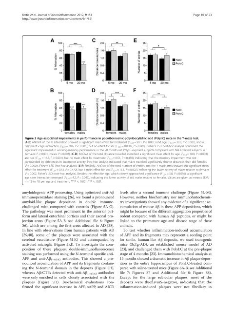
Figure 3 Age-associated impairments in performance in polyriboinosinic-polyribocytidilic acid (PolyI:C) mice in the Y-maze test.<br />
(A-B) ANOVA <strong>of</strong> the % alternation showed a significant main effect for treatment (F 1,54 = 90.1, P < 0.001) and age (F 1,54 = 39.8, P < 0.001), and a<br />
treatment × age interaction (F3,54 = 70.6, P < 0.001), but no effect for sex (F1,54 = 0.0002, P = 0.988). Fisher's LSD post-hoc analysis confirmed the<br />
significant impairment in working-memory performance in the 20 month-old PolyI:C-exposed subjects compared with NaCI-treated subjects in<br />
(females: P < 0.001, males: P = 0.003). (C-D) ANOVA <strong>of</strong> the total distance travelled identified a significant main effect for age (F1,54 = 10.0, P = 0.003)<br />
and sex (F 1,54 = 16.5, P < 0.001), but no main effect for treatment (F 1,54 = 0.51, P = 0.480), indicating that the memory impairment was not<br />
confounded by differences in locomotor activity. Post-hoc analysis indicated that males travelled significantly shorter distances than did females<br />
(P = 0.0003, Fisher's LSD Post-hoc analysis). (E-F) Similarly, ANOVA <strong>of</strong> the total number <strong>of</strong> entries into the Y-maze arms showed no significant main<br />
effect for treatment (F 1,54 = 0.53, P = 0.470), but a main effect for sex (F 1,54 = 11.1, P = 0.002), reflecting the lower activity <strong>of</strong> males relative to females<br />
(P = 0.002, Fisher's LSD post-hoc analysis). Besides the effect for age, which closely approached significance (F1,54 = 3.8, P = 0.056), a significant<br />
age × sex interaction emerged (F3,54 = 4.2, P = 0.045), indicating the lower activity <strong>of</strong> old males relative to females. Values are given as mean ± SEM;<br />
n = 13 to 18 per age and treatment; ***P < 0.001, **P < 0.01.<br />
amyloidogenic APP processing. Using optimized anti-Aβ<br />
immunoperoxidase staining [26], we found a pronounced<br />
amyloid-like plaque deposition in double immunechallenged<br />
mice compared with controls (Figure 5A-G).<br />
The pathology was most prominent in the anterior piriform<br />
and lateral entorhinal cortices and their axonal projection<br />
areas (Figure 5A-B; see Additional file 6: Figure<br />
S6), which are among the first areas affected in AD [38].<br />
In line with observations from human patients with AD<br />
[39,40], some <strong>of</strong> the plaques were associated with the<br />
cerebral vasculature (Figure 5I-K) and accompanied by<br />
activated microglia (Figure 5F,I). To investigate the composition<br />
<strong>of</strong> these plaques, double-immun<strong>of</strong>luorescence<br />
staining was performed using the N-terminal specific anti-<br />
APP and anti-Aβ 1–40/42 antibodies. This showed a pronounced<br />
accumulation <strong>of</strong> APP and its fragments containing<br />
the N-terminal domain in the deposits (Figure 5H),<br />
whereas Aβ/CTFs detected with anti-Aβ 1–40/42 antibodies<br />
were only enriched in cells closely associated with the<br />
plaques (Figure 5H). Biochemical evaluations confirmed<br />
the significant increase in APP, sAPP, and AICD<br />
levels after a second immune challenge (Figure 5L-M).<br />
However, neither biochemistry nor immunohistochemistry<br />
investigations showed any evidence <strong>of</strong> a significant accumulation<br />
<strong>of</strong> mouse Aβ in these APP depositions, which<br />
might be because <strong>of</strong> the different aggregation properties <strong>of</strong><br />
rodent compared with human Aβ peptides, or might be<br />
linked to the premature age and disease stage <strong>of</strong> these<br />
animals.<br />
To test whether inflammation-induced accumulation<br />
<strong>of</strong> APP and its fragments may represent a seeding point<br />
for senile, human-like Aβ deposits, we used transgenic<br />
mice (3xTg-AD), an established mouse model <strong>of</strong> AD<br />
[23], and challenged them with PolyI:C at the pre-plaque<br />
stage <strong>of</strong> 4 months [23]. Immunohistochemical analysis at<br />
15 months showed a dramatic increase in Aβ plaque deposition<br />
in the entire hippocampus <strong>of</strong> PolyI:C-treated compared<br />
with saline-treated mice (Figure 6A-B; see Additional<br />
file 7: Figures S7 and Additional file 8: Figure S8).<br />
Except for the large subicular plaques, most <strong>of</strong> the<br />
deposits were thi<strong>of</strong>lavinS-negative, indicating that the<br />
inflammation-induced plaques were not fibrillary in