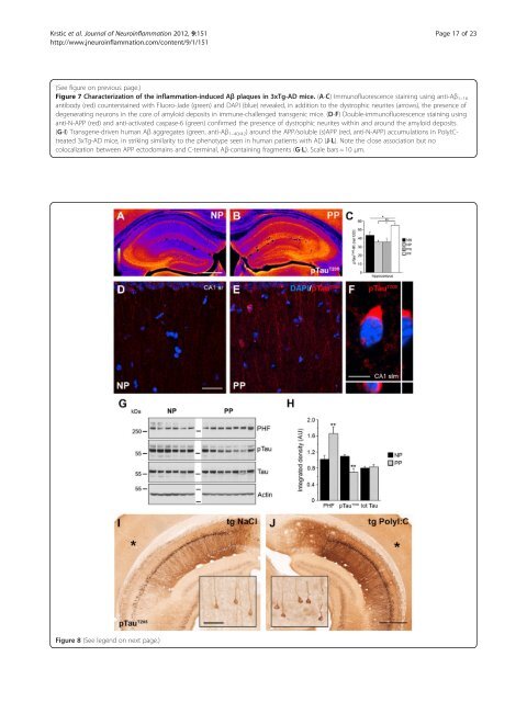
Krstic et al. <strong>Journal</strong> <strong>of</strong> <strong>Neuroinflammation</strong> 2012, 9:151 Page 17 <strong>of</strong> 23 http://www.jneuroinflammation.com/content/9/1/151 (See figure on previous page.) Figure 7 Characterization <strong>of</strong> the inflammation-induced Aβ plaques in 3xTg-AD mice. (A-C) Immun<strong>of</strong>luorescence staining using anti-Aβ 1–16 antibody (red) counterstained with Fluoro-Jade (green) and DAPI (blue) revealed, in addition to the dystrophic neurites (arrows), the presence <strong>of</strong> degenerating neurons in the core <strong>of</strong> amyloid deposits in immune-challenged transgenic mice. (D-F) Double-immun<strong>of</strong>luorescence staining using anti-N-APP (red) and anti-activated caspase-6 (green) confirmed the presence <strong>of</strong> dystrophic neurites within and around the amyloid deposits. (G-I) Transgene-driven human Aβ aggregates (green, anti-Aβ1–40/42) around the APP/soluble (s)APP (red, anti-N-APP) accumulations in PolyI:Ctreated 3xTg-AD mice, in striking similarity to the phenotype seen in human patients with AD (J-L). Note the close association but no colocalization between APP ectodomains and C-terminal, Aβ-containing fragments (G-L). Scale bars = 10 μm. Figure 8 (See legend on next page.)
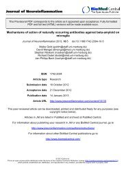
Krstic et al. <strong>Journal</strong> <strong>of</strong> <strong>Neuroinflammation</strong> 2012, 9:151 Page 18 <strong>of</strong> 23 http://www.jneuroinflammation.com/content/9/1/151 (See figure on previous page.) Figure 8 Aggravated Tau pathology after systemic infection in adult mice. Representative images <strong>of</strong> coronal brain sections obtained from (A,D) NP (NaCl at gestation day (GD)17, PolyI:C at 12 months) and (B,E,F) PP mice (PolyI:C at GD17 and at 12 months), processed for (A,B) immunoperoxidase and (D-F) immun<strong>of</strong>luorescence staining using anti-phosphorylated Tau T205 antibodies. (A,B) Images are color-coded for visual display, with white/yellow indicating highest and blue/purple lowest staining intensity. (C) Quantitative analysis <strong>of</strong> the optical density (OD) <strong>of</strong> pTau IR in the hippocampus <strong>of</strong> all treatment groups. Values represent mean OD values corrected for non-specific background labeling (mean ± SEM), n = 6 to 7. **P < 0.01, *P < 0.05; statistical significance based on ANOVA/Fisher’s LSD post-hoc analysis. (D-F) Increase in pTau IR in CA1 is accompanied by mis-sorting into dendrites and intraneuronal aggregation in non-transgenic PP mice. (F) Enlarged view <strong>of</strong> the CA1 stratum lacunosum moleculare subfield showing the typical somatic pTau aggregation. (G) Biochemical analysis <strong>of</strong> Tau phosphorylation using anti-paired helical filaments (PHFs), anti-pTau T205 , and anti-total Tau antibodies. Lanes represent individual mice. (H) Quantitative analysis <strong>of</strong> the immunoreactive signals. Values represent mean relative optical density normalized to β-Actin and expressed in arbitrary units (AU) (mean ± SEM, n = 6 to 7). **P < 0.01; Mann–Whitney U test. Please note that the pTau T205 antibody was used for (A,B) immunohistochemistry (IHC) and (G) immunoblotting (IB) experiments. Because this antibody can crossreact with Ser199 in mouse Tau, an epitope that can be detected with the AT100 (PHF) antibody [63], the increase <strong>of</strong> pTau IR in (A,B) is potentially due to an increase in aggregated pTau in sections. (I-J) Anti-pTau T205 IR in the ventral hippocampus <strong>of</strong> 15-month-old 3xTg-AD mice injected with NaCI or PolyI:C at 4 months. Insets show higher magnifications <strong>of</strong> the cortical areas (asterisk) with low transgene expression, and indicate formation <strong>of</strong> distinct neur<strong>of</strong>ibrillary tangle-like structures after a single systemic infection in transgenic AD mice. Scale bars: (A) = 500 μm, (D)=30 μm, (F)=5 μm, (I, inset) = 50 μm, (J) = 500 μm. [23] and challenged them with PolyI:C at the pre-plaque stage. Subsequent analysis at 15 months showed a dramatic increase in Aβ plaque deposition in the entire hippocampus <strong>of</strong> the PolyI:C-treated mice. These data are in line with recent findings <strong>of</strong> accelerated Aβ pathology in transgenic AD mice after induction <strong>of</strong> osteoarthritis, which is accompanied by significant upregulation <strong>of</strong> inflammation-related genes and astrocyte and microglial activation in the brain [56]. Importantly, inflammationinduced plaques contained significant amounts <strong>of</strong> proteolytic APP fragments, as seen in double immunechallenged WT mice (Figure 9A-D). These APP-derived aggregates were surrounded by human Aβ peptides, showing a striking similarity to the morphology <strong>of</strong> Aβ plaques seen in human patients with AD (Figure 9E-F). Importantly, other laboratories have also reported rodent APP in the core <strong>of</strong> Aβ deposits in variuos transgenic AD mouse strains [55,57]. Hence, we conclude that accumulation <strong>of</strong> APP and its N-terminus-containing fragments precedes the formation <strong>of</strong> senile plaques, representing a crucial and conserved precursor stage <strong>of</strong> this typical neuropathologic hallmark. In agreement with recent experimental evidence showing that hyperphosphorylated, non-fibrillary Tau may have a key role in eliciting behavioral impairments in vivo [34,58], we were able to show in this study that the Tau hyperphosphorylation and mislocalization into somatodendritic compartments is accompanied by severe deficit in prenatally challenged mice compared with salinetreated controls. However, it would be interesting to confirm if a second immune challenge during aging results in progressive decline <strong>of</strong> cognitive functions, as indicated by the recent findings in patients with AD, providing evidence that acute and chronic systemic inflammation is associated with an increase in cognitive decline [42]. Although we did not observe the formation <strong>of</strong> NFTs in immune-challenged mice, a second immune challenge in our model elicited the intraneuronal aggregation <strong>of</strong> Tau, probably involving the formation <strong>of</strong> PHFs. Further studies will be required to characterize the nature <strong>of</strong> these and potentially other phosphorylated epitopes affected by the chronic inflammatory state. We have recently shown that NFT-like structures can form in the brain <strong>of</strong> AD mice lacking tau transgene expression, by genetic reduction <strong>of</strong> the reelin gene [27]. Interestingly, formation <strong>of</strong> these NFT-like structures was associated with a high Aβ plaque burden, and prominent neuroinflammation and neurodegeneration [27]. Hence, it is reasonable to suggest that the formation <strong>of</strong> PHFs we saw in double immune-challenged WT mice would eventually develop into NFTs at older age and later stages <strong>of</strong> the disease. In summary, and based on the results presented and discussed here, we propose a model (Figure 9G) in which the mendelian mutations underlying familial AD cause pr<strong>of</strong>ound changes in APP metabolism, inducing a neuroinflammatory response capable <strong>of</strong> driving and aggravating AD neuropathology during aging. Repeated systemic immune challenges, by contrast, induce chronic neuroinflammation and may accelerate senescence <strong>of</strong> microglia cells. Reduction in their neuroprotective function is expected to impair both APP and pTau homeostasis. This in turn could trigger a vicious cycle that will lead, over the course <strong>of</strong> aging, to the formation <strong>of</strong> senile Aβ plaques and NFTs, induce degeneration, and ultimately result in clinical dementia. We suggest, therefore, that systemic infections and persistent neuroinflammatory conditions represent major risk factors for the development <strong>of</strong> sporadic AD in older persons. Importantly, our model complements the long-standing amyloid hypothesis <strong>of</strong> AD [59,60], and is in accordance with the recent proposal to revise the current view on the etiology <strong>of</strong> sporadic AD [61].