View PDF - Journal of Neuroinflammation
View PDF - Journal of Neuroinflammation
View PDF - Journal of Neuroinflammation

Create successful ePaper yourself
Turn your PDF publications into a flip-book with our unique Google optimized e-Paper software.
Krstic et al. <strong>Journal</strong> <strong>of</strong> <strong>Neuroinflammation</strong> 2012, 9:151 Page 13 <strong>of</strong> 23<br />
http://www.jneuroinflammation.com/content/9/1/151<br />
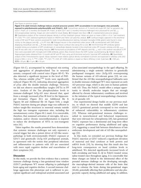
(See figure on previous page.)<br />
Figure 5 An adult immune challenge induces amyloid precursor protein (APP) accumulation in non-transgenic mice prenatally<br />
exposed to polyriboinosinic-polyribocytidilic acid (PolyI:C). (A,B) Coronal brain sections <strong>of</strong> 15-month-old single (NP, NaCl at gestation day<br />
(GD)17, PolyI:C at 12 months) and double (PP, PolyI:C at both GD17 and 12 months) immune-challenged mice processed for anti-Aβ 1–16<br />
immunoperoxidase staining. Images are color-coded for visual display. (C) Enlarged view (box in B) <strong>of</strong> a representative precursor plaque.<br />
(D) Quantitative analysis <strong>of</strong> the numerical plaques density in all four treatment groups. Values are given as mean ± SEM, n = 6 to 7 per treatment.<br />
*P < 0.05, **P < 0.01; statistical significance based on ANOVA and Fisher LSD post-hoc analysis. (E-F) Confocal images captured in the entorhinal<br />
cortex <strong>of</strong> NP and PP brain sections processed for triple immun<strong>of</strong>luorescence staining. Activated microglia (CD68, green) were found in close<br />
vicinity to the plaques (anti-Aβ 1–16) in PP mice. (G) Enlarged neuron in the entorhinal cortex (arrow in F) counterstained with DAPI (blue)<br />
displaying intracellular anti-Aβ1–16 IR (red). Bottom image shows confocal view along the xz axis. (H) Anti-N-terminal APP antibody (green)<br />
revealed strong accumulation <strong>of</strong> APP ectodomains in extracellular deposits located in the entorhinal cortex (PP mouse), whereas anti-Aβ1–40/42<br />
(red) IR was seen only in surrounding neurons. (I-K) Pronounced accumulations <strong>of</strong> APP deposits (anti-Aβ 1–16, red) in the cerebral vasculature in PP<br />
mice were associated with microglia (anti-CD68 antibody, green). Blue channel shows the DAPI nuclear counterstaining. (L) Western blots <strong>of</strong><br />
hippocampal lysates obtained from 12-month-old NP and PP mice using anti-N-terminal and anti-C-terminal APP antibodies. (M) Quantitative<br />
analysis <strong>of</strong> the immunoreactive signals. Values represent mean relative optical density normalized to β-Actin and expressed in arbitrary units (AU)<br />
(mean ± SEM, n = 6 to 7). ***P < 0.001, **P < 0.01, *P < 0.05, #P = 0.08; statistics is based on Mann–Whitney U test. Scale bars: (B) = 500 μm,<br />
(C,F,I)=20 μm, (G)=5 μm, (H)=10 μm.<br />
(Figure 8A-C), accompanied by widespread mis-sorting<br />
and aggregation <strong>of</strong> phosphorylated Tau in neuronal<br />
somata, compared with control mice (Figure 8D-F). We<br />
also detected a significant increase in the level <strong>of</strong> PHF-<br />
Tau, whereas soluble pTau T205 levels were significantly<br />
reduced (Figure 8G-H), indicating abnormal aggregation<br />
<strong>of</strong> pTau after the double immune challenge. However,<br />
we did not observe neur<strong>of</strong>ibrillary tangles (NFTs) in PP<br />
mice. Analysis <strong>of</strong> the Tau phosphorylation levels in<br />
immune-challenged 3xTg-AD mice showed that, apart<br />
from a strongly increased pTau IR level in the hippocampal<br />
formation (Figure 8I-J; see Additional file 8:<br />
Figures S8 and Additional file 10: Figure S10), a single<br />
PolyI:C injection during pre-plaque stage was sufficient to<br />
induce tangle-like structures in neuronal somata outside<br />
the high tau transgene expression sites, including the<br />
somatosensory cortex (Figure 8I-J, inset). It is conceivable,<br />
therefore, that sustained activation <strong>of</strong> microglia, Aβ accumulation,<br />
and/or chronic neuroinflammation is required<br />
to induce the development <strong>of</strong> NFTs in non-transgenic<br />
mice [41].<br />
Taken together, the results presented here demonstrate<br />
that systemic immune challenges not only represent a<br />
crucial trigger but also a potent driver <strong>of</strong> AD-like neuropathology<br />
in both environmentally (PolyI:C exposure at<br />
GD17) and genetically (3xTg-AD) predisposed animals,<br />
in agreement with the notion that peripheral infections<br />
and inflammation in patients with AD are associated<br />
with more rapid cognitive decline and exacerbation <strong>of</strong><br />
their symptoms [42].<br />
Discussion<br />
In this study, we provide the first evidence that a systemic<br />
immune challenge during a late-gestational time window<br />
(GD17) predisposes WT mouse <strong>of</strong>fspring to pathological<br />
brain aging and cognitive decline. A second immune challenge<br />
aggravates this phenotype and is sufficient to precipitate<br />
significant and widespread amyloid-associated and<br />
pTau-associated neuropathology in the aged <strong>of</strong>fspring. By<br />
administering a single systemic infection to genetically<br />
predisposed transgenic mice (3xTg-AD) overexpressing<br />
the human variants <strong>of</strong> AD-relevant genes [23], we confirmed<br />
that the AD-like neuropathological hallmarks seen<br />
in double immune-challenged WT mice represent an early<br />
AD precursor stage, which is also seen in human patients<br />
with AD. Thus, the PolyI:C model <strong>of</strong>fers a unique opportunity<br />
to identify molecular targets that are strongly<br />
affected by chronic inflammatory conditions and involved<br />
in the initiation <strong>of</strong> the typical neuropathology characteristic<br />
<strong>of</strong> sporadic AD.<br />
Our experimental design builds on our previous studies,<br />
in which we showed that middle (GD9) and late<br />
(GD17) gestational periods correspond to two windows<br />
<strong>of</strong> enhanced vulnerability to inflammatory cytokines<br />
[19]. Whereas mid-gestational immune challenges resulted<br />
in neurochemical and behavioral impairments<br />
that were releveant for schizophrenia [20], late-gestational<br />
PolyI:C exposure has a detrimental and long-term effect<br />
on cognitive performance during adulthood and aging<br />
[19], suggesting a causal link between disturbances <strong>of</strong> late<br />
embryonic development and risk <strong>of</strong> AD-like neuropathology<br />
[22].<br />
In this study, we extended our previous findings that<br />
PolyI:C injection at GD17 induced a significant elevation<br />
<strong>of</strong> maternal and fetal cytokines at both protein and<br />
mRNA levels [19], by showing that this insult also has<br />
long-term consequences on basal cytokine levels in<br />
adulthood. We detected significantly increased levels <strong>of</strong><br />
both circulating and brain IL-1β in PolyI:C-treated versus<br />
saline-treated mice throughout aging. It is conceivable that<br />
these changes are linked to the detrimental effect <strong>of</strong> the<br />
prenatal immune challenge on the developing microglia,<br />
the macrophage-derived immune cells <strong>of</strong> the brain that<br />
start to colonize the brain during late-gestational stages<br />
[43,44], potentially affecting their proliferative and also phagocytotic<br />
capacity during adulthood and aging. In addition,