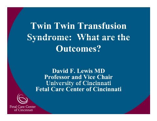
Twin Twin Transfusion Syndrome: What are the ... - March of Dimes
Twin Twin Transfusion Syndrome: What are the ... - March of Dimes
Twin Twin Transfusion Syndrome: What are the ... - March of Dimes

Create successful ePaper yourself
Turn your PDF publications into a flip-book with our unique Google optimized e-Paper software.
<strong>Twin</strong> <strong>Twin</strong> <strong>Transfusion</strong><br />
<strong>Syndrome</strong>: <strong>What</strong> <strong>are</strong> <strong>the</strong><br />
Outcomes?<br />
David F. Lewis MD<br />
Pr<strong>of</strong>essor and Vice Chair<br />
University <strong>of</strong> Cincinnati<br />
Fetal C<strong>are</strong> Center <strong>of</strong> Cincinnati
TWIN-TWIN TRANSFUSION SYNDROME<br />
Unsolved problem<br />
• Most common complication in MC<br />
twins<br />
• 44-35% 35% <strong>of</strong> all MC gestations in US<br />
• 0.1-0.9 per 1000 births<br />
• 17% <strong>of</strong> all perinatal p mortality yin<br />
twins<br />
• Mortality <strong>of</strong> 80-100% if untreated<br />
• Mortality <strong>of</strong> 15-63% 15 63% even with<br />
treatment
Fetal C<strong>are</strong> Center <strong>of</strong> Cincinnati<br />
TWIN TROUBLES<br />
1118 sets <strong>of</strong> twins<br />
27 Conjoined twins<br />
198 Anomalous Co-twins<br />
67 TRAP Sequence q<br />
826 TTTS<br />
<strong>Twin</strong>-<strong>Twin</strong> <strong>Transfusion</strong> <strong>Syndrome</strong>
<strong>Twin</strong>-<strong>Twin</strong> <strong>Transfusion</strong> <strong>Syndrome</strong><br />
Diagnostic Criteria<br />
• Neonatal criteria<br />
− Discordant cord blood Hgb > 5 g/dl<br />
− Discordant birth weight > 20%<br />
• Non-TTTS Non TTTS meeting criteria<br />
− 19/130 had Hgb > 5 g/dl<br />
− Growth restriction without TTTS<br />
− Fetal acidosis acidosis, hypoxemia<br />
− Polycy<strong>the</strong>mia<br />
• Discordant Hgb r<strong>are</strong> in TTTS<br />
• Discordant weight eight common in TTTS<br />
− ? threshold <strong>of</strong> >20%
Quintero Staging <strong>of</strong> TTTS<br />
Stage I: Discordant amniotic fluid: DVP >8 cm & < 2 cm<br />
Stage II: Absent bladder in <strong>the</strong> donor<br />
Stage III: Doppler velocimetry changes in UA, UV, DV<br />
Stage IV: Hydrops<br />
St Stage V: V DDeath th <strong>of</strong> f one ffetus t
<strong>Twin</strong>-<strong>Twin</strong> <strong>Transfusion</strong> <strong>Syndrome</strong><br />
Diagnostic g Evaluation<br />
•Echocardiogram<br />
•Fetal MRI<br />
•Ultrasound<br />
• Monochorionic gestation<br />
• Oligohydramnios DVP < 2cm<br />
• Polyhydramnios DVP > 8cm<br />
• Doppler velocimetry changes<br />
• Donor<br />
• AEDF in UA<br />
• Complete absence <strong>of</strong> DF in<br />
UA<br />
• RRecipient i i t<br />
• Pulsatile UV<br />
• AEDF in UA<br />
• DV: decreased, absent,<br />
reversed a wave<br />
• Growth discordance
TTTS DIAGNOSIS OF EXCLUSION<br />
•15% cases not TTTS<br />
•15% cases not TTTS<br />
•TTTS mimicked by<br />
• Placental insufficiency<br />
• Dichorionic gestation<br />
• Discordant anomaly<br />
• Discordant viral<br />
infection
Hydrops/<br />
Ascites<br />
Fetal MRI Findings in TTTS<br />
Recipient p Donor<br />
Cases<br />
DCVS<br />
Cases<br />
DCVS<br />
15% 6%<br />
B Brain i Bleed/ Bl d/ 15% 3%<br />
Ischemia<br />
Demise 4% 6%
<strong>Twin</strong>-<strong>Twin</strong> <strong>Transfusion</strong> <strong>Syndrome</strong><br />
TTTS Cardiomyopathy<br />
• Cardiac decompensation<br />
• More afterload pathology p gy<br />
• Not volume stress<br />
• Isolated tricuspid regurgitation<br />
• RV hypertension yp Systolic y P> 80<br />
mmHg<br />
• Pulmonary outflow tract<br />
obstruction<br />
• Pulmonary insufficiency<br />
• Pulmonary atresia/intact<br />
ventricular septum p
EEchocardiographic h di hi Fi Findings di iin TTTS<br />
•Myocardial Myocardial hypertrophy RV ><br />
LV<br />
•Decreased compliance RV > LV<br />
−Monophasic inflow<br />
•AV AV valve l iincompetence t TV ><br />
MV<br />
•Poor compliance/ AV valve<br />
incompetence reflected in<br />
•DV abnormalities<br />
−Decrease in a wave<br />
−Absence <strong>of</strong> a wave<br />
−Reversal <strong>of</strong> a wave<br />
•Acquired congenital heart<br />
disease<br />
−Pulmonic valve stenosis, insufficiency,<br />
atresia<br />
−Not reversible with treatment <strong>of</strong> TTTS
Treatments for <strong>Twin</strong>-<strong>Twin</strong><br />
<strong>Transfusion</strong> <strong>Transfusion</strong> <strong>Syndrome</strong><br />
<strong>Syndrome</strong><br />
•Maternal Digoxin<br />
•Maternal Maternal Indocin<br />
•Amnioreduction<br />
•Selective fetocide<br />
•Hysterotomy Hysterotomy for cord ligation<br />
•Blood letting <strong>of</strong> recipient<br />
•Septostomy<br />
•Microseptostomy<br />
Microseptostomy<br />
•Non-selective Laser<br />
photocoagulation<br />
•Selective Laser photocoagulation<br />
•Fetoscopic cord ligation<br />
•Fetoscopic RFA<br />
•Fetoscopic cord coagulation
<strong>Twin</strong>-<strong>Twin</strong> <strong>Transfusion</strong> <strong>Syndrome</strong><br />
Amniocentesis<br />
• Standard <strong>the</strong>rapy in US<br />
• IInitially iti ll treated t t dmaternal t l<br />
symptoms<br />
• Survival <strong>of</strong> 50%<br />
• Moise Semin Perinatol 1993<br />
• Survival in aggressive<br />
amnioreduction<br />
• 37% to 83%<br />
• Minimal maternal or fetal risk<br />
• Incidence <strong>of</strong> neurologic ne rologic<br />
abnormalites<br />
• 18 to 26 % <strong>of</strong> survivors
<strong>Twin</strong>-<strong>Twin</strong> <strong>Transfusion</strong> <strong>Syndrome</strong><br />
• International Registry on<br />
TTTS Treated by<br />
Amnioreduction<br />
• 223 pregnancies < 28 weeks<br />
• Average g number <strong>of</strong><br />
amnioreductions 2<br />
• Complications-mostly fetal<br />
− PPROM 6.2% 6 2%<br />
− Labor 3.1%<br />
− Fetal demise 3.4%<br />
− Placental abruption 1.3%<br />
− Chorioamnionitis 0.9%<br />
− Miscarriage 0.4%<br />
• Mari a et al a Am J Obstet Gynecol Gy eco<br />
185:708-715,2001<br />
Amniocentesis
<strong>Twin</strong>-<strong>Twin</strong> <strong>Twin</strong> <strong>Twin</strong> <strong>Transfusion</strong> <strong>Transfusion</strong> <strong>Syndrome</strong><br />
IInternational t ti l Amnioreduction A i d ti Registry R i t<br />
Mari et al<br />
• Survival to birth 78%<br />
• Survival to 4 weeks <strong>of</strong> age 60%
SEPTOSTOMY<br />
• Single amnioreduction paradox<br />
• Inadvertent septostomy<br />
• Saade et al Am J Obstet Gynecol 172: 429, 1995<br />
• Saade et al Fetal Diagn Ther 13:868-93<br />
− 12 patient ti t<br />
− Survival 83% at GA <strong>of</strong> 31 wks<br />
• Hubinot et al Euro J Obstet Gynecol Reprod Biol 92: 14, 2000<br />
− 7patients 7 patients<br />
− Survival 57%<br />
• Johnson et al Am J Obstet Gynecol 185: 1044, 2001<br />
− Single institution: 7 cases AR; 7 cases septostomy<br />
− Survival for AR 64%:<br />
− Survival for septostomy 71%<br />
• Moise et prospective trial comparing AR to septostomy<br />
− 65% survival with AR and with septostomy<br />
Septostomy
TEMPORIZING MICROSEPTOSTOMY<br />
•Eliminates repeated amnioreductions<br />
•Decreased risk <strong>of</strong> chorioamniotic<br />
separation<br />
•Resuscitation <strong>of</strong> “stuck” twin<br />
•Temporizing measure in TTTS<br />
•Lose oligo/poly as sign<br />
•TTTS’s hemodynamic derangement<br />
− Serial ultrasounds<br />
− Serial echocardiograms<br />
•Microseptostomies<br />
Mi t t i<br />
− 1/3 stabilized<br />
− 2/3 progressed hemodynamically<br />
Microseptostomy
DETECTING PROGRESSION OF TTTS<br />
•ULTRASOUND<br />
FINDINGS<br />
Ductus Venosus<br />
− Serial examinations<br />
−<br />
−<br />
Hydrops y p<br />
Progressive growth<br />
discordance-2 wk<br />
Flow Reversal<br />
•DOPPLER<br />
VELOCIMETRY<br />
− Absent or reversed<br />
diastolic flow in UA<br />
Umbilical Artery<br />
− Reversal <strong>of</strong> flow in<br />
Ductus Venosus Absent Diastolic flow
FETAL ECHOCARDIOGRAPHIC<br />
SIGNS OF PROGRESSION OF TTTS<br />
•Development or<br />
worsening TR<br />
•Development or<br />
worsening MR<br />
•Development <strong>of</strong> PI/PS<br />
•Worsening ventricular<br />
hypertrophy<br />
•Reduced ventricular<br />
fractional shortening<br />
•Tei myocardial<br />
performance index<br />
TR jet
<strong>Twin</strong>-<strong>Twin</strong> <strong>Transfusion</strong> <strong>Syndrome</strong><br />
Non-Selective Non Selective Fetoscopic<br />
Laser Photocoagulation<br />
• 1990 DeLia<br />
− Ob Obstet GGynecol l 75 75:1046, 1046 1990<br />
• 1995 DeLia<br />
− Am J Obstet Gynecol<br />
172:1202,1995<br />
− 53% survival<br />
− 96% “developing<br />
normally”<br />
• 1995 995 Ville V e<br />
− N Engl J Med 332;224,<br />
1995<br />
− 53% survival<br />
− Survivors “developing developing<br />
normally”<br />
Inter-twin Membrane
<strong>Twin</strong>-<strong>Twin</strong> <strong>Transfusion</strong> <strong>Syndrome</strong><br />
Non-Selective or Semi-Selective<br />
Fetoscopic Laser Photocoagulation<br />
Fetal Survival At least 1 Survivor<br />
DeLia ‘95 52.8% 69.2%<br />
DeLia ‘99 69% 82%<br />
Ville ‘98 54.5% 73.5%<br />
Senat ‘04 57% 76%<br />
Inter-twin Membrane
<strong>Twin</strong>-<strong>Twin</strong> <strong>Transfusion</strong> <strong>Syndrome</strong><br />
Sequential Selective Fetoscopic Laser<br />
Photocoagulation<br />
Quintero et al J Matern Fetal Neonatal Med 2007<br />
• Sequential selective laser photocoagulation <strong>of</strong> communicating<br />
vessels in twin-twin transfusion syndrome Quintero et al J<br />
Matern Fetal Neonatal Med 10: 763-768, 2007<br />
• Donor AV first, <strong>the</strong>n Recepient AV <strong>the</strong>n A-A <strong>the</strong>n VV<br />
• Favorable results<br />
• Likely due to detailed mapping<br />
• Shorter duration <strong>of</strong> laser use<br />
• CComparable bl results lt to t FCC
<strong>Twin</strong>-<strong>Twin</strong> <strong>Transfusion</strong> <strong>Syndrome</strong><br />
SelectiveFetoscopic Laser Photocoagulation<br />
•1998 Quintero et al<br />
− Obstet Gynecol Surv<br />
53:597 53:597,1998 1998<br />
•1999 Hecher et al<br />
− Am J Obstet Gynecol 180:<br />
717, 1999<br />
• 73 Fetoscopic laser<br />
• 43 Amnioreduction<br />
• Survival: Laser 61% AR<br />
51%<br />
− Both in same pregnancy<br />
54%<br />
− One or both survive 79% vs<br />
60%<br />
− Abnormal brain US 6% vs<br />
18%<br />
Vascular Equator<br />
Inter-twin Membrane
<strong>Twin</strong>-<strong>Twin</strong> <strong>Transfusion</strong> <strong>Syndrome</strong><br />
Selective Fetoscopic Laser Photocoagulation<br />
Fetal Survival At least 1 Survivor<br />
Hecher ‘99 61% 79%<br />
• Hecher ‘00 68% 81%<br />
• Quintero ‘00 61.3% 83%<br />
• Quintero ‘03 64.2% 83.2%<br />
• Huber ‘04 70% 83%<br />
• Huber ‘06 71.5% 83.5%<br />
• C Crombleholme ‘0 ‘07 77% % 91 91.7%<br />
%<br />
Vascular Equator<br />
Inter-twin Membrane
TTeam<br />
Pediatric surgeon<br />
Maternal Fetal<br />
Medicine<br />
Sonographer<br />
Cincinnati Mapping protocol<br />
1 st<br />
2 nd<br />
Identification <strong>of</strong> all vascular connection and<br />
recorded <strong>the</strong> findings.<br />
Confirmation <strong>of</strong> <strong>the</strong> vascular connection and<br />
laser photocoagulation.<br />
3rd No vascular connection was missed and<br />
Recorder no vessel had recanalized recanalized.
Positioning g and Set Up p
<strong>Twin</strong>-<strong>Twin</strong> <strong>Transfusion</strong> <strong>Syndrome</strong><br />
Anes<strong>the</strong>tic Options<br />
• Local with IV sedation<br />
• Posterior placentas only<br />
• Psychologically prep<strong>are</strong>d<br />
• Calm and cooperative disposition<br />
• English speaking<br />
• Epidural with IV sedation<br />
• Standard approach<br />
• General anes<strong>the</strong>s<strong>the</strong>tic<br />
• Reserved for those with contraindications to regional<br />
or local anes<strong>the</strong>sia
<strong>Twin</strong>-<strong>Twin</strong> <strong>Transfusion</strong> <strong>Syndrome</strong><br />
• All have sequential compression stockings<br />
• All pressure points well padded<br />
• PPosterior t i placenta l t<br />
− Bump for uterine displacement<br />
− Side <strong>of</strong> recipient up<br />
• Anterior Placenta<br />
− Full lateral decubitus position<br />
− Flex <strong>the</strong> bed<br />
− Approach from side opposite donor with a window<br />
− Window between Pelvis<br />
and dth <strong>the</strong> costal t lmargin i
Fetoscopes Used in Laser Procedure<br />
• Storz fetoscopes<br />
• 3.3 mm<br />
• 1.8 mm semirigid lens<br />
• Must be hand curved to ~ 3- 3 degrees<br />
• Laser port 600µ endostat<br />
• Side port for Level I Rapid Infusor<br />
• Remote head scope requires req ires an<br />
Humanitarian exemption from <strong>the</strong> FDA<br />
• Can be inserted with or without a<br />
separate sheath 10 Fr Cook Introducer
Choice <strong>of</strong> Laser Used in Fetoscopic p<br />
Procedure<br />
• Neodymium-doped yttrium aluminium garnet<br />
− nd: YAG (Nd:Y (Nd:Y3Al Al5OO 12) ) crystal<br />
− Wavelength 1164 nm<br />
• Diode laser<br />
− semiconductor<br />
− Wavelength 980 nm<br />
nd:YAG<br />
Diode
Intraoperative Complications<br />
• Bleeding<br />
obscuring view<br />
• Level I<br />
amnioinfusion<br />
− Trocar insertion<br />
n=1<br />
− Chorionic plate n=1<br />
− Vessel laceration<br />
• 3 all controlled with<br />
Level I and Laser
Obscured Fetoscopic View<br />
• Mountainous topography<br />
• May be seen in prior AR<br />
• Amnioinfusion may flatten surface<br />
and facilitate visualization<br />
• Amnioinfusion may move donor twin<br />
or fl flatten tt <strong>the</strong> th iintertwin t t i membrane b onto t th <strong>the</strong><br />
chorionic plate<br />
• Facilitate visualization <strong>of</strong> anterior<br />
placentas<br />
Level I Rapid<br />
Inf Infusion sion Device De ice
Intraoperative Complications<br />
• Loss <strong>of</strong> amniotic cavity volume<br />
• Fracture <strong>of</strong> laser fiber<br />
• Loss <strong>of</strong> laser efficiency<br />
− Tangential angle<br />
− Th Through h iintertwin t t i<br />
membrane<br />
− Endostat problem<br />
• Debris<br />
• Fracture <strong>of</strong> <strong>the</strong> tip<br />
− Laser mal-alignment<br />
mal alignment<br />
600 µ Endostat fiber<br />
Amniotic litter
Intraoperative Complications<br />
• Laser Microseptostomy?<br />
• Risk <strong>of</strong> monoamniotic<br />
gestation<br />
− Thought to be high with<br />
ultrasound guided<br />
needle<br />
• Risk <strong>of</strong> laser<br />
microseptostomy<br />
• 600 µ diameter width <strong>of</strong><br />
laser fiber<br />
• MMonoamniotic i ti gestation t ti<br />
− Fetoscopy alone 1.3%<br />
− Microseptostomy in<br />
addition dd o 1.3% .3%<br />
− Does it help?<br />
− Restores fluid more<br />
quickly<br />
− Change in survival ?
Minimizing Vascular Shifts During<br />
Laser Photocoagulation<br />
• Sequential SFLP<br />
• AV AV, AA AA, VV<br />
• Requires detailed mapping<br />
• Improved p survival<br />
• Cincinnati mapping protocol<br />
• No laser <strong>of</strong> any vessel until <strong>the</strong> mapping is complete<br />
• Minimize laser time (usually ~ 5 minutes<br />
• Similar results
PPost-operative t ti Course C<br />
• Admit to FCC for<br />
tocodynomometric monitoring<br />
• Sequential compression<br />
stockings, foley<br />
• Clear liquid and advance<br />
• Tocolysis nifedipine with rate<br />
patient having Terbutaline<br />
• Ultrasound POD # 1<br />
• Discharge to from FCC in<br />
Cincinnati<br />
• Follow up fetal echocardiogram<br />
and ultrasound at POD #3
Quintero Staging <strong>of</strong> TTTS<br />
Stage I: Discordant amniotic fluid: DVP >8 cm & < 2 cm<br />
Stage II: Absent bladder in <strong>the</strong> donor<br />
Stage III: Doppler velocimetry changes in UA, UV, DV<br />
Stage IV: Hydrops<br />
St Stage V: V DDeath th <strong>of</strong> f one ffetus t
Acute Cardiac Changes in TTTS<br />
Quintero Staging<br />
I II III IV V<br />
Donor Recipient<br />
AEDF in UA Severe biventricular failure<br />
Reversal <strong>of</strong> flow in DV<br />
Severe TR/MR<br />
Reversal <strong>of</strong> flow in UA rec<br />
Early TTTS Endstage TTTS<br />
Staging is heavily weighted toward <strong>the</strong> donor<br />
Staging is heavily weighted toward <strong>the</strong> donor<br />
Recipient findings on in advance stage III and IV
10/10<br />
NIH TTTS Trial<br />
Logistic g Regression g Analysis y <strong>of</strong> Covariates<br />
Cardiovascular Pr<strong>of</strong>ile Score (CVPS)<br />
Normal -1 point -2 points<br />
Hydrops None Ascites, pleural<br />
pericardial i di leffusion ff i<br />
skin edema<br />
Venous<br />
Dopplers Normal DV atrial reversals Venous pulsations<br />
CT Ratio < 0.35 > 0.35< 0.5 > 0.5<br />
Abnormal myo- Vent SF > 0.28 SF < 0.28<br />
cardial di l ffunction ti No AV regurg TR or semi lunar TR +dysfunction<br />
valve regurg any MR<br />
Abnormal Normal AEDF REDF<br />
Arterial<br />
Dopplers<br />
Cardiovascular Pr<strong>of</strong>ile Score (CVPS). From Huhta, J Perinat Med 29:390-398, 2001
NIH Sponsored TTTS Trial<br />
LLogistic i i RRegression i AAnalysis l i <strong>of</strong> fC Covariates i on Survival S i l<br />
• Recipients<br />
− CCardiovascular di l Pr<strong>of</strong>ile P fil Score* S *<br />
− Most predictive <strong>of</strong> poor recipient outcome<br />
• OR = 3.025/point<br />
• p = 0.05<br />
• Donors<br />
− Most predictive models <strong>of</strong> poor outcome<br />
− Increased Stage<br />
• OR = 0.446/stage p = 0.124<br />
− Earlier Gestational Age<br />
• OR = 1.052/day p = 0.097<br />
*Huhta et al<br />
J Perinat Med 29:390-398, 2001
Impact <strong>of</strong> Fetal Echocardiographic Findings<br />
on Recipient Survival<br />
•Cardiovascular Pr<strong>of</strong>ile Score (CVPS)<br />
•62 consecutive recipient twins with TTTS<br />
• All patients treated by SFLP<br />
• All ll patients i had h d pre-operative i echo h evaluated l d<br />
• Blinded to outcome<br />
Michelfelder et al Ultrasound Obstet Gynecol<br />
30: 965-71, 2007<br />
Shah AD, et al:J Am Soc Echocardiogr 2008<br />
TR jet
SFLP<br />
100<br />
90<br />
80<br />
70<br />
60<br />
50<br />
40<br />
30<br />
20<br />
10<br />
0<br />
Recipient Survival Based on<br />
Pre-Operative CVPS<br />
p< 0.03<br />
p< 00.008 008<br />
10 <strong>of</strong> 10 9 <strong>of</strong> 10 8 <strong>of</strong> 10<br />
CVPS
Problem with <strong>the</strong> CVPS<br />
Normal -1 point -2 points<br />
Hydrops y p<br />
Venous<br />
None Ascites, ,ppleural<br />
pericardial effusion<br />
skin edema<br />
Dopplers Normal DV atrial reversals Venous pulsations<br />
CT Ratio < 0.35 > 0.35< 0.5 > 0.5<br />
Abnormal myo- y Vent SF > 0.28 SF < 0.28<br />
cardial function No AV regurg TR or semi lunar TR +dysfunction<br />
valve regurg any MR<br />
Abnormal<br />
Arterial<br />
Dopplers<br />
Normal AEDF REDF<br />
Cardiovascular Pr<strong>of</strong>ile Score (CVPS). From Huhta, J Perinat Med 29:390-398, 2001<br />
Not sensitive enough
TTTS Cardiomyopathy<br />
•Fetal F lh hypertensive i cardiomyopathy<br />
di h<br />
− AV valve competence<br />
• Mild, moderate, severe TR/MR<br />
− HHypertrophy t h<br />
• End-diastolic LV, RV, Septum<br />
M di ld f ti<br />
− Myocardial dysfunction<br />
• Tei myocardial performance index
Fetal C<strong>are</strong> Center <strong>of</strong> Cincinnati<br />
Cincinnati Modification <strong>of</strong> Quintero TTTS Staging<br />
Stage g I<br />
Stage II<br />
Stage III<br />
IIIA Mild cardiomyopathy<br />
IIIB Moderate cardiomyopathy<br />
IIIC S di h<br />
IIIC Severe cardiomyopathy<br />
Stage IV<br />
Stage V
Fetal C<strong>are</strong> Center <strong>of</strong> Cincinnati<br />
Cincinnati Modification <strong>of</strong> Quintero TTTS Staging<br />
Stage g I<br />
Stage II<br />
Stage III<br />
IIIA RV MPI > 0.5 (Z+2) / LV MPI > 0.42 (Z+2)<br />
Mild ventricular hypertrophy (Z for RV/LV >2)<br />
Mild AV valve regurgitation<br />
IIIB RV MPI > 0.56 0 56 (Z+3) (Z 3) / LV MPI > 0.53 0 53 (Z+3) (Z 3)<br />
Moderate ventricular hypertrophy (Z for RV/LV >3)<br />
> Mod AV valve regurgitation<br />
IIIC RV/LV > Z+4Critical Doppler changes in RT<br />
(reversed DV, rUA)<br />
Severe TR/MR TR/MR, severe dysfunction<br />
Stage IV<br />
Stage V
Echocardiographic Features <strong>of</strong> TTTS by Stage<br />
Incidence <strong>of</strong> TR and MR<br />
100<br />
90<br />
80<br />
70<br />
60<br />
50<br />
40<br />
30<br />
20<br />
10<br />
0<br />
III IIIA IIIB IIIC IV<br />
Ci Cincinnati i ti Stage St<br />
Cincinnati Stage<br />
Tricuspid<br />
Regurgitation<br />
Mitral<br />
Regurgitation
Echocardiographic Features <strong>of</strong> TTTS by Stage<br />
Biventricular Hypertrophy<br />
Percent<br />
100<br />
90<br />
80<br />
70<br />
60<br />
50<br />
40<br />
30<br />
20<br />
10<br />
0<br />
Incidence <strong>of</strong> Biventricular<br />
Hypertrophy<br />
III IIIA IIIB IIIC IV<br />
Cincinnati Stage<br />
Biventricular<br />
Hypertrophy
Tei Myocardial<br />
Performance Index<br />
Assessing Recipient TTTS Cardiomyopathy<br />
Tei C, et al. New index <strong>of</strong> combined systolic and diastolic myocardial performance: a<br />
simple and reproducible measure <strong>of</strong> cardiac function--a study in normals and dilated<br />
cardiomyopathy cardiomyopathy. J Cardiol 1995;26:357 1995;26:357-66 66<br />
Eidem BW, et al: Quantitative assessment <strong>of</strong> fetal ventricular function: establishing normal<br />
values <strong>of</strong> <strong>the</strong> myocardial performance index in <strong>the</strong> fetus. Echocardiography 2001;18:9-13<br />
Ichizuka K et al, The Tei index for evaluation <strong>of</strong> fetal myocardial performance in sick<br />
fetuses. Early Hum Dev 2005;81:273-9
Assessing Recipient TTTS Cardiomyopathy<br />
Tei Myocardial Performance Index<br />
*Identify Identify subtle alteration in ventricular function<br />
*Difficult to accurately measure<br />
*Inter-observer variability can be high<br />
*FCC echocardiographers h di h 10% variation i ti<br />
*Minimal change is >10%<br />
*Method to detect progression <strong>of</strong> TTTS during trial <strong>of</strong> AR<br />
*Underestimates dysfunction in moderate TR/MR<br />
*Can confirm post-treatment arrest <strong>of</strong> TTTS
Echocardiographic Features <strong>of</strong> TTTS by Stage<br />
Tei Index<br />
Myoccardial<br />
Peerformancce<br />
Index<br />
0.8<br />
0.7<br />
0.6<br />
0.5<br />
0.4<br />
0.3<br />
0.2<br />
01 0.1<br />
0<br />
III IIIA IIIB IIIC<br />
Cincinnati Stage<br />
IV<br />
LV MPI<br />
RV MPI
Fetal C<strong>are</strong> Center <strong>of</strong> Cincinnati<br />
TTTS cases since NIH Trial<br />
Closed May 2005<br />
570 TTTS patients<br />
Stage<br />
I 17.2%<br />
II 10 10.3% 3%<br />
III 11.5%<br />
IIIA 25.3%<br />
IIIB 18.4%<br />
IIIC 4.6%<br />
IV 69% 6.9%<br />
V 5.7%<br />
48.3%<br />
59 59.8% 8%
Distribution by Quintero Stage <strong>of</strong> TTTS<br />
Patients: The The Fetal Fetal C<strong>are</strong> C<strong>are</strong> Center <strong>of</strong> Cincinnati<br />
May 2005-January 2009<br />
570 patients<br />
300<br />
250<br />
200<br />
150<br />
100<br />
50<br />
0<br />
Quintero Stag<br />
0 I II III IV V<br />
Stag
Shift in Stage Distribution Comparing<br />
Quintero and Cincinnati Staging Systems<br />
May 2005-January 2009<br />
570 patients p<br />
250<br />
200<br />
100<br />
Quintero vs Cinc<br />
150 Quintero<br />
50<br />
0<br />
0 I II III IIIa IIIb IIIc IV V<br />
Stag<br />
Cincinna
Percentage <strong>of</strong> Cases by Stage<br />
Upstaged by Echocardiographic Findings<br />
May 2005-January 2009<br />
570 patients 0<br />
90<br />
80<br />
70250<br />
60<br />
50<br />
40<br />
30<br />
20<br />
10<br />
0<br />
200<br />
150<br />
100<br />
50<br />
0<br />
Percent Upstaged<br />
0 I II III III IIIa<br />
Stage<br />
IIIb III IIIc IV V<br />
Quintero Staging<br />
0 I II III IV V<br />
St Stage<br />
I II III
Echocardiographic Upstaging <strong>of</strong> Stage I<br />
Upstaging <strong>of</strong> Quintero's Stage I<br />
St Stage I<br />
39%<br />
70 patients<br />
Upstaged<br />
61%<br />
Stage I Stage IIIA Stage IIIB Stage IIIC<br />
Stage IIIA<br />
27%<br />
Stage IIIB<br />
14%<br />
Stage g IIIC<br />
20%
Echocardiographic Upstaging <strong>of</strong> Stage I<br />
Upstaging <strong>of</strong> Quintero Quintero'ss Stage II<br />
Stage II<br />
36%<br />
40 Patients<br />
Upstaged<br />
64%<br />
Stage IIIA<br />
16%<br />
Stage IIIB<br />
25%<br />
Stage IIIC<br />
23%<br />
Stage II Stage IIIA Stage IIIB Stage IIIC
Trial <strong>of</strong> Amnioreduction vs<br />
Selective Fetoscopic Laser Photocoagulation<br />
�175 patients stages I-IIIA<br />
�Normal �Normal fetal echocardiograms<br />
�Trial <strong>of</strong> AR with serial fetal echocardiographic assessmen<br />
�40% response to AR alone<br />
�60% progressed
Trial <strong>of</strong> Amnioreduction vs<br />
Selective Fetoscopic Laser Photocoagulation<br />
�AR responders survival<br />
�82.2% overall survival<br />
�90.7% 1 or both surviving<br />
�75% both surviving<br />
�AR non-responders-SFLP<br />
�79 �79.5% 5% overall survival<br />
�89.9% 1 or both surviving<br />
�70% both twin survive
<strong>Twin</strong>-<strong>Twin</strong> <strong>Transfusion</strong> <strong>Syndrome</strong><br />
Selective Fetoscopic Laser Photocoagulation<br />
�Stage Some IIIB, IIIC, IV<br />
�F �Fail iltil trial <strong>of</strong> fAR AR<br />
�Severe placental insufficiency
Acute Effect <strong>of</strong> SLFP on<br />
Tei e Myocardial yoc d Performance e o ce Index de<br />
•Pre and Post SFLP Echos<br />
•>10% improvement in Tei Index<br />
P 10% fall in Tei Index No Fall in Tei<br />
Habli et al<br />
Am J Obstet Gynecol Nov 2008
Overall Survival and Survival <strong>of</strong><br />
One One or or Both Both <strong>Twin</strong>s by Cincinnati Staging<br />
Selective Fetoscopic Laser Photocoagulation<br />
Cincinnati Stage
Post-Operative Complications in TTTS<br />
PPROM < 7 day : 5%<br />
PPROM > 7 day d : 22%<br />
Chorioamnionitis : 2.3%<br />
Transplacental trocar : 47%<br />
Yamamoto et al.<br />
Am J Obstet Gynecol. 2005 Sep;193(3 Pt 2):1110-6<br />
Iatrogenic monoamniocity<br />
Vascular accident<br />
Amniotic band syndrome<br />
Recurrent TTTS : 14%<br />
TAPS : 13%<br />
PPROM : 17%<br />
RRobyr b et al. l<br />
Am J Obstet Gynecol. 2006 Mar;194(3):796-803.<br />
?
AIMS<br />
• Incidence <strong>of</strong> complications after SFLP.<br />
• Distinguish early and late complications.
77<br />
Methods<br />
249 patient evaluated<br />
Sept. 2005 Feb. 2008<br />
Retrospective review<br />
14 6 SFLP (152) ( )<br />
1152 2<br />
Amnioreduction alone (77)<br />
Radi<strong>of</strong>recuency ablation (14)<br />
No treatment (6)
Methods<br />
Complications (maternal and fetal):<br />
• NNo complication li ti<br />
• Early complication: < 7 day after <strong>the</strong><br />
procedure.<br />
• LLate t complication: li ti > >7 7 day d after ft <strong>the</strong> th<br />
procedure.<br />
• Early and late.
SFLP = 152 patients<br />
Results<br />
3<br />
(2%)<br />
149<br />
(98%)<br />
<strong>Twin</strong>s<br />
Triplets
SFLP<br />
Result<br />
Serious complication or maternal deaths (0%)<br />
Overall Fetal survival (77%)<br />
Survival <strong>of</strong> one or both twins (88%)<br />
Recipient survival (83%)<br />
Donor survival (73%)
10%<br />
40%<br />
Complication<br />
29%<br />
21%<br />
No complication<br />
Early complication<br />
Late complication<br />
Early and late<br />
complication
10%<br />
40%<br />
Complication<br />
29%<br />
21%<br />
Early complication<br />
8% (12/152) PPROM.<br />
1.3% (2/152) Chorioamnionitis.<br />
11.3% 3% (2/152) Maternal<br />
peritoneal leak.
10%<br />
40%<br />
Complication<br />
29%<br />
Late complication<br />
• 17 17.8% 8% (27/152) PPROM<br />
• 8% (12/152) Abruption<br />
21%<br />
• 3.3% (5/152) ABS<br />
• 2% (3/152) TAPS<br />
• 2% (3/152) TTTS<br />
• 1.3% (2/152) Iatrogenic<br />
monoamniotic i i sac<br />
• 0.7% (1/152) Fetal hand necrosis<br />
due to vascular disruption p<br />
sequence
Impact <strong>of</strong> Complications on Survival<br />
No complication Early complication Late complication Both p value<br />
GA at procedure 21.0 + 2.5 21.3 + 2.6 21.1 + 2.4 22.2 + 2.5 N.S<br />
GA at delivery<br />
(weeks)<br />
Numbers DA-RV<br />
anastomoses<br />
Total number <strong>of</strong><br />
anastomses<br />
coagulated<br />
31.4 + 4.1 28.3 + 5.7 30.1 + 3.1 29.8 + 3.8 p
Conclusions<br />
• SFLP Early, late and both early and late<br />
complications impact survival<br />
• Stringent placental mapping protocol Low rate <strong>of</strong><br />
− TAPS (2%),<br />
− Recurrent/persistent TTTS (2%). (2%)<br />
• This information is important for counseling patients.
Pathophysiology <strong>of</strong> Recipient<br />
•Volume over load<br />
•Fetal systemic<br />
hypertension<br />
Cardiomyopathy<br />
− Estimates <strong>of</strong><br />
ventricular ti l pressures<br />
based flow velocity <strong>of</strong><br />
tricuspid regurgitation<br />
jt jet
Will Treating g TTTS Cardiomyopathy y p y<br />
Change Survival in Recipients?<br />
• Empiric treatment <strong>of</strong> TTTS cardiomyopathy<br />
with nifedipine 20 mg q 6 hours<br />
• Stages IIIA IIIA, IIIB IIIB, IIIC IIIC, IV<br />
• 20 mg q 6 hours 24-48 hours<br />
• 141 TTTS cases<br />
• 152 stage and GA matched controls<br />
• Significant reduction in tocolytics
•<br />
•<br />
Nifedipine 20 mg q 6 h<br />
Pre-op treatment 24-48<br />
100<br />
h<br />
90<br />
•<br />
•<br />
Case control study<br />
Matched for GA and<br />
80<br />
Ci Cincinnati i ti stage t 70<br />
•<br />
•<br />
Survival to birth<br />
Significant effect on<br />
60<br />
overall ll survival i l 50<br />
• Significant effect on<br />
recipient survival<br />
40<br />
• NNo effect ff ton DDonor<br />
survival<br />
30<br />
Effect <strong>of</strong> Nifedipine on Survival in<br />
TTTS Treated by SLFP<br />
20<br />
10<br />
0<br />
Nifedipine Control
Overall Survival and Survival <strong>of</strong><br />
One or or Both Both <strong>Twin</strong>s <strong>Twin</strong>s in Stage IV IV TTTS<br />
TTTS<br />
Selective Fetoscopic Laser Photocoagulation<br />
Nifedipine effect?
Challenges g in TTTS<br />
• Define <strong>the</strong> etiology<br />
• Improving survival<br />
• Develop targeted medical <strong>the</strong>rapies<br />
• Adj Adjunctive ti medical di l th <strong>the</strong>rapy iin AR<br />
• Reducing morbidity<br />
• Defining cases to treat<br />
• Extend <strong>the</strong> length <strong>of</strong> gestation<br />
• Convincing <strong>the</strong> Obstetrical community
•Maternal-Fetal Medicine<br />
−Mounira Habli MD<br />
−William Polzin, MD<br />
−Kim Brady, MD<br />
−Jim VanHook, , MD<br />
−Ronald Jaekle,MD<br />
−David Lewis, MD<br />
•Fetal Echocardiography<br />
−Erik Michelfelder, MD<br />
−James |Cnota,MD | ,<br />
−William Gottliebson, MD<br />
−Allison Divanovic,MD<br />
−Haleh Hadarian, MD<br />
•Urology/Nephrology<br />
−Pramod Reddy,MD<br />
−Robert Defore,MD<br />
−Elizabeth Jackson,MD<br />
•ENT<br />
−Ravi Ellarhu,MD<br />
−Paul Willging,MD<br />
•Neurosurgery<br />
−Karin Bierbrauer,MD<br />
−Francesco Mangano,MD<br />
•Cardiac Surgery<br />
−Pirooz Eghtesady,MD<br />
P M i MD<br />
Fetal C<strong>are</strong> Center <strong>of</strong> Cincinnati<br />
•Pediatric Surgery<br />
•Timothy Crombleholme,MD<br />
•Foong-Yen Lim,MD<br />
•Sundeep Keswani MD<br />
•Anes<strong>the</strong>siology<br />
–Ann Boat,MD<br />
–Mohammad Mahmoud,MD<br />
–Bonnie Hugus<br />
•Radiology<br />
•Connie Bitters<br />
•Linda Martin<br />
–Beth Kline-Fath, MD<br />
–Maria M i Calvo, C l MD<br />
–Eva Rubio, MD<br />
–Leann Linam, MD<br />
–Kyuran Choe, MD<br />
•Operating Room Nurses<br />
•Missy Ritter<br />
•Stacy Ruth<br />
•Tracy Heidrich<br />
•Latressa Latressa Ratner<br />
•Curtis Johnson<br />
•Neonatology<br />
− James Greenberg, MD<br />
− Tanya Cahill, MD<br />
− Kurt Schibler, MD<br />
− Beth Haberman, , MD<br />
− Paul Kingma, MD<br />
− Amy Nathan, MD<br />
•Nursing Director<br />
− Kelli Young<br />
•Coordinators<br />
− Gina Sharp, RN<br />
− Deb Voet, RN<br />
− Jenni Mason<br />
− Steve Imh<strong>of</strong>f<br />
− Judy Hostiuck<br />
•Genetics<br />
− Rob Hopkin,MD<br />
− Howard Saul,MD<br />
− Christine Spaeth, CGC<br />
− Diana Smith, CGC<br />
•Social Services<br />
− Erin Hartman, MSW<br />
•Administrative<br />
− Rachel Jones<br />
− Emmie Beyer<br />
Ch l S ll<br />
•Level III L&D Nurses<br />
• Good Samaritan Hospita<br />
•Nurse Midwife<br />
K<strong>are</strong>n McGirr<br />
−Peter Manning,MD − Cheryl Snell