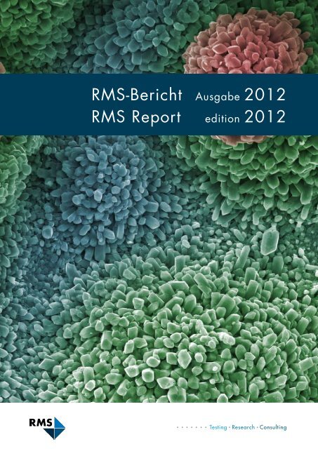
RMS-Bericht 2012 - RMS Foundation
RMS-Bericht 2012 - RMS Foundation
RMS-Bericht 2012 - RMS Foundation

You also want an ePaper? Increase the reach of your titles
YUMPU automatically turns print PDFs into web optimized ePapers that Google loves.
<strong>RMS</strong>-<strong>Bericht</strong><br />
<strong>RMS</strong> Report<br />
Ausgabe <strong>2012</strong><br />
edition <strong>2012</strong>
Einleitung<br />
Jubiläumsfeier «25 Jahre <strong>RMS</strong>»<br />
Teilnahme an Fachmessen und Publikation eines Newsletters<br />
Ringversuch «Biegeversuch an Metallen nach DIN EN ISO 7438»<br />
Charakterisierung von nichtmetallischen Einschlüssen nach EN 10247<br />
Trägergasschmelzextraktionsanalyse von H, N, O und Ar in Metallen<br />
Partikelanalyse in der <strong>RMS</strong><br />
Kontaktwinkelmessung in der <strong>RMS</strong><br />
Verschleissreduktion durch Kavitäten<br />
Künstliche Gelenkflüssigkeit für die Verschleisstestung<br />
Machbarkeitsstudie: Wasserstrahlstrukturierte Oberflächen<br />
Gewebeschonende Eigenschaften abgerundeter Weichteilhebel<br />
Neue Erkenntnisse über den Mechanismus der initialen Zelladhäsion<br />
Tierstudie mit β-TCP und DCPA-Granulaten<br />
Rekristallisation von amorphisiertem α-TCP<br />
Röntgenbeugung unter simulierten physiologischen Bedingungen<br />
Synthese von β-Tricalciumphosphat-Nanoplättchen<br />
Potenzielle Anwendungen von pHEMA-Hydrogelen in der Orthopädie<br />
Auswirkungen von Nanofasern auf das Skelettgewebe<br />
Biomimetisches Reaktorsystem für Knorpelgewebe<br />
Zellen und Trägerstrukturen für die Knorpeltherapie<br />
Publikationen<br />
Publikationen aus externen Projekten (EFO) mit <strong>RMS</strong>-Unterstützung<br />
Abstracts / Kurzfassungen, präsentiert als Vortrag (V) oder Poster (P)<br />
Externe Vorträge durch <strong>RMS</strong>-Mitarbeitende<br />
Seminarvorträge in der <strong>RMS</strong><br />
Lehrtätigkeiten<br />
Patente<br />
Personal<br />
<strong>RMS</strong>-<strong>Bericht</strong> Ausgabe <strong>2012</strong> – Inhaltsverzeichnis<br />
3<br />
5<br />
6<br />
7<br />
8<br />
9<br />
10<br />
11<br />
12<br />
13<br />
14<br />
15<br />
16<br />
17<br />
18<br />
19<br />
20<br />
21<br />
22<br />
23<br />
24<br />
25<br />
25<br />
26<br />
29<br />
30<br />
30<br />
31<br />
32<br />
1
2<br />
<strong>RMS</strong> Report edition <strong>2012</strong> – Table of contents<br />
Introduction<br />
Celebration of the 25 th anniversary of the <strong>RMS</strong><br />
Participation at trade shows and edition of newsletters<br />
Interlaboratory test «Bend test of metals according to DIN EN ISO 7438»<br />
Characterisation of non-metallic inclusions according to EN 10247<br />
Melt extraction analysis of H, N, O and Ar in metals<br />
Particle analysis in the <strong>RMS</strong><br />
Contact angle measurement in the <strong>RMS</strong><br />
Wear reduction due to cavities<br />
Artificial synovia for the testing of materials for hip replacements<br />
Feasibility study: structuring of surfaces using waterjet<br />
Tissue preserving properties of rounded retractors<br />
Novel insights in the mechanism of initial cellular adhesion<br />
Animal study with β-TCP and DCPA granules<br />
Recrystallization of amorphized α-TCP<br />
X-ray diffraction under simulated physiological conditions<br />
Synthesis of β-tricalcium phosphate nano-platelets<br />
Potential pHEMA hydrogel application in orthopedics<br />
Effect of nanofibers on skeletal tissues<br />
Physiological Robot Reactor System (PRRS) for cartilage tissue<br />
Cells and scaffolds for cartilage repair<br />
Publications<br />
Publications on external projects (EFO) with <strong>RMS</strong> grant<br />
Abstracts presented as talk (V) or poster (P)<br />
External presentations by <strong>RMS</strong> staff<br />
Seminary talks at the <strong>RMS</strong><br />
Teaching activities<br />
Patents<br />
Personnel<br />
4<br />
5<br />
6<br />
7<br />
8<br />
9<br />
10<br />
11<br />
12<br />
13<br />
14<br />
15<br />
16<br />
17<br />
18<br />
19<br />
20<br />
21<br />
22<br />
23<br />
24<br />
25<br />
25<br />
26<br />
29<br />
30<br />
30<br />
31<br />
32
Einleitung<br />
Es ist mir eine Freude, Ihnen nach den ersten<br />
beiden Jahren als Geschäftsführer in gewohnter<br />
Manier über die Aktivitäten der <strong>RMS</strong><br />
in der Forschung, im Technologietransfer und<br />
im Aufbau von Dienstleistungen berichten<br />
zu dürfen.<br />
Mit einer neuen und erweiterten Geschäftsleitung<br />
haben wir Organisation und Abläufe<br />
in ausgewählten Punkten optimiert, um den<br />
fortwährend grösser werdenden Herausforderungen<br />
an unsere Stiftung und die Mitarbeitenden<br />
gerecht zu werden.<br />
Basierend auf unserem Stiftungszweck und<br />
Leitbild haben wir die Strategie überarbeitet<br />
und einen detaillierten Mittelfristplan erarbeitet,<br />
um die <strong>RMS</strong> auf dem eingeschlagenen<br />
dualen Weg mit Forschung und Dienstleistung<br />
weiter auf Kurs zu halten und zu stärken.<br />
Personelle Mutationen waren primär<br />
durch drei Pensionierungen und die Zusammenarbeit<br />
mit Studierenden in Praktika und<br />
zur Erstellung von Semester-, Bachelor- oder<br />
Masterarbeiten bedingt. Neben den bereits<br />
bestehenden Lehraufträgen an verschiedenen<br />
Hochschulinstituten engagieren sich<br />
mehrere Mitarbeitende der <strong>RMS</strong> mit einer<br />
Vorlesung über «Applied Biomaterials» neu<br />
auch im Masterstudium an der Universität<br />
Bern. Im Qualitätsmanagement sind laufend<br />
Aktivitäten zum Erhalt und Ausbau unserer<br />
Zertifizierung nach ISO 9001 und Akkreditierung<br />
nach ISO/IEC 17025 im Gange.<br />
Aus unseren Forschungsprojekten sind wiederum<br />
einige Beiträge für die wissenschaftliche<br />
Literatur und Fachkongresse entstanden<br />
oder wurden sogar zum Patent angemeldet.<br />
Wir haben wiederholt Projektanträge an den<br />
Schweizerischen Nationalfonds eingereicht,<br />
uns im Rahmen des EU-FP7-Programms oder<br />
an den von der Kommission für Technologie<br />
und Innovation (KTI) gestarteten Fördermassnahmen<br />
«Starker Franken» beteiligt.<br />
<strong>RMS</strong>-<strong>Bericht</strong> Ausgabe <strong>2012</strong> – Einleitung<br />
Die Bemühungen, so unsere Forschungsaufwendungen<br />
durch Drittmittel zu entlasten,<br />
haben vermehrt zählbare Erfolge gebracht.<br />
Schliesslich haben wir im vergangenen Jahr<br />
unsere Forschungsaktivitäten anlässlich der<br />
Sonderschau «MedTech – HighTech» der<br />
Herbstausstellung HESO in Solothurn einmal<br />
einem breiteren Publikum präsentiert.<br />
Wir konnten unser Dienstleistungsangebot<br />
weiter ausbauen: Besonders hervorzuheben<br />
sind etwa die Trägergas-Heissextraktionsanalyse,<br />
das optische Deformationsmesssystem<br />
und die verbesserten Möglichkeiten zur<br />
Partikelanalyse. Insgesamt konnte in den<br />
letzten beiden Jahren auch der Dienstleistungsumfang<br />
leicht gesteigert werden, was<br />
mit guten Arbeitsleistungen, aber sicher auch<br />
mit unseren Auftritten an Fachmessen und<br />
Kongressen oder den Werbeaktivitäten mittels<br />
Website und Newsletter zu begründen ist.<br />
Erfreulich ist zudem, dass wir vereinzelt umfangreichere,<br />
mehrmonatige Aufträge, zum<br />
Teil sogar von neuen Kunden, genauso wie<br />
vermehrt Aufträge aus dem Nicht-MedTech-<br />
Sektor zugesprochen erhalten haben.<br />
Die <strong>RMS</strong> <strong>Foundation</strong> darf erneut auf zwei<br />
erfolgreiche Jahre zurückblicken, was nur mit<br />
dem engagierten und motivierten Einsatz aller<br />
Mitarbeitenden zu erklären ist. Natürlich<br />
gilt es, auch dem Wissenschaftlichen Rat und<br />
dem Stiftungsrat sowie allen unseren Partnern<br />
und Kunden, von deren Mitarbeitenden<br />
uns viele seit mehreren Jahren treu begleiten,<br />
ganz herzlich zu danken.<br />
Beat Gasser<br />
Bettlach, 31. Dezember 2011<br />
3
Abbildung:<br />
Die <strong>RMS</strong>-Mitarbeitenden im<br />
Sommer 2011.<br />
4<br />
Figure:<br />
The <strong>RMS</strong> employees in the<br />
summer of 2011.<br />
<strong>RMS</strong> Report edition <strong>2012</strong> – Introduction<br />
Introduction<br />
After the first two years as managing director,<br />
I am pleased to present you the biannual<br />
report on the activities of the <strong>RMS</strong> in<br />
research, technology transfer and on novel<br />
services in the usual manner.<br />
With a new and adjusted business management,<br />
we have optimised selected points of<br />
the organisation and operational sequences<br />
in order to cope with the constantly increasing<br />
challenges to our foundation and our<br />
employees.<br />
On the basis of our foundation’s scope and<br />
concept, we have revised our strategy and<br />
developed a detailed medium-term plan,<br />
in order to continue keeping the <strong>RMS</strong> on<br />
the current dual course with research and<br />
services and to strengthen it. The personnel<br />
changes were primarily due to three retirements<br />
and the cooperation with students in<br />
work experiences and in the preparation of<br />
semester, bachelor or master theses. In addition<br />
to the already existing teaching assignments<br />
at various university institutes, several<br />
<strong>RMS</strong> employees are involved in lectures on<br />
«applied biomaterials» in the master studies<br />
at the University of Berne. The quality management<br />
is constantly active to maintain and<br />
upgrade our certification according to ISO<br />
9001 and accreditation to ISO/IEC 17025.<br />
Some inputs emerged again from our research<br />
projects for the scientific literature<br />
and professional congresses or have even<br />
been applied for a patent. We repeatedly<br />
have submitted project applications to the<br />
Swiss National Science <strong>Foundation</strong>, to participate<br />
in the EU-FP7 programme or in the<br />
initiative «strong Swiss franc» started by the<br />
Commission for Technology and Innovation<br />
(CTI). These efforts will relieve our research<br />
expenditures by means of third-party funds.<br />
And finally last year, we have presented our<br />
research activities to a broader public on the<br />
occasion of the special exhibit «MedTech –<br />
HighTech» of the autumn exhibition HESO<br />
in Solothurn.<br />
We were able to further expand our service<br />
offer: particularly emphasised should be<br />
the carrier gas hot extraction analysis, the<br />
optical deformation measuring system and<br />
the improved possibilities<br />
for the<br />
particle analysis.<br />
All in all, we were<br />
also able to slightly<br />
increase the<br />
service volume in<br />
the last two years,<br />
which was based<br />
on good working<br />
performances but<br />
certainly also on<br />
our appearances<br />
at trade fairs and<br />
congresses or on<br />
the promotion activities<br />
by means<br />
of the website and<br />
the newsletters. Furthermore, it is gratifying<br />
that we were assigned isolated more comprehensive,<br />
multi-month mandates. As well<br />
we are proud that our services are more<br />
and more used by new customers with an<br />
increasing number being active in the nonmedtech<br />
sector.<br />
The <strong>RMS</strong> foundation can look back again<br />
on two successful years, which can only be<br />
explained by the dedicated and motivated<br />
commitment of all the <strong>RMS</strong> employees. Of<br />
course we also wish to thank sincerely the<br />
scientific committee and the board of trustees<br />
as well as our partners and customers,<br />
many of whose employees have been committed<br />
to us for several years.<br />
Beat Gasser<br />
Bettlach, 31 December 2011
Jubiläumsfeier «25 Jahre <strong>RMS</strong>»<br />
Celebration of the 25 th anniversary of the <strong>RMS</strong><br />
25 Jahre nach der Gründung durch Dr. h. c.<br />
Robert Mathys durfte die nach ihm benannte,<br />
heute <strong>RMS</strong> <strong>Foundation</strong> genannte Stiftung im<br />
März 2010 ihren Geburtstag mit einem Fachseminar<br />
und Festakt begehen. Das Jubiläum<br />
«25 Jahre <strong>RMS</strong>» wurde im Kreise der Stifterfamilien<br />
Mathys und Marzo, der Mitarbeitenden,<br />
des Stiftungsrates, des Wissenschaftlichen<br />
Rates sowie Forschungspartnern und langjährigen<br />
Kunden begangen.<br />
Das Tagesseminar mit 14 Fachreferaten über<br />
Themen aus Orthopädie, Traumatologie und<br />
Zahnimplantologie sowie von Biomechanik<br />
über Biomaterialien bis Biologie aus den<br />
Reihen der Mitglieder des Wissenschaftlichen<br />
Rates und der Gruppenleiter der <strong>RMS</strong> ermöglichte<br />
einen Rückblick auf ausgewählte Aktivitäten<br />
und Forschungsprojekte aus der <strong>RMS</strong><br />
und der Zusammenarbeit mit ihren Partnern.<br />
Natürlich wurden auch neue Fragestellungen<br />
aufgegriffen, die in Zukunft einer Lösung<br />
bedürfen und in die sich die <strong>RMS</strong> einbringen<br />
kann. Nach der Präsentation der Geschichte<br />
und der heutigen <strong>RMS</strong> durch den Stiftungsratspräsidenten<br />
respektive den Geschäftsführer<br />
folgte der eigentliche Höhepunkt und Start<br />
des offiziellen Festaktes mit der Grussbotschaft<br />
durch die Solothurner Regierungsrätin Frau<br />
Esther Gassler.<br />
Der Abend stand dann im Zeichen der Mitarbeitenden<br />
mit Partnerinnen und Partnern<br />
sowie der geladenen Gäste. Mit einem ausgedehnten<br />
Apéro wurde zum Abendessen<br />
mit gemütlichem Gedankenaustausch und<br />
Beisammensein gestartet. Begleitet wurde<br />
der ganze Abend durch humoristische Zaubernummern<br />
eines auch als Kellner aktiven<br />
Komikers.<br />
In March 2010, 25 years after Dr Robert<br />
Mathys created the foundation named after<br />
him, today’s <strong>RMS</strong> <strong>Foundation</strong> celebrated its<br />
anniversary with a seminar and a ceremonial<br />
act. The founder families Mathys and Marzo,<br />
the employees, the board of trustees, the scientific<br />
committee as well as the research partners<br />
and customers of long standing celebrated the<br />
«25 years of <strong>RMS</strong>» jubilee.<br />
The day seminar included 14 professional<br />
presentations on subjects such as orthopedics,<br />
traumatology, dental implantology as well as<br />
biomechanics and biomaterials as far as biology.<br />
The members of the scientific committee<br />
and group leaders of the <strong>RMS</strong> provided ret-<br />
<strong>RMS</strong> Report edition <strong>2012</strong> – General<br />
rospection on selected activities and research<br />
projects of the <strong>RMS</strong> as well as collaborations<br />
with its partners. Needless to say, also new<br />
questions were picked up that might require<br />
a solution in the future and possibly the cooperation<br />
of the <strong>RMS</strong>. The real highlight and<br />
start of the official ceremonial act was the<br />
greeting speech by the Solothurn Cantonal<br />
Council Mrs Esther Gassler. She had her say<br />
after the presentation of the history and today’s<br />
<strong>RMS</strong> by the president of the board and the<br />
managing director respectively.<br />
The evening was under the sign of the employees<br />
with partners as well as of the invited<br />
guests. A protracted aperitif heralded the<br />
dinner with informal exchanges of ideas and<br />
social gathering. A comedian who was also<br />
active as waiter accompanied the evening with<br />
his humoristic magic tricks.<br />
B. Gasser<br />
R. Mathys<br />
J. Meister<br />
Abbildungen:<br />
Oben: Grussbotschaft der<br />
Solothurner Regierungsrätin<br />
Esther Gassler.<br />
Unten: Dinner im Foyer des<br />
Restaurants Parktheater,<br />
Grenchen.<br />
Figure:<br />
Top: Greeting speech by the<br />
Solothurn Cantonal Council<br />
Mrs Esther Gassler.<br />
Bottom: Dinner at the foyer<br />
of the Restaurant Parktheater<br />
in Grenchen.<br />
5
Abbildungen:<br />
L. Eschbach<br />
E. Meister<br />
Messeteilnahme an<br />
der Medtec Europe in<br />
Stuttgart im März 2011 (©<br />
Medtech Switzerland).<br />
Beispiele für thematische<br />
Newsletter der <strong>RMS</strong>. Anmeldung<br />
in die Versandliste mit<br />
dem Mobile Tag Barcode.<br />
Figures:<br />
Participation at the trade<br />
fair Medtec Europe in<br />
Stuttgart in March 2011 (©<br />
Medtech Switzerland).<br />
6<br />
Examples of the topical<br />
newsletters of the <strong>RMS</strong>.<br />
Subscription to the mailing<br />
list with the mobile tag<br />
barcode.<br />
<strong>RMS</strong>-<strong>Bericht</strong> Ausgabe <strong>2012</strong> – Marketing<br />
Teilnahme an Fachmessen und Publikation eines Newsletters<br />
Participation at trade shows and edition of newsletters<br />
Die Marketingstrategie der <strong>RMS</strong> beschreibt verschiedene<br />
Instrumente zum Aufbau und zur<br />
Pflege der Kontakte zu neuen und bestehenden<br />
Kunden. Ein wichtiges Marketinginstrument ist<br />
die Teilnahme an nationalen und internationalen<br />
Fachmessen.<br />
In den <strong>Bericht</strong>sjahren<br />
war<br />
die <strong>RMS</strong> jeweils Teil<br />
des Gemeinschaftsstands<br />
des Medical Cluster (Medtech<br />
Switzerland) an der Medtec Europe<br />
in Stuttgart. Der Vorteil des Gemeinschaftsstands<br />
liegt darin, dass man als kleiner<br />
Aussteller von einer grösseren Infrastruktur<br />
profitieren kann und ein einheitliches und markanteres<br />
Erscheinen erzielt. An der in Regensdorf<br />
(ZH) stattfindenden Ausstellung OrthoTec<br />
Europe waren wir ebenfalls in beiden Jahren<br />
präsent, an diesem Anlass sogar mit einem eigenen<br />
Stand. Ebenso an der im Mai 2011 ausgetragenen<br />
Messe mediSIAMS in Moutier. Die<br />
Teilnahme an Fachmessen erlaubt es, neben<br />
den Kontakten zu den bisherigen Kunden, die<br />
sehr oft an diesen Anlässen anzutreffen sind,<br />
mit unseren Angeboten auch neue potenzielle<br />
Abnehmer von Labordienstleistungen anzusprechen.<br />
Ein weiteres Standbein des Marketings sind Flyer<br />
(Newsletter), in denen wir jeweils ein Thema unserer<br />
Arbeit beleuchten. In den <strong>Bericht</strong>sjahren wurden<br />
folgende Themen behandelt: Ver schleiss test<br />
im Hüftsimulator, Kontakt winkelmes sung, keramische<br />
Pulvertechnologie, Oberflächenuntersuchung<br />
an einem ver färbten Medizinalinstrument<br />
und Reinheitsgradbestimmung an Stäh len. Diese<br />
Newsletter werden jeweils zum Erscheinungszeitpunkt<br />
an einen interessierten Empfängerkreis per<br />
E-Mail verschickt und dienen ausgedruckt auch<br />
als Flyer zum Verteilen an den Messen und bei<br />
Kundenbesuchen. Interessierte können sich auf<br />
der <strong>RMS</strong>-Website zum Empfang der Newsletter<br />
in die Versandliste eintragen.<br />
The <strong>RMS</strong>' marketing strategy describes various<br />
instruments for the development and care of the<br />
contacts to new and current customers. An important<br />
marketing instrument is attending national<br />
and international trade fairs.<br />
In the reporting years, the <strong>RMS</strong> was in each<br />
case part of the collective booth of the Medical<br />
Cluster (Medtech Switzerland) at the Medtec<br />
Europe in Stuttgart. The advantage of a collective<br />
booth is due to the fact that a small<br />
exhibitor can benefit from a larger<br />
infrastructure and achieves a<br />
consistent and distinctive<br />
appearance. Both years, we<br />
were present at the OrthoTec<br />
Europe exhibition taking place<br />
in Regensdorf (ZH), at this trade<br />
fair even with our own booth. This<br />
was also the case in May 2011 at the<br />
mediSIAMS fair in Moutier. Attending a<br />
trade show allows us to use our offers to<br />
approach as well new potential consumers<br />
of our lab services, and this in addition<br />
to the contacts to the current customers encountered<br />
very often at these events.<br />
Additional marketing pillars are flyers (newsletters)<br />
that we use to highlight a topic of our<br />
work at a time. In the reporting years, we dealt<br />
with the following topics: wear tests in the hip<br />
simulator, contact angle measurement, surface<br />
examination of a discoloured medical instrument,<br />
ceramic powder technology and determination<br />
of the degree of purity of steels. In each<br />
case, we send these newsletters at the time of<br />
appearance per e-mail to an interested circle of<br />
consignees. We use the printed newsletters as<br />
well as flyers for distribution at exhibitions and<br />
in business calls. To receive the newsletters, the<br />
interested parties can subscribe to the mailing<br />
list on our website.
<strong>RMS</strong> Report edition <strong>2012</strong> – Services<br />
Ringversuch «Biegeversuch an Metallen nach DIN EN ISO 7438»<br />
Interlaboratory test «Bend test of metals according to DIN EN ISO 7438»<br />
Das wohl wichtigste Instrument in der internen<br />
Qualitätskontrolle sowie der Kompetenzbewertung<br />
von Prüflaboratorien ist<br />
die Teilnahme an Ringversuchen. Solche<br />
Eignungsprüfungen werden vor dem Hintergrund<br />
der gegenseitigen internationalen<br />
Anerkennung immer wichtiger. Auch im Rahmen<br />
der Begutachtung von Prüflaboratorien<br />
zur Akkreditierung nach ISO/IEC 17025 werden<br />
Ringversuche als Mittel zur Sicherung der<br />
Qualität von Prüf- und Kalibrierergebnissen<br />
sowie der Messunsicherheit angesehen.<br />
Die <strong>RMS</strong> nimmt jährlich an diversen Ringversuchen<br />
teil. Ein Beispiel dafür ist der «Biegeversuch<br />
an Metallen nach DIN EN ISO<br />
7438», welcher 2011 erstmals vom Institut<br />
für Eignungsprüfung (Marl / D) organisiert<br />
wurde. Insgesamt nahmen 21 Prüflaboratorien<br />
aus 13 Nationen an dieser Bestandesaufnahme<br />
teil. Eine repräsentative Beurteilung<br />
der internationalen Umsetzung dieses<br />
Normversuchs war damit gewährleistet. In<br />
der Durchführung mussten mittig verschweisste<br />
Proben auf einer Universalprüfmaschine<br />
(die <strong>RMS</strong> verwendete eine Zwick 1474 / 100<br />
kN) bei Raumtemperatur bis zu einem Biegewinkel<br />
von α = 120° gebogen werden. Dabei<br />
wurden folgende Kenngrössen ermittelt:<br />
Biegewinkel α, Vorschubweg f und das Vorhandensein<br />
von Rissen, beurteilt ohne Einsatz<br />
von Vergrösserungsmitteln.<br />
Insgesamt wies das Ergebnis der Eignungsprüfung<br />
eine sehr gute Qualität der Durchführung<br />
dieses Biegeversuchs auf. Die Normanforderungen<br />
wurden richtig interpretiert<br />
und umgesetzt. Das erwartete Rissverhalten<br />
der eingesetzten Proben wurde in vielen Labors<br />
gefunden. Die Ergebnisse bestätigen,<br />
dass die Ausbildung der Prüfer, der Zustand<br />
und die Kalibrierung der eingesetzten Prüfgeräte<br />
ein hohes Niveau erreicht haben. Sehr<br />
erfreulich ist, dass die von der <strong>RMS</strong> <strong>Foundation</strong><br />
erarbeiteten Ergebnisse ebenfalls als<br />
sehr gut beurteilt wurden.<br />
Probably the most important instrument in the<br />
internal quality control and the expertise validation<br />
of test laboratories is the participation<br />
in interlaboratory tests. These qualification<br />
tests are becoming more and more important<br />
against the background of the mutual international<br />
acceptance. Also in the assessment<br />
of test laboratories for the accreditation according<br />
to ISO/IEC 17025, the interlaboratory<br />
tests are considered a means to ensure<br />
the quality of test and calibration results as<br />
well as of measuring uncertainty.<br />
The <strong>RMS</strong> participates every year on different<br />
interlaboratory tests. An example is the<br />
«bend test of metals according to DIN EN ISO<br />
7438», which the Institut für Eignungsprüfung<br />
(Marl/D) organised for the first time in 2011.<br />
A total of twenty-one test laboratories from<br />
thirteen nations participated in this appraisal.<br />
This ensured a representative evaluation of<br />
the international realisation of the standard<br />
test. The implementation included bending<br />
the specimens<br />
welded in the<br />
middle up to a<br />
bending angle<br />
of α = 120° at<br />
room tempera<br />
ture on a univer<br />
sal testing<br />
machine (the<br />
<strong>RMS</strong> used a<br />
Zwick 1474 /<br />
100 kN). The<br />
following characteristics<br />
were<br />
established in<br />
the process:<br />
ben ding angle α, feed rate f and the existence<br />
of cracks, assessed without the use of<br />
magnifying aids.<br />
The result of these bending tests showed a<br />
very good quality of the performance. The<br />
standard requirements were interpreted and<br />
implemented correctly. The expected crack<br />
behaviour of the specimens used was found<br />
in many laboratories. The results confirm that<br />
the formation of the investigators, the state<br />
and the calibration of the testing devices used<br />
have reached a high level.<br />
It was very gratifying that the results worked<br />
out by the <strong>RMS</strong> <strong>Foundation</strong> were considered<br />
excellent as well.<br />
Ph. Däster<br />
F. Bigolin<br />
L. Radicic<br />
Abbildungen:<br />
Links: Kraft-Weg-Diagramm<br />
einer Biegeprobe mit den<br />
Angaben Fmax und Weg bis<br />
Biegewinkel α = 120°.<br />
Rechts: Versuchsaufbau mit<br />
einer gebogenen Probe bei<br />
Biegewinkel α = 120°.<br />
Figures:<br />
Left: Force-displacement<br />
chart of a bending specimen<br />
with the details Fmax and<br />
displacement up to a bending<br />
angle of α = 120°.<br />
Right: Test setup with a<br />
curved specimen at a<br />
bending angle α = 120°.<br />
7
α<br />
M. Gergs<br />
A. Lau<br />
M. Himmer<br />
J.-P. Flury<br />
Gestreckte, regellose<br />
einzelne Einschlüsse<br />
Elongated, non-ordered<br />
single inclusions<br />
8<br />
Abbildung:<br />
Einschlussarten nach EN<br />
10247.<br />
Figure:<br />
Types of inclusions<br />
according to EN 10247.<br />
<strong>RMS</strong>-<strong>Bericht</strong> Ausgabe <strong>2012</strong> – Werkstoffe / Oberflächen<br />
Charakterisierung von nichtmetallischen Einschlüssen nach EN 10247<br />
Characterisation of non-metallic inclusions according to EN 10247<br />
Die Anzahl der nichtmetallischen Einschlüsse<br />
(NME) bestimmt den Reinheitsgrad eines<br />
Stahls. Da sie durch den Erschmelzungsprozess<br />
(Desoxidationsprodukte, Sulfide) in den<br />
Werkstoff gelangen, kommen NME auch in<br />
hochreinen Stählen vor, wie sie in der Medizinaltechnik<br />
verwendet werden. Sie haben<br />
einen grossen Einfluss auf die mechanischen<br />
Eigenschaften und somit auf die Lebensdauer<br />
eines Produkts und müssen deshalb stets bei<br />
der Qualitätskontrolle der Rohmaterialien /<br />
Halbzeuge überprüft werden.<br />
Diese Überprüfung erfolgt durch Anwendung<br />
internationaler Normen wie ASTM E 45 und<br />
ISO 4967, die in der Regel zur manuellen<br />
Auswertung unter Zuhilfenahme von Richtreihenbildern<br />
angewendet werden. Seit 2007<br />
gibt es die europäische Norm EN 10247<br />
«Metallografische Prüfung des Gehaltes der<br />
γ<br />
Gestreckte, zeilenförmige<br />
Einschlüsse<br />
Elongated,<br />
aligned inclusions<br />
NME in Stählen». Da feste mathematische<br />
Regeln hinterlegt sind, kann mit dieser Norm<br />
neben einer manuellen Auswertung auch eine<br />
automatisierte, bildanalytische Auswertung<br />
erfolgen. Dadurch wird eine bessere Reproduzierbarkeit<br />
der Ergebnisse gewährleistet.<br />
Neben der Morphologie und Anordnung der<br />
NME werden zusätzlich die Länge, Breite und<br />
Fläche berücksichtigt. Dadurch können NME in<br />
physikalischen Einheiten wie Fläche/mm 2 und<br />
Länge/mm 2 angeben werden. Dies ermöglicht<br />
nicht nur eine bessere Vergleichbarkeit der<br />
Ergebnisse untereinander, sondern auch ein<br />
besseres allgemeines Verständnis für Fachkräfte<br />
ohne metallografischen Hintergrund. In den<br />
vorhergehenden Normen wurden die NME<br />
bezüglich ihrer chemischen Zusammensetzung<br />
klassifiziert. Da die Kerbwirkung eines NME<br />
nicht in Zusammenhang mit der chemischen<br />
Zusammensetzung steht, wird dieser Aspekt in<br />
der EN 10247 nur sekundär (nach Kundenbedarf)<br />
berücksichtig. Die Norm ist anwendbar<br />
für Einschlüsse mit einer Länge von 3 µm bis<br />
1410 µm und ab einer Breite von 2 µm.<br />
Unsere Dienstleistungen zur Prüfung der NME<br />
β<br />
Runde, zeilenförmige<br />
Einschlüsse<br />
Globular,<br />
aligned inclusions<br />
in Stählen umfassen die manuelle Auswertung<br />
nach ISO 4967 und ASTM E45 sowie die<br />
halbautomatische, bildanalytische Auswertung<br />
nach EN 10247. Auf Wunsch können die Resultate<br />
nach mehreren Normen ausgegeben<br />
werden.<br />
The number of non-metallic inclusions (NMI)<br />
determines the degree of purity of steel. The<br />
NMI appear as well in high-purity steels, used<br />
in medical technology, as they end up in the<br />
material through the melting process (desoxidation<br />
products, sulphides). They have a<br />
great influence on the mechanical properties<br />
and therefore on the fatigue life of a product.<br />
Hence, they should always be checked in the<br />
quality control of the raw materials / semifinished<br />
products.<br />
This check should be done by applying<br />
the international standards<br />
such as ASTM E 45 and ISO 4967<br />
which normally are used for the<br />
manual analysis with the help of<br />
comparison charts. Since 2007,<br />
the European EN 10247 standard<br />
«Micrographic examination of the<br />
δ<br />
Runde, regellose<br />
einzelne Einschlüsse<br />
NMI content of steels» is available.<br />
This standard allows for an automated<br />
image analysis in addition<br />
to the manual analysis, as defined<br />
mathematical rules are given. This<br />
ensures a better reproducibility<br />
of the results. The length, width and surface<br />
of the NMI are assessed in addition to their<br />
morphology and arrangement. This permits<br />
to specify the NMI in physical entities such as<br />
surface/mm 2 and length/mm 2 . This not only<br />
allows for a better comparability of the results<br />
among one another, but also for a better<br />
general comprehension for specialists without<br />
micrographic background. In the previous<br />
standards the NMI were classified according<br />
to their chemical composition. As the notch effect<br />
of a NMI has no bearing on the chemical<br />
composition the EN 10247 provides for this<br />
aspect only on a secondary basis (according to<br />
customer requirements). The standard applies<br />
to inclusions with a length of 3 µm to 1410 µm<br />
and from a width of 2 µm.<br />
Our services for the examination of the NMI<br />
in steels include the manual processing according<br />
to ISO 4967 and ASTM E45 as well<br />
as the semi-automatic, image-recognising<br />
analysis according to EN 10247. On demand,<br />
it is possible to issue the results according to<br />
several standards.<br />
Globular, non-ordered<br />
single inclusions
<strong>RMS</strong> Report edition <strong>2012</strong> – Materials / Surfaces<br />
Trägergasschmelzextraktionsanalyse von H, N, O und Ar in Metallen<br />
Melt extraction analysis of H, N, O and Ar in metals<br />
Die Elemente Sauerstoff, Stickstoff und Wasserstoff<br />
können die Eigenschaften fester Materialien<br />
in positiver, aber auch in negativer Weise<br />
beeinflussen. Mit steigendem Sauerstoff- und<br />
Stickstoffgehalt kann die Festigkeit und Härte<br />
von Titan erhöht werden. Entsprechend<br />
nimmt aber die Duktilität ab. Sauerstoff und<br />
Wasserstoff können grundsätzlich als Stahlschädlinge<br />
bezeichnet werden. Insbesondere<br />
die mechanischen Eigenschaften, wie die<br />
Kerbschlagzähigkeit, respektive die Dehnung<br />
und Einschnürung werden durch Wasserstoffversprödung<br />
verringert. Stickstoff kann<br />
sowohl als Stahlschädling als auch als Legierungsbestandteil<br />
in Erscheinung treten: Durch<br />
Ausscheidungsvorgänge wird die Zähigkeit<br />
herabgesetzt. Als Legierungselement erweitert<br />
Stickstoff das Austenitgebiet und erhöht<br />
vor allem die Streckgrenze. Für spezifische<br />
Anwendungen der Pulvermetallurgie ist der<br />
Argongehalt entscheidend, da in heissisostatisch<br />
ge press ten Stählen höhere Argongehalte<br />
ein Anzeichen für ungenügende Versinterung<br />
darstellen können.<br />
Seit einigen Monaten kann die <strong>RMS</strong> neu<br />
auch Trägergasschmelzextraktionsanalysen<br />
durchführen. Bei der Bestimmung von Sauerstoff,<br />
Stickstoff und Wasserstoff in Metallen<br />
wird die Probe in einem Graphittiegel bei<br />
Temperaturen bis 2700 °C in inerter oder<br />
reaktiver Gasatmosphäre aufgeschmolzen.<br />
Die entstehenden Messgase werden von<br />
einem Detektorsystem erfasst. Die Messung<br />
von Wasserstoff und Stickstoff erfolgt durch<br />
einen Wärmeleitfähigkeitsdetektor und beruht<br />
auf der Änderung der Wärmeleitfähigkeit des<br />
Trägergases durch das Messgas im Vergleich<br />
zum Referenzgasstrom aus dem Trägergas.<br />
Bei der Sauerstoffbestimmung wird das entstandene<br />
CO mittels IR-Detektion gemessen.<br />
Zur Bestimmung von Argon wird die Probe<br />
bei einer Temperatur bis 2000 °C unter inerter<br />
Gasatmosphäre auf geschmolzen. Der Argongehalt<br />
wird anschliesend über ein Quadrupol-<br />
Massenspektrometer gemessen.<br />
Messbereiche:<br />
Sauerstoff / Stickstoff 0.5 µg/g bis 0.5 %<br />
Wasserstoff 0.5 bis 1000 µg/g<br />
Argon 2 ng/g bis 1000 µg/g<br />
Oxygen, nitrogen, and hydrogen can influence<br />
the properties of solid materials in a positive,<br />
but also in a negative way. An increasing<br />
oxygen and nitrogen content can augment<br />
the strength and hardness of titanium. But<br />
the ductility decreases accordingly. Oxygen<br />
and hydrogen can be designated basically as<br />
steel pests. Notably, the mechanical properties<br />
such as the notch impact resistance, the<br />
elongation and contraction are decreased<br />
through hydrogen embrittlement. Nitrogen can<br />
appear both as steel pest and as alloying component:<br />
Precipitation processes decrease the<br />
toughness. As an alloying element, nitrogen<br />
enlarges the austenite<br />
area and<br />
mainly increases<br />
the elastic limit.<br />
For specific powder<br />
metallurgy<br />
applications, the<br />
Argon content is<br />
crucial, as in hot<br />
isostatic pressed<br />
steels higher Ar<br />
contents may be<br />
a sign for insufficient<br />
sintering.<br />
For a few months,<br />
the <strong>RMS</strong> has been<br />
able to perform<br />
melt extraction<br />
analysis. In the<br />
determination of<br />
oxygen, nitrogen<br />
and hydrogen<br />
contents in metals<br />
the specimen<br />
is melted in a<br />
graphite crucible<br />
in an inert or reactive<br />
gas atmosphere<br />
at a temperature of up to 2700 °C. A<br />
detector system collects the resulting sample<br />
gases. The measurement of hydrogen and<br />
nitrogen takes place through a heat conductivity<br />
detector and is based on the change of the<br />
heat conductivity of the carrying gas through<br />
the sample gas in comparison to the reference<br />
gas flow of the carrying gas. In the oxygen<br />
determination, the produced CO is measured<br />
by means of the IR detection. To determine<br />
argon, the specimen is melted in an inert gas<br />
atmosphere at a temperature of up to 2000<br />
°C. Then a quadrupole mass spectrometer is<br />
used to measure the argon content.<br />
Measuring ranges:<br />
Oxygen / nitrogen 0.5 µg/g to 0.5 %<br />
Hydrogen 0.5 to 1000 µg/g<br />
Argon 2 ng/g to 1000 µg/g<br />
F. Bigolin<br />
I. Delfini<br />
L. Radicic<br />
L. Eschbach<br />
Abbildung:<br />
Trägergasschmelzextraktions-<br />
Analysator mit gekoppeltem<br />
Massenspektrometer. Vergrösserter<br />
Ausschnitt: glühender<br />
Grafittiegel (ca. 2700 °C).<br />
Figure:<br />
Melt extraction analyser with<br />
coupled mass spectrometer.<br />
Magnified section: red-hot<br />
graphite crucible (about<br />
2700 °C).<br />
9
10<br />
J. Krieg<br />
L. Eschbach<br />
R. Heuberger<br />
R. Luginbühl<br />
Abbildung:<br />
Monocalciumphosphat-<br />
Partikel (REM-Aufnahme),<br />
Partikelgrössenverteilung<br />
gemessen mit dem LS<br />
13320.<br />
Figure:<br />
Mono-calcium phosphate<br />
particles (SEM picture),<br />
particle size distribution<br />
measured with the<br />
LS 13320.<br />
<strong>RMS</strong>-<strong>Bericht</strong> Ausgabe <strong>2012</strong> – Werkstoffe / Oberflächen<br />
Partikelanalyse in der <strong>RMS</strong><br />
Particle analysis in the <strong>RMS</strong><br />
Partikel spielen in der Medizintechnik eine bedeutende<br />
Rolle. Aussagen über eingebrachte<br />
Partikellasten sowie Informationen über Abriebpartikel,<br />
resultierend aus Artikulationen<br />
oder Mikrobewegungen, sind sehr wichtig.<br />
Partikelanalysen sind aber auch in anderen Bereichen<br />
notwendig. So ist es oft entscheidend,<br />
die Eigenschaften von Strahl-, Sinter- oder<br />
Schleifpulvern zu kennen. Mit spezifischen<br />
Partikelmessgeräten kann auf einfache Art die<br />
Partikelgrössenverteilung und je nach Methode<br />
auch die Partikelanzahl bestimmt werden.<br />
In der <strong>RMS</strong> bestehen unterschiedliche Bedürfnisse<br />
für Partikelanalysen – von keramischen<br />
Pulvern über Verschleissprodukte bis hin zu<br />
Bakterien. Die Evaluation eines Partikelmessgerätes<br />
war dementsprechend komplex und es<br />
war nicht möglich, eine einzelne Messtechnik<br />
für alle Anforderungen auszuwählen. Die<br />
Entscheidung fiel auf zwei Geräte der Firma<br />
Beckman Coulter GmbH: einen Coulter Coun-<br />
ter (Multisizer 4) und ein Partikelgrössenanalysator<br />
mittels Laserbeugung (LS 13320 mit<br />
PIDS). Durch die Kombination beider Geräte<br />
können Partikel qualitativ wie auch quantitativ<br />
bestimmt werden und zudem wird ein breiter<br />
Messbereich abgedeckt (17 nm bis 2000 µm).<br />
Der modulare Aufbau des LS 13320 ermöglicht<br />
das Messen von Pulvern, Suspensionen<br />
und Emulsionen.<br />
Die Geräte werden vorerst hauptsächlich für<br />
Forschungszwecke im Bereich der Partikelanalyse<br />
eingesetzt. Für spezielle Anwendungen,<br />
wie die Untersuchung der Abriebpartikel aus<br />
Verschleisstestungen (Hüftsimulator, Pin-on-<br />
Disk-Test) und dynamischen Tests, müssen die<br />
Methoden noch validiert werden. Sobald das<br />
entsprechende Know-how in diesem Bereich<br />
aufgebaut worden ist, wird auch dieses als<br />
Dienstleistung angeboten.<br />
Particles play a significant role in medical<br />
technology. Statements about deposited<br />
particles as well as information about wear<br />
particles resulting from articulations or micromovements<br />
are very important. But particle<br />
analyses are also necessary in other fields. It<br />
is often crucial to know the exact particle size<br />
distribution of blasting, sintering or grinding<br />
powders. Specific particle measuring devices<br />
permit to determine the particle size distribution<br />
and, depending on the method used, the<br />
exact number of particles as well.<br />
The <strong>RMS</strong> has different requirements<br />
– from ceramic<br />
powders to abrasion products<br />
up to bacteria. Accordingly,<br />
the evaluation<br />
of the devices was complex<br />
and it was not possible to<br />
meet all the requirements<br />
with one technique only.<br />
Hence, we decided on two<br />
devices made by Beckman<br />
Coulter GmbH, the Coulter<br />
Counter (Multisizer 4) and<br />
a laser diffraction device<br />
(LS 13320 with PIDS). The<br />
combination of both techniques<br />
permits to assess<br />
the particles qualitatively as<br />
well as quantitatively and<br />
furthermore to cover a wide<br />
measuring range (17 nm<br />
to 2000 µm). The modular<br />
build-up of the LS 13320 allows measuring dry<br />
powders, suspensions and emulsions.<br />
First we will use the devices primarily for<br />
research purposes in the field of particle<br />
analysis. For special applications, such as<br />
the investigation of wear particles from abrasion<br />
tests (hip simulator, pin-on-disk test)<br />
and from dynamic tests, the methods have<br />
to be validated. As soon as the appropriate<br />
know-how has been established in this field,<br />
we will offer such complex measurements for<br />
customer orders.
Kontaktwinkelmessung in der <strong>RMS</strong><br />
Contact angle measurement in the <strong>RMS</strong><br />
Der Kontaktwinkel eines Wassertropfens auf<br />
einer Oberfläche zeigt, ob diese wasserliebend<br />
(hydrophil) oder wasserabstossend (hydrophob)<br />
ist. Besitzt die Oberfläche viele polare<br />
Gruppen, so breitet sich der Wassertropfen<br />
auf dieser aus. Bei einem Kontaktwinkel von<br />
unter 45° spricht man von hydrophil. Sind<br />
weniger polare Gruppen vorhanden, so perlt<br />
der Wassertropfen auf der Oberfläche. Ab<br />
einem Kontaktwinkel von 90° spricht man von<br />
einer hydrophoben Oberfläche, ab 160° von<br />
superhydrophob. Verwendet man zusätzlich<br />
eine apolare Testflüssigkeit, beispielsweise<br />
Methyleniodid, so kann die Oberflächenenergie<br />
des Materials berechnet<br />
werden. Die Oberflächeneigenschaften<br />
sind für Implantate entscheidend,<br />
so führte ein hydrophileres Dentalimplantat<br />
zu einem schnelleren Anwachsen<br />
des Knochens1 .<br />
Seit 2010 ist die <strong>RMS</strong> im Besitz eines<br />
Kontaktwinkelmessgeräts (Abbildung<br />
oben, Surftens universal von OEG<br />
GmbH, D). Neben Kundenaufträgen<br />
wurde dieses auch für die Erforschung<br />
von Poly(HEMA-co-MMA) Kunststoffen<br />
als Stütz- und Dämpfungsimplant verwendet<br />
(Abbildung unten). Mit zunehmendem<br />
MMA-Gehalt im Copolymer<br />
änderte sich die Oberfläche von hydrophil zu<br />
hydrophob.<br />
Im Allgemeinen kann eine Oberfläche durch<br />
die Bestimmung des Kontaktwinkels schnell<br />
und einfach charakterisiert werden. Dies<br />
ist aber nur ein Aspekt in der Oberflächenanalytik<br />
und wird idealerweise durch XPS<br />
(Röntgen-Photoelektronenspektroskopie) zur<br />
Bestimmung der Oberflächenchemie und<br />
REM (Rasterelektronenmikroskopie) für die<br />
Topographie ergänzt.<br />
<strong>RMS</strong> Report edition <strong>2012</strong> – Materials / Surfaces<br />
The contact angle of a water droplet on a<br />
surface shows if this surface is hydrophilic or<br />
hydrophobic. If there are many polar groups<br />
on the surface, wetting is good and the drop<br />
spreads out on the surface. Such surfaces with<br />
a contact angle θ smaller than 45° are called<br />
hydrophilic. If there are only few polar groups,<br />
the contact angle of water will be higher.<br />
Above a contact angle of 90° such a surface<br />
is called hydrophobic. On superhydrophobic<br />
surfaces (θ >160°), the water drop balls up to<br />
a spherical shape enclosing air underneath.<br />
Applying an additional, non-polar test fluid,<br />
e.g. methylene iodide, allows calculating the<br />
surface energy of the material. For implants,<br />
surface properties are crucial for the acceptance<br />
in the body. A hydrophilic dental implant<br />
for example leads to a faster osseointegration1 .<br />
Since 2010 <strong>RMS</strong> owns a contact angle measuring<br />
device (top figure, Surftens universal<br />
from OEG GmbH, D). Besides contract work<br />
it was used to investigate poly(HEMA-co-MMA)<br />
polymers for a spacer implant (bottom figure).<br />
With an increasing MMA-fraction of the copolymer,<br />
the surface changed from<br />
hydrophilic to hydrophobic.<br />
In general, a surface can be characterised<br />
quickly by measuring the<br />
contact angle. But this is only one<br />
aspect of the surface analysis and<br />
it is ideally accompanied by XPS<br />
(X-ray photoelectron spectroscopy)<br />
for the surface chemistry and SEM<br />
(scanning electron microscopy) for<br />
the topography.<br />
1 Schwarz F. et al. Journal of Biomedical<br />
Materials Research, Part B 88, 544-557,<br />
2009.<br />
R. Heuberger<br />
J. Sague<br />
Abbildungen:<br />
Oben: Kontaktwinkelmessgerät.<br />
Unten: Kontaktwinkel von<br />
poly(HEMA-co-MMA)<br />
(poly(2-hydroxyethylmethacrylate-co-methylmethacrylate))Mischpolymeren.<br />
Figures:<br />
Top: Device for measuring<br />
the contact angle.<br />
Bottom: Contact angle<br />
of poly(HEMA-co-MMA)<br />
(poly(2-hydroxyethylmethacrylate-co-methylmethacrylate))<br />
co-polymers.<br />
11
R. Heuberger<br />
J. Krieg<br />
L. Radicic<br />
Abbildungen:<br />
Oben: Darstellung eines<br />
Inlays mit Kavitäten wie im<br />
Hüftsimulator getestet.<br />
Rechts: Abrieb in Abhängigkeit<br />
des Durchmessers der<br />
Kavität von Pins Ø 9.52 mm<br />
aus UHMWPE oder vernetztem<br />
PE vitamys ® .<br />
Figures:<br />
Top: Drawing of an inlay<br />
with cavities as they were<br />
tested in the hip simulator.<br />
Right: Wear rate as a function<br />
of the diameter of the<br />
cavity of the Ø 9.52 mm<br />
pins made of UHWMPE or<br />
cross-linked PE vitamys ® .<br />
12<br />
<strong>RMS</strong>-<strong>Bericht</strong> Ausgabe <strong>2012</strong> – Werkstoffe / Oberflächen<br />
Verschleissreduktion durch Kavitäten<br />
Wear reduction due to cavities<br />
Abriebpartikel von künstlichen Hüftgelenken<br />
können Entzündungen hervorrufen, welche<br />
möglicherweise zu einem Knochenabbau und<br />
somit zu einer Lockerung des Implantats führen<br />
(Osteolyse). Um den Abrieb zu verringern,<br />
wurden einerseits Polyethylen-Hohlpins mit<br />
einer Kavität zur Flächenreduktion und/oder<br />
als Schmiertasche in der Mitte und andererseits<br />
Hüftpfannen mit vielen Kavitäten hergestellt<br />
und die Abriebeigenschaften untersucht.<br />
Pins aus ultrahochmolekularem<br />
Polyethylen<br />
(UHMWPE) und vernetztem<br />
Polyethylen<br />
vitamys ® rieben auf<br />
CoCrMo-Scheiben in<br />
einem 6-Stationen-<br />
Verschleisstester<br />
(AMTI OrthoPOD) in<br />
einer auf Serum basierenden<br />
Testflüssigkeit<br />
bei 37 °C. Dabei wurde<br />
eine rechteckige<br />
Bewegung (10 x 5 mm)<br />
mit Kräften von 10,<br />
250, 10 und 125 N in<br />
den Ecken abgefahren. Ein Zyklus dauerte 1<br />
Sekunde und der Test wurde nach 2 Millionen<br />
Zyklen (MC) beendet. Die Pfannen wurden<br />
gegenüber CoCrMo-Köpfen für 5 MC im<br />
Hüftsimulator nach ISO 14242 getestet.<br />
Je grösser die Kavitäten der UHMWPE-Hohlpins<br />
waren, desto tiefer war der Abrieb. Um<br />
zu untersuchen, ob dieser Effekt durch die<br />
Schmiertasche oder durch die kleinere Fläche<br />
zustande kam, wurden Pins (ohne Kavität) mit<br />
einem kleineren Durchmesser getestet und<br />
Hohlpins mit einer um die Hälfte reduzierten<br />
Last belastet. Dabei zeigte sich, dass der Abrieb<br />
proportional zur Kontaktfläche war. Bei<br />
den Pins aus vernetztem Polyethylen gab es<br />
keine systematische Änderung des Abriebs<br />
durch die Kavitäten.<br />
Durch die Kavitäten in den UHMWPE-Pfannen<br />
reduzierte sich der Abrieb im Hüftsimulator<br />
von 34±2 mg/MC auf 28±3 mg/MC oder<br />
um 17 %. Da die Kontaktfläche der Pfanne<br />
durch die Kavitäten um 43 % abnahm, war<br />
dies weniger als erwartet. Das kann dadurch<br />
erklärt werden, dass die Pfannen nicht am<br />
Pol, sondern äquatorial belastet wurden. Dies<br />
führte zu einer höheren Flächenpressung<br />
und somit zu erschwerten Testbedingungen<br />
verglichen mit den Hohlpins.<br />
Wear particles of artificial hip joints can lead<br />
to inflammations and herewith to a resorption<br />
of bone and thus to a loosening of the implant<br />
(osteolysis). In this investigation, cavities were<br />
drilled in the articulating surface of polyethylene<br />
pins and inlays of hip implants. The aim<br />
was to reduce the amount of wear by first<br />
the smaller contact area and second by the<br />
lubrication pockets.<br />
Ultra-high-molecular-weight polyethylene<br />
(UHMWPE) and cross-linked polyethylene<br />
vitamys ® pins were tested vs. CoCrMo discs in<br />
a 6 station wear testing device (AMTI Ortho-<br />
POD) in a serum-based test fluid at 37 °C. A<br />
rectangular movement (10 x 5 mm) with varying<br />
loads (10, 250, 10, 125 N at the edges)<br />
was repeated with a frequency of 1 Hz. The<br />
test was finished after 2 million cycles (MC).<br />
The UHMWPE inlays were tested vs. CoCrMo<br />
heads during 5 MC in a hip simulator according<br />
to ISO 14242.<br />
The larger the cavity of the UHMWPE pins was,<br />
the lower was the resulting wear. In order to investigate<br />
if this was an effect of the lubrication<br />
pocket or due to the smaller area of contact,<br />
pins with a reduced diameter (no cavity) were<br />
tested and pins with cavities were stressed with<br />
half of the load. The results showed that the<br />
wear was actually proportional to the contact<br />
area. The wear of the cross-linked PE pins did<br />
not depend on the cavities.<br />
With the cavities, the wear rate of the UHMW-<br />
PE inlays decreased from 34±2 mg/MC to<br />
28±3 mg/MC, which was 17 % lower. Since<br />
the contact area decreased by 43 % with<br />
the cavities, this was less than expected. The<br />
reason might be that the inlays were loaded<br />
equatorial and not on the pole, which led to<br />
higher contact pressures and thus to harsher<br />
test conditions compared to the pins.
<strong>RMS</strong> Report edition <strong>2012</strong> – Materials / Surfaces<br />
Künstliche Gelenkflüssigkeit für die Verschleisstestung<br />
Artificial synovia for the testing of materials for hip replacements<br />
Bei der Testung von Materialien für künstliche<br />
Gelenke besteht das Problem, dass die<br />
Zusammensetzungen der verwendeten Testflüssigkeiten<br />
stark variieren und nicht wirklich<br />
der realen Gelenkflüssigkeit entsprechen. Im<br />
Rahmen einer Diplomarbeit wurde daher die<br />
bestehende, auf Rinderserum (NCS) basierende<br />
Hüftsimulator-Testflüssigkeit (HiSi) verändert<br />
und der Gelenksflüssigkeit angeglichen. Die<br />
Viskosität wurde durch Zugabe von Hyaluronsäure<br />
(HA) erhöht, und mittels Ringerlösung<br />
(RS) wurde die Ionenkonzentration an die<br />
natürlichen Verhältnisse angepasst. Da die<br />
Zusammensetzung von NCS von Lot zu Lot<br />
variiert, wurde NCS durch die Rinderserumproteine<br />
Albumin (BSA) und Globulin (IgG)<br />
ersetzt und mit Phospholipiden (PL) ergänzt.<br />
Die tribologischen Eigenschaften der künstlichen<br />
Gelenkflüssigkeiten wurden anhand<br />
der Gleitpaarung mit Pins aus ultrahochmolekularem<br />
Polyethylen (UHMWPE) gegenüber<br />
CoCrMo getestet. Der Reibungskoeffizient<br />
wurde bei Geschwindigkeiten von 1 – 20 mm/s<br />
(lineare 10 mm Bewegung und 1 N Last) und<br />
der Abrieb auf dem OrthoPOD1 getestet.<br />
Erwartungsgemäss war die Reibung bei höheren<br />
Geschwindigkeiten kleiner, vor allem<br />
bei den hochviskosen Testflüssigkeiten mit<br />
HA. Durch den Ersatz des NCS mit den<br />
einzelnen Proteinen stieg die Reibung leicht<br />
an; auch die Zugabe der Phospholipide änderte<br />
nichts daran. Folglich muss ein Teil der<br />
zahlreichen weiteren Proteine im NCS gute<br />
Schmiereigenschaften aufweisen. Der Abrieb<br />
war im Vergleich zur HiSi durch die Zugabe<br />
von HA 20 % tiefer und mit HA + RS wieder<br />
etwa gleich. Durch die Verwendung von BSA<br />
sank der Abrieb um 40 %, während bei den<br />
IgG-haltigen Testflüssigkeiten der Abrieb leicht<br />
höher ausfiel.<br />
Die herkömmliche Testflüssigkeit kann somit<br />
durch eine realistischere künstliche Gelenkflüssigkeit<br />
bestehend aus HA, RS, BSA, IgG und<br />
PL ersetzt werden. Dies würde dank Standardprodukten<br />
eine erhöhte Reproduzierbarkeit bei<br />
etwa gleichbleibenden Kosten gewährleisten.<br />
1 Details im <strong>Bericht</strong> «Verschleissreduktion durch Kavitäten»,<br />
Seite 12.<br />
The test fluids for the tribological testing of<br />
materials for artificial joints vary strongly and<br />
are quite different from the real synovial fluid.<br />
Therefore an artificial synovial test fluid was<br />
developed in a step by step approach starting<br />
with the existing test fluid for the hip simulator<br />
(HiSi) based on newborn calf serum (NCS)<br />
based. First, hyaluronic acid (HA) was added<br />
in order to increase the viscosity. With respect<br />
to the ion level in the body, ringer solution (RS)<br />
was used instead of deionized water. Because<br />
the proteins in serum vary from lot to lot,<br />
the pure proteins albumin (BSA, bovine) and<br />
immunoglobulin G (IgG) were added instead<br />
of the NCS. Finally phospholipids (PL) were<br />
added since they are known as boundary<br />
lubricants.<br />
The viscosity of the test fluids and their tribological<br />
properties lubricating γ-irradiated<br />
ultra-high-molecular-weight polyethylene<br />
(UHMWPE) pins on CoCrMo discs were tested.<br />
The friction coefficient was determined on a<br />
reciprocal tribometer applying different sliding<br />
speeds ranging from 1 to 20 mm/s (10 mm<br />
@ 1 N load). The wear was investigated on<br />
an OrthoPOD device1 .<br />
The higher the sliding speed, the lower was the<br />
resulting friction, especially with the test fluids<br />
containing the highly-viscous HA. Substituting<br />
the NCS by the single proteins led to an increase<br />
of the friction forces, surprisingly also in the<br />
presence of the phospholipids. Thus some of<br />
the additional proteins in NCS must exhibit excellent<br />
lubrication properties. Compared to the<br />
HiSi, the wear<br />
was 20 % lower<br />
adding HA<br />
but about the<br />
same adding<br />
salts (RS) to the<br />
solution. Using<br />
albumin only<br />
led to a drop<br />
of the wear by<br />
40 % while in<br />
the presence<br />
of the globulin<br />
the wear was<br />
slightly higher.<br />
In this work it<br />
was shown that<br />
the existing test fluids can be replaced by more<br />
realistic synovial-like test fluids consisting of<br />
HA, RS, BSA, IgG and PL. Thanks to standardized<br />
products this would allow at the same<br />
costs a higher reproducibility and to compare<br />
the results of different laboratories.<br />
1 see report «Wear reduction due to cavities», page 12.<br />
R. Heuberger<br />
E. Bortel<br />
J. Krieg<br />
J. Sague<br />
Abbildung:<br />
Reibungskoeffizient von<br />
UHMWPE Pins vs. CoCrMo-<br />
Scheiben in verschiedenen<br />
Testflüssigkeiten.<br />
Figure:<br />
Friction coefficient of<br />
UHMWPE pins vs. CoCrMo<br />
discs in different test fluids.<br />
13
L. Eschbach<br />
R. Heuberger<br />
O. Loeffel<br />
F. Pude<br />
in collaboration with<br />
W. Maurer, Microwaterjet<br />
AG, Aarwangen<br />
Abbildungen:<br />
Rechts: Topographien von<br />
wasserstrahlstrukturierten<br />
und partikelgestrahlten<br />
Titan oberflächen.<br />
Links: Oberflächenstruktur<br />
einer mit Wasserstrahl<br />
bearbeiteten Keramik (ATZ<br />
Mischkeramik).<br />
Figures:<br />
Right: Topographies of<br />
waterjet structured and grit<br />
blasted titanium alloy surfaces.<br />
Left: Surface structure of<br />
a waterjet treated ceramic<br />
(alumina toughened<br />
zirconia).<br />
14<br />
<strong>RMS</strong>-<strong>Bericht</strong> Ausgabe <strong>2012</strong> – Werkstoffe / Oberflächen<br />
Machbarkeitsstudie: Wasserstrahlstrukturierte Oberflächen<br />
Feasibility study: structuring of surfaces using a waterjet<br />
Die Rauheit von Implantatoberflächen stellt ein<br />
wichtiges Merkmal mit grossem Einfluss auf die<br />
biologische Reaktion im Körper dar. So wird<br />
eine gezielte Strukturierung der Oberfläche bei<br />
Metallen eingesetzt, um das Anwachsen von<br />
Knochengewebe zu fördern. Metalle werden<br />
dazu oft durch Strahlprozesse mit keramischen<br />
Partikeln aufgeraut. Dies hat aber zur Folge,<br />
dass Strahlmittelrückstände die Oberfläche<br />
kontaminieren. Zudem ist bei keramischen<br />
Werkstoffen diese Art der Strukturierung kaum<br />
möglich.<br />
In dieser externen Studie wurde die Machbarkeit<br />
von Strukturierungen mittels partikelfreiem<br />
Hochdruckwasserstrahl auf Metallen<br />
und Keramiken untersucht. Dazu wurde ein<br />
Wasserstrahl in stumpfem Winkel einfach oder<br />
gekreuzt über die Oberfläche gezogen. Auf<br />
Probeplättchen aus einer Titanlegierung konnten<br />
damit Rauheiten in der gleichen Grössenordnung<br />
erzielt<br />
werden wie mit<br />
einem Strahlprozess<br />
mit Partikeln.<br />
Bei den gekreuzten<br />
Spuren<br />
war die resultierende<br />
Rauheit<br />
sogar grösser.<br />
Bei Plättchen aus<br />
spröder Keramik<br />
(Al 2 O 3 ) führte<br />
die Bearbeitung<br />
mit dem Wasserstrahl<br />
zu grösseren Abplatzungen und damit<br />
zu einer Zerstörung der Oberflächen. Die Anwendung<br />
auf weniger spröde Mischkeramiken<br />
erwies sich hingegen als durchführbar: Hier<br />
führte die Wasserstrahlbearbeitung zu einer<br />
homogenen Strukturierung.<br />
Damit konnte in dieser Studie gezeigt werden,<br />
dass sich ein partikelfreier Wasserstrahl zur<br />
Strukturierung von Implantaten eignet und<br />
damit eine kontaminationsfreie Oberfläche<br />
entsteht. Zudem bietet das Verfahren Möglichkeiten<br />
zur Aufrauung von Keramikoberflächen,<br />
was ein direktes Einwachsen in den Knochen<br />
begünstigen und damit für das Design von<br />
Implantatsystemen neue Perspektiven eröffnen<br />
könnte.<br />
The roughness of implant surfaces represents<br />
an important feature and has significant influence<br />
on the biological reaction in the human<br />
body. Therefore, a specific structuring of the<br />
metal surfaces is utilized to promote ongrowth<br />
of bony tissue. For this purpose, metals are<br />
often treated by grit blasting processes using<br />
ceramic particles to roughen the surface. The<br />
consequences are residual blasting media and<br />
a contamination of the surface. Additionally,<br />
these processes will not work well on ceramic<br />
surfaces.<br />
In this external study, the feasibility of structuring<br />
metal and ceramic surfaces by particle<br />
free high pressure waterjet treatment was<br />
investigated. For this purpose, a waterjet was<br />
dragged in an obtuse angle in single and<br />
crossed paths over the surface. On sample<br />
plates made of a titanium alloy, it was possible<br />
to produce roughnesses in the same order than<br />
with blasting using particles. For the crossed<br />
paths, the resulting roughness was even<br />
higher. Using plates of brittle ceramic (Al 2 O 3 )<br />
lead to substantial chipping and therefore to<br />
a destruction of the surface. The application to<br />
less brittle dispersion ceramics showed to be<br />
viable: Here, the waterjet treatment lead to a<br />
homogeneous structuring.<br />
The conclusion of this study is that a particle<br />
free waterjet can successfully be used to structure<br />
an implant surface without any particle<br />
contamination. Additionally, this process offers<br />
the possibility to roughen ceramic surfaces,<br />
which could enable a direct ingrowth in the<br />
bone and hence open up new perspectives for<br />
the design of future implant systems.
<strong>RMS</strong> Report edition <strong>2012</strong> – Mechanics / Biomechanics<br />
Gewebeschonende Eigenschaften abgerundeter Weichteilhebel<br />
Tissue preserving properties of rounded retractors<br />
Weichteil- oder Knochenhebel dienen in der<br />
Chirurgie dazu, während einer Operation<br />
das Operationsfeld offen zu halten und<br />
eine gute Sicht zu gewährleisten. Die hierbei<br />
konventionell zur Anwendung kommenden<br />
Hebel können das Gewebe durch ihre kantigen<br />
Querschnittsgeometrien in erheblichem<br />
Masse schädigen, insbesondere bei lang<br />
andauernden chirurgischen Eingriffen. Mögliche<br />
Folgen sind irreversible Nekrosen durch<br />
Quetschen des Gewebes und eine erhöhte<br />
Infektionsgefahr.<br />
Ziel dieses Projektes war es, im Rahmen einer<br />
Finite-Elemente-Analyse die Gewebebeanspruchung<br />
verschiedener Querschnittsprofile<br />
zu quantifizieren. Die Vorstudie beinhaltete<br />
ein vereinfachtes, 2-dimensionales Modell für<br />
eine erste Beurteilung der Designmerkmale.<br />
Hierzu wurden flache, elliptische und abgerundete<br />
Profile in fünf verschiedenen Breiten<br />
hinsichtlich der maximalen im Gewebe auftretenden<br />
Vergleichsspannungen untersucht.<br />
Bei einer Gewebeverschiebung von 11 mm<br />
ergab sich im Fall des abgerundeten Profils<br />
für alle berechneten Breiten eine Reduktion<br />
der maximalen Vergleichsspannung um 50<br />
% gegenüber dem flachen Profil.<br />
Anhand dieser Ergebnisse konnte der nicht zu<br />
vernachlässigende Einfluss der Querschnittsgeometrie<br />
von Weichteilhebeln aufgezeigt und<br />
in einem vereinfachten Modell quantifiziert<br />
werden. Die abgerundeten Hebelprofile ergaben<br />
bezüglich gewebeschonender Eigenschaften<br />
die besten Ergebnisse.<br />
Tissue or bone retractors are designed to keep<br />
the region of interest accessible during surgery<br />
and allow visual inspection. In many cases,<br />
conventionally used retractors can inflict severe<br />
damage on the surrounding tissue due to their<br />
edged cross sectional profile, especially during<br />
extensive surgeries. As a consequence, irreversible<br />
necroses due to overly squeezed tissue<br />
and an elevated risk of infections may arise.<br />
The aim of this project was the quantification of<br />
the stresses induced in the tissue by retractors<br />
of different cross sectional designs by means<br />
of a finite element analysis. Thr pre-study was<br />
based on a simplified, 2-dimensional model<br />
used for a first assessment of design features.<br />
To that end, flat, elliptic and round<br />
profiles of five different widths<br />
were compared with respect to the<br />
maximum von Mises stress generated<br />
in the tissue. At a surface displacement<br />
of 11 mm, the rounded<br />
profile yielded a von Mises stress<br />
reduction of 50 % compared to the<br />
flat profile for all retractor widths<br />
taken into account.<br />
We concluded that the cross sectional<br />
profile of tissue retractors has<br />
a significant influence on inflicted<br />
tissue damages, which we quantified<br />
within a simplified model. The<br />
rounded profile showed the most<br />
tissue preserving qualities.<br />
C. May<br />
Y. Loosli<br />
S. Honold<br />
A. Butscher<br />
in collaboration with<br />
J. Küffer, Accuratus AG,<br />
Bern, and R. Ganz, Bern<br />
Abbildungen:<br />
Oben: Abgerundete Weichteilhebel<br />
während einer<br />
Operation (hier: Subtilis ® ).<br />
Links: Maximale im Gewebe<br />
induzierte Vergleichsspannung<br />
für verschiedene<br />
Querschnittsgeometrien in<br />
Abhängigkeit der Hebelbreite.<br />
Figures:<br />
Top: Rounded retractors<br />
used during surgery (here:<br />
Subtilis ® ).<br />
Left: Maximum von Mises<br />
stress calculated in the tissue<br />
for different cross sections<br />
as a function of retractor<br />
width.<br />
15
Y. Loosli<br />
R. Luginbühl<br />
in collaboration with<br />
J. G. Snedeker, Institute<br />
for Biomechanics, ETH<br />
Zurich, as well as C. Labouesse<br />
and B. Vianay,<br />
Laboratory of Cell Biophysics,<br />
EPF Lausanne<br />
Abbildung:<br />
Aktin-Zytoskelett einer Zelle<br />
auf einem 10 µm-Muster<br />
(gelbe Linie) mit stabilen<br />
Aktinbündeln.<br />
Figure:<br />
Actin cytoskeleton of a cell<br />
spread on 10 µm pattern<br />
(yellow line) with stable<br />
actin bundles.<br />
16<br />
<strong>RMS</strong>-<strong>Bericht</strong> Ausgabe <strong>2012</strong> – Mechanik / Biomechanik<br />
Neue Erkenntnisse über den Mechanismus der initialen Zelladhäsion<br />
Novel insights in the mechanism of initial cellular adhesion<br />
Zellen als kleinste Einheiten des Lebens interagieren<br />
auf effiziente Weise mit ihrer Umgebung<br />
und sind die Bausteine für Gewebe und<br />
Organe. Eine fundamentale Interaktion stellt<br />
die mechanische Kopplung des Zytoskeletts<br />
mit seiner Umgebung durch fokale Adhäsion<br />
dar. Der Mechanismus, über den Zellen eine<br />
langlebige Adhäsion aufbauen und stabile<br />
Zytoskelett-Elemente (stabile Aktinbündel)<br />
bilden, wird jedoch noch nicht verstanden.<br />
Neuere numerische Untersuchungen legen<br />
einen neuartigen Prozess nahe, in dem Zellen<br />
auf Kontraktion reagieren und damit eine Stabilisierung<br />
der Aktinbündel und der Adhäsion<br />
auslösen. Dieser Mechanismus beruht auf<br />
einem Aktinfilament, das an der Zellwand<br />
ausgerichtet ist und in Abhängigkeit seiner<br />
Länge lamelläre Kräfte kumuliert. Wenn die<br />
Länge des Filaments einen Grenzwert überschreitet,<br />
sind genügend Kräfte kumuliert, um<br />
die Stabilisierung des Bündels auszulösen und<br />
den Adhäsionsprozess abzuschliessen. Das in<br />
diesem Projekt theoretisch hergeleitete Modell<br />
wird experimentell mit 3T3-Fibroblasten auf<br />
adhäsiven Fibronektin-Mikromustern validiert.<br />
Die scheibenförmigen Muster beinhalten nicht<br />
adhäsive Bereiche<br />
verschiedener Breite,<br />
die mittels Fluoreszenz-Bildgebung<br />
zusammen mit den<br />
Aktin-Filamenten<br />
sichtbar gemacht<br />
werden können.<br />
Gemäss Annahme<br />
sollten sich Aktinbündel<br />
stabilisieren,<br />
sobald die Breite<br />
der Lücke einen<br />
Schwellwert überschreitet.Interessanterweise<br />
treten<br />
stabile Aktinbündel<br />
auf allen Strukturen<br />
auf, einschliesslich<br />
derjenigen mit schmalen, nicht adhäsiven<br />
Bereichen. Die Längen der auch auf den<br />
kleinen Mustern nachgewiesenen Bündel<br />
sind jedoch alle über 4 µm und stehen daher<br />
nicht im Widerspruch zur vorgeschlagenen<br />
Grenzwert-Hypothese. Diese wird weiter durch<br />
einen drastischen Anstieg stabiler Aktinbündel<br />
auf Muster mit nicht adhäsiven Bereichen, die<br />
breiter als 6 µm sind, untermauert. Somit legen<br />
auch die Experimente die Existenz des numerisch<br />
gezeigten Stabilisierungsprozesses nahe.<br />
Cells, the smallest units of life, interact very<br />
efficiently with their surroundings and are the<br />
building blocks for tissues and organs. One<br />
essential interaction occurs through the mechanical<br />
coupling of the cytoskeleton with its<br />
environment via focal adhesions. The mechanisms<br />
by which cells establish long lasting<br />
adhesion sites and form stable cytoskeleton<br />
elements (stable actin bundle) is only poorly<br />
understood.<br />
Recent numerical investigations have suggested<br />
a novel process, in which cells respond<br />
to contraction by stabilizing actin bundles and<br />
adhesions. This mechanism relies on an actin<br />
filament that is aligned with the cell boundary<br />
and cumulates lamellar forces as a function of<br />
its length. If this filament exceeds a threshold<br />
length, sufficient forces are accumulated to<br />
trigger the stabilization of the bundle and its<br />
terminating adhesions. The theoretical model<br />
derived in this project is validated experimentally<br />
with 3T3 fibroblasts seeded on adhesive<br />
fibronectin micropatterns. The patterns are<br />
circular with non adhesive gaps of various<br />
widths. Fluorescence imaging reveals actin<br />
filaments and adhesion sites layouts. According<br />
to the assumption, actin bundles are<br />
expected to become stable once a threshold<br />
gap width is exceeded. Interestingly such stable<br />
actin bundles occur on all patterns including<br />
the ones with limited non-adhesive gap widths.<br />
However, the lengths of the bundles, detected<br />
on the small patterns, are all above 4 µm<br />
reconciling the presented experiments with the<br />
proposed thresholding aspect. This is further<br />
supported by a radical increase of the stable<br />
actin bundles on patterns with gaps wider than<br />
6 µm. These experiments support the existence<br />
of the stabilization process revealed by the<br />
numerical model.
Tierstudie mit β-TCP und DCPA-Granulaten<br />
Animal study with β-TCP and DCPA granules<br />
Um die Heilung von Knochendefekten zu<br />
beschleunigen, werden viele Materialien<br />
verwendet. In der praktischen Anwendung<br />
wird ein Knochenersatzmaterial in den Defekt<br />
implantiert, wo es von Knochen umwachsen<br />
wird. Kalziumphosphat-Keramiken sind dazu<br />
besonders geeignet, da sie eine schnelle Heilung<br />
ermöglichen ohne negative Reaktionen<br />
im Körper hervorzurufen. Es gibt jedoch noch<br />
zahlreiche offene Fragen in Bezug auf die<br />
Anwendung und das biologische Verhalten<br />
von Kalziumphosphaten. So ist zum Beispiel<br />
bekannt, dass ein Zusammenhang zwischen<br />
der Bildung von neuem Knochen und der<br />
Abbaurate des Implantats besteht, aber die<br />
zugrundeliegenden Mechanismen sind noch<br />
wenig erforscht. Antworten auf diese offenen<br />
Fragen können Tierstudien liefern.<br />
Die hier vorgestellte Studie soll (i) Möglichkeiten<br />
zeigen, um die Knochenheilung weiter<br />
zu beschleunigen, und (ii) dazu beitragen,<br />
dass wir die Zusammenhänge zwischen Eigenschaften<br />
des Knochenersatzmaterials und<br />
der Heilung besser verstehen. Dazu wurden<br />
8 zylindrische oder granulare Proben während<br />
3 verschiedenen Zeiträumen (2, 4 und<br />
8 Wochen) an 8 verschiedenen Stellen in<br />
Schafe implantiert. Pro Schaf wurden 8 Defekte<br />
erzeugt, und jedes Material wurde 6-mal implantiert,<br />
was insgesamt 18 Schafe erforderte.<br />
Umfangreiche Untersuchungen wurden vor,<br />
während und nach der Implantation durchgeführt.<br />
Von besonderem Interesse war eine<br />
präzise Analyse der 3D-Struktur der Proben<br />
mittels «micro-computed tomography» (μCT).<br />
Die Studie wurde erfolgreich abgeschlossen.<br />
Zurzeit werden die Daten ausgewertet und<br />
in Form einer wissenschaftlichen Publikation<br />
veröffentlicht.<br />
<strong>RMS</strong> Report edition <strong>2012</strong> – Skeletal substitutes<br />
Many materials can be used to accelerate<br />
the healing of bone defects. Practically, the<br />
material (= bone graft substitute) is implanted<br />
into the defect and bone grows on top of the<br />
material. Calcium phosphate ceramics are<br />
particularly interesting because they allow<br />
fast bone healing without adverse reactions.<br />
However, there are still many questions relative<br />
to the use and performance of calcium<br />
phosphate ceramics. For example, it is likely<br />
that bone formation is influenced by the rate<br />
of degradation of the implanted material, but<br />
little is known about this relationship. There-<br />
fore, it is very important to perform new animal<br />
studies and specifically address some of the<br />
open questions.<br />
The present project aims at (i) finding a way to<br />
accelerate bone healing and (ii) better understanding<br />
the link between bone graft substitute<br />
properties and healing. For that purpose, 8 cylindrical<br />
or granular materials were implanted<br />
for 3 different durations (2, 4, and 8 weeks) in<br />
8 different locations in sheep. Since 8 defects<br />
were created in each sheep and since each<br />
material was implanted 6 times, 18 sheep<br />
were used. Extensive characterizations were<br />
performed prior, during and after implantation.<br />
A large emphasis was set on a precise 3D<br />
morphological analysis of the scaffolds using a<br />
technique called micro-computed tomography<br />
(μCT). The study was completed successfully.<br />
Currently, data is analyzed and summarized<br />
in the form of a scientific publication.<br />
M. Bohner<br />
S. Grünenfelder<br />
L. Galea<br />
N. Döbelin<br />
P. Brotschi<br />
in collaboration with<br />
B. von Rechenberg,<br />
MSRU, University Zurich,<br />
and R. Müller, Institute<br />
for Biomechanics, ETH<br />
Zurich<br />
Abbildungen:<br />
Links: Histologischer Querschnitt<br />
eines Implantats<br />
(runde farbige Zone im<br />
Zentrum) unter fluoreszierendem<br />
Licht, 8 Wochen<br />
nach Implantation. Neu<br />
gebildeter Knochen nach 2,<br />
4 und 8 Wochen ist jeweils<br />
grün, rot und gelb dargestellt.<br />
Der Ausschnitt (oben<br />
rechts) zeigt ein Röntgenbild<br />
der gleichen Stelle.<br />
Rechts (gleicher Ausschnitt<br />
wie links, aber unter weissem<br />
Licht): Die Probe wurde<br />
mit Toluidin-Blau eingefärbt.<br />
Das keramische Implantat<br />
erscheint rosa/braun.<br />
Figures:<br />
Left: Histological section<br />
of an implant (spherical<br />
colored zone in the middle)<br />
under fluorescence light after<br />
8 weeks of implantation.<br />
Bone formed at 2, 4 and 8<br />
weeks is colored in green,<br />
red and yellow, respectively.<br />
The insert (on the top right<br />
corner of the image) is the<br />
x-ray image of this histological<br />
section.<br />
Right (same image as figure<br />
left but under white light):<br />
Staining was done with toluidine<br />
blue. Bone is coloured<br />
in blue. The ceramic implant<br />
is coloured in purple/<br />
brown.<br />
17
N. Döbelin<br />
M. Bohner<br />
L. Galea<br />
in collaboration with U.<br />
Eggenberger, Institute of<br />
Geological Sciences,<br />
University of Berne<br />
Abbildung:<br />
Ein intensiv gemahlenes<br />
und teilweise amorphes<br />
α-TCP-Pulver rekristallisiert<br />
vollständig, bis die Zieltemperatur<br />
(hier 600 °C)<br />
erreicht ist. Anschliessend<br />
findet eine schnelle Umwandlung<br />
in β-TCP statt.<br />
Figure:<br />
An intensely milled partially<br />
amorphous α-TCP powder<br />
recrystallizes completely<br />
before reaching the target<br />
temperature (here 600°C).<br />
Afterwards it is rapidly transformed<br />
to β-TCP.<br />
18<br />
<strong>RMS</strong>-<strong>Bericht</strong> Ausgabe <strong>2012</strong> – Skelettersatzmaterialien<br />
Rekristallisation von amorphisiertem α-TCP<br />
Recrystallization of amorphized α-TCP<br />
Kalziumphosphat-Zemente werden schon<br />
seit Jahrzehnten als Knochenersatzmaterial<br />
verwendet. Eine bekannte Aushärtereaktion<br />
basiert auf der Hydratation von α-TCP zu<br />
Ca-defizitärem Hydroxylapatit (CDHA), wenn<br />
es mit Wasser gemischt wird. Für die Herstellung<br />
von reaktivem α-TCP-Pulver werden oft<br />
mehrere Mahl- und Kalzinierungsprozesse<br />
(thermische Behandlung) nacheinander durchgeführt,<br />
um ein feinkörniges aber dennoch<br />
kristallines Pulver zu erhalten. Die optimale<br />
Kalzinierungszeit und -temperatur zu finden<br />
ist kritisch, da bei zu tiefer Temperatur keine<br />
Rekristallisation stattfindet und bei zu hoher<br />
Temperatur ein Kristallwachstum auftritt und<br />
eine Umwandlung des metastabilen α-TCPs in<br />
das stabile β-TCP droht. In dieser Studie wurde<br />
die Rekristallisation von amorphem α-TCP als<br />
Funktion der Kalzinierungszeit, -temperatur<br />
sowie der vorhergehenden Mahlintensität<br />
untersucht.<br />
α-TCP Pulver wurde in einer Planetenmühle<br />
gemahlen, um partiell amorphes Material<br />
zu erhalten. Dieses wurde bei Temperaturen<br />
zwischen 350 und 600 °C kalziniert, und die<br />
Phasenentwicklung wurde mittels in situ Hochtemperatur-XRD<br />
als Funktion der Temperatur<br />
und Zeit beobachtet. Bei allen Temperaturen<br />
rekristallisierte der amorphe Anteil mehrheitlich<br />
zu α-TCP, bei nur geringer Bildung von<br />
β-TCP. Bei tiefen Temperaturen (≤ 450 °C)<br />
stellte sich eine stabile Zusammensetzung<br />
mit 85 wt-% α-TCP ein. Bei Temperaturen ><br />
450 °C wurde nach der Rekristallisation eine<br />
langsame Umwandlung in β-TCP beobachtet.<br />
Die Umwandlungsrate war abhängig von der<br />
Kalzinierungstemperatur und der Intensität der<br />
Mahlung (Abbildung).<br />
Diese Studie hat aufgezeigt, dass teilweise<br />
amorphisiertes α-TCP ohne massive Umwandlung<br />
in β-TCP rekristallisiert werden kann. Die<br />
Kalzinierungsbedingungen müssen allerdings<br />
sorgfältig gewählt und der vorherigen Bearbeitung<br />
(z. B. Mahlung) des Materials angepasst<br />
werden.<br />
Calcium phosphate cements have been used<br />
for bone substitution for several decades.<br />
A common setting reaction is based on<br />
the hydration of α-TCP to Ca-deficient hydroxyapatite<br />
(CDHA) when mixed with water.<br />
Preparation of the reactive α-TCP powder often<br />
involves combined milling and calcination<br />
(thermal treatment) procedures to obtain a<br />
fine-grained but crystalline material. Finding<br />
the optimum calcination temperature and<br />
time is critical, because too low temperatures<br />
are not effective, and too high temperatures<br />
cause crystal growth and transformation of the<br />
metastable α-TCP to the stable β-TCP phase.<br />
This study analyzed the recrystallization reaction<br />
of partially amorphous α-TCP as a function<br />
of calcination time and temperature, as well<br />
as milling intensity.<br />
Phase-pure α-TCP powder was milled using<br />
a high-energy planetary mill to obtain a<br />
partially amorphous material. Calcination at<br />
temperatures between 350 and 600 °C was<br />
employed to recrystallize the powder, while the<br />
phase composition as a function of calcination<br />
time and temperature was determined in<br />
situ using high-temperature X-ray diffraction<br />
(XRD) equipment. It was found that at all temperatures<br />
the amorphous fraction recrystallized<br />
mainly to α-TCP, with only small amounts of<br />
β-TCP being formed. At low temperatures (≤<br />
450 °C), a stable composition with 85 wt-%<br />
α-TCP was found. For temperatures > 450 °C<br />
a slow transformation to β-TCP was observed<br />
after reaching full crystallinity. The transformation<br />
rate to β-TCP depended on the calcination<br />
temperature and milling intensity (figure).<br />
This study has shown that it is possible to<br />
recrystallize partially amorphous α-TCP with<br />
only minor formation of β-TCP, provided the<br />
calcination temperature and time are chosen<br />
carefully. The optimum calcination time<br />
and temperature strongly depend on the<br />
processing of the powder, namely on milling<br />
procedures.
<strong>RMS</strong> Report edition <strong>2012</strong> – Skeletal substitutes<br />
Röntgenbeugung unter simulierten physiologischen Bedingungen<br />
X-ray diffraction under simulated physiological conditions<br />
Seit einigen Jahren schon werden Zemente<br />
auf der Basis von Kalziumphosphaten zum<br />
Füllen von Knochendefekten verwendet. Eine<br />
Möglichkeit, selbsthärtende Zementpasten<br />
herzustellen, basiert auf Mischungen von<br />
MCPM (Monokalziumphosphat Monohydrat)<br />
und β-TCP Pulvern, die unter Zugabe von<br />
Wasser zu DCPD (Brushit) oder DCP (Monetit)<br />
aushärten. Welche dieser beiden Phasen sich<br />
bildet, hängt unter anderem von den Umgebungsbedingungen<br />
(Temperatur, Feuchtigkeit)<br />
während der Aushärtereaktion ab. Gemessen<br />
wird die Phasenzusammensetzung mittels<br />
Röntgenbeugung (XRD).<br />
Um überprüfen zu können, welche der beiden<br />
Phasen sich nach der Implantation in<br />
vivo bildet, musste eine Möglichkeit gefunden<br />
werden, eine Zementprobe unter physiologischen<br />
Bedingungen aushärten zu lassen und<br />
die Zusammensetzung ohne Trocknung und<br />
Mahlung der Probe messen zu können. Dazu<br />
wurde ein spezieller Probenhalter entwickelt,<br />
der es ermöglicht, die Zementprobe in einer<br />
temperierten Phosphat-Pufferlösung zu inkubieren<br />
und anschliessend in nassem Zustand<br />
zu messen (Abbildung). Die Inkubationslösung<br />
kann so gewählt werden, dass Zusammensetzung<br />
und Temperatur möglichst genau die<br />
Situation im Körper simulieren. Der Zement<br />
wird in den ringförmigen Probenhalter gespritzt,<br />
der fest auf eine Grundplatte geklemmt<br />
ist. Nach initialer Aushärtung wird die Probe<br />
mitsamt der Grundplatte in die vorgewärmte<br />
Inkuba tionslösung gelegt und zur Messung<br />
wieder entnommen. Da eine Oberfläche<br />
abgedeckt ist, die andere hingegen in freiem<br />
Austausch mit der Inkubationslösung steht,<br />
können sowohl Flächen in Kontakt mit Knochen<br />
als auch frei umspülte Oberflächen des<br />
Implantats simuliert und analysiert werden.<br />
Die zerstörungsfreie Messung erlaubt zudem,<br />
an einer Probe durch mehrfache, zeitlich<br />
aufgelöste Messungen den Fortschritt der<br />
Aushärtereaktion zu analysieren. Experimente<br />
haben gezeigt, dass eine Probe nach konventioneller<br />
Aufbereitung (trocknen, mahlen)<br />
grosse Anteile DCP enthielt. Die gleiche Probe<br />
enthielt ausschliesslich DCPD, wenn sie mit der<br />
Inkubationsmethode präpariert worden war.<br />
Cements based on calcium phosphates have<br />
been used for several years to fill bone defects.<br />
One possibility to obtain a self-hardening cement<br />
paste is based on a mixture of MCPM<br />
(monocalcium phosphate monohydrate) and<br />
β-TCP powder, which reacts and sets to DCPD<br />
(brushite) or DCP (monetite) when mixed with<br />
water. Which of the two phases forms largely<br />
depends on the conditions (temperature,<br />
humidity) in the proximity of the cement body<br />
during the setting reaction. The final phase<br />
composition is normally determined by X-ray<br />
diffraction (XRD).<br />
In order to be able to predict the phase composition<br />
in vivo after implantation, namely the<br />
formation of DCPD or DCP, a sample preparation<br />
technique was developed that lets the<br />
cement cure under simulated physiological<br />
conditions. After curing, the sample is analyzed<br />
in wet condition in order to avoid altering of<br />
the phase composition by drying and milling.<br />
For this purpose, a new sample holder was<br />
developed, allowing to incubate the cement<br />
in a tempered phosphate buffer solution and<br />
to subsequently measure the phase composition<br />
on the wet sample (figure). Temperature<br />
and composition of the incubation solution<br />
can be chosen to match the in vivo situation<br />
as closely as possible. The cement is injected<br />
into the ring-shaped sample holder, which<br />
itself is clamped to a base plate. After initial<br />
setting, the entire sample is submerged in<br />
the tempered solution. The covered and the<br />
open surfaces allow to simulate surfaces in<br />
direct contact with the surrounding bone, as<br />
well as surfaces in open contact with body<br />
fluid and hence possibly washed out. The<br />
non-destructive analysis furthermore allows to<br />
perform multiple, time-resolved analyses on<br />
one sample, and thus monitor the progress of<br />
the setting reaction. Experiments have shown<br />
that conventionally prepared samples (dried<br />
and milled) contained large amounts of DCP,<br />
whereas the same sample prepared with the<br />
incubation method only contained DCPD.<br />
N. Döbelin<br />
M. Bohner<br />
P. Brotschi<br />
in collaboration with U.<br />
Eggenberger, Institute of<br />
Geological Sciences,<br />
University of Berne,<br />
and M. Fisch, Holcim<br />
(Schweiz) AG, Würenlingen<br />
Abbildung:<br />
Der Zement wird in einen<br />
ringförmigen Probenhalter<br />
gespritzt und anschliessend<br />
in einer wässrigen Lösung<br />
bei 37 °C inkubiert.<br />
Figure:<br />
The cement is injected into<br />
the ring-shaped sample<br />
holder, and subsequently<br />
immersed in aqueous solution<br />
tempered at 37 °C.<br />
19
L. Galea<br />
M. Bohner<br />
S. Grünenfelder<br />
O. Loeffel<br />
in collaboration with T.<br />
Graule, EMPA, Dübendorf,<br />
and M. Niederberger,<br />
ETH Zurich<br />
Abbildung:<br />
β-TCP-Plättchen hergestellt<br />
durch Ausfällung in Ethylenglykol.<br />
Figure:<br />
β-TCP platelets obtained<br />
by precipitation in ethylene<br />
glycol.<br />
20<br />
<strong>RMS</strong>-<strong>Bericht</strong> Ausgabe <strong>2012</strong> – Skelettersatzmaterialien<br />
Synthese von β-Tricalciumphosphat-Nanoplättchen<br />
Synthesis of β-tricalcium phosphate nano-platelets<br />
Synthetische Knochenersatzmaterialien sind<br />
Alternativen zu Auto- und Allografts, die wesentliche<br />
Nachteile haben. Derzeit bietet kein<br />
Knochenersatzmaterial die Kombination von<br />
Bioresorbierbarkeit, Biokompatibilität und<br />
lasttragenden Eigenschaften, die erforderlich<br />
wären, um temporär Knochen ohne zusätzliche<br />
Verstärkung (Schrauben, Platten) zu ersetzen.<br />
Ziel dieses Projektes ist die Herstellung einer<br />
bioinspirierten Kompositstruktur aus mineralischen<br />
Plättchen. Um ein starkes und zähes,<br />
bioresorbierbares Material zu erzeugen, sollen<br />
die Plättchen wie in Perlmutt durch dünne<br />
Schichten organischen Materials zusammengehalten<br />
werden.<br />
Kalziumphosphate (CaP) sind chemisch dem<br />
mineralischen Anteil von Knochen sehr ähnlich,<br />
weshalb sie als Knochenersatz geeignet<br />
sind. β-Trikalziumphosphat (β-TCP) ist von<br />
besonderem Interesse, da es von Osteoklasten<br />
aktiv abgebaut wird. Es ist deshalb<br />
als keramischer Anteil in Kompositmaterialien<br />
besonders geeignet.<br />
Eine organisierte Schichtstruktur mit hohem<br />
keramischem Anteil wird durch die Verwendung<br />
von nicht-agglomerierten, monodispersen<br />
Plättchen in definierter Form und Grösse<br />
erreicht. Zurzeit ist kein CaP-Material kommerziell<br />
erhältlich, das alle Anforderungen<br />
erfüllt. Einzig Tao et al. 1 haben eine Methode<br />
publiziert, um hexagonale β-TCP-Plättchen in<br />
Ethylenglykol auszufällen. In unserer Studie<br />
wurde diese Methode weiter optimiert, um die<br />
Grösse, das Seitenverhältnis und die Menge<br />
der hergestellten Plättchen zu verbessern.<br />
Insbesondere wurden die Reaktionskinetik,<br />
der Einfluss der Kalzium- und Phosphat-<br />
Konzentration, das molare Ca/P-Verhältnis, die<br />
Reaktionstemperatur sowie Verunreinigungen<br />
(Mg-Ionen) untersucht. Es gelang, Plättchen mit<br />
900 nm Durchmesser, 160 nm Dicke und damit<br />
einem Seitenverhältnis von 6 herzustellen.<br />
Die Streuung der Grösse lag unter 10 %, womit<br />
die Plättchen beinahe monodispers sind.<br />
Synthetic bone graft substitutes are alternatives<br />
to bone auto- and allografts which have<br />
important drawbacks. However, presently, no<br />
material offers the combination of bioresorbability,<br />
biocompatibility and load-bearing<br />
properties that would be required to temporarily<br />
replace bone without additional reinforcement<br />
(screws, metal plates). The aim of this<br />
project is to create a bio-inspired composite<br />
structure made of ceramic platelets glued together<br />
by thin layers of organic material, like<br />
in nacre, in order to get a strong and tough<br />
bioresorbable material.<br />
Calcium phosphates (CaP) have a chemical<br />
composition close to that of the mineral part<br />
of bone, making them attractive as bone<br />
substitutes. β-tricalcium phosphate (β-TCP) is<br />
of particular interest because it is actively degraded<br />
by osteoclasts. It is thus the preferred<br />
choice for the ceramic part of the composite<br />
material.<br />
In order to be able to build well organized<br />
layer structures with high ceramic fractions, it<br />
is necessary to use non-agglomerated, monodisperse<br />
platelets with well-defined shape<br />
and size. Unfortunately, no commercial CaP<br />
material is presently available in this form.<br />
Only Tao et al. 1 reported the precipitation of<br />
hexagonal β-TCP platelets in ethylene glycol<br />
(hot injection technique). In our study, their<br />
method was adapted to improve the size, aspect<br />
ratio and amount of platelets produced.<br />
In particular, the reaction kinetics, the influence<br />
of calcium and phosphate concentration, Ca/P<br />
molar ratio, reaction temperature and presence<br />
of impurities like magnesium ions were<br />
investigated. About 900 nm wide and 160<br />
nm thick hexagonal platelets were produced.<br />
So, the aspect ratio was close to 6. The size<br />
dispersion was smaller than 10 %, i.e. nearly<br />
monodisperse particles were produced.<br />
1 Tao J. et al.; Crystal growth and design. 8(7), 2227-<br />
2234, 2008.
Das menschliche Skelettsystem erleidet im<br />
Laufe des Lebens immer wieder diverse Verletzungen.<br />
Während viele dieser Verletzungen<br />
vom Körper selbst geheilt werden können,<br />
bedürfen andere einer medizinischen Behandlung.<br />
Eine Herausforderung sind trotz<br />
intensiver Forschung bis heute Verletzungen<br />
des Gelenkknorpels. Beschädigtes Knorpelgewebe<br />
tendiert dazu, im Laufe der Zeit immer<br />
mehr zu degenerieren. Bei betroffenen Patienten<br />
besteht daher eine erhöhte Gefahr der<br />
Osteoarthritis, die einen totalen Gelenkersatz<br />
nötig machen kann.<br />
Idealerweise könnte ein solcher Eingriff durch<br />
Verhinderung des Knorpelabbaus vermieden<br />
werden. In der <strong>RMS</strong> wird ein neues Implantat<br />
entwickelt, welches diese Funktion indirekt<br />
durch einen Dämpfungseffekt übernehmen<br />
könnte und mechanische Last ähnlich einem<br />
Knorpel oder Meniskus absorbiert und verteilt.<br />
Potenzielle Materialien hierfür sind Hydrogel-<br />
Polymere. Die entwickelten Materialien können<br />
wie der Knorpel grosse Mengen an Wasser<br />
absorbieren. Ihre mechanischen Eigenschaften<br />
können durch Änderung der molekularen<br />
Bestandteile gezielt variiert werden; dies ermöglicht<br />
einen Gradienten der mechanischen<br />
Eigenschaften im Implantat, das dadurch ein<br />
verbessertes Aufprallabsorptionsverhalten<br />
aufweisen soll.<br />
Eine wichtige Eigenschaft bei der Entwicklung<br />
dieses Materials ist das tribologische Verhalten<br />
gegenüber Knorpel. Die von uns entwickelten<br />
Materialien weisen um ein vielfaches kleinere<br />
Reibungskoeffizienten auf als andere in der<br />
Orthopädie etablierten Materialien (Abbildung).<br />
Implantate aus diesem Material besitzen das<br />
Potenzial, als innovative Lösung beschädigtes<br />
natives Knorpelmaterial im Patienten für<br />
mehrere Jahre zu schützen – vorausgesetzt,<br />
die für <strong>2012</strong> geplanten Ermüdungstests des<br />
Implantats werden erfolgreich absolviert.<br />
<strong>RMS</strong> Report edition <strong>2012</strong> – Chemistry & Biology<br />
Potenzielle Anwendungen von pHEMA-Hydrogelen in der Orthopädie<br />
Potential pHEMA hydrogel application in orthopedics<br />
Our skeletal system is exposed to many injuries<br />
with different degrees of severity during<br />
lifetime. Many of these injuries are repaired by<br />
the body itself, while others require medical intervention.<br />
Despite intense research, there are<br />
still injuries, which cannot successfully heal, i.e.<br />
injuries of the articular cartilage. Injured cartilage<br />
tends to degenerate over time. Patients<br />
suffering from this condition are more likely<br />
to develop osteoarthritis, being candidates for<br />
total joint replacement procedures.<br />
Ideally, such procedures could be delayed if<br />
the degeneration of the cartilage is avoided.<br />
At the <strong>RMS</strong>, we are developing a new implant<br />
that could fulfill such a function through a<br />
cushioning effect. This implant might be able<br />
to achieve an analogous function in the body<br />
to the cartilage and the meniscus, absorbing<br />
and uniformly distributing the mechanical<br />
load in the articulation. As potential implant<br />
materials we have chosen hydrogel polymers.<br />
The hydrogels under development can absorb<br />
a high amount of water, similar to cartilage<br />
tissue, and their mechanical properties can be<br />
tuned by varying their molecular components.<br />
This is of vital importance,<br />
because the<br />
implant is intended<br />
to posses a gradient<br />
of mechanical properties<br />
across its body<br />
in order to increase<br />
its shock absorption<br />
performance.<br />
An important feature<br />
of the material is its<br />
tribological behavior<br />
when articulating<br />
versus cartilage. The<br />
new material blends<br />
show friction coefficient<br />
values that are<br />
several times lower than already established<br />
materials used in the orthopedic field (figure).<br />
Implant design optimization and fatigue tests<br />
on the new implant are planned to begin in<br />
<strong>2012</strong>. If these tests are successful, this implant<br />
might represent an innovative solution to<br />
protect a damaged native cartilage tissue in<br />
patients for several years.<br />
J. Sague<br />
B. Andreatta<br />
C. Wälti<br />
Y. Loosli<br />
C. May<br />
R. Luginbühl<br />
in collaboration with<br />
J. Vogt, PolyPhysConsult,<br />
Flüh<br />
Abbildung:<br />
Reibungskoeffizienten von<br />
pHEMA-basierten Hydrogelen,<br />
UHMWPE und CoCrMo<br />
gegen Knorpel (Ergebnisse<br />
einer in vitro Studie der<br />
<strong>RMS</strong>).<br />
Figure:<br />
Friction coefficient values of<br />
pHEMA based hydrogels,<br />
UHMWPE and CoCrMo alloy<br />
versus cartilage (results<br />
from an in vitro study performed<br />
at <strong>RMS</strong>).<br />
21
E. Mulky<br />
R. Luginbühl<br />
in collaboration with<br />
K. Maniura, EMPA, St.<br />
Gallen, and W. Hofstetter,<br />
Department Clinical<br />
Research, University<br />
of Berne<br />
Abbildungen:<br />
Links: Elektronenmikroskopaufnahme<br />
von Polyε-CaprolactoneFaserbündeln,<br />
die zur Verstärkung<br />
von Kalziumphosphaten<br />
verwendet werden können.<br />
Diese Fasern können vom<br />
menschlichen Körper abgebaut<br />
werden.<br />
Rechts: Querschnitt von<br />
Glasfasern, die in einer<br />
Kalziumphosphatmatrix<br />
eingebettet sind. Diese<br />
Kombination ist ein Beispiel<br />
eines faserverstärkten Verbundwerkstoffes.<br />
Figures:<br />
Left: Electron microscope<br />
image of poly-εcaprolactone<br />
fiber bundles<br />
that can be used to reinforce<br />
calcium phosphates.<br />
The human body can absorb<br />
these fibers.<br />
Right: Cross section of glass<br />
fibers, embedded in a calcium<br />
phosphate matrix. This<br />
combination is an example<br />
of a fiber reinforced<br />
composite.<br />
22<br />
<strong>RMS</strong>-<strong>Bericht</strong> Ausgabe <strong>2012</strong> – Chemie & Biologie<br />
Auswirkungen von Nanofasern auf das Skelettgewebe<br />
Effect of nanofibers on skeletal tissues<br />
Kalziumphosphate (CaP) werden seit Anfang<br />
des 20. Jahrhunderts als körperverträgliche<br />
und knochenwachstumsfördernde Materialien<br />
verwendet. Der zunehmende Fortschritt der<br />
Chirurgie in den letzten Jahren und die zunehmende<br />
Nachfrage nach künstlichen Knochenersatzmaterialien<br />
hat den CaPs erhöhte<br />
Aufmerksamkeit beschert.<br />
Aktuell beschränkt sich der Einsatz dieser<br />
Materialien auf nichtlasttragende Anwendungsgebiete.<br />
Der Grund dafür ist, dass<br />
diese Materialien eine geringe Zähigkeit und<br />
Bruchfestigkeit sowie kurze kritische Rissfortpflanzungslänge<br />
besitzen; Eigenschaften,<br />
welche alle mineralischen und keramischen<br />
Kalziumphosphate teilen. Es ist zu erwarten,<br />
dass nanostrukturierte Keramiken, insbesondere<br />
nanofaserverstärk te Verbundwerkstoffe<br />
(FRC) in Zukunft eine grosse Rolle spielen<br />
werden, unter anderem weil sie verbesserte<br />
thermische, mechanische und Abriebeigenschaften<br />
aufweisen. Die Addition von künstlichen<br />
und natürlichen Fasern bietet jedoch<br />
nicht nur Chancen, sondern kann auch<br />
grosse Risken für den Organismus bergen,<br />
wenn sie ähnliche Eigenschaften wie Asbest<br />
aufweisen. Dieses Risiko wurde sichtbar, als<br />
Aramid- und Kohlefaserverbundwerkstoffe<br />
als Asbestersatz in industriellen Anwendungen<br />
eingesetzt wurden. Es ist das Ziel dieses Projektes,<br />
durch Herstellung solcher Materialien<br />
die Möglichkeiten von FRCs zu untersuchen,<br />
aber auch die Risiken auf das Skelettgewebe<br />
mittels fortgeschrittener Zell- und Tierstudien<br />
abzuschätzen. Dieses Projekt wird durch NFP<br />
64 unterstützt und ist eine Zusammenarbeit<br />
zwischen der <strong>RMS</strong>, der EMPA St. Gallen und<br />
der Universität Bern.<br />
Calcium phosphates (CaP) compounds have<br />
been used as osteoconductive materials since<br />
the early 20th century, but they have received<br />
more attention only with advances in surgical<br />
techniques and the increased demand for synthetic<br />
bone substitute materials in recent years.<br />
The application of such materials is limited<br />
to non-load-bearing situations since these<br />
porous materials exhibit low strength, fracture<br />
toughness, and short critical crack propagation<br />
length, which is common to all mineral<br />
and ceramic CaP materials. It is expected that<br />
nano-structured ceramics, in particular nanocomposites<br />
reinforced with fibers, will play<br />
an important role in the future since they are<br />
known to exhibit unusual properties such as<br />
creep resistance, improved thermomechanical<br />
or wear behaviour, or superplasticity. The<br />
addition of synthetic and natural fibers may,<br />
however, not only represent opportunities for<br />
material engineers, but may also entail high<br />
risks for the organism as they could have properties<br />
similar to asbestos, a fact which became<br />
apparent when aramid or carbon fibers were<br />
used as asbestos substitutes in industrial applications.<br />
It is the aim of this project to elucidate<br />
the possibilities of nano-fibre reinforcement<br />
by engineering model nano-composites and<br />
assess the risks on skeletal tissues in advanced<br />
in vitro and in vivo studies. This project is supported<br />
by the NRP 64 and is a collaboration<br />
between <strong>RMS</strong>, EMPA St Gallen and the University<br />
of Bern.
<strong>RMS</strong> Report edition <strong>2012</strong> – Chemistry & Biology<br />
Biomimetisches Reaktorsystem für Knorpelgewebe<br />
Physiological Robot Reactor System (PRRS) for cartilage tissue<br />
Erfolge im Züchten von künstlichem Knorpel<br />
und in der Reparatur von Knorpeldefekten<br />
basieren auf der Bildung von hyalinem<br />
Knorpelgewebe. Hierbei spielen die Wechselwirkung<br />
zwischen Zellen, Trägerstrukturen,<br />
biochemischen und biomechanischen Faktoren<br />
eine entscheidende Rolle. Um aussagekräftige<br />
Laboruntersuchungen im Bereich der<br />
Knorpeltherapie machen zu können, müssen<br />
Experimente unter ähnlich herausfordernden<br />
Bedingungen durchgeführt werden, wie sie in<br />
realen Gelenken auch anzutreffen sind.<br />
Um eine solche Umgebung simulieren zu können,<br />
wurde in Zusammenarbeit mit der Berner<br />
Fachhochschule in Biel ein biomimetisches Reaktorsystem<br />
entwickelt. Eine mechanische Stimulationseinheit<br />
mit drei orthogonalen Achsen<br />
und einem rotatorischen Freiheitsgrad (um die<br />
z-Achse) erlaubt die Übertragung von Lasten<br />
bis zu 500 N über einen anatomisch geformten<br />
Stempel auf die in Probehaltern befestigten<br />
Gewebeproben. Die Bewegungen und erzeugten<br />
Kräfte werden kontinuierlich in Echtzeit<br />
gemessen und überwacht. Ein integriertes Probenkarussell<br />
bietet Platz für 24 Probenhalter,<br />
welche individuell mit verschiedenen Stimulationsmustern<br />
angesteuert werden können.<br />
Das System ist in einen Inkubator integriert, in<br />
welchem Feuchtigkeit, Temperatur, und Gaszusammensetzung<br />
(O , CO ) aktiv gesteuert<br />
2 2<br />
werden. Darüber hinaus ist eine automatisierteKulturflüssigkeitsaustauschstation<br />
in das System<br />
eingebaut, so dass eine<br />
kontinuierliche Kultivierung<br />
von Gewebeproben<br />
über mehrere Wochen<br />
möglich ist. Der modulare<br />
Aufbau des Reaktorsystems<br />
erlaubt eine grosse Flexibilität<br />
und ermöglicht auch<br />
die Stimulation anderer<br />
Gewebeproben unter Berücksichtigung<br />
der entsprechenden mechanischen<br />
und Umgebungsparameter.<br />
Success in cartilage tissue engineering and<br />
cartilage repair strategies depends on the<br />
formation of a hyaline cartilage tissue. The<br />
interplay of cells, scaffold/matrix, biochemical,<br />
and biomechanical factors is of utmost<br />
importance to induce or maintain the cells<br />
in a chondrocytic phenotype (which is a prerequisite<br />
to form hyaline-like cartilage tissue).<br />
Any noteworthy in vitro assessment has to be<br />
performed under the harsh conditions encountered<br />
in vivo within synovial joints.<br />
To address near physiological testing and<br />
cultivation conditions, a Physiological Robot<br />
Reactor System (PRRS) was designed in collaboration<br />
with the Bern University of Applied<br />
Sciences, Biel. The mechanical stimulation unit<br />
(MSU) is designed with three linear (orthogonal<br />
axes) and one rotational degree of freedom<br />
(around z-axis). Loads of up to 500 N generated<br />
by the MSU are transferred via an anatomically<br />
shaped plunger on a sample tissue<br />
placed in a sample holder. The motion systems<br />
and the highly accurate force-feedback are<br />
controlled by ultra-fast Field Programmable<br />
Gate Array (FPGA) and real-time components<br />
which continuously monitor all system<br />
parameters. An<br />
integrated sample<br />
carrousel provides<br />
space for 24 sample<br />
holders, each of it individually addressable<br />
with its own stimulation pattern. The system is<br />
integrated in an environment in which humidity,<br />
temperature, and gas composition (O 2 ,<br />
CO 2 ) are actively controlled. In addition, an<br />
automated media exchange unit is implemented<br />
in the system, which enables a prolonged<br />
uninterrupted cultivation of sample tissues.<br />
The PRRS is designed modularly, thus it is a<br />
very flexible system that may also be used for<br />
the stimulation of tissues samples other than<br />
cartilage, with much different mechanical and<br />
environmental parameters.<br />
R. Luginbühl<br />
R. Egli<br />
in collaboration with<br />
Veit Schmid, Jörn Justiz,<br />
Microtechnology,<br />
BFH-TI, Biel,<br />
and A. Tekari,<br />
Department Clinical<br />
Research, University<br />
of Berne<br />
Abbildungen:<br />
Das Reaktorsystem mit<br />
mechanischer Stimulationseinheit<br />
(links) und Karussell<br />
(oben; vergrösserter Ausschnitt),<br />
integriert in einen<br />
Inkubator.<br />
Figures:<br />
The reactor system with the<br />
mechanical stimulation unit<br />
(left) and the carrousel (top;<br />
magnified section) integrated<br />
in an environmental<br />
control box.<br />
23
R. Egli<br />
J. Sague<br />
B. Andreatta<br />
R. Luginbühl<br />
in collaboration with<br />
W. Hofstetter, Department<br />
Clinical Research,<br />
University of Berne<br />
Abbildung:<br />
Humane mesenchymale<br />
Stammzellen wurden auf<br />
2D-Polymeren kultiviert, um<br />
den Einfluss der chemischen<br />
und mechanischen Eigenschaften<br />
auf das Wachstum<br />
und die Differenzierung der<br />
Zellen zu untersuchen. Immunfluoreszenzmikroskopie<br />
(Aktin: grün, Zellkern: blau).<br />
Figure:<br />
Human mesenchymal stem<br />
cells are cultured on 2D<br />
polymers to investigate<br />
the effect of chemical and<br />
mechanical properties on<br />
growth and differentiation<br />
of the cells. Immunofluorescence<br />
microscopy (actin:<br />
green, nucleus: blue).<br />
24<br />
<strong>RMS</strong>-<strong>Bericht</strong> Ausgabe <strong>2012</strong> – Chemie & Biologie<br />
Zellen und Trägerstrukturen für die Knorpeltherapie<br />
Cells and scaffolds for cartilage repair<br />
Isolierte Verletzungen des Gelenkknorpels<br />
treten meist als Folge von Unfällen auf. Wegen<br />
fehlender Selbstheilung degeneriert der<br />
geschädigte Knorpel bis hin zum Endstadium<br />
der Arthrose. Um dies zu verhindern oder<br />
zumindest das Fortschreiten der Erkrankung<br />
einzudämmen, ist es notwendig, Defekte in<br />
einem frühen Stadium zu behandeln. Ein<br />
Therapiekonzept, welches durch die <strong>RMS</strong><br />
<strong>Foundation</strong> verfolgt wird, ist die Versorgung<br />
des Defektes und das Einbringen eines geeigneten<br />
Trägermaterials in einem einzigen<br />
operativen Eingriff mit oder ohne vorgängiger<br />
Zellbesiedelung. Von besonderem Interesse als<br />
potenzielle Zellquellen sind aus einer Knorpel- Damage to articular cartilage may arise from<br />
traumatic injuries and leads to further degeneration<br />
with development of osteoarthritis,<br />
since cartilage has limited self-healing potential.<br />
Therefore, it is necessary to treat defects<br />
in an early stage to prevent or delay progression<br />
to osteoarthritis. One of several repair<br />
strategies, which is closely followed by the <strong>RMS</strong><br />
<strong>Foundation</strong>, includes filling the defect with an<br />
appropriate scaffold either with or without cells<br />
in a single operation (single stage procedure).<br />
Of significant interest as a potential cell source<br />
are articular chondrocytes and bone marrow<br />
derived mesenchymal stem cells. A prerequisite<br />
for a successful application of such cells is their<br />
potency to produce a cartilaginous matrix.<br />
However, upon the necessary expansion to<br />
increase cell number, they lose their capacity to<br />
differentiate towards or maintain chondrocyte<br />
phenotype. Therefore, both cell types have to<br />
biopsie gewonnene Knorpelzellen sowie aus<br />
dem Knochenmark angereicherte mesenchymale<br />
Stammzellen. Eine Voraussetzung für<br />
eine erfolgreiche Anwendung solcher Zellen<br />
ist ihre Fähigkeit, eine Knorpelsubstanz zu<br />
produzieren. Um genügend Zellen zu erhalten,<br />
müssen diese in einer Zellkultur vervielfacht<br />
werden. Dies bringt jedoch den Nachteil mit<br />
sich, dass die Fähigkeit zur Bildung von Knorpelgewebe<br />
reduziert wird. Um das Verhalten<br />
solcher Zellen vorherzusagen, sowie zur Standardisierung<br />
von zukünftigen Experimenten,<br />
werden diese beiden Zelltypen im Detail<br />
charakterisiert. Von besonderem Interesse<br />
sind die Bestimmung ihrer Funktion nach der<br />
Zellvermehrung und ihre Fähigkeit zur Bildung<br />
eines knorpelähnlichen Gewebes. Hierzu werden<br />
molekularbiologische Techniken wie PCR,<br />
ELISA (Enzyme-linked immunosorbent Assay)<br />
und Durchflusszytometrie sowie Histologie<br />
angewendet.<br />
Derzeit wird in der <strong>RMS</strong> <strong>Foundation</strong> eine polymere<br />
Trägerstruktur entwickelt, welche zur<br />
Knorpelreparatur verwendet werden soll. Mehrere<br />
Parameter sind hierbei von besonderem<br />
Interesse, wie strukturelle Organisation, chemische<br />
Zusammensetzung und physikalische<br />
Eigenschaften. Das Potenzial dieser Träger zur<br />
Unterstützung der Bildung von Knorpelgewebe<br />
wird mit Hilfe der oben beschriebenen Zellen<br />
im Bioreaktorsystem 1 untersucht werden.<br />
1 Details im <strong>Bericht</strong> Seite 23.<br />
be characterized in detail in order to be able<br />
to predict cell behavior and standardize future<br />
experiments on cartilage repair strategies. Of<br />
particular interest are the determination of<br />
their phenotype after the expansion phase and<br />
their ability to form a cartilage-like tissue. For<br />
that purpose, molecular biology techniques<br />
such as real-time PCR, ELISA (Enzyme-linked<br />
immunosorbent Assay) and flow cytometry, as<br />
well as histology, are applied.<br />
Currently, the <strong>RMS</strong> <strong>Foundation</strong> is developing<br />
scaffolds based on polymers intended to be<br />
used for cartilage repair. Several parameters<br />
of particular interest are investigated, such as<br />
structural organization, chemical composition<br />
and physical characteristics. The potential of<br />
these scaffolds to support formation of a cartilaginous<br />
tissue will be investigated using the<br />
cells described above within the physiologic<br />
robot reactor system 1 .<br />
1 see report page 23.
Publikationen<br />
Publications<br />
<strong>RMS</strong> Report edition <strong>2012</strong> – Appendix<br />
2010<br />
– Bohner M.; Resorbable biomaterials as bone graft substitutes. Materials Today, 13(1), 24-30, 2010.<br />
– Bohner M., Gasser B., Baroud G.; Critical aspects in the use of injectable calcium phosphates in spinal surgery. Biomaterials, 31(16),<br />
4609-11, 2010.<br />
– Bohner M.; Design of ceramic-based cements and putties for bone graft substitution. European Cells & Materials Journal, 20, 1-12,<br />
2010.<br />
– Doebelin N., Luginbuehl R., Bohner M.; Synthetic calcium phosphate ceramics healing bone fractures. Chimia, 64(10), 1-7, 2010.<br />
– Doebelin N., Brunner T. J., Stark W. J., Fisch M., Conforto E., Bohner M.; Thermal treatment of flame-synthesized amorphous tricalcium<br />
phosphate nanoparticles. Journal of the American Ceramic Society, 93(10), 3455-63, 2010.<br />
– Eschbach L., Heuberger R., Loeffel O.; Warum sich medizinische Zangen bei der Reinigung dunkelgrau verfärben. Oberflächen<br />
Polysurfaces, 6, 14-16, 2010.<br />
– Habib M., Baroud G., Gitzhofer F., Bohner M.; Mechanisms underlying the limited injectability of hydraulic calcium phosphate paste.<br />
Part II: particle separation study. Acta Biomaterilia, 6, 250-256, 2010.<br />
– Körner L., Sonnenfeld A., Heuberger R., Waller J. H., Leterrier Y., Manson J. A. E., von Rohr, P. R.; Oxygen permeation, mechanical<br />
and structural properties of multilayer diffusion barrier coatings on polypropylene. Journal of Physics D: Applied Physics, 43(11),<br />
article no 115301, 2010.<br />
– Loosli Y., Luginbuehl R., Snedeker J. G.; Cytoskeleton reorganization of spreading cells on micro-patterned islands: a functional<br />
model. Philosophical Transactions of the Royal Society, A: Mathematical, Physical and Engineering Sciences, 368, 2629-2652, 2010.<br />
– Slenters T. V., Sagué J. L., Brunetto P. S., Zuber S., Fleury A., Mirolo L., Robin A. Y., Meuwly M., Gordon O., Landmann R., Daniels A.<br />
U., Fromm K. M.; Of chains and rings: synthetic strategies and theoretical investigations for tuning the structure of silver coordination<br />
compounds and their applications (review). Materials, 3(5), 3407-3429, 2010.<br />
– Stähli C., Bohner M., Bashoor Zadeh M., Doebelin N., Baroud G.; Aqueous impregnation of porous β-tricalcium phosphate scaffolds.<br />
Acta Biomaterialia, 6(6), 2760-72, 2010.<br />
– Wandrey C., Bohner M., Richards G.; The 22nd European Conference on Biomaterials - Retrospective view, facts and figures. Journal<br />
of Materials Science: Materials in Medicine, 21, 843-845, 2010.<br />
2011<br />
– Bohner M., Loosli Y., Baroud G., Lacroix D.; Deciphering the link between architecture and biological response of a bone graft substitute.<br />
Acta Biomaterialia, 7 (2), 478-484, 2011.<br />
– Bruinink A., Luginbuehl R.; Evaluation of biocompatibility using in vitro methods: Interpretation and limitations. In: Advances in Biochemical<br />
Engineering/Biotechnology – Tissue Engineering III: Cell – Surface Interactions for Tissue Culture, Editors: Cornelia Kasper,<br />
Frank Witte, Ralf Pörtner, 2011.<br />
– Butscher A., Bohner M., Hoffmann S., Gauckler L., Müller R.; Structural and material approaches to bone tissue engineering in<br />
powder-based three-dimensional printing. Acta Biomaterialia, 7, 907-920, 2011.<br />
– Butscher A., Bohner M., Roth C., Ernstberger A., Heuberger R., Doebelin N., Rudolf von Rohr P., Müller R.; Printability of calcium<br />
phosphate powders for three-dimensional printing of tissue engineering scaffolds. Acta Biomaterialia, doi:10.1016/j.actbio.2011.08.027,<br />
2011.<br />
– Egli R. J., Gruenenfelder S., Doebelin N., Hofstetter W., Luginbuehl, R., Bohner, M.; Thermal treatments of calcium phosphate biomaterials<br />
to tune the physico-chemical properties and modify the in vitro osteoclast response. Advanced Engineering Materials, 13(3),<br />
B102-B107, 2011.<br />
– Egli R. J., Wernike E., Grad S., Luginbuehl R.; Physiological Cartilage Tissue Engineering: Effect of Oxygen and Biomechanics. International<br />
Review of Cell and Molecular Biology, 289, 37-87, 2011.<br />
Publikationen aus externen Projekten (EFO) mit <strong>RMS</strong>-Unterstützung<br />
Publications on external projects (EFO) with <strong>RMS</strong> grant<br />
2010<br />
– Bashoor-Zadeh M., Baroud G., Bohner M.; Geometric analysis of porous bone substitutes using micro-computed tomography and<br />
fuzzy distance transform. Acta Biomaterialia, 6, 864-875, 2010.<br />
– Bastian J. D., Egli R. J., Ganz R., Hofstetter W., Leunig M.; Chondrocyte function after osteochondral transfer: Comparison of concave<br />
and plane punches. Archives of Orthopaedic and Trauma Surgery, 130(3), 341-346, 2010.<br />
25
<strong>RMS</strong>-<strong>Bericht</strong> Ausgabe <strong>2012</strong> – Anhang<br />
– Bastian J. D., Egli R. J., Ganz R., Hofstetter W., Leunig M.; Chondrocytes within osteochondral grafts are more resistant than osteoblasts<br />
to tissue culture at 37°C. Journal of Investigative Surgery, ePub, 2010.<br />
– Baumgartner D.; Optimisation of fixation techniques for proximal humeral fractures. ETH diss. no. 18744, 2010.<br />
– Clauss M., Trampuz A., Borens O., Bohner M., Ilchmann T.; Biofilm formation on bone grafts and bone graft substitutes: comparison<br />
of different materials by a standard in vitro test and microcalorimetry. Acta Biomaterialia, 6, 3791-3797, 2010.<br />
– Janicki P., Kasten P., Kleinschmidt K., Luginbuehl R., Richter W.; Chondrogenic pre-induction of human mesenchymal stem cells on<br />
ß-TCP: enhanced bone quality by endochondral heterotopic bone formation. Acta Biomaterialia, 6(8), 3292-3301, 2010.<br />
– Kasten P., Beyen I., Bormann D., Luginbuehl R., Plöger F., Richter W.; The effect of two point mutations in GDF-5 on ectopic bone<br />
formation in a ß-tricalciumphosphate scaffold. Biomaterials, 31, 3878-3884, 2010.<br />
– Mohamed R., Silbermann C., Ahmari A., Bohner M., Becker S., Baroud G.; Cement filling control and bone marrow removal in<br />
vertebral body augmentation by unipedicular aspiration technique: an experimental study using leakage model. Spine, 35(3), 353-<br />
360, 2010.<br />
– Niemeyer P., Szalay K., Luginbuehl R., Südkamp N. P., Kasten P.; Transplantation of human mesenchymal stem cells in a non-autogenous<br />
setting for bone regeneration in a rabbit critical-size defect model. Acta Biomaterialia, 6, 900-908, 2010.<br />
– Nijs S.; The rotator cuff insufficient shoulder in trauma patients. Katholieke Universiteit Leuven, Doctoral thesis in Biomedical Sciences,<br />
2010.<br />
2011<br />
– Bashoor-Zadeh M., Baroud G., Bohner M.; Simulation of the in vivo resorption rate of four β-tricalcium phosphate bone graft substitutes<br />
implanted in a sheep model. Biomaterials, 32 (27), 6362-6373, 2011.<br />
– Bashoor-Zadeh M., Baroud G., Bohner M.; Effect of subvoxel process on non-destructive characterization of bone substitutes. Acta<br />
Biomaterialia, 7(11), 4045-56, 2011.<br />
– Baumgartner D., Nolan B. M., Mathys R., Lorenzetti S. R., Stüssi E.; Review of fixation techniques for the four-part fractured proximal<br />
humerus in hemiarthroplasty. Journal of Orthopaedic Surgery and Research, 6, 36, 2011.<br />
– Bretin P., O’Loughlin P. F., Suero E. M., Kendoff D., Ostermeier S., Hüfner T., Krettek C., Citak M.; Influence of femoral malrotation<br />
on knee joint alignment and intra-articular contract pressures. Archives of Orthopaedic and Trauma Surgery, 131 (8), 1115-1120,<br />
2011.<br />
– Frère J., Göpfert B., Nüesch C., Huber C., Fischer M., Wirz D., Friederich N. F.; Kinematical and EMG-classification of a fencing<br />
attack. International Journal of Sports Medicine, 32, 28-34, 2011.<br />
– Lopez-Heredia M. A., Bohner M., Zhou W., Winnubst L. A. J. A., Wolke J. G. C., Jansen J. A.; The effect of ball milling grinding pathways<br />
on the bulk and reactivity properties of calcium phosphate cements. Journal of Biomedical Materials Research, 98B(1), 68-79,<br />
2011.<br />
– Mohn D., Doebelin N., Tadier S., Bernabei R. E., Luechinger N. A., Stark W. J., Bohner M.; Reactivity of calcium phosphate nanoparticles<br />
prepared by flame spray synthesis as precursors for calcium phosphate cements. Journal of Materials Chemistry, 21, 13963-72,<br />
2011.<br />
– Osterhoff G., Baumgartner D., Favre P., Wanner G. A., Gerber H., Simmen H., Werner C. M. L.; Medial support by fibula bone graft<br />
in angular stable plate fixation of proximal humeral fractures: an in vitro study with synthetic bone. Journal of Shoulder and Elbow<br />
Surgery, 20 (5), 740-746, 2011.<br />
– Stufkens S. A., van Bergen C. J., Blankevoort L., van Dijk C. N., Hintermann B., Knupp M.; The role of the fibula in varus and valgus<br />
deformity for the tibia. Journal of Bone and Joint Surgery (British Volume), 93-B, 1232-9, 2011.<br />
– Thimm B. W., Wüst S., Hofmann S., Hagenmüller H., Müller R.; Initial cell pre-cultivation can maximize ECM mineralization by human<br />
mesenchymal stem cells on silk fibroin scaffolds. Acta Biomaterialialia 7, 2218-2228, 2011.<br />
– Yachouh J., Domergue S., Loosli Y., Goudot P.; Correlation of images: technique for mandible biomechanics analysis. Journal of<br />
Craniofacial Surgery, 22 (5), 1893-1997, 2011.<br />
Abstracts / Kurzfassungen, präsentiert als Vortrag (V) oder Poster (P)<br />
Abstracts presented as lecture (V) or poster (P)<br />
2010<br />
– Bashoor Zadeh M., Bohner M., Baroud G.; (V) Resorption simulation of porous substitutes for repair. GRIBOI 2010, Turin, Italy, 25.<br />
– 27.3.2010.<br />
– Bashoor Zadeh M., Stähli C., Doebelin N., Bohner M., Baroud G.; (V) Characterization of porous calcium phosphate substitutes using<br />
micro-computed tomography and fuzzy image processing methods. GRIBOI 2010, Turin, Italy, 25. – 27.3.2010.<br />
– Bohner M., Stähli C., Bashoor-Zadeh M., Doebelin N., Baroud G.; (V) Aqueous impregnation of porous β-tricalcium phosphate<br />
scaffolds. TERMIS 2010, Galway, Ireland, 13. – 17.6.2010.<br />
26
<strong>RMS</strong> Report edition <strong>2012</strong> – Appendix<br />
– Bohner M., Stähli C., Bashoor-Zadeh M., Doebelin N., Baroud G.; (V) Aqueous impregnation of porous β-tricalcium phosphate<br />
scaffolds, ESBiomat 2010, Tampere, Finland, 11. – 15.9.2010.<br />
– Butscher A., Hirsiger W., Doebelin N., Bohner M., Müller R.; (V) Printability of tricalcium phosphate powder for bone tissue engineering<br />
scaffolds. GRIBOI 2010, Turin, Italy, 25. – 27.3.2010.<br />
– Butscher A., Hirsiger W., Doebelin N., Bohner M., Müller R.; (P) Printability of tricalcium phosphate powder for bone tissue engineering<br />
scaffolds. ESBiomech 2010, Edinburgh, Scotland, 5. – 8.7.2010.<br />
– Clauss M., Bohner M., Borens O., Trampuz A., Ilchmann T.; (V) Infection – Implant infection. 11 th EFORT Congress, Madrid, Spain,<br />
2.6. – 5.6.2010.<br />
– Clauss M., Bohner M., Borens O., Trampuz A., Ilchmann Th.; (V) Real-time quantitative monitoring of staphylococcal biofilm formation<br />
on bone grafts and bone substitutes. A comparison between microcalorimetry and a standard method. 11th EFORT Congress,<br />
Madrid, Spain, 2. – 5.6.2010.<br />
– Clauss M., Bohner M., Borens O., Trampuz A., Ilchmann Th.; (P) In vitro testing of biofilm formation on injected bone grafts and bone<br />
substitutes. 70. Jahreskongress der Schweizerischen Gesellschaft für Orthopädie und Traumatologie, St. Gallen, Switzerland, Swiss<br />
Medical Weekly 140 (Suppl 181), 41S, 30.6. – 2.7.2010.<br />
– Doebelin N., Bohner M.; (P) Brushite cement samples for XRD phase analysis prepared under simulated physiological conditions. SSB<br />
2010, Dübendorf, Switzerland, 5.5.2010.<br />
– Doebelin N., Bohner M.; (V) Detection limit of XRD phase quantification. CIMTEC 2010, Montecatini Terme, Italy, 13. – 18.6.2010.<br />
– Doebelin N., Bohner M.; (P) Formation of monetite in brushite cements in aqueous environments. ESBiomat 2010, Tampere, Finland,<br />
11. – 15.9.2010.<br />
– Egli R. J., Gruenenfelder S., Doebelin N., Hofstetter W., Luginbuehl R., Bohner M.; (V) Dead-burning calcium phosphate bone substitutes:<br />
a physical approach to tune chemical and biological properties. EORS, Davos, Switzerland, 30.6. – 2.7.2010.<br />
– Galea L., Habib M., Baroud G., Bohner M.; (V) Influence of sintering temperature, sintering type and Ca/P ratio on injectability of<br />
CaP-based pastes. GRIBOI 2010, Turin, Italy, 25. – 27.3.2010.<br />
– Galea L., Habib M., Baroud G., Bohner M.; (P) Influence of particle size distribution on injectability of CaP based pastes. SSBiomat<br />
2010, Dübendorf, Switzerland, 5.5.2010.<br />
– Galea L., Baroud G., Bohner M.; (V) Influence of sintering temperature, sintering type and CaP ratio on injectability of CaP-based<br />
pastes. ESBiomat 2010, Tampere, Finland, 11. – 15.9.2010.<br />
– Habib M., Gitzhofer F., Bohner M., Baroud G.; (V) Investigating the effect of the permeability on the injectability of calcium phosphate<br />
powder. GRIBOI 2010, Turin, Italy, 25. – 27.3.2010.<br />
– Janicki P., Kasten P., Kleinschmidt K., Luginbuehl R., Richter W.; (P) Chondrogenic pre-induction of human mesenchymal stem cells<br />
on β-TCP: enhanced bone quality by endochondral heterotopic bone formation. Orthopedic Research Society Conference, New Orleans,<br />
USA, 6. – 9.3.2010.<br />
– Loosli Y., Luginbuehl R., Snedeker J. G.; (P) A predictive model of cytoskeleton reorganization during initial cell adhesion to bioactive<br />
substrate. Orthopedic Research Society Conference, New Orleans, USA, 6. – 9.3.2010.<br />
– Loosli Y., Luginbuehl R., Snedeker J. G.; (V) Predicting cell morphology and cytoskeletal organization of cells adhering on different<br />
patterned substrates. 6th World Congress of Biomechanics, Singapore 1. – 6.8.2010.<br />
– Loosli Y., Luginbuehl R., Snedeker J. G.; (V) An integrative approach using numerical models to bridge spatiotemporal interactions<br />
of subcellular processes: cell spreading. Gordon Research Conference, Biointerface Science, Les Diablerets, Suisse, 5. – 10.9.2010.<br />
– Luginbuehl R., Al-Bataineh S. A., Kuehl R., Sigrist H., Textor M., Landmann R.; (P) Loss of antimicrobial effect of covalently immobilized<br />
furanones on implant surfaces. Orthopedic Research Society Conference, New Orleans, USA, 6. – 9.3.2010.<br />
– Luginbuehl R., Textor M., Sigrist H., Hofstetter W., Landmann R.; (V) Antimicrobial implant coatings based on immobilized furanones:<br />
good tissue integration but loss of bacteriostatic properties. EORS 2010, Davos, Switzerland, 30.6. – 2.7.2010.<br />
– Luginbuehl R., Textor M., Sigrist H., Hofstetter W., Landmann R.; (P) The importance of understanding the mechanistic action: surface<br />
bound furanones loose their bacteriostatic properties. Biointerface Science, Les Diablerets, Suisse, 5. – 10.9.2010.<br />
– Millar J., Clarke S. A., Nelson J., Dunne N., Bohner M., Buchanan F.; (V) Cell mediated bioresorption of calcium phosphate bone<br />
substitutes. 6th Bioengineering in Ireland Conference, Malahide, Ireland, 22. –23.1.2010.<br />
– Sague J., Luginbuehl R., Honold S., Loosli Y., Vogt J.; (P) Modulating the properties of poly(HEMA-co-MMA) materials and their potential<br />
use as meniscal substitutes. EORS, Davos, Switzerland, 30.6. – 2.7.2010.<br />
– Sague J., Luginbuehl R., Loosli Y., Honold S., Vogt J., Egli R.; (P) Tribological and mechanical properties of poly(Hema-Co-MMA)<br />
materials as meniscus substitutes. International Cartilage Repair Society, Barcelona, Spain, 26. – 29.9.2010.<br />
– Schmid V., Egli R. J., Luginbuehl R., Justiz J.; (P) A physiologic robot reactor system to simulate in vivo conditions. 11th European Cells<br />
and Materials conference, Davos, Switzerland, 28. – 30.6.2010.<br />
– Schmid V., Egli R. J., Luginbuehl R., Justiz J.; (V) Cartilage tissue engineering using a novel physiologic robot reactor system to simulate<br />
in vivo conditions. BMT 2010, Rostock, Germany, 5. – 8.10.2010.<br />
– Wüthrich A., Siegrist M., Montjovent M. O., Siegrist H., Luginbuehl R., Hofstetter W.; (V) Osseointegration of model implants with<br />
antibacterial surfaces. Swiss Bone and Mineral Society Meeting, Davos, 17.3.2010.<br />
27
<strong>RMS</strong>-<strong>Bericht</strong> Ausgabe <strong>2012</strong> – Anhang<br />
2011<br />
– Bashoor-Zadeh M., Baroud G., M. Bohner M.; (P) Simulation of the cell-mediated resorption of bone substitutes. GRIBOI 2011,<br />
Boston, USA, 5. – 7.4.2011.<br />
– Bernstein A., Südkamp N., Bohner M., Mayr H. O.; (V) Investigation on bone integration of a microporous β-tricalcium phosphate<br />
scaffold. ESBiomat 2011, Dublin, Ireland, 4. – 9.9.2011.<br />
– Bernstein A., Südkamp N., Bohner M., Mayr H. O.; (V) Histological and histomorphometric investigation on bone integration of a<br />
microporous β-tricalcium phosphate scaffolds. BMT 2011, Freiburg, Germany, 27. – 30.9.2011.<br />
– Bohner M.; (V) Ion-substituted calcium phosphates: a difficult path to bioinorganics. ESBiomat 2011, Dublin, Ireland, 4. – 9.9.2011.<br />
– Doebelin N., Galea L., Eggenberger U., Bohner M., Ferreira J.; (V) Recrystallization of amorphized α-TCP. Bioceramics 23, Istanbul,<br />
6. – 9.11.2011.<br />
– Döbelin N., Bohner M., Fisch M., Eggenberger U.; (V) XRD unter simulierten physiologischen Bedingungen. Fraunhofer ITC Workshop<br />
«Zeit- und temperaturaufgelöste Röntgen-Pulver-Diffraktometrie (X) − In-situ-Untersuchungen und moderne Auswerteverfahren»,<br />
Pfinztal, Deutschland, 10. – 11.11.2011.<br />
– Galea L., Doebelin N., Bohner M.; (V) Effect of sintering conditions on physical properties of TCP ceramics. SSBiomat 2011, Yverdon,<br />
4.5.2011.<br />
– Galea L., Doebelin N., Bohner M.; (P) Effect of sintering conditions on physical properties of TCP ceramics. ESBiomat 2011, Dublin,<br />
Ireland, 4. – 9.9.2011.<br />
– Galea L., Niederberger M., Graule Th., Bohner M.; (V) Hydrothermal treatment to improve the mechanical properties of CaP ceramics.<br />
Biofuture 2011, Ghent, Belgium, 16. – 18.11.2011.<br />
– Habib M., Bohner M., Baroud G.; (P) Novel sonication to improve the delivery of calcium phosphate cements and putties for bone<br />
grafting procedures. GRIBOI 2011, Boston, USA, 5. – 7.4.2011.<br />
– Loosli Y., Luginbuehl R., Snedeker J.; (P) An integrative approach using numerical models to bridge spatiotemporal interactions of<br />
subcellular processes: cell spreading. Biophysical Society 55th Annual Meeting, Baltimore, USA, 5. – 9.3.2011.<br />
– Bartalena G., Loosli Y., Zambelli T., Muff R., Snedeker J.; (P) Tensile mechanical characterization of cell stiffness improves correlation<br />
to metastatic potential in models of osteosarcoma. Biophysical Society 55th Annual Meeting, Baltimore, USA, 5. – 9.3.2011.<br />
– Loosli Y., Ganz R., Küffer J., Gasser B.; (P) Retractors with rounded profile preserve soft tissues from excessive loading during surgical<br />
exposure. 71. Jahreskongress der Schweizerischen Gesellschaft für Orthopädie und Traumatologie, Lausanne, 22. – 24.6.2011.<br />
– Luginbuehl R., Acikgöz C., Klenke F., Khanna N., Charnley M., Griesser H., Hofstetter W., Textor M., Landmann R.; (P) Fighting implant<br />
associated infections: review and outlook of concepts based on natural antimicrobials and silver coatings. ISTA Bruges, Belgium,<br />
21. – 23.9.2011.<br />
– Luginbuehl R., Spiegelberg S.; (V) Cleanliness: How clean is clean enough – guidance for industry. Meet the Expert: Material- und<br />
Oberflächentechnologie für Implantate, Interlaken, Schweiz, 12.4. 2011.<br />
– Mayr H. O., Suedkamp N. P., Bohner M., Klehm J., Bernstein A.; (P) Microporous β-tricalcium phosphate scaffolds populated with<br />
autologous chondrocytes for osteochondral reconstruction. ISAKOS 2011, Rio de Janeiro, Brazil, 15. – 19.5.2011.<br />
– Millar J., Clarke S. A., Nelson J., Dunne N., Galea L., Descamps M., Bohner M., Hofstetter W., Buchanan F.; (V) A Comparison of<br />
osteoclast resorption on calcined and non-calcined β-TCP. ESBiomat 2011, Dublin, Ireland, 4. – 9.9.2011.<br />
– Mohn D., Doebelin N., Tadier S., Bernabei R. E., Luechinger N. A., Stark W. J., Bohner M.; (V) Reactivity of calcium phosphate nanoparticles<br />
prepared by flame spray synthesis as precursors for calcium phosphate cements. Bioceramics 23, Istanbul, 6. – 9.11.2011.<br />
– Mulky E., Luginbuehl R., Maniura K., Hofstetter W.; (P) Absorbable nanofiber reinforced bone substitutes. EMPA, Dübendorf, Switzerland,<br />
20.9.2011.<br />
– Sague J., Andreatta B., Egli R., Luginbuehl R., Vogt J.; (V) Material properties of crosslinked HEMA-MMA hydrogels as potential meniscal<br />
substitutes. ISTA Bruges, Belgium, 21. – 23.9.2011.<br />
– Schmid V., Egli R.J., Tekari A., Luginbühl R., Justiz J.; (P) A physiologic robot reactor system to simulate in vivo conditions. OARSI, San<br />
Diego, CA, USA, 15. – 18.9.2011.<br />
– Tadier S., Galea L., Gruenenfelder S., Bohner M.; (V) Investigation of the phase separation occurring during the injection of<br />
β-tricalcium phosphate – water – glass beads pastes. Bioceramics 23, Istanbul, 6. – 9.11.2011.<br />
– Tekari A., Dolder S., Luginbuehl R., Hofstetter W., Egli R. J.; (P) Persistence of intracellular collagen type II expression in monolayer<br />
cultures of bovine articular chondrocytes. Day of Clinical Research, University of Bern, Bern, Switzerland, 2.11.2011.<br />
– Thimm B. W., Hofmann S., Wechsler O., Bohner M., Gasser B., Müller R.; (P) Histomorphometric analysis of tricalcium phosphate<br />
scaffolds for bone tissue engineering: comparison of histological vs. micro-computed tomographical analysis. ESBiomat 2011, Dublin,<br />
Ireland, 4. – 9.9.2011.<br />
– Thimm B. W., Hofmann S., Wechsler O., Bohner M., Gasser B., Müller R.; (P) Monitoring of ECM mineralization on tricalcium phosphate<br />
scaffolds by means of micro-computed tomography. ESBiomat 2011, Dublin, Ireland, 4. – 9.9.2011.<br />
28
Externe Vorträge durch <strong>RMS</strong>-Mitarbeitende<br />
External presentations by <strong>RMS</strong> staff<br />
<strong>RMS</strong> Report edition <strong>2012</strong> – Appendix<br />
2010<br />
– Bohner M.; Synthetic bone substitutes: failures and hopes. EORS 2010, Davos, Switzerland, 30.6. – 2.7.2010.<br />
– Bohner M.; Nanodefects to tune the chemical and biological properties of calcium phosphate biomaterials. NanoBio 2010, Zurich,<br />
Switzerland, 24. – 27.8.2010.<br />
– Bohner M.; Knochenersatz mittels synthetischer CaP-Keramik. 48. Jahrestagung der Deutschen Gesellschaft für plastische und Wiederherstellungschirurgie<br />
(DGPW), Leipzig, Deutschland, 8. – 11.9.2010.<br />
– Bohner M.; Reactivity of calcium phosphates. BiomMedD 2010, Bucharest, Romania, 23. – 25.9.2010.<br />
– Bohner M.; Materials science approaches in biomaterials. Symposium S «Bio-Medical Materials», IUMRS-ICA 2010, Qingdao, China,<br />
25. – 28.9.2010.<br />
– Bohner M.; Calcium phosphates as bone graft substitutes. Shanghai Institute of Ceramics, Shanghai, China, 29.9.2010.<br />
– Bohner M.; Calcium phosphates as bone graft substitutes: where do we stand? University of Pittsburgh, Pittsburgh, USA, 19.10.2010.<br />
– Gasser B.; Die <strong>RMS</strong> – ihre Forschungstätigkeiten und Zusammenarbeiten mit der ETH Zürich. Gastvorlesung «Biomechanik IV» FS10<br />
(Dr. H. Gerber), ETH Zürich-Hönggerberg, Schweiz, 27.4.2010.<br />
– Heuberger R.; Implantatoberflächen – Charakterisierung mit XPS und REM/EDX. Medical Cluster: Insight KKS Ultraschall AG, Steinen,<br />
Schweiz, 4.11.2010.<br />
– Luginbühl R.; How will the new ASTM «standard for reporting and assessment of residuals on implants» impact Synthes? Synthes<br />
GmbH Hägendorf, Schweiz, 9.6.2010.<br />
2011<br />
– Bohner M.; Ceramic-based bone graft substitutes. GRIBOI 2011, Boston, USA, 5. – 7.4.2011.<br />
– Bohner M.; Calcium phosphate bone graft substitutes: failures and hopes. 12th Conference of the European Ceramic Society, Stockholm,<br />
Sweden, 19. – 23.6.2011.<br />
– Bohner M.; Can a physical change of the surface of a calcium phosphate bone graft substitute modify its chemical and biological<br />
properties? 8th International Workshop on Interfaces, Santiago de Compostela, Spain, 26. – 30.6.2011.<br />
– Bohner M.; A career in Biomaterials: scientific curiosity as drive. DGBM, Giessen, Germany, 11. – 12.11.2011.<br />
– Bohner M.; Sind In-vitro-Testmethoden geeignet für die Vorhersage des In-vivo-Verhaltens von Biokeramiken? Zwei Beispiele: «Bioaktivität»<br />
und «Zellkulturversuche». Jahrestreffen des Arbeitskreises Biokeramik der DGM und DKG, Seefeld (München), Deutschland,<br />
18.11.2011.<br />
– Döbelin N.; Synthetic bone substitutes. Seminar am GeoZentrum Nordbayern, Erlangen, Germany, 10.10.2011.<br />
– Gasser B.; My Journey with Marcus across Titanium Surfaces. Farewell Symposium «Light metals to biointerfaces: Marcus Textor at<br />
the ETH», ETH Zürich, Switzerland, 11.3.2011.<br />
– Gasser B.; Die <strong>RMS</strong> – ihre Forschungstätigkeiten und Zusammenarbeiten mit der ETH Zürich. Gastvorlesung «Biomechanik IV», FS11,<br />
ETH Zürich-Hönggerberg, Schweiz, 12.4.2011.<br />
– Gasser B.; Biomechanics from a non-academic perspective – History, achievements and wishes. Mini-Symposium, 50 Years of Biomechanics<br />
at ETH Zürich, ETH Zürich-Hönggerberg, Switzerland, 17.6.2011.<br />
– Gasser B.; Materialprüfung. Veranstaltung des Präzisionsclusters mit Betriebsbesichtigung der Firma Imoberdorf AG, Oensingen,<br />
Schweiz, 13.9.2011.<br />
– Luginbuehl R.; Nanofibres reinforced bone substitute materials: effect of delayed fibre degradation on cells and tissues. NFP64-<br />
Tagung, Nottwil, Switzerland, 4.3.2011.<br />
– Luginbuehl R.; Perspectives of bone and cartilage tissue engineering in an industrial or clinical setting. EMPA, St. Gallen, Switzerland,1.4.2011.<br />
– Luginbuehl R.; Exploiting natural antimicrobial compounds for the prevention of implant-associated infection. CCMX Annual Meeting,<br />
Bern, Switzerland, 5.4.2011.<br />
– Luginbuehl R.; Antimikrobielle Oberflächen in der Medizintechnik: Konzepte, Anwendungen und Limitierung bei Implantaten. SVMT<br />
Biomaterial-Tagung «Beschichtungen und Schichtsysteme für medizinische Anwendungen», Olten, Schweiz, 25.10.2011.<br />
29
Seminarvorträge in der <strong>RMS</strong><br />
Seminary talks at the <strong>RMS</strong><br />
<strong>RMS</strong>-<strong>Bericht</strong> Ausgabe <strong>2012</strong> – Anhang<br />
2010<br />
19.01.2010 Jakob Kübler; Labor für Hochleistungskeramik, EMPA, Dübendorf: Bruchmechanik spröder Werkstoffe (Keramik).<br />
27.02.2010 Ralf Schumacher; Institut für Medizinal- und Analysetechnik, Fachhochschule Nordwestschweiz, Muttenz: Rapid Prototyping<br />
in der Medizinaltechnik.<br />
17.02.2010 Daniel Baumgartner; Institut für Biomechanik, ETH Zürich: Eine biomechanische Simulation der humanen Schulter zur<br />
Analyse bestehender und neuartiger Techniken der Frakturfixierung am Oberarm.<br />
18.02.2010 Dr. Sandra Hofman; Institut für Biomechanik, ETH Zürich: Seide als Trägermaterial und als Drug Delivery System.<br />
29.07.2010 Prof. Dr. André Studart; Complex Materials, Department of Materials, ETH Zürich: Tailor the structure of porous materials.<br />
01.09.2010 Prof. Jose Maria Ferreira; Ceramic and Glass Engineering Department, University Aveiro, Portugal: Research activities on<br />
biomaterials – an overview.<br />
2011<br />
02.02.2011 Prof. Dr. Patrik Schmuki; Universität Erlangen-Nürnberg, Deutschland: Self-ordered anodic TiO nanotube layers: forma-<br />
2<br />
tion, properties and potential biomedical use.<br />
02.02.2011 Dr. Tuli Dey; Universität Erlangen-Nürnberg, Deutschland: Highly ordered mesoporous titania coatings and some biorelevant<br />
aspects.<br />
28.03.2011 Detlef Heinen, Tamaris Bucher, Dr. Florian Bielefeldt; BOHEST AG, Patentanwaltskanzlei, Basel: Von der Erfindung zum<br />
Patent – in der Schweiz, in Europa und im Rest der Welt.<br />
06.04.2011 Dr. Markus Stöckli; Novartis Institutes for BioMedical Research, Basel: Label-free molecular imaging using mass spectrometry.<br />
28.04.2011 Benjamin Thimm; Institut für Biomechanik, ETH Zürich: PhD-Arbeit: The importance of multi-modality imaging for the<br />
assessment of combined bone and vascular tissue engineering.<br />
25.05.2011 Dr. Zhidao Xia; Centre for Nanohealth, Institute of Life Science, School of Medicine, Swansea, UK: Macrophage mediated<br />
degradation of implantable biomaterials.<br />
25.05.2011 Roger Mathys; Institute for Surgical Technology and Biomechanics, Universität Bern: Simulation of the effects of different<br />
pilot helmets on the neck during «air combat».<br />
29.08.2011 Dr. Johan Foster; Adolph Merkle Institute, Universität Fribourg: Bio-inspired mechanically-adaptive polymer – cellulose<br />
nanofiber nanocomposites.<br />
23.09.2011 Dr. Paul Bowen; Powder Technology Laboratory, EPF Lausanne: Particle size measurement – from nanometers to millimeters<br />
and from rods to platelets.<br />
16.12.2011 Dr. med. Shahram Ghanaati; Institute of Pathology, repairLab, Johannes Gutenberg Universität, Mainz, Deutschland: The<br />
influence of physico-chemical characteristics of a substitute material on hosts cellular inflammatory response.<br />
Lehrtätigkeiten<br />
Teaching activities<br />
2010<br />
– Bohner M., Gasser B., Luginbuehl R.; Osteosynthesis, Orthopaedics, Dental Implants, Surfaces, Testing, Workshop (16 Lektionen).<br />
Modul 13: Medical Devices, Masterstudiengang in Medicinal and Industrial Pharmaceutical Sciences (Koordinator: Bohner M.),<br />
Dept. of Chemistry and Applied Biosciences, ETH Zürich-Hönggerberg, Mai 2010.<br />
– Gasser B.; Orthopädische Implantate (12 Lektionen). Nachdiplomstudium CAS Medizin-Technik, Berner Fachhochschule – Technik<br />
und Informatik, Medical Technology Center – Managementzentrum, Bern, November 2009 – Januar 2010.<br />
– Gasser B.; Orthopädische Implantate (29 Lektionen). Vertiefung Medizinaltechnik 2, BSc-Studiengang Mikrotechnik und Medizintechnik,<br />
Berner Fachhochschule – Technik und Informatik, Biel, Februar 2010 – Mai 2010.<br />
– Gasser B.; Orthopädische Implantate (12 Lektionen). Nachdiplomstudium DAS/MAS Medical Technology Management, Berner<br />
Fachhochschule – Technik und Informatik, Medical Technology Center – Managementzentrum, Bern, Oktober 2010 – Januar 2011.<br />
30
Patente<br />
Patents<br />
<strong>RMS</strong> Report edition <strong>2012</strong> – Appendix<br />
2011<br />
– Gasser B.; Orthopädische Implantate (23 Lektionen). Vertiefung Medizinaltechnik 2, BSc-Studiengang Mikrotechnik und Medizintechnik,<br />
Berner Fachhochschule – Technik und Informatik, Biel, Februar 2011 – April 2011.<br />
– Gasser B.; Orthopädische Implantate (12 Lektionen). Nachdiplomstudium DAS/MAS Medizin-Technik, Berner Fachhochschule –<br />
Technik und Informatik, Medical Technology Center – Managementzentrum, Bern, Oktober 2011 – Dezember 2011.<br />
– Luginbuehl R., Bohner M., Eschbach L., Heuberger Sague J.; Applied Biomaterials (26 Lektionen). Vorlesung für das Biomedical<br />
Engineering Masterprogramm der Universität Bern (Koordinator: Luginbuehl R.), Herbstsemester 2011.<br />
– PCT-Anmeldung: PCT/EP99/00684 (1713/PCT; A00G053)<br />
Erfinder / Eigentümer: M. Bohner / Dr. h. c. Robert Mathys Stiftung und Stratec Medical<br />
Titel: Implant comprising calcium cement and hydrophobic liquid (chronOS block)<br />
Kanada: Erteilung / granting 20.04.2010 (No 2361847)<br />
– PCT-Anmeldung: PCT/CH 2004/00134 (2112/PCT; A04G009)<br />
Erfinder / Eigentümer: M. Bohner / Dr. h. c. Robert Mathys Stiftung<br />
Titel: Hydraulic cement based on calcium phosphate for surgical use (ACP)<br />
USA: Erteilung / granting 02.03.2010 (US 7’670’419 B2)<br />
– PCT-Anmeldung: PCT/CH07/000181 (2322/PCT; A08_0268)<br />
Erfinder / Eigentümer: M. Bohner / Dr. h. c. Robert Mathys Stiftung<br />
Titel: Method for producing pyrogene-free calcium phosphate<br />
Japan: Application No 2010-502397; publication number No 2010-523232<br />
USA: Veröffentlichung / publication 04.03.2010 (US 2010/0055018)<br />
– PCT-Anmeldung: PCT/EP2009/058431 (P35549EP00; S09_0009)<br />
Erfinder / Eigentümer: R. Luginbühl, J. Vogt, R. Mathys, B. Gasser, Y. Loosli<br />
Titel: Implant Device<br />
PCT: Veröffentlichung / publication 07.01.2010 (WO 2010/000844)<br />
– PCT-Anmeldung: PCT/CH09/000976 (2500/CH; S09_0052)<br />
Erfinder / Eigentümer: R. Mathys, D. Baumgartner, B. Gasser / Dr. h. c. Robert Mathys Stiftung<br />
Titel: Vorrichtung zur Führung und Fixierung zweier sich kreuzender Kabel<br />
Deutschland: Offenlegung / publication 09.06.2010 (Nr. 10 2010 023 146.0)<br />
Schweiz: Veröffentlichung / publication 31.12.2010 (CH701346 A2)<br />
– PCT-Anmeldung: PCT/CH2010/000063 (2596/PCT; A10_1505)<br />
Erfinder / Eigentümer: M. Bohner, R. Luginbühl / Mathys AG Bettlach<br />
Titel: Bone graft substitute<br />
PCT: Anmeldung / application 09.03.2010<br />
– PCT-Anmeldung: PCT/CH01/00210 (1864/PCT; S09_0003)<br />
Erfinder / Eigentümer: V. Frauchiger, F. Schlottig, M. Textor, B. Gasser / Dr. h. c. Robert Mathys Stiftung und Stratec Medical<br />
Titel: Bioaktive Oberflächenschicht, insbesondere für medizinische Implantate und Prothesen (APC)<br />
Kanada: Erteilung / granting 04.01.2011 (CA 2442582)<br />
– PCT-Anmeldung: PCT/CH08/000200 (2425/PCT; S09_0031)<br />
Erfinder / Eigentümer: M. Bohner / Dr. h. c. Robert Mathys Stiftung<br />
Titel: Calcium phosphate particles and hydraulic cements based thereon<br />
Europa: Veröffentlichung / publication 12.01.2011 (EP 2’271’585 A1)<br />
USA: Veröffentlichung / publication 24.01.2011 (US 2011/0041735 A1)<br />
Japan: Veröffentlichung / publication 27.07.2011 (JP 2011-506544)<br />
– CH-Anmeldung: CH20090000976 (2500/CH; S09_0052)<br />
Erfinder / Eigentümer: R. Mathys, D. Baumgartner, B. Gasser / Dr. h. c. Robert Mathys Stiftung<br />
Titel: Vorrichtung zur Führung und Fixierung zweier sich kreuzender Kabel<br />
Deutschland: Offenlegung / publication 10.02.2011 (DE 10 2010 023 146 A1)<br />
31
<strong>RMS</strong>-<strong>Bericht</strong> Ausgabe <strong>2012</strong> – Anhang<br />
Personal<br />
Personnel<br />
Stiftungsrat / Board of Trustees<br />
Mathys Robert; Präsident / President<br />
Dr. Kaiser Urs, Mathys Lisbeth / Mathys Gabriela, Mathys Reinhold, Stuber Urs<br />
Wissenschaftlicher Rat / Scientific Board<br />
Bamert Peter Dr. med.<br />
Gasser Beat Dr. rer. nat., dipl. Ing. ETH<br />
Gautier Emanuel Prof. Dr. med.<br />
Hofstetter Willy Prof. Dr. phil. nat.<br />
Mathys Robert Dipl. Ing. ETH<br />
Perren Stephan Prof. Dr. med., Dr. sci. (h. c.)<br />
Schlapbach Louis Prof. Dr. sc. nat.<br />
Schmoker Roland Dr. med. et Dr. med. dent.<br />
Stüssi Edgar Prof. Dr. phil. nat.<br />
Textor Hans Marcus Prof. Dr. phil. II<br />
Geschäftsleitung / Management<br />
Gasser Beat Dr. rer. nat., dipl. Ing. ETH, Geschäftsführer / PhD, Engineer, Managing Director 01.06.1990<br />
Bohner Marc Dr. sc. techn., Leiter Gruppe Skelettersatzmaterialien / PhD, Head of the Skeletal Substitutes Group 01.03.1998<br />
Eschbach Lukas Dr. sc. techn., Leiter Gruppe Werkstoffe / PhD, Head of the Materials Group 01.11.1997<br />
Luginbühl Reto Dr., dipl. lic. phil. nat., Leiter Gruppe Chemie & Biologie / PhD Head of the Chemistry & Biology Group 01.10.2001<br />
Lüthi-Studer Beatrice Leiterin Finanzen, Leiterin Q-Management / Head of Finances, Head of QMS 19.02.1996<br />
Wissenschaftliches, technisches und kaufmännisches Personal / Scientific, technical, and administrative employees<br />
Andreatta Benjamin Chemielaborant / Laboratory Technician for Chemistry 01.06.2010<br />
Bigolin Fabrizio Leiter Analytik & Werkstoffprüfung / Head of Analytics & Materials Testing 01.08.1985<br />
Bigolin Gianni Testmechaniker / Testing mechanic 15.04.1985<br />
Brandt Yannick Lernender Physiklaborant / Physics laboratory apprentice 01.08.2009<br />
Brotschi Paul Versuchsmechanik / Prototype mechanic 01.06.2006<br />
Bucher Patrick Leiter Informatik / Head of IT 01.03.2002<br />
Butscher André Dipl. Ing. ETH / Mechanical engineer 01.05.2001<br />
Däster Philippe Physiklaborant in technischer Richtung / Physics laboratory technician 08.08.2005<br />
De Gasparo Alex Dr. phil. nat., Geologe / PhD, geologist 14.11.2011<br />
Delfini-Inniger Irène Laborantin / Laboratory technician 06.03.2006<br />
Döbelin Nicola Dr. phil. nat., Mineraloge / PhD, mineralogist 01.01.2006<br />
Egli Rainer Dr. med. et phil. nat. / MD-PhD 01.04.2008<br />
Erzer Anita Rechnungswesen / Accounting 01.12.2006<br />
Fan Xiaoou MSc ETH Mat. Sc. 01.01.2009 – 31.07.2010<br />
Flury Jean-Pierre Laborant / Laboratory technician 01.12.2000<br />
Galea Laëtitia MSc ETH Mat. Sc. 21.01.2008<br />
Gergs Myriam Dipl.-Ing. (FH), Verantwortliche Materialografie/Produktschäden / Manager materialography / Product failure analysis 01.05.2010<br />
Grünenfelder Sonja Laborantin / Laboratory technician 20.01.2003<br />
Haldi Cornelia Sekretärin / Secretary 01.10.2011<br />
Heuberger Roman Dr. sc., dipl. Werkstoff-Ing. ETH / PhD, MSc ETH 01.11.2007<br />
Himmer Marina Metallografin / Metallographer 05.05.1997<br />
Hirsiger Werner Chemielaborant / Laboratory technician for chemistry 01.02.1987 – 30.06.2010<br />
Honold Sibylle Dipl.-Ing. (FH) / BSc Bioengineering 19.10.2009 – 31.03.2011<br />
Jedrinovic Lucija Reinigungsfachfrau / Charwoman 01.10.2008<br />
Krieg Jeannine Dipl.-Ing. (FH), Verantwortliche Werkstoffe / Dipl.-Ing. Materials Science (FH), Project leader materials 14.06.2010<br />
Kruklis Samuel Netzwerk- und Systemingenieur / IT Systems engineer 01.12.2007<br />
Lau Alexandra Laborantin Metallografie und Analytik / Laboratory technician 01.02.2011<br />
Loeffel Oliver XPS-Spezialist / XPS-operator 01.11.2006<br />
Loosli Yannick Dipl. Ing. ETH / Mechanical engineer 12.10.2005<br />
May Christian Dr. sc. ETH, Dipl.-Phys. / Dr. sc. ETH, Physicist (Dipl.-Phys.) 09.05.2011<br />
Meister Eva Sachbearbeiterin Marketing & Verkauf / Assistant Marketing & Sales 12.04.2011<br />
Meister Josiane Leiterin Adm. & Personal, Assistentin GF / Head of Adm. & HR, assistant to Managing Director 05.04.1988<br />
Mettan Heidi Laborantin / Laboratory technician 01.07.1999 – 22.03.2011<br />
Niggemann Lotti Sekretärin / Secretary 01.07.1998 – 31.05.2010<br />
Radicic Ljubisa Laborant / Laboratory technician 16.02.2009<br />
Sague Jorge Dr. phil. nat., Chemiker / PhD chemist 16.04.2009<br />
Tadier Solène Dr. / PhD, MSc Materials Science 20.09.2010 – 30.09.2011<br />
Wälti Christian Testmechaniker / Testing mechanic 21.04.1987<br />
Wingeier Hans-Rudolf Produktschäden / Failure analysis specialist 27.02.1984 – 31.07.2010<br />
Gastforscher, Studenten / Guest scientists, students<br />
Bonnard Alexandre Werkstoffingenieur / MSc ETH Mat. Sc., ETH Lausanne 09.08.2010 – 11.02.2010<br />
Bortel Emely Lea Studentin (Spezialisierung Medizinalingenieurwesen / Medical Engineering), TU Hamburg-Harburg 04.04.2011 – 31.08.2011<br />
Clauss Martin Dr. med. / MD (Orthopaedic Surgery), Kantonsspital Liestal 01.04.2009 – 31.03.2010<br />
Ernstberger Annika Studentin (Bio- und Umweltingenieurwissenschaft / Environmental Engineering), University Bayreuth 18.10.2010 – 04.03.2011<br />
Hiltpold Patrick Student (Bewegungswissenschaften), ETH Zürich 06.04.2010 – 09.07.2010<br />
Knittel Charlotte Studentin (Chemie / Chemistry), University Aachen 31.05.2010 – 03.09.2010<br />
Moser Lukas Student (Maschineningenieurwissenschaften, Mechanical Engineering), ETH Zürich 21.02.2011 – 10.05.2011<br />
Mouser Vivian Studentin, (Biomedical Engineering), Technical University Eindhoven 16.08.2011 – 17.11.2011<br />
Mulky Elias MSc ETH, Doktorand / PhD student 01.08.2011<br />
Olsson Mirja Studentin, University Uppsala 12.09.2011<br />
Phuntsok Nyima Student (Sport- und Bewegungswissenschaften), ETH Zürich 14.06.2011 – 09.09.2011<br />
Pude Frank Dr.-Ing. 16.11.2009 – 31.03.2010<br />
32