DNA Extraction - archeologia.unifg.it
DNA Extraction - archeologia.unifg.it
DNA Extraction - archeologia.unifg.it

Create successful ePaper yourself
Turn your PDF publications into a flip-book with our unique Google optimized e-Paper software.
CASE REPORT<br />
<strong>DNA</strong> <strong>Extraction</strong><br />
An Anthropologic Aspect of Bone Remains From Sixth- to<br />
Seventh-Century AD Bone Remains<br />
Nunzio Di Nunno, MD, PhD,* Sandro Sublimi Saponetti, BSc,‡ V<strong>it</strong>o Scattarella, BSc,‡<br />
Patrizia Emanuel, BSc,‡ Stefania Lonero Baldassarra, BSc,† Giuliano Volpe, BSc,§<br />
and Cosimo Di Nunno, MD†<br />
Abstract: In the archeological s<strong>it</strong>e of the early Christian Episcopal<br />
complex of Saint Peter, in Canosa di Puglia (Bari, Italy), during the<br />
operations of archaeological excavations, tombs were discovered.<br />
They were dated between the sixth and seventh centuries AD w<strong>it</strong>h<br />
carbon 14 methodology. Five skeletons were found in the 5 tombs:<br />
28A: male individual, 43 years old. The height was 170 cm; the<br />
biomass was 65.7 kg. The analysis of the bones indicated several<br />
noteworthy pathologies, such as a number of hypoplasia lines of the<br />
enamel, the presence of Schmorl hernias on the first 2 lumbar<br />
vertebrae, and the outcome of subacromial impingement syndrome.<br />
28E was a male individual, w<strong>it</strong>h a biologic age of death of between<br />
44 and 60 years. The height was 177 cm. He had a posttraumatic<br />
fracture callus of the medial third of the clavicle, w<strong>it</strong>h an oblique<br />
fracture rima. 29B was a female individual, 44–49 years old. The<br />
height was 158.8 cm; the biomass was 64.8 kg. There was Wells<br />
burs<strong>it</strong>is on the ischial tuberos<strong>it</strong>y on both sides. 29E was a male<br />
individual, 45–50 years old. The height was 169.47 cm; the biomass<br />
was 70.8 kg. The third and the fourth vertebrae showed Baastrup<br />
syndrome (compression of the vertebral spine). There were radiologic<br />
signs of deform<strong>it</strong>y on the higher edge of the acetabula and<br />
results of frequent sprains of the ankles. 31A was a male individual,<br />
47–54 years old. The height was 178.65 cm; the biomass was 81 kg.<br />
The vertebral index showed a heavy overloading in the thoracic<br />
lumbar region. There were bony formations under the periosteum on<br />
both on the higher and medium facets of the first metatarsus and on<br />
the higher and lateral facets of the fifth metatarsus on both sides.<br />
As the topography indicates, these small ossifications coincided w<strong>it</strong>h<br />
the contact points between the back of the foot and parts of the upper<br />
shoe. From the osseous remains, in particular from the teeth (central<br />
Manuscript received December 19, 2005; accepted January 5, 2006.<br />
From the *Dipartimento di Scienze Pedagogiche, Psicologiche e Didattiche,<br />
Univers<strong>it</strong>à degli Studi del Salento, Lecce, Italy; †Sezione di Medicina<br />
Legale, Di.M.I.M.P., ‡Sezione di Antropologia, Dipartimento di Zoologia,<br />
Univers<strong>it</strong>à degli Studi di Bari, Bari, Italy; and the §Dipartimento di Scienze<br />
Umane, Univers<strong>it</strong>à degli Studi di Foggia, Foggia, Italy.<br />
Reprints: Nunzio Di Nunno, MD, PhD, Dipartimento di Scienze Pedagogiche,<br />
Psicologiche e Didattiche, Univers<strong>it</strong>à degli Studi del Salento,<br />
Via Mario Stampacchia 45, 73100 Lecce, Italy. E-mail: forem@ateneo.<br />
unile.<strong>it</strong>.<br />
Copyright © 2007 by Lippincott Williams & Wilkins<br />
ISSN: 0195-7910/07/2804-0333<br />
DOI: 10.1097/PAF.0b013e3181405f35<br />
incisors), the <strong>DNA</strong> was extracted and typed to identify potential<br />
family ties among all the subjects. The extraction technique used<br />
came from the <strong>DNA</strong> Promega technique, partially modified by the<br />
authors. Stay times of the sample in the extraction buffer were<br />
increased and were increased the polymerase chain reaction (PCR)<br />
cycles.<br />
Key Words: ancient bone remains, <strong>DNA</strong> extraction, <strong>DNA</strong><br />
fingerprint, anthropology<br />
(Am J Forensic Med Pathol 2007;28: 333–341)<br />
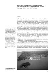
As of 2001, systematic archeological digs have been carried<br />
out in the area of the hill at San Pietro in Canosa, in<br />
the suburbs to the southeast of the town. 1–5 These archeological<br />
investigations are being conducted jointly by the Univers<strong>it</strong>ies<br />
of Foggia and Bari and by the Soprintendenza per i Beni<br />
archeologici della Puglia (Apulian Superintendency of Archeological<br />
Her<strong>it</strong>age). During the digs, many tombs have<br />
come to light, containing bone remains. These have recently<br />
started to be analyzed from the anthropological standpoint,<br />
performing <strong>DNA</strong> typing. First, a brief geographic and historical<br />
outline of the town of Canosa: It is s<strong>it</strong>uated in the south<br />
of Italy, in the Apulian region, approximately 400 km to the<br />
southeast of Rome, and is one of the most important archeological<br />
centers in Apulia and in south Italy in general. The<br />
town was already important in pre-Roman times, when<br />
Canosa was one of the richest and most powerful Daunian<br />
settlements, and <strong>it</strong> emerged even more during the Roman era.<br />
In the late part of the era, the town became the seat of the<br />
governors of the provincia Apulia et Calabria; thus, the<br />
regional cap<strong>it</strong>al. It very early assumed a leading function<br />
from the religious point of view, too, thanks to the presence<br />
of a large Christian commun<strong>it</strong>y guided by powerful bishops,<br />
involved in important councils and diplomatic activ<strong>it</strong>ies. The<br />
greatest moment for the Canosa church was in the sixth<br />
century AD, when <strong>it</strong> was presided over by the famous bishop<br />
Sabino, who is trad<strong>it</strong>ionally held to have governed the diocese<br />
for over 50 years (AD 514-566) and who set in motion the<br />
construction of a great number of holy buildings.<br />
The American Journal of Forensic Medicine and Pathology Volume 28, Number 4, December 2007 333
Di Nunno et al The American Journal of Forensic Medicine and Pathology Volume 28, Number 4, December 2007<br />
MATERIALS AND METHODS<br />
Anthropological Study<br />
An anthropological study was made of 5 adult individuals<br />
found in 3 tombs (nos. 28, 29, 31) at the Paleo-Christian<br />
archeological dig at San Pietro, in Canosa (Figs. 1–4). The<br />
skeletal remains were in a mediocre state of preservation and<br />
various parts were missing, especially in 2 of the tombs that<br />
showed taphonomic cond<strong>it</strong>ions referable to secondary depos<strong>it</strong>s.<br />
The morphometric examinations were made according to<br />
the indications specified by Martin and Saller. 6 Determination<br />
of age and sex was based on the methodologies reported<br />
by Ferembach et al, 7 and Lovejoy et al. 8 The calculations of<br />
stature and biomass were made according to the methods<br />
established by Trotter and Gleser 9 and Ruff et al, 10 respectively.<br />
Assessment of the markers of nutr<strong>it</strong>ional and/or disease<br />
stress on the dental apparatus, in terms of periodontal<br />
disease and caries, was based on the indications by Brothwell<br />
11 ; tartar depos<strong>it</strong>s and the degree of dental wear were<br />
FIGURE 1. The Paleo-Christian archeological dig at San Pietro, in Canosa.<br />
334<br />
analyzed following the indications given by Dobney and<br />
Brothwell 12 and Molnar, 13 respectively. The study of hypoplasia<br />
lines on the enamel was performed according to<br />
Goodman et al. 14 The investigation of skeletal markers of<br />
biomechanical stress, in terms of assessment of syndesmopathy,<br />
enthesopathy, supernumerary articular faces, and degenerative<br />
joint disease was carried out according to the methods<br />
suggested by Kennedy, 15 Lai and Lovell, 16 Robb and Mallegni,<br />
17 and Rogers et al 18 ; assessment of alterations of the<br />
spinal column was made following the indications by Borgognini<br />
Tarli and Repetto. 19 Geometrical techniques were<br />
applied on the transverse sections of the humerus and femur<br />
according to Larsen, 20 Capasso et al, 21 Ledger et al, 22 Stock<br />
and Pfeiffer, 23 and Tracey et al. 24<br />
<strong>DNA</strong> Typing<br />
The present study was conducted on 5 samples typed<br />
w<strong>it</strong>h short tandem repeat (STR) belonging to the CODIS<br />
system: CSF1PO, FGA, TH01, TPOX, vWA, D3S1358,<br />
© 2007 Lippincott Williams & Wilkins
The American Journal of Forensic Medicine and Pathology Volume 28, Number 4, December 2007 Ancient Bone Remains<br />
FIGURE 2. Particulars of the dig.<br />
FIGURE 3. The Paleo-Christian archeological dig at San Pietro,<br />
in Canosa, the tombs.<br />
D5S818, D7S820, D8S1179, D13S317, D16S539, D18S51,<br />
and D21S11. For samples w<strong>it</strong>h teeth, the teeth were treated as<br />
follows. They were first extracted from the dental arch and<br />
then washed first in water and then in a 10% ethanol solution<br />
to eliminate any bacterial contamination. Subsequently, a<br />
long<strong>it</strong>udinal incision was made of each tooth, using the<br />
laboratory drill KAVO EWL 3–36 V � 25.000/min 50 W,<br />
equipped w<strong>it</strong>h a diamond-point mill disk Mesinger 94 off HP<br />
220. The hemidental elements, w<strong>it</strong>h the root canal exposed,<br />
presented brownish residues of dental pulp; these were removed<br />
from the root canals using an odontic curette, ASA<br />
Stainless 1807-12, and were collected in 1.5-mL Eppendorf<br />
test tubes. As a preliminary step in the skeletal investigations,<br />
the femur shafts were cleaned w<strong>it</strong>h a brush and detergent, to<br />
remove fragments of earth and any tree roots adhering to the<br />
bone. After abundant washing to remove any residual detergent,<br />
the femur shafts were left to dry at room temperature.<br />
Later, electromechanical scraping of their surfaces was performed,<br />
using a Dremel. The bone structure was drilled and<br />
a window was made, from which a piece of compact bone<br />
8 � 3 cm in size was taken. The piece of bone was first<br />
broken into smaller pieces about 1 � 2 cm, using the Dremel.<br />
These pieces were placed in a Falcon test tube and repeatedly<br />
washed in deionized water until the washing water was<br />
perfectly clear. They were then washed in ethanol 90°C and<br />
then in ethanol 95°C. After completely drying the samples,<br />
they were reduced to powder. <strong>DNA</strong> extraction from the<br />
dental pulp samples was performed following a modified<br />
protocol of the Promega K<strong>it</strong> “SV Total RNA Isolation System,”<br />
su<strong>it</strong>able for <strong>DNA</strong> extraction from samples containing<br />
only a small number of nucleated cells to begin w<strong>it</strong>h. The<br />
same method was used for the bone samples. The protocol<br />
was partially modified by lengthening the incubation time of<br />
the cell lysis step: each sample of dental pulp was placed,<br />
overnight, at room temperature, in a single microtube containing<br />
350 L of SV RNA Lysis Buffer.<br />
<strong>DNA</strong> extraction from the bone powder was performed<br />
using the commercial k<strong>it</strong> Geneclean for Ancient <strong>DNA</strong><br />
Q-Biogene, following a protocol that we modified only as<br />
regards the first step: a quant<strong>it</strong>y of 100–500 mg of bone<br />
powder is added to a 1.5-mL Eppendorf test tube containing<br />
1 mL of DeHybernation Solution. The microtube is left to<br />
incubate at a temperature of 45°C–60°C overnight and ag<strong>it</strong>ated<br />
occasionally. This step may sometimes need to be<br />
protracted for 24 hours. Amplification was obtained using a<br />
thermal cycler-<strong>DNA</strong> Gene Amp polymerase chain reaction<br />
(PCR) system 9700 (Applied Biosystems, Foster C<strong>it</strong>y, CA) and<br />
the AmpFlSTR Identifiler PCR Amplification K<strong>it</strong> (Applied Biosystems).<br />
25,26 Analysis of the amplified allele fragments was<br />
performed by capillary electrophoresis using the ABI Prism 310<br />
Genetic Analyzer (Applied Biosystems) and the 310 Genetic<br />
Analyzer performance optimized polymer 4 (POP-4; Applied<br />
Biosystems), 10� Genetic Analyzer Buffer w<strong>it</strong>h EDTA, allele<br />
ladders, and GeneScan-500 (internal standard). 27–30<br />
Data Analysis of the <strong>DNA</strong> Typing<br />
The results obtained for the 5 samples were used to<br />
make a comparison w<strong>it</strong>h the haplotypes of present-day subjects<br />
belonging to the Apulian population, to assess genotypical<br />
and allelic homologies and differences. For this purpose,<br />
the genotypes of all the systems analyzed were inserted in the<br />
computer program S.H.S. Hardware software systems that<br />
© 2007 Lippincott Williams & Wilkins 335
Di Nunno et al The American Journal of Forensic Medicine and Pathology Volume 28, Number 4, December 2007<br />
FIGURE 4. A, Tomb T28. B, Tomb T29.<br />
C, Tomb T31.<br />
allows estimation of the frequency of the haplotype (genotype<br />
combinations) of each sample from the Apulian population<br />
(reference population). Many cases of alleles no longer<br />
present in the reference population, at least in the samples<br />
subm<strong>it</strong>ted to statistical analysis, were found (Table 1); the<br />
calculation was therefore based only on the frequency of the<br />
alleles present in the Apulian population. For all the samples,<br />
the differences between the haplotypes of the 5 individuals<br />
and of the present-day Apulian population were statistically<br />
significant (Table 2).<br />
RESULTS<br />
Tomb 28<br />
This is a multiple tomb in which 7 adults were found:<br />
6 male adults and 1 male child. The identification of the<br />
single individuals was made by explo<strong>it</strong>ing morphometric,<br />
336<br />
articular, sex, and bone robustness compatibil<strong>it</strong>ies, as well as<br />
pathologic signs.<br />
Individual 28A<br />
This male subject was aged 40–43 years. He appears to<br />
have had a robust const<strong>it</strong>ution, w<strong>it</strong>h a well-developed muscular<br />
frame. The estimated stature is 170 cm and the body<br />
biomass, 66 kg. Examination of the teeth showed severe<br />
periodontosis, moderate dental wear, and absence of caries<br />
but 2 odontogenic abscesses. There was a cond<strong>it</strong>ion of weight<br />
overload of the cervical vertebrae, as well as Schmorl hernias<br />
of the lumbar vertebrae. The femurs showed Poirier facets<br />
and flattening of the subtrochanteric shaft sections, and the<br />
tibias showed lateral accessory facets. There was moderate<br />
osteoarthr<strong>it</strong>ic degeneration in the form of p<strong>it</strong>ting. From the<br />
pathologic standpoint, phlogosis of the os zygomaticum (Fig.<br />
© 2007 Lippincott Williams & Wilkins
The American Journal of Forensic Medicine and Pathology Volume 28, Number 4, December 2007 Ancient Bone Remains<br />
TABLE 1. Data Analysis of the <strong>DNA</strong> Typing<br />
D8S1179 D21S11 D7S820 CSF1PO D19S433 vWA TPOX FGA<br />
Bone Canosa T28A 8–9 26–31 15–15 11–13 – 13–17 8–10 –<br />
Bone Canosa T28E 13–18 31.2–33 11–11 12–13 10–10 14–19 6–7 27–27<br />
Bone Canosa T29B 12–13 26–26 7–7 �6–�6 – – 6–9 –<br />
Bone Canosa T29E 12–13 26–26 7–7 �6–�6 – – – –<br />
Bone Canosa T31A 9–10 33–36 8–9 6–7 16–17 12–15 7–7 –<br />
Lab. anthropology personnel 15–15 28–28 8–10 11–13 14–14.2 16–19 8–10 20–22<br />
Lab anthropology personnel 13–14 29–32.2 9–9 10–11 14–14 16–20 8–8 23.2–24.2<br />
Lab forensic genetics personnel 12–14 29–30 8–12 10–10 13–14 17–18 8–11 21–22<br />
Lab forensic genetics personnel 13–14 29–29 12–12 10–12 13–15 17–19 8–11 21–22<br />
Lab forensic genetics personnel 11–13 29–32.2 10–11 10–12 13–14 17–17 8–9 20–25<br />
Lab forensic genetics personnel 11–13 28–30 8–10 11–12 12–15 14–16 8–8 21–24<br />
D18S51 D3S1358 TH01 D13S317 D16S539 D2S1338 D5S818 Amel<br />
Bone Canosa T28A 15–20 16–16 6–6 14–15 9–9 – 7–8 X-Y<br />
Bone Canosa T28E – 20–20 4–8 9–9 5–8 16–17 10–10 X-Y<br />
Bone Canosa T29B 15–17 – 6–9.3 8–12 9–12 19–21 9–12 X-Y<br />
Bone Canosa T29E 15–17 – 6–9 �8–10 �8–9 16–18 9–12 X-Y<br />
Bone Canosa T31A – 13–17 9.3–9.3 8–13 10–15 24–24 13–16 X-Y<br />
Lab anthropology personnel 14–16 15–17 6–7 12–12 11–11 17–19 9–12 X-X<br />
Lab anthropology personnel 13–16 15–16 8–9.3 9–12 11–12 17–18 11–12 X-Y<br />
Lab forensic genetics personnel 15–16 15–17 8–9.3 8–11 9–11 20–24 11–12 X-Y<br />
Lab forensic genetics personnel 15–16 15–17 6–9.3 8–12 9–12 20–23 9–12 X-Y<br />
Lab forensic genetics personnel 13–18 16–17 6–10 8–11 11–13 24–25 12–12 X-X<br />
Lab forensic genetics personnel 16–16 15–17 6–9 9–12 9–11 17–19 12–12 X-Y<br />
TABLE 2. Frequency of Haplotypes W<strong>it</strong>h Respect to the<br />
Apulian Population<br />
Sample Haplotype Frequency<br />
T28A 3.89/10 22<br />
T28E 3.65/10 23<br />
T29B 8.8/10 19<br />
T29E 2.87/10 16<br />
T31A 3.54/10 18<br />
5), loss of substance in a half-moon shape was evident on the<br />
clavicula, corresponding to the area of insertion of the conoid<br />
ligament, together w<strong>it</strong>h a bone alteration of the acromial joint<br />
facet. These lesions are compatible w<strong>it</strong>h a past acromionclavicular<br />
dislocation. The person had tibial periost<strong>it</strong>is (Fig. 6).<br />
Individual 28E<br />
This subject was judged to be a male aged between 44<br />
and 60 years. His stature was calculated as 177 cm. There<br />
were many lines of enamel hypoplasia, which had formed<br />
between the ages of 1 and 6 years (Fig. 7). Assessment of the<br />
postcranial indexes revealed a cond<strong>it</strong>ion of eurybrachia of<br />
both humeri and of mesocnemia of both tibias; the bone<br />
robustness index was average in both humeri. The vertebral<br />
load index showed weight overload in the lumbar and cervical<br />
tracts and mild overload in the thoracic tract. Vertebral<br />
osteolysis (Fig. 8). There was disk herniation of the 12th and<br />
10th thoracic vertebrae and the second lumbar vertebra. The<br />
FIGURE 5. Phlogosis of the os zygomaticum.<br />
few musculoskeletal markers available indicate intense muscular<br />
activ<strong>it</strong>y. Osteoarthrosis is present in the form of moderate<br />
lipping and p<strong>it</strong>ting. The patellas show the incisura and<br />
the fossa of the lateral vastus, both tibias have a kneeling<br />
facet. The left astragalus presents the os trigonum. On the left<br />
clavicula (Fig. 9), a bone callous due to fracture of the third<br />
clavicular median along an oblique line was observed, w<strong>it</strong>h<br />
shortening of the segment due to raising of the sternal stump<br />
over the acromial stump. The fracture was not compound and<br />
could have been due to falling on the palm of the hand or on<br />
the shoulder, w<strong>it</strong>h the arm abducted.<br />
© 2007 Lippincott Williams & Wilkins 337
Di Nunno et al The American Journal of Forensic Medicine and Pathology Volume 28, Number 4, December 2007<br />
FIGURE 6. Tibial periost<strong>it</strong>is.<br />
FIGURE 7. Enamel hypoplasia.<br />
Tomb 29<br />
This is a multiple tomb in which 5 individuals were<br />
found: 3 adults, 1 male, and 2 females, as well as 1 young<br />
female and 1 child. The identification of the single individuals<br />
was made by explo<strong>it</strong>ing morphometric, articular, sex,<br />
and bone robustness compatibil<strong>it</strong>ies.<br />
Individual 29B<br />
This is a female subject aged between 50 and 65 years.<br />
This was the only case in which diagnosis of the sex from the<br />
bone remains was discordant w<strong>it</strong>h the <strong>DNA</strong> result. There was<br />
complete fusion between the manubrium and the body of the<br />
sternum. The stature was calculated to be 159 cm, and the<br />
body biomass, 65 kg. Assessment of the postcranial indexes<br />
showed flattening of the diaphyses of the right humerus, left<br />
ulna, and both femurs. The index of robustness of the bones<br />
showed a moderate mean score. The lumbar and thoracic<br />
vertebrae showed a load w<strong>it</strong>hin normal lim<strong>it</strong>s, while <strong>it</strong> was<br />
not possible to estimate the cervical load; herniation of the<br />
first, third, and fourth lumbar vertebrae was observed (Fig.<br />
338<br />
FIGURE 8. Verteblal osteolysis.<br />
FIGURE 9. On the left clavicula, a bone callous due to fracture<br />
of the third clavicular median along an oblique line.<br />
10). Analysis of the degree of enthesopathies showed moderate<br />
muscular activ<strong>it</strong>y. There was osteoarthr<strong>it</strong>is in the form<br />
of mild lipping of the upper limbs, moderate lipping of the<br />
lower limbs, and mild p<strong>it</strong>ting. Osteochondr<strong>it</strong>is dissecans<br />
(O.D.) was observed at the level of the hip joint in both<br />
acetabular cav<strong>it</strong>ies, while the ischiatic tuberos<strong>it</strong>ies presented<br />
traces of Wells burs<strong>it</strong>is. Humeral osteolysis (Fig. 11).<br />
Individual 29E<br />
This individual was diagnosed as being of male sex and<br />
aged approximately 45 years. His stature was calculated to be<br />
169 cm, and body biomass, 71 kg. Dental examination revealed<br />
mild dental wear, the presence of 2 caries, an odontogenic<br />
abscess, and a large amount of tartar. Outcome of<br />
sinus<strong>it</strong>is (Fig. 12). The medial and lateral incisors were spade<br />
shaped. The axial skeleton showed moderate vertebral load in<br />
the cervical tract and the presence of herniation of the last<br />
part of the thoracic tract. The third and fourth lumbar vertebrae<br />
presented Baastrup syndrome (Fig. 13). The sacrum<br />
© 2007 Lippincott Williams & Wilkins
The American Journal of Forensic Medicine and Pathology Volume 28, Number 4, December 2007 Ancient Bone Remains<br />
FIGURE 10. Herniation of the first, third, and fourth lumbar<br />
vertebrae was observed.<br />
presents a bridge vertebra. Examination of the musculoskeletal<br />
markers of the postcranial skeleton reveals strong tendon<br />
impression on all the segments examined. There were signs<br />
of moderate osteoarthrosis, in terms of moderate lipping, mild<br />
p<strong>it</strong>ting, but no eburnation. Poirier facet, Charles facet, and<br />
Allen fossa were present on the left femur; the right femur<br />
showed only Allen fossa and Poirier facet; both tibias presented<br />
accessory lateral and medial facets. Accessory facets<br />
were also present on both astragals. The right patella presented<br />
the fossa of the lateral vastus muscle. There was<br />
evident syndesmopathy in virtually all the ligament insertion<br />
areas. The iliac bone showed a rough surface bilaterally in the<br />
right region above the acetabulum. These roughened, wrinkled<br />
surfaces were more extensive and evident on the right, w<strong>it</strong>h the<br />
presence of bone neoformations of syndesmopathic type in the<br />
hip joint capsule insertion zone. The superior part of the anterior<br />
sacroiliac ligament was completely ossified. The posterior face<br />
of the ischiatic tuberos<strong>it</strong>y presented clear signs of Wells burs<strong>it</strong>is.<br />
The acetabulum had a marginal bone formation bilaterally, w<strong>it</strong>h<br />
extension of the superior joint surface and the presence of O.D.<br />
cav<strong>it</strong>ation.<br />
Tomb 31<br />
This tomb contained 2 individuals of the male sex, 1<br />
adult and 1 adolescent.<br />
FIGURE 11. Humeral osteolysis.<br />
FIGURE 12. Outcome of sinus<strong>it</strong>is.<br />
Individual 31A<br />
This male subject was aged approximately 54 years.<br />
His stature was 179 cm, and body biomass, 81 kg. Dental<br />
examination showed the presence of 2 caries and an abscess<br />
extending to the left maxilla in the malar zone; there was also<br />
severe periodont<strong>it</strong>is, moderate tooth wear, and lines of hypoplasia<br />
attributable to formation between the ages of 2 years<br />
and 6 and a half years. Assessment of the postcranial indexes<br />
© 2007 Lippincott Williams & Wilkins 339
Di Nunno et al The American Journal of Forensic Medicine and Pathology Volume 28, Number 4, December 2007<br />
FIGURE 13. The third and fourth lumbar vertebrae presented<br />
Baastrup syndrome.<br />
FIGURE 14. There are bone neoformations of the feet and<br />
subperiosteal formations of the superior medial face of the<br />
first metatarsal.<br />
demonstrated a cond<strong>it</strong>ion of eurybrachia of the humeri, platolenia<br />
of the ulnas, eurymeria of the left femur, and eurycnemia<br />
of the tibias; a moderate robustness index of both<br />
radiuses, the right femur, and both tibias; and high robustness<br />
of the left clavicle. The pillar index was zero in the right<br />
femur and weak in the left. The vertebral load index showed<br />
TABLE 3. Distribution of Skeletal Alterations in the Study Sample<br />
Tomb Sex Age<br />
Endocr.<br />
Alterations<br />
Vertebr.<br />
Ipervasc<br />
Vertebr.<br />
Cistic<br />
General<br />
Ipervasc<br />
Long<br />
Bones<br />
Periost<strong>it</strong>is<br />
a moderate load in the cervical region and overload in the<br />
thoracolumbar region. There were bone neoformations of the<br />
feet and subperiosteal formations of the superior medial face<br />
of the first metatarsal and the superior lateral face of the fifth<br />
metatarsal (Fig. 14). These small bone formations, due to<br />
continual, mild irr<strong>it</strong>ant stimuli, were located in correspondence<br />
w<strong>it</strong>h the points of contact between the back of the foot<br />
and parts of the soles of the shoes worn at the time, such as<br />
sandals. The observation of enthesopathic traces demonstrates<br />
intense muscular activ<strong>it</strong>y, as also evinced from the<br />
syndesmopathic traces and osteoarticular degenerations. Both<br />
femurs presented an extensive Poirier facet and Charles facet;<br />
both tibias had a kneeling facet. Accessory facets were<br />
present on the astragals. The hip bones showed the calcar of<br />
the iliac crest.<br />
Concluding Pathology Notes<br />
The paleopathological investigation of the 5 individuals<br />
revealed a whole series of skeletal alterations, involving the<br />
skull, ribs, vertebral column, and long bones, attributable to<br />
inflammatory foci (Table 3), whose distribution and morphology<br />
could likely indicate a specific nature, probably of tubercular<br />
origin. This finding is of particular interest because <strong>it</strong><br />
can be generalized to other individuals found in the same<br />
cemetery area.<br />
CONCLUSIONS<br />
Genetic and anthropologic investigations have yielded<br />
a series of new data on the bone remains found in the area<br />
around San Pietro in Canosa di Puglia. From the genetic<br />
standpoint, we can state that significant differences were<br />
observed between the STR sequences analyzed in the presentday<br />
inhab<strong>it</strong>ants of the Apulian region and their medieval<br />
ancestors. These findings may be justified by the fact that,<br />
during the Middle Ages, there were strong gene flows from<br />
the Germanic and Asian peoples (Goths, Lombards, Avars)<br />
that may have cond<strong>it</strong>ioned the genome of the native populations.<br />
In the subsequent centuries, these gene pools may have<br />
been diluted, thus strongly altering the gene status of the<br />
Italian population. From the anthropologic standpoint, <strong>it</strong> must<br />
be borne in mind that part of the cemetery in the archeological<br />
s<strong>it</strong>e at San Pietro shows selective cond<strong>it</strong>ions, in terms of<br />
alterations indicating inflammatory disease, likely of a specific<br />
nature and probably of tubercular origin.<br />
Ribs<br />
Phlogosis/Alterations Hyperostosis<br />
Periart<strong>it</strong>is<br />
Humerus<br />
Alterat.<br />
Sternum<br />
Alterations<br />
Turbinate<br />
Bones<br />
Phlogosis<br />
e28 A M 40–43 x x x x x x n.r.<br />
28 E M 44–60 n.r. x x x x x n.r. x n.r. n.r.<br />
29 B F 44–65 x x x x n.r. x x<br />
29 E M 45–49 x x x x x x<br />
31 A M 47–54 x x x x x x x x n.r.<br />
340<br />
© 2007 Lippincott Williams & Wilkins
The American Journal of Forensic Medicine and Pathology Volume 28, Number 4, December 2007 Ancient Bone Remains<br />
ACKNOWLEDGMENTS<br />
The authors would like to thank M. V. C. Pragnell, BA,<br />
for help w<strong>it</strong>h translation of the text.<br />
REFERENCES<br />
1. Volpe G, Annese C, Ciminale M, et al. Il complesso episcopale paleocristiano<br />
di san Pietro a Canosa: prima relazione preliminare (campagna<br />
di scavi 2001). Vetera Christianorum. 2002;39:133–190.<br />
2. Volpe G, Annese C, Corrente M, et al. Il complesso episcopale paleocristiano<br />
di san Pietro a Canosa : seconda relazione preliminare (campagna<br />
di scavi 2002). Archeologia Medievale. 2003;30:107–164.<br />
3. Volpe G. Il complesso episcopale di San Pietro nel quadro di Canosa<br />
paleocristiana, in Atti del Convegno Canosa. Ricerche Storiche 2003<br />
(Canosa 14. 12. 2002), a cura di L. Bertoldi Lenoci, Fasano 2003:89–104.<br />
4. Volpe G. Nuovi dati sul complesso episcopale paleocristiano di san<br />
Pietro a Canosa, in Atti del Convegno Canosa, Ricerche storiche 2004<br />
(Canosa 7. 2.2004), a cura di L. Bertoldi Lenoci, Fasano 2005:15–34.<br />
5. Volpe G, Annese C, Leone D, et al. I mosaici pavimentali del complesso<br />
paleocristiano di San Pietro a Canosa (Ba), in Atti del X Colloquio<br />
dell’Associazione Italiana per lo Studio e la Conservazione del Mosaico<br />
(AISCOM) (Lecce 18–21 febbraio 2004), Roma 2005.<br />
6. Martin R, Saller K. Lehrbuch der Anthropologie. Stuttgart: G. Fischer;<br />
1959.<br />
7. Ferembach D, Schwidetzky I, Stloukal M. Raccomandazioni per la<br />
determinazione dell’età e del sesso sullo scheletro. Rivista Antropologia.<br />
1979;60:5–51.<br />
8. Lovejoy CO, Meindl S, Pryzbeck TR, et al. Chronological metamorphosis<br />
of the auricolar surface of the ilium: a new method for the determination of<br />
adult skeletal age at death. Am J Phys Anthropol. 1985;68:15–28.<br />
9. Trotter M, Gleser GC. Corrigenda to estimation of stature from limb<br />
bones of American wh<strong>it</strong>es and Negroes. Am J Phys Anthropol. 1997;<br />
47:355–356.<br />
10. Ruff CR, Trinkaus E, Holliday TW. Body mass and encephalization in<br />
Pleistocene Homo. Nature. 1997;387:173–176.<br />
11. Brothwell DR. Digging Up Bones. Oxford: Univers<strong>it</strong>y Press; 1981.<br />
12. Dobney K, Brothwell DR. A method for evaluating the amont of dental<br />
calculus on teeth from archaeological s<strong>it</strong>e. J Archaeol Sci. 1987;14:343–<br />
351.<br />
13. Molnar S. Human tooth wear, tooth function and cultural variabil<strong>it</strong>y.<br />
Am J Phys Anthropol. 1971;34:175–189.<br />
14. Goodman AH, Martin DL, Armelagos GJ. Indications of stress from<br />
bone and teeth. In: Cohen MN, Armelagos GJ, eds. Paleopathology at<br />
the Origin of the Agriculture. London: Academic Press; 1984:13–49.<br />
15. Kennedy KAR. Skeletal markers of occupational stress. In: Iscan MY,<br />
Kennedy KAR, eds. Reconstruction of Life from the Skeleton. New<br />
York: Liss; 1989:129–160.<br />
16. Lai P, Lovell NC. Skeletal markers of occupational stress in the fur<br />
trade: a case study from Hudson’s Bay Company Fur Trade Post. Int J<br />
Osteoarchaeol. 1992;2:221–234.<br />
17. Robb J, Mallegni F. Anthropology and paleopathology of Neol<strong>it</strong>hic<br />
human remains from Catignano (Pescara, Italy). Rivista Antropologia.<br />
1994;72:197–224.<br />
18. Rogers J, Waldron T, Dieppe P, et al. Arthropathies in paleopathology:<br />
the basis of the classification according to most probable cause.<br />
J Archaeol Sci. 1987;14:179–193.<br />
19. Borgognini Tarli SM, Repetto E. Skeletal indicators of subsistence<br />
patterns and activ<strong>it</strong>y regime in the Mesol<strong>it</strong>hic sample from Grotta<br />
dell’Uzzo (Trapani, Sicily): a case in study. Hum Evol. 1986;4:331–352.<br />
20. Larsen CS. Bioarchaeology: Interpreting Behaviour from the Human<br />
Skeleton. Cambridge: Univers<strong>it</strong>y Press; 1999.<br />
21. Capasso L, Kenneth AR, Kennedy C, et al. Atlas of Occupational<br />
Markers on Human Remains. Teramo: Edigraf<strong>it</strong>al S.p.A.; 1999.<br />
22. Ledger M, Holtzhausen L, Constant D, et al. Biomechanical beam<br />
analysis of long bones from a late 18 th century slave cemetery in Cape<br />
Town, South Africa. Am J Phys Anthropol. 2000;112:207–216.<br />
23. Stock J, Pfeiffer S. Linking structural variabil<strong>it</strong>y in long bone diaphyses<br />
to hab<strong>it</strong>ual behaviors: foragers from the southern African Later Stone<br />
Age and the Andaman Isles. Am J Phys Anthropol. 2001;115:337–348.<br />
24. Tracey AR, Ruff CB, Plato CC. Hand dominance and bilateral asymmetry<br />
in the structure of the second metacarpal. Am J Phys Anthropol.<br />
1994;94:203–211.<br />
25. Kimpton CP, Oldroyd NJ, Watson SK, et al. Validation of highly<br />
discriminating multiplex short tandem repeat amplification systems for<br />
individual identification. Electrophoresis. 1996;17:1283–1293.<br />
26. PE Biosystems. AmpFlSTR Cofiler PCR Amplification K<strong>it</strong> User Bulletin.<br />
Foster C<strong>it</strong>y, CA: PE Biosystems; 1998.<br />
27. PE Biosystems. ABI PRISM 310 Genetic Analyzer User’s Manual: Rev.<br />
1. Foster C<strong>it</strong>y, CA: PE Biosystems; 1995.<br />
28. PE Biosystems. ABI PRISM 310 Genetic Analyzer User’s Manual.<br />
Foster C<strong>it</strong>y, CA: PE Biosystems; 1998.<br />
29. PE Biosystems. ABI PRISM GeneScan Analysis Software 2.1 User’s<br />
Manual. Foster C<strong>it</strong>y, CA: PE Biosystems; 1996.<br />
30. Issaq H, Chan K, Muschik G. The effect of column length, applied<br />
voltage, gel type, and concentration on the capillary electrophoresis<br />
separation of <strong>DNA</strong> fragments and polymerase chain reaction products.<br />
Electrophoresis. 1997;18:1153–1158.<br />
© 2007 Lippincott Williams & Wilkins 341