pathfinder™ corneal analysis - Carl Zeiss Meditec
pathfinder™ corneal analysis - Carl Zeiss Meditec
pathfinder™ corneal analysis - Carl Zeiss Meditec
Create successful ePaper yourself
Turn your PDF publications into a flip-book with our unique Google optimized e-Paper software.
14<br />
INTRODUCTION<br />
PATHFINDER CORNEAL ANALYSIS<br />
STATISTICAL INDICES AND<br />
NORMAL POPULATIONS<br />
HUMPHREY ATLAS AND ATLAS ECLIPSE<br />
PN 55430 REV. A FEB 2003<br />
ADDENDUM TO REV. C PN 48113-1<br />
PathFinder Corneal Analysis is a software program designed<br />
specifically for <strong>corneal</strong> topography to identify abnormal, pathologic,<br />
and normal corneas based on statistical indices derived from the<br />
Humphrey ATLAS, ATLAS Eclipse and MasterVue Corneal<br />
Topography systems. Some of these indices are exclusive to<br />
Humphrey <strong>corneal</strong> topography, and therefore make this software<br />
<strong>analysis</strong> unique when compared to other software programs available<br />
that examine the cornea for pathology or abnormal surface conditions.<br />
The three statistical indices that PathFinder uses to gauge the shape,<br />
regularity, and toricity of the <strong>corneal</strong> surface are Corneal Irregularity<br />
Measurement (CIM), Shape Factor (SF), and Mean Toric<br />
Keratometry(TKM). Isolating and identifying normal versus<br />
abnormalpopulations using these three parameters by examining<br />
hundreds oftopography exams has been done in order to achieve the<br />
high level ofsensitivity and specificity that the software is able to<br />
accomplish.<br />
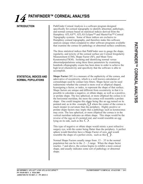
Shape Factor (SF) is a measure of the asphericity of the cornea, and<br />
aderivative of eccentricity, which is a well known calculation of<br />
<strong>corneal</strong>shape used by contact lens fitters. Shape factor can be used<br />
todetermine whether the <strong>corneal</strong> is more oval or elliptical shaped,<br />
byassigning a factor, or index, to represent the shape of that surface.<br />
Shape factors are unique and different from eccentricity in that it is<br />
possible to calculate a negative, or oblate shape, as well as a positive,<br />
or prolate shape. The less spherical, or more elliptical the cornea is in<br />
the horizontal meridian, the more the cornea will resemble a prolate<br />
shape. One could imagine this shape being like an egg turned on its<br />
pointed end, as in this example, where the center of the cornea is<br />
much steeper in curvature than the periphery. Highly positive or<br />
prolate shape factors may imply that a pathology such as keratoconus<br />
may exist. The less spherical, or more elliptical the cornea is in the<br />
vertical meridian indicates an oblate shape. This shape would be the<br />
reverse of the egg on it's pointed end, and would resemble an egg<br />
lying on its side, such as this .<br />
This type of negative or oblate shape would mimic a post refractive<br />
surgery eye, with the center being flatter than the periphery. A perfect<br />
sphere would therefore have a Shape Factor of zero, and would<br />
resemble the shape of a perfect circle, such as this .<br />
Normal Shape Factors usually range from .13 - .35 in the normal<br />
population but can be in the .2 - .3 range. When the shape factor<br />
reaches .7 and above, the cornea begins to exhibit a more conical<br />
shape, and usually indicates some sort of pathology or abnormal<br />
shape.<br />
PATHFINDER CORNEAL ANALYSIS / 14-1<br />
PATHFINDER CORNEAL ANALYSIS
Shape Factor ranges occur in the human population as follows:<br />
Normal - .13 to .35<br />
Borderline - .02 to .12, .36 to .46<br />
Abnormal - - 1.0 to .01, .47 to 1.0<br />
The normal distribution in the human population appears as a bell<br />
shaped curve where the mean value is .24 while 96% of the<br />
population falls between 0 and .46.<br />
The mathematical formula for shape factor is:<br />
Ep = e 2 = 1 - pp where Ep is a prolate shape<br />
and<br />
Eo = _-e 2 _<br />
1 - e 2 where Eo is an oblate shape.<br />
Corneal Irregularity Measurement (CIM) is a number or index<br />
assigned to represent the irregularity of the <strong>corneal</strong> surface. The<br />
higher the irregularity index, the more uncorrectable or uneven the<br />
surface is optically, thereby highlighting irregular astigmatism that<br />
often results in visual distortions. CIM uses the thousands of data<br />
points within the first ten rings of the <strong>corneal</strong> topography data to<br />
determine the difference in "height" or elevation between the patients<br />
cornea and a perfect model toric cornea. The difference between the<br />
perfect model and the actual cornea is measured in microns and the<br />
standard deviation is taken. This is defined as CIM. Higher CIM<br />
values, then, would tend to indicate a worsening pathology such as<br />
keratoconus, due to the inherent <strong>corneal</strong> thinning and "wrinkling" of<br />
the <strong>corneal</strong> surface that the pathology causes. High CIM indices can<br />
also be observed in post refractive surgery corneas due to the<br />
irregularity that the surgical procedure and subsequent healing process<br />
cause. CIM normal values appear in the table below; they range from<br />
14-2 / PATHFINDER CORNEAL ANALYSIS HUMPHREY ATLAS AND ATLAS ECLIPSE<br />
PN 55430 REV. A FEB 2003<br />
ADDENDUM TO REV. C PN 48113-1
HUMPHREY ATLAS AND ATLAS ECLIPSE<br />
PN 55430 REV. A FEB 2003<br />
ADDENDUM TO REV. C PN 48113-1<br />
0.4 to 0.8 or 0.9 microns, depending on the model. The higher the<br />
number becomes, the more abnormal it is. When CIM values are<br />
measured at 1.0 microns and above, severe <strong>corneal</strong> distortion is<br />
usually present.<br />
Models 991 & 993 Models 992 & 995<br />
Normal - 0 to 0.80 0 to 0.90<br />
Borderline - 0.81 to 1.00 0.91 to 1.10<br />
Abnormal - >1.00 >1.10<br />
67% of the population falls in the normal range. The infrared Models<br />
992 & 995 have more noise, which accounts for the difference in the<br />
ranges. A bell shaped distribution curve is not seen due to the fact<br />
that no negative numbers can be calculated using CIM, resulting in a<br />
skewed distribution plot.<br />
Models 991 & 993 Models 992 & 995<br />
Mean Toric "K" (TKM) is the value which is derived using<br />
elevation data from a best fit toric reference surface as compared to<br />
the actual cornea. Two values are calculated at the apex of the flattest<br />
meridian and their mean is determined. This is described as the mean<br />
value of apical curvature. The higher the TKM index becomes, the<br />
greater likelihood that excessive <strong>corneal</strong> toricity exists, which often<br />
leads to keratoconus. By fitting the topography of the patient's cornea<br />
to a best fit reference toric, all of the correctable sphere and cylinder<br />
can be accounted for in the topographical data, thereby giving the<br />
most accurate toric representation of the patient's cornea possible.<br />
PATHFINDER CORNEAL ANALYSIS / 14-3<br />
PATHFINDER CORNEAL ANALYSIS
Definition of Conditions<br />
Identified by PathFinder<br />
The ranges for TKM in the population are as follows:<br />
Normal - 43.1 to 45.9<br />
Borderline - 41.8 to 43, 46 to 47.2<br />
Abnormal - 36 to 41.7, 47.3 to 60<br />
The distribution appears as a bell shaped curve where the mean value<br />
for TKM is 44.5 and 96% of the population falls between 41.25 and<br />
47.25 diopters.<br />
Isolating and identifying normal versus abnormal populatioins has<br />
been determined using the three statistical indices (CIM, SF, MTK) in<br />
the examination of hundreds of topography exams. These exams act<br />
as a control group for the software to identify when a cornea is<br />
normal, just as when there is an abnormal condition.<br />
Normal- The cornea is typically aspherical in shape with TKM, CIM<br />
and Shape Factor in the normal range. No abnormal topography<br />
patterns such as inferior steepening or abnormal elevation or<br />
curvature can be seen. These corneas may have a history of soft<br />
contact lens wear but no family history of keratoconus, excessive or<br />
irregular astigmatism, or rigid lens wear.<br />
Corneal Distortion- (also known as pseudokeratoconus) is a<br />
condition caused by excessive RGP lens wear, where the poor fitting<br />
lens actually causes the inferior cornea to bulge outward, mimicking<br />
keratoconus on the topographical map, while no clinical or slit lamp<br />
findings are evident. The abnormal topography pattern may resolve or<br />
improve in a relatively short time period after removing the lens. The<br />
topographic pattern usually shows the cornea "bulging" rather than<br />
steepening abnormally, and there may also be signs of a compression<br />
ring, where the lens is "biting" or digging into the cornea. Some<br />
visual distortion symptoms may accompany this condition but usually<br />
diminish quickly after discontinuing lens wear. CIM may be outside<br />
normal limits, while Shape Factor and TKM remain within normal<br />
limits.<br />
14-4 / PATHFINDER CORNEAL ANALYSIS HUMPHREY ATLAS AND ATLAS ECLIPSE<br />
PN 55430 REV. A FEB 2003<br />
ADDENDUM TO REV. C PN 48113-1
To Access the PathFinder<br />
Corneal Analysis<br />
HUMPHREY ATLAS AND ATLAS ECLIPSE<br />
PN 55430 REV. A FEB 2003<br />
ADDENDUM TO REV. C PN 48113-1<br />
Sub-Clinical Keratoconus-(also known as forme fruste keratoconus)<br />
is usually distinguishable with the typical inferior nasal steepening<br />
topography pattern, but the cornea does not yet exhibit slit lamp<br />
findings typical of keratoconus. The steepening is noticeable on<br />
topography but can be well under 50 diopters at the apex of the cone.<br />
Often there is a history of the disease in the family, or the refractive<br />
history is unstable with a measurable increase in both myopia and<br />
astigmatism over the past several years. There may also be an<br />
inability to wear contact lenses, along with visual symptoms (reduced<br />
VA, ghosting, light sensitivity). CIM and TKM are likely to be<br />
outside normal limits in this condition, while Shape Factor will be<br />
within normal limits to slightly outside normal limits.<br />
Keratoconus- is defined as the true pathological condition which<br />
causes thinning and wrinkling of the cornea, along with a cone-like<br />
protrusion of the cornea in it's later stages. Keratoconus can also<br />
produce high regular astigmatism, irregular astigmatism, and inferior<br />
steepening topographical patterns (usually nasal). Abnormal <strong>corneal</strong><br />
findings observed under biomicroscopy to confirm a true keratoconus<br />
diagnosis may include but are not limited to: Vogt's striae, Munson's<br />
sign, Flesicher's ring, <strong>corneal</strong> ectasia, stromal thinning and superficial<br />
scars in Descemet's membrane. All three statistical indices, CIM,<br />
Shape Factor, and TKM will likely be outside normal limits with this<br />
condition.<br />
Note: Decisions involving surgical procedures should be made<br />
only after considering total clinical information, and not on the<br />
basis of a single index or measurement.<br />
PathFinder can be accessed two ways within the software. From the<br />
Main Menu screen, click on "Review", then select the patient and<br />
exam that you wish to evaluate. Select PathFinder Corneal Analysis<br />
from the drop down “Views” list box. Click on "OK" and the<br />
software will run and instantly provide the <strong>analysis</strong> using the standard<br />
Axial Map. The software can also be run from the MultiVue<br />
Displays/Optional Modules Screen. By clicking on the entry labeled<br />
"PathFinder Corneal Analysis" after selecting the desired patient and<br />
exam, the software will display the findings. Scaling can be changed<br />
from .25 diopter to .50 diopter increments simply by clicking on the<br />
AutoSize or Standard buttons respectively. To print the <strong>analysis</strong>,<br />
return to the Main Menu, or cancel the selection, click on Options,<br />
and follow the onscreen prompts.<br />
PATHFINDER CORNEAL ANALYSIS / 14-5<br />
PATHFINDER CORNEAL ANALYSIS
PATHFINDER CORNEAL<br />
ANALYSIS SCREEN<br />
EXAMPLES<br />
NORMAL RESULT<br />
There are four different results that can be displayed when using the<br />
software, depending on the <strong>analysis</strong>:<br />
The final result of the PathFinder<br />
Corneal Analysis is shown here.<br />
CIM is a measure of irregularity<br />
taken from the difference between<br />
a best fit toric model and the<br />
actual <strong>corneal</strong> surface itself using<br />
highly sensitive elevation data.<br />
Shape Factor is a measure of the<br />
asphericity of the cornea, and a<br />
derivative of eccentricity, a well<br />
known measurement of <strong>corneal</strong><br />
shape.<br />
TKM is the mean toric keratometry<br />
measured as the difference<br />
between a best fit toric surface and<br />
the mean apical <strong>corneal</strong> curvature.<br />
NORMAL RESULT USING THE PATHFINDER CORNEAL ANALYSIS (FIG. 1)<br />
The red area of the bar indicates<br />
the abnormal range, while the<br />
yellow is borderline and the green<br />
is within normal limits. The index<br />
for this particular cornea is denoted<br />
above the bar and it’s location on<br />
the bar is indicated by an arrow.<br />
The results of this <strong>analysis</strong> confirm a normal diagnosis as the<br />
statistical indicators CIM and SF are within normal limits while the<br />
TKM index is only borderline or slightly abnormal. If the TKM<br />
continues to increase or the CIM rises to an abnormal level over time,<br />
this cornea could be identified as <strong>corneal</strong> distortion. This may<br />
indicate a need to follow the patient long term to evaluate any<br />
changes that may be taking place.<br />
14-6 / PATHFINDER CORNEAL ANALYSIS HUMPHREY ATLAS AND ATLAS ECLIPSE<br />
PN 55430 REV. A FEB 2003<br />
ADDENDUM TO REV. C PN 48113-1


