Givaudan-Roure Lecture - Association for Chemoreception Sciences
Givaudan-Roure Lecture - Association for Chemoreception Sciences
Givaudan-Roure Lecture - Association for Chemoreception Sciences
Create successful ePaper yourself
Turn your PDF publications into a flip-book with our unique Google optimized e-Paper software.
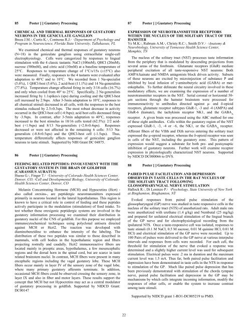
85 Poster [ ] Gustatory Processing<br />
CHEMICAL AND THERMAL RESPONSES OF GUSTATORY<br />
NEURONS IN THE GENICULATE GANGLION<br />
Breza J.M. 1, Curtis K. 1, Contreras R.J. 1 1Department of Psychology and<br />
Program in Neuroscience, Florida State University, Tallahassee, FL<br />
We examined chemical and thermal responses of gustatory neurons<br />
(N=18) in the geniculate ganglion using extracellular single-cell<br />
electrophysiology. Cells were categorized by responses to lingual<br />
stimulation with the 4 classic tastants: NaCl (100mM), QHCl (20mM),<br />
sucrose (500mM), and citric acid (10mM) at a baseline temperature of<br />
25°C. Responses to temperature change of 1°C/s/15s (±15°C) also<br />
were measured. Finally, responses to the 4 tastants were evaluated after<br />
adaptation to 40°C and to 10°C. We recorded from 1 Na-specialist<br />
(5.6%), 1 QHCl-best (5.6%), 2 acid-best (11.1%) and 14 Na-generalists<br />
(77.8%). Temperature change affected firing in only 3/18 cells (16.7%)<br />
and only when cooled from 40° to 25°C. Specifically, 2 Na-generalists<br />
increased firing by 1.1spikes/s (sps) during cooling and the QHCl-best<br />
cell increased by 2.9sps. After 3-5min adaptation to 10°C, responses to<br />
all chemical stimuli decreased in all cells, with the responses to the best<br />
stimulus reduced by 2.5±0.3sps. The most robust decrease in activity<br />
occurred in the QHCl-best cell (-4.9sps); acid best cells decreased firing<br />
by -3.9sps. In contrast, after 3-5min adaptation to 40°C, responses<br />
increased to the best stimulus in 10/16 cells tested (62.5%): 2/2 acidbest<br />
(+1.9sps) and 8/13 Na-generalists (+2.0±0.4sps). Responses<br />
decreased or were not affected in the remaining 6 cells: 5/13 Nageneralists<br />
(-0.8±0.5sps) and the QHCl-best cell (-1.5sps). Thus,<br />
temperature differentially affects responses of geniculate ganglion<br />
neurons to taste stimuli. Supported by NIH Grant DC 04875.<br />
86 Poster [ ] Gustatory Processing<br />
FEEDING RELATED PEPTIDES: INVOLVEMENT WITH THE<br />
GUSTATORY SYSTEM IN THE BRAIN OF GOLDFISH<br />
(CARASSIUS AURATUS)<br />
Huesa G. 1, Finger T. 2 1University of Colorado Health <strong>Sciences</strong> Center,<br />
Denver, CO; 2Cell and Developmental Biology, University of Colorado<br />
Health <strong>Sciences</strong> Center, Denver, CO<br />
Melanin Concentrating Hormone (MCH) and Hypocretins (Hcrt) -<br />
also called orexins-, are peptidergic neurotransmitters expressed<br />
primarily in neurons located in the lateral hypothalamus. This region is<br />
known to have a critical role in control of feeding and these peptides<br />
actively participate in the modulation (stimulation) of food intake. To<br />
test whether these orexigenic peptidergic systems are involved in the<br />
gustatory in<strong>for</strong>mation processing we examined their distribution in<br />
gustatory nuclei of the CNS of goldfish. For this purpose we employed<br />
immunocytochemical techniques using polyclonal antisera directed<br />
against MCH or Hcrt2. The reaction was developed with<br />
diaminobenzidine to enhance the intensity of the labeling. The<br />
distribution of these two peptides was similar to those described in<br />
mammals, with cell bodies in the hypothalamic region and fibers<br />
projecting rostrally and caudally. Hcrt2 immunoreactive fibers are<br />
located mainly in preoptic areas, hypothalamus, a few mesencephalic<br />
regions and the dorsal horn in the spinal cord, but are scarce in taste<br />
related brainstem nuclei. In contrast, MCH fibers were present in many<br />
encephalic regions including the vagal gustatory lobe. These MCH<br />
fibers occur mainly in layer IV of the sensory zone of the vagal lobe,<br />
where many primary gustatory afferents terminate. In addition,<br />
occasional MCH fibers could be observed crossing the sensory zone, in<br />
layer IX and also in fiber and motor layers. These results support the<br />
concept that MCH but not Hypocretins may act as a central modulator<br />
of gustatory processing in goldfish. Supported by NIDCD Grant:<br />
DC00147<br />
22<br />
87 Poster [ ] Gustatory Processing<br />
EXPRESSION OF NEUROTRANSMITTER RECEPTORS<br />
WITHIN THE NUCLEUS OF THE SOLITARY TRACT OF THE<br />
HAMSTER<br />
Ye M.K. 1, Rubrum A.M. 1, Christy R.C. 1, Smith D.V. 1 1Anatomy &<br />
Neurobiology, University of Tennessee Health Science Center,<br />
Memphis, TN<br />
The nucleus of the solitary tract (NST) processes taste in<strong>for</strong>mation<br />
from the periphery that is modulated by descending projections from<br />
several areas of the <strong>for</strong>ebrain. Glutamate receptors (GluR) mediate<br />
peripheral excitation of all taste-responsive NST neurons: Both<br />
AMPA/kainate and NMDA antagonists block driven activity. Subsets<br />
of these neurons are excited by microinjection of substance P and<br />
inhibited by local infusion of γ-aminobutyric acid (GABA) or metenkephalin.<br />
To further delineate the neural circuitry involved in these<br />
modulatory effects, we are examining the expression of a number of<br />
neurotransmitter receptors in the NST. Serial coronal or horizontal 50-<br />
µm sections through the hamster brainstem were processed <strong>for</strong><br />
immunoreactivity to antibodies directed against µ- and δ-opioid<br />
receptors, glutamate receptor subtypes GluR-1, -3 and -4 (AMPA) and<br />
GluR-6 (kainate), the tachykinin receptor NK1, and the GABAA receptor. A given brain was processed using the ABC method <strong>for</strong> one<br />
of these eight antibodies. Cells within the gustatory region of the NST<br />
expressed GluR-1, -3, -4 or –6, the NK1, or the GABA receptor.<br />
A<br />
Afferent fibers of the VIIth and IXth nerves entering the solitary tract<br />
expressed the µ-opioid receptor, whereas the δ-opioid receptor was seen<br />
in cells of the NST, including the gustatory zone. This pattern of<br />
expression would suggest a substrate <strong>for</strong> both pre- and postsynaptic<br />
inhibition of gustatory neurons. Further work will examine receptor<br />
expression in physiologically characterized NST neurons. Supported<br />
by NIDCD DC000066 to DVS.<br />
88 Poster [ ] Gustatory Processing<br />
PAIRED PULSE FACILITATION AND DEPRESSION<br />
OBSERVED IN TASTE CELLS IN THE RAT NUCLEUS OF<br />
THE SOLITARY TRACT FOLLOWING<br />
GLOSSOPHARYNGEAL NERVE STIMULATION<br />
Hallock R. 1, Di Lorenzo P. 1 1Psychology, State University of New York<br />
at Binghamton, Binghamton, NY<br />
Evoked responses from paired pulse stimulation of the<br />
glossopharyngeal (GP) nerve was studied in taste responsive cells in the<br />
nucleus of the solitary tract (NTS) of anesthetized rats. Adult male rats<br />
were anesthetized with urethane (1.4 g/kg) and Nembutal (25 mg/kg)<br />
and prepared <strong>for</strong> unilateral electrical stimulation of the lingual branch<br />
of the GP nerve and <strong>for</strong> electrophysiological recording from the<br />
ipsilateral NTS. Once a taste-responsive cell was isolated, responses to<br />
taste stimuli (0.1 M NaCl, 0.5 M sucrose, 0.01 M quinine HCl, 0.01 M<br />
HCl) and electrical stimulation of the GP nerve were recorded. Up to<br />
100 Pairs of pulses were delivered to the GP nerve at various interpulse<br />
intervals and responses from cells were recorded. For each cell, the<br />
threshold <strong>for</strong> stimulation of the nerve that evoked a response was<br />
determined and a slightly higher current level was used <strong>for</strong> subsequent<br />
stimulation. Electrical pulses were .2 ms in duration and the maximum<br />
current level was 1.5 mA. Thus far, both paired pulse facilitation and<br />
depression have been demonstrated in taste cells in the NTS in response<br />
to stimulation of the GP. Much like paired pulse depression that has<br />
been previously demonstrated with stimulation of the chorda tympani<br />
nerve, paired pulse facilitation and depression in the GP may be<br />
mechanisms by which cells integrate incoming in<strong>for</strong>mation, modify the<br />
responses of other cells, or enable the system to increase contrast<br />
among taste stimuli.<br />
Supported by NIDCD grant 1-RO1-DC005219 to PMD.


