

Isolated lymphoid follicles in colon - World Journal of Gastroenterology
Isolated lymphoid follicles in colon - World Journal of Gastroenterology
Isolated lymphoid follicles in colon - World Journal of Gastroenterology

Create successful ePaper yourself
Turn your PDF publications into a flip-book with our unique Google optimized e-Paper software.
Sipos F et al . Colonic isolated <strong>lymphoid</strong> <strong>follicles</strong><br />
*<br />
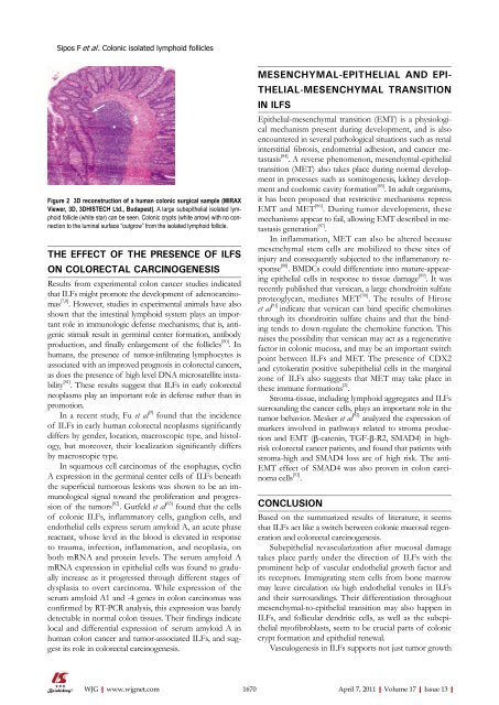
Figure 2 3D reconstruction <strong>of</strong> a human <strong>colon</strong>ic surgical sample (MIRAX<br />
Viewer, 3D, 3DHISTECH Ltd., Budapest). A large subepithelial isolated <strong>lymphoid</strong><br />
follicle (white star) can be seen. Colonic crypts (white arrow) with no connection<br />
to the lum<strong>in</strong>al surface “outgrow” from the isolated <strong>lymphoid</strong> follicle.<br />
THE EFFECT OF THE PRESENCE OF ILFS<br />
ON COLORECTAL CARCINOGENESIS<br />
Results from experimental <strong>colon</strong> cancer studies <strong>in</strong>dicated<br />
that ILFs might promote the development <strong>of</strong> adenocarc<strong>in</strong>omas<br />
[7,8] . However, studies <strong>in</strong> experimental animals have also<br />
shown that the <strong>in</strong>test<strong>in</strong>al <strong>lymphoid</strong> system plays an important<br />
role <strong>in</strong> immunologic defense mechanisms; that is, antigenic<br />
stimuli result <strong>in</strong> germ<strong>in</strong>al center formation, antibody<br />
production, and f<strong>in</strong>ally enlargement <strong>of</strong> the <strong>follicles</strong> [80] . In<br />
humans, the presence <strong>of</strong> tumor-<strong>in</strong>filtrat<strong>in</strong>g lymphocytes is<br />
associated with an improved prognosis <strong>in</strong> colorectal cancers,<br />
as does the presence <strong>of</strong> high level DNA microsatellite <strong>in</strong>stability<br />
[81] . These results suggest that ILFs <strong>in</strong> early colorectal<br />
neoplasms play an important role <strong>in</strong> defense rather than <strong>in</strong><br />
promotion.<br />
In a recent study, Fu et al [9] found that the <strong>in</strong>cidence<br />
<strong>of</strong> ILFs <strong>in</strong> early human colorectal neoplasms significantly<br />
differs by gender, location, macroscopic type, and histology,<br />
but moreover, their localization significantly differs<br />
by macroscopic type.<br />
In squamous cell carc<strong>in</strong>omas <strong>of</strong> the esophagus, cycl<strong>in</strong><br />
A expression <strong>in</strong> the germ<strong>in</strong>al center cells <strong>of</strong> ILFs beneath<br />
the superficial tumorous lesions was shown to be an immunological<br />
signal toward the proliferation and progression<br />
<strong>of</strong> the tumors [82] . Gutfeld et al [83] found that the cells<br />
<strong>of</strong> <strong>colon</strong>ic ILFs, <strong>in</strong>flammatory cells, ganglion cells, and<br />
endothelial cells express serum amyloid A, an acute phase<br />
reactant, whose level <strong>in</strong> the blood is elevated <strong>in</strong> response<br />
to trauma, <strong>in</strong>fection, <strong>in</strong>flammation, and neoplasia, on<br />
both mRNA and prote<strong>in</strong> levels. The serum amyloid A<br />
mRNA expression <strong>in</strong> epithelial cells was found to gradually<br />
<strong>in</strong>crease as it progressed through different stages <strong>of</strong><br />
dysplasia to overt carc<strong>in</strong>oma. While expression <strong>of</strong> the<br />
serum amyloid A1 and -4 genes <strong>in</strong> <strong>colon</strong> carc<strong>in</strong>omas was<br />
confirmed by RT-PCR analysis, this expression was barely<br />
detectable <strong>in</strong> normal <strong>colon</strong> tissues. Their f<strong>in</strong>d<strong>in</strong>gs <strong>in</strong>dicate<br />
local and differential expression <strong>of</strong> serum amyloid A <strong>in</strong><br />
human <strong>colon</strong> cancer and tumor-associated ILFs, and suggest<br />
its role <strong>in</strong> colorectal carc<strong>in</strong>ogenesis.<br />
WJG|www.wjgnet.com<br />
MESENCHYMAL-EPITHELIAL AND EPI-<br />
THELIAL-MESENCHYMAL TRANSITION<br />
IN ILFS<br />
Epithelial-mesenchymal transition (EMT) is a physiological<br />
mechanism present dur<strong>in</strong>g development, and is also<br />
encountered <strong>in</strong> several pathological situations such as renal<br />
<strong>in</strong>terstitial fibrosis, endometrial adhesion, and cancer metastasis<br />
[84] . A reverse phenomenon, mesenchymal-epithelial<br />
transition (MET) also takes place dur<strong>in</strong>g normal development<br />
<strong>in</strong> processes such as somitogenesis, kidney development<br />
and coelomic cavity formation [85] . In adult organisms,<br />
it has been proposed that restrictive mechanisms repress<br />
EMT and MET [86] . Dur<strong>in</strong>g tumor development, these<br />
mechanisms appear to fail, allow<strong>in</strong>g EMT described <strong>in</strong> metastasis<br />
generation [87] .<br />
In <strong>in</strong>flammation, MET can also be altered because<br />
mesenchymal stem cells are mobilized to these sites <strong>of</strong><br />
<strong>in</strong>jury and consequently subjected to the <strong>in</strong>flammatory response<br />
[88] . BMDCs could differentiate <strong>in</strong>to mature-appear<strong>in</strong>g<br />
epithelial cells <strong>in</strong> response to tissue damage [89] . It was<br />
recently published that versican, a large chondroit<strong>in</strong> sulfate<br />
proteoglycan, mediates MET [90] . The results <strong>of</strong> Hirose<br />
et al [91] <strong>in</strong>dicate that versican can b<strong>in</strong>d specific chemok<strong>in</strong>es<br />
through its chondroit<strong>in</strong> sulfate cha<strong>in</strong>s and that the b<strong>in</strong>d<strong>in</strong>g<br />
tends to down-regulate the chemok<strong>in</strong>e function. This<br />
raises the possibility that versican may act as a regenerative<br />
factor <strong>in</strong> <strong>colon</strong>ic mucosa, and may be an important switch<br />
po<strong>in</strong>t between ILFs and MET. The presence <strong>of</strong> CDX2<br />
and cytokerat<strong>in</strong> positive subepithelial cells <strong>in</strong> the marg<strong>in</strong>al<br />
zone <strong>of</strong> ILFs also suggests that MET may take place <strong>in</strong><br />
these immune formations [2] .<br />
Stroma-tissue, <strong>in</strong>clud<strong>in</strong>g <strong>lymphoid</strong> aggregates and ILFs<br />
surround<strong>in</strong>g the cancer cells, plays an important role <strong>in</strong> the<br />
tumor behavior. Mesker et al [92] analyzed the expression <strong>of</strong><br />
markers <strong>in</strong>volved <strong>in</strong> pathways related to stroma production<br />
and EMT (β-caten<strong>in</strong>, TGF-β-R2, SMAD4) <strong>in</strong> highrisk<br />
colorectal cancer patients, and found that patients with<br />
stroma-high and SMAD4 loss are <strong>of</strong> high risk. The anti-<br />
EMT effect <strong>of</strong> SMAD4 was also proven <strong>in</strong> <strong>colon</strong> carc<strong>in</strong>oma<br />
cells [93] .<br />
CONCLUSION<br />
Based on the summarized results <strong>of</strong> literature, it seems<br />
that ILFs act like a switch between <strong>colon</strong>ic mucosal regeneration<br />
and colorectal carc<strong>in</strong>ogenesis.<br />
Subepithelial revascularization after mucosal damage<br />
takes place partly under the direction <strong>of</strong> ILFs with the<br />
prom<strong>in</strong>ent help <strong>of</strong> vascular endothelial growth factor and<br />
its receptors. Immigrat<strong>in</strong>g stem cells from bone marrow<br />
may leave circulation via high endothelial venules <strong>in</strong> ILFs<br />
and their surround<strong>in</strong>gs. Their differentiation throughout<br />
mesenchymal-to-epithelial transition may also happen <strong>in</strong><br />
ILFs, and follicular dendritic cells, as well as the subepithelial<br />
my<strong>of</strong>ibroblasts, seem to be crucial parts <strong>of</strong> <strong>colon</strong>ic<br />
crypt formation and epithelial renewal.<br />
Vasculogenesis <strong>in</strong> ILFs supports not just tumor growth<br />
1670 April 7, 2011|Volume 17|Issue 13|


