

Isolated lymphoid follicles in colon - World Journal of Gastroenterology
Isolated lymphoid follicles in colon - World Journal of Gastroenterology
Isolated lymphoid follicles in colon - World Journal of Gastroenterology

You also want an ePaper? Increase the reach of your titles
YUMPU automatically turns print PDFs into web optimized ePapers that Google loves.
Table 2 Percentage <strong>of</strong> CD3+ lymphocyte populations <strong>in</strong> patients with normal livers, mild hepatic steatosis, and moderate to severe<br />
steatosis n (%)<br />
Lymphocyte population N steatosis M steatosis MS steatosis P value<br />
(N vs M)<br />
by ELISA, compared to peripheral lymphocytes, though<br />
no differences were noted between patient cohorts (data<br />
not shown). In the majority <strong>of</strong> samples, IL-4 secretion<br />
rema<strong>in</strong>ed undetectable.<br />
Expression <strong>of</strong> CD161+ on NKT cells is <strong>in</strong>creased <strong>in</strong> patients<br />
with moderate to severe steatosis<br />
CD161 (NKR-P1A) is a receptor that is primarily associ-<br />
WJG|www.wjgnet.com<br />
ated with NK cells, but is also expressed on NKT cells,<br />
and may <strong>in</strong>dicate an effector and memory subset <strong>of</strong> such<br />
cells [19] . We therefore assessed the expression <strong>of</strong> CD161<br />
on the CD3+/CD56+ populations <strong>in</strong> the liver and blood<br />
(Figure 1). Aga<strong>in</strong>, <strong>in</strong> each cohort, there were a higher percentage<br />
<strong>of</strong> CD3+/CD56+ cells that expressed CD161<br />
<strong>in</strong> the liver, compared to the blood (Table 2). Further, the<br />
percentage <strong>of</strong> CD161-express<strong>in</strong>g CD3+/CD56+ cells<br />
<strong>in</strong> the liver (35.8% ± 9.1%) and blood (9.6% ± 4.9%) <strong>of</strong><br />
subjects with moderate-to-severe hepatic steatosis were<br />
significantly <strong>in</strong>creased compared to those without steatosis<br />
(liver: 15.5% ± 12.6%, P = 0.01, blood: 2.5% ± 3.8%, P =<br />
0.03) and those with mild hepatic steatosis (liver: 18.9% ±<br />
12.5%, P = 0.02, blood: 1.2% ± 1.1%, P = 0.02).<br />
Moderate-to-severe steatosis alters the percentages <strong>of</strong><br />
non NKT cell lymphocyte population<br />
In addition to <strong>in</strong>creases <strong>in</strong> the percentages <strong>of</strong> NKT cells,<br />
other m<strong>in</strong>or lymphocyte subsets were significantly affected<br />
<strong>in</strong> patients with moderate-to-severe hepatic steatosis. Intrahepatic<br />
percentages <strong>of</strong> double negative T cells (CD3+,<br />
CD4-, CD8-) were <strong>in</strong>creased <strong>in</strong> the liver <strong>of</strong> subjects with<br />
moderate-severe steatosis (26.6% ± 17.0%), compared to<br />
those without steatosis [12.6% ± 10.4%, P = 0.05 (Table 2)].<br />
The CD3+/CD8+ lymphocytes were the only lymphocyte<br />
population found to significantly decrease <strong>in</strong> patients<br />
with moderate-to-severe steatosis. In these patients,<br />
the percentage <strong>of</strong> CD3+/CD8+ lymphocytes (27.3%<br />
± 9.6%) decreased significantly as compared to patients<br />
with mild steatosis (49.%3 ± 10.7%, P < 0.001) or without<br />
steatosis (55.6% ± 14.3%, P < 0.001). CD3+/CD8+<br />
lymphocytes also decreased <strong>in</strong> the peripheral blood <strong>in</strong><br />
patients with moderate-to-severe steatosis as compared to<br />
normal livers and approached significance (17.4% ± 8.5%<br />
vs 26.2% ± 7.0%, P = 0.06).<br />
DISCUSSION<br />
P value<br />
(N vs MS)<br />
P value<br />
(M vs MS)<br />
CD3+/CD4-/CD8- PBMC 8.13 3.62 22.88 0.22 0.1 0.04 a<br />
Correlation<br />
coefficient<br />
CD3+/CD4-/CD8- IHL 12.61 9.12 26.58 0.4 0.1 0.05 a<br />
0.76<br />
CD3+/CD56+ PBMC 5.09 2.45 12.32 0.22 0.049 a<br />
0.016 a<br />
0.71<br />
CD3+/CD56+ IHL 21.49 24.13 38.62 0.7 0.03 a<br />
0.048 a<br />
0.93<br />
CD3+/CD56+/CD161+ PBMC 2.45 1.15 9.64 0.3 0.027 a<br />
0.017 a<br />
0.79<br />
CD3+/CD56+/CD161+ IHL 15.50 18.90 35.81 0.6 0.006 a<br />
0.017 a<br />
0.93<br />
CD3+/Vα24+ PBMC 0.60 0.53 0.57 0.48 0.23 0.14 -0.43<br />
CD3+/Vα24+ IHL 0.43 0.42 0.76 0.9 0.37 0.36 0.85<br />
CD3+/CD8+ IHL 55.59 49.30 26.58 0.51 0.0003 a<br />
0.006 a<br />
-0.95<br />
Each percentage is the proportion <strong>of</strong> a specific CD3+ lymphocyte population out <strong>of</strong> all CD3+ lymphocytes. a P < 0.05. PBMC: Peripheral blood mononuclear<br />
cell; IHL: Intrahepatic lymphocyte. N: Normal; M: Mild; MS: Mod/sev.<br />
A<br />
CD56 FITC<br />
B<br />
CD56 FITC<br />
Adler M et al . Natural killer T cells <strong>in</strong> human hepatic steatosis<br />
10 5<br />
10 4<br />
10 3<br />
10 2<br />
0<br />
10 5<br />
10 4<br />
10 3<br />
10 2<br />
0<br />
8.3%<br />
46.0%<br />
6.6%<br />
39.1%<br />
0 10 2 10 3 10 4 10 5<br />
CD161 PE<br />
11.9% 20.1%<br />
36.0%<br />
32.1%<br />
0 10 2 10 3 10 4 10 5<br />
CD161 PE<br />
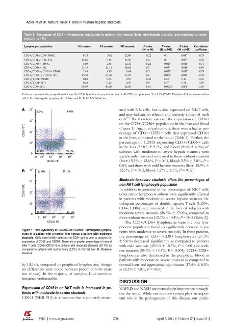
Figure 1 Flow cytometry <strong>of</strong> CD3+/CD56+/CD161+ <strong>in</strong>trahepatic lymphocytes<br />
<strong>in</strong> a patient with a normal liver versus a patient with moderate<br />
steatosis. Cells were <strong>in</strong>itially selected via CD3+ gat<strong>in</strong>g prior to analysis for<br />
expression <strong>of</strong> CD56 and CD161. There are a greater percentage <strong>of</strong> natural<br />
killer T cells (CD56+/CD161+) <strong>in</strong> patients with moderate steatosis (20.1%) as<br />
compared to patients with normal livers (6.6%). A: Normal liver; B: Moderate<br />
steatosis.<br />
NAFLD and NASH are <strong>in</strong>creas<strong>in</strong>g <strong>in</strong> importance throughout<br />
the world. While our immune system plays an important<br />
role <strong>in</strong> the pathogenesis <strong>of</strong> this disease, our under-<br />
1728 April 7, 2011|Volume 17|Issue 13|<br />
0.73


