

6 - World Journal of Gastroenterology
6 - World Journal of Gastroenterology
6 - World Journal of Gastroenterology

Create successful ePaper yourself
Turn your PDF publications into a flip-book with our unique Google optimized e-Paper software.
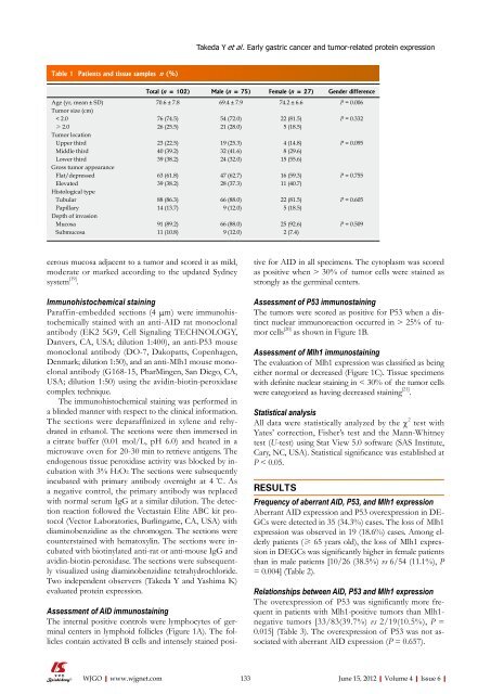
Table 1 Patients and tissue samples n (%)<br />
cerous mucosa adjacent to a tumor and scored it as mild,<br />
moderate or marked according to the updated Sydney<br />
system [19] .<br />
Immunohistochemical staining<br />
Paraffin-embedded sections (4 μm) were immunohistochemically<br />
stained with an anti-AID rat monoclonal<br />
antibody (EK2 5G9, Cell Signaling TECHNOLOGY,<br />
Danvers, CA, USA; dilution 1:400), an anti-P53 mouse<br />
monoclonal antibody (DO-7, Dakopatts, Copenhagen,<br />
Denmark; dilution 1:50), and an anti-Mlh1 mouse monoclonal<br />
antibody (G168-15, PharMingen, San Diego, CA,<br />
USA; dilution 1:50) using the avidin-biotin-peroxidase<br />
complex technique.<br />
The immunohistochemical staining was performed in<br />
a blinded manner with respect to the clinical information.<br />
The sections were deparaffinized in xylene and rehydrated<br />
in ethanol. The sections were then immersed in<br />
a citrate buffer (0.01 mol/L, pH 6.0) and heated in a<br />
microwave oven for 20-30 min to retrieve antigens. The<br />
endogenous tissue peroxidase activity was blocked by incubation<br />
with 3% H2O2. The sections were subsequently<br />
incubated with primary antibody overnight at 4 ℃. As<br />
a negative control, the primary antibody was replaced<br />
with normal serum IgG at a similar dilution. The detection<br />
reaction followed the Vectastain Elite ABC kit protocol<br />
(Vector Laboratories, Burlingame, CA, USA) with<br />
diaminobenzidine as the chromogen. The sections were<br />
counterstained with hematoxylin. The sections were incubated<br />
with biotinylated anti-rat or anti-mouse IgG and<br />
avidin-biotin-peroxidase. The sections were subsequently<br />
visualized using diaminobenzidine tetrahydrochloride.<br />
Two independent observers (Takeda Y and Yashima K)<br />
evaluated protein expression.<br />
Assessment <strong>of</strong> AID immunostaining<br />
The internal positive controls were lymphocytes <strong>of</strong> germinal<br />
centers in lymphoid follicles (Figure 1A). The follicles<br />
contain activated B cells and intensely stained posi-<br />
Takeda Y et al . Early gastric cancer and tumor-related protein expression<br />
Total (n = 102) Male (n = 75) Female (n = 27) Gender difference<br />
Age (yr, mean ± SD) 70.6 ± 7.8 69.4 ± 7.9 74.2 ± 6.6 P = 0.006<br />
Tumor size (cm)<br />
< 2.0 76 (74.5) 54 (72.0) 22 (81.5) P = 0.332<br />
≥ 2.0 26 (25.5) 21 (28.0) 5 (18.5)<br />
Tumor location<br />
Upper third 23 (22.5) 19 (25.3) 4 (14.8) P = 0.095<br />
Middle third 40 (39.2) 32 (41.6) 8 (29.6)<br />
Lower third 39 (38.2) 24 (32.0) 15 (55.6)<br />
Gross tumor appearance<br />
Flat/depressed 63 (61.8) 47 (62.7) 16 (59.3) P = 0.755<br />
Elevated 39 (38.2) 28 (37.3) 11 (40.7)<br />
Histological type<br />
Tubular 88 (86.3) 66 (88.0) 22 (81.5) P = 0.605<br />
Papillary 14 (13.7) 9 (12.0) 5 (18.5)<br />
Depth <strong>of</strong> invasion<br />
Mucosa 91 (89.2) 66 (88.0) 25 (92.6) P = 0.509<br />
Submucosa 11 (10.8) 9 (12.0) 2 (7.4)<br />
tive for AID in all specimens. The cytoplasm was scored<br />
as positive when > 30% <strong>of</strong> tumor cells were stained as<br />
strongly as the germinal centers.<br />
Assessment <strong>of</strong> P53 immunostaining<br />
The tumors were scored as positive for P53 when a distinct<br />
nuclear immunoreaction occurred in > 25% <strong>of</strong> tumor<br />
cells [20] as shown in Figure 1B.<br />
Assessment <strong>of</strong> Mlh1 immunostaining<br />
The evaluation <strong>of</strong> Mlh1 expression was classified as being<br />
either normal or decreased (Figure 1C). Tissue specimens<br />
with definite nuclear staining in < 30% <strong>of</strong> the tumor cells<br />
were categorized as having decreased staining [21] .<br />
Statistical analysis<br />
All data were statistically analyzed by the χ 2 test with<br />
Yates’ correction, Fisher’s test and the Mann-Whitney<br />
test (U-test) using Stat View 5.0 s<strong>of</strong>tware (SAS Institute,<br />
Cary, NC, USA). Statistical significance was established at<br />
P < 0.05.<br />
RESULTS<br />
Frequency <strong>of</strong> aberrant AID, P53, and Mlh1 expression<br />
Aberrant AID expression and P53 overexpression in DE-<br />
GCs were detected in 35 (34.3%) cases. The loss <strong>of</strong> Mlh1<br />
expression was observed in 19 (18.6%) cases. Among elderly<br />
patients (≥ 65 years old), the loss <strong>of</strong> Mlh1 expression<br />
in DEGCs was significantly higher in female patients<br />
than in male patients [10/26 (38.5%) vs 6/54 (11.1%), P<br />
= 0.004] (Table 2).<br />
Relationships between AID, P53 and Mlh1 expression<br />
The overexpression <strong>of</strong> P53 was significantly more frequent<br />
in patients with Mlh1-positive tumors than Mlh1negative<br />
tumors [33/83(39.7%) vs 2/19(10.5%), P =<br />
0.015] (Table 3). The overexpression <strong>of</strong> P53 was not associated<br />
with aberrant AID expression (P = 0.657).<br />
WJGO|www.wjgnet.com 133<br />
June 15, 2012|Volume 4|Issue 6|


