3+4+Supplementum/2012 - Společnost pro pojivové tkáně
3+4+Supplementum/2012 - Společnost pro pojivové tkáně
3+4+Supplementum/2012 - Společnost pro pojivové tkáně
- TAGS
- www.pojivo.cz

You also want an ePaper? Increase the reach of your titles
YUMPU automatically turns print PDFs into web optimized ePapers that Google loves.
Pokroky ve výzkumu, diagnostice a terapii<br />
Vydává <strong>Společnost</strong> <strong>pro</strong> <strong>pojivové</strong> <strong>tkáně</strong> ČLS J. E. Purkyně<br />
Ambulantní centrum <strong>pro</strong> vady pohybového aparátu<br />
Odborná společnost ortopedicko-<strong>pro</strong>tetická ČLS J. E. Purkyně<br />
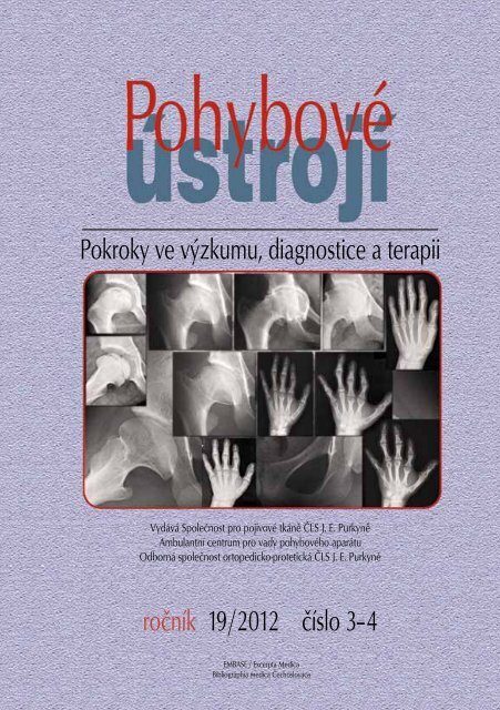
ročník 19/<strong>2012</strong> číslo 3–4<br />
EMBASE / Excerpta Medica<br />
Bibliographia medica Čechoslovaca
technicko<strong>pro</strong>tetická péče • výroba a servis <strong>pro</strong>téz, ortéz, korzetů • poradenská činnost<br />
Bolevecká 38, 301 00 Plzeň • Tel. 377 529 060-1 • <strong>pro</strong>tetikaplzen@volny.cz • www.<strong>pro</strong>tetika-plzen.cz
POHYBOVÉ ÚSTROJÍ<br />
ročník 19, <strong>2012</strong>, číslo 1+2<br />
datum vydání 28. 2. <strong>2012</strong><br />
RedakčnÍ Rada<br />
VEDOUCÍ REDAKTOR: Doc. MUDr. Ivo Mařík, CSc.<br />
ZÁSTUPCI VEDOUCÍHO REDAKTORA: Prof. Ing. Miroslav Petrtýl, DrSc.<br />
RNDr. Martin Braun, PhD.<br />
VĚDECKÝ SEKRETÁŘ: MUDr. Miloslav Kuklík, CSc.<br />
ODPOVĚDNÝ REDAKTOR: Ing. Pavel Lorenc<br />
Prof. MUDr. Jaroslav Blahoš, DrSc. Prof. Ing. František Maršík, DrSc.<br />
Doc. RNDr. Pavel Bláha, CSc. Doc. RNDr. Ivan Mazura, CSc.<br />
Prof. Ing. Jan Čulík, DrSc. MUDr. Pavel Novosad<br />
Doc. MUDr. Ivan Hadraba, CSc. Prof. MUDr. Ctibor Povýšil, DrSc.<br />
Ing. Hana Hulejová RNDr. Petr Sedlak, PhD.<br />
Prof. MUDr. Josef Hyánek, DrSc. Doc. MUDr. Václav Smrčka, CSc.<br />
Prof. MUDr. Jaromír Kolář, DrSc. Prof. PhDr. Jiří Straus, DrSc.<br />
Doc. MUDr. Petr Korbelář, CSc. MUDr. Jan Všetička<br />
Doc. MUDr. Vladimír Kříž RNDr. Daniela Zemková, CSc.<br />
ediTORial BOaRd<br />
Prof. Dr. Ing. Romuald Bedzinski, Politechnika Doc. Dr. Med. Kazimierz S. Kozlowski,<br />
Wroclawska, Poland M.R.A.C.R., Westmead NSW 2145, Sydney<br />
Dr. Michael Bellemore, F.R.A.C.S., Ass. Prof. Jacques Cheneau, MD, Saint Orens, France<br />
Westmead NSW 2145, Sydney Prof. Dr. Med. Zoran Vukasinovic, Belgrade,<br />
Prof. Tomasz Karski, MD, PhD, Lublin, Poland Yugoslavia<br />
Pohybové ústrojí. Pokroky ve výzkumu, diagnostice a terapii.<br />
ISSN 1212-4575<br />
Vydává <strong>Společnost</strong> <strong>pro</strong> <strong>pojivové</strong> <strong>tkáně</strong> ČLS J.E.Purkyně,<br />
Ambulantní centrum <strong>pro</strong> vady pohybového aparátu,<br />
& Odborná společnost ortopedicko – <strong>pro</strong>tetická ČLS J. E. Purkyně<br />
Excerpováno v Excerpta Medica. Tiskne PeMa, Černokostelecká 1168/90, Praha 10<br />
Návrh a grafická úprava obálky Rudolf Štorkán<br />
Časopis vychází 4krát ročně, nebo jako dojčíslo 2× ročně. Každá práce je recenzována.<br />
Objednávky přijímá Ambulantní centrum <strong>pro</strong> vady pohybového aparátu, s. r. o.<br />
Olšanská 7, 130 00 Praha 3, tel./fax: (+420) 222 582 214,<br />
http://www.pojivo.cz.<br />
Rukopisy zasílejte na adresu Doc. MUDr. Ivo Mařík, CSc., Olšanská 7, 130 00 Praha 3,<br />
(ambul_centrum@volny.cz) ve formátu doc, rtf. Vydavatel<br />
upozorňuje, že za obsah inzerce odpovídá výhradně inzerent. Časopis jakožto<br />
nevýdělečný neposkytuje honoráře za otištěné příspěvky<br />
POHYBOVÉ ÚSTROJÍ, ročník 19, <strong>2012</strong>, č. 3+4 165
166<br />
lOCOMOTOR SYSTeM<br />
advances in Research, diagnostics and Therapy<br />
Published by The Society for Connective Tissues, Czech Medical Association of<br />
J. E. Purkyně, Prague, Ambulant Centre for Defects of Locomotor Apparatus Prague<br />
& Society for Prosthetics and Orthotics, Czech Medical Association of J. E. Purkyně, Prague,<br />
Czech Republic<br />
Call for papers<br />
Support this journal by sending in your best and most interesting papers. Publication<br />
will normally be within six months of acceptance. The journal appears four times in a year.<br />
Chief editor: Ivo Mařík<br />
Associate Editors: Miroslav Petrtýl<br />
Martin Braun<br />
Scientific Secretary: Miloslav Kuklík<br />
Responsible Editor: Pavel Lorenc<br />
editorial board<br />
Romuald Bedzinski Vladimír Kříž<br />
Michael Bellemore Kazimierz Kozlowski<br />
Jaroslav Blahoš František Maršík<br />
Pavel Bláha Ivan Mazura<br />
Jacques Cheneau Pavel Novosad<br />
Jan Čulík Ctibor Povýšil<br />
Ivan Hadraba Petr Sedlak<br />
Hana Hulejová Václav Smrčka<br />
Josef Hyánek Jiří Straus<br />
Tomasz Karski Zoran Vukasinovic<br />
Jaromír Kolář Jan Všetička<br />
Petr Korbelář Daniela Zemková<br />
Submitted papers: Locomotor System will review for publication manuscripts concerned<br />
with <strong>pro</strong>gress in research of connective tissue diagnostics, me dical and surgical<br />
therapy mainly in the fields of orthopaedic surgery, dysmorphology (multiple congenital<br />
abnormalities of skeleton) and plastic surgery, biomechanics and biorheology, clinical<br />
anthropology and paleopathology.<br />
The journal has an interdisciplinary character which gives possibilities for complex<br />
a<strong>pro</strong>ach to the <strong>pro</strong>blematics of locomotor system. The journal belongs to clinical, preclinical<br />
and theoretical medical branches which connect various up-to-date results and disco veries<br />
concerned with locomotor system. You can find the volumes of Locomotor System journal<br />
at www.pojivo.cz since 1998.<br />
Papers published in the journal are excerpted in EMBASE / Excerpta Medica and<br />
Bibliograhia medica Čechoslovaca. We prefer the manuscripts to be prepared according<br />
to Uniform Requirements for Manuscripts Submitted to Biomedical Journals (Vancouver<br />
Declaration, Brit med J 1988; 296, pp. 401–405)<br />
LOCOMOTOR SYSTEM vol. 19, <strong>2012</strong>, No. 3+4
Devatenáctý ročník časopisu Pohybové ústrojí<br />
je věnován životnímu jubileu<br />
<strong>pro</strong>fesora Ing. Františka Maršíka, DrSc.<br />
The 19 th volume of Locomotor System journal<br />
is dedicated to the anniversary of<br />
Professor Eng František Maršík, DSc.<br />
POHYBOVÉ ÚSTROJÍ, ročník 19, <strong>2012</strong>, č. 3+4 167
POHYBOVÉ ÚSTROJÍ,<br />
19, <strong>2012</strong>, č. 3+4<br />
Pokroky ve výzkumu, diagnostice<br />
a terapii<br />
OBSaH<br />
SLOVO ČTENÁŘŮM –<br />
EDITORIAL . . . . . . . . . . . . . . . . . . . . . . . 171<br />
OBRÁZEK NA TITULNÍ STRANĚ A POPIS<br />
Trichorinofalangeální dysplazie . . . . . 173<br />
SOUBORNÉ REFERÁTY<br />
Kolář J.<br />
Překlad: Nosologie a klasifikace<br />
genetických kostních poruch –<br />
revise 2010 Am J Med Genet A.<br />
2011 May; 155A(5): 943-968 . . . . . . . . . 177<br />
PařízKová J.<br />
Pohybová aktivita a složení těla . . . . . . 205<br />
PŮVODNÍ PRÁCE<br />
KocoureK r., HlaváčeK P., GoGela J.,<br />
JuřičKa M., odenwald S.<br />
Studium jímavosti energie měkké části<br />
vkládacích stélek u sportovní obuvi . . 217<br />
čulíK J.<br />
Simulace v medicíně . . . . . . . . . . . . . . . . 224<br />
168<br />
lOCOMOTOR SYSTeM ,<br />
19, <strong>2012</strong>, no. 3+4<br />
Advances in Research, Diagnostics<br />
and Therapy<br />
COnTenT<br />
A WORD TO READERS –<br />
EDITORIAL . . . . . . . . . . . . . . . . . . . . . . . 171<br />
TITLE PICTURE AND DESCRIPTION<br />
Trichorhinophalangeal dysplasia . . . . . 173<br />
REVIEWS<br />
Kolar J.<br />
Translation of Nosology and Classification<br />
of Genetic Skeletal Disorders – 2010<br />
Revision&. Am J Med Genet A.<br />
2011 May; 155A(5): 943-968 . . . . . . . . . 177<br />
ParizKova J.<br />
Physical activity and body<br />
composition . . . . . . . . . . . . . . . . . . . . . . . . 205<br />
ORIGINAL PAPERS<br />
KocoureK r., HlavaceK P., GoGela J.,<br />
JuricKa M., odenwald S.<br />
Study of absorption energy of the soft<br />
insoles for sport shoes . . . . . . . . . . . . . . 217<br />
culiK J.<br />
Simulation in medicine . . . . . . . . . . . . . 224<br />
LOCOMOTOR SYSTEM vol. 19, <strong>2012</strong>, No. 3+4
ScHindlerová n., Foltýnová B.,<br />
HlaváčeK P., StaňKová c.<br />
Základní antropometrické znaky nohou<br />
gerontů v České republice. . . . . . . . . . . 231<br />
KASUISTIKY<br />
MaříK i., ŠorMová l., zeMKová d.,<br />
KuKlíK M., PovýŠil c.<br />
Enchondromatóza a Maffuciho syndrom –<br />
diagnostika a symptomatické léčení<br />
závažných deformit DK . . . . . . . . . . . . . 239<br />
ZPRÁVY<br />
KarSKi t., KędzierSKi z., BartoSzczyK<br />
a., JaBłońSKi M., KarSKi J., SłowińSKa B.,<br />
KaliSz a.<br />
History of orthopaedics in Lublin.<br />
Founding of the Orthopaedic Department<br />
of Medical University in 1954. Problems,<br />
the team, didactics, clinic and research<br />
aim in the years 1954–<strong>2012</strong> . . . . . . . . . . 256<br />
Diskuse k publikované recenzované<br />
práci – „Monitorování „jemných“<br />
pohybů pacienta na lůžku? . . . . . . . . . . 280<br />
Informace o <strong>Společnost</strong>i <strong>pro</strong> <strong>pojivové</strong><br />
<strong>tkáně</strong> ČLS JEP . . . . . . . . . . . . . . . . . . . . . . 282<br />
Přihláška řádného člena SPT . . . . . . . . . 283<br />
SMĚRNICE AUTORŮM . . . . . . . . . . . . 286<br />
ScHindlerova n., Foltynová B.,<br />
HlavaceK P., StanKova c.<br />
The Basic Anthropometric Characteristics<br />
of the Feet in Geronts in the Czech<br />
Republic . . . . . . . . . . . . . . . . . . . . . . . . . . . 231<br />
CASE REPORTS<br />
MariK i., SorMova l., zeMKova d.,<br />
KuKliK M., PovySil c.<br />
Enchondromatosis and Maffuci<br />
syndrome – diagnostics and<br />
symptomatic treatment of severe<br />
leg deformities. . . . . . . . . . . . . . . . . . . . . . 239<br />
NEWS<br />
KarSKi t., KędzierSKi z., BartoSzczyK<br />
a., JaBłońSKi M., KarSKi J., SłowińSKa B.,<br />
KaliSz a.<br />
History of orthopaedics in Lublin.<br />
Founding of the Orthopaedic Department<br />
of Medical University in 1954. Problems,<br />
the team, didactics, clinic and research<br />
aim in the years 1954–<strong>2012</strong> . . . . . . . . . 256<br />
Discusion about published paper<br />
„Monitoring „fine“ movements<br />
of patient on the bed? . . . . . . . . . . . . . . . 280<br />
Membership application of The Society<br />
for Connective Tissues, Czech Medical<br />
Association J.E. Purkynje, Prague, CZ 283<br />
Information on the Society for<br />
Connective Tissues, Czech Medical<br />
Association J.E. Purkynje, Prague, CZ 284<br />
INSTRUCTIONS FOR AUTHORS . . . 288<br />
POHYBOVÉ ÚSTROJÍ, ročník 19, <strong>2012</strong>, č. 3+4 169
Oznámení úmrtí<br />
MUDr. Jindřiška Galliová . . . . . . . . . . . . 291<br />
Prof. MUDr. Jaroslav Masopust, DrSc. 292<br />
OBSAH ROČNÍKU 2011. . . . . . . . . . . . . 294<br />
OBSAH ROČNÍKU <strong>2012</strong> . . . . . . . . . . . . 300<br />
SUPPLEMENTUM<br />
The 14 th Prague-Lublin-Sydney<br />
Symposium: Highlights in orthopaedics,<br />
anthropology and human biomechanics,<br />
September 16–23, <strong>2012</strong>, Rehabilitation<br />
Centre Sophia-Bryza in Sarbinowo,<br />
Poland . . . . . . . . . . . . . . . . . . . . . . . . . . . . . 131<br />
170<br />
Obituary<br />
Jindriska Galliova, MD . . . . . . . . . . . . . . 291<br />
Professor<br />
MUDr. Jaroslav Masopust, MD, DSc. . . 292<br />
CONTENTS OF VOLUME 2011 . . . . . . . 297<br />
CONTENTS OF VOLUME <strong>2012</strong> . . . . . . 302<br />
SUPPLEMENTUM<br />
The 14 th Prague-Lublin-Sydney<br />
Symposium: Highlights in orthopaedics,<br />
anthropology and human biomechanics,<br />
September 16–23, <strong>2012</strong>, Rehabilitation<br />
Centre Sophia-Bryza in Sarbinowo,<br />
Poland . . . . . . . . . . . . . . . . . . . . . . . . . . . . . 131<br />
LOCOMOTOR SYSTEM vol. 19, <strong>2012</strong>, No. 3+4
SlOVO čTenářůM � a wORd TO ReadeRS<br />
Vážení čtenáři, autoři a inzerenti,<br />
děkujeme za Vaši pomoc při tvorbě mezioborového odborného recenzovaného<br />
časopisu „Pohybové ústrojí – pokroky ve výzkumu, diagnostice a terapii“.<br />
Dostává se Vám do rukou dvojčíslo časopisu 3–4/<strong>2012</strong>, kde kromě odborných a vědeckých<br />
referátů je uvedena historie ortopedie v Lublinu v letech 1954–<strong>2012</strong> zásluhou pana<br />
<strong>pro</strong>fesora Tomasze Karskiho, MD, PhD, emeritního přednosty the Paediatric Orthopaedic<br />
and Rehabilitation Department of the Medical University in Lublin. V suplementu časopisu<br />
jsou publikována abstrakta sdělení z The 14 th Prague-Lublin-Sydney Symposia: Highlights<br />
in orthopaedics, anthropology and human biomechanics, které je organizováno v tomto<br />
roce ve spolupráci s panem <strong>pro</strong>fesorem Karskim, MD, PhD ve dnech 16.–23. 9. <strong>2012</strong><br />
v Sarbinowu v Polsku (Rehabilitation Centre Sophia-Bryza).<br />
Informace o odborných akcích <strong>Společnost</strong>i <strong>pro</strong> <strong>pojivové</strong> <strong>tkáně</strong> ČLS JEP a Odborné<br />
společnosti ortopedicko-<strong>pro</strong>tetické ČLS JEP jsou dostupné na webové doméně <strong>Společnost</strong>i<br />
<strong>pro</strong> <strong>pojivové</strong> <strong>tkáně</strong> ČLS JEP www.pojivo.cz, kterou úspěšně vede a v tomto roce inovoval<br />
pan Ing. Alexander Lichý. V záložce Info/Pohybové ústrojí/Archivní čísla ke stažení jsou<br />
dostupná jednotlivá čísla a dvojčísla časopisu (včetně Suplement) od roku 1997 do roku<br />
<strong>2012</strong> ve formátu PDF.<br />
Jako v dřívějších letech je předmětem a hlavním posláním časopisu publikovat práce<br />
vycházející z výzkumu pojivových tkání, práce orientované na biochemickou, morfologickou,<br />
genetickou a molekulární diagnostiku a kostní metabolismus chorob genetických<br />
i získaných vad pohybového ústrojí. Dále práce klinické, týkající se symptomatické<br />
léčby metabolických kostních chorob, osteoporózy, sekundární osteoporózy, osteo/<br />
spondyloartrózy, kostních dysplazií, končetinových anomálií, dysmorfických vad pohybového<br />
aparátu a genetických syndromů, ale i jiných chorob, které ve svých důsledcích<br />
negativně ovlivňují pohybové ústrojí v průběhu lidského života. Pozornost patří pracím<br />
z oblasti biomechaniky, neuroadaptivním změnám skeletu, řízené remodelaci pojivových<br />
tkání v závislosti na léčebných metodách (kalciotropní léky, rehabilitace, ortoticko-<br />
-<strong>pro</strong>tetické a operační léčení), studiím muskuloskeletálních a neuronálních interakcí,<br />
v neposlední řadě sdělením antropologickým, paleopatologickým a pod. Významné jsou<br />
především interdisciplinárně zaměřené práce. V anglickém jazyce jsou publikována sdělení<br />
zahraničních, ale i našich autorů. Žádaným doplněním obsahu časopisu jsou zprávy<br />
ze sjezdů a konferencí. V rubrice zprávy zveřejňujeme oznámení o životním výročí členů<br />
RR časopisu, SPT ČLS JEP a významných osobností, sdělení o prioritních pozorováních,<br />
ze studijních a poznávacích cest aj.<br />
Dne 26. března <strong>2012</strong> v plné duševní svěžesti opustila tento svět v nedožitém věku<br />
99 let paní MUDr. Jindřiška Galliová. Vzpomínky z jejího bohatého <strong>pro</strong>fesního života<br />
byly publikovány v časopise Pohybové ústrojí (ročník 18, 2011, 107–124) z popudu<br />
doc. MUDr. I. Maříka, CSc., který s <strong>pro</strong>f. MUDr. R. Kubátem, DrSc. v roce 1981 rozpoznal<br />
POHYBOVÉ ÚSTROJÍ, ročník 19, <strong>2012</strong>, č. 3+4 171
u nás první případ BCG gonitidy u 3letého chlapce očkovaného tehdejší sovětskou BCG<br />
vakcínou.<br />
Dne 23. července <strong>2012</strong> navždy odešel pan <strong>pro</strong>f. MUDr. Jaroslav Masopust, DrSc., čestný<br />
člen <strong>Společnost</strong>i <strong>pro</strong> <strong>pojivové</strong> <strong>tkáně</strong> ČSL JEP, který se v roce 2004 zasloužil o transformaci<br />
<strong>Společnost</strong>i <strong>pro</strong> výzkum a využití pojivových tkání (založenou a vedenou 12 let panem<br />
<strong>pro</strong>f. MUDr. Milanem Adamem, DrSc.) v dnešní <strong>Společnost</strong>. V roce 2009 při příležitosti<br />
jeho 80. životního výročí byl naší <strong>Společnost</strong>í oceněn Medailí za zásluhy o rozvoj vědy. Pan<br />
<strong>pro</strong>f. Masopust byl jednou z klíčových osobností nejen biochemie, ale i 2. lékařské fakulty<br />
Karlovy Univerzity. Naposledy se s námi podělil o své životní zkušenosti a vědomosti hodinovou<br />
přednáškou na téma „Obezita jako nemoc“ přednesenou při zahájení 17. Kubátova<br />
podologického dne (Lékařský dům, 2. 3.–3. 3. <strong>2012</strong>, www.pojivo.cz).<br />
Jako každoročně uvádíme směrnice <strong>pro</strong> autory příspěvků, kterým věnujte <strong>pro</strong>sím náležitou<br />
pozornost při přípravě Vašich vědeckých sdělení.<br />
Časopis PÚ byl v roce 2008 zařazen Radou <strong>pro</strong> výzkum, vývoj a inovace vlády ČR na<br />
Seznam recenzovaných neimpaktovaných periodik vydávaných v České republice a je<br />
uveden v tomto Seznamu i po aktualizaci <strong>pro</strong>vedené v roce 2010 (po revizi 2 dvojčísel PÚ<br />
vydaných v roce 2009).<br />
Souhrny prací publikovaných v časopisu jsou excerpovány v EMBASE / Excerpta<br />
Medica (od r. 1994) a v Bibliographia medica Čechoslovaca (od r. 2010).<br />
K <strong>pro</strong>sazení časopisu Pohybové ústrojí mezinárodně je velmi významné citovat práce<br />
uveřejněné v časopisu v příspěvcích posílaných do zahraničních časopisů. Pro zvýšení<br />
úrovně časopisu PÚ je nezbytné získávat původní kvalitní práce. Původní práce a kasuistiky<br />
doporučujeme publikovat v angličtině s cílem zvýšit zájem o náš časopis v odborném světě.<br />
Souhrny původních prací doporučujeme psát co nejvýstižněji, strukturovaně česky a anglicky<br />
(objectives, methods, results and discussion), s klíčovými slovy.<br />
Těšíme se na Vaši tvůrčí spolupráci během roku <strong>2012</strong>.<br />
Redakční rada<br />
172<br />
LOCOMOTOR SYSTEM vol. 19, <strong>2012</strong>, No. 3+4
OBRázek na TiTulnÍ STRaně čaSOPiSu deMOnSTRuJe<br />
Obrázek na titulní straně časopisu demonstruje charakteristické rentgenologické <strong>pro</strong>jevy<br />
Tricho-rhino-falangeálního syndromu, typ 1 (TRPS 1, MiM 190350), na základě<br />
kterých lze s jistotou potvrdit genetickou diagnózu (stejně jako např. u achondroplazie<br />
nebo spondylometafyzární dysplazie, typ Kozlowski). Určení správné diagnózy již v batolecím<br />
a předškolním věku je významné nejen <strong>pro</strong> <strong>pro</strong>gnózu, ale zabrání dalšímu finančně<br />
nákladnému vyšetřování.<br />
Na obrázku jsou zobrazeny patognomonicky významné rentgenologické změny<br />
na snímcích rukou a kyčlí matky a syna a jedné nepříbuzných mladé ženy. Diagnóza pacientů<br />
byla stanovena nebo potvrzena v Ambulantním centru <strong>pro</strong> vady pohybového aparátu<br />
v Praze, kde jsou dlouhodobě sledováni.<br />
Ruka<br />
TiTle PiCTuRe deMOnSTRaTeS<br />
Na obrázku dole a u<strong>pro</strong>střed jsou RTG<br />
snímky levé ruky <strong>pro</strong>banda ve věku 7, 10<br />
a 14 let, které ukazují typické konické epifýzy<br />
(Giedion typ 12) i slonovinové epifýzy<br />
na distálních článcích 1.–4. prstu (snímek<br />
ve věku 7 a 10 let), nápadný je zkrat středních<br />
článků zvláště 2.–4. prstu a jejich desaxace<br />
v úrovni PIP kloubů (důsledek deformit<br />
a předčasné fúze konických epifýz).<br />
Dále zkrat 2., 3. a 5. paprsku, na kterém se<br />
podílí významně i předčasná fúze distálních<br />
epifýz metakarpů. V pravém dolním rohu je<br />
RTG snímek levé ruky jeho matky ve věku<br />
24 let se zkratem 2. a 5. paprsku a desaxací<br />
v úrovni PIP kloubu 3. a 4. prstu. (biomechanická<br />
příčina zkratu a desaxací je stejná jako<br />
u jejího syna). V pravém horním rohu je snímek<br />
RTG levé ruky nepříbuzné pacientky<br />
ve věku 24 let, kde je mírné zkrácení středních<br />
článků 2.,3. a 5. prstu a málo vyjádřená<br />
desaxace 2. a 3. prstu v úrovni PIP kloubu.<br />
kyčelní kloub<br />
Na obrázku vlevo jsou snímky pravého<br />
kyčelního kloubu v AP a axiální <strong>pro</strong>jekci<br />
<strong>pro</strong>banda ve věku 7 a 18 let. Na snímku<br />
v 7 letech jsou acetabulum a hlavice relativně<br />
velké, acetabulum je strmější, hlavice<br />
mírně oploštělá, krček krátký varosní.<br />
Epifýza hlavice je plochá a fragmentovaná,<br />
V 18letech v obou <strong>pro</strong>jekcích je acetabulum<br />
mělké a strmé, hlavice je velká a plochá,<br />
hřibovitého tvaru se zobákovitým <strong>pro</strong>tažením<br />
mediálně.<br />
Nahoře u<strong>pro</strong>střed jsou pravé kyčelní<br />
klouby matky <strong>pro</strong>banda a nepříbuzné pacientky<br />
ve věku 24 let. Na obou jsou známky<br />
pokročilé koxartrózy. Na snímku u<strong>pro</strong>střed<br />
u matky <strong>pro</strong>banda je výrazně zúžená kloubní<br />
štěrbina, <strong>pro</strong>truse acetabula do pánve<br />
a okrajové osteofyty na hlavici. Pokročilá<br />
koxartróza a obtíže pacientky byly indikací<br />
k náhradě kyčelních kloubů totální endo<strong>pro</strong>tézou<br />
již po 40. roce věku.<br />
Rentgenologická diagnostika TRPS-1 je<br />
možná již v prvních letech života. Hlavní<br />
RTG příznaky jsou zkrácení jedné nebo více<br />
falang anebo metakarpů. Charakteristická<br />
je epifyzární dysplazie postižených dlouhých<br />
kostí s konickými epifýzami (Giedion<br />
typ 12), předčasná fúze epifýz a slonovinové<br />
epifýzy na distálních článcích prstů<br />
rukou. Epifýzy hlavic femurů jsou malé,<br />
někdy se vyvíjí morbus pseudo-Perthes,<br />
krčky femurů bývají varosní (pozorová-<br />
POHYBOVÉ ÚSTROJÍ, ročník 19, <strong>2012</strong>, č. 3+4 173
ní autora) a vzniká předčasná koxartróza.<br />
Distální epifýza femuru bývá mírně oploštělá.<br />
U novorozenců se typické RTG změny<br />
nezjistí. Určení diagnózy radiologickým<br />
vyšetřením je možné u dětí i dospělých při<br />
současném zhodnocení klinického nálezu.<br />
K typickým klinickým <strong>pro</strong>jevům patří<br />
krátká postava, řídké, pomalu rostoucí<br />
jemné vlasy, předčasné <strong>pro</strong>řídnutí vlasů<br />
v čelní krajině, řídké obočí zevně, někdy<br />
tenké a křehké nehty. Prominující bambulovitý<br />
nos, dlouhé a široké filtrum, úzký<br />
horní ret, někdy odstávající uši, nadpočetné<br />
zuby a retrognacie. Brachydaktylie<br />
jednoho nebo více prstů, otok mezičlánkových<br />
kloubů s nebo bez distální osové<br />
deviace postižených prstů.<br />
Diferenciální diagnóza. Podobné RTG<br />
změny skeletu jsou u tricho-rhino-phalangeálního<br />
syndromu, typ 2 (TRPS-2), který<br />
se ale odlišuje přítomností mnohočetných<br />
kartilaginosních exostóz a mentálním<br />
defektem. U Albrightovy osteodystrofie<br />
a akrodysostózy jsou metakarpy a falangy<br />
obvykle kratší a širší než u TRPS-1 a epifyzární<br />
změny jsou odlišné. Thiemannova<br />
174<br />
choroba se manifestuje bolestivým vřetenovitým<br />
otokem postižených kloubů.<br />
Postižené epifýzy článků prstů jsou nepravidelné<br />
denzity, někdy fragmentované.<br />
Ostatní syndromy brachydaktylie se liší<br />
typem abnormalit prstů a chyběním faciální<br />
stigmatizace (vlasy, nos, uši, rty).<br />
Genetický přenos onemocnění je autozomálně<br />
dominantní s významnou variabilitou<br />
exprese fenotypu. TRPS-1 je způsoben<br />
mutacemi genu TRPS1, které jsou lokalizované<br />
na chromosomu 8q24.12. Tento gen<br />
kóduje zinek-<strong>pro</strong>užkový transkripční faktor.<br />
Prognóza. Vývoj inteligence u většiny<br />
postižených a životní <strong>pro</strong>gnóza jsou normální.<br />
Faciální dimorfismus a deformity<br />
prstů mohou být kosmetickým <strong>pro</strong>blémem<br />
a indikací k plastickým výkonům.<br />
Oploštění a deformace hlavice femuru je<br />
příčinou předčasné koxartrózy a je indikací<br />
k řešení totální endo<strong>pro</strong>tézou v dospělém<br />
nebo středním věku, kdy se často<br />
diagnostikuje i osteopenie a osteoporóza<br />
(pozn. autora). Výška dospělých je obvykle<br />
pod 10. percentilem, zřídka dosáhne<br />
150 cm u mužů a 140 cm u žen.<br />
Title picture demonstrates Tricho-Rhino-Phalangeal Syndrome, Type i (TRPS-<br />
1, MiM 190350). This disorder is important for the radiologists because of distinctive<br />
radiographic findings usually in first years of life. On the basis of these findings the<br />
genetic diagnosis can be definitely confirmed (identically as e.g. at Achondroplasia or<br />
Spondylometaphyseal Dysplasia, Kozlowski type). Determination of genetic diagnosis in<br />
first years of life is important not only for <strong>pro</strong>gnosis but prevent next financially expensive<br />
examination.<br />
At the title picture are shown pathognomonic skeletal changes at X-rays of the hands<br />
and hip joints of a mother and son and two affected unrelated women.<br />
The major clinical findings are small stature – the adult height is usually below the<br />
tenth centile and may occasionally reach 150 cm in males and 140 cm in females, sparse,<br />
slowly growing fine hair, premature frontal balding, sparse eyebrows laterally. Prominent<br />
nose with bulbous end, long and wide philtrum, thin upper lip, sometimes <strong>pro</strong>minent<br />
ears, supernumerary teeth and retrognathia. Brachydactyly in one or more fingers, swelling<br />
of the interphalangeal joints with or without distal axial deviation in affected fingers.<br />
Intellectual development and life expectancy are normal.<br />
LOCOMOTOR SYSTEM vol. 19, <strong>2012</strong>, No. 3+4
RTG snímky z archivu Ambulantního centra <strong>pro</strong> vady pohybového aparátu s.r.o., Olšanská 7, Praha 3<br />
X-rays from archive of Ambulant Centre for Defects of Locomotor Apparatus Prague<br />
The major radiographic features<br />
are shortening of one or more phalanges<br />
and/or metacarpals, characteristic epiphyseal<br />
dysplasia of affected tubular bones<br />
with cone-shaped epiphyses, Giedion type<br />
12, premature epiphyseal fusion and ivory<br />
epiphyses in distal phalanges. Small capital<br />
femoral epiphyses are sometimes associated<br />
with Perthes-like changes.<br />
differential diagnosis<br />
The tricho-rhino-phalangeal syndrome,<br />
type II, differs by the presence of multiple<br />
cartilaginous exostoses and mental<br />
deficiency. In Albright osteodystrophy and<br />
Acrodysostosis the metacarpals and phalanges<br />
are usually shorter and broader<br />
than in TRPS-1, and the epiphyseal abnor-<br />
malities are different. Other brachydactyly<br />
syndromes differ by the type of digital<br />
abnormalities and absence of the facial and<br />
hair changes characterizing tricho-rhino-<br />
-phalangeal dysplasia.<br />
Genetics and molecular pathology<br />
The genetic transmission is autosomal<br />
dominant with considerable variability of<br />
phenotypic expression. TRPS-1 is caused<br />
by mutations of the TRPS1 gene located on<br />
chromosome 8q24.12. The gene encodes<br />
a zinc-finger transcription factor.<br />
Course and <strong>pro</strong>gnosis<br />
Facial and digital abnormalities are<br />
rarely handled by plastic and hand sur-<br />
POHYBOVÉ ÚSTROJÍ, ročník 19, <strong>2012</strong>, č. 3+4 175
geons. Premature coxarthrosis is sometimes<br />
indication for hip arthroplasty in<br />
adulthood. Early commencement of osteoporosis<br />
should be controlled by densitometric<br />
examination (DEXA).<br />
key words: skeletal dysplasia, Tricho-<br />
Rhino-Phalangeal Syndrome, Type I, radiologic<br />
diagnosis<br />
RefeRenCeS<br />
1. MOMENI P, GLöCKNER G, SCHMIDT O,<br />
VON HOLTUM D et al. Mutations in a new gene,<br />
encoding a zinc-finger <strong>pro</strong>tein, cause tricho-rhino-phalangeal<br />
syndrome type I. Nature Genetics<br />
24, 2000, p. 71–74.<br />
2. SPRANGER JW, BRILL PW, POZNANSKI A.<br />
Tricho-Rhino-Phalangeal Syndrome, Type I. In:<br />
Bone Dysplasias. An Atlas of Genetic Disorders<br />
of Skeletal Development, 2nd Ed. Oxford, New<br />
York: Oxford University Press, 2002, p. 380 –382.<br />
3. TAYBI H, LACHMAN RS. Tricho-Rhino-<br />
Phalangeal Dysplasia, Type I. In: Radiology of<br />
Syndromes, Metabolic Disorders, and Skeletal<br />
Dysplasias, 4th ed., St. Louis, Baltimore: Mosby,<br />
1996, p. 943–946.<br />
Author‘s address<br />
assoc. Professor ivo Marik, Md, Phd<br />
Ambulant Centre for Defects of Locomotor<br />
Apparatus<br />
Olšanská 7<br />
130 00 Prague, Czech Republic<br />
E-mail: ambul_centrum@volny.cz<br />
176<br />
LOCOMOTOR SYSTEM vol. 19, <strong>2012</strong>, No. 3+4
SOuBORnÉ RefeRáTY � ReViewS<br />
nOSOlOGie a klaSifikaCe GeneTiCkýCH<br />
kOSTnÍCH PORuCH – ReViSe 2010 aM J Med<br />
GeneT a. 2011 MaY; 155a(5): 943-968<br />
TRanSlaTiOn Of nOSOlOGY and<br />
ClaSSifiCaTiOn Of GeneTiC SkeleTal<br />
diSORdeRS – 2010 ReViSiOn&. aM J Med<br />
GeneT a. 2011 MaY; 155a(5): 943-968<br />
PŘEKLAD: kOlář JaROMÍR<br />
AUTHORS: MaTTHew l. waRMan 1) , ValeRie CORMieR-<br />
daiRe 2) , CHRiSTine Hall 3) , deBORaH kRakOw 4, 5) ,<br />
RalPH laCHMan 4) , MaRTine leMeRReR 2) , GeeRT<br />
MORTieR 6) , STefan MundlOS 7) , Gen niSHiMuRa 8) ,<br />
daVid l. RiMOin 4) , STePHen ROBeRTSOn 9) , RaVi<br />
SaVaRiRaYan 10) , daVid SillenCe 11) , JueRGen<br />
SPRanGeR 12) , SHeila unGeR 12, 13) , BeRnHaRd zaBel 12) ,<br />
and andRea SuPeRTi-fuRGa12, 14)<br />
1) Orthopaedic Research Laboratories, Department of Orthopaedic<br />
Surgery, and the Howard Hughes Medical Institute, Children’s Hospital,<br />
Boston<br />
2) Paris Descartes University, Department of Genetics and INSERM U781,<br />
Hôpital Necker Enfants Malades, 75015 Paris, France<br />
3) Institute of Child Health, University of London, London, UK<br />
4) Medical Genetics Institute, Steven Spielberg Building, Cedars-Sinai<br />
Medical Center, 8700 Beverly Blvd, PACT 4, Los Angeles, CA 90048<br />
5) Departments of Orthopaedic Surgery and Human Genetics, UCLA, 615<br />
Charles E. Young Drive South, Los Angeles, CA 90048<br />
POHYBOVÉ ÚSTROJÍ, ročník 19, <strong>2012</strong>, č. 3+4 177
6) Department of Medical Genetics, University Hospital of Antwerp,<br />
University of Antwerp, Edegem, Belgium<br />
7) Institut für Medizinische Genetik, Charité Universitätsmedizin Berlin,<br />
and Max-Planck-Institut für Molekulare Genetik, Berlin, Germany<br />
8) Department of Pediatric Imaging, Tokyo Metropolitan Children’s<br />
Medical Center, Fuchu, Tokyo, Japan<br />
9) Department of Paediatrics and Child Health, Dunedin School of<br />
Medicine, Otago University, Dunedin, New Zealand<br />
10) Murdoch Children’s Research Institute, Royal Children’s Hospital, and<br />
Department of Paediatrics, University of Melbourne, Victoria, Australia,<br />
3052<br />
11) Discipline of Genetic Medicine, The Children’s Hospital at Westmead<br />
Clinical School, The University of Sydney, Westmead, Australia 2145<br />
12) Centre for Pediatrics and Adolescent Medicine, Freiburg University<br />
Hospital, University of Freiburg, 79106 Freiburg, Germany<br />
13) Medical Genetics Service, University of Lausanne, CHUV – Centre<br />
Hospitalier Universitaire Vaudois, 1011 Lausanne, Switzerland<br />
14) Department of Pediatrics, University of Lausanne, CHUV – Centre<br />
Hospitalier Universitaire Vaudois, 1011 Lausanne, Switzerland<br />
aBSTRakT<br />
Genetické poruchy, postihující kostní systém nastávají odchylkami komplikovaných<br />
dějů kosterního vývoje, růstu a homeostázy a svou pestrostí zůstávají diagnostickou<br />
výzvou. Nosologie a klasifikace genetických kostních poruch podává přehled uznaných<br />
diagnostických jednotek a skupin podle klinických a radiologických <strong>pro</strong>jevů a molekulární<br />
patogenezy. Jejím záměrem je, poskytnout genetickým, pediatrickým a radiologickým<br />
oborům seznam uznávaných genetických poruch, jež by usnadnila diagnostiku jednotlivých<br />
případů a vyhranění nových poruch při přemosťování kontaktů mezi kliniky a vědci,<br />
pracujícími na poli kostní biologie.<br />
Do revize roku 2010 bylo zařazeno 456 jednotek, rozložených do 40 skupin, definovaných<br />
molekulárními,biochemickými a radiologickými hledisky. Z těchto stavů bylo 316 spojeno<br />
s mutacemi jednoho nebo více z 226 různých genů, v rozmezí „běžných“, opakujících<br />
se mutací až po jakési „soukromé“, jež byly nalezeny v jednotlivých rodinách, nebo<br />
u jednotlivců. Nosologie je <strong>pro</strong>to hybridem, zahrnujícím seznam klinicky definovaných<br />
chorob, čekajících na molekulární vysvětlení a záznamem v databázi, komentujícím<br />
spektrum změn, jež jsou vyvolány mutací daného genu.<br />
178<br />
LOCOMOTOR SYSTEM vol. 19, <strong>2012</strong>, No. 3+4
Nosologie by měla být užitečná <strong>pro</strong> diagnostiku nemocných s geneticky podmíněnými<br />
kosterními poruchami, zejména v očekávání přívalu informací, jež poskytnou nové<br />
technologie k definování klinických jednotek a nových poruch a v tom, že poskytne<br />
přehled už zavedených nosologických jednotek; vědeckým pracovníkům pak tím, že<br />
informuje o klinických korelacích genů, <strong>pro</strong>teinů a dějů, odehrávajících se v kostní biologii.<br />
ÚVOd<br />
V šedesátých letech 20. století se<br />
hromadily doklady o tom, že genetické<br />
poruchy kostry jsou klinicky a geneticky<br />
různorodé, což podnítilo skupinu mezi národních<br />
odborníků k přípravě dokumentu,<br />
jímž by se dosáhl souhlas v nomenklatuře,<br />
jež byla nazvána „Constitutional (or<br />
Intrinsic) Disorders of Bone“ (1970, 1971a,<br />
1971b, 1971c, 1971d; McKusick a Sčoty<br />
1971). Záměrem „Nomenklatury“ bylo sdružit<br />
radiology, klinické genetiky a pediatry,<br />
aby souhlasili s pojmenováním a klasifikací<br />
kostních poruch, syndromů a metabolických<br />
chorob, které byly nedáv no popsány.<br />
Revise byla připravena v roce 1977,<br />
1983, 1992 a 1997 (1978, 1979; Rimoin<br />
1979, 1983; Spranger 1992, 1998; Lachman<br />
1998). Po ustavení International Skeletal<br />
Dysplasia Society (ISDS) v r. 1999 a aby<br />
bylo možné se vypořádat s lavinou nově<br />
přibývajících informací byla v rámci ISDS<br />
pověřena komise expertů <strong>pro</strong>vedením revize<br />
Nosologie (2001 a 2006) (Hall 2002;<br />
Superti-Furga a Unger 2007).<br />
MeTOdY<br />
Nosologická skupina ISDS se sešla<br />
v srpnu 2009. Dosáhlo se souhlasu se změnami,<br />
které byly učiněny v kategorizaci<br />
skupin chorob a začlenění jednotlivých<br />
poruch do nich. Po zasedání byly poslány<br />
cirkuláře návrhů se snahou, dostat infor-<br />
mace o novinkách do zasedání v říjnu 2010.<br />
Proti předešlé revizi nedoznala nové změny<br />
kriteria užívaná <strong>pro</strong> zařazení jednotlivých<br />
poruch, tyto byly:<br />
1. Podstatné postižení kostry, odpovídající<br />
definici kostních dysplazií, metabolických<br />
kostních odchylek, dysostóz<br />
a kostních malformací a/nebo redukčních<br />
syndromů,<br />
2. Publikace a/nebo uvedení v seznamu<br />
MIM (což značí, že pozorování nemají<br />
být dříve uveřejněna, než budou<br />
zásadně oponována a nabudou status<br />
v Nosologii a tím dosáhnou publikační<br />
status,<br />
3. Třetí genetické baze se musí opírat<br />
o rodokmen, nebo být vysoce pravděpodobná<br />
z homogenity fenotypů<br />
v rodinách,jež nejsou příbuzné,<br />
4. Nosologická autonomie musí být potvrzena<br />
molekulární nebo vazebnou analýzou<br />
a/nebo přítomností diagnostických<br />
charakteristických rysů a nálezy<br />
u mnoha jedinců nebo rodin.<br />
VýSledkY<br />
Do nosologie bylo zařazeno 456 různých<br />
stavů, vyčleněných do 40 skupin,<br />
definovaných hledisky molekulárními,<br />
biochemickými a/nebo radiologickými.<br />
Z těchto stavů 316 (podle revize z r. 2006<br />
pak 215) bylo spojeno s jedním nebo více<br />
než 226 (podle revize 2006 pak 140) různý-<br />
POHYBOVÉ ÚSTROJÍ, ročník 19, <strong>2012</strong>, č. 3+4 179
mi geny. Výsledky jsou uvedeny v tabulce<br />
č. 1. Skuina poruch se známou molekulární<br />
poruchou byla předřazena před ony, kde je<br />
porucha známa méně, nicméně byly ponechány<br />
při sobě variety téže poruchy. Ve<br />
srovnání s verzí 2006 došlo k další změně<br />
v uspořádání skupin. Byly utvořeny dvě<br />
nové skupiny, opírající se o běžněji postižené<br />
molekuly nebo biochemické cesty<br />
(skupina TRPV4 a skupina Aggrecanová).<br />
Skupina TRPV4 zahrnuje relativně běžné<br />
poruchy a tvoří nové spektrum <strong>pro</strong>totypů,<br />
sahajících od mírných až po letální.<br />
Aggrecan je jednou z důležitých molekul<br />
struktury chrupavky a nemělo by být překvapením,<br />
kdyby více chorob našlo své<br />
zařazení do této skupiny v budoucnosti.<br />
Skupiny 1 až 8 jsou opřeny o běžné fundamentální<br />
geny nebo patologické děje.<br />
Skupiny 9–17 se opírají o lokalizaci<br />
radiologických odchylek ve specifických<br />
kostních strukturách (obratle, epifýzy,<br />
metafýzy, diafýzy nebo jejich kombinace)<br />
nebo postiženého segmentu (rhino, meso<br />
nebo acro). Skupiny 18–20 jsou definovány<br />
makroskopickými hledisky v kombinaci<br />
s klinickými <strong>pro</strong>jevy (ohyby kostí,<br />
štíhlé kosti, přítomnost mnohotných luxací.<br />
Skupiny 21 až 25 a 28 berou v úvahu<br />
<strong>pro</strong>jevy mineralizace (zvýšená nebo snížená<br />
kostní denzita, různorodá mineralizace,<br />
tečkovitost, osteolýza). Skupina 27<br />
zahrnuje skupinu lysozomálnch poruch<br />
s postižením kostry. Skupina 29 s tak zvaným<br />
abnormálním vývojem (dříve „anarchickým)<br />
kostních složek, jako jsou exostózy,<br />
enchondromy a ektopické kalcifikace,<br />
je částečně heterogenní a v budoucnosti<br />
může vyžadovat revizi, s pomocí nových<br />
molekulárních údajů.<br />
Skupina 23 zahrnuje osteopetrózu<br />
(OP), její variety a příbuzné stavy a byla<br />
rozšířena po definici daných genetických<br />
180<br />
defektů, biochemických a/nebo histologických<br />
rysů variet typů OP, čímž je oprávněno<br />
rozdělení „OP fenotypu“ na četné<br />
subtypy.<br />
Skupina 25 (osteogenesis imperfekta<br />
a skupina snížené kostní hutnoty<br />
byla předmětem zvláštní pozornosti.<br />
Klasifikace dle Sillence, jez byla uveřejněna<br />
před 30 lety, zajistila prvou systemtickou<br />
klasifikaci a vybudovala vztahy mezi formami<br />
dědičnosti jednotlivých klinických<br />
typů (Sillence a Rimoin 1978; Sillence<br />
a ost. 1979a, Silence a ost. 1979b). V současnosti<br />
byla odhalena překvapivě složitá<br />
komplexnost genetiky molekulárních<br />
základů OI a současně se zřetelně dokumentovala<br />
variabilita fenotypů, vycházející<br />
z jednotlivých míst. Jevila se <strong>pro</strong>to<br />
neudržitelná snaha pokoušet se udržovat<br />
tuhé vazby vztahů mezi „Sillencovskými<br />
typy“ a jejich molekulárním základem. Byl<br />
vysloven souhlas se zachováním klasifikace<br />
dle Silence jako <strong>pro</strong>totypové a obecně přijatelné<br />
cesty ke klasifikaci stupně a závažnosti<br />
u OI; a o<strong>pro</strong>stit klasifikaci Sillence<br />
z jakékoliv přímé molekulární spojitosti.<br />
Proto byl vydán separátně seznam genů,<br />
které mohou vyvolat OI. Proliferace „typů<br />
OI“ k objasnění jednotlivých genů separátně,<br />
tak jak ji obhajovali někteří účastníci<br />
sezení, je více zavádějící, než <strong>pro</strong>spěšná<br />
v klinické praxi.<br />
Skupina 26 se dožila identifikace několika<br />
nových molekulárních mechanismů,<br />
vedoucích k hypofosfatemické křivici.<br />
Do skupiny 29 (Poruchy vývoje kostních<br />
složek) byla začleněna neurofibromatóza<br />
1. typu, podle hledisek Stevensona<br />
a jiných, přestože hlavní klinické příznaky<br />
NF1 jsou neurologické a kožní, jsou<br />
u ní časté kostní změny, jež jsou cenné<br />
<strong>pro</strong> diagnostiku a mají klinický význam<br />
(Stevenson a ost. 2007).<br />
LOCOMOTOR SYSTEM vol. 19, <strong>2012</strong>, No. 3+4
Skupina 30 (Syndromy nadměrného<br />
vzrůstu s významným postižením kostí)<br />
a skupina 31 (Genetické zánětlivé a revmatoidní<br />
osteoartropatie) byly doplněny<br />
nově. Skupina 30 zahrnuje poruchy, manifestující<br />
se jako syndromy nadměrného<br />
vzrůstu s významnou kostní složkou, jež je<br />
částí diagnostických kriterií <strong>pro</strong> specifický<br />
stav. Jedna afekce byla zavzata pokusně<br />
kvůli svým zřetelným kostním <strong>pro</strong>jevům<br />
(Nishimura a ost. 2004; Schmidt a ost.<br />
2007); tato afekce však zůstává neúplně<br />
vyhraněná. Skupina 31 zahrnuje poruchy<br />
se zánětlivými rysy a postižením kostry.<br />
Vytvoření těchto dvou skupin bylo iniciováno<br />
častým překrýváním těchto diagnostických<br />
stavů s primárními kosterními chorobami,<br />
jakož i identifikací genetické baze<br />
těchto chorob v nedávných letech, což<br />
umožnilo přesnější definování fenotypů.<br />
Skupiny 32 až 40 jsou nakonec určeny<br />
dysostózám a řídí se rovněž anatomickými<br />
hledisky (lebeční klenba, obličej, axiální<br />
kostra, končetiny) s přídatnými hledisky,<br />
která respektují principy embryonálního<br />
vývoje, jako je zkrácení končetiny nebo<br />
hypoplazie (<strong>pro</strong>ximo-distální růst) o<strong>pro</strong>ti<br />
terminální diferenciaci, tvarování prstů<br />
a utváření kloubů. V těchto skupinách<br />
významně narostl počet jednotek s identifikovaným<br />
molekulárním základem a ukazuje<br />
se, že jejich heterogenita bude ještě<br />
větší než dosud.<br />
Vypuštěna byla jediná skupina<br />
Brachyvolemií (dříve skupina 13). Po začlenění<br />
skupiny dominantní brachyvolemie<br />
do skupiny TRPV4 byla zařazena nadpočetná<br />
zbývající skupina stavů s krátkým<br />
trupem do skupiny SED.<br />
diSkuSe<br />
Proč „skupiny“?<br />
Určení jednotlivých poruch do skupin<br />
se dělo od prvého znění „Nomenklatury“.<br />
V oné době omezených biochemických<br />
a molekulárních znalostí, jež byly k dispozici,<br />
odráželo vytváření skupin mínění, že<br />
choroby s podobnými fenotypickými rysy<br />
(např. dysostosis multiplex) by mohly být<br />
vyvolány poruchami podobných metabolických<br />
dějů nebo genetické sítě (v případě<br />
dysostosis multiplex degradace lysozomů).<br />
Tento směr byl potvrzen nálezem<br />
biochemicky příbuzných skupin, jako jsou<br />
poruchy mineralizace nebo lysozomální<br />
poruchy a genetických rodin, jako je rodina<br />
kolagenu 2, rodina FGFR3 a rodina DTDST.<br />
Sdružování chorob do skupin je nezbytností,<br />
ježto na<strong>pro</strong>stý počet zařazených chorob<br />
může být diagnostikován a diferenciálně<br />
diagnosticky rozlišen podle hlavního fenotypického<br />
nálezu, kupř. u mesomelické<br />
dysplazie nebo chondrodysplasia punctata.<br />
Některé skupiny jsou nadále charakterizovány<br />
hlavními rentgenologickými <strong>pro</strong>jevy<br />
při postižení určitého anatomického<br />
místa. Navíc, nosologická komise uznává,<br />
že někteří čtenáři nemusí souhlasit s naším<br />
zařazením klinické jednotky do dané skupiny,<br />
jestliže se stejně tak hodí do skupiny<br />
jiné.<br />
Jaká klasifikační kriteria je vhodné<br />
používat?<br />
Kritika předešlých verzí „Nosologie“<br />
se soustředila na její „hybridní“ charakter<br />
v tom, že se nedrží jednoho systémového<br />
přístupu, ať už klinického nebo molekulárního.<br />
Takové hybridní pojetí je dáno<br />
rozšifrováváním základů kostních cho-<br />
POHYBOVÉ ÚSTROJÍ, ročník 19, <strong>2012</strong>, č. 3+4 181
ob; poruchy se nejprve klasifikují podle<br />
podobnosti fenotypu a tak, jak se postupně<br />
objasňovala jejich povaha molekulární<br />
mohly být reklasifikovány podle určitého<br />
genu nebo patologické cesty, která se ukázala<br />
být nenormální. Hlavním záměrem<br />
„Nosologie“ je sestavit seznam odkazů a až<br />
na druhém místě je napomoci <strong>pro</strong>cesu diagnostiky.<br />
Musí <strong>pro</strong>to koexistovat s větším<br />
počtem klasifikací odlišných, které se opírají<br />
při diagnostice o přístup klinický nebo<br />
radiologický, nebo o změny molekulární<br />
a dějové. Podle toho, jak se publikují stále<br />
nové prameny na webu, může překrytí<br />
s jinými klasifikacemi a databázemi usnadňovat<br />
jejich souběžné využití.<br />
Ačkoliv se dbalo o to, aby byla kriteria<br />
začleněna uniformně, existují odchylky bez<br />
ověřených defektů molekulárních nebo<br />
biochemických, u nichž se jeví hlediska<br />
jejich zařazení do definovaných jednotek<br />
poněkud neodůvodněná. Pro tento druh<br />
poruch je diskuse v komisi Nosologie velmi<br />
zásadní, <strong>pro</strong>tože v ní lze harmonizovat jednotlivá<br />
mínění a pokud to je nutné, lze je<br />
opravit kolektivní expertízou. Kromě toho<br />
existují poruchy, které neodpovídají našim<br />
hlediskům k začlenění a to ve většině případů<br />
<strong>pro</strong>to, že pozorování je příliš málo,<br />
nebo <strong>pro</strong>to, že některé znaky jim chybí,<br />
aby byly rozlišitelné <strong>pro</strong>ti jiným poruchám<br />
a přitom jsou zařazeny v MIM. Je pravděpodobné,<br />
že hlediska přídatná nebo poznání<br />
specifického molekulárního základu bude<br />
umožňovat v budoucnosti začlenění těchto<br />
poruch buďto do samostatných jednotek<br />
nebo jako „variet“ stavů již existujících.<br />
dysplazie <strong>pro</strong>ti dysostózám<br />
Dysostózy jsou poruchy, postihující jednotlivé<br />
kosti, nebo jejich skupiny, což je<br />
odlišné <strong>pro</strong>ti „dysplaziím“, které se často<br />
182<br />
odvíjejí z defektů strukturálních <strong>pro</strong>teinů,<br />
metabolických <strong>pro</strong>cesů nebo regulace růstových<br />
chrupavek; dysostózy často vznikají<br />
z embryonálních morfogenetických defektů<br />
a jsou <strong>pro</strong>to intimněji příbuzné se syndromy<br />
mnohotných malformací. Od doby<br />
prvotního začlenění dysostóz (resp. znovu<br />
začlenění, <strong>pro</strong>tože v prvních třech versích<br />
byly dysostózy uvedeny – pozn. překladatele)<br />
do revize z r. 2001, vzrostl počet<br />
dysostóz začleněných do Nosologie velmi<br />
významně. Současná revize zahrnuje ještě<br />
větší počet dysostóz následkem pokroků,<br />
kterých se dosáhlo v objasnění jejich molekulárního<br />
základu. Vazby mezi kostními<br />
dysplaziemi a dysostózami, metabolickými<br />
a molekulárními poruchami a syndromy<br />
mnohotných vrozených anomalií se stávají<br />
stále neostřejšími a <strong>pro</strong>ces jejich rozlišení<br />
vyžaduje více znalostí, přesahujících do<br />
okrsků těchto subspecializací; skupina kranio-frontonazálních<br />
vad a syndrom Franck<br />
det Haar se zde dají vzpomenout jako příklad.<br />
Katalog MIM obsahuje mnohem více<br />
jednotek, jako jsou syndromy početných<br />
malformací, které zahrnují některé stupně,<br />
v nichž jsou kosterní složky nápadné a/<br />
nebo <strong>pro</strong> diagnózu podstatné.<br />
OMiM a nOSOlOGie<br />
Protože koexistence paralelních databází<br />
je důležitá, byl revidován vztah mezi<br />
nosologií a databází OMIM. Obsáhlejší charakter<br />
souboru dat a jejich náplně v MIM<br />
a odlišná povaha <strong>pro</strong>cesu její revize může<br />
vyústit v rozdílnosti mezi začleněním nosologických<br />
jednotek a jejich pojmenováním.<br />
MIM je všeobecně přístupná k více apozicím,<br />
kdežto Nosologie se pokouší držet jednotky<br />
ve větším souboru tím, že je shrnuje<br />
do skupin a vypouští z nich ty, které byly<br />
LOCOMOTOR SYSTEM vol. 19, <strong>2012</strong>, No. 3+4
přiřazeny mezi jiné. Probíhají pokusy jak<br />
MIM a Nosologii harmonizovat.<br />
VýHled<br />
Rostoucí dostupnost masivních paralelních<br />
řazení a nových řadících technologií<br />
nejspíše povede k identifikaci nových chorobu<br />
vyvolávajících genů, ale také k novým<br />
fenotypům, spojeným s mutacemi genů,<br />
které už byly přiřazeny k jiným fenotypům.<br />
V blízké budoucnosti může katalog<br />
kostních fenotypů, podložených geneticky,<br />
narůst do tak velkých rozměrů, že překročí<br />
rámec záměru Nosologie tak, jak jej v současnosti<br />
pojímáme a Nosologie se změní<br />
v anotovanou databázi.<br />
V takovém případě opakované revize<br />
Nosologie, jak lze doufat, upravily cestu stanovením<br />
standardů <strong>pro</strong> poznání a definici<br />
kostních fenotypů. Minulá znění Nosologie<br />
byla přeložena do mnoha jazyků a našly<br />
své cesty do mnoha monografií a učebnic<br />
pediatrie a genetiky. V současnosti může<br />
Nosologie pomoci klinikovi, který usiluje<br />
o diagnózu už tím, že vypracovala jednoduchý<br />
seznam poruch, sestavený podle<br />
hlavních <strong>pro</strong>jevů. Nosologie umožňuje<br />
rychlou informaci o možných diferenciálních<br />
diagnózách u daného případu. Jako<br />
seznam poruch, uznávaných v současnosti,<br />
který byl sestaven znalci, snad přestavuje<br />
Nosologie standard, s kterým má být<br />
porovnána pravděpodobná „nová porucha“.<br />
Nosologie také představuje katalog<br />
genů, podílejících se na vývoji a homeostáze<br />
kostry, který může být užitečný <strong>pro</strong><br />
všechny, kdo pracují na poli kostní patologie<br />
i medicíny.<br />
OCeněnÍ a uznánÍ<br />
M.L.W., výzkumnému pracovníkovi<br />
a Howard Hughes Medical Institute<br />
a A.S.F a podpoře Leonards Foundation<br />
(Lausanne, Švýcarsko) a Faculté de Biologie<br />
et Medicine Lausanne University.<br />
CiTaCe liTeRaTuRY<br />
1. International nomenclature of constitutional<br />
diseases of bones. Ann Radiol (Paris). 1970;<br />
13(7):455–64. [PubMed: 5449618]<br />
2. A nomenclature for constitutional<br />
(intrin sic) diseases of bones. J Pediatr. 1971a;<br />
78(1):177–9.<br />
3. International nomenclature of constitutional<br />
bone diseases. Constitutional bone diseases<br />
without known pathogenesis. Arch Fr<br />
Pediatr. 1971b; 28(5):553–7.<br />
4. Nomenclature for constitutional (intrinsic)<br />
diseases of bones. Pediatrics. 1971c; 47(2):431–4.<br />
5. Nomenclature for the constitutional<br />
(intrinsic) diseases of bone. Radiology. 1971d;<br />
99(3):699–702.<br />
6. International nomenclature of constitutional<br />
diseases of bone. Revision--May, 1977. J<br />
Pediatr. 1978; 93(4):614–6. [PubMed: 702238]<br />
7. International nomenclature of Constitutional<br />
Diseases of bone: revision--May 1977. Am<br />
J Med Genet. 1979; 3(1):21–6. [PubMed: 474615]<br />
8. International Nomenclature of Constitutional<br />
Diseases of Bone. Revision, May, 1983.<br />
Ann Radiol (Paris). 1983; 26(6):457–62.<br />
[PubMed: 6651181]<br />
9. International nomenclature and classification<br />
of the osteochondrodysplasias (1997).<br />
International Working Group on Constitutional<br />
Diseases of Bone. Am J Med Genet. 1998;<br />
79(5):376–82. [PubMed: 9779805]<br />
10. HALL CM. International nosology and classification<br />
of constitutional disorders of bone<br />
POHYBOVÉ ÚSTROJÍ, ročník 19, <strong>2012</strong>, č. 3+4 183
(2001). Am J Med Genet. 2002; 113(1):65–77.<br />
[PubMed: 12400068]<br />
11. LACHMAN RS. International nomenclature<br />
and classification of the osteochondrodysplasias<br />
(1997). Pediatr Radiol. 1998; 28(10):737–44.<br />
[PubMed: 9799294]<br />
12. MCKUSICK VA, SCOTT CI. A nomenclature<br />
for constitutional disorders of bone. J Bone<br />
Joint Surg Am. 1971; 53(5):978–86. [PubMed:<br />
5557608]<br />
13. NISHIMURA G, HASEGAWA T, KINOSHITA<br />
E, TANAKA Y, KUROSAWA K, YOSHIMOTO M.<br />
Newly recognized syndrome of metaphyseal<br />
undermodeling, spondylar dysplasia, and overgrowth:<br />
report of two adolescents and a child.<br />
Am J Med Genet A. 2004; 128A(2):204–8.<br />
[PubMed: 15214018]<br />
14. RIMOIN DL. International nomenclature<br />
of constitutional diseases of bone with bibliography.<br />
Birth Defects Orig Artic Ser. 1979;<br />
15(10):30 p. [PubMed: 389306]<br />
15. SCHMIDT H, KAMMER B, GRASSER M,<br />
ENDERS A, ROST I, KIESS W. Endochondral<br />
gigantism: a newly recognized skeletal dysplasia<br />
with pre- and postnatal overgrowth and endocrine<br />
abnormalities. Am J Med Genet A. 2007;<br />
143A(16):1868–75. [PubMed: 17618504]<br />
16. SILLENCE DO, RIMOIN DL. Classification<br />
of osteogenesis imperfect. Lancet. 1978;<br />
1(8072):1041–2. [PubMed: 76956]<br />
17. SILLENCE DO, RIMOIN DL, DANKS DM.<br />
Clinical variability in osteogenesis imperfecta-<br />
-variable expressivity or genetic heterogeneity.<br />
Birth Defects Orig Artic Ser. 1979a; 15(5B):113–<br />
29. [PubMed: 393318]<br />
18. SILLENCE DO, SENN A, DANKS DM.<br />
Genetic heterogeneity in osteogenesis imperfecta.<br />
J Med Genet. 1979b; 16(2):101–16.<br />
[PubMed: 458828]<br />
19. SPRANGER J. International classification<br />
of osteochondrodysplasias. The International<br />
Working Group on Constitutional Diseases<br />
184<br />
of Bone. Eur J Pediatr. 1992; 151(6):407–15.<br />
[PubMed: 1628667]<br />
20. STEVENSON DA, VISKOCHIL DH, CAREY<br />
JC. Neurofibromatosis type 1 is a genetic<br />
skeletal disorder. Am J Med Genet A. 2007;<br />
143A(17):2082–3. author reply 2084. [PubMed:<br />
17702004]<br />
21. SUPERTI-FURGA A, UNGER S. Nosology<br />
and classification of genetic skeletal disorders:<br />
2006 revision. Am J Med Genet A. 2007;<br />
143(1):1–18. [PubMed: 17120245]<br />
LOCOMOTOR SYSTEM vol. 19, <strong>2012</strong>, No. 3+4
group/Name of Disorder Inheritance MIM No. Locus Gene Protein<br />
1. FGFR3 chondrodysplasia group<br />
Thanatophoric dysplasia type 1 (TD1) AD 187600 4p16.3 FGFR3 FGFR3<br />
Thanatophoric dysplasia type 2 (TD2) AD 187601 4p16.3 FGFR3 FGFR3<br />
Severe achondroplasia with developmental delay and acanthosis nigricans (SADDAN) AD see 187600 4p16.3 FGFR3 FGFR3<br />
Achondroplasia AD 100800 4p16.3 FGFR3 FGFR3<br />
Hypochondroplasia AD 146000 4p16.3 FGFR3 FGFR3<br />
Camptodactyly, tall stature and hearing loss syndrome (CATSHL) AD 187600 4p16.3 FGFR3 FGFR3<br />
Hypochondroplasia–like dysplasia(s) AD, SP<br />
See also group 33 for craniosynostoses syndromes linked to FGFR3 mutations, as well<br />
as LADD syndrome in group 39 for another FGFR3-related phenotype<br />
2. Type 2 collagen group and similar disorders<br />
Achondrogenesis type 2 (ACG2; Langer-Saldino) AD 200610 12q13.1 COL2A1 Type 2 collagen<br />
Platyspondylic dysplasia, Torrance type AD 151210 12q13.1 COL2A1 Type 2 collagen<br />
Hypochondrogenesis AD 200610 12q13.1 COL2A1 Type 2 collagen<br />
Spondyloepiphyseal dysplasia congenital (SEDC) AD 183900 12q13.1 COL2A1 Type 2 collagen<br />
Spondyloepimetaphyseal dysplasia (SEMD) Strudwick type AD 184250 12q13.1 COL2A1 Type 2 collagen<br />
Kniest dysplasia AD 156550 12q13.1 COL2A1 Type 2 collagen<br />
Spondyloperipheral dysplasia AD 271700 12q13.1 COL2A1 Type 2 collagen<br />
POHYBOVÉ ÚSTROJÍ, ročník 19, <strong>2012</strong>, č. 3+4 185<br />
Mild SED with premature onset arthrosis AD 12q13.1 COL2A1 Type 2 collagen<br />
SED with metatarsal shortening (formerly Czech dysplasia) AD 609162 12q13.1 COL2A1 Type 2 collagen<br />
Stickler syndrome type 1 AD 108300 12q13.1 COL2A1 Type 2 collagen<br />
Stickler-like syndrome(s)<br />
3. Type 11 collagen group<br />
Stickler syndrome type 2 AD 604841 1p21 COL11A1 Type 11 collagen alpha-1 chain<br />
Marshall syndrome AD 154780 1p21 COL11A1 Type 11 collagen alpha-1 chain<br />
Fibrochondrogenesis AR 228520 1p21 COL11A1 Type 11 collagen alpha-1 chain<br />
Otospondylomegaepiphyseal dysplasia (OSMED), recessive type AR 215150 6p21.3 COL11A2 Type 11 collagen alpha-2 chain
group/Name of Disorder Inheritance MIM No. Locus Gene Protein<br />
AD 215150 6p21.3 COL11A2 Type 11 collagen alpha-2 chain<br />
Otospondylomegaepiphyseal dysplasia (OSMED), dominant type (Weissenbacher-<br />
Zweymüller syndrome, Stickler syndrome type 3)<br />
186<br />
See also Stickler syndrome type 1 in group 2<br />
4. Sulphation disorders group<br />
Achondrogenesis type 1B (ACG1B) AR 600972 5q32-33 DTDST SLC26A2 sulfate transporter<br />
Atelosteogenesis type 2 (AO2) AR 256050 5q32-33 DTDST SLC26A2 sulfate transporter<br />
Diastrophic dysplasia (DTD) AR 222600 5q32-33 DTDST SLC26A2 sulfate transporter<br />
MED, autosomal recessive type (rMED; EDM4) AR 226900 5q32-33 DTDST SLC26A2 sulfate transporter<br />
SEMD, PAPSS2 type AR 603005 10q23-q24 PAPSS2 PAPS-Synthetase 2<br />
AR 608637 10q22.1 CHST3 Carbohydrate sulfotransferase 3; chondroitin<br />
6-sulfotransferase<br />
Chondrodysplasia with congenital joint dislocations, CHST3 type (recessive Larsen<br />
syndrome)<br />
Ehlers-Danlos syndrome, CHST14 type (“musculo-skeletal variant”) AR 601776 15q14 CHST14 Carbohydrate sulfotransferase 14; dermatan<br />
4-sulfotransferase<br />
See also group 7 and group 26 for other conditions with multiple dislocations.<br />
5. Perlecan group<br />
Dyssegmental dysplasia, Silverman-Handmaker type AR 224410 1q36-34 PLC (HSPG2) Perlecan<br />
LOCOMOTOR SYSTEM vol. 19, <strong>2012</strong>, No. 3+4<br />
Dyssegmental dysplasia, Rolland-Desbuquois type AR 224400 1q36-34 PLC (HSPG2) Perlecan<br />
Schwartz-Jampel syndrome (myotonic chondrodystrophy) AR 255800 1q36-34 PLC (HSPG2) Perlecan<br />
6. Aggrecan group<br />
SED, Kimberley type AD 608361 15q26 AGC1 Aggrecan<br />
SEMD, Aggrecan type AR 612813 15q26 AGC1 Aggrecan<br />
Familial osteochondritis dissecans AD 165800 15q26 AGC1 Aggrecan<br />
7. Filamin group and related disorders<br />
Frontometaphyseal dysplasia XLD 305620 Xq28 FLNA Filamin A<br />
Osteodysplasty Melnick-Needles XLD 309350 Xq28 FLNA Filamin A<br />
Otopalatodigital syndrome type 1 (OPD1) XLD 311300 Xq28 FLNA Filamin A<br />
Otopalatodigital syndrome type 2 (OPD2) XLD 304120 Xq28 FLNA Filamin A<br />
Terminal osseous dysplasia with pigmentary defects (TODPD) XLD 300244 Xq28 FLNA Filamin A<br />
Atelosteogenesis type 1 (AO1) AD 108720 3p14.3 FLNB Filamin B<br />
Atelosteogenesis type 3 (AO3) AD 108721 3p14.3 FLNB Filamin B<br />
Larsen syndrome (dominant) AD 150250 3p14.3 FLNB Filamin B
group/Name of Disorder Inheritance MIM No. Locus Gene Protein<br />
Spondylo-carpal-tarsal dysplasia AR 272460 3p14.3 FLNB Filamin B<br />
Spondylo-carpal-tarsal dysplasia AR 272460<br />
Franck - ter Haar syndrome AR 249420 5q35.1 SH3PXD2B TKS4<br />
Serpentine fibula - polycystic kidney syndrome AD? 600330<br />
(see also group 4 for recessive Larsen syndrome and group 26 for conditions with<br />
multiple dislocations)<br />
8. TRPV4 group<br />
Metatropic dysplasia AD 156530 12q24.1 TRPV4 Transient receptor potential cation channel,<br />
subfamily V, member 4<br />
Spondyloepimetaphyseal dysplasia, Maroteaux type (Pseudo-Morquio syndrome type 2) AD 184095 12q24.1 TRPV4 Transient receptor potential cation channel,<br />
subfamily V, member 4<br />
Spondylometaphyseal dysplasia, Kozlowski type AD 184252 12q24.1 TRPV4 Transient receptor potential cation channel,<br />
subfamily V, member 4<br />
Brachyolmia, autosomal dominant type AD 113500 12q24.1 TRPV4 Transient receptor potential cation channel,<br />
subfamily V, member 4<br />
Familial digital arthropathy with brachydactyly AD 606835 12q24.1 TRPV4 Transient receptor potential cation channel,<br />
subfamily V, member 4<br />
9. Short-ribs dysplasias (with or without polydactyly) group<br />
Chondroectodermal dysplasia (Ellis-van Creveld) AR 225500 4p16 EVC1 EvC gene 1<br />
4p16 EVC2 EvC gene 2<br />
POHYBOVÉ ÚSTROJÍ, ročník 19, <strong>2012</strong>, č. 3+4 187<br />
Short ribs – polydactyly syndrome (SRPS) type 1/3 (Saldino-Noonan/Verma-Naumoff) AR 263510 11q22.3 DYNC2H1 Dynein, cytoplasmic 2, heavy chain 1<br />
SRPS type 1/3 (Saldino-Noonan/Verma-Naumoff) AR 263510 3q25.33 IFT80 Intraflagellar transport 80 (homolog of)<br />
SRPS type 1/3 (Saldino-Noonan/Verma-Naumoff) AR 263510<br />
SRPS type 2 (Majewski) AR 263520<br />
SRPS type 4 (Beemer) AR 269860<br />
Oral-facial-digital syndrome type 4 (Mohr-Majewski) AR 258860<br />
Asphyxiating thoracic dysplasia (ATD; Jeune) AR 208500 3q25.33 IFT80 intraflagellar transport 80 (homolog of)<br />
Asphyxiating thoracic dysplasia (ATD; Jeune) AR 208500 11q22.3 DYNC2H1 Dynein, cytoplasmic 2, heavy chain 1<br />
Asphyxiating thoracic dysplasia (ATD; Jeune) AR 208500<br />
Thoracolaryngopelvic dysplasia (Barnes) AD 187760<br />
See also paternal UPD14 and cerebro-costo-mandibular syndrome<br />
10. Multiple epiphyseal dysplasia and pseudoachondroplasia group
group/Name of Disorder Inheritance MIM No. Locus Gene Protein<br />
Pseudoachondroplasia (PSACH) AD 177170 19p12-13.1 COMP COMP<br />
Multiple epiphyseal dysplasia (MED) type 1 (EDM1) AD 132400 19p13.1 COMP COMP<br />
188<br />
Multiple epiphyseal dysplasia (MED) type 2 (EDM2) AD 600204 1p32.2-33 COL9A2 Collagen 9 alpha-2 chain<br />
Multiple epiphyseal dysplasia (MED) type 3 (EDM3) AD 600969 20q13.3 COL9A3 Collagen 9 alpha-3 chain<br />
Multiple epiphyseal dysplasia (MED) type 5 (EDM5) AD 607078 2p23-24 MATN3 Matrilin 3<br />
Multiple epiphyseal dysplasia (MED) type 6 (EDM6) AD 120210 6q13 COL9A1 Collagen 9 alpha-1 chain<br />
Multiple epiphyseal dysplasia (MED), other types<br />
Stickler syndrome, recessive type AR 120210 6q13 COL9A1 Collagen 9 alpha-1 chain<br />
Familial hip dysplasia (Beukes) AD 142669 4q35<br />
Multiple epiphyseal dysplasia with microcephaly and nystagmus (Lowry-Wood) AR 226960<br />
See also Multiple Epiphyseal Dysplasia, recessive type (rMED; EDM4) in sulphation<br />
disorders (group 4), Familial osteochondritis dissecans in the Aggrecan group, as well as<br />
ASPED in the Acromelic group<br />
11. Metaphyseal dysplasias<br />
Metaphyseal dysplasia, Schmid type (MCS) AD 156500 6q21-22.3 COL10A1 Collagen 10 alpha-1 chain<br />
Cartilage-hair hypoplasia (CHH; metaphyseal dysplasia, McKusick type) AR 250250 9p13 RMRP RNA component of RNAse H<br />
LOCOMOTOR SYSTEM vol. 19, <strong>2012</strong>, No. 3+4<br />
Metaphyseal dysplasia, Jansen type AD 156400 3p22-21.1 PTHR1 PTH/PTHrP receptor 1<br />
Eiken dysplasia AR 600002 3p22-21.1 PTHR1 PTH/PTHrP receptor 1<br />
Metaphyseal dysplasia with pancreatic insufficiency and cyclic neutropenia<br />
AR 260400 7q11 SBDS SBDS <strong>pro</strong>tein<br />
(Shwachman-Bodian-Diamond syndrome, SBDS)<br />
Metaphyseal anadysplasia type 1 AD, AR 309645 11q22.2 MMP13 Matrix metallo<strong>pro</strong>teinase 13<br />
Metaphyseal anadysplasia type 2 AR 20q13.12 MMP9 Matrix metallo<strong>pro</strong>teinase 9<br />
Metaphyseal dysplasia, Spahr type AR 250400<br />
Metaphyseal acroscyphodysplasia (various types) AR 250215<br />
Genochondromatosis (type 1/type 2) AD/SP 137360<br />
Metaphyseal chondromatosis with D-2-hydroxyglutaric aciduria AR/SP see 271550<br />
12. Spondylometaphyseal dysplasias (SMD)<br />
Spondyloenchondrodysplasia (SPENCD) AR 271550 19p13.2 ACP5 Tartrate-resistant acid phosphatase (TRAP)<br />
Odontochondrodysplasia (ODCD) AR 184260<br />
Spondylometaphyseal dysplasia, Sutcliffe type or corner fractures type AD 184255<br />
SMD with severe genu valgum AD 184253
group/Name of Disorder Inheritance MIM No. Locus Gene Protein<br />
SMD with cone-rod dystrophy AR 608940<br />
SMD with retinal degeneration, axial type AR 602271<br />
Dysspondyloenchondromatosis SP<br />
Cheiro-spondyloenchondromatosis SP<br />
See also SMD Kozlowski (group TRPV4) disorders in group 11 as well as SMD<br />
Sedaghatian type in group 12; there are many individual reports of SMD variants.<br />
13. Spondylo-epi-(meta)-physeal dysplasias (SE(M)D)<br />
Dyggve-Melchior-Clausen dysplasia (DMC) AR 223800 18q12-21.1 DYM Dymeclin<br />
Immuno-osseous dysplasia (Schimke) AR 242900 2q34-36 SMARCAL1 SWI/SNF-related regulator of chromatin<br />
subfamily A-like <strong>pro</strong>tein 1<br />
SED, Wolcott-Rallison type AR 226980 2p12 EIF2AK3 Translation initiation factor 2-alpha kinase-3<br />
SEMD, Matrilin type AR 608728 2p23-p24 MATN3 Matrilin 3<br />
SEMD, short limb – abnormal calcification type AR 271665 1q23 DDR2 Discoidin domain receptor family, member 2<br />
SED tarda, X-linked (SED-XL) XLR 313400 Xp22 SEDL Sedlin<br />
Spondylo-megaepiphyseal-metaphyseal dysplasia (SMMD) AR 613330 4p16.1 NKX3-2 NK3 Homeobox 2<br />
Spondylodysplastic Ehlers-Danlos syndrome AR 612350 11p11.2 SLC39A13 Zinc transporter ZIP13<br />
SPONASTRIME dysplasia AR 271510<br />
SEMD with joint laxity (SEMD-JL) leptodactylic or Hall type AD 603546<br />
POHYBOVÉ ÚSTROJÍ, ročník 19, <strong>2012</strong>, č. 3+4 189<br />
SEMD with joint laxity (SEMD-JL) Beighton type AR 271640<br />
Platyspondyly (brachyolmia) with amelogenesis imperfecta AR 601216<br />
Late onset SED, autosomal recessive type AR 609223<br />
Brachyolmia, Hobaek and Toledo types AR 271530, 271630<br />
See also: Brachyolmia (group 8), Opsismodysplasia (group 14), SEMDs (group 11),<br />
mucopolysaccharidosis type 4 (Morquio syndrome) and other conditions in group 26, as<br />
well as PPRD (SED with <strong>pro</strong>gressive arthropathy) in group 31<br />
14. Severe spondylodysplastic dysplasias<br />
Achondrogenesis type 1A (ACG1A) AR 200600 14q32.12 TRIP11 Golgi-microtubule-associated <strong>pro</strong>tein, 210-<br />
KD; GMAP210<br />
Schneckenbecken dysplasia AR 269250 1p31.3 SLC35D1 solute carrier family 35 member D1; UDPglucuronic<br />
acid/UDP-N-acetylgalactosamine<br />
dual transporter<br />
Spondylometaphyseal dysplasia, Sedaghatian type AR 250220<br />
Severe spondylometaphyseal dysplasia (SMD Sedaghatian-like) AR 7q11 SBDS SBDS gene, function still unclear
group/Name of Disorder Inheritance MIM No. Locus Gene Protein<br />
Opsismodysplasia AR 258480<br />
190<br />
See also: Thanatophoric dysplasia, types 1 and 2 (group 1); ACG2 and Torrance<br />
dysplasia (group 2); Fibrochondrogenesis (group 3); Achondrogenesis type 1B (ACG1B,<br />
group 4); and Metatropic Dysplasia (TRPV4 group)<br />
15. Acromelic dysplasias<br />
Trichorhinophalangeal dysplasia types 1/3 AD 190350 8q24 TRPS1 Zinc finger transcription factor<br />
Trichorhinophalangeal dysplasia type 2 (Langer-Giedion) AD 150230 8q24 TRPS1 and EXT1 Zinc finger transcription factor and<br />
Exostosin 1<br />
Acrocapitofemoral dysplasia AR 607778 2q33-q35 IHH Indian hedgehog<br />
Cranioectodermal dysplasia (Levin-Sensenbrenner) type 1 AR 218330 3q21 IFT122 Intraflagellar transport 122<br />
(Chlamydomonas, homolog of)<br />
Cranioectodermal dysplasia (Levin-Sensenbrenner) type 2 AR 613610 2p24.1 WDR35 WD repeat-containing <strong>pro</strong>tein 35<br />
Geleophysic dysplasia AR 231050 9q34.2 ADAMTSL2 ADAMTS-like <strong>pro</strong>tein 2<br />
Geleophysic dysplasia, other types AR<br />
Acromicric dysplasia AD 102370<br />
Acrodysostosis AD 101800<br />
LOCOMOTOR SYSTEM vol. 19, <strong>2012</strong>, No. 3+4<br />
Angel-shaped phalango-epiphyseal dysplasia (ASPED) AD 105835<br />
Saldino-Mainzer dysplasia AR 266920<br />
See also: Short rib dysplasias group<br />
16. Acromesomelic dysplasias<br />
Acromesomelic dysplasia type Maroteaux (AMDM) AR 602875 9p13-12 NPR2 Natriuretic peptide receptor 2<br />
Grebe dysplasia AR 200700 20q11.2 GDF5 Growth and Differentiation Factor 5<br />
Fibular hypoplasia and complex brachydactyly (Du Pan) AR 228900 20q11.2 GDF5 Growth and Differentiation Factor 5<br />
Acromesomelic dysplasia with genital anomalies AR 609441 4q23-24 BMPR1B Bone morphogenetic <strong>pro</strong>tein receptor 1B<br />
Acromesomelic dysplasia, Osebold-Remondini type AD 112910<br />
17. Mesomelic and rhizo-mesomelic dysplasias<br />
Dyschondrosteosis (Leri-Weill) Pseudo-AD 127300 Xpter-p22.32 SHOX Short stature – homeobox gene<br />
Langer type (homozygous dyschondrosteosis) Pseudo-AR 249700 Xpter-p22.32 SHOX Short stature – homeobox gene<br />
Omodysplasia AR 258315 13q31-q32 GPC6 Glypican 6<br />
Robinow syndrome, recessive type AR 268310 9q22 ROR2 Receptor tyrosine kinase-like orphan<br />
receptor 2<br />
Robinow syndrome, dominant type AD 180700
group/Name of Disorder Inheritance MIM No. Locus Gene Protein<br />
Mesomelic dysplasia, Korean type AD 2q24-32 Duplication in HOXD gene cluster<br />
Mesomelic dysplasia, Kantaputra type AD 156232 2q24-32 Duplications in HOXD gene cluster<br />
Mesomelic dysplasia, Nievergelt type AD 163400<br />
Mesomelic dysplasia, Kozlowski-Reardon type AR 249710<br />
Mesomelic dysplasia with acral synostoses (Verloes-David-Pfeiffer type) AD 600383 8q13 SULF1 and SLCO5A1 Heparan sulfate 6-O-endosulfatase 1 and<br />
solute carrier organic anion transporter<br />
family member 5A1<br />
Mesomelic dysplasia, Savarirayan type (Triangular Tibia-Fibular Aplasia) SP 605274<br />
18. Bent bones dysplasias<br />
Campomelic dysplasia (CD) AD 114290 17q24.3-25.1 SOX9 SRY-box 9<br />
Stüve-Wiedemann dysplasia AR 601559 5p13.1 LIFR Leukemia Inhibitory Factor Receptor<br />
Kyphomelic dysplasia, several forms 211350<br />
Bent bones at birth can be seen in a variety of conditions, including osteogenesis<br />
imperfecta, Antley-Bixler syndrome, cartilage-hair hypoplasia, Cummings syndrome,<br />
hypophosphatasia, dyssegmental dysplasia, TD, ATD, and others<br />
19. Slender bone dysplasia group<br />
3-M syndrome (3M1) AR 273750 6p21.1 CUL7 Cullin 7<br />
3-M syndrome (3M2) AR 612921 2q35 OBSL1 Obscurin-like 1<br />
Kenny-Caffey dysplasia type 1 AR 244460 1q42-q43 TBCE Tubulin-specific chaperone E<br />
POHYBOVÉ ÚSTROJÍ, ročník 19, <strong>2012</strong>, č. 3+4 191<br />
Kenny-Caffey dysplasia type 2 AD 127000<br />
Microcephalic osteodysplastic primordial dwarfism type 1/3 (MOPD1) AR 210710 2q<br />
Microcephalic osteodysplastic primordial dwarfism type 2 (MOPD2; Majewski type) AR 210720 21q PCNT2 Pericentrin 2<br />
XL/AD 300290<br />
IMAGE syndrome (intrauterine growth retardation, metaphyseal dysplasia, adrenal<br />
hypoplasia, and genital anomalies)<br />
Osteocraniostenosis SP 602361<br />
Hallermann-Streiff syndrome AR 234100<br />
See also Cerebro-arthro-digital dysplasia<br />
20. Dysplasias with multiple joint dislocations<br />
Desbuquois dysplasia (with accessory ossification centre in digit 2) AR 251450 17q25.3 CANT1<br />
Desbuquois dysplasia with short metacarpals and elongated phalanges (Kim type) AR 251450 17q25.3 CANT1<br />
Desbuquois dysplasia (other variants with or without accessory ossification centre) AR<br />
Pseudodiastrophic dysplasia AR 264180
group/Name of Disorder Inheritance MIM No. Locus Gene Protein<br />
See also: SED with congenital dislocations, CHST3 type (group 4); Atelosteogenesis<br />
type 3 and Larsen syndrome (group 6); SEMDs with joint laxity (group 11)<br />
192<br />
21. Chondrodysplasia punctata (CDP) group<br />
CDP, X-linked dominant, Conradi-Hünermann type (CDPX2) XLD 302960 Xp11 EBP Emopamil-binding <strong>pro</strong>tein<br />
CDP, X-linked recessive, brachytelephalangic type (CDPX1) XLR 302950 Xp22.3 ARSE Arylsulfatase E<br />
CHILD (congenital hemidysplasia, ichthyosis, limb defects) XLD 308050 Xp11 NSDHL NAD(P)H steroid dehydrogenase-like<br />
<strong>pro</strong>tein<br />
CHILD (congenital hemidysplasia, ichthyosis, limb defects) XLD 308050 Xq28 EBP Emopamil-binding <strong>pro</strong>tein<br />
Greenberg dysplasia AR 215140 1q42.1 LBR Lamin B receptor, 3-beta-hydroxysterol<br />
delta (14)-reductase<br />
Rhizomelic CDP type 1 AR 215100 6q22-24 PEX7 Peroxisomal PTS2 receptor<br />
Rhizomelic CDP type 2 AR 222765 1q42 DHPAT Dihydroxyacetonephosphate acyltransferase<br />
Rhizomelic CDP type 3 AR 600121 2q31 AGPS Alkylglycerone-phosphate synthase (AGPS)<br />
CDP tibial-metacarpal type AD/AR 118651<br />
Astley-Kendall dysplasia AR?<br />
Note that stippling can occur in several syndromes such as Zellweger, Smith-Lemli-<br />
Opitz and others. See also desmosterolosis as well as SEMD short limb - abnormal<br />
calcification type in group 11.<br />
22. Neonatal osteosclerotic dysplasias<br />
Blomstrand dysplasia AR 215045 3p22-21.1 PTHR1 PTH/PTHrP receptor 1<br />
LOCOMOTOR SYSTEM vol. 19, <strong>2012</strong>, No. 3+4<br />
Desmosterolosis AR 602398 1p33-31.1 DHCR24 3-beta-hydroxysterol delta-24-reductase<br />
Caffey disease (including infantile and attenuated forms) AD 114000 17q21-22 COL1A1 Collagen 1, alpha-1 chain<br />
Caffey disease (severe variants with prenatal onset) AR 114000<br />
Raine dysplasia (lethal and non-lethal forms) AR 259775 7p22 FAM20C<br />
See also Astley-Kendall dysplasia and CDPs in group 21<br />
23. Increased bone density group (without modification of bone shape)<br />
Osteopetrosis, severe neonatal or infantile forms (OPTB1) AR 259700 11q13 TCIRG1 Subunit of ATPase <strong>pro</strong>ton pump<br />
Osteopetrosis, severe neonatal or infantile forms (OPTB4) AR 611490 16p13 CLCN7 Chloride channel 7<br />
Osteopetrosis, infantile form, with nervous system involvement (OPTB5) AR 259720 6q21 OSTM1 Grey lethal/Osteopetrosis associated<br />
transmembrane <strong>pro</strong>tein<br />
Osteopetrosis, intermediate form, osteoclast-poor (OPTB2) AR 259710 13q14.11 RANKL (TNFSF11) Receptor activator of NF-kappa-B ligand<br />
(Tumor necrosis factor ligand superfamily,<br />
member 11)
group/Name of Disorder Inheritance MIM No. Locus Gene Protein<br />
Osteopetrosis, infantile form, osteoclast-poor with immunoglobulin deficiency (OPTB7) AR 612302 18q21.33 RANK (TNFRSF11 Receptor A) activator of NF-kappa-B<br />
Osteopetrosis, intermediate form (OPTB6) AR 611497 17q21.3 PLEKHM1 Pleckstrin homology domain-containing<br />
<strong>pro</strong>tein, family M, member 1<br />
Osteopetrosis, intermediate form (OPTA2) AR 259710 16p13 CLCN7 Chloride channel pump<br />
Osteopetrosis with renal tubular acidosis (OPTB3) AR 259730 8q22 CA2 Carbonic anhydrase 2<br />
Osteopetrosis, late-onset form type 1 (OPTA1) AD 607634 11q13.4 LRP5 Low density lipo<strong>pro</strong>tein receptor-related<br />
<strong>pro</strong>tein 5<br />
Osteopetrosis, late-onset form type 2 (OPTA2) AD 166600 16p13 CLCN7 Chloride channel 7<br />
Osteopetrosis with ectodermal dysplasia and immune defect (OLEDAID) XL 300301 Xq28 IKBKG (NEMO) Inhibitor of kappa light polypeptide gene<br />
enhancer, kinase of<br />
Osteopetrosis, moderate form with defective leucocyte adhesion (LAD3) AR 612840 11q12 FERMT3 (KIND3)Fermitin 3 (Kindlin 3)<br />
Osteopetrosis, moderate form with defective leucocyte adhesion AR 612840 11q13 RASGRP2 (CalDAG- Ras GEF1) guanyl nucleotide-releasing <strong>pro</strong>tein 2<br />
Pyknodysostosis AR 265800 1q21 CTSK Cathepsin K<br />
Osteopoikilosis AD 155950 12q14 LEMD3 LEM domain-containing 3<br />
Melorheostosis with osteopoikilosis AD 155950 12q14 LEMD3 LEM domain-containing 3<br />
Osteopathia striata with cranial sclerosis (OSCS) XLD 300373 Xq11.1 WTX FAM123B<br />
Melorheostosis SP<br />
Dysosteosclerosis AR 224300<br />
Osteomesopyknosis AD 166450<br />
POHYBOVÉ ÚSTROJÍ, ročník 19, <strong>2012</strong>, č. 3+4 193<br />
Osteopetrosis with infantile neuroaxonal dysplasia AR? 600329<br />
24. Increased bone density group with metaphyseal and/or diaphyseal involvement<br />
Craniometaphyseal dysplasia, autosomal dominant type AD 123000 5p15.2-14.2 ANKH Homolog of mouse ANK (ankylosis) gene<br />
Diaphyseal dysplasia Camurati-Engelmann AD 131300 19q13 TGFbeta1 Transforming growth factor beta 1<br />
Hematodiaphyseal dysplasia Ghosal AR 231095 7q34 TBXAS1 Thromboxane A synthase 1<br />
Hypertrophic osteoarthropathy AR 259100 4q34-35 HPGD 15-alpha-hydroxy<strong>pro</strong>staglandin dehydrogenase<br />
Pachydermoperiostosis (hypertrophic osteoarthropathy, primary, autosomal dominant) AD 167100<br />
Oculodentoosseous dysplasia (ODOD) mild type AD 164200 6q22-23 GJA1 Gap junction <strong>pro</strong>tein alpha-1<br />
Oculodentoosseous dysplasia (ODOD) severe type AR 257850<br />
Osteoectasia with hyperphosphatasia (juvenile Paget disease) AR 239000 8q24 OPG Osteo<strong>pro</strong>tegerin<br />
Sclerosteosis AR 269500 17q12-21 SOST Sclerostin<br />
Endosteal hyperostosis, van Buchem type AR 239100 17q12-21 SOST Sclerostin
group/Name of Disorder Inheritance MIM No. Locus Gene Protein<br />
Trichodentoosseous dysplasia AD 190320 17q21 DLX3 Distal-less homeobox 3<br />
Craniometaphyseal dysplasia, autosomal recessive type AR 218400 6q21-22<br />
194<br />
Diaphyseal medullary stenosis with bone malignancy AD 112250 9p21-p22<br />
Craniodiaphyseal dysplasia AD 122860<br />
Craniometadiaphyseal dysplasia, Wormian bone type AR --<br />
Endosteal sclerosis with cerebellar hypoplasia AR 213002<br />
Lenz-Majewski hyperostotic dysplasia SP 151050<br />
Metaphyseal dysplasia, Braun-Tinschert type XL 605946<br />
Pyle disease AR 265900<br />
25. Osteogenesis Imperfecta and decreased bone density group<br />
For comments the classification of Osteogenesis Imperfecta, please refer to the text<br />
Osteogenesis imperfecta, non-deforming form (OI type 1) AD COL1A1, COL1A2COL1A1: Collagen 1 alpha-1 chain,<br />
COL1A2: Collagen 1 alpha-<br />
Osteogenesis imperfecta, perinatal lethal form (OI type 2) AD, AR COL1A1,<br />
COL1A2,<br />
CRTAP,<br />
LEPRE1,<br />
PPIB<br />
Osteogenesis imperfecta, <strong>pro</strong>gressively deforming type (OI type 3) AD, AR COL1A1,<br />
COL1A2,<br />
CRTAP,<br />
LEPRE1,<br />
PPIB,<br />
FKBP10,<br />
SERPINH1<br />
Osteogenesis imperfecta, moderate form (OI type 4) AD, AR COL1A1,<br />
COL1A2,<br />
CRTAP,<br />
FKBP10, SP7<br />
LOCOMOTOR SYSTEM vol. 19, <strong>2012</strong>, No. 3+4<br />
AD 610967<br />
Osteogenesis imperfecta with calcification of the interosseous membranes and/or<br />
hypertrophic callus (OI type 5)<br />
Osteogenesis imperfecta, other types<br />
Bruck syndrome type 1 (BS1) AR 259450 17p12 FKBP10 FK506 binding <strong>pro</strong>tein 10<br />
Bruck syndrome type 2 (BS2) AR 609220 3q23-24 PLOD2 Procollagen lysyl hydroxylase 2<br />
Osteoporosis-pseudoglioma syndrome AR 259770 11q12-13 LRP5 LDL-receptor related <strong>pro</strong>tein 5<br />
Calvarial doughnut lesions with bone fragility AD 126550
group/Name of Disorder Inheritance MIM No. Locus Gene Protein<br />
Idiopathic juvenile osteoporosis SP 259750<br />
Cole-Carpenter dysplasia (bone fragility with craniosynostosis) SP 112240<br />
Spondylo-ocular dysplasia AR 605822<br />
Osteopenia with radiolucent lesions of the mandible AD 166260<br />
Ehlers-Danlos syndrome, <strong>pro</strong>geroid form AR 130070 5q35 B4GALT7 Xylosyl<strong>pro</strong>tein 4-beta- galactosyltransferase<br />
deficiency<br />
Geroderma osteodysplasticum AR 231070 1q24.2 GORAB SCYL1-binding <strong>pro</strong>tein 1<br />
Cutis laxa, autosomal recessive form, type 2B (ARCL2B) AR 612940 17q25.3 PYCR1 Pyrroline-5-carboxylate reductase 1<br />
Cutis laxa, autosomal recessive form, type 2A (ARCL2A) (Wrinkly skin syndrome) AR 278250, 21920012q24.3 ATP6VOA2 ATPase, H+ transporting, lysosomal, V0<br />
subunit A2<br />
Singleton-Merten dysplasia AD 182250<br />
26. Abnormal mineralization group<br />
Hypophosphatasia, perinatal lethal and infantile forms AR 241500 1p36.1-p34 ALPL Alkaline phosphatase, tissue non-specific<br />
(TNSALP)<br />
Hypophosphatasia, adult form AD 146300 1p36.1-p34 ALPL Alkaline phosphatase, tissue non-specific<br />
(TNSALP)<br />
Hypophosphatemic rickets, X-linked dominant XLD 307800 Xp22 PHEX X-linked hypophosphatemia membrane<br />
<strong>pro</strong>tease<br />
Hypophosphatemic rickets, autosomal dominant AD 193100 12p13.3 FGF23 Fibroblast growth factor 23<br />
Hypophosphatemic rickets, autosomal recessive, type 1 (ARHR1) AR 241520 4q21 DMP1 Dentin matrix acidic phospho<strong>pro</strong>tein 1<br />
POHYBOVÉ ÚSTROJÍ, ročník 19, <strong>2012</strong>, č. 3+4 195<br />
Hypophosphatemic rickets, autosomal recessive, type 2 (ARHR2) AR 613312 6q23 ENPP1 Ectonucleotide pyrophosphatase/phospho<br />
diesterase 1<br />
Hypophosphatemic rickets with hypercalciuria, X-linked recessive XLR 300554 Xp11.22 ClCN5 Chloride channel 5<br />
Hypophosphatemic rickets with hypercalciuria, autosomal recessive (HHRH) AR 241539 9q34 SLC34A3 Sodium-phosphate cotransporter<br />
Neonatal hyperparathyroidism, severe form AR 239200 3q13.3-21 CASR Calcium-sensing receptor<br />
Familial hypocalciuric hypercalcemia with transient neonatal hyperparathyroidism AD 145980 3q13.3-21 CASR Calcium-sensing receptor<br />
Calcium pyrophosphate deposition disease (familial chondrocalcisnosis) type 2 AD 118600 5p15.2-14.2 ANKH Homolog of mouse ANK (ankylosis) gene<br />
See also Jansen dysplasia and Eiken dysplasia<br />
27. Lysosomal Storage Diseases with Skeletal Involvement (Dysostosis Multiplex<br />
group)<br />
Mucopolysaccharidosis type 1H/1S AR 607014 4p16.3 IDA Alpha-1-Iduronidase<br />
Mucopolysaccharidosis type 2 XLR 309900 Xq27.3-28 IDS Iduronate-2-sulfatase<br />
Mucopolysaccharidosis type 3A AR 252900 17q25.3 HSS Heparan sulfate sulfatase
group/Name of Disorder Inheritance MIM No. Locus Gene Protein<br />
Mucopolysaccharidosis type 3B AR 252920 17q21 NAGLU N-Ac-beta-D-glucosaminidase<br />
Mucopolysaccharidosis type 3C AR 252930 8p11-q13 HSGNAT Ac-CoA: alpha-glucosaminide N-acetyltransferase<br />
196<br />
Mucopolysaccharidosis type 3D AR 252940 12q14 GNS N-Acetylglucosamine 6-sulfatase<br />
Mucopolysaccharidosis type 4A AR 253000 16q24.3 GALNS Galactosamine-6-sulfate sulfatase<br />
Mucopolysaccharidosis type 4B AR 253010 3p21.33 GLBI beta-Galactosidase<br />
Mucopolysaccharidosis type 6 AR 253200 5q13.3 ARSB Arylsulfatase B<br />
Mucopolysaccharidosis type 7 AR 253220 7q21.11 GUSB beta-Glucuronidase<br />
Fucosidosis AR 230000 1p34 FUCA alpha-Fucosidase<br />
alpha-Mannosidosis AR 248500 19p13.2-12 MANA alpha-Mannosidase<br />
beta-Mannosidosis AR 248510 4q22-25 MANB beta-Mannosidase<br />
Aspartylglucosaminuria AR 208400 4q23-27 AGA Aspartyl-glucosaminidase<br />
GMI Gangliosidosis, several forms AR 230500 3p21-14.2 GLB1 beta-Galactosidase<br />
Sialidosis, several forms AR 256550 6p21.3 NEU1 Neuraminidase (sialidase)<br />
Sialic acid storage disease (SIASD) AR 269920 6q14-q15 SLC17A5 Sialin (sialic acid transporter)<br />
Galactosialidosis, several forms AR 256540 20q13.1 PPGB beta-Galactosidase <strong>pro</strong>tective <strong>pro</strong>tein<br />
LOCOMOTOR SYSTEM vol. 19, <strong>2012</strong>, No. 3+4<br />
Multiple sulfatase deficiency AR 272200 3p26 SUMF1 Sulfatase-modifying factor-1<br />
Mucolipidosis II (I-cell disease), alpha/beta type AR 252500 4q21-23 GNPTAB N-Acetylglucosamine 1-phosphotransferase,<br />
alpha/beta subunits<br />
Mucolipidosis III (Pseudo-Hurler polydystrophy), alpha/beta type AR 252600 4q21-23 GNPTAB N-Acetylglucosamine 1-phosphotransferase,<br />
alpha/beta subunits<br />
Mucolipidosis III (Pseudo-Hurler polydystrophy), gamma type AR 252605 4q21-23 GNPTG N-Acetylglucosamine 1-phosphotransferase,<br />
gamma subunit<br />
28. Osteolysis group<br />
Familial expansile osteolysis AD 174810 18q22.1 RANK (TNFRSF11 A)<br />
Mandibuloacral dysplasia type A AD 248370 1q21.2 LMNA Lamin A/C<br />
Mandibuloacral dysplasia type B AR 608612 1p34 ZMPSTE24 Zinc metallo<strong>pro</strong>teinase<br />
Progeria, Hutchinson-Gilford type AD 176670 1q21.2 LMNA Lamin A/C<br />
Torg-Winchester syndrome AR 259600 16q13 MMP2 Matrix metallo<strong>pro</strong>teinase 2<br />
Hajdu-Cheney syndrome AD 102500<br />
Multicentric carpal-tarsal osteolysis with and without nephropathy AD 166300
group/Name of Disorder Inheritance MIM No. Locus Gene Protein<br />
AR 221770 6p21.2 TREM2 Triggering receptor expressed on myeloid<br />
cells 2<br />
Lipomembraneous osteodystrophy with leukoencephalopathy (presenile dementia with<br />
bone cysts; Nasu-Hakola)<br />
AR 221770 19q13.1 TYROBP Tyro <strong>pro</strong>tein tyrosine kinase-binding <strong>pro</strong>tein<br />
Lipomembraneous osteodystrophy with leukoencephalopathy (presenile dementia with<br />
bone cysts; Nasu-Hakola)<br />
See also Pycnodysostosis, cleidocranial dysplasia and Singleton-Merten syndrome.<br />
Note: several neurologic conditions may cause acroosteolysis<br />
29. Disorganized development of skeletal components group<br />
Multiple cartilaginous exostoses 1 AD 133700 8q23-24.1 EXT1 Exostosin-1<br />
Multiple cartilaginous exostoses 2 AD 133701 11p12-11 EXT2 Exostosin-2<br />
Multiple cartilaginous exostoses 3 AD 600209 19p<br />
Cherubism AD 118400 4p16 SH3BP2 SH3 domain-binding <strong>pro</strong>tein 2<br />
Fibrous dysplasia, polyostotic form SP 174800 20q13 GNAS1 Guanine nucleotide-binding <strong>pro</strong>tein, alphastimulating<br />
activity subunit 1<br />
Progressive osseous heteroplasia AD 166350 20q13 GNAS1 Guanine nucleotide-binding <strong>pro</strong>tein, alphastimulating<br />
activity subunit 1<br />
Gnathodiaphyseal dysplasia AD 166260 11p15.1-14.3 TMEM16E Transmembrane <strong>pro</strong>tein 16E<br />
Metachondromatosis AD 156250 12q24 PTPN11 Protein-tyrosine phosphatase nonreceptortype<br />
11<br />
Osteoglophonic dysplasia AD 166250 8p11 FGFR1 Fibroblast growth factor receptor 1<br />
Fibrodysplasia ossificans <strong>pro</strong>gressiva (FOP) AD, SP 135100 2q23-24 ACVR1 Activin A (BMP type 1) receptor<br />
POHYBOVÉ ÚSTROJÍ, ročník 19, <strong>2012</strong>, č. 3+4 197<br />
Neurofibromatosis type 1 (NF1) AD 162200 17q11.2 NF1 Neurofibromin<br />
Carpotarsal osteochondromatosis AD 127820<br />
Cherubism with gingival fibromatosis (Ramon syndrome) AR 266270<br />
Dysplasia epiphysealis hemimelica (Trevor) SP 127800<br />
Enchondromatosis (Ollier) SP 166000<br />
Enchondromatosis with hemangiomata (Maffucci) SP 166000<br />
See also: Proteus syndrome in group 30<br />
30. Overgrowth syndromes with skeletal involvement<br />
Weaver syndrome SP/AD 277590<br />
Sotos syndrome AD 117550 5q35 NSD1 Nuclear receptor-binding su-var, enhancer of<br />
zeste, and trithorax domain <strong>pro</strong>tein 1<br />
Marshall-Smith syndrome SP 602535 19p13.3 NFIX nuclear factor I/X<br />
Proteus syndrome SP 176920
group/Name of Disorder Inheritance MIM No. Locus Gene Protein<br />
Marfan syndrome AD 154700 15q21.1 FBN1 Fibrillin 1<br />
Congenital contractural arachnodactyly AD 121050 5q23.3 FBN2 Fibrillin 2<br />
198<br />
Loeys-Dietz syndrome types 1A and 2A AD 609192, 610168, 9q22 TGFBR1, TGFbeta receptor subunit 1<br />
Loeys-Dietz syndrome types 1B and 2B AD 608967, 6103803p22 TGFBR2 TGFbeta receptor subunit 2<br />
Overgrowth syndrome with 2q37 translocations SP -- 2q37 NPPC Natriuretic peptide precursor C<br />
SP?<br />
Overgrowth syndrome with skeletal dysplasia (Nishimura-Schmidt, endochondral<br />
gigantism)<br />
See also: Shprintzen-Goldberg syndrome in Craniosynostosis group<br />
31. Genetic inflammatory/rheumatoid-like osteoarthropathies<br />
Progressive pseudorheumatoid dysplasia (PPRD; SED with <strong>pro</strong>gressive arthropathy) AR 208230 6q22-23 WISP3 WNT1-inducible signaling pathway <strong>pro</strong>tein<br />
3<br />
Chronic infantile neurologic cutaneous articular syndrome (CINCA)/neonatal onset AD 607115 1q44 CIAS1 Cryopyrin<br />
multisystem inflammatory disease (NOMID)<br />
Sterile multifocal osteomyelitis, periostitis, and pustulosis (CINCA/NOMID-like) AR 147679 2q14.2 IL1RN Interleukin 1 receptor antagonist<br />
Chronic recurrent multifocal osteomyelitis with congenital dyserythropoietic anemia AR 609628 18p11.3 LPIN2 Lipin 2<br />
(CRMO with CDA; Majeed syndrome)<br />
Hyperostosis/hyperphosphatemia syndrome AR 610233 2q24-q31; GALNT3 UDP-N-acetyl-alpha-Dgalactosamine:polypeptideNacetylgalactosaminyltransfe<br />
rase 3<br />
LOCOMOTOR SYSTEM vol. 19, <strong>2012</strong>, No. 3+4<br />
Infantile systemic hyalinosis/Juvenile hyaline fibromatosis (ISH/JHF) AR 236490 4q21 ANTXR2 Anthrax toxin receptor 2<br />
32. Cleidocranial dysplasia and isolated cranial ossification defects group<br />
Cleidocranial dysplasia AD 119600 6p21 RUNX2 Runt related transcription factor 2<br />
AR 603116 22q12-q13<br />
CDAGS syndrome (craniosynostosis, delayed fontanel closure, parietal foramina,<br />
imperforate anus, genital anomalies, skin eruption)<br />
Yunis-Varon dysplasia AR 216340<br />
Parietal foramina (isolated) AD 168500 11q11.2 ALX4 Aristaless-like 4<br />
Parietal foramina (isolated) AD 168500 5q34-35 MSX2 Muscle segment homeobox 2<br />
See also: pycnodysostosis, wrinkly skin syndrome, and several others<br />
33. Craniosynostosis syndromes<br />
Pfeiffer syndrome (FGFR1-related) AD 101600 8p12 FGFR1 Fibroblast growth factor receptor 1<br />
Pfeiffer syndrome (FGFR2-related) AD 101600 10q26.12 FGFR2 Fibroblast growth factor receptor 2<br />
Apert syndrome AD 101200 10q26.12 FGFR2 Fibroblast growth factor receptor 2<br />
Craniosynostosis with cutis gyrata (Beare-Stevenson) AD 123790 10q26.12 FGFR2 Fibroblast growth factor receptor 2
group/Name of Disorder Inheritance MIM No. Locus Gene Protein<br />
Crouzon syndrome AD 123500 10q26.12 FGFR2 Fibroblast growth factor receptor 2<br />
Crouzon-like craniosynostosis with acanthosis nigricans (Crouzonodermoskeletal AD 612247 4p16.3 FGFR3 Fibroblast growth factor receptor 3<br />
syndrome)<br />
Craniosynostosis, Muenke type AD 602849 4p16.3 FGFR3 Fibroblast growth factor receptor 3<br />
Antley-Bixler syndrome AR 201750 7q11.23 POR Cytochrome P450 oxidoreductase<br />
Craniosynostosis Boston type AD 604757 5q35.2 MSX2 MSX2<br />
Saethre-Chotzen syndrome AD 101400 7p21.1 TWIST1 TWIST<br />
Shprintzen-Goldberg syndrome AD 182212<br />
Baller-Gerold syndrome AR 218600 8q24.3 RECQL4 RECQ Protein-like 4<br />
Carpenter syndrome AR 201000 RAB23<br />
See also Cole-Carpenter syndrome in group 24, CDAGS syndrome in group 29, and<br />
Craniofrontonasal syndrome in group 34<br />
34. Dysostoses with predominant craniofacial involvement<br />
Mandibulo-facial dysostosis (Treacher Collins, Franceschetti-Klein) AD 154500 5q32 TCOF1 Treacher Collins-Franceschetti syndrome 1<br />
Mandibulo-facial dysostosis (Treacher-Collins, Franceschetti-Klein) AD 154500 13q12.2 POLR1D Polymerase (RNA) I polypeptide D<br />
Mandibulo-facial dysostosis (Treacher-Collins, Franceschetti-Klein) AR 154500 6p21.1 POLR1C Polymerase (RNA) I polypeptide C<br />
Oral-facial-digital syndrome type I (OFD1) XLR 311200 Xp22.3 CXORF5 chr. X open reading frame 5<br />
Weyer acrofacial (acrodental) dysostosis AD 193530 4p16 EVC1 Ellis-van Creveld 1 <strong>pro</strong>tein<br />
POHYBOVÉ ÚSTROJÍ, ročník 19, <strong>2012</strong>, č. 3+4 199<br />
Endocrine-cerebro-osteodysplasia (ECO) AR 612651 6p12.3 ICK Intestinal cell kinase<br />
Craniofrontonasal syndrome XLD 304110 Xq13.1 EFNB1 Ephrin B1<br />
Frontonasal dysplasia, type 1 AR 136760 1p13.3 ALX3 Aristaless-like-3<br />
Frontonasal dysplasia, type 2 AR 613451 11p11.2 ALX4 Aristaless-like-4<br />
Frontonasal dysplasia, type 3 AR 613456 12q21.3 ALX1 Aristaless-like 1<br />
Hemifacial microsomia SP/AD 164210<br />
Miller syndrome (postaxial acrofacial dysostosis) AR 263750 16q22 DHODH Dihydroorotate dehydrogenase<br />
Acrofacial dysostosis, Nager type AD/AR 154400<br />
Acrofacial dysostosis, Rodriguez type AR 201170<br />
See also Oral-facial-digital syndrome type IV in the Short Rib Dysplasias group<br />
35. Dysostoses with predominant vertebral with and without costal involvement<br />
Currarino triad AD 176450 7q36 HLXB9 Homeobox gene HB9
group/Name of Disorder Inheritance MIM No. Locus Gene Protein<br />
Spondylocostal dysostosis type 1 (SCD1) AR 277300 19q13 Dll3 Delta-like 3<br />
Spondylocostal dysostosis type 2 (SCD2) AR 608681 15q26 MESP2 Mesoderm posterior (expressed in) 2<br />
200<br />
Spondylocostal dysostosis type 3 (SCD3) AR? 609813 7p22 LFNG Lunatic fringe<br />
Spondylocostal dysostosis type 4 (SCD4) AR 17p13.1 HES7 Hairy-and-enhancer-of- split-7<br />
Spondylothoracic dysostosis AR 15q26 MESP2 Mesoderm posterior (expressed in) 2<br />
Klippel-Feil anomaly with laryngeal malformation AD 148900 8q22.1 GDF6 Growth and differentiation factor 6<br />
Spondylocostal/thoracic dysostosis, other forms AD/AR<br />
Cerebro-costo-mandibular syndrome (rib gap syndrome) AD/AR 117650<br />
Cerebro-costo-mandibular-like syndrome with vertebral defects AR 611209 17q25 COG1 Component of oligomeric Golgi complex 1<br />
Diaphanospondylodysostosis AR 608022 7p14 BMPER Bone morphogenetic <strong>pro</strong>tein-binding<br />
endothelial cell precursor-derived regulator<br />
See also Spondylocarpotarsal dysplasia in group 7 and Spondylo-metaphysealmegaepiphyseal<br />
dysplasia in group 13<br />
36. Patellar dysostoses<br />
Ischiopatellar dysplasia (small patella syndrome) AD 147891 17q21-q22 TBX4 T-box gene 4<br />
Small patella - like syndrome with clubfoot AD 5q31 PITX1 Paired-like homeodomain transcription<br />
factor 1 (pituitary homeobox 1)<br />
LOCOMOTOR SYSTEM vol. 19, <strong>2012</strong>, No. 3+4<br />
Nail-patella syndrome AD 161200 9q34.1 LMX1B LIM homeobox transcription factor 1<br />
Genitopatellar syndrome AR? 606170<br />
Ear-patella-short stature syndrome (Meier-Gorlin) AR 224690<br />
See also MED group for conditions with patellar changes as well as ischio-pubic-patellar<br />
dysplasia as mild expression of campomelic dysplasia<br />
37. Brachydactylies (with or without extraskeletal manifestations)<br />
Brachydactyly type A1 AD 112500 2q35-36 IHH Indian Hedgehog<br />
Brachydactyly type A1 AD 5p31<br />
Brachydactyly type A2 AD 112600 4q23 BMPR1B Bone Morphogenetic Protein Receptor, 1B<br />
Brachydactyly type A2 AD 112600 BMP2 Bone Morphogenetic Protein Type 2<br />
Brachydactyly type A2 AD 112600 20q11.2 GDF5 Growth and Differentiation Factor 5<br />
Brachydactyly type A3 AD 112700<br />
Brachydactyly type B AD 113000 9q22 ROR2 Receptor Tyrosine Kinase-like Orphan<br />
Receptor 2<br />
Brachydactyly type B2 AD 611377 17q NOG Noggin
group/Name of Disorder Inheritance MIM No. Locus Gene Protein<br />
Brachydactyly type C AD, AR 113100 20q11.2 GDF5 Growth and Differentiation Factor 5<br />
Brachydactyly type D AD 113200 2q31 HOXD13 Homeobox D13<br />
Brachydactyly type E AD 113300 12p11.22 PTHLH Parathyroid hormone-like hormone<br />
(Parathyroid hormone related peptide,<br />
PTHRP)<br />
Brachydactyly type E AD 113300 2q31 HOXD13 Homeobox D13<br />
Brachydactyly - mental retardation syndrome AD 600430 2q37.3 HDAC4 Histone deacetylase 4<br />
Hyperphosphatasia with mental retardation, brachytelephalangy, and distinct face AR 1p36.11 PIGV Phosphatidylinositol-glycan biosynthesis<br />
class V <strong>pro</strong>tein (GPI mannosyltransferase 2)<br />
Brachydactyly-hypertension syndrome (Bilginturian) AD 112410 12p12.2-11.2<br />
Brachydactyly with anonychia (Cooks syndrome) AD 106995 17q24.3 SOX9<br />
Microcephaly-oculo-digito-esophageal-duodenal syndrome (Feingold syndrome) AD 164280 2p24.1 MYCN nMYC oncogene<br />
Hand-foot-genital syndrome AD 140000 7p14.2 HOXA13 Homeobox A13<br />
Brachydactyly with elbow dysplasia (Liebenberg syndrome) AD 186550<br />
Keutel syndrome AR 245150 12p13.1-12.3 MGP Matrix Gla <strong>pro</strong>tein<br />
Albright hereditary osteodystrophy (AHO) AD 103580 20q13 GNAS1 Guanine nucleotide binding <strong>pro</strong>tein of<br />
adenylate cyclase – subunit<br />
Rubinstein-Taybi syndrome AD 180849 16p13.3 CREBBP CREB-Binding Protein<br />
Rubinstein-Taybi syndrome AD 180849 22q13 EP300 E1A-Binding Protein, 300-KD<br />
POHYBOVÉ ÚSTROJÍ, ročník 19, <strong>2012</strong>, č. 3+4 201<br />
Catel-Manzke syndrome XLR? 302380<br />
Brachydactyly, Temtamy type AR 605282<br />
Christian type brachydactyly AD 112450<br />
Coffin-Siris syndrome AR 135900<br />
Mononen type brachydactyly XLD? 301940<br />
Poland anomaly SP 173800<br />
See also group 20 for other conditions with brachydactyly as well as<br />
brachytelephalangic CDP.<br />
38. Limb hypoplasia – reduction defects group<br />
Ulnar-mammary syndrome AD 181450 TBX3 T-box gene 3<br />
de Lange syndrome AD 122470 5p13.1 NIPBL Nipped-B-like<br />
Fanconi anemia (see note below) AR 227650 (several) (several)<br />
Thrombocytopenia-absent radius (TAR) AR?/AD? 274000 1q21.1 (several)
group/Name of Disorder Inheritance MIM No. Locus Gene Protein<br />
Thrombocythemia with distal limb defects AD 3q27 THPO Thrombopoietin<br />
Holt-Oram syndrome AD 142900 12q24.1 TBX5 T-box gene 5<br />
202<br />
Okihiro syndrome (Duane – radial ray anomaly) AD 607323 20q13 SALL4 SAL-like 4<br />
Cousin syndrome AR 260660 1p13 TBX15 T-box gene 15<br />
Roberts syndrome AR 268300 8p21.1 ESCO2 Homolog of Establishment of Cohesion - 2<br />
Split-hand-foot malformation with long bone deficiency (SHFLD1) AD 119100 1q42.2-q43<br />
Split-hand-foot malformation with long bone deficiency (SHFLD2) AD 610685 6q14.1<br />
Split-hand-foot malformation with long bone deficiency (SHFLD3) AD 612576 17p13.1<br />
Tibial hemimelia AR 275220<br />
Tibial hemimelia-polysyndactyly-triphalangeal thumb AD 188770<br />
Acheiropodia AR 200500 7q36 LMBR1 Putative receptor <strong>pro</strong>tein<br />
Tetra-amelia XL 301090<br />
Tetra-amelia AR 273395 17q21 WNT3 Wingless-type MMTV integration site<br />
family, member 3<br />
Ankyloblepharon-ectodermal dysplasia-cleft lip/palate (AEC) AD 106260 3q27 P63 (TP63) Tumor Protein p63<br />
LOCOMOTOR SYSTEM vol. 19, <strong>2012</strong>, No. 3+4<br />
Ectrodactyly-ectodermal dysplasia cleft-palate syndrome Type 3 (EEC3) AD 604292 3q27 P63 (TP63) Tumor Protein p63<br />
Ectrodactyly-ectodermal dysplasia cleft-palate syndrome type 1 (EEC1) AD 129900 7q11.2-12.3<br />
Ectrodactyly-ectodermal dysplasia-macular dystrophy syndrome (EEM) AR 225280 16q22 CDH3 Cadherin 3<br />
Limb-mammary syndrome (including ADULT syndrome) AD 603273 3q27 P63 (TP63) Tumor Protein p63<br />
Split hand-foot malformation, isolated form, type 4 (SHFM4) AD 605289 3q27 P63 (TP63) Tumor Protein p63<br />
Split hand-foot malformation, isolated form, type 1 (SHFM1) AD 183600 7q21.3-22.1<br />
Split hand-foot Malformation, isolated form, type 2 (SHFM2) XL 313350 Xq26<br />
Split hand-foot malformation, isolated form, type 3 (SHFM3) AD 600095 10q24 FBXW4 Dactylin<br />
Split hand-foot malformation, isolated form, type 5 (SHFM5) AD 606708 2q31<br />
Al-Awadi Raas-Rothschild limb-pelvis hypoplasia-aplasia AR 276820 3p25 WNT7A Wingless-type MMTV integration site<br />
family, member 7A<br />
Fuhrmann syndrome AR 228930 3p25 WNT7A Wingless-type MMTV integration site<br />
family, member 7A<br />
RAPADILINO syndrome AR 266280 8q24.3 RECQL4 RECQ Protein-like 4<br />
Adams-Oliver syndrome AD/AR 100300<br />
Femoral hypoplasia-unusual face syndrome (FHUFS) SP/AD? 134780
group/Name of Disorder Inheritance MIM No. Locus Gene Protein<br />
Femur-fibula-ulna syndrome (FFU) SP? 228200<br />
Hanhart syndrome (Hypoglossia-hypodactylia) AD 103300<br />
Scapulo-iliac dysplasia (Kosenow) AD 169550<br />
Note: the particularly complex genetic basis of Fanconi anemia and its complementation<br />
groups is acknowledged but not further listed in this Nosology. The Reader is referred to<br />
MIM or to specialized reviews. - See also CHILD in group 20 and the mesomelic and<br />
acromesomelic dysplasias.<br />
39. Polydactyly-Syndactyly-Triphalangism group<br />
Preaxial polydactyly type 1 (PPD1) AD 174400 7q36 SHH Sonic Hedgehog<br />
Preaxial polydactyly type 1 (PPD1) AD 174400<br />
Preaxial polydactyly type 2 (PPD2)/Triphalangeal thumb (TPT) AD 174500 7q36 SHH Sonic Hedgehog<br />
Preaxial polydactyly type 3 (PPD3) AD 174600<br />
Preaxial polydactyly type 4 (PPD4) AD 174700 7p13 GLI3 Gli-Kruppel Family Member 3<br />
Greig cephalopolysyndactyly syndrome AD 175700 7p13 GLI3 Gli-Kruppel Family Member 3<br />
Pallister-Hall syndrome AD 146510 7p13 GLI3 Gli-Kruppel Family Member 3<br />
Synpolydactyly (complex, fibulin1 - associated) AD 608180 22q13.3 FBLN1 Fibulin 1<br />
Synpolydactyly AD 186000 2q31 HOXD13 Homeobox D13<br />
Townes-Brocks syndrome (Renal-Ear-Anal-Radial syndrome) AD 107480 16q12.1 SALL1 SAL-like 1<br />
Lacrimo-auriculo-dento-digital syndrome (LADD) AD 149730 10q26.12 FGFR2 Fibroblast growth factor receptor 2<br />
POHYBOVÉ ÚSTROJÍ, ročník 19, <strong>2012</strong>, č. 3+4 203<br />
Lacrimo-auriculo-dento-digital syndrome (LADD) AD 149730 4p16.3 FGFR3 Fibroblast growth factor receptor 3<br />
Lacrimo-auriculo-dento-digital syndrome (LADD) AD 149730 5p13-p12 FGF10 Fibroblast growth factor 10<br />
Acrocallosal syndrome AR 200990 7p13<br />
Acro-pectoral syndrome AD 605967 7q36<br />
Acro-pectoro-vertebral dysplasia (F-syndrome) AD 102510 2q36<br />
Mirror-image polydactyly of hands and feet (Laurin-Sandrow syndrome) AD 135750 7q36 SHH Sonic Hedgehog<br />
Mirror-image polydactyly of hands and feet (Laurin-Sandrow syndrome)<br />
Cenani-Lenz syndactyly AR 212780 11p11.2 LRP4 low density lipo<strong>pro</strong>tein receptor-related<br />
<strong>pro</strong>tein 4<br />
Cenani-Lenz like syndactyly SP (AD?) 15q13-q14 GREM1, FMN1 Gremlin 1, Formin 1<br />
Oligosyndactyly, radio-ulnar synostosis, hearing loss and renal defects syndrome SP (AR?) 15q13-q14 FMN1 Formin 1<br />
Syndactyly, Malik-Percin type AD 609432 17p13.3
group/Name of Disorder Inheritance MIM No. Locus Gene Protein<br />
STAR syndrome (syndactyly of toes, telecanthus, ano- and renal malformations) XL 300707 Xq28 FAM58A<br />
Syndactyly type 1 (III–IV) AD 185900 2q34-36<br />
204<br />
Syndactyly type 3 (IV–V) AD 185900 6q21-23 GJA1<br />
Syndactyly type 4 (I–V) Haas type AD 186200 7q36 SHH Sonic Hedgehog<br />
Syndactyly type 5 (syndactyly with metacarpal and metatarsal fusion) AD 186300 2q31 HOXD13<br />
Syndactyly with craniosynostosis (Philadelphia type) AD 601222 2q35-36.3<br />
Syndactyly with microcephaly and mental retardation (Filippi syndrome) AR 272440<br />
Meckel syndrome type 1 AR 249000 17q23 MKS1<br />
Meckel syndrome type 2 AR 603194 11q<br />
Meckel syndrome type 3 AR 607361 8q21 TMEM67<br />
Meckel syndrome type 4 AR 611134 12q CEP290<br />
Meckel syndrome type 5 AR 611561 16q12.1 RPGRIP1L<br />
Meckel syndrome type 6 AR 612284 4p15 CC2D2A<br />
Note: the Smith-Lemli-Opitz syndrome can present with polydactyly and/or syndactyly.<br />
See also the SRPS group.<br />
LOCOMOTOR SYSTEM vol. 19, <strong>2012</strong>, No. 3+4<br />
40. Defects in joint formation and synostoses<br />
Multiple synostoses syndrome type 1 AD 186500 17q22 NOG Noggin<br />
Multiple synostoses syndrome type 2 AD 186500 20q11.2 GDF5 Growth and Differentiation Factor 5<br />
Multiple synostoses syndrome type 3 AD 612961 13q11-q12 FGF9<br />
Proximal symphalangism type 1 AD 185800 17q22 NOG Noggin<br />
Proximal symphalangism type 2 AD 185800 20q11.2 GDF5 Growth and Differentiation Factor 5<br />
Radio-ulnar synostosis with amegakaryocytic thrombocytopenia AD 605432 7p15-14.2 HOXA11 Homeobox A11
SOuBORnÉ RefeRáTY � ReViewS<br />
POHYBOVá akTiViTa a SlOženÍ Těla<br />
PHYSiCal aCTiViTY and BOdY COMPOSiTiOn<br />
PařÍzkOVá J.<br />
Centrum <strong>pro</strong> diagnostiku a léčení obezity, Endokrinologický ústav,<br />
Praha 1, Národní 8., 116 94<br />
SuMMaRY<br />
Recent changes of lifestyle including inadequate nutrition, and reduced physical activity<br />
and work load have had a significant impact on somatic development – especially body<br />
adiposity along with functional capacity and motor abilities, especially in the growing<br />
organism. Inap<strong>pro</strong>priate relationship between energy intake and output has contributed<br />
to the increase of depot fat, and reduced also the development of desirable functional<br />
capacity – cardiorespiratory fitness, skill, endurance, muscle strength and others. This situation<br />
was revealed not only in school children, adolescents and adult subjects, but already<br />
in young, preschool age. Mentioned changes have further contributed to an increasing<br />
prevalence of overweight and obesity, which have concerned also young children and<br />
adolescents. Previous cross-sectional and longitudinal studies revealed positive development<br />
in body composition and functional capacity, <strong>pro</strong>vided certain character, intensity,<br />
frequency and duration of physical activity was implemented. Such changes concerned all<br />
age categories, including also advanced age. Therefore, adequately increased physical activity<br />
and exercise contributed also to successful treatment and prevention of obesity, along<br />
with monitored diet; such a treatment was especially suitable during growth. - The results<br />
of experimental model showed significant positive changes of body composition, lipid<br />
metabolism in adipose tissue, skeletal and heart muscles, etc. in laboratory animals adapted<br />
to an increased level of physical activity, especially in comparison with those adapted to<br />
life-long reduction of activity - which was always more marked during growth. Adaptation<br />
to increased exercise manifested as delayed positive effects of lipid metabolism, microstructure<br />
and resistance of the heart etc. in adult offspring of pregnant mothers exercised<br />
during pregnancy.<br />
key words: Physical activity – body composition – secular changes – motor abilities –<br />
adiposity – children<br />
POHYBOVÉ ÚSTROJÍ, ročník 19, <strong>2012</strong>, č. 3+4 205
ÚVOd<br />
Pohyb lze zvažovat z řady ohledů –<br />
nejen jako schopnost lokomoce a tělesné<br />
aktivity, ale také z hlediska energie na uskutečňovaný<br />
pohyb potřebné. Pohybová aktivita<br />
má nejdůležitější a tím rozhodující vliv<br />
na výdej energie nad základní, bazální spotřebu<br />
(basální metabolismus –BM). Úroveň<br />
a množství pohybové aktivity (PA) za určitou<br />
časovou jednotku, její styl a časové rozložení<br />
v průběhu dne patří k význačným<br />
individuálním charakteristikám jednotlivce<br />
(4), v závislosti na genetických faktorech,<br />
výchově a adaptaci na fyzickou zátěž od<br />
samého počátku života. Tyto charakteristiky<br />
PA se signifikantně mění v průběhu<br />
jednotlivých etap života, a mají vztah ke<br />
stavbě, složení těla a jeho funkční kapacitě,<br />
a dále i nutričním, metabolickým, hormonálním,<br />
biochemickým, psychologickým<br />
a dalším vlastnostem organizmu (9, 11,<br />
21). Pohybová aktivita, její druh a struktura<br />
patří dále k důležitým formativním,<br />
výchovným i léčebným faktorům – působí<br />
však signifikantně jen při určitém charakteru,<br />
intenzitě, pravidelnosti působení<br />
a délce trvání vlivu (15, 19, 20). Pohybová<br />
aktivita je dále významně závislá na sociálních,<br />
ekonomických, kulturních a dalších<br />
vlivech <strong>pro</strong>středí, působících především na<br />
začátku života.<br />
SlOženÍ Těla a POHYB<br />
Vzhledem k energetickým požadavkům<br />
PA ovlivňuje příjem potravy, metabolické<br />
<strong>pro</strong>cesy v organizmu, aktivuje a podmiňuje<br />
aktivitu řady hormonálních systémů (např.<br />
činnost nadledvinek). Toto má dále dopad<br />
i na tělesné složení a řadu morfologických<br />
změn týkajících se především svalů a kost-<br />
206<br />
ry, které jsou pohybem stimulovány ke zvýšenému<br />
rozvoji. Pohybová aktivita zvyšující<br />
výdej energie omezuje naopak ukládání<br />
tukových zásob organizmu; nejvýrazněji<br />
se to <strong>pro</strong>jevuje u vysoce aktivních jedinců,<br />
jako jsou sportovci různého zaměření.<br />
PA, její charakter a úroveň tak mezi jiným<br />
ovlivňuje složení těla (absolutní a relativní<br />
množství aktivní, tuku<strong>pro</strong>sté hmoty – lean,<br />
fat-free body mass, a depotního tuku) a lipidový<br />
metabolizmus u každého jedince (9,<br />
11, 17).<br />
Složení těla lze posuzovat z různých<br />
hledisek i počtu složek (jednotlivé orgány<br />
a <strong>tkáně</strong>, chemické složení atd.). Z hlediska<br />
funkčního je v posledních desetiletích uvažováno<br />
složení dělící organizmus na depotní<br />
tuk, a na aktivní, tuku<strong>pro</strong>stou hmotu,<br />
která zahrnuje především vitální orgány,<br />
kosterní svalstvo, a kostru. Z hlediska používaných<br />
názvů je však nutno zdůraznit, že<br />
tuková tkáň je též mimořádně metabolicky<br />
aktivní, ale její úloha především v rámci<br />
PA není srovnatelná s rolí aktivní, beztuké<br />
hmoty; a i v této souvislosti lze také uvést,<br />
že aktivní hmota není zcela beztuká (bohužel<br />
se nepodařilo zavést v češtině obdobný<br />
vhodný termín jako v angličtině – lean<br />
znamená hubený nebo libový). PA nelze<br />
uskutečňovat bez aktivní hmoty, ale bez<br />
depotní tukové <strong>tkáně</strong> ano – jak ukazují příklady<br />
řady sportovců (11).<br />
Uvedené funkční dělení organizmu<br />
nabylo ještě další důležitosti především<br />
v méně dávné době, kdy dochází ke zvýšené<br />
prevalenci nadváhy a obezity (1, 3,<br />
5, 40) nejen v průmyslově rozvinutých<br />
zemích, ale také v zemích tranzitních,<br />
i ve vybraných sociálních skupinách zemí<br />
rozvojových (27). Tento <strong>pro</strong>blém postihuje<br />
všechny věkové kategorie včetně dětí<br />
a dospívajících, kdy se výskyt nadváhy<br />
a obezity stále více zvyšuje, a predisponuje<br />
LOCOMOTOR SYSTEM vol. 19, <strong>2012</strong>, No. 3+4
např. k metabolickému syndromu a kardiovaskulárním<br />
nemocem. Obezita s do<strong>pro</strong>vázejícími<br />
komorbiditami se tak stala velmi<br />
závažným zdravotním, a tím i ekonomickým<br />
<strong>pro</strong>blémem (27, 28, 41). Jako jeden<br />
z hlavních činitelů této situace – především<br />
v průběhu růstu – je redukce pohybové<br />
aktivity omezujícím výdej energie; výsledná<br />
energetická nerovnováha tak vyplývá<br />
z nadměrného příjmu potravy nesprávného<br />
složení, které u většiny neaktivních<br />
jedinců neodpovídá energetickým potřebám<br />
organizmu, a tím vede i ke zvýšenému<br />
ukládání tuku.<br />
změny pohybové aktivity<br />
a tělesného složení v průběhu<br />
růstu a vývoje<br />
Jak <strong>pro</strong>kázala měření s použitím různých<br />
metod (pedometry, accelerometry<br />
atd.), PA je spontánně nejvyšší u dětí předškolního<br />
věku (25, 38), který je nazýván<br />
„zlatým věkem motoriky“ (20). V tomto<br />
období dochází především k růstu do délky<br />
(dle pediatrů je to „období první dětské<br />
vytáhlosti“), index tělesné hmotnosti (body<br />
mass index – BMI) se snižuje až do období<br />
„adiposity rebound“ (BMI – 35), ukládání<br />
tuku stagnuje u dívek a poněkud klesá<br />
u chlapců (8, 9, 11, 20), a paralelně PA<br />
stále stoupá, resp. udržuje se na vysoké<br />
úrovni (38). Toto trvá do cca 6 let, kdy se<br />
hodnoty BMI začínají znovu zvyšovat, PA<br />
ale spontánně klesá, a ukládání tělesného<br />
tuku se opět zvyšuje (11); není vyloučeno,<br />
že empirické zkušenosti o všech těchto<br />
změnách přispěly k tomu, že začátek školní<br />
docházky byl stanoven na tento věk –<br />
děti se uklidňují a lépe soustředí na výuku.<br />
Signifikantní změny spontánní PA a tělesného<br />
složení nastávají tak v období začátku<br />
školního věku.<br />
V průběhu předškolního období (cca<br />
od 3–6 let) se ukázaly též výrazné věkové<br />
změny nejen z hlediska růstu a změn tělesného<br />
složení, ale také výkonnosti kardiorespiračního<br />
aparátu (zlepšování výsledků<br />
modifikovaného step testu), pohybových<br />
schopností – zlepšování výsledků motorických<br />
testů (jako je např. běh na 20 m,<br />
skok do dálky, hod míčkem atd.). Pohybové<br />
schopnosti byly vždy lepší u chlapců stejně<br />
jako později ve školním věku, dospívání<br />
i dospělosti (20, 30).<br />
Také v tomto období se uplatňoval vliv<br />
PA a tělesných cvičení (20); u spontánně<br />
více aktivních dětí ve věku 4,6 let se ve<br />
srovnání s méně aktivními dětmi stejného<br />
věku <strong>pro</strong>jevila tendence k nižším hodnotám<br />
tuku a BMI, lepším výsledkům step<br />
testu při vyšším příjmu energie; hladina<br />
HDL byla však signifikantně vyšší. Hodnoty<br />
celkového cholesterolu a triacylglycerolů<br />
korelovaly signifikantně s množství tuku<br />
v organizmu již v tomto útlém věku (29,<br />
30), tj. děti s vyšší PA byly na tom zdravotně<br />
výhodněji. Děti účastnící se organizované<br />
tělesné výchovy rodičů a dětí v předškolním<br />
věku vykazovaly lepší výkony v motorických<br />
testech (20).<br />
Průřezové i longitudinální studie v průběhu<br />
dalšího růstu ukázala nižší úroveň<br />
PA než v raném dětství, BMI se dále zvyšuje,<br />
podíl tělesného tuku stoupá – s výjimkou<br />
dočasného omezení, event. snížení<br />
u chlapců v prepubertě a pubertě (8, 9,<br />
11). Od postpubertálního období podíl<br />
tuku v organismu dále stoupá až do vyššího<br />
věku. U ženského pohlaví je od narození<br />
tělesného tuku více, a má rozdílné rozložení<br />
na povrchu těla již od narození (tj.<br />
více na boku a břiše (9, 11). Nejvýraznější<br />
pohlavní rozdíl v množství a rozložení tuku<br />
se objevuje v období puberty, později se<br />
tento rozdíl stále více snižuje (11).<br />
POHYBOVÉ ÚSTROJÍ, ročník 19, <strong>2012</strong>, č. 3+4 207
Měření spontánní PA v dospělosti a vyšším<br />
věku ukázalo další výrazné snižování<br />
a velkou interindividuální variabilitu.<br />
Genetické faktory se významně uplatňují,<br />
jak ukázala např. srovnání množství spontánní<br />
PA v průběhu jednoho týdne u jedno-<br />
a dvouvaječných dvojčat, a u jedinců bez<br />
příbuznosti. Z hlediska množství spontánního<br />
pohybu se sobě nejvíce podobala<br />
jednovaječná dvojčata (4). Také ve struktuře<br />
pohybů obecně i z hlediska biomechanického<br />
<strong>pro</strong>vedení různých motorických<br />
úkonů se jednovaječná dvojčata podobají<br />
nejvíce, jak ukázala filmová sledování dvojčat<br />
<strong>pro</strong>vádějících stejné sportovní úkony.<br />
Empirická pozorování ukazují charakteristickou<br />
podobnost např. ve způsobu chůze,<br />
gestikulace atd. jednotlivých osob, které<br />
lze dle toho rozeznat z dálky dříve než dle<br />
detailní podoby.<br />
Vliv adaptace na zvýšenou<br />
pohybovou aktivitu<br />
Adaptace na různou pohybovou aktivitu<br />
a její úroveň a charakter výrazně mění<br />
řadu tělesných parametrů počínaje specifickou<br />
výkonností v daném druhu cvičení<br />
a sportu. Jak uvedeno výše, vliv vhodně zvýšené<br />
PA se <strong>pro</strong>jevuje od předškolního věku<br />
(20); vliv zvýšené PA se může uplatňovat již<br />
ve fetálním období – u novorozených dětí<br />
matek, které měly v průběhu těhotenství<br />
vyšší PA a dále cvičily byla zjištěna nižší<br />
hmotnost a menší množství tuku při stejné<br />
délce těla (2).<br />
Nejnázornějším příkladem jsou sportovci<br />
nejrůznějšího věku. V současné době<br />
dochází k těmto adaptacím již ve velmi<br />
raném věku (plavání, tenis, gymnastika<br />
atd.), kdy jsou výsledky z hlediska pozdější<br />
výkonnosti nejvýraznější, i když při nevhodných<br />
postupech může dojít i k poškozování<br />
208<br />
rostoucího organizmu. Morfologické utváření<br />
organizmu včetně tělesného složení<br />
lze tímto způsobem signifikantně změnit<br />
(14, 15). Longitudinální a průřezová sledování<br />
např. skupin chlapců od 10,8 do 17,8<br />
let ukázala výrazné rozdíly v absolutních<br />
a relativních množstvích tuku<strong>pro</strong>sté, aktivní<br />
hmoty a depotního tuku v závislosti na<br />
pohybovém režimu a účasti na sportovním<br />
tréninku. Ukládání celkového tuku v průběhu<br />
růstu u pravidelně intenzivně sportujících<br />
chlapců bylo nižší, spolu se zvýšenou<br />
aerobní kapacitou a výkonností, ale jen za<br />
podmínek odborně vedeného intenzivního<br />
tréninku (více než 6 hodin týdně) v lehké<br />
atletice a basketbalu, po celou dobu 8 let<br />
(11). Rozdíly v tělesném složení s nižším<br />
absolutním i relativním množstvím tuku<br />
byly spolu s úrovní tělesné zdatnosti nejvýraznější<br />
charakteristikou působení úrovně<br />
PA: rozdíly v BMI byly velmi málo patrné<br />
– v průběhu studie se charakter rozdílů<br />
poněkud měnil, a nevystihoval rozdíly<br />
tělesného složení. Hodnoty nejen celkového,<br />
ale jen podkožního tuku měřeného<br />
jednoduššími metodami (caliper) a jejich<br />
změny se <strong>pro</strong>to často ve sportovní praxi<br />
používaly jako jedno z kritérií stavu adaptace<br />
na danou zátěž v různých fázích tréninku,<br />
které nebyly vždy vystiženy změnami<br />
hmotnosti nebo BMI. V současné době se<br />
používají k tomuto účelu další metody jako<br />
bioimpedanční analýza (BIA).<br />
Změny podkožního tuku v závislosti na<br />
PA byly nejlépe dokumentovány výsledky<br />
pětiletého longitudinálního sledování děvčat<br />
od cca 11 do 15 let v různých fázích<br />
gymnastického tréninku: změny výšky,<br />
hmotnosti a BMI narůstaly v průběhu<br />
pěti let růstu a vývoje rovnoměrně, pouze<br />
množství tuku kolísalo v positivním nebo<br />
negativním smyslu způsobem odpovídajícím<br />
úrovni PA a gymnastického trénin-<br />
LOCOMOTOR SYSTEM vol. 19, <strong>2012</strong>, No. 3+4
ku. Ten se během školního roku význačně<br />
měnil (tj. normální trénink ve sportovní<br />
škole během školního roku, zvýšená intenzita<br />
tréninku na letním soustředění, přerušení<br />
během zbytku prázdnin do začátku<br />
školního roku – kdy se tuk zvýšeně ukládal).<br />
Sledování příjmu potravy nevysvětlovalo<br />
změny tělesného složení: např. při<br />
zvyšování množství tuku v období přerušení<br />
tréninku docházelo k signifikantnímu<br />
snižování příjmu energie (11). Vliv intenzity<br />
PA byl vždy rozhodující, a nejvíce patrný<br />
v obsahu depotního tuku.<br />
Vliv PA se <strong>pro</strong>jevil signifikantně z hlediska<br />
tělesného složení v mnoha dalších<br />
případech, např. u hokejistů, gymnastů lehkých<br />
atletů atd. (11, 14). U všech těchto<br />
mladých sportovců byla zaručena patřičná<br />
úroveň intenzity, pravidelnosti a trvání<br />
fyzické zátěže, vedoucí ke zvýšenému podílu<br />
aktivní hmoty, sníženému ukládání tuku,<br />
a zvýšené úrovni funkční kapacity a tělesné<br />
zdatnosti. Současná sledování příjmu potravy<br />
ukázaly vždy zvýšený příjem energie<br />
i všech komponent stravy, většinou značně<br />
nad úroveň doporučených dávek (RDA).<br />
Také výsledky v rámci Mezinárodního<br />
Biologického <strong>pro</strong>gramu (International<br />
Biological Programme IBP) ukázal významnou<br />
závislost tělesného složení a PA (37).<br />
K optimálnímu růstu a vývoji tělesného<br />
složení docházelo tehdy, kdy příjem a výdej<br />
energie byly na vysoké úrovni – podíl<br />
a množství aktivní hmoty vysoký a podíl<br />
tuku byl nízký, tělesná zdatnost na vysoké<br />
úrovni, příjem potravy též vyšší (26).<br />
Tato situace z hlediska výživy znamenala<br />
výhodu, tj. k omezení možnosti vzniku deficiencí<br />
různých esenciálních složek stravy<br />
(vitaminy, minerály atd.). K tomu často<br />
dochází při snaze udržet vhodnou hmotnost<br />
a optimální tělesné složení různými<br />
monitorovanými nebo redukčními dietami,<br />
jejichž optimální složení lze dlouhodobě –<br />
a především u rostoucích jedinců zajistit<br />
přesněji pouze s pomocí nutričních specialistů,<br />
ale značně obtížně v normálních<br />
podmínkách rodiny apod.<br />
Adaptace na zvýšenou úroveň PA je<br />
snadná při využití normálních spontánních<br />
tendencí vývoje – tj. vysoké úrovně<br />
spontánní PA v raném dětství, kdy je vhodné<br />
dosáhnout osvojení vhodných motorických<br />
návyků a žádoucí úroveň PA co<br />
nejdříve (18, 20). Toto pokračuje i později<br />
v období růstu, <strong>pro</strong>tože z fyziologického<br />
hlediska je <strong>pro</strong> rostoucí organizmus snadnější<br />
přechod z klidu do aktivity. Toto<br />
<strong>pro</strong>kázala studie na základě výsledků IBP<br />
– při stejné fyzické zátěži (2 W/kg hmotnosti,<br />
na veloergometru) je relativní zvýšení<br />
výdeje energie vyjádřené jako MET<br />
(<strong>pro</strong>cento BM) nižší než v pozdějších stadiích<br />
života až do 55 let (14). Zvyšování<br />
PA v dětství a dospívání je fyziologicky<br />
snadnější, a nezpůsobuje takové zvýšení<br />
únavy jako u dospělého a stárnoucího<br />
organizmu. Také adaptace na zvýšenou PA<br />
usnadňuje tyto přechody z klidu do stavu<br />
fyzické zátěže, a tím i větší ochotě a zálibě<br />
ve zvýšené PA. Proto je vhodné vhodným<br />
a individuálně odpovídajícím způsobem<br />
rostoucí organizmus adaptovat a přizpůsobovat<br />
co nejdříve, tj. v raném stadiu<br />
vývoje (18).<br />
Vliv specifické adaptace na Pa<br />
vrcholové úrovně<br />
Nejvýraznějším příkladem jsou vrcholoví<br />
sportovci: jejich příjem potravy je<br />
často velmi vysoký včetně příjmu nepříliš<br />
vhodných živin, jako jsou tuky a cukry. To<br />
platí především u těch, kteří jsou adaptováni<br />
na dynamickou vytrvaleckou aerobní<br />
zátěž, nebo na zátěž intermitentní (spor-<br />
POHYBOVÉ ÚSTROJÍ, ročník 19, <strong>2012</strong>, č. 3+4 209
tovní hry). Adaptace je vždy z těchto hledisek<br />
velmi specifická, a závisí na potřebném<br />
výkonu různých funkčních systémů,<br />
především kardiorespiračního aparátu<br />
(např. běžci na dlouhé tratě), nebo více<br />
koordinačních schopností a obratnosti<br />
(gymnastika), nebo svalové síly (zápas,<br />
vzpírání, atd.). Např. gymnastky nedosahují<br />
příliš zvýšené kardiorespirační zdatnosti<br />
ani zvýšeného objemu svalů (příliš<br />
zvýšená hmotnost není <strong>pro</strong> tento výkon<br />
žádoucí), ale význačně je snížen podíl<br />
tělesného tuku, dosahovaného dietami.<br />
Toto platí i <strong>pro</strong> lehké atlety; naopak vyšší<br />
podíl tuku nevadí výkonům ve statických<br />
sportech, jako je zápas (u vyšších hmotnostních<br />
kategorií) nebo vzpírání (11).<br />
Charakter adaptace na různou pohybovou<br />
aktivitu je nejvíce patrný ve vrcholovém<br />
sportu kdy nejen velmi výraznou úlohu<br />
hraje primární genetická predisposice, ale<br />
adaptace na různý typ fyzické zátěže, která<br />
dále zvýrazňuje rozdíly v tělesném složení<br />
a funkční schopnosti organizmu.<br />
Vliv dlouhodobé adaptace na<br />
pohybovou aktivitu ve vysokém<br />
věku<br />
Vliv PA se uplatňuje do vysokého věku,<br />
kdy se nejvýrazněji uplatňuje celoživotní<br />
dostatečně intenzivní tělesná cvičení<br />
a sporty <strong>pro</strong>vozovaná pravidelně až do<br />
stáří. Srovnání mužů v 7. a 8. dekádě života<br />
ukázala opět snížené ukládání depotního<br />
tuku a vyšší podíl aktivní hmoty u aktivních<br />
mužů, kteří byli charakterizováni<br />
také signifikantně vyšší aerobní kapacitou<br />
(charakterizovanou vyššími hodnotami<br />
absolutní a relativní spotřeby kyslíku<br />
při maximální zátěži na veloergometru)<br />
a vyšší úrovní výsledků motorických testů.<br />
Aktivní muži v 8. dekádě života měli<br />
210<br />
všechny tyto charakteristiky na úrovni<br />
neaktivních, kontrolních mužů o deset let<br />
mladších. Zavedení režimu zvýšené PA<br />
i v pozdějším věku se uplatňovalo z hlediska<br />
vybraných charakteristik příznivě (11).<br />
Úloha pohybové aktivity v rozvoji<br />
obezity, její prevenci a léčení<br />
Zvýšení PA je jedním z faktorů prevence<br />
a léčení nadváhy a obezity, která je mezi<br />
jiným charakterizována také sníženou<br />
spontánní PA (6, 16, 18, 41). Vybraná sledování<br />
obézních dětí ukázala, že výsledky<br />
sledování PA pomocí dotazníků (které jsou<br />
<strong>pro</strong> tyto účely nejčastěji používány) může<br />
dávat sporné výsledky: filmování jedinců<br />
s normální a zvýšenou hmotností ukázalo<br />
sice stejné výsledky z hlediska času<br />
stráveného PA, ale filmový záznam <strong>pro</strong>kázal<br />
velmi výrazný rozdíl v charakteristice<br />
a intenzitě PA, které byly na významně<br />
nižší úrovni u obézních. Dotazníkové akce<br />
tak často zkreslují závěry o vlivu PA na<br />
nadváhu a obezitu, a ukazují na nutnost<br />
použití přesnějších metod (akcelerometry,<br />
pedometry, dlouhodobé záznamy srdeční<br />
frekvence, atd.). Tyto postupy jsou však<br />
náročnější než dotazníky, ale jsou podstatně<br />
spolehlivější. Jak je často uváděno<br />
v literatuře, pouze PA určitého charakteru,<br />
intenzity, frekvence a délky působení<br />
může mít signifikantní vliv na organizmus,<br />
včetně tělesného složení.<br />
Zvyšování úrovně PA je především<br />
vhodné <strong>pro</strong> prevenci a léčbu dětské<br />
a adolescentní obezity, kdy nelze používat<br />
stejných postupů jako u léčby dospělých:<br />
především nadměrná redukce příjmu<br />
potravy může vést ke zpomalení růstu do<br />
výšky (11, 16). Výsledky komplexní léčby<br />
dětí a dospívajících na prázdninových letních<br />
táborech trvajících několik týdnů,<br />
LOCOMOTOR SYSTEM vol. 19, <strong>2012</strong>, No. 3+4
s použitím zvýšené PA, monitorované<br />
diety (s omezením tučných a sladkých<br />
jídel, a nápojů) a behaviorální intervencí<br />
vedl opětovaně nejen ke snížení hmotnosti,<br />
BMI, redukce absolutního a relativního<br />
množství depotního tuku, ale také ke<br />
snížení hladiny sérových lipidů, leptinu<br />
a dalších hormonálních a biochemických<br />
ukazatelů, Zároveň se zvyšovala úroveň<br />
funkčních ukazatelů – aerobní kapacity,<br />
zlepšení motorických testů, atd. Na pozitivní<br />
vliv PA ukázalo také např. dlouhodobé<br />
sledování BMI a složení těla během<br />
čtyř let – po návratu do domácích podmínek,<br />
kdy nebylo možno dodržet žádoucí<br />
režim s dostatečnou PA, docházelo pravidelně<br />
ke zvýšené BMI a podílu tělesného<br />
tuku, spolu se snížením úrovně funkční<br />
kapacity (11, 16, 17, 31). Opětovný<br />
pobyt na dalších letních táborech opět<br />
omezila hodnoty BMI a množství tuku<br />
v organizmu, s následným vzestupem po<br />
návratu do obvyklých podmínek režimu<br />
snížené PA v průběhu školního roku (27).<br />
Toto kolísání tělesného složení spolu se<br />
změnami BMI a funkčních ukazatelů se<br />
opakovalo pravidelně v průběhu čtyř<br />
let sledování skupiny stejných jedinců;<br />
výsledky tak ukazují na nutnost opakovaných<br />
léčebných zásahů s pomocí PA <strong>pro</strong><br />
zlepšení stavu obézních v průběhu růstu<br />
(27, 28). V tomto období je zvýšený výdej<br />
energie pomocí PA nejvíce fyziologickým<br />
přístupem k nápravě, a umožňuje také<br />
přirozenější tj. především usměrněný příjem<br />
potravy. Účelem léčby obézních dětí<br />
a dospívajících jsou především změny složení<br />
těla, tj. snížení množství tuku za současného<br />
růstu do výšky a rozvoje aktivní<br />
hmoty – vitálních orgánů, svalstva, kostry<br />
atd. Zajišťování optimální diety splňující<br />
tyto požadavky v podmínkách normální<br />
rodiny je velice obtížné z řady důvodu,<br />
a velmi těžko splnitelné. Proto je využití<br />
PA tím nejvhodnějším postupem.<br />
PA je vhodným léčebným <strong>pro</strong>středkem<br />
i <strong>pro</strong> redukční terapii dospělých a starších,<br />
její využití je však často omezováno<br />
fyzickým stavem obézních pacientů<br />
a neschopností podstupovat intenzivnější<br />
režim PA a tělesná cvičení. I když použití<br />
např. velmi nízkokalorické diety (VNKD)<br />
je u dospělých možné, je vhodně zvýšená<br />
PA také použitelným postupem. Jak u dětí<br />
tak u dospělých je volba velmi důležitá –<br />
<strong>pro</strong> morbidně obézní se používají především<br />
cvičení v bazénu, a teprve po zlepšení<br />
stavu – tj. úbytku BMI a množství tuku za<br />
současné adaptace na fyzickou zátěž je<br />
možno přistoupit na cviky na suchu, a to<br />
vleže, v sedě, kleče atd. aby byl vyloučen<br />
vliv nadměrné zátěže na páteř, klouby atd.<br />
(27, 28).<br />
Využití dalších tělesných cvičení,<br />
nebo veloergometru je možné po dalším<br />
celkovém zlepšení situace a stupni adaptace<br />
na PA. Pro redukci tuku se doporučují<br />
především různé druhy dynamické,<br />
aerobní zátěže, při současném přenosu<br />
vlastní hmotnosti po dráze, kdy dochází<br />
nejsnadněji ke zvýšené utilizaci tuku<br />
jako zdroje energie, a tím i ke snižování<br />
depotního tuku v těle. PA charakteristicky<br />
mění lipidový metabolismus: při fyzické<br />
zátěži především dynamického aerobního<br />
charakteru se zvyšuje utilizace lipidů,<br />
a především dlouhodobá adaptace na ni<br />
umožňuje při současném zvýšení aerobní<br />
kapacity zvýšenou spotřebu lipidových<br />
metabolitů (volné mastné kyseliny a dalších)<br />
jako zdroje energie, což umožňuje<br />
vyšší a delší výkon, přednostně vytrvaleckého<br />
charakteru (11, 27). Toto bylo zjištěno<br />
např. u obézních rostoucích jedinců<br />
po redukční terapie s použitím PA (viz<br />
výše).<br />
POHYBOVÉ ÚSTROJÍ, ročník 19, <strong>2012</strong>, č. 3+4 211
experimentální model<br />
morfologických a metabolických<br />
důsledků Pa<br />
Vzhledem k etickým aspektům nebylo<br />
možno vztahy mezi PA, složením těla a lipidovým<br />
metabolismem a dalšími faktory<br />
sledovat u humánních objektů, především<br />
v období růstu (invazivní postupy s krevními<br />
odběry, biopsiemi apod.). Další analýzy<br />
byly <strong>pro</strong>váděny v rámci experimentů<br />
s laboratorními zvířaty, adaptovanými jak<br />
na zvýšenou PA, tak na hypokinezu (pobytem<br />
v malých <strong>pro</strong>storech, 11). Adaptace na<br />
různý pohybový režim měnila významně<br />
i metabolické vlastnosti: tuková tkáň zvířat<br />
adaptovaných dlouhodobě na zvýšenou<br />
PA (každodenní běh na běhacím koberci<br />
až po dobu jednoho roku) uvolňovala in<br />
vitro jak spontánně tak po působení adrenalinu<br />
signifikantně více volných mastných<br />
kyselin ve srovnání s zvířaty kontrolními,<br />
a významně nejvíce ve srovnání se<br />
zvířaty adaptovanými na omezení pohybu<br />
(celoživotní pobyt v malých <strong>pro</strong>storech),<br />
u kterých bylo množství tuku v těle tak<br />
nejvyšší. Na zvýšenou utilizaci tuku ukázaly<br />
i pokusy s mastnými kyselinami značenými<br />
14 C, která se <strong>pro</strong>jevila signifikantně i v klidových<br />
podmínkách (11). Vliv PA se <strong>pro</strong>jevil<br />
významně ve vztahu k věku: zvýšení<br />
množství tuku v organizmu a snížení jeho<br />
metabolické aktivity se u rostoucích zvířat<br />
(kdy je PA spontánně nejvyšší) <strong>pro</strong>jevil po<br />
adaptaci na omezení pohybu, a u dospělých<br />
(kdy se PA snižuje) se naopak <strong>pro</strong>jevil<br />
vliv adaptace na každodenní fyzickou zátěž.<br />
Spotřeba potravy byla vždy nejvyšší u zvířat<br />
adaptovaných na vyšší PA (11).<br />
Vliv pohybové aktivity se <strong>pro</strong>jevoval<br />
i prenatálně – u potomků matek laboratorních<br />
zvířat, které byly v celém průběhu<br />
březosti adaptované na zvýšenou PA (denní<br />
212<br />
běh na běhacím koberci po 1 hodinu) byly<br />
zjištěny určité rozdíly v lipidovém metabolizmu<br />
jejich potomků sledovaných v dospělosti<br />
(32, 33). Také mikrostruktura srdečního<br />
svalu (počet kapilár a svalových vláken,<br />
jejich poměr a difuzní distance) dospělých<br />
potomků matek takto adaptovaných byla<br />
příznivější (10, 12, 13). Vliv PA v průběhu<br />
fetálního období se signifikantně <strong>pro</strong>jevil<br />
i z hlediska pozdních důsledků u potomků<br />
v dospělosti. Navazující zvýšená postnatální<br />
PA potomků ještě dále zdůraznila tyto<br />
vlivy z fetálního období (12, 13).<br />
Zvířata adaptovaná do puberty na snížený<br />
příjem bílkovin, které vedlo ke zvýšené<br />
ekonomičnosti růstu, spontánně zvýšené<br />
PA, snížené hmotnosti a obsahu tuku, ale<br />
stejnému rozvoji vitálních orgánů a vybraných<br />
svalů v dospělosti vykazoval vyšší<br />
odolnost k působení isoprenalinu, který<br />
vyvolává experimentální nekrózu myokardu.<br />
Usměrněná dieta vedoucí ke změnám<br />
spontánní PA se <strong>pro</strong>jevila nejen různými<br />
metabolickými a morfologickými důsledky,<br />
ale také z hlediska rezistence srdce<br />
k noxám (24). Adaptační změny týkající se<br />
jak výživy tak PA byly vždy ve vzájemném<br />
vztahu, a bylo možno z těchto modelů získat<br />
určité informace o možných optimálních<br />
usměrněních jak PA, tak výživy.<br />
Sekulární trend změn pohybové<br />
aktivity a složení těla<br />
Současný způsob života je charakterizován<br />
– mezi jinými změnami, hlavně<br />
výživy – také redukcí PA – „sedentarizmu“,<br />
která přispívá ke zvýšené prevalenci<br />
obezity ve všech věkových kategoriích.<br />
Tato změna PA působí v souhlasu s výše<br />
uvedenými výsledky především v období<br />
růstu a vývoje. Sledování změn adipozity<br />
(hodnocené jako tricipitální kožní řasa<br />
LOCOMOTOR SYSTEM vol. 19, <strong>2012</strong>, No. 3+4
měřená kaliperem) ukázalo od šedesátých<br />
let do začátku tohoto milénia u subjektů<br />
od 6 do 18 let stálé postupné zvyšování<br />
(7). Souběžně s touto změnou tělesného<br />
složení se snižovala také úroveň tělesné<br />
zdatnosti – aerobní a anaerobní kapacita,<br />
a později i síla atd. (39).<br />
Nedávná měření u českých předškolních<br />
dětí <strong>pro</strong>kázala obdobné tendence<br />
již u dětí předškolního věku, charakterizované<br />
zvyšováním hodnot kožních řas,<br />
a zhoršováním výsledků motorických<br />
testů u dětí od 4 do 6 let, sledovaných<br />
v padesátých a sedmdesátých letech, a pak<br />
v r. 2009 a 2011 (22, 23). Vliv redukce PA<br />
se tak <strong>pro</strong>jevil již ve velmi raném vývojovém<br />
období; u dětí, které měly v mateřské<br />
škole zahrnutou zvýšený režim PA, byly<br />
tyto změny mnohem menší (36, 42). Jak<br />
vyplývá z řady klinických studií, zvýšená<br />
adipozita v raném věku vedoucí ke snadnějšímu<br />
rozvoji nadváhy a obezity v následujících<br />
obdobích života znamená výrazné<br />
zdravotní riziko a rozvoji dalších komplikací<br />
jako je metabolický syndrom, kardiovaskulární<br />
onemocnění, diabetes 2 typu,<br />
orthopedické a psychologické <strong>pro</strong>blémy<br />
atd. (27, 28). Proto je péče o žádoucí PA,<br />
možnost vhodných cvičení a sportu podstatně<br />
důležitá již od začátku života, kdy<br />
je třeba zajistit možnost za bezpečných<br />
podmínek nejen spontánní pohybové aktivity<br />
a her, ale i potřebné tělesné výchovy<br />
vedoucí k adaptaci na zvýšený pohyb, osvojení<br />
vhodných pohybových návyků a vytrvalosti<br />
(19). I v poněkud pozdějším věku je<br />
třeba začít věnovat PA více pozornosti, ale<br />
nejvhodnějším věkem je rané období; později<br />
docházet k nápravám obtížněji, které<br />
někdy již nejsou v plném rozsahu možné.<br />
Neobratnost, snížená vytrvalost, předčasná<br />
únavnost díky zvýšené zátěži nadměrným<br />
tukem odrazuje od PA, a s narůstajícím<br />
věkem se sedentarismus stále více udržuje<br />
a rozvíjí dále. Tělesnému složení ve vztahu<br />
k PA je dále věnována značná pozornost,<br />
jak vyplývá ze současné i dřívější literatury<br />
(6, etc.).<br />
záVěRY<br />
PA jako faktor stimulující řadu dalších<br />
charakteristik organizmu jako např. příjem<br />
potravy, stav výživy, metabolické, biochemické,<br />
hormonální a další ukazatele působí<br />
signifikantně i na složení těla – absolutní<br />
a relativní množství aktivní, tuku<strong>pro</strong>sté<br />
hmoty a depotního tuku. PA a její vlivy je<br />
vždy nutno přesně specifikovat z hlediska<br />
charakteru, intenzity, frekvence a trvání<br />
jejího působení, a tím definovat její<br />
možný vliv; nedostatečná úroveň všech<br />
těchto faktorů omezuje vliv PA, nebo se<br />
vůbec ne<strong>pro</strong>jeví – především v adipozitě<br />
a tělesné zdatnosti. Jak zvýšení objemu<br />
aktivity a změny dalších jejich vlastností,<br />
tak její redukce se signifikantně uplatňují<br />
z řady hledisek, a to ve vztahu k věkovému<br />
období jedince: zvýšení PA se uplatňuje<br />
v dospělosti a starším věku, kdy se<br />
spontánní hladina PA snižuje, a naopak<br />
redukce PA se <strong>pro</strong>jeví především v období<br />
růstu a vývoje, kdy je hladina PA spontánně<br />
vysoká. Změny tělesného složení<br />
pod vlivem zvýšené PA spočívá v omezeném<br />
ukládání tuku a zdůrazněném rozvoji<br />
aktivní hmoty, především svalstva. Zároveň<br />
se mění i metabolické vlastnosti tukové<br />
<strong>tkáně</strong> a even tuálně i řada dalších ukazatelů.<br />
Jak ukázaly výsledky experimentů na<br />
těhotných laboratorních zvířatech, vliv PA<br />
v průběhu fetálního období se <strong>pro</strong>jevil<br />
i z hlediska pozdních důsledků řady ukazatelů<br />
u jejich dospělých potomků (lipidový<br />
metabolizmus, morfologické změny a zvý-<br />
POHYBOVÉ ÚSTROJÍ, ročník 19, <strong>2012</strong>, č. 3+4 213
šená rezistence srdečního svalu k noxám<br />
atd.). PA se tak uplatňuje nejen jako činitel<br />
přispívající k optimálnímu morfologickému,<br />
funkčnímu, metabolickému atd.<br />
vývoji organizmu, ale dále i jako léčebný<br />
<strong>pro</strong>středek při léčbě obezity, především<br />
u dětí a dospívajících; v tomto ohledu se<br />
PA uplatňuje také jako nejfyziologičtější<br />
<strong>pro</strong>středek prevence obezity a všech <strong>pro</strong>vázejících<br />
nemocí. I když jde o všeobecně<br />
známé závěry – které jsou obsaženy již<br />
v učení J. A. Komenského, je třeba je dále<br />
zdůrazňovat především z hlediska zvyšující<br />
prevalence obezity a jejich zdravotních<br />
komplikací ve všech věkových kategoriích<br />
ve všech částech světa.<br />
liTeRaTuRa<br />
1. BLÁHA P, VIGNEROVÁ J. Long-term<br />
observations of body parameters of Czech children<br />
and adolescents. In Bodszár E. B. and C.<br />
Suzanne (Eds.): Physique and body composition<br />
Variability and sources of variation.<br />
Biennal Books of European Anthropological<br />
Assoc. Budapest, Eőtvős University Press, 4,<br />
p. 157–, 2000<br />
2. CLAPP J.F., CAPEKESS E.L. Neonatal morphometrics<br />
after endurance exercise during<br />
pregnancy, American Journal of Obstetrics and<br />
Gynecology 163, 1990, 1805–1811.<br />
3. KUNEŠOVÁ M., J. VIGNEROVÁ, J.<br />
PAŘÍZKOVÁ J. et al. Long-term changes in prevalence<br />
of overweight and obesity in Czech 7-year-<br />
-old children : evaluation of different cut-off<br />
criteria of childhood obesity. Obesity Reviews<br />
12, 2011, 483–491.<br />
4. LEDOVSKAYA, N.M. Experience in the assessment<br />
of physical activity in twins. In Slonim<br />
A.D. and K.M. Smirnov K.M. (Eds.): Physical<br />
activity in man and hypokinesia, Academy<br />
of Sciences of USSR, Siberian Department,<br />
214<br />
Institute of Physiology, Novosibirsk, 1972, p. 30,<br />
1972 (v ruštině).<br />
5. LOBSTEIN, T, FRELUT, ML. Prevalence of<br />
overweight among children in Europe. Obesity<br />
Reviews 4, 2003, 195–200.<br />
6. LOHMAN, TG, RING, PFEIFFER, K, CAMH,<br />
S, ARREDONDO, E. PRATT, S, PATE, R, WEBBER,<br />
L.S. Relationship among fitness, body composition<br />
and physical activity. Med. Sci. Sports Exerc.<br />
40, 2008, 1163–70.<br />
7. OLDS, TS. One million of skinfolds: secular<br />
trends in the fatness of young people 1951-<br />
2004, European Journal of Clinical Nutrition 63,<br />
2009, 934–946.<br />
8. PAŘÍZKOVÁ, J. Age trends in fat in normal<br />
and obese children. Journal of Applied<br />
Physiology 16, 1961, 1734–1735.<br />
9. PAŘÍZKOVÁ, J.: The impact of age, diet and<br />
exercise on man´s body composition. Annals of<br />
the New York Academy of Sciences 110, 1963,<br />
661–674.<br />
10. PAŘÍZKOVÁ, J. Impact of daily workload<br />
during pregnancy on the microstructure of the<br />
rat heart in male offspring, European Journal of<br />
Applied Physiology 34, 1975, 323–326.<br />
11. PAŘÍZKOVÁ, J. Body fat and physical fitness.<br />
Body composition and lipid metabolism in<br />
different regimes of physical activity, Martinus<br />
Nijhoff B.V./Medical Division, The Hague, 1977<br />
(279 s.).<br />
12. PAŘÍZKOVÁ, J. The impact of daily work<br />
load during pregnancy and/or postnatal life on<br />
the heart microstructure of rat male offspring,<br />
Basic Research in Cardiology 73, 1978,433–441.<br />
13. PAŘÍZKOVÁ, J. Cardiac microstructure in<br />
female and male offspring of exercised rat<br />
mothers, Acta Anatomica 105, 1979, 382–387.<br />
14. PAŘÍZKOVÁ, J. Adaptation of functional<br />
capacity and exercise. In Blaxter K, J.C.<br />
Waterlow: Nutritional adaptation in man,<br />
Plenum Press, New York 1985, p. 127–140.<br />
15. PAŘÍZKOVÁ, J. Age-dependent changes in<br />
dietary intake related to work output, physi-<br />
LOCOMOTOR SYSTEM vol. 19, <strong>2012</strong>, No. 3+4
cal fitness and body composition, American<br />
Journal of Clinical Nutrition, 49, 1989, 962–967.<br />
16. PAŘÍZKOVÁ, J. Obesity and its treatment by<br />
exercise. In Simopoulos A., (Ed): Nutrition and<br />
Fitness in Health and Disease, World Review of<br />
Nutrition and Dietetics, Vol. 72. S. Karger, Basel,<br />
p. 78–91, 1993.<br />
17. PAŘÍZKOVÁ, J. Changes in ap<strong>pro</strong>ach to<br />
the measurements of body composition. In<br />
Proc. Int. Symp Body composition. Techniques<br />
and Assessment in Health and Disease, Symp.<br />
Soc. Study Human Biol., Cambridge 1993,<br />
Cambridge, Cambridge University Press, 1995,<br />
s. 222.<br />
18. PAŘÍZKOVÁ, J. Interaction between physical<br />
activity and nutrition early in life and<br />
their impact on later development, Nutrition<br />
Research Reviews 11, (1), 1998, 71–90.<br />
19. PAŘÍZKOVÁ, J. Impact of education on<br />
food behavior, body composition and physical<br />
fitness in children, British Journal of Nutrition<br />
99, Suppl 1, 2008, S26–S32.<br />
20. PAŘÍZKOVÁ, J. Nutrition, physical activity<br />
and health in early life. CRC Press, 1. vyd. 1995,<br />
2. vyd. Taylor & Francis Group, Boca Raton –<br />
London – New York, 2010 (404 s.).<br />
21. PAŘÍZKOVÁ, J. The role of motor and<br />
nutritional individuality in childhood obesity.<br />
Collegium Anthropol. 36, 2011, 23–29.<br />
22. PAŘÍZKOVÁ, J, DVOŘÁKOVÁ, H,<br />
BABOULKOVÁ, V.: Development of morphological<br />
and motor characteristics during preschool<br />
age, Biometrie Humaine et Anthropologie ,<br />
Paris, 29 (1–2) , 2011, 1–6.<br />
23. PAŘÍZKOVÁ, J, DVOŘÁKOVÁ H., SEDLÁK<br />
P., BLÁHA P. Secular changes in adiposity and<br />
motor performance in pre-school age. Med.<br />
Sport Boh. Slov. 21, <strong>2012</strong>, 67–75.<br />
24. PAŘÍZKOVÁ, J, FALTOVÁ,E, MRÁZ, M,<br />
ŠPÁTOVÁ, M: Growth, food intake, motor activity<br />
and experimental cardiac necrosis in early<br />
malnourished male rats, Ann. Nutr. Metab. 26,<br />
121–128, 1982<br />
25. PAŘÍZKOVÁ , J, HAINER, V. Exercise in<br />
growing an adult obese individuals, In Torg J.S.,<br />
R.P. Welsh, R.J. Shephard (Eds): Current Therapy<br />
in Sports Medicine – 2., Decker H.B.C., Inc.,<br />
Toronto, 1995, s. 22–6.<br />
26. PAŘÍZKOVÁ, J, HELLER, J. Relationship<br />
of dietary intake to work output and physical<br />
performance in Czechoslovak adolescents adapted<br />
to various work loads. In: Shephard R.J. a J.<br />
Pařízková (Eds): Human growth, physical fitness<br />
and nutrition. Medicine and Sport Science,<br />
Vol. 31, S. Karger, Basel, 1991, s. 156–167.<br />
27. PAŘÍZKOVÁ, J, HILLS, AP. Childhood obesity.<br />
Prevention and Treatment, CRC Press Boca<br />
Raton – London – New York – Washington D.C,<br />
1. vyd. 2000, 2nd Ed. 2005 (522 pp).<br />
28. PAŘÍZKOVÁ J., LISÁ L. Eds. Obezita v dětství<br />
a dospívání. Terapie a prevence. Galén<br />
Karolinum, Praha 2007, 239 s.<br />
29. PAŘÍZKOVÁ, J, MACKOVÁ, E, MACKOVÁ, J,<br />
ŠKOPKOVÁ, M. Blood lipids as related to food<br />
intake, body composition and cardiorespiratory<br />
efficiency in preschool children, Journal<br />
of Pediatric Gastroenterology and Nutrition 5,<br />
295–298, 1986a.<br />
30. PAŘÍZKOVÁ, J, MACKOVÁ, E, KÁBELE, J,<br />
MACKOVÁ, J, ŠKOPKOVÁ, M. Body composition,<br />
food intake, cardiorespiratory fitness, blood<br />
lipids and psychological development in highly<br />
active and inactive preschool children, Human<br />
Biology 58, 1986b, 261-5.<br />
31. PAŘÍZKOVÁ, J, MAFFEIS, C, POSKITT, ME.<br />
Management through activity. In Burniat W.<br />
T.J. Cole, L. Lissau (Eds): Child and adolescent<br />
obesity, cases and consequences, prevention<br />
and treatment, Cambridge University Press,<br />
Cambridge, 2002, s. 307- 326.<br />
32. PAŘÍZKOVÁ, J, PETRÁSEK, R. The impact of<br />
daily work load during pregnancy on lipid metabolism<br />
in the liver of the offspring, European<br />
Journal of Applied Physiology 39, 1978, 81-87.<br />
33. PAŘÍZKOVÁ, J, PETRÁSEK, R. Impact of<br />
early nutrition on later development of spon-<br />
POHYBOVÉ ÚSTROJÍ, ročník 19, <strong>2012</strong>, č. 3+4 215
taneous physical activity and lipid metabolism.<br />
Nutrition et Metabolism, 23, 1979, 266–274.<br />
34. PAŘÍZKOVÁ, J, POLEDNE, R. Consequences<br />
of long-term hypokinesia as compared to mild<br />
exercise in lipid metabolism of the heart, skeletal<br />
muscle and adipose tissue, European Journal<br />
of Applied Physiology 33, 1974, 331–338.<br />
35. ROLLAND-CACHERA, MF, BELLISLE, F,<br />
SEMPÉ, M, GUILLAUD-BATAILLE, M, PATOIS, E.<br />
Adiposity rebound in children. A simple indicator<br />
for predicting obesity, American Journal of<br />
Clinical Nutrition 39, 1984, 129–135.<br />
36. SEDLAK, P. Růstová dynamika a vybrané<br />
aspekty motorického vývoje českých dětí předškolního<br />
a školního věku. Habilitatační práce,<br />
Přírodovědecká fakulta, Karlova Univerzita,<br />
Praha, 2010, 309 s.<br />
37. SELIGER, V. Survey of physical fitness on<br />
the inhabitants of Czechoslovakia. In Collins<br />
K.J. and J. Weiner (Eds): Human adaptability –<br />
International Biological Programme. Taylor and<br />
Francis, London, 1977, s. 88–90.<br />
38. SIGMUND, E, DE STE CROIX, M,<br />
MIKLÁNKOVÁ, L, FRöMEL, K. Physical activity<br />
patterns in kindergarten children in comparison<br />
to teenagers and young adults, European<br />
Journal of Public Health (Advanced Access ) 17,<br />
2007, 646–651.<br />
39. TOMKINSON GR, OLDS TS. Secular changes<br />
in pediatric aerobic fitness test performance:<br />
the global picture. In: Tomkinson G.R. a T.S.<br />
Olds (Eds): Pediatric fitness. Secular trends and<br />
geographic variability, Medical Sports Sciences,<br />
Karger, Basel, 59, 2007, 46.<br />
40. VIGNEROVÁ J., L. HUMENÍKOVÁ, M.<br />
PAULOVÁ et al.: Long-term changes in body height,<br />
BMI, and adiposity rebound among children<br />
and adolescents in the Czech Republic,<br />
Economics and Human Biology 5, 2007, 409–425.<br />
41. WHO infobase https: // apps.who.int/infobase/report.aspx.,<br />
2011<br />
42. World health Organization, 1995. European<br />
Health Promoting School, Resource Manual,<br />
216<br />
WHO, Council of Europe and Commission of<br />
the European Communities. schoolforhealth.eu.<br />
Adresa autorky<br />
Prof. Jana Pařízková M.d., Ph.d., d.Sc.<br />
Centre for Obesity Management, Institute of<br />
Endocrinology<br />
Národní 8<br />
11694 Prague 1<br />
Czech Republic<br />
jparizkova@endo.cz<br />
jana.parizkova@iex.cz<br />
LOCOMOTOR SYSTEM vol. 19, <strong>2012</strong>, No. 3+4
PůVOdnÍ PRáCe � ORiGinal PaPeRS<br />
STudiuM POHlCenÉ eneRGie MěkkÉ čáSTi<br />
VkládaCÍCH STÉlek u SPORTOVnÍ OBuVi<br />
STudY Of aBSORPTiOn eneRGY Of THe SOfT<br />
inSOleS fOR SPORT SHOeS<br />
kOCOuRek R. 1) , HlaVaCek P. 1) , GOGela J. 2) , Juřička M. 2) ,<br />
Odenwald S. 2)<br />
1) Universita Tomáše Bati ve Zlíně, Fakulta technologická, Ústav fyziky<br />
a materiálového inženýrství.<br />
2) Technická univerzita v Chemnitzu<br />
1) Tomas Bata University in Zlín, Faculty of Technology, Institute of<br />
Physics and Materials Engineering.<br />
2) Technical University of Chemnitz<br />
aBSTRakT<br />
ráce se pokouší experimentálně měřit a vyhodnotit průběh deformace obuvnických<br />
spodkových dílců v kombinaci podešví a vkládacích stélek obuvi. U stélkových materiálů<br />
byl ověřován vliv plniv a barviv. Průběh zkoušek byl stanoven tak aby imitoval chůzi i běh.<br />
Výsledky experimentu dokazují možnost měření deformačních vlastností měkkých<br />
vkládacích stélek obuvi, z hlediska pohlcené energie a vysvětluje jejich význam v současné<br />
nabídce obuvnických výrobců obuvi.<br />
klíčová slova: Vkládací stélky obuvi, latexové stélky, jímavost energie.<br />
aBSTRaCT<br />
This work attempts to experimentally measure and evaluate the <strong>pro</strong>gress of deformation<br />
elements combination of shoe soles and insoles of shoes. The shoe insoles was tested<br />
the influence of fillers and pigments. During the tests was set so that imitated the walking<br />
and running.<br />
Experiment results show the possibility of measuring the deformation <strong>pro</strong>perties of<br />
soft shoe insoles, in terms of adsorption capacity of energy and explain their importance<br />
in the current offer shoe manufacturers.<br />
keywords: Shoe Insoles, Latex Insoles, Absorption Energy.<br />
POHYBOVÉ ÚSTROJÍ, ročník 19, <strong>2012</strong>, č. 3+4 217
ÚVOd<br />
Z porovnání současné nabídky obuvi<br />
s obuví <strong>pro</strong>dávanou před třemi desítkami<br />
let jednoznačně vyplývá, že mezi jednu<br />
z hlavních pozorovatelných změn bezesporu<br />
patří vybavování obuvi měkkými vnitřními<br />
dílci zejména vkládacími a vlepovacími<br />
stélkami. Dnes je obtížné stanovit prvotní<br />
podnět k této změně, ale obecně jej lze spojovat<br />
s pracemi JUNGA (1), který se zabýval<br />
<strong>pro</strong>blematikou možnosti snížení počtu pracovních<br />
úrazů skeletu nohy při nečekaném<br />
pádu. Na základě této studie vznikla první<br />
norma DIN 4843, která se poměrně rychle<br />
rozšířila i do jiných evropských norem,<br />
např. ČN 79 5600 a posléze se stala základem<br />
<strong>pro</strong> světovou ISO normu ČSN ISO<br />
20344. Tato norma je zaměřena na stanovení<br />
pohlcené energie patní částí spodkových<br />
dílců obuvi, především podešví, případně<br />
podpatkem. Teprve později publikoval<br />
CAVANAGH (2) názor, že schopnost pohlcovat<br />
energii (související s nášlapnými silami)<br />
obuví je vhodné rozdělit do tří rozsahů,<br />
které odpovídají schopnost neobuté nohy<br />
tuto energii pohlcovat. První rozsah srovnává<br />
s funkcí patního tukového polštáře,<br />
druhý rozsah s funkcí pružné nožní klenby<br />
a třetí (nejvyšší) je stanovena na základě<br />
schopností tlumit silové rázy svalovými<br />
aktivitami přes skelet dolní končetiny. Toto<br />
dělení umožnilo designérům a návrhářům<br />
obuvi hledat konstrukční a materiálové<br />
řešení dolní části obuvi (podešví, případně<br />
218<br />
podpatků), jinými slovy vývoj se zaměřil na<br />
<strong>pro</strong>blematiku pohlcování energie, které při<br />
chůzi naboso zajišťuje nožní klenba. Dosud<br />
nebyly publikovány práce, které by popisovaly<br />
nejnižší rozsah tlumení nášlapných sil<br />
v rozsahu, který zajišťuje při chůzi na boso<br />
tukový polštář. Tlumení je chápáno jako<br />
pohlcování energie nášlapných sil, vznikajících<br />
při chůzi (8, 11).<br />
Dnešní populaci je z výše zmíněného<br />
pohledu možné označit jako populaci pravidelně<br />
obouvanou a pohybující se převážně<br />
na vytvrzených površích (4, 5, 6). Proto<br />
byl navržen nový experiment, jehož cílem<br />
bylo ověřit míru tlumení obutých nohou<br />
zajišťovanou měkkými vkládacími dílci. Pro<br />
experiment byly zvoleny různé druhy latexových<br />
zkušebních těles na bázi syntetického<br />
kaučuku s různými poměry plniv, barviv<br />
a jejich kombinacemi. Latexové měkké<br />
materiály jsou v obuvnickém průmyslu<br />
využívány právě k výrobě vkládacích nebo<br />
vlepovacích stélek, popřípadě k výrobě<br />
ortopedických korekčních dílců nebo polštářování<br />
vnitřního vybavení obuvi. Cílem<br />
výzkumu je ověření možnosti měření účinnosti<br />
tlumících vlastností vkládacích stélek<br />
obuvi z měkkých materiálů (10, 15, 16, 17).<br />
MaTeRiál a MeTOdika<br />
Pro <strong>pro</strong>vedení výzkumu bylo vyrobeno<br />
78 zkušebních těles za syntetického<br />
kaučuku SBR. Pro experiment byly použi-<br />
zkušební tělesa Přídavek plniv [%]<br />
čistý latex -<br />
latex + barva 0.2 0.5 0,7 1.4<br />
latex + korek 2,0 5,0 7,0 10,0<br />
latex + 2g korek + barva 0.2 0.5 0,7 1.0<br />
Tab. 1. Zkušební tělesa s jednotlivými poměry plniv.<br />
LOCOMOTOR SYSTEM vol. 19, <strong>2012</strong>, No. 3+4
ty čtyři druhy materiálů s různými poměry<br />
plniv: SBR latex, SBR latex s přídavky<br />
pigmentové barvy, SBR latex s přídavky<br />
přírodního korkového prášku a jejich kombinace,<br />
viz tab. 1. Zkušebními tělesy byly<br />
ve skutečnosti obuvnické vkládací stélky,<br />
v patní části miskovitého tvaru s průměrnou<br />
tloušťkou celé plochy stélky 3,00 mm<br />
± 0,2 mm. Tyto vkládací stélky byly vkládány<br />
do reálné sportovní obuvi s monolitními<br />
PUR podešvemi s výškou v patní<br />
části 24,3 mm ± 0,1 mm. Pro testování byly<br />
zvoleny dvě dynamické zkoušky tak, aby<br />
imitovaly podmínky chůze a běhu a v obou<br />
případech byly vzorky zatěžovány 1000<br />
cykly namáhání. Tyto hodnoty vycházely<br />
ze zkušeností biomechanické laboratoře<br />
Technické univerzity Chemnitz, které publikoval<br />
ODENWALD (12).<br />
Zkouška padajícím závažím Hydraulic<br />
Impact Tests (HIT), je metoda založena na<br />
principu opakovaného tlakového zatěžování<br />
v patní části obuvi. Viz obr. 1. Zdvih<br />
strojního zařízení byl 1 mm a doba maximálního<br />
zatížení byla 1,0 sec. Rychlost zatěžování<br />
<strong>pro</strong> imitaci chůze byla 1500 mm/min. při<br />
síle zatížení 950 N. Pro imitaci běhu byla<br />
působící rychlost strojního zařízení nastavena<br />
na 3500 mm/min. a zatížení 1500 N.<br />
Falling Weight Tests („FWT“) je metoda<br />
založena na principu tlumení účinku defi-<br />
Obr. 1: Stroj <strong>pro</strong> Hydraulict Impact Tests („HIT“). Obr. 2: Stroj <strong>pro</strong> Falling Weight Tests („FWT“).<br />
POHYBOVÉ ÚSTROJÍ, ročník 19, <strong>2012</strong>, č. 3+4 219
Deformace [mm]<br />
16<br />
14<br />
12<br />
10<br />
8<br />
6<br />
4<br />
2<br />
0<br />
0 200 400 600 800 1000 1200 1400 1600<br />
Graf 1. „HIT“ Průběh deformace imitující chůzi, v rozsahu působení síly 0-950 N.<br />
novaného tělesa volným pádem z výšky. Viz<br />
obr. 2. Zdvih strojního zařízení byl v tomto<br />
případě 30 mm. Rychlost <strong>pro</strong> imitaci chůze<br />
byla 1000 mm/min. a zatížení 950 N. Pro<br />
imitaci běhu byla působící rychlost strojního<br />
zařízení 300 mm/min. a zatížení 1500 N.<br />
U <strong>pro</strong>váděných dynamických zkoušek byla<br />
senzoricky zaznamenávána změna tloušťky<br />
materiálu v celém průběhu deformace (9).<br />
VýSledkY<br />
U zkoušky Hydraulic Impact Tests imitující<br />
chůzi činila průměrná deformace<br />
220<br />
Zkouška Hydraulic Impact Tests, imitace běhu.<br />
podešev<br />
min. rozsah testovaných materiálů<br />
stlačených podešví se stélkami 29 % z jejich<br />
původní tloušťky a u zkoušky imitující běh<br />
je 35 %.<br />
U zkoušky Falling Weight Tests je průměrná<br />
deformace podešví se stélkami <strong>pro</strong><br />
imitaci chůze 30 % a u zkoušky imitující<br />
běh 62 %. Pro obě zkušební metody byly<br />
ze získaných výsledků zhotoveny grafické<br />
průběhy křivek, které znázorňují závislost<br />
postupného zatěžování a odlehčování [N]<br />
na deformaci [mm] zkoušených materiálů.<br />
U obou zkoušek byla testována nejdříve<br />
pouze samostatná podešev bez stélky<br />
a poté byly do obuvi vkládány jednotlivé<br />
stélky z různých druhů materiálů. Výsledky<br />
LOCOMOTOR SYSTEM vol. 19, <strong>2012</strong>, No. 3+4<br />
Zatížení [N]<br />
průměr testovaných materiálů<br />
max. rozsah testovaných materiálů
Deformace [mm]<br />
8<br />
7<br />
6<br />
5<br />
4<br />
3<br />
2<br />
1<br />
0<br />
0 100 200 300 400 500 600 700 800 900 1000<br />
Graf 2. „HIT“ Průběh deformace imitující běh, v rozsahu působení síly 0-1500 N.<br />
odpovídající namáhání samostatné podešve<br />
je v grafech 1 a 2 znázorněna jako plná<br />
křivka. Průměrná hodnota jednotlivých<br />
zkoušených materiálů (zkušebních těles<br />
s podešví) je označena tečkovanou křivkou.<br />
Minimální rozsah zkoušených materiálů<br />
je znázorněn pomocí čárkované křivky<br />
a maximální rozsah je znázorněn pomocí<br />
čerchované křivky. Při porovnání výsledků<br />
obou zkoušek imitující chůzi tak i běh,<br />
nebyla zjištěna statistická shoda pohlcené<br />
energie stélek měkkého charakteru mezi<br />
obuvi se stélkou a bez ní. (Indipendent<br />
Samples Test t (4)=2,133, p > 0,05).<br />
Hydraulic Impact Tests, imitace chůze.<br />
podešev<br />
min. rozsah testovaných materiálů<br />
diSkuze<br />
Zatížení [N]<br />
průměr testovaných materiálů<br />
max. rozsah testovaných materiálů<br />
Z grafických zápisů hodnot HIP testu<br />
je zřejmé, že po překročení maximálního<br />
zatížení 1000 N již nedochází k výraznému<br />
navyšování hodnot deformace spodkových<br />
dílců. Proto doporučujeme <strong>pro</strong> případné<br />
další experimentální práce z praktického<br />
hlediska snížit maximální zatížení na hodnotu<br />
1000 N, jelikož při <strong>pro</strong>váděném měření<br />
jednotlivých zkušebních těles se pohlcená<br />
energie <strong>pro</strong>jevuje minimálně a nárůst<br />
deformace je téměř lineární.<br />
Ze srovnání výsledků zkoušky Hydraulic<br />
Impact Tests dále vyplývá, že při experimentálně<br />
imitované chůzi je množství<br />
POHYBOVÉ ÚSTROJÍ, ročník 19, <strong>2012</strong>, č. 3+4 221
pohlcené energie vyšší, než při zkouškách<br />
imitujících běh. Viz graf č. 1 a 2.<br />
U současně nabízené obuvi je vybavení<br />
měkkými vkládacími stélkami samozřejmostí,<br />
Nicméně o jejich smyslu, významu,<br />
případně popisu fyzikálně-mechanickém<br />
chování se vedou diskuze. Otázkou je <strong>pro</strong>č<br />
došlo k rozšíření měkkých vkládacích stélek<br />
původně určených pouze <strong>pro</strong> sportovní<br />
obuv především i u obuvi vycházkové a pracovní.<br />
Experimentálně lze považovat za<br />
<strong>pro</strong>kázané, že deformační vlastnosti měkkých<br />
vkládacích stélek se <strong>pro</strong>jevují v celém<br />
rozsahu <strong>pro</strong>váděného zatěžování. Jinými<br />
slovy, rozšířená obliba měkkých stélek<br />
může být výsledek subjektivně vnímaných<br />
změn deformace v celém rozsahu a podvědomě<br />
spojována se subjektivním vnímáním<br />
zlepšení komfortu obuvi.<br />
záVěR<br />
Experiment <strong>pro</strong>kázal, že vliv použitých<br />
plniv a barviv je <strong>pro</strong> pohlcení energie<br />
měkkých vkládacích stélek obuvi při<br />
zvolených podmínkách zkoušek zanedbatelný.<br />
Dále se podařilo <strong>pro</strong>kázat, že<br />
deformační účinky měkkých materiálů<br />
vkládacích stélek jsou měřitelné<br />
i při vyšších hodnotách silového zatěžování.<br />
Lze tím vysvětlit dnes tolik rozšířenou<br />
oblibu vybavování obuvi měkkými<br />
vkládacími stélkami, které přispívají k subjektivnímu<br />
vnímání lepšího komfortu. To<br />
může být hlavní příčina obliby u zákazníků<br />
a upřednostňování výběru měkkými<br />
stélkami vybavené obuvi. Zejména <strong>pro</strong>to,<br />
že v tržních podmínkách se vyrábí především<br />
to zboží, které je úspěšně <strong>pro</strong>dáváno.<br />
Schází však přesvědčivé studie, které by se<br />
zabývaly možnostmi nežádoucího poškozování<br />
zdravotního stavu nohou v případech<br />
222<br />
minimální nebo vysoké jímavosti energie<br />
v patě obuvi a <strong>pro</strong>to je třeba se <strong>pro</strong>blematice<br />
deformačních vlastností obuvi se<br />
i nadále věnovat.<br />
liTeRaTuRa<br />
1. JUNG, K., SCHMITT, D. Mechanical energy<br />
and effective foot mass during impact loading of<br />
walking and running. Journal of Biomechanics,<br />
Volume 38, Issue 7, Supplement 2, 2005, 1387–<br />
1395 p.<br />
2. CAVANAGH, P.R. In-Shoe Plantar Pressure<br />
During Activites of Daily Living Implications<br />
for Therapeutic Footwear Design. Journal of<br />
Biomechanics, Volume 38, Issue 9, September<br />
2005, Pages 1798–1806.<br />
3. HIMAN, M.R. Effect of Magnetic Insoles<br />
on Balance in Older Adults. Journal of the<br />
American Geriatrics Society. Vol. 52, No. 1,<br />
January 2004.<br />
4. HYBLEROVÁ, M. Diplomová práce:<br />
Fyzikálně-mechanické vlastnosti vkládacích<br />
stélek obuvi. UTB Zlín : Fakulta Technologická,<br />
2011. 78 p.<br />
5. KADLEC, S., at al., Zkoušení obuvnických<br />
materiálů. Státní nakladatelství technické literatury,<br />
Praha, 1964. 340 p. 04-835-64.<br />
6. STOLWIJK, NIKI M., et. al., Plantar Pressure<br />
With and Without Custom Insoles i Patients<br />
With Common Foot Complaints. Amer orthopaedic<br />
foot & ankle. 2517 EASTLAKE AVE EAST,<br />
STE 200, SEATTLE, WA 98102 USA, IDS Number:<br />
703NR, ISSN: 1071–1007.<br />
7. VAŘEKA, Ivan; VAŘEKOVÁ, Renata.<br />
Kineziologie nohy. 1. vydání. Olomouc :<br />
Univerzita Palackého v Olomouci, 2009. 189<br />
p. ISBN 978-80-244-2432-3.<br />
8. ROOT, M. L., ORIEN, W. P., WEED, J. H.,<br />
HUGHES, R. J. Biomechanical exami nation of<br />
the foot. 1971. Volume 1. Los Angeles: Clinical<br />
Biomechanics Corporation.<br />
LOCOMOTOR SYSTEM vol. 19, <strong>2012</strong>, No. 3+4
9. OLFAT, M. et al. Effect of Terrain on Foot<br />
Pressure During Walking. Online: http://gait.<br />
aidi.udel.edu/gaitlab/ gcma/info/ abstracts/<br />
P50.abs20062.pdf. [cit. 2011-01-09].<br />
10. NIGG, B., GEOPFERT, B., STEFANYSHYN,<br />
D.: Changes in EMG signals for the muscle tibialis<br />
anterior while running barefoot or with<br />
shoes resolved by non-linearly scaled wevelets.<br />
Journal of Biomechanics, Volume 36, Issue 8,<br />
August 2003, Pages 1169–1176.<br />
11. DUNGL,P.:Ortopedie a traumatologie<br />
nohy. Praha:Avicenum,1989.ISBN 08-082-89.<br />
12. KRAUS, A., ODENWALD, S.: Determining<br />
Ground Reaction Forces Using a Preassure<br />
Distribution Measuring systém P 156. [online].<br />
[cit.<strong>2012</strong>-08-31]. Dostupný na http://books.<br />
google.cz/books?id=QhoTizAhspEC&pg=PA-<br />
111&lpg=PA111 &dq=Stephan+Odenwald+deformation&source=bl&ots=QiV0vEMSwR&sig-=FD_5pKkJdfDO5E8PwOKx67T1yhM&hl=cs#v=onepage&q=Stephan%20Odenwald%20<br />
deformation&f=false<br />
13. DĚRDA, J. Testování tlumících a užitných<br />
vlastností speciálních vkládacích stélek.<br />
Diplomová práce. Zlín: Fakulta technologická,<br />
Vysoké učení technické v Brně, 2000.<br />
14. MORAVEC, P. Konstrukce výrobků a forem<br />
I <strong>pro</strong> VOŠ. SPŠK Zlín. 2001.<br />
15. HIMAN, M.R. Effect of Magnetic Insoles<br />
on Balance in Older Adults. Journal of the<br />
American Geriatrics Society. Vol. 52, No. 1,<br />
January 2004.<br />
16. BÁBEK, M.; LIŠKA, J.; VALENTA, V.<br />
Gumárenská technologie IX : latexy – jejich<br />
druhy a zpracování. Vydání 1. Praha: SNTL,<br />
1973. 260 p. 04-619-73.<br />
17. RAAB, M. Materiály a člověk: (Netradiční<br />
úvod do současné materiálové vědy). 1st ed. Praha, Encyklopedický dům, 1999. ISBN<br />
80-86044-13-0.<br />
Adresa autora:<br />
ing. Radim kocourek<br />
Nad Stráněmi 4511<br />
760 05 Zlín<br />
POHYBOVÉ ÚSTROJÍ, ročník 19, <strong>2012</strong>, č. 3+4 223
224<br />
PůVOdnÍ PRáCe � ORiGinal PaPeRS<br />
SiMulaTiOn in MediCine<br />
čulÍk J.<br />
Faculty of Biomedical Engineering, Czech Technical University in Prague,<br />
Czech Republic<br />
aBSTRaCT<br />
The simulation (computer modeling) is used at many fields as the science method and<br />
for completing of simulator on which medical doctors makes his praxis training. The simulation<br />
software makes easy to compose simulation model on computer. The article presents<br />
software CDCSIS using on the examples: the simulation <strong>pro</strong>gram for gripe infection model<br />
(time continuous <strong>pro</strong>cess), the simulation of hospital beds occupation (discrete <strong>pro</strong>cess –<br />
the model states are changed at the finite time points) and the computing of spine bend<br />
under scoliosis loading as example of <strong>pro</strong>cess which change its states continuous in time<br />
and changes its structure at finite time points (combined discrete continuous simulation).<br />
The article purpose is to show how a simulation <strong>pro</strong>gram compilation is easy.<br />
keywords: simulation, continuous simulation, discrete simulation, combined simulation,<br />
gripe infection model, hospital beds occupancy model, spine bending<br />
inTROduCe<br />
Simulation (computer modeling) is<br />
a method for solving the tasks changing<br />
their states in time (dynamic model). The<br />
model of real experiments is composed<br />
on a computer and the course of model<br />
behavior is compared with the real experiment<br />
(real <strong>pro</strong>cess course). Parameters<br />
and model structure are changes to be the<br />
model behavior the same with observed<br />
real <strong>pro</strong>cess (model verification). The<br />
result is recognition of the real <strong>pro</strong>cess<br />
rightfulness and further the ap<strong>pro</strong>ved computer<br />
model can be used for real authenticity<br />
<strong>pro</strong>gnosis. The simulation software<br />
makes possible easy simulation models<br />
compilation. The simulation methods are<br />
used at many science fields in this time.<br />
The simulation is used at medicine praxis<br />
for simulators compare for example for<br />
surgical execution training.<br />
The simulation software is divided<br />
according its using in follow way:<br />
1. Continuous simulation makes possible<br />
to compare a model changing their states<br />
continuous in time if the model do<br />
not change its structure, for example<br />
software MATLAB and SIMULING. The<br />
differential equation solving is typical<br />
for the continuous simulation.<br />
2. Discrete simulation makes possible<br />
to compare a model of system witch<br />
LOCOMOTOR SYSTEM vol. 19, <strong>2012</strong>, No. 3+4
change their states and structure in the<br />
finite time points, for example the software<br />
GPSS which is used for economic<br />
and administrative tasks.<br />
3. Combined discrete continuous simulation<br />
makes possible to compare model<br />
witch change their states continuous in<br />
time and its structure in the finite time<br />
points, for example software CDCSIS,<br />
which will be used in this article.<br />
Combined simulation makes possible<br />
to compare continuous and discrete<br />
models too. The combined simulation<br />
is used for solving of differential equations<br />
witch change their structure in<br />
time.<br />
The software CDCSIS is assembled as<br />
library of <strong>pro</strong>cedures at language C++.<br />
A simulation model in language C++ is<br />
compiled as set of discrete, continuous and<br />
check blocks:<br />
z discrete block makes disposable calculations<br />
at specific situations,<br />
z continuous block makes particular calculation<br />
under certain situation, for<br />
example calculation of right side of<br />
differential equation,<br />
z check block watches ever the model<br />
structure could have be changed.<br />
A calling of the library <strong>pro</strong>cedures<br />
schedules calculation according to the<br />
block and/or finishing of the calculation<br />
according to the block, starting and/or<br />
finishing of integration, graph output and<br />
animation.<br />
The top of article is not to learn any<br />
software and/or get to know a simulation<br />
using at some branch but to show a simulation<br />
advantage and simplicity on 3 examples<br />
and makes a <strong>pro</strong>pagation of this scientific<br />
method. The continuous simulation<br />
will be performed on the model of grippe<br />
infection (eventually the other infect<br />
illness). The discrete simulation will be<br />
shown on hospital beds exploitation. The<br />
combined simulation will be performed on<br />
the model of spine bending under orthesis<br />
loading. The algorithms will be described<br />
by calculation diagrams. The scheduling of<br />
calculation according to block is <strong>pro</strong>vided<br />
by the <strong>pro</strong>cedure “schedule(starting time,<br />
calling block, continuous or discrete block,<br />
parameters)”. The <strong>pro</strong>cedure „cancelblock(canceled<br />
block)“ finishes calculation<br />
according to continuous block and the <strong>pro</strong>cedure<br />
„integr(vector length, integrated<br />
vector, output vector, integration name)“<br />
starts integration and the <strong>pro</strong>cedure „cancelint(integration<br />
name)“ the integration<br />
finishes.<br />
Grippe infection model<br />
The first example is continuous simulation,<br />
it means the structure model is fixed<br />
and the signals are changed continuous<br />
in time. The infection is simulated at closed<br />
community with n persons. The time<br />
unit is coincident with illness time include<br />
incubation period. Each person after grippe<br />
illness is resistant.<br />
Let’s designate:<br />
n number of persons at community<br />
State number of persons endangered by<br />
grippe<br />
Infect number of infected persons<br />
The velocity change of number of persons<br />
endangered by grippe (derivation) is<br />
<strong>pro</strong>portional to their number and number<br />
of infected persons, in mathematic formulation:<br />
State´ = -r.State.Infect, (1)<br />
POHYBOVÉ ÚSTROJÍ, ročník 19, <strong>2012</strong>, č. 3+4 225
where r is a parameter. The velocity of<br />
number of infected persons (derivation) is<br />
<strong>pro</strong>portional of number of persons endangered<br />
by grippe minus number of infected<br />
persons, in mathematic formulation:<br />
Infect´ = r.State – a.Infect, (2)<br />
Where a is parameter.<br />
The equation (1) a (2) is the differential<br />
equation system with initial conditions:<br />
State = n, Infect = 1.<br />
The simulation <strong>pro</strong>gram consists of<br />
Block1, calculated the right side of (1), (2)<br />
in universal time point:<br />
Block1: derState = -r.State.Infect; derInfect<br />
= r.State-a.Infect;<br />
The main <strong>pro</strong>gram set initial conditions<br />
to values State, Infect, the parameter values,<br />
the scheduling of calculation according<br />
to block1 and integration of differential<br />
equations:<br />
schedule(start time, blok1, continuous,…);<br />
integr(1, derState, State,…);<br />
integr(1,derInfect, Infect,…);<br />
where integrate vector has unit length.<br />
The graphic output can be schedule by<br />
statement<br />
graph(State,…); graph(Infect,…);<br />
The simulation system is called by statement:<br />
wait(time of end simulation,…).<br />
226<br />
Model of hospital beds occupancy<br />
The example of discrete simulation<br />
model will be performed. The model<br />
structure will be changed in finite time<br />
points only. Let’s the hospital has n<br />
departments. The free beds are infill step<br />
by step. If all beds are occupied then the<br />
queue are created of patients waiting for<br />
cure at home. The model will show a course<br />
of bed occupation at real hospital. The<br />
task is to calculate average queue length<br />
and perceptual bed occupation. The convenient<br />
bed number can be determined by<br />
simulation experiments.<br />
Each hospital department has the<br />
record:<br />
n number of beds,<br />
i number of occupied beds,<br />
patient pointer to the patient data<br />
structure with the first bed starting,<br />
queue pointer to the patient queue<br />
structure with the last patient at<br />
queue starting,<br />
utilizing bed utilizing,<br />
d queue length of patient waiting<br />
on cure,<br />
daverage average queue length,<br />
tbed last time of bed occupation changing,<br />
tqueue last time of queue length changing.<br />
The list of hospital departments are at<br />
fig. 1. Each record has the pointer to the<br />
next record.<br />
depbeg Department 1 Department 2<br />
fig. 1. Hospital departments list.<br />
LOCOMOTOR SYSTEM vol. 19, <strong>2012</strong>, No. 3+4
Each patient has the record:<br />
name name and other patient data,<br />
tcure cure time,<br />
start cure start time.<br />
Now the simulation <strong>pro</strong>gram will be<br />
build. The current time is denoted time.<br />
The each data list has the record structure;<br />
each record has a pointer to the next<br />
record. The record is on the fix place and<br />
its move, delay and ads are <strong>pro</strong>vided as<br />
change of its pointer.<br />
The hospital state is changed with<br />
help block; each type change has its own<br />
block. The patient arrival is implemented<br />
by block1 and the patient departure makes<br />
block2. The compute diagrams of blocks<br />
are showed at the appendix.<br />
The main <strong>pro</strong>gram reads from input<br />
monitoring time, number of hospital<br />
departments and for each department<br />
build up the record (see the data list of<br />
hospital department). If the patient or<br />
queue list is empty then the pointer is zero.<br />
The statement schedule of block1 calls the<br />
first patient coming. The statement wait is<br />
a jump to simulation system. After return<br />
from simulation are calculated percent of<br />
bed utilizing and average queue length<br />
from formulas:<br />
utilizing.100/observing time;<br />
daverage/observing time;<br />
Spine bending<br />
Let’s show a combined simulation<br />
example; the continuous solving of differential<br />
equations witch changing their<br />
structure. Let’s determine a bend curve<br />
w(x) of the spine axis loaded by orthesis.<br />
The task will be solved as a 2D system at<br />
frontal plane. Let’s suppose that it is known<br />
function f(x) which calculates spine loading<br />
and where x is distance from the<br />
upper spine end. The inter-vertebrae disc<br />
is supposed ideal elastic and the vertebrae<br />
stiff. The differential equations according<br />
to linear elasticity theory for the bending<br />
moment M and spine bend curve w are:<br />
M" = –f (x), (3)<br />
w ''<br />
M(<br />
x)<br />
−<br />
EI<br />
= , (4)<br />
where E is the elasticity modulus and<br />
I the inertia moment, it is the values which<br />
can be determined for inter-vertebrae disc.<br />
The lineal curve w at stiff vertebra are<br />
given by differential equations:<br />
w´(x) = w 0´, (5)<br />
where w 0´ is a initial condition at the<br />
vertebra start.<br />
The simulation <strong>pro</strong>gram will be written<br />
now. The camber w and its derivation w’,<br />
w’’ at last point is stored at the vector w,<br />
the bending moment M, cross-sectional<br />
force Q and loading –f(x) are stored as<br />
vector M. The zero initial conditions of w,<br />
w’, M, M’=Q are set at spine beginning. The<br />
last inter-vertebrae disc values are initial<br />
condition for next vertebra and vice versa.<br />
The coordinate x is the distance from spine<br />
beginning and it will be interpreted as<br />
simulation time. The right sides of differential<br />
equation (3), (4) calculate continuous<br />
blocks:<br />
block1: M[2] = –f(t);<br />
block2: w[2] = –M[0]/EI;<br />
POHYBOVÉ ÚSTROJÍ, ročník 19, <strong>2012</strong>, č. 3+4 227
The change from vertebra to inter-vertebrae<br />
disc makes block:<br />
block3:schedule(time,block2,continuous,…);<br />
//scheduling of right side calculation<br />
cancelint(„integral2“);<br />
//vertebra integration finishing<br />
integr(2,w[1],w[0],”integral1”);<br />
// integration of inter-vertebrae disc<br />
schedule(time+d,block4,discrete);<br />
//jump on vertebra in time equal disc thin d<br />
The block for jump from inter-vertebrae<br />
disc to vertebra:<br />
block4:cancelint(„integral1“);<br />
//end of disc integration<br />
integr(1,w[1],w[0],”integral2”);<br />
//vertebra integration<br />
cancelblock(block2);<br />
//end of right site calculation (3)<br />
schedule(time+tvertebra,block3,diskrete);<br />
//jump to disc <strong>pro</strong>gram after time equal<br />
vertebra high<br />
The main <strong>pro</strong>gram read parameters,<br />
sets initial conditions. The calculation is<br />
started by calling of block4 for vertebra<br />
solving (the block4 cancels not yet existing<br />
integration “integral1” and not existing<br />
block2 but it is not error). The statement<br />
wait jumps on simulation system and starts<br />
the simulation calculation:<br />
schedule(0.0, block4, diskrete) ;<br />
schedule(0.0, block1, continuous) ;<br />
integr(2, M[1], M[0],” moment”);<br />
228<br />
wait(finish time,…);<br />
The calculation according to presented<br />
<strong>pro</strong>gram is for zero initial condition. But<br />
the initial condition w´(0) is not zero and<br />
at the spine end must be w = 0. The calculation<br />
for w´(0)=0 gives a not zero finish<br />
value w end = w[0]. The calculation will be<br />
repeated with new initial conditions:<br />
w[0] = 0, w[1]= –w end/l, M[0] = 0, M[1] = 0,<br />
where l is spine length.<br />
COnCluSiOn<br />
The simulation is mainly recognition<br />
method. The parameters for algorithms<br />
presented above can be determined just<br />
roughly. If the simulation <strong>pro</strong>gram is completed<br />
then the real experiments are modeled<br />
on computer and the computer and real<br />
signal course are compared. The parameters<br />
and eventual model structure are changed<br />
to be mutually in concordance (model<br />
verification). The known parameters value<br />
can be used at next science research. The<br />
check model can be used for <strong>pro</strong>gnosis of<br />
the other experiment course.<br />
Presented models are simple. If the<br />
comparison the model with reality shows<br />
that the more detail model is necessary<br />
to be the other influences respected. For<br />
example by grippe infection model can be<br />
respected incubation period, illness period,<br />
community structure (city or country<br />
community, age structure, degree of mutual<br />
people contact etc.). The spine bending<br />
model can respected free beds for acute<br />
cases, aspect of chose a patient from queue<br />
according to illness parameters etc. The<br />
spine bending model can be respected soft<br />
LOCOMOTOR SYSTEM vol. 19, <strong>2012</strong>, No. 3+4
lock1 patient arrival to hospital bed or patient queue<br />
Patient is moved<br />
from queue to<br />
bed, pointer<br />
correction<br />
patient …<br />
queue …<br />
Input: department number, name, cure time<br />
Patient record definition (pointer pacp):<br />
name, tcure, start time<br />
S<br />
Is the bed<br />
free?<br />
i < n<br />
no therecord is placed to queue end<br />
The record is placed at yes<br />
„queue“<br />
0<br />
list end of patienton<br />
bed „pacient“ 0<br />
daverage=daverage +d(time-tqueue)<br />
utilizing =utilizing + i(time-tbed)/n<br />
tbed= time<br />
schedule (time+tcure, block2, pacp,….)<br />
Input: T … time of next patient coming<br />
schedule(T,blok1,…)<br />
block2 – bed release<br />
E<br />
Z<br />
Is the queue<br />
empty?<br />
End<br />
no<br />
0<br />
yes bed release, pointer correction<br />
patient …<br />
... 0<br />
appendix 1. Program blocks „Model of hospital bed occupation“<br />
POHYBOVÉ ÚSTROJÍ, ročník 19, <strong>2012</strong>, č. 3+4 229
tissue influence, rib influences, complex<br />
spine model, non lineal elasticity model,<br />
orthesis deformation etc.<br />
used terms<br />
software … computer <strong>pro</strong>gram equipment,<br />
simulation … modeling on computer,<br />
continuous simulation … model changes<br />
its states continuous in time and don’t<br />
change its structure,<br />
discrete simulation … model changes its<br />
states and structure at finite time points,<br />
combined simulation … model changes its<br />
states continuous in time and its structure<br />
at finite time points,<br />
simulation <strong>pro</strong>gram … computer <strong>pro</strong>gram<br />
for model behavior,<br />
record … data list,<br />
pointer … pointer to record<br />
<strong>pro</strong>cedure … sub<strong>pro</strong>gram<br />
block … <strong>pro</strong>cedure which defines model<br />
part behavior under certain situation<br />
continuous block … according this <strong>pro</strong>cedure<br />
is calculated in each time point<br />
discrete block … the calculation is <strong>pro</strong>vided<br />
only once<br />
liTeRaTuRe<br />
1. ČULÍK J. (2008), Simulation software<br />
CDCSIS (Czech), publication Faculty of<br />
Biomedical Engineering, Czech Technical<br />
University in Prague<br />
2. GPSS – free software. www.gpss.force9.<br />
co.uk<br />
3. HERINGOVÁ B., HORA P. Matlab, lesson<br />
1–9, uprt.vscht.cz/majorova,matlab/<br />
4. KŘIVÝ I, KINDLER E. (2001) Simulation<br />
and modeling. Scriptum Technical University<br />
Ostrava. Prf.osu.cz/Hip/dokumenty/msm.pdf<br />
230<br />
5. RUDOLPH JW, SIMON R, RAEMER DB<br />
(2007). Which Reality Matters? Questions on<br />
the Road to High Engagement in Healthcare<br />
Simulation.<br />
2007;2:161–3.<br />
Simulation in Healthcare<br />
Adresa autora:<br />
Professor eng. Jan čulík, drSc.<br />
Volutova 2522/16<br />
158 00 Praha 5, Czech Republic<br />
E-mail: culik.j@upcmail.cz<br />
LOCOMOTOR SYSTEM vol. 19, <strong>2012</strong>, No. 3+4
PůVOdnÍ PRáCe � ORiGinal PaPeRS<br />
MOžnOSTi MěřenÍ zMěn HOdnOT OBVOdu<br />
nOHOu SeniORů V OBlaSTi MeTaTaRSO-<br />
PHalanGeálnÍHO SklOuBenÍ na zařÍzenÍ<br />
iMiTuJÍCÍ nOšenÍ ÚzkÉ OBuVi<br />
POSSiBiliTY Of MeaSuRinG CHanGeS<br />
CiRCuMfeRenCe ValueS On feeT Of SeniORS<br />
in MeTHaTaRSOPHalanGeal JOinTS On new<br />
deVelOPed deViCe iMiTaTinG weaRinG<br />
naRROw SHOeS<br />
SCHindleROVá n., fOlTýnOVá B., HlaVáček P.,<br />
STaňkOVá C.<br />
Univerzita Tomáše Bati ve Zlíně, Fakulta technologická, Nám. T.G.M 5555,<br />
760 01 Zlín<br />
aBSTRakT<br />
Práce pojednává o studii seniorů v rámci antropometrického měření, vyhodnocení<br />
a zpracování naměřených výsledků. U každého <strong>pro</strong>banda bylo zjišťováno pohlaví, věk, váha<br />
a výška. Byly zaznamenávány případné deformity a jiné anomálie na nohách daného jedince.<br />
Také se zhodnotil stav a vhodnost výběru obuvi u seniorů. Při samotném odebírání měrných<br />
podkladů se pomocí obuvnického měřidla získávaly tyto informace: obvod prstních<br />
kloubů v zatíženém a nezatíženém stavu, obvod nártu a paty. Dalším nezbytným údajem byl<br />
otisk a obrys (plantogram) získaný pomocí jednoduchého přístroje – plantografu. Poslední<br />
část měření se uskutečnila na zařízení <strong>pro</strong> měření odezvy chodidla. Princip spočíval v tom,<br />
že noha stojícího <strong>pro</strong>banda byla vsunuta do smyčky z měřícího pásku, který byl měřitelnou<br />
silou na noze utahován. Míra utažení byla určena maximem 65 N (6,5 kg), vzhledem k vyššímu<br />
věku <strong>pro</strong>bandů.<br />
Tímto měřením byly získány zcela nové údaje o deformačních vlastnostech chodidel<br />
u starší populace. Výstupy z <strong>pro</strong>vedených měření upozorňují na nutnost změn pravidel při<br />
navrhování speciální obuvi a jejího materiálového řešení zejména <strong>pro</strong> seniory.<br />
klíčová slova: senior, antropometrická měření, deformační vlastnosti chodidel, metatarzofalangeální<br />
oblast<br />
POHYBOVÉ ÚSTROJÍ, ročník 19, <strong>2012</strong>, č. 3+4 231
aBSTRaCT<br />
The work deals with the study of seniors in the study of anthropometric measurements,<br />
evaluation and <strong>pro</strong>cessing of measured results. For each tested person were registered sex,<br />
age, weight and height. At the same time were recorded all deformities and other anomalies<br />
on the feet of the individual. It also assessed the status and ap<strong>pro</strong>priateness of the selection<br />
of shoes in the elderly. Before examination was gathered other information focused on<br />
their feet: circumference of finger joints in loaded and unloaded position, the circuit instep<br />
and heel. Important information was a print and contour of feet (plantogram) obtained<br />
using a simple device - plantograf. The last part of the measurement was carried out on the<br />
device for measuring the response of the foot. The principle consisted in the fact that the<br />
standing leg of tested voluntaries was inserted into the loop of the measuring tape, which<br />
was measurable force on the leg tightened. Degree of tightening was determined by maximum<br />
65 N, due to the increased age of the tested persons.<br />
This measurement was obtained completely new data on deformation <strong>pro</strong>perties feet<br />
in the elderly population. Outcomes of the measurements indicate the need to change the<br />
rules in the design of special shoes and material solutions, especially for the senior.<br />
keywords: senior, anthropometric measurements, deformation <strong>pro</strong>perties feet, metatarsophalangeal<br />
area<br />
ÚVOd<br />
Individuální tvar nohou se i u zdravé<br />
populace významně liší od tvarů nabízené<br />
obuvi (1). Je to dáno výrobními možnostmi,<br />
módními trendy a technologickými<br />
výrobními omezeními (11). Tvar obuvi lze<br />
označit jako jistou estetizaci tvaru nohou.<br />
Ve svém důsledku je celkem běžné, že tvarové<br />
rozdíly mezi vybranou nohou a tvarem<br />
zakoupené (nošené) obuvi se ve většině<br />
případů natolik neshodují, že obutá noha je<br />
obuví více méně deformována (2, 5, 9, 11,<br />
13). Tato skutečnost je mnohem výraznější<br />
u skupiny starších lidí (10, 15), kde byl opakovaně<br />
<strong>pro</strong>kázán zvýšený výskyt deformit<br />
(6, 8, 14) a tvarových odchylek (3, 4, 7, 12).<br />
Z tohoto důvodu byl vyvinut speciální<br />
přístroj, kterým by bylo možné měřit deformační<br />
vlastnosti nohou v rozsahu sil působících<br />
na nohy v případě nošení tvarově<br />
odlišné obuvi.<br />
232<br />
Poznání průběhu deformace obutých<br />
nohou starších lidí lze označit jako prakticky<br />
významné, <strong>pro</strong>tože znalosti průběhu<br />
deformace nohou mohou být využity<br />
<strong>pro</strong> zlepšení kvality obouvání a následné<br />
usnadnění možnosti pohybu. Významné<br />
uplatnění výše popisovaných zákonitostí<br />
lze očekávat i v oblasti prevence zejména<br />
v případě obouvání diabetiků s neuropatickým<br />
postižením dolní končetiny. Průběh<br />
deformace může být využit při vývoji<br />
nových druhů obuvi účelově upravených<br />
<strong>pro</strong> populaci starších lidí apod.<br />
MaTeRiál a MeTOdika<br />
Měření seniorů <strong>pro</strong>běhlo ve třech sociálních<br />
zařízeních (DD Burešov, CDS Charity<br />
Zlín a v Domově s chráněným bydlením).<br />
V každém z nich byla nejdříve <strong>pro</strong>jedná-<br />
LOCOMOTOR SYSTEM vol. 19, <strong>2012</strong>, No. 3+4
Obr. 1. Zařízení <strong>pro</strong> měření deformační odezvy chodidla<br />
na možnost měření s vedením zařízení.<br />
Následně byl vysvětlen klientům sociálních<br />
zařízení smysl experimentu. Všichni měli<br />
možnost účast na experimentu odmítnout.<br />
Celkově s měřením souhlasilo 33 <strong>pro</strong>bandů,<br />
z toho 27 žen a 6 mužů. Věk se pohyboval od<br />
62 do 97 let. Nejvíce seniorů bylo ve věku<br />
66 až 70 let. U každého seniora se zjišťovaly<br />
základní údaje (věk, hmotnost a tělesná<br />
výška). Dalším zájmem bylo antropometrické<br />
měření nohou, konkrétně byly měřeny<br />
obvody prstních kloubů v zatíženém a nezatíženém<br />
stavu, obvod nártu a paty, otisky<br />
nohou na plantogramu. Poslední částí experimentu<br />
bylo měření hodnot obvodu prstních<br />
kloubů v závislosti na síle utažení. Toto<br />
bylo <strong>pro</strong>váděno na speciálním zařízení <strong>pro</strong><br />
měření deformační odezvy chodidla.<br />
Při vyhodnocení naměřených dat se<br />
u každého <strong>pro</strong>banda vypočítal z výšky<br />
a váhy BMI. Z plantografů byly zjišťovány<br />
přímé délky chodidla, šířka nohy, šířka<br />
istmu a další údaje, které byly korelovány<br />
s hodnotami získanými z měření deformační<br />
odezvy chodidla na speciálně zkonstruovaném<br />
zařízení. Tento přístroj se skládá ze<br />
dvou desek a pomocného zařízení (obr. 1).<br />
Na základové plošině (1) se nachází napínací<br />
zařízení s klikou (2), na které se navíjí<br />
lanko, utahující deformační pásek opatřený<br />
délkovými údaji. Mezi lankem a páskem<br />
se nachází siloměr (3), který je uložen na<br />
vyvýšené ližině (4), opatřené podélnými<br />
drážkami, na které dosedá spodní díl siloměru<br />
a slouží k vedení měřicího přístroje<br />
při utahování pákou (6). Tvar a tuhost pružné<br />
části siloměru byl zvolen s ohledem na<br />
měřicí rozsah siloměru a na způsobu odečítání<br />
hodnoty dané deformace. Na digitální<br />
siloměr je připevněn konvenční metr (7),<br />
který se větví v kladce (8) na dvě ramena<br />
a je přes ní veden do vrchní části přístrojové<br />
desky (5). V této etáži je rozdělen<br />
přes dvě kladky (10) a obepíná PVC trubku<br />
(9) – ta slouží k aretaci metru, aby nedošlo<br />
k jeho povolení.<br />
POHYBOVÉ ÚSTROJÍ, ročník 19, <strong>2012</strong>, č. 3+4 233
Měření se uskutečnilo na zmíněném<br />
zařízení <strong>pro</strong> měření odezvy chodidla.<br />
Princip měření spočívá v tom, že se utahuje<br />
noha měřícím páskem (konvenčním metrem)<br />
v oblasti prstních kloubů. Samotné<br />
utažení se <strong>pro</strong>vádí pomocí páky a hodnota<br />
utažení se odečítá na displeji, který je součástí<br />
přístroje. Utahuje se zvlášť levá a pravá<br />
noha. Maximální hodnota utahování nohou<br />
byla vzhledem k věku <strong>pro</strong>bandů stanovena<br />
na úrovni 65 N. Pokud bylo utahování<br />
<strong>pro</strong> některého z <strong>pro</strong>bandů bolestivé, nebo<br />
jej nebylo možné ze zdravotních důvodů<br />
dokončit, byla evidována taková hodnota,<br />
která byla v daný okamžik <strong>pro</strong> seniora snesitelná.<br />
234<br />
VýSledkY<br />
Z 67 klientů ve vybraných ústavech<br />
s měřením souhlasilo pouze 33 <strong>pro</strong>bandů.<br />
Z toho byla pozorována větší ochota<br />
u skupiny žen, kde souhlasilo 82 %, kdežto<br />
u mužů jen 18 %. Z tohoto počtu jen<br />
40 % <strong>pro</strong>bandů spadalo do kategorie „ideální“<br />
hmotnosti s hodnotami BMI v rozmezí<br />
18,5–25,0. Probandi s nadváhou byli<br />
zastoupeni v 39 %, kde se BMI pohybuje<br />
od 25–30. Mírnou obezitou trpělo 21 %<br />
<strong>pro</strong>bandů, u této skupiny je BMI v rozmezí<br />
30–35. Viz tab. 1 a obr. 2. Při vyhodnocování<br />
Indexu plochonoží (16) měřených<br />
seniorů bylo zjištěno, že 80 % <strong>pro</strong>bandů má<br />
Proband 1 2 3 4 5 6 7 8 9 10 11<br />
Rok nar. 1940 1923 1943 1917 1927 1931 1914 1949 1926 1938 1921<br />
Věk 71 88 68 94 84 80 97 62 85 73 90<br />
Pohlaví žena žena žena muž žena žena žena žena žena žena žena<br />
Výška 155 168 140 170 167 150 140 160 162 165 164<br />
Hmotnost 68 92 71 77 70 65 45 65 58 90 55<br />
BMI 28,30 32,60 36,22 26,64 25,10 28,89 22,96 25,39 22,10 33,06 20,45<br />
Proband 12 13 14 15 16 17 18 19 20 21 22<br />
Rok nar. 1941 1927 1935 1923 1925 1948 1945 1943 1930 1933 1947<br />
Věk 70 84 76 88 86 63 66 68 81 78 64<br />
Pohlaví žena žena muž muž žena žena žena žena žena žena žena<br />
Výška 156 146 171 165 160 164 170 165 168 168 160<br />
Hmotnost 55 72 70 60 70 98 67 65 64 84 64<br />
BMI 22,60 33,78 23,94 22,04 27,34 36,44 23,18 23,88 22,68 29,76 25,00<br />
Proband 23 24 25 26 27 28 29 30 31 32 33<br />
Rok nar. 1935 1938 1932 1945 1937 1946 1923 1936 1944 1945 1933<br />
Věk 76 73 79 66 74 65 88 75 67 66 78<br />
Pohlaví žena žena žena muž žena žena muž žena muž žena žena<br />
Výška 156 164 173 195 159 158 163 160 176 164 158<br />
Hmotnost 48 80 87 90 85 65 60 75 75 70 85<br />
BMI 19,72 29,74 29,07 23,67 33,62 26,04 22,58 29,30 24,21 26,03 34,05<br />
Tab. 1. Hodnoty BMI u 33 měřených <strong>pro</strong>bandů<br />
LOCOMOTOR SYSTEM vol. 19, <strong>2012</strong>, No. 3+4
Obr. 2. Četnosti hodnot BMI u 33 <strong>pro</strong>bandů<br />
nohy zdravé – normálně klenuté. Celkem<br />
8 % <strong>pro</strong>bandů mělo oboustranně mírně<br />
Změna obvodu prstních kloubů [mm]<br />
18,5–24,9<br />
40 %<br />
35<br />
30<br />
25<br />
20<br />
15<br />
10<br />
5<br />
Četnost BMI<br />
25,0–29,9<br />
39 %<br />
30,0–34,9<br />
21 %<br />
0–18,49<br />
0 %<br />
Obr. 3. Průběh deformačních křivek u 33 <strong>pro</strong>bandů<br />
plochou nohu. Středně plochá noha byla<br />
zjištěna u 10 % seniorů. Silně plochá noha<br />
se vyskytla jen u jednoho <strong>pro</strong>banda.<br />
Z hodnot získaných na výše popisovaném<br />
přístroji bylo možné sestrojit grafické<br />
zápisy deformačních křivek. Viz obr. 3.<br />
Téměř všechny měly lineární charakter.<br />
Toto zjištění nabízelo možnost ověřit, zda<br />
směrnice vzniklých křivek nekorelují s jinými<br />
evidovanými hodnotami, jako je věk,<br />
hmotnost, tělesná výška, dále obvody prstních<br />
kloubů v zatíženém a nezatíženém<br />
stavu, obvod nártu a paty, BMI atd.<br />
Patrně vzhledem k vyššímu výskytu<br />
různých druhů a rozdílných stadií vývoje<br />
nožních deformit měřených seniorů, a to<br />
hlavně „halluxu valgusu“, vyšším výskytem<br />
hodnot plochonoží se nepodařilo potvrdit<br />
nějakou závislost mezi zkoumanými parametry<br />
a směrnicemi deformačních křivek.<br />
Závislost změny OPK na zatížení<br />
0<br />
0 15 25 35 45 55 65<br />
Zatížení [N]<br />
Řady1 Řady2 Řady3 Řady4 Řady5 Řady6 Řady7 Řady8 Řady9 Řady10 Řady11<br />
Řady12 Řady13 Řady14 Řady15 Řady16 Řady17 Řady18 Řady19 Řady20 Řady21 Řady22<br />
Řady23 Řady24 Řady25 Řady26 Řady27 Řady28 Řady29 Řady30 Řady31 Řady32 Řady33<br />
Řady34 Řady35 Řady36 Řady37 Řady38 Řady39 Řady40 Řady41 Řady42 Řady43 Řady44<br />
Řady45 Řady46 Řady47 Řady48 Řady49 Řady50 Řady51 Řady52 Řady53 Řady54 Řady55<br />
Řady56 Řady57 Řady58 Řady59 Řady60 Řady61 Řady62 Řady63 Řady64 Řady65 Řady66<br />
POHYBOVÉ ÚSTROJÍ, ročník 19, <strong>2012</strong>, č. 3+4 235
205<br />
0 15 25 35 45 55 65<br />
Levá noha 235 229 225 220 218<br />
Pravá noha 225 222 220 215 211 208 205<br />
Obr. 4. Grafické znázornění změny OPK na zatížení u vybraného <strong>pro</strong>banda<br />
I když se nepodařilo potvrdit existenci<br />
obecné zákonitosti deformačního chování<br />
nohou seniorů utahovaných v oblasti<br />
prstních kloubů, poskytlo měření velmi<br />
zajímavé výsledky. Pro tyto účely byl vybrán<br />
případ <strong>pro</strong>banda č. 1 (žena, 71 let, výška –<br />
155 cm, hmotnost – 68 kg, BMI – 28,3). Viz<br />
obr. 4.<br />
Na ose y je adekvátní hodnota obvodu<br />
prstních kloubů při konkrétním zatížení,<br />
jehož maximální hodnota v tomto případě<br />
činila 65 N <strong>pro</strong> pravou nohu. Snížení<br />
původní hodnoty obvodu prstních kloubů<br />
z nezatíženého stavu činí při tahu o síle<br />
65 N 20 mm (pokles z 225 na 205 mm).<br />
diSkuSe<br />
Obecně je třeba zdůraznit, že antropometrické<br />
měření seniorů je nejen organizačně,<br />
ale i fyzicky náročné. Organizačně,<br />
236<br />
Obvod prstních kloubů [mm]<br />
235<br />
230<br />
225<br />
220<br />
215<br />
210<br />
Proband č. 1<br />
ani technicky nebylo možné <strong>pro</strong>vést měření<br />
většího počtu <strong>pro</strong>bandů. Rovněž ochota<br />
oslovených seniorů zapojit se do experimentu<br />
byla relativně nízká.<br />
V rámci získaných údajů z přístroje<br />
určeného na měření deformační odezvy<br />
chodidel lze vyvodit následující zjištění.<br />
Jestliže rozdíl mezi šířkovými skupinami<br />
vyráběné obuvi je 6 mm, pak pouhou silou<br />
65 N dojde ke snadnému zúžení nohou<br />
o více jak tři šířkové skupiny. Toto zjištění<br />
potvrzuje běžně rozšířený jev (17) vybírat<br />
obuv bez znalosti o jejich hodnotách vnitřního<br />
obvodu (šířky). Přesto, že je v teoretických<br />
pracích a odborných publikacích (18,<br />
19) doporučováno vyrábět obuv ve více<br />
šířkách, prakticky se na pultech obchodů<br />
obuvi ve více šířkách nenabízí. Snížení<br />
hodnot obvodů nohou v oblasti metatarzophalangeálního<br />
skloubení lze dosáhnout<br />
relativně malými silami. Vzhledem k tomu,<br />
že se nepodařilo sehnat publikaci zabývají-<br />
LOCOMOTOR SYSTEM vol. 19, <strong>2012</strong>, No. 3+4<br />
Zatížení [N]
cí se podobným <strong>pro</strong>blémem, nebylo možné<br />
<strong>pro</strong>vést porovnání. V současné době kolegyně<br />
B. Foltýnová dokončuje měření míry<br />
deformace u skupiny mladých fotbalistů<br />
a kolegyně J. Běláková u předškolních dětí.<br />
Výsledky nebyly dosud publikovány, ale<br />
ústní sdělení potvrzuje přibližně podobný<br />
průběh.<br />
záVěR<br />
Provedená studie <strong>pro</strong>kázala, že deformační<br />
vlastnosti nohou u starší populace<br />
jsou překvapivě vysoké. Pouhým stahováním<br />
65 N se změní hodnota obvodu<br />
prstních kloubů v průměru o 20 mm. Pro<br />
upřesnění je třeba dodat, že šířka obuvi je<br />
podle norem stupňována po 6 mm. Jinými<br />
slovy běžnými silami pod prahem bolesti<br />
může obuv stahovat nohu o 2 až 3 šířkové<br />
skupiny. Toto zjištění je varující, <strong>pro</strong>tože<br />
může být příčinou dalších deformací,<br />
diskomfortu a zdrojem zdravotních komplikací.<br />
Z <strong>pro</strong>vedeného experimentu vyplývá,<br />
že deformační parametry nohou seniorů<br />
jsou porovnatelné s deformačními vlastnostmi<br />
zdravých aktivních jedinců. Tyto<br />
výsledky lze považovat za první krok ke<br />
studiu zákonitostí mechanického komfortu<br />
obouvání.<br />
Poděkování<br />
Tento experiment byl finančně podpořen<br />
díky Interní grantové agentuře číslo<br />
IGA/FT/<strong>2012</strong>/030/ přiznaný Univerzitou<br />
Tomáše Bati ve Zlíně, Fakultou technologickou.<br />
liTeRaTuRa<br />
1. Australasian journal of podiatric medicine:<br />
AJPM [online]. Collingwood, Vic.: Australian:<br />
Podiatry Council, 1997, ISSN 1328-0694.<br />
2. BAEHLER, ANDRÉ. Orthopädietechnische<br />
Indikationen. 1. Aufl. Bern: Hans Huber, 1996,<br />
592 s. ISBN 3-456-82784-9.<br />
3. CAPEZUTI, Liz. Evidence-based geriatric<br />
nursing: <strong>pro</strong>tocols for best practice. 3rd<br />
ed. New York: Springer Publishing Company,<br />
c2008, xxxiii, 698 s. ISBN 978-0-8261-1103-6.<br />
4. ELIOPOULOS, Charlotte. Gerontological<br />
nursing. 6th ed. Philadelphia: Lippincott<br />
Williams & Wilkins, c2005, xix,601 s. ISBN<br />
0-7817-4428-8.<br />
5. HOHMANN, DIETRICH a RALF UHLIG.<br />
Orthopädische Technik. 9., überarbeitete und<br />
neu gestaltete Aufl. Stuttgart: Georg [Stuttgart],<br />
c2005, viii,567 s. ISBN 3-13-135929-3.<br />
6. KALVACH, Zdeněk. Geriatrické syndromy<br />
a geriatrický pacient. Vyd. 1. Praha: Grada, 2008,<br />
336 s. ISBN 978-80-247-2490-4.<br />
7. KALVACH, ZDENĚK. Vybrané kapitoly<br />
z geriatrie a medicíny chronických stavů: (<strong>pro</strong><br />
studující lékařských fakult a praktické lékaře).<br />
Praha: Karolinum, 1995. ISBN 80-7184-001-7.<br />
8. KANE, ROBERT L. Essentials of clinical geriatrics.<br />
6th ed. New York: McGraw-Hill Medical,<br />
c2009, xxii, 576 s. ISBN 978-0-07-149822-7.<br />
9. KAPANDJI, IBRAHIM A. Funktionelle<br />
Anatomie der Gelenke: schematisierte und<br />
kommentierte Zeichnungen zur menschlichen<br />
Biomechanik. 2. unveränderte Aufl. Přeložil<br />
Jürgen Koebke. Stuttgart: Enke, 1992, x, 245 s.<br />
ISBN 3-432-94642-2.<br />
10. KIRTLEY, CHRISTOPHER. Clinical gait analysis:<br />
theory and practice. 1st pub. Edingurgh:<br />
Elsevier, 2006, xii, 316 s. ISBN 0-443-10009-8.<br />
11. LARSEN, Christian. Zdravá chůze po celý<br />
život. Olomouc: Poznání, 2005, 154 s. ISBN<br />
80-86606-38-4.<br />
POHYBOVÉ ÚSTROJÍ, ročník 19, <strong>2012</strong>, č. 3+4 237
12. PERRY, JACQUELIN. Gait analysis: nor-<br />
mal and pathological function. Thorofare, NJ:<br />
SLACK, c1992, xxxii, 524 s. ISBN 1-55642-192-3.<br />
13. VALMASSY, RONALD L. Clinical biomechanics<br />
of the lower extremities. Saint Louis: C. V.<br />
Mosby, c1996, xviii, 510 s. ISBN 0-8016-7986-9.<br />
14. VAŘEKA, IVAN A RENATA VAŘEKOVÁ.<br />
Kineziologie nohy. 1. vyd. Olomouc: Univerzita<br />
Palackého v Olomouci, 2009, 189 s. ISBN 978-<br />
80-244-2432-3.<br />
15. WHITTLE, M. Gait analysis: an introduction.<br />
4th ed. Edinburgh: Butterworth-Heinemann,<br />
2007, 255 s. ISBN 978-0-7506-8883-3.<br />
16. SVAJČÍKOVÁ, J.: Metody hodnocení plantogramu<br />
u plochonoží. Přehled metod hodnocení<br />
plantogramu z hlediska diagnostiky plochonoží.<br />
Univerzita Palackého, Fakulta tělesné kultury,<br />
Olomouc, 2000, s. 191.<br />
17. MICHAEL BROWMAN, M.D., CHAIRMAN,<br />
AOFAS FOOTWEAR, DENIS JANISSE, C. Ped.<br />
The Basic of Professional Shoe Fitting. USA:<br />
National Shoe Retailers Association, Pedorthic<br />
Footwear Association, American Orthopedic<br />
Foot and Ankle Society, 2008.<br />
18. TENNANT, Ross a William A ROSSI.<br />
Professional Shoe Fitting. New York: National<br />
Shoe Retailers Association, 1993, 160 s.<br />
19. MAIER, Erne, Gustav Hoffmann GmbH,<br />
Kleve (ed.). Der Fuss. Bielefeld: Gundlach, 1977.<br />
Adresa autora:<br />
nina Schindlerová<br />
E-mail: schindlerova@ft.utb.cz<br />
238<br />
LOCOMOTOR SYSTEM vol. 19, <strong>2012</strong>, No. 3+4
kazuiSTikY � CaSe RePORTS<br />
OllieROVa CHOROBa a MaffuCCiHO<br />
SYndROM – diaGnOSTika a lÉčenÍ BiOMe-<br />
CHaniCkY záVažnýCH defORMiT dOlnÍCH<br />
kOnčeTin<br />
OllieR diSeaSe and MaffuCCi SYndROMe –<br />
diaGnOSiS and TReaTMenT Of BiOMeCHani-<br />
CallY SeVeRe lOweR liMB defORMiTieS<br />
MařÍk i. 1) , šORMOVá l. 2) , POVýšil C. 3) , zeMkOVá d. 4) ,<br />
kuklÍk M. 5)<br />
1) Ambulantní centrum <strong>pro</strong> vady pohybového aparátu s.r.o., Olšanská 7,<br />
130 00 Praha 3<br />
2) 1. Lékařská fakulta UK, Kateřinská 32, 128 00 Praha 2<br />
3) Ústav patologie 1. LF UK a VFN, Studničkova 2, 128 00 Praha 2<br />
4) Pediatrická klinika UK a FN v Motole, V Úvalu 84, 150 06 Praha 5<br />
5) Genetická ambulance, Olšanská 7, 130 00 Praha 3,<br />
ambul_centrum@volny.cz<br />
SOuHRn<br />
Ollierova choroba (OCH) a Maffucciho syndrom (MaSy) jsou dobře dokumentované<br />
nejčastěji se vyskytující nedědičné podtypy enchondromatózy, které nepostihují páteř<br />
postižených jedinců. OCH a MaSy se řadí podle poslední verse Nosologie a klasifikace genetických<br />
chorob skeletu z roku 2011 do 29. skupiny označené jako skupina deorganizovaného<br />
vývoje komponent skeletu. Bylo zjištěno, že aktivní hedgehog signalizace je důležitá <strong>pro</strong><br />
vznik enchondromů. Mutace PTH1R byly identifikovány u přibližně 10 % pacientů s OCH.<br />
Autoři na dvou kazuistických sděleních demonstrují vývoj biomechanicky závažných<br />
deformit skeletu v období růstu u pacientů s OCH a MaSy a úskalí při rekonstrukčním<br />
chirurgickém léčení. Etapové léčení u prezentovaných <strong>pro</strong>bandů vyžadovalo opakované<br />
korekční osteotomie a <strong>pro</strong>dlužovací výkony s cílem obnovit biomechanickou osu a korigovat<br />
nestejnou délku dolních končetin (DK).<br />
POHYBOVÉ ÚSTROJÍ, ročník 19, <strong>2012</strong>, č. 3+4 239
U pacienta s OCH ve věku 21 let a 7 měsíců byl v rostoucím tumoru pravého femuru<br />
histologickým vyšetřením <strong>pro</strong>kázán chondrom hraniční biologické povahy velmi pravděpodobně<br />
se známkami počínající malignizace. U pacienta s MaSy ve věku 30 let a 2 měsíce<br />
byly v rostoucích tumorech na 2. a 4. prstu levé ruky <strong>pro</strong>kázány atypicky <strong>pro</strong>liferující<br />
enchondromy a low-grade chondrosarkom, později i v jiných lokalitách.<br />
Studie upozorňuje na typickou klinickou a radiologickou symptomatologii a vysoké<br />
riziko malignizace enchondromů (grade I – III) v dospělosti, kdy je nezbytné onkologické<br />
sledování (RTG plic, MRI scany postižených oblastí, onkologický a biochemický screening)<br />
a chirurgické léčení. Pacienty je nezbytné seznámit s příznaky možného maligního růstu<br />
(náhlé zvětšování stávajících enchondromů, lokalizované a zvětšující se bolesti) a resekovanou<br />
tkáň histologicky vyšetřit na přítomnost atypických buněk.<br />
klíčová slova: enchondromatóza, Ollierova choroba, Maffucciho syndrom, diagnostika,<br />
rekonstrukční chirurgie<br />
SuMMaRY<br />
Ollier´s disease (OD) and Maffucci syndrome (MaSy) are most common non-hereditary<br />
subtypes of enchondromatosis. The other subtypes (metachondromatosis, genochondromatosis,<br />
spondyloenchondrodysplasia, dysspondyloenchondromatosis and cheirospondyloenchondromatosis)<br />
are extremely rare and some of them are autosomal dominant or<br />
recessive. For a long time enchondromas have been considered to be developmental disorders<br />
caused by the failure of normal endochondral bone formation. According to Nosology<br />
and Classification of Genetic Skeletal Disorders – 2010 Revision OD & MaSy belong to the<br />
29 th group called “Disorganized development of skeletal comonents group”. Active hedgehog<br />
signaling is reported to be important for enchondroma development and PTH1R mutations<br />
have been identified in ~10% of Ollier patients.<br />
Both case reports – the 1 st patient with Ollier´s disease and the 2nd one with Maffuci<br />
syndrome - demonstrate typical clinical features with biomechanically severe deformities<br />
of the lower limbs. The authors describ treatment options for patients affected by these<br />
diseases. Long term comprehensive treatment requires corrective osteotomies and lengthening<br />
<strong>pro</strong>cedures with the aim to restore biomechanical axis and the length of legs.<br />
The histological investigation of growing tumour at the right femur <strong>pro</strong>ved a chondroma<br />
with suspicion on incipient malignisation in the patient with OD in the age 21 years<br />
and 7 months. In the patient with MaSy an atypical <strong>pro</strong>liferative enchondroma and low<br />
grade chondrosarcomas (grade I) at the 2nd and the 4 th digit of the left hand were <strong>pro</strong>ved<br />
in the age 30 years and 2 months.<br />
The case reports advertise on typical clinical findings and radiological features and high<br />
risk of malignant transformation of enchondromas into chondrosarcomas (grade I- III)<br />
especially in adulthood when oncologic monitoring (X-ray of lungs, MRI scans of affected<br />
regions, oncologic and biochemical screening, etc.) and surgical treatment is imperative.<br />
The patients should be familiarise with features of malignant growth (unexpected enlar-<br />
240<br />
LOCOMOTOR SYSTEM vol. 19, <strong>2012</strong>, No. 3+4
ging of enchondromas, localized and increasing pains). Taken samples must be histologically<br />
investigated (atypical cells).<br />
key words: enchondromatosis, Ollier disease, Maffucci syndrome, diagnostics, reconstructive<br />
surgery<br />
ÚVOd<br />
enchondromatóza (eCH) je heterogenní<br />
nedědičné onemocnění postihující<br />
skelet v oblastech epifýz, metafýz<br />
a diafýz tubulárních kostí končetin, které<br />
se vyznačuje tvorbou enchondromů (na<br />
povrchu kosti) a chondromů (uvnitř<br />
kosti). Prevalence v populaci se odhaduje<br />
na 1:100 000. Výskyt nádorů způsobuje<br />
deformace a zkrácení postižených<br />
kostí, zejména kostí ruky a nohy, s následným<br />
asymetrickým zkrácením končetin.<br />
V současnosti je v rámci enchondromatózy<br />
rozlišováno devět podtypů, a to<br />
na základě zasažení oblasti páteře a typu<br />
dědičnosti (11, 8). Dobře dokumentované<br />
podtypy ECH jsou Ollierova choroba<br />
a Maffucciho syndrom. Raritně se vyskytuje<br />
Metachondromatóza, Genochondromatóza<br />
I a II (5), Spondyloenchondrodysplazie,<br />
Spondyloenchondromatóza, Spon dy lo -<br />
en chondromatóza s kalcifikacemi<br />
v bazál ních gangliích, Cheiro spon dyloenchondromatóza<br />
(3), Dysspondyloenchondromatóza<br />
(4). ECH se podle<br />
poslední verse Nosologie a klasifikace<br />
genetických chorob skeletu z roku 2011<br />
(12) řadí do 29. skupiny označené jako skupina<br />
deorganizovaného vývoje komponent<br />
skeletu. Ollierova choroba a Maffucciho<br />
syndrom jsou nedědičnými podtypy<br />
enchondromatózy, které nepostihují páteř<br />
jedinců.<br />
RTG vyšetření obvykle dokumentuje<br />
asymetrické změny v kostře, které jsou<br />
charakteristické oválnými, pyramidálními,<br />
pruhovitými nebo sloupcovitými ostrůvky<br />
hyperplastické neosifikované chrupavky<br />
vycházejícími z metafýz a šířícími se někdy<br />
do diafýz dlouhých kostí, ale i v plochých<br />
kostech (pánev). Dochází k rozšíření postižených<br />
oblastí a zeslabení kortikalis. Kulaté<br />
radiolucentní (jasné, průhledné) masy<br />
v diafýze vznikají z periostu (4).<br />
Molekulární patologie: V enchondromatosní<br />
tkání byl identifikován mutovaný<br />
receptor PTH1R přibližně u 10 % pacientů<br />
s Ollierovou chorobou. Mutovaný receptor<br />
podstatně aktivuje signalizaci Indian<br />
Hedgehog genu, který reguluje normální<br />
enchondrální kostní formaci a v růstové<br />
ploténce může vyvolat enchondromatosní<br />
růst (2). Lze předpokládat, že geny způsobující<br />
různé podtypy ECH jsou zahrnuty<br />
v Hedgehog/PTH1R signalizaci.<br />
U Maffucciho syndromu nebyla objevena<br />
mutace v PTH1R (9).<br />
Histologické vyšetření se indikuje<br />
u vybraných případů solitárních enchondromů<br />
a je povinné u pacientů s klinickými<br />
příznaky, jako např. reaktivace růstu stávajících<br />
enchondromů v dospělosti a lokalizované<br />
zvětšující se bolesti anebo rentgenologicky<br />
doložené změny, které budí<br />
podezření na zhoubný nádor. Histologické<br />
vyšetření potvrdí jednoznačně maligní<br />
transformaci v chondrosarkom.<br />
Léčení je symptomatické. V období<br />
růstu je operační léčení zaměřeno na<br />
korekci deformit na dolních (DK) a horních<br />
končetinách (HK) na základě zhodno-<br />
POHYBOVÉ ÚSTROJÍ, ročník 19, <strong>2012</strong>, č. 3+4 241
Obr. 1a Prodlužování pravého femuru kruhovým<br />
ZF ve 4 letech. RTG snímek v bočné<br />
<strong>pro</strong>jekci ukazuje šesticentimetrový osifikovaný<br />
regenerát v distální polovině diafýzy.<br />
cení úhlových deformit a zkratů a nestejné<br />
délky končetin (10, 13). V dospělosti je<br />
nezbytné onkologické sledování <strong>pro</strong> vysoké<br />
riziko malignizace enchondromů (RTG<br />
plic, MRI scany postižených oblastí, onkologický<br />
a biochemický screening) a adekvátní<br />
chirurgické léčení.<br />
Ollierova choroba (OCH) (synonyma:<br />
dyschondroplazie, mnohočetná chrupav-<br />
242<br />
Obr. 1B. Zkrat PDK o 14 cm, rekurvace <strong>pro</strong>ximální<br />
krajiny a valgozita distální třetiny P bérce<br />
ve 14 letech.<br />
čitá enchondromatóza, enchondromatóza<br />
– Spranger typ I) je nejčastějším podtypem<br />
LOCOMOTOR SYSTEM vol. 19, <strong>2012</strong>, No. 3+4
Obr. 1C. RTG snímek ukazuje dvouúrovňovou<br />
<strong>pro</strong>longaci pravého bérce kruhovým ZF o 13 cm<br />
v 17 letech.<br />
enchondromatózy s výskytem odhadovaným<br />
na 1:100 000. Tato zvláštní topografická<br />
forma enchondromatózy byla poprvé uveřejněna<br />
L. Ollierem v roce 1900 pod názvem<br />
„La dyschondroplasie“ (cit. sec. 11). Termín<br />
OCH se dodnes užívá <strong>pro</strong> případy s jednostranným<br />
postižením skeletu. Kostní léze<br />
jsou patrné již v raném dětství, obvykle mezi<br />
2.–10. rokem života, rostou sporadickým<br />
způsobem do konce puberty. Unilaterální<br />
výskyt (en)chondromů lze vysvětlit působením<br />
čerstvé dominantní mutace v období<br />
časné blastogeneze (postzygoticky). Hlavní<br />
klinické příznaky jsou asymetrické zkrácení<br />
DK, zduření prstů rukou a nohou, někdy<br />
zlomeniny v postižených krajinách. Ve vzácných<br />
případech může být zasažena lebka<br />
Obr. 1d. RTG kolen a bérců v AP <strong>pro</strong>jekci ve 13<br />
letech ukazuje anterolaterální zakřivení distální<br />
krajiny pravého femuru. Typické je lahvovité<br />
rozšíření konců dlouhých kostí a kulatá radiolucentní<br />
ložiska v kostní struktuře, úzká kortikalis.<br />
s páteří. Riziko změny benigních (en)chondromů<br />
v maligní nádor (chondrosarkom)<br />
je vysoké. K transformaci (malignizaci)<br />
dochází zejména v období adolescence, a to<br />
v 15–57 % případů (7).<br />
POHYBOVÉ ÚSTROJÍ, ročník 19, <strong>2012</strong>, č. 3+4 243
Obr. 1e. Histologické vyšetření <strong>pro</strong>kázalo laločnatě uspořádaný enchondrom na periferii ohraničený<br />
lemem červené kostní <strong>tkáně</strong> (<strong>pro</strong>fesor Povýšil, zvětšeno 400x).<br />
Maffucciho syndrom (MaSy) (synonyma:<br />
dyschondroplazie s mnohočetnými<br />
hemangiomy, enchondromatóza s mnohočetnými<br />
kavernózními hemangiomy,<br />
Kastův syndrom, hemangiomatóza chondrodystrofica,<br />
enchondromatóza Spranger<br />
typ II) je vzácným typem ECH vyznačujícím<br />
se tvorbou mnohočetných benigních (en)<br />
chondromů a mnohočetných hemangiomů.<br />
Syndrom byl poprvé popsán v roce 1881<br />
(6). U některých pacientů lze vzácně pozorovat<br />
výskyt lymfagiomů. Venózní angiomy<br />
mají charakter modrých či načervenalých<br />
nodulů, vyskytujících se zejména v měkké<br />
tkáni, ojediněle však také ve stěnách vnitřních<br />
orgánů. Onemocnění je obvykle patr-<br />
244<br />
né při nástupu puberty, u 25 % případů<br />
jsou charakteristické znaky pozorovatelné<br />
již v prvním roce života. Riziko transformace<br />
v sarkom je 25% (1) – 30 % (11).<br />
kaSuiSTikY<br />
1. případ: Ollierova choroba<br />
Uveden je vývoj deformit, komplexní<br />
léčení a péče o pacienta s Ollierovou chorobou<br />
od 3 do 25 let. Proband pochází<br />
z 2. gravidity (1. gravidita spontánní potrat)<br />
zdravých nepříbuzných rodičů. Porod byl<br />
v termínu, spontánní, záhlavím. Porodní<br />
LOCOMOTOR SYSTEM vol. 19, <strong>2012</strong>, No. 3+4
Obr. 1f. rigidní equinovarosní kontraktura P<br />
nohy po <strong>pro</strong>longaci P bérce v 17 letech.<br />
hmotnost byla 3 200 g. Chlapec byl postižen<br />
unilaterálně – pouze pravá dolní končetina.<br />
Ollierova choroba byla diagnostikována ve<br />
věku 3 let na základě zjištěné deformity<br />
pravé DK a rentgenologického průkazu<br />
typické kostní struktury v oblasti metafýz.<br />
První příznaky poruchy růstu byly pozorovány<br />
již v prvním roce, a to zkrat pravé<br />
DK. Ve 3 letech života dosáhl tento zkrat<br />
4 cm. Nestejná délka DK byla u pacienta<br />
Obr. 1G, H. Rekonstrukčním operačním léčením<br />
byla uspokojivě vyrovnána délka DK, bylo<br />
dosaženo funkční plantigrádní postavení P nohy<br />
a zachována flexe 90 ° v P kolenním kloubu – H.<br />
POHYBOVÉ ÚSTROJÍ, ročník 19, <strong>2012</strong>, č. 3+4 245
Obr. 1H.<br />
řešena ve 3 etapách, a to nejprve <strong>pro</strong>longací<br />
pravého femuru o 6 cm ve 4 letech<br />
(obr. 1a). Epifyzeodéza levého kolenního<br />
kloubu ve 12,5 letech vedla ke zkrácení<br />
zdravé kontralaterální DK o 4–5 cm.<br />
Čtrnácticentimetrový zkrat, rekurvace a valgozita<br />
distální třetiny P bérce (obr. 1B)<br />
byly indikací k dvouúrovňové <strong>pro</strong>longaci<br />
v 17 letech – pravý bérec byl <strong>pro</strong>dloužen<br />
o 13 cm (obr. 1C). Rozvíjející Sudeckův<br />
syndrom byl úspěšně léčen bisfosfonáty<br />
(Fosamax tbl. 70 mg 1x týdně), vitaminem<br />
D (Cholecalciferol) a kalciem. Původně<br />
predikovaný zkrat pravé DK 23–24 cm byl<br />
korigován na 3,5 cm. Recidivující anterolaterální<br />
zakřivení distální krajiny pravého<br />
femuru (obr. 1d) bylo během růstu korigováno<br />
suprakondylickými korekčními osteo-<br />
246<br />
tomiemi v 7 a 9,5 letech, kdy ve stejné době<br />
byla korekční osteotomií řešena i rekurvace<br />
distální třetiny bérce. Suprakondylická<br />
korekční OT byla naposledy <strong>pro</strong>vedena v 16<br />
letech a 8 měsících se současnou ablací 2<br />
enchondromů. Histologické vyšetření <strong>pro</strong>kázalo<br />
laločnatě uspořádané enchondromy<br />
na periferii ohraničené lemem červené<br />
kostní <strong>tkáně</strong>, jejíž přítomnost dokládá<br />
benigní charakter léze bez známek invaze<br />
do intertrabekulárních <strong>pro</strong>storů. (obr.<br />
1e). Dvouúrovňová <strong>pro</strong>longace P bérce<br />
měla za následek rigidní equinovarosní<br />
kontrakturu P nohy (obr. 1f). Klínovitou<br />
artrodézou tarsu pravé nohy <strong>pro</strong>vedenou<br />
v 18 letech a valgizační OT <strong>pro</strong>ximální<br />
tibie se současně v jedné době <strong>pro</strong>vedenou<br />
extenční osteotomií distální tibie v 19<br />
letech bylo docíleno plantigrádní postavení<br />
nohy (obr 1G, H). Zkrat PDK 3,0–3,5 cm je<br />
korigován speciální vložkou a upravenou<br />
obuví. Antropometrický nález v 19 letech:<br />
výška ve stoje 172 cm (s podložením PDK<br />
o 3,5 cm), hmotnost 76 kg, rozpětí HK<br />
178 cm. Pravá noha byla kratší o 3,8 cm.<br />
Rostoucí tumor pravého femuru<br />
(obr. 1 CH) byl ve věku 21 let a 7 měsíců<br />
indikací k ablační operaci. Histologickým<br />
vyšetřením byl <strong>pro</strong>kázán chondrom hraniční<br />
biologické povahy, velmi pravděpodobně<br />
se známkami počínající malignizace<br />
(<strong>pro</strong>f. MUDr. Ctibor Povýšil, DrSc.).<br />
Na základě denzitometrickým vyšetřením<br />
verifikované nízké kostní denzity<br />
ve věku 21 let a 10 měs. bylo zavedeno<br />
dlouhodobé léčení bisfofonáty (Fosavance,<br />
Bonviva), současně užívá vitamin D<br />
(Vigantol) a kalcium. Poslední denzitometrické<br />
vyšetření v 24 letech ukázalo kostní<br />
denzitu (BMD) v <strong>pro</strong>ximální oblasti pravého<br />
i levého femuru a levého předloktí<br />
v pásmu osteopenie, BMD bederní páteře<br />
byla při dolní hranici normy.<br />
LOCOMOTOR SYSTEM vol. 19, <strong>2012</strong>, No. 3+4
Obr. 1CH. RTG snímek P femuru v bočné <strong>pro</strong>jekci<br />
v 19 letech, mnohočetné kulaté radiolucentní<br />
defekty s expanzemi v <strong>pro</strong>ximální<br />
a distální krajině femuru, ventrálně rostoucí<br />
tumor <strong>pro</strong>ximálního konce femuru. Stav po<br />
korekční suprakondylické osteotomii femuru,<br />
fixace AO dlahou.<br />
Potencionální růst rozsáhlého enchondromu<br />
P femuru a tibie je monitorován<br />
MR vyšetřováním PDK (obr. 1i), pravidelně<br />
je sledován laboratorně a rentgenologicky.<br />
Laboratorní vyšetření onkogenních<br />
markerů v období 13,5–15 let<br />
<strong>pro</strong>kázalo zvýšenou hladinu CA 19-9. Při<br />
posledním vyšetření krve ve věku 24 let<br />
byly hodnoty biochemického vyšetření,<br />
markerů kostního obratu a onkogenních<br />
markerů v normě. DNA analýza <strong>pro</strong>vedená<br />
v 24 letech odhalila přítomnost Leidenské<br />
mutace G1691A v heterozygotním stavu,<br />
mutaci MTHFR A1298C v heterozygotním<br />
stavu a polymorfizmus 7TA/7TA<br />
(insTA) genu UGT 1A1 podmiňující vznik<br />
Gilbertova syndromu.<br />
Obr. 1i. MR P stehna ve 25 letech <strong>pro</strong>kázala stacionární<br />
obraz tumorosní masy P femuru a tibie<br />
(srovnání s vyšetřením v 17 a 19,5 letech).<br />
Patologické změny jsou intramedulárně, kortikálně<br />
a expandují do měkkých struktur okolí.<br />
Kloubní štěrbina kyčelní a kolenní je intaktní.<br />
Strukturální přestavba květákovitého či morušovitého<br />
vzhledu odpovídá enchondromatóze.<br />
závěr<br />
U pacienta byl vyřešen zkrat pravé DK<br />
jednak <strong>pro</strong>longací pravého femuru o 6 cm,<br />
jednak dvouúrovňovou <strong>pro</strong>longací pravého<br />
bérce o 13 cm a epifyzeodézou v krajině<br />
levého kolenního kloubu. Epifyzeodéza<br />
<strong>pro</strong>vedená ve 12,5 letech vedla ke zkrácení<br />
zdravé kontralaterální DK o 4–5 cm.<br />
Původní predikovaný zkrat pravé DK<br />
odhadovaný na 23–24 cm byl chirurgickým<br />
rekonstrukčním léčením zmenšen na<br />
3,0–3,5 cm. Korekčními osteotomiemi bylo<br />
úspěšně řešeno anterolaterální zakřivení<br />
distální části pravého femuru, rekurvace<br />
a valgozita <strong>pro</strong>ximální oblasti pravého<br />
POHYBOVÉ ÚSTROJÍ, ročník 19, <strong>2012</strong>, č. 3+4 247
érce. Korekční osteotomií tarsu bylo docíleno<br />
plantigrádní postavení pravé nohy,<br />
která je kratší o 3,8 cm.<br />
Mladý muž se přes svůj tělesný handicap<br />
zařadil do společnosti, úspěšně ukončil<br />
VŠ zemědělskou v Praze, kde získal titul<br />
Ing.<br />
2. případ: Maffucciho syndrom<br />
Prezentujeme vývoj a komplexní léčení<br />
deformit končetin pacienta s MaSy ve<br />
věku od 10 do 35 let. V tomto případě<br />
jde o bilaterální postižení s <strong>pro</strong>gresí zkratu<br />
levé DK. Pacient pochází z 3. gravidity zdravých<br />
nepříbuzných rodičů. V 16 letech<br />
podstoupil operaci za účelem odstranění<br />
kavernózních angiomů z oblasti krku<br />
(jugulum) a levého předloktí.<br />
Obr. 2a. Aplikace ZF typu Ilizarov a dvouúrovňová<br />
osteotomie (OT) levého bérce <strong>pro</strong>vedená<br />
v 18 letech a 8 měsících.<br />
248<br />
Od dětství byl u pacienta pozorován<br />
zkrat levé DK, který ve věku 16 let dosahoval<br />
4,5 cm. Přestože v tomto věku již je růst<br />
DK ukončen, <strong>pro</strong>gredovala deformita bérce,<br />
takže ve věku 17 let jsme zjistili zkrat levé<br />
DK 7 cm. RTG snímky v tomto věku zobrazily<br />
biomechanicky závažnou valgozitu levé<br />
nohy v důsledku zkratu levé fibuly distálně,<br />
která byla <strong>pro</strong>ximálně i distálně rozšířená<br />
s patrnými strukturálními změnami. Snímky<br />
ukázaly typické lahvovité rozšíření <strong>pro</strong>ximální<br />
a distální metafýzy levého femuru,<br />
disekát chrupavky na mediál ním kondylu<br />
pravého femuru, klinodaktylii prstů levé<br />
ruky a zkrácení pravého humeru o 2 cm.<br />
Zkrat levé DK byl řešen <strong>pro</strong>longací levé<br />
tibie a fibuly. V 18 letech a 8 měsících<br />
byla <strong>pro</strong>vedena aplikace ZF typu Ilizarov<br />
a dvouúrovňová osteotomie (OT) bérce -<br />
OT tibie a fibuly <strong>pro</strong>ximálně a korekční<br />
OT tibie a fibuly distálně (obr. 2a) s cílem<br />
vyřešit valgozitu levého hlezenního kloubu.<br />
Z důvodu počínající flegmóny v okolí<br />
Kirschnerových drátů byla zavedena ATB<br />
léčba. Prolongace levého bérce o 6,5 cm<br />
(<strong>pro</strong>ximálně 5 cm, distálně 1,5 cm) byla<br />
ukončena po 2 měsících, zkrat levé DK byl<br />
snížen na 1,3 cm (zkrat femuru). Z RTG<br />
snímků vyhotovených měsíc po ukončení<br />
<strong>pro</strong>dlužování je patrná velmi dobrá osifikace<br />
kostního regenerátu tibie (obr. 2B). ZF<br />
byl sundán za 8 měsíců po operaci, pacient<br />
bez omezení zatěžoval LDK díky pooperační<br />
ortéze dle Sarmienta. Následovalo<br />
komplexní RHB léčení v Janských Lázních.<br />
RTG snímek za 1 rok po <strong>pro</strong>longační operaci<br />
<strong>pro</strong>kázal dobrou remodelaci kostních<br />
regenerátů (obr. 2C, d). Během <strong>pro</strong>longace<br />
vznikla 20 st. equinózní kontraktura<br />
nohy a 20 st. flekční kontraktura v kolenním<br />
kloubu v důsledku desaxace <strong>pro</strong>ximální<br />
tibie. Před <strong>pro</strong>longací přítomná 20 st.<br />
vnitřní torze bérce nebyla korigována.<br />
LOCOMOTOR SYSTEM vol. 19, <strong>2012</strong>, No. 3+4
Obr. 2B. RTG dobrá osifikace kostního regenerátu<br />
<strong>pro</strong>ximální tibie 1 měsíc po skončení<br />
<strong>pro</strong>dlužování. Velký enchondrom <strong>pro</strong>x. fibuly<br />
má tzv. „pop corn“ strukturu.<br />
Ve 20 letech a 12 měsících podstoupil<br />
extenční a derotační OT v <strong>pro</strong>ximální<br />
diametafýze levé tibie. OT byla fixována<br />
AO dlahou (obr. 2 e). Současně byla <strong>pro</strong>vedena<br />
resekce <strong>pro</strong>ximální třetiny fibuly<br />
<strong>pro</strong> enchondrom (velikosti 4x3x2,5<br />
cm). Histologické vyšetření resekované<br />
kostní <strong>tkáně</strong> potvrdilo nález typický <strong>pro</strong><br />
enchondromatózu. Během operace došlo<br />
ke zhmoždění n. femoralis a n. tibialis<br />
a k přerušení n. fibularis v krajině resekované<br />
hlavičky fibuly. Z toho důvodu byla ve<br />
věku 21 let a 6 měsíců <strong>pro</strong>vedena rekonstrukční<br />
operace levé nohy s transpozicí<br />
m. tibialis posterior ventrálně na dorzální<br />
stranu 2. metatarzu a náhradou n. fibularis<br />
surálním štěpem. Při operaci byla odstraněna<br />
AO dlaha z levé tibie, <strong>pro</strong>dloužena<br />
Achillova šlacha a resekována distální část<br />
levé fibuly, aby bylo možné <strong>pro</strong>vést trans-<br />
Obr. 2C, d. RTG snímek bérců v AP <strong>pro</strong>jekci<br />
(C) a bérce v bočné <strong>pro</strong>jekci (D) za 1 rok po<br />
<strong>pro</strong>longační operaci (4 měsíce po extrakci ZF)<br />
<strong>pro</strong>kázal dobrou remodelaci kostních regenerátů.<br />
Během <strong>pro</strong>longace 20 st. flekční kontraktura<br />
v kolenním kloubu v důsledku desaxace <strong>pro</strong>ximální<br />
tibie (D).<br />
pozici m. tibialis posterior ventrálně skrze<br />
membrana interossea (obr. 2 f). Po zhojení<br />
následovala intenzivní RHB s velmi dobrým<br />
funkčním výsledkem (obr. 2 G, H).<br />
POHYBOVÉ ÚSTROJÍ, ročník 19, <strong>2012</strong>, č. 3+4 249
Obr. 2d.<br />
Další operace v dospělosti se týkaly rostoucích<br />
chondromů a hemangiomů.<br />
Z paty levé nohy byla ve věku 23 let<br />
a 7 měsíců odstraněna exostóza – histologické<br />
vyšetření <strong>pro</strong>kázalo diferencovaný<br />
chondrosarkom.<br />
Ve 24 letech a 9 měsících byly pacientovi<br />
odstraněny vazivové tumory z oblasti<br />
levé lopatky, levé hýždě, levého lýtka a levé<br />
250<br />
Obr. 2e. Ve 20 letech a 12 měsících byla <strong>pro</strong>vedena<br />
extenční a derotační OT v <strong>pro</strong>ximální<br />
dia-metafýze levé tibie. OT byla fixována AO<br />
dlahou. Současně byla resekována <strong>pro</strong>ximální<br />
třetina fibuly <strong>pro</strong> enchondrom.<br />
nohy. Z histologického hlediska se jednalo<br />
o benigní hemangiomy a fibrolipom<br />
(oblast lopatky).<br />
Scintigrafické vyšetření skeletu ve věku<br />
25 let a 4 měsíce <strong>pro</strong>kázalo akumulaci<br />
radiofarmaka v diafýze levé tibie, distální<br />
metafýze levého femuru, <strong>pro</strong>ximální metafýze<br />
levého femuru, laterálním kondylu<br />
levého femuru a tibie, v oblasti 3.–5. prstu<br />
levé nohy, malleolus lateralis levé DK, os<br />
naviculare levé nohy a distální části sterna.<br />
Zvýšené <strong>pro</strong>krvení měkkých tkání bylo<br />
zjištěno v oblasti 3.–5. prstu, os naviculare<br />
a Achillovy šlachy levé nohy. Na základě<br />
tohoto vyšetření ve věku 25 let a 5 měsíců<br />
byly odstraněny 2 hemangiomy levé<br />
nohy, hemangiom za levým vnitřním<br />
kotníkem, hemangiom na dorzální stra-<br />
LOCOMOTOR SYSTEM vol. 19, <strong>2012</strong>, No. 3+4
Obr. 2f. Resekce diafýzy v distální části levé<br />
fibuly umožnila <strong>pro</strong>vedení transpozice m. tibialis<br />
posterior ventrálně skrze membrana interossea.<br />
Obr. 2G, H. Velmi dobrý funkční výsledek<br />
rekonstrukční operace levé nohy (transpozice<br />
m. tibialis posterior ventrálně na dorzální stranu<br />
2. metatarzu a náhrada n. fibularis surálním<br />
štěpem) a intenzivní RHB.<br />
ně levého lýtka, enchondrom distálního<br />
článku 2. prstu levé nohy, enchondrom<br />
a Schlofferův granulom nártu levé nohy<br />
a enchondrom základního článku 5. prstu<br />
pravé nohy. Ve všech případech se jednalo<br />
o benigní tumory.<br />
Ve věku 27 let byl pacientovi odstraněn<br />
hemangiom a dva enchondromy na 5. prstu<br />
POHYBOVÉ ÚSTROJÍ, ročník 19, <strong>2012</strong>, č. 3+4 251
Obr. 2CH. RTG snímek L ruky ve věku 29 let a 9 měsíců zobrazuje ostře ohraničené enchondromy<br />
v oblasti epifýz <strong>pro</strong>ximálních článků 1.–5. prstu a mediálních článků 2.–5. prstu, které se rozšiřují do<br />
okolních tkání. Na radiální straně karpu, při bazi 1. metakarpu a <strong>pro</strong>ximálního článku 2. prstu byly<br />
přítomny drobné kalcifikace v měkké tkáni.<br />
levé nohy (chirurgické zkrácení prstu),<br />
současně resekována hlavička 3. metatarsu,<br />
excidován hemangiom z přední axilární<br />
čáry, hemangiom z pektorální krajiny<br />
(na okraji mamilly), hemangiom z oblasti<br />
levé spiny scapulae a hemangiom z oblasti<br />
levé gluteální fascie. Histologické vyšetření<br />
252<br />
bylo typické <strong>pro</strong> enchondromatózu, ve<br />
všech případech se jednalo o benigní léze<br />
(konkrétně o kapilárně kavernosní hemangiomy<br />
a enchondromy). Další odstranění<br />
hemangiomů <strong>pro</strong>běhlo ve věku 27 let<br />
a 1 měsíc, kdy byly pacientovi exstirpovány<br />
2 hemangiomy na levé ruce (dorsální<br />
LOCOMOTOR SYSTEM vol. 19, <strong>2012</strong>, No. 3+4
Obr. 2i. Po rekonstrukčním etapovém léčení<br />
rezultuje hypotrofie svalů levého bérce, zkrat<br />
LDK o 1,3 cm a mírná valgozita paty L nohy.<br />
strana 1. prstu a radiální strana 2 metakarpu),<br />
2 hemangiomy na pravé noze (z oblasti<br />
baze 5. metatarsu a dorzální strany nohy)<br />
a 2 hemangiomy z pravého bérce (oblast<br />
malleolus lateralis a zevní strana Achillovy<br />
šlachy).<br />
V 27 letech a 10 měsících byly odstraněny<br />
mnohočetné hemangiomy z oblasti<br />
palce levé ruky, 2. metakarpu levé ruky,<br />
dorzální strany pravé nohy, baze 5. metatarzu<br />
pravé nohy, pravého zevního kotníku<br />
a zevní strany bérce pravé DK. Histologicky<br />
se ve všech uvedených excisních jednalo<br />
o benigní kapilárně-kavernosní a kavernosní<br />
hemangiomy.<br />
V 29 letech a 9 měsících byl u pacienta<br />
zjištěn omezený pohyb v <strong>pro</strong>ximálním<br />
interphalangeálním kloubu 2., 3. a 4.<br />
prstu levé ruky. Z RTG snímků byly patrné<br />
ostře ohraničené enchondromy v oblasti<br />
epifýz <strong>pro</strong>ximálních článků 1.–5. prstu<br />
a mediálních článků 2.–5. prstu, které se<br />
rozšiřují do okolních tkání. Zároveň na<br />
radiální straně karpu, při bázi 1. metakarpu<br />
a <strong>pro</strong>ximálního článku 2. prstu byly<br />
přítomny drobné kalcifikace v měkké<br />
tkáni (obr. 2CH). Ve věku 30 let a 2<br />
měsíce byl odstraněn enchondrom z dorzoradiální<br />
oblasti 4. prstu, 2 enchondromy<br />
a 2 hemangiomy z oblasti 2. prstu levé<br />
ruky. Zároveň byla <strong>pro</strong>vedena ablace 2<br />
hemangiomů (v podkoží a subfasciálně)<br />
z dorzolaterální strany levého lýtka a varixu<br />
levé paže na vena basilica. Histologické<br />
vyšetření <strong>pro</strong>kázalo ve vzorcích z 2. prstu<br />
atypicky <strong>pro</strong>liferující enchondromy,<br />
v resekátu ze 4. prstu se jednalo o low-grade<br />
chondrosarkom. Všechny hemangiomy<br />
obsahovaly okrsky kavernózních <strong>pro</strong>stor<br />
a flebolity.<br />
Antropometrický nález v 19 letech<br />
a 8 měs.: Normostenická postava, výška<br />
ve stoje 175 cm (pánev byla horizontální),<br />
hmotnost 75 kg, rozpětí HK 177 cm.<br />
Dis<strong>pro</strong>porcionalita důsledkem strukturálních<br />
změn kostí je patrná z rozdílu obvodů<br />
dolních končetin. Ve 23 letech a 1 měsíci<br />
byla stanovena hypotrofie levé DK (obvod<br />
stehna menší o 2,5 cm, lýtka o 3 cm). Zkrat<br />
levé DK ve stoje o 1,3 cm je v důsledku<br />
zkrácení levého femuru. Mírná valgozita<br />
paty L nohy (obr. 2i).<br />
Biochemické vyšetření krevního séra<br />
včetně markerů kostního obratu bylo opakovaně<br />
v mezích referenčních hodnot.<br />
DNA analýza nebyla u pacienta <strong>pro</strong>vedena.<br />
POHYBOVÉ ÚSTROJÍ, ročník 19, <strong>2012</strong>, č. 3+4 253
závěr<br />
U pacienta byl v adolescentním věku<br />
vyřešen zkrat levé DK <strong>pro</strong>longací levého<br />
bérce o 6,5 cm s výsledným rozdílem délek<br />
DK 1,3 cm. Po <strong>pro</strong>longaci v další etapě byla<br />
korekčními OT vyřešena desaxace a vnitřní<br />
rotace levého bérce, hypervalgozita hlezenního<br />
kloubu a iatrogenní paréza n. fibularis<br />
communis l. sin. (transpozicí m. tibialis<br />
posterior a náhradou n. fibularis surálním<br />
štěpem.).<br />
V dospělosti jsou pacientovi opakovaně<br />
odstraňovány četné hemangiomy a (en)<br />
chondromy horních a dolních končetin.<br />
Ve většině případů se jednalo o benigní<br />
nádory. Vzhledem k diagnóze a z důvodu<br />
opakované identifikace chondrosarkomu<br />
v resekované tkáni je pacient dispenzarizován<br />
na onkologické ambulanci ortopedické<br />
kliniky IPVZ Na Bulovce.<br />
Pacientovi byl přiznám DI, pracuje na<br />
částečný úvazek jako mechanik hudebních<br />
nástrojů.<br />
diSkuSe<br />
OCH a MaSy jsou choroby postihující<br />
strukturu kostní hmoty tvorbou chondromů<br />
a enchondromů. Jedinci s diagnózou<br />
Maffucciho syndrom zároveň trpí tvorbou<br />
četných hemangiomů. Jedná se o nedědičné<br />
formy onemocnění s asymetrickou<br />
distribucí tumorů, z čehož vyplývá, že<br />
původem onemocnění jsou dominantní<br />
mutace vzniklé během blastogeneze a způsobující<br />
mozaicismus somatických buněk<br />
(8). U nízkého počtu pacientů postižených<br />
Ollierovou chorobou byly identifikovány<br />
mutace genu PTH1R v germální linii či<br />
nádorové tkáni, jejichž následkem je porušena<br />
enchondrální osifikace (2). Doposud<br />
254<br />
však nebyly odhaleny kauzativní geny,<br />
jejichž mutace vyvolávají klinický obraz<br />
typický <strong>pro</strong> tyto choroby. Jednou z hypotéz<br />
je, že se může jednat o geny, jejichž mutace<br />
postihují heparan sulfát dependentní<br />
signální dráhy (8). Tato hypotéza vychází<br />
z genetické podstaty syndromu multiple<br />
osteochondromas, který je vyvolán mutacemi<br />
genů ovlivňujících biosyntézu heparan<br />
sulfátu (HS), látky důležité <strong>pro</strong> vazbu<br />
FGF (fibroblast growth factor) k receptoru<br />
a <strong>pro</strong> šíření morfogenů hedgehog (2).<br />
Riziko malignizace tumorů (zvláště po<br />
ukončení růstu) je v rámci obou těchto podtypů<br />
enchondromatózy velmi vysoké (7).<br />
záVěR<br />
OCH a MaSy jsou nedědičné nejčastější<br />
podtypy enchondromatózy s mnohostrannou<br />
klinickou a radiologickou podobou,<br />
variabilním a často nepředvídatelným<br />
klinickým průběhem a biomechanicky<br />
závažnými deformitami končetin. Z hlediska<br />
léčby a prevence maligní transformace<br />
podstupují pacienti opakovaně chirurgické<br />
zákroky za účelem odstranění četných (en)<br />
chondromů, hemangiomů a rekonstrukční<br />
operace deformit končetin (desaxace,<br />
nestejná délka DK), které vznikají z porušeného<br />
růstu. Pro vysoké riziko malignizace<br />
tumorů je nezbytné pacienty s ECH<br />
seznámit s příznaky možného maligního<br />
růstu (náhlé zvětšování stávajících enchondromů,<br />
lokalizované a zvětšující se bolesti)<br />
a resekovanou tkáň suspektní z maligní<br />
transformace histologicky vyšetřit na přítomnost<br />
atypických buněk. Nezbytné je<br />
sledování postižených jedinců na onkologických<br />
ambulancích či specializovaných<br />
pracovištích a laboratorní monitorování<br />
onkogenních markerů a ukazatelů kostní-<br />
LOCOMOTOR SYSTEM vol. 19, <strong>2012</strong>, No. 3+4
ho obratu i kostní denzity. Cílené vyšetřování<br />
postiženého skeletu rentgenologicky<br />
a dnes i MR patří ke komplexní péči<br />
o postižené pacienty. Měla by se <strong>pro</strong>vádět<br />
i DNA analýza na detekci trombofilních<br />
mutací, aby bylo sníženo možné riziko<br />
tromboembolické choroby během chirurgických<br />
zákroků.<br />
liTeRaTuRa<br />
1. AMEZYANE T, BASSOU D, ABOUZAHIR A,<br />
FATIHI J, AKHADDAR A, MAHASSIN F, GHAFIR<br />
D, OHAYON V. A Young Woman with Maffucci<br />
Syndrome. Inter Med 49, 2010, s. 85–86.<br />
2. COUVINEAU A, WOUTERS V, BERTRAND<br />
G, ROUYER C, GÉRARD B, BOON LM. PTHR1<br />
mutations associated with Ollier disease result<br />
in receptor loss of function. Hum Mol Gen, 17,<br />
2008 Jun, č. 18, s. 2766–2775.<br />
3. IGNYS A, KRAJEWSKA-WALASEK M,<br />
MARIKOVA O, MARIK I, KOZLOWSKI K.<br />
Cheirospondyloenchondromatosis. Hungarian<br />
Radiol, 77, 2003, No. 4, p. 162–168.<br />
4. KOWALSKA A, KOZLOWSKI K. Localised<br />
enchondromatosis. Pohybové ústrojí, 18, 2011,<br />
č. 1–2, s. 65–68.<br />
5. KOZLOWSKI K, JARRETT J.<br />
Genochondromatosis II. Pediatr Radiol 1992;<br />
22: 593-5.<br />
6. MAFFUCCI A. Di un caso encondroma ed<br />
angioma multiplo. Movimento medico-chirurgico,<br />
Napoli 1881; 3: 399-412; 565-575.<br />
7. MATĚJOVSKÝ Z, POVÝŠIL C, KOLÁŘ J.<br />
Mnohočetné enchondromy, enchondromatóza.<br />
In: Kostní nádory, 1. vydání. Praha, Avicenum,<br />
1988, s. 134–139.<br />
8. PANSURIYA TC, KROON HM, BOVÉE<br />
JVMG. Enchondromatosis: insights on the different<br />
subtypes. Int J Clin Exp Pathol, 3, 2010 Jun,<br />
č. 6, s. 557–569.<br />
9. ROZEMAN LB, SANGIORGI L, BRUIJN<br />
IH, MAINIL-VARLET P, BERTONI F, CLETON-<br />
JANSEN AM, HOGENDOORN PC, BOVÉE JVMG.<br />
Enchondromatosis(Ollier disease, Maffucci syndrome)<br />
is not caused by the PTHR1 mutation<br />
p.R150C. Hum Mutat 2004; 24: 466–73.<br />
10. SHAPIRO F. Ollier‘s Disease. An assessment<br />
of angular deformity, shortening, and pathological<br />
fracture in twenty-one patients. J Bone Joint<br />
Surg Am 1982; 64: 95–103.<br />
11. SPRANGER JW, BRILL PW, POZNANSKI A.<br />
Enchondromatosis. In: Bone Dysplasias. An Atlas<br />
of Genetic Disorders of Skeletal Developement,<br />
2nd Ed. Oxford, New York: Oxford University<br />
Press, 2002, s. 554–557.<br />
12. WARMAN ML, CORMIER-DAIRE V, HALL<br />
CH, KRAKOW D, LACHMAN R, LEMERRER M.<br />
et al. Nosology and Classification of Genetic<br />
Skeletal Disorders – 2010 Revision&. Am J Med<br />
Genet, 155A, 2011, č. 5, s. 943–968.<br />
13. WATANABE K, TSUCHIYA H,<br />
SAKURAKICHI K, YAMASHIRO T, MATSUBARA<br />
H, TOMITA K. Treatment of lower limb deformities<br />
and limb-length discrepancies with the<br />
external fixator in Ollier‘s disease. J Orthop Sci<br />
2007; 12: 471–5.<br />
Adresa <strong>pro</strong> korespondenci:<br />
doc. Mudr. ivo Mařík , CSc.<br />
Ambulantní centrum <strong>pro</strong> vady pohybového<br />
aparátu s.r.o.<br />
Olšanská 7<br />
130 00 Praha 3<br />
Tel./fax: 222 582 214<br />
POHYBOVÉ ÚSTROJÍ, ročník 19, <strong>2012</strong>, č. 3+4 255
256<br />
zPRáVY � newS<br />
HiSTORY Of ORTHOPaediCS in luBlin.<br />
fOundinG Of THe ORTHOPaediC<br />
dePaRTMenT Of MediCal uniVeRSiTY in<br />
1954. PROBleMS, THe TeaM, didaCTiCS, CliniC<br />
and ReSeaRCH aiM in THe YeaRS 1954–<strong>2012</strong><br />
kaRSki TOMaSz, kędzieRSki zBiGniew, BaRTOSzCzYk<br />
adaM, JaBłOńSki MiROSlaw, kaRSki JaCek, SłOwińSka<br />
BeaTa, kaliSz aGaTa<br />
inTROduCTiOn<br />
There are two important events connected with Orthopaedics in Lublin and Lublin<br />
District as well as with Lublin University.<br />
The first event has to do with the founding of the Military Hospital in Lublin during the<br />
first days of September 1939.<br />
The second event deals with the founding of Orthopaedics for Lublin (as a Department<br />
of Medical University) and the Lublin region by Professor Stanisław Piątkowski in 1954 (7 th<br />
December 1954). Prof. Stanisław Piątkowski came from Poznań where he was a student of<br />
Prof. Wiktor Dega. He arrived from Poznan to Lublin together with As. Prof. Józef Kamiński.<br />
The Department for Adults was situated in the Military Hospital (Racławickie Avenue<br />
23), the Department for Children in Staszica Street 11. On the 7 th of December 1954 the<br />
first orthopaedic operation of club foot of a small child was performed in Lublin.<br />
So, initially the Orthopaedic Department was operating in cooperation with the<br />
Military Hospital for the first 10 years – till June 1964.<br />
At the beginning the Faculty of Medicine was situated in the University of Maria<br />
Skłodowska – Curie (decree of the Polish Committee of National Liberation of 23 rd of<br />
October 1944). In 1950 Medical Academy as a separate institution was assigned. And on<br />
the 23 rd of January 2008 its name was changed into Medical University, and it is the name<br />
operating till now.<br />
LOCOMOTOR SYSTEM vol. 19, <strong>2012</strong>, No. 3+4
THe TeaM Of THe<br />
ORTHOPaediC<br />
dePaRTMenT in THe<br />
MiliTaRY HOSPiTal<br />
(1954–1964 – fiG. 1–33)<br />
In the first decade, the Department for<br />
Adults (a Part of the whole Department)<br />
was situated in the Military Hospital - there<br />
worked civil doctors as well soldier doctors.<br />
The team was as follows: Prof. S. Piątkowski,<br />
Ass. Prof. Józef Kamiński (1923–1982),<br />
Dr. Ignacy Wośko, Dr. Józef Kozak, Dr. Jerzy<br />
Purski, Dr. Józef Skrzypczak, Dr. Emil<br />
Zatoński, Dr. Stanisław Król, Dr. Tomasz<br />
Karski, Dr. Edward Warda, Dr. Władysław<br />
Lipiński (a soldier).<br />
In the beginnings of orthopaedic activities<br />
in Lublin the surgeons themselves performed<br />
anaesthesia (3 years T. Karski and<br />
E. Warda). The first anaesthetist in Lublin<br />
1a 1b<br />
1c<br />
fig. 1a, 1b, 1c History of orthopaedics in Poland, in Poznan, in Lublin.<br />
In 1913 in Poznań was grounded first in Poland „Orthopaedics Ward” under the name Gąsiorowski. In<br />
1921 was grounded in Poznań University Orthopaedic Department. The head of the Department was<br />
Prof. Irenausz Wierzejewski (1a) – called „Father of Polish Orthopaedics”. The successor was Prof. F.<br />
Raszeja (1b) [dead during 2 nd War in Getto in Warsaw], The next successor was Prof. Wiktor Dega<br />
(1c). In 1954 two Professor Dega’s Assistants were designed to Lublin.<br />
was Dr Wojciechowski (a soldier). He was<br />
able to perform <strong>pro</strong>fessional anaesthesia in<br />
the years 1954–1964, which enabled longer<br />
and more precise operations.<br />
In the years 1954 till 2001 the Children<br />
Orthopaedic Department was situated in<br />
Lublin, Staszica Street 11 (the distance<br />
between these both Departments was<br />
3 km).<br />
In 1970 the Children Department<br />
was acknowledged as a separate Children<br />
University Department in the Paediatric<br />
Institute of Medical University. The chief of<br />
this Department has been Prof. I. Wośko till<br />
1995. In the time 1995–2009 the chief of<br />
this Department was Prof. T. Karski, that’s<br />
means till the retirement.<br />
In 2001 the Children Orthopaedic<br />
Department (but only in 50 % of beds) was<br />
moved from Staszica Street 11 to a new<br />
Hospital (DSK) in Chodzki Street 2. The<br />
whole Department moved to DSK in 2007.<br />
POHYBOVÉ ÚSTROJÍ, ročník 19, <strong>2012</strong>, č. 3+4 257
In 2010 (1 st October 2010) the chief of the<br />
Paediatric Orthopaedic and Rehabilitation<br />
Department was Prof. G. Kandzierski as<br />
a successor of Prof. T. Karski. Initially, the<br />
whole Department had 45 beds. Since<br />
2005 this Department (of Prof. Karski<br />
and now of Prof. Kandzierski) has had<br />
25 beds.<br />
In 2005 – the Paediatric Orthopaedic<br />
Department (as a University Department<br />
since 1970) was divided by the Authorities<br />
fig. 2 In this building was grounded in 1954 the Orthopaedic Department (for adults patients)<br />
of Medical Academy (later Medical University). The exactly date of Orthopaedics in Lublin is: 7 th<br />
December 1954.<br />
History of the building (Photo from <strong>2012</strong>)<br />
(1) Monastery of „Society of Jesus” constructed before 2 nd War<br />
(2) Hospital for Polish Soldier in September 1939 (till 16. 09. 1939)<br />
(3) Hospital for Wehrmacht till 1944<br />
(4) Military Hospital with many Departments till now (1944–<strong>2012</strong>)<br />
(5) Orthopaedic Department of Medical Academy (1954–1964)<br />
258<br />
of Medical University into two separate<br />
Departments - the chief of the second<br />
(20 beds) Department is Prof. A.<br />
Gregosiewicz.<br />
The chief of Orthopaedic Department<br />
for adults were successively:<br />
Prof. S. Piątkowski, Prof. J. Kozak,<br />
Prof. K. Modrzewski and now Prof. T.<br />
Mazurkiewicz. In the years from 1964 till<br />
now the Department for adults is situated<br />
in Jaczewskiego Street 8.<br />
LOCOMOTOR SYSTEM vol. 19, <strong>2012</strong>, No. 3+4
fig. 3a, 3b, 3c Wiktor Dega (3a) Head of the Orthopaedic Department of Medical Academy in<br />
Poznań. Prof. St. Piątkowski (3b) Head of the Orthopaedic Department of Medical Academy in Lublin<br />
1954–1984. Ass. Prof. Józef Kamiński (3c) – Vice Chief of the Orthopaedic Department of Medical<br />
Academy in Lublin.<br />
fig. 4 Ignacy Wośko, chief<br />
of Paediatric Orthopaedic<br />
Department (1970–1995) use<br />
to speak: the bad result of treatment<br />
is „if the doctor is not<br />
sufficient educated” but more<br />
worse „if the doctor is overeducated”.<br />
3a 3b<br />
3c<br />
In 2004 (in other part of Hospital in the basis<br />
of own beds) the third University Orthopaedic<br />
Department in Lublin was founded – namely,<br />
the Orthopaedic and Rehabilitation Department<br />
of Medical University in Jaczewskiego Street 8.<br />
The chief of this Department is Prof. Mirosław<br />
Jabłoński. In <strong>2012</strong> he was chosen a Vice-Rector<br />
for Clinical Problems at the Medical University.<br />
THe CliniCal and SCien-<br />
TifiC aiMS Of ORTHOPaediCS<br />
in luBlin and in<br />
luBlin ReGiOn (1954–<strong>2012</strong>)<br />
There were three main aims of orthopaedics:<br />
scientific, therapeutical and didactic.<br />
The therapeutic aim<br />
The operative and conservative treatment<br />
of adults and children, civilians and soldiers.<br />
POHYBOVÉ ÚSTROJÍ, ročník 19, <strong>2012</strong>, č. 3+4 259
260<br />
5a 5b<br />
5c<br />
fig. 5a, 5b, 5c Chiefs of Orthopaedic Ward in Miltary Hospital cooperating with Orthopaedic<br />
Department of Medical Academy (later Medical University). (5a) Dr. Henryk Jabłoński, (5b) Dr. Jan<br />
Stachurski and (5c) Dr. Zbigniew Kędzierski – actually Commandant of the Hospital and chief of<br />
Orthopaedic and Rehabilitation Wards in Military Hospital<br />
fig. 6 Anniversary picture of Team of all Orthopaedic Departments of Medical Academy in Lublin<br />
(1968)<br />
LOCOMOTOR SYSTEM vol. 19, <strong>2012</strong>, No. 3+4
fig. 7 Building of Paediatric Hospital of Medical University in Lublin. Orthopaedic and Rehabilitation<br />
Department on the 6th floor. History:<br />
z 1997 (6. 05. 1997) – part of Department was moved from Staszica Street 11 to DSK Chodzki Street<br />
2 (20 beds).<br />
z 2007 (13.06.2001) – full Department was moved from Staszica Street 11 to DSK Chodzki Street 2<br />
(45 beds)<br />
Mostly, there were patients from Lublin<br />
region (60%) but also from Podkarpacie<br />
Province (30%) and from other parts of<br />
Poland 10%.<br />
In those first years of the orthopaedic<br />
activities in Lublin district and now,<br />
the Orthopaedic Departments has a cooperating<br />
Sanatorium in Iwonicz Zdrój in<br />
Podkarpacie Province (near Slovak border).<br />
This Rehabilitation Centre was<br />
and is mainly for post–operative treatment<br />
of the patients of the Orthopaedic<br />
Department in Lublin. The leader doctors<br />
of this Rehabilitation Ward were also assistants<br />
of Lublin Orthopaedic Department –<br />
in the years 1964–1967 it was Dr Gustaw<br />
Szpecht, in the years 1967–1984 it was Dr<br />
Janusz Lubaś and from 1984 it is Dr Jacek<br />
Patoła.<br />
The main <strong>pro</strong>blems dealt with in<br />
the Departments for adult patients were<br />
and are: coxarthrosis, gonarthrosis, foot<br />
deformities, back pain <strong>pro</strong>blems, oncology<br />
<strong>pro</strong>blems and every day trauma related<br />
<strong>pro</strong>blems.<br />
The clinical <strong>pro</strong>blems of patients in<br />
the both Children Departments until 2005<br />
are: hip dysplasia without or with dislocation<br />
of femoral head, Perthes disease, cerebral<br />
palsy, feet deformities – congenital<br />
and static, neurogenical and others, Blount<br />
disease, X knee deformity, spina bifida<br />
children, so-called idiopathic scoliosis and<br />
every day traumas of children.<br />
POHYBOVÉ ÚSTROJÍ, ročník 19, <strong>2012</strong>, č. 3+4 261
fig. 8 Team of the Department. History: 1970 – is grounded the Paediatric Orthopaedic Department<br />
(first in Poland) as a part of Paediatric Institute of Medical Academy (later Medical University) (13.<br />
06. 2001 – change of the name of Department – from Paediatric Orthopaedic Department into<br />
Paediatric Orthopaedic and Rehabilitation Department of Medical University in Lublin) 17. 01. 2005 –<br />
the Department is divided into two Departments: (1) Paediatric Orthopaedic and Rehabilitation<br />
Department (25 beds) and (2) Paediatric Orthopaedic Department (20 beds). Picture made in 2003.<br />
The Paediatric Orthopaedic and<br />
Rehabilitation Department also has its own<br />
Rehabilitation Wards in Krasnobród (near<br />
to Zamość, from 1977 – till now) and in<br />
Rymanów Zdrój (Podkarpacie Province,<br />
from 1995 – till now). The leader doctors<br />
of both Wards are assistants of our<br />
Department.<br />
The research aim<br />
The research aims of all Lublin<br />
Orthopaedic, Traumatology and<br />
Rehabilitations Departments of Medical<br />
University refer to the <strong>pro</strong>blems of hips,<br />
262<br />
congenital dislocation in children and<br />
arthrosis in adults, fractures and pseudoarthrosis<br />
(specially in years 1954–1964) as<br />
a result of im<strong>pro</strong>per treatment, poliomyelitis<br />
(till 1964), scoliosis, club feet, <strong>pro</strong>blems<br />
of hand, traumatology and others.<br />
The Paediatric Orthopaedic and<br />
Rehabilitation Department, since 1970 till<br />
now, has developed a new treatment of<br />
many disorders, abnormalities and illnesses<br />
of children movement apparatus.<br />
Torticollis muscularis (wry neck) –<br />
we developed a very effective stretching<br />
therapy for contracted (shortened) musculus<br />
sterno – cleino – mastoideus in 1974<br />
LOCOMOTOR SYSTEM vol. 19, <strong>2012</strong>, No. 3+4
9c 9a<br />
9b<br />
fig. 9a, 9b, 9c Guests in Lublin: In September 1980 Prof. Pauli Raunio (9a) from Heinola / Finland –<br />
Chief in Reumasaation Sairaala (on middle) visited Children Orthopaedic Department. On right<br />
Prof. I. Wośko (9b), on left Ass. Prof. T. Karski (9c). Picture taken before Department’s building in<br />
Lublin, Staszic Street 11 (Photo: 17 th September 1980)<br />
(publication in “Orthopädische Praxis” in<br />
Germany in 1991). It bases on permanently<br />
retaining the same “turned position to the<br />
wry neck side”. The results are only positive.<br />
Congenital hips dislocation – in 1982<br />
we discovered the possibility to stabilize<br />
the dislocated (after reduction) femoral<br />
head in acetabulum. According to Prof. R.<br />
Graf from Stolzalpe (by Murau in Austria) –<br />
in the therapy of dislocated hip we need<br />
3 Rs – 1/Reposition (Germ.) – Redaction,<br />
2/Retention (Germ.) – keeping of the head<br />
in acetabulum and 3/ Reifung (Germ.) –<br />
the <strong>pro</strong>per development of hip. It is possible<br />
to achieve the second R in a simple way<br />
presented in the lecture.<br />
Blount disease (crus varum) – 30 years<br />
ago, I was researching the <strong>pro</strong>blem of<br />
shank varus deformity in connection of<br />
the Heuter – Volkmann low (other words –<br />
Wolff – Delpeche low). Overstressing and<br />
overloading cause varus deformity. I put<br />
the following question – if the Heuter<br />
Volkmann low will work in “the reverse<br />
direction” – if so will the varus disappear<br />
or not. The answer was positive – the child<br />
POHYBOVÉ ÚSTROJÍ, ročník 19, <strong>2012</strong>, č. 3+4 263
fig. 10 Celebration of 50 years of Orthopaedics in Lublin October 2003 (1954–2004). Guest<br />
from Australia (Prof. Kozłowski), Hungary (Dr. Thot), Slovak Republic (Prof. Makai), Germany<br />
(Dr. Thielemann) and Sweden (Prof. Sevastik) and others person (J. Szydlo-Hanus – Director of<br />
Sanatorium in Rymanów Zdrój).<br />
with varus deformity when <strong>pro</strong>hibited<br />
from standing or walking for 2–4 months<br />
will be cured from the deformity.<br />
Our method of therapy we called<br />
“reverse Heuter – Volkmann law method”.<br />
Explanation: overstress causes the deformity,<br />
when relieved it corrects the axis.<br />
In the years 1970 till 1980 we operated<br />
from 1 to 2 children per week because of<br />
shank varus deformity, from 1980 we operated<br />
from 1 o 2 children per year.<br />
Knee valgus deformity and other<br />
pathology of patello – femoral joint<br />
We found an important role of shortening<br />
(of contracture) of ilio – tibial band<br />
264<br />
(tractus iliotibialis – ITB) in pathology of<br />
knee – in valgus deformity, in overstressing<br />
of patello – femoral joint and in subluxation<br />
(partially dislocation) of patella.<br />
The research in Paediatric Orthopaedic<br />
and Rehabilitation Department in years<br />
1995–<strong>2012</strong> explains (J. Karski – experiments<br />
on animals) that even in cases<br />
with normal Mikulicz line but – in the<br />
situation of coexisting varus deformity in<br />
upper part of shank and at the same time<br />
valgus deformity on the level of knee, the<br />
deciding factor in the pathology is valgus<br />
of knee with various clinical symptoms.<br />
Because of this we introduced the new<br />
LOCOMOTOR SYSTEM vol. 19, <strong>2012</strong>, No. 3+4
surgery - the release of ITB in 1995 as<br />
a method to diminish valgus knee deformity<br />
or to cure pain in arthrosis of patello –<br />
femoral joint or to <strong>pro</strong>tect before partial<br />
dislocation of patella.<br />
Pethes disease – in the years 1970 –<br />
1980 we found and confirmed (T. Karski) –<br />
that the necrosis of femoral head is the<br />
result of permanent trauma of hips in<br />
some children aged from 4 to 11. The frequent<br />
jumping onto a hard surface (floor,<br />
ground) – leads to minimal fractures of<br />
spongy bones of femoral head. Therefore<br />
we confirmed the observations of Perthes,<br />
of Calve and of Salter – but insufficiently<br />
recognized orthopaedic surgeons and others<br />
doctors on the ward in past time.<br />
Which children are especially threatened?<br />
Answer – the children with MBD (minimal<br />
brain dysfunction) and ADHD (attention<br />
deficit hyperactivity disorders) because<br />
11a<br />
of hyperactivity. This knowledge was confirmed<br />
in researches in our Department.<br />
A known aetiology of the disorder is very<br />
important for <strong>pro</strong>phylactics of children in<br />
all countries around the world.<br />
In 1995 for the first time was described<br />
the biomechanical aetiology of so-called<br />
idiopathic scoliosis by T. Karski.<br />
The chronology of the findings are as<br />
follow:<br />
z 1995 – the first presentation concerning<br />
the “biomechanical aetiology of idiopathic<br />
scoliosis” – during Orthopaedic<br />
Congress in Szeged / Hungary.<br />
z 1997 – the discovery of the importance<br />
of permanent standing “at ease” on the<br />
right leg in all children with scoliosis.<br />
It has been <strong>pro</strong>ven that every child<br />
in the world with scoliosis habitually<br />
stands ‘at ease’ only on the right leg.<br />
Explanation: adduction of the right hip<br />
11b<br />
fig. 11a, 11b, 11c, 11d Guests in Lublin / Orthopaedic surgeons on Symposiums and Congresses in<br />
Poland and abroad. On picture from left: Prof. John Sevastik ((11a), Prof. Bo Sevastik (11b) [Sweden<br />
/ Stockholm), Prof. T. Karski (11c) and Dr. Eberhard Eckert (11d) – „Direktor der Meisterschule in<br />
Siebenlehn” (Germany).<br />
POHYBOVÉ ÚSTROJÍ, ročník 19, <strong>2012</strong>, č. 3+4 265<br />
11c<br />
11d
fig. 12 Orthopaedics / Team & Friends. Prague – Lublin – Sydney Symposium in Prague (2009).<br />
Cooperation with Czech and German team of Orthopaedics (Photo 2009)<br />
fig. 13 Orthopaedics / Team & Friends. Prague – Lublin – Sydney Symposium in Prague (2009).<br />
Cooperation with Czech and Egypt team of Orthopaedics (2009)<br />
266<br />
LOCOMOTOR SYSTEM vol. 19, <strong>2012</strong>, No. 3+4
fig. 14 Orthopaedics / Team & Friends. Prague – Lublin – Sydney Symposium in Prague (2009)<br />
(asymmetry of movement and positions<br />
of body parts explain “seven contractures<br />
Syndrome” according Prof. Hans<br />
Mau) is smaller or even (–) 5 or (–) 10<br />
degree and because of this standing on<br />
this leg is safer, easier, more comfortable<br />
but it is the cause of the development<br />
of curves (of scoliosis).<br />
z 2001 – the new classification – description<br />
of two types of scoliosis: “S” scoliosis<br />
in I epg, “C” scoliosis in II/A epg &<br />
“S ”scoliosis in II/B epg – later “degenerative<br />
scoliosis”. It has been confirmed<br />
that it is important to do stretching<br />
exercises (karate) as a therapy and <strong>pro</strong>phylactics.<br />
z 2004 – the description of third type<br />
“I” scoliosis in III epg – this type without<br />
curves & without gibbous or with<br />
very small one. It was discussed at an<br />
Orthopaedic Department in Hong Kong<br />
with Prof. Keith Luk and Prof. Kenneth<br />
Cheung.<br />
z 2006 (October) – new contributions to<br />
the classification of scoliosis. I was able<br />
to describe and confirm the “model of<br />
hips movements” in the I epg “S”, II/A<br />
epg “C” and II/B epg “S” and III epg<br />
“I” scoliosis precisely. Every type of<br />
scoliosis starts to develop at the age of<br />
2–4 years!<br />
z In “S” scoliosis I epg – the spine is<br />
stiff, both curves develop very early at<br />
POHYBOVÉ ÚSTROJÍ, ročník 19, <strong>2012</strong>, č. 3+4 267
fig. 15 Orthopaedics / Team & Friends. Prague – Lublin – Sydney Symposium in Prague (2009).<br />
Cooperation with Czech and Egypt team of Orthopaedics (2009)<br />
2–4 years of life. This scoliosis is <strong>pro</strong>gressive.<br />
The cause: function though<br />
“gait” and “standing on the right leg”.<br />
z In “C scoliosis II/A epg – it is only one<br />
curve scoliosis – lumbar, or sacro –<br />
lumbar or lumbar – thoracic to the left.<br />
Cause: only standing on the right leg.<br />
z In “S scoliosis II/B epg – it is primary<br />
one curve scoliosis – lumbar to the left<br />
and secondary after some years thoracic<br />
to the right side. The cause: standing<br />
on the right leg and additionally “laxity<br />
of joints” and / or harmful exercises<br />
(therapy). In this type of scoliosis the<br />
spine is flexible (not stiff).<br />
z In “I” scoliosis III epg – the spine is<br />
without curves or they are very small.<br />
The spine is totally stiff. The cause:<br />
function though “gait”.<br />
z 2007 (January) – I was able to give the<br />
answer to why “blind children” do not<br />
268<br />
have scoliosis. Their gait without lifting<br />
the feet <strong>pro</strong>tects against “pathological<br />
influences” operating though gait and<br />
influencing the pelvis and spine. This<br />
special gait <strong>pro</strong>tects against scoliosis.<br />
z 2007 (May) – I <strong>pro</strong>vided the description<br />
of the indirect influences of CNS<br />
on scoliosis. There are: a/ “extension<br />
contracture of the trunk” (in I epg and<br />
III epg), b/ anterior tilt of pelvis, and c/<br />
“laxity of joints”.<br />
The didactic aim<br />
Teaching of doctors and students of 4th<br />
and 5 th year of Medical University.<br />
In the years between 1954–1970 teaching<br />
meetings for doctors concerning the<br />
<strong>pro</strong>blems of the Department were held<br />
every Thursday, and teaching meetings<br />
concerning practical patient <strong>pro</strong>blems<br />
LOCOMOTOR SYSTEM vol. 19, <strong>2012</strong>, No. 3+4
fig. 16 Guests in Lublin / Lublin orthopaedic surgeons in Symposiums and Congresses in Poland<br />
and in abroad. Orthopaedic Team from Prague visited Lublin / photo taken on Urocza Street in 2009<br />
were held every Tuesday (also in years<br />
1954–1970).<br />
The Rehabilitation Ward (as part of<br />
Orthopaedic Department for Adults)<br />
founded in 1965 was of special importance<br />
for orthopaedics patients.). The chief of<br />
this Ward was initially Dr. Cezary Rejzner<br />
(1932–1992), then Prof. A. Skwarcz. Now<br />
the chief of this Department is Prof. M.<br />
Jabłoński (as mentioned above).<br />
The teaching of doctors in the last years<br />
(till now) had to be changed dramatically<br />
in Poland. One stage five year long speciality<br />
replaced two stage orthopaedic speciality.<br />
In the field of rehabilitation there exists<br />
a big disorientation. Very often the rehabilitation<br />
doctors recommend the wrong and<br />
harmful therapy as a modern, new therapy.<br />
For example doctors recommend carrying<br />
the babies directed towards the front that<br />
is the street, the shops, without abduction<br />
of hips and without the eye contact of the<br />
mother and the child.<br />
The various method of treatment of<br />
newborns (nonsense!) and babies causes<br />
the children only stress and no good<br />
results.<br />
I contacted the Polish Ministry of<br />
Health and I gave three times lectures for<br />
the Ministry about the <strong>pro</strong>blem in contemporary<br />
orthopaedics and about the<br />
organisation of therapy but there are no<br />
responses and no results. My colleagues<br />
from Warsaw and other cities are against<br />
any changes in the organisation of orthopaedic<br />
treatment of children and adults.<br />
I <strong>pro</strong>posed one speciality for “orthopaedic,<br />
traumatology and rehabilitation of<br />
POHYBOVÉ ÚSTROJÍ, ročník 19, <strong>2012</strong>, č. 3+4 269
fig. 17 Orthopaedics / Team & Friends. Bratislava / Slovak Republic (2004). Orthopaedic Congress<br />
in Bratislava – „Cervenansky Days”. Organizer of Congresses – Prof. Frantisek Makai. From left:<br />
lady Vizkelety, Prof. O. Cech, Dr Weissinger, Prof. P. Dungl, Prof. Fr. Makai, Prof. L. Rehak, Prof. T.<br />
Vizkelety, Dr. J. Karska, Prof. M. Kokavec.<br />
270<br />
18a 18b<br />
fig. 18 Guests in Lublin / Lublin orthopaedic surgeons in Symposiums and Congresses in Poland and<br />
in abroad. Team & Friends. „Good bay Symposiumin Kozłówka” (2009) [18a]. Karate Conference in<br />
Łodź (2009) [18b]<br />
LOCOMOTOR SYSTEM vol. 19, <strong>2012</strong>, No. 3+4
19a 19b 19c<br />
fig. 19 Professors from Krakow – creators of Polish Geothermal Associations. Cooperation with<br />
Paediatric Orthopaedic Department in Lublin for 21 years. From left Prof. Ryszard Kozłowski [19a],<br />
Prof. Julian Sokołowski [19b], Prof. Jacek Zimny [19c].<br />
fig. 20 Prof. T. Karski and Assistants every year have the lectures for students of High Schools in<br />
Krakow in subject: „Geothermal water in treatment of orthopaedics patients”. Picture taken in<br />
Krakow on 10th November 2007. On the Photo: Prof. Jacek Zimny, Dr Jolanta Karska, Prof. Tomasz<br />
Karski and students<br />
POHYBOVÉ ÚSTROJÍ, ročník 19, <strong>2012</strong>, č. 3+4 271
fig.21 Guests in Lublin / Lublin orthopaedic surgeons in Symposiums and Congresses in Poland<br />
and in abroad. Cooperation of Lublin Orthopaedics Departments with Orthopaedics Departments<br />
in Bratislava (Lady Cervenansky, Prof. Makai, Prof. Kokavec, Prof. Rehak), Budapest (Prof. Vizkelety,<br />
Prof. Szendroi), Haifa (Prof. Bialik), Lubljana (Prof. S. Herman). Photo taken during Congress in<br />
Bratislava<br />
movement apparatus” – the <strong>pro</strong>position<br />
was rejected by other <strong>pro</strong>fessors and the<br />
Ministry authorities.<br />
Organisation of Orthopaedics in Lublin<br />
in the years 1964–<strong>2012</strong>.<br />
iMPORTanT daTeS<br />
(fiG. 1–33)<br />
In 1964 the Department of Adult<br />
Orthopaedics had to move to Clinical<br />
Hospital in Jaczewskiego Street 8.<br />
272<br />
In 1970 the Children Ward was reconstructed<br />
as the Paediatric Orthopaedic<br />
Department of the Paediatric Institute.<br />
In 1991 the Paediatric Orthopaedic<br />
Department moved to a new Children<br />
Hospital in Chodzki Street 2.<br />
The Head of the Adult Department<br />
in Lublin were successively: Prof. S.<br />
Piątkowski (born 1914 – dead 1988), Prof. J.<br />
Kozak (born 1927 – dead 1994), Prof. K.<br />
Modrzewski (born 1935) and now Prof. T.<br />
Mazurkiewicz (born 1951).<br />
The head of the Paediatric Orthopaedic<br />
Department were Prof. I. Wośko (born<br />
LOCOMOTOR SYSTEM vol. 19, <strong>2012</strong>, No. 3+4
1925 – dead 1997), Prof. T. Karski (born<br />
1939), now Prof. G. Kandzierski (born<br />
1952) and in other part of the Paediatric<br />
Orthopaedic Department – Prof. A.<br />
Gregosiewicz (born 1948).<br />
Now the team of the Paediatric<br />
Orthopaedic and Rehabilitation<br />
Department (my Department where<br />
I was the chief in the years 1995–2009)<br />
is: Prof. Grzegorz Kandzierski, Dr. Jacek<br />
Karski, Dr. Marek Okoński, Dr Mariusz<br />
Długosz, Dr Jarek Kalakucki, Dr. Leszek<br />
Gil, Dr. Jerzy Ostrowski, Dr Anna Wójcik,<br />
Dr Norbert Kwaśniewski and others<br />
(Fig. 1–20).<br />
Prof. Tomasz Karski retired from the<br />
Medical University in October 2009 and<br />
works in the Orthopaedic Department of<br />
the Military Hospital.<br />
fig. 22 Guests in Lublin / Lublin orthopaedic surgeons in Symposiums and Congresses in Poland and<br />
in abroad. Cooperation with Czech Slovak, Austrian, German and Hungarian Teams of many <strong>pro</strong>blems<br />
concerning Orthopaedics.<br />
23a 23b<br />
fig. 23a, 23b Guests in Lublin / Lublin orthopaedic surgeons in Symposiums and Congresses in<br />
Poland and in abroad. Cooperation with German and Czech team of many <strong>pro</strong>blems concerning<br />
Orthopaedics (2009). Picture on left (23a): Quitmann (Essen / Germany), Karski, Bednarek (Lublin).<br />
Picture on right (23b): Pavel Trc, Jola Karska, Tomasz Karski<br />
POHYBOVÉ ÚSTROJÍ, ročník 19, <strong>2012</strong>, č. 3+4 273
274<br />
24a<br />
24c<br />
In the Military Hospital the heads of<br />
the Orthopaedic Department were successively:<br />
Dr Henryk Jabłoński (1964 –<br />
dead), next Dr Jan Stachurski, Dr Antoni<br />
Widawski, Dr Mieczysław Bryk and presently<br />
Dr Zbigniew Kędzierski and Dr Adam<br />
Bartoszczyk (both last chiefs – are active<br />
soldiers). Dr Bartoszczyk became the chief<br />
of the Sub-Department of Orthopaedics in<br />
April <strong>2012</strong>.<br />
The Rehabilitation Ward of the Military<br />
Hospital is very important for post operative<br />
treatment of many patients. The<br />
chief of this Department is Dr. Zbigniew<br />
fig. 24 Guests in Lublin / Lublin orthopaedic<br />
surgeons in Symposiums and Congresses in<br />
Poland and in abroad. Orthopaedic Congress in<br />
Szekesfehervar / Hungary / 2008 / On the picture<br />
on left [24a]: Prof. Laslo Bucsi – President<br />
of Hungarian Orthopaedic and Traumatology<br />
Association and Prof. T. Karski. Picture on<br />
right [24b]: Prof. Janos Rigo, Dr Jola Karska<br />
and Prof. T. Karski. Pictures on left down [24c]:<br />
Prof. Tomas Maszaros, Dr Edit Meszaros, Dr Jola<br />
Karska, Prof. Tomasz Karski<br />
Kędzierski (Commandant of the Hospital)<br />
and as physiotherapist Mgr Beata<br />
Słowińska (civil person) as the head for<br />
Physiotherapy Team.<br />
This Department plays a very important<br />
role for treatment of patients with<br />
“movement apparatus <strong>pro</strong>blems” from<br />
whole city of Lublin.<br />
The Military Orthopaedic Department<br />
cooperates with the University of Vincent<br />
Pol (here Prof. T. Karski is a lecturer since<br />
2009) and also as training place and research<br />
facility for students of physiotherapy of this<br />
University (for MA dissertations).<br />
LOCOMOTOR SYSTEM vol. 19, <strong>2012</strong>, No. 3+4<br />
24b
fig. 25 Guests in Lublin / Lublin orthopaedic surgeons in Symposiums and Congresses in Poland and<br />
in abroad. Lecture of T. Karski about scoliosis during SICOT Istanbul (2005). On the picture from<br />
left: Prof. Albert Zaratsjan (Armenia), Dr. Jola Karska and Prof. Tomasz Karski. Photo taken in Istanbul<br />
during SICOT Congress.<br />
26a 26b<br />
fig. 26 Guests in Lublin / Lublin orthopaedic surgeons in Symposiums and Congresses in Poland and<br />
in abroad. Picture taken during IRSSD Congress in Poznań / Poland (1–4. 07. <strong>2012</strong>). On the picture<br />
Prof. Dudin and Dr Shashko from Petersburg, Lady Dudin, Prof. Karski and Dr. Jola Karska.<br />
POHYBOVÉ ÚSTROJÍ, ročník 19, <strong>2012</strong>, č. 3+4 275
fig. 27 Guests in Lublin / Lublin orthopaedic<br />
surgeons in Symposiums and Congresses in<br />
Poland and in abroad. SICOT (2004) / Hawana<br />
/ Cuba / from left: Prof. Galal Zaki Said (Egypt),<br />
Prof. Tomasz Karski ( Poland), Profesor Rodrigo<br />
Alvarez Cambras (Cuba).<br />
COOPeRaTiOn wiTH<br />
ORTHOPaediC CenTReS<br />
aBROad (fiG. 1–33)<br />
Lublin Orthopaedic Department<br />
in years 1954–1970 cooperated with<br />
Orthopaedic Department in Hungary<br />
(Prof. Tibor Vizkelety), as well as some<br />
doctors form German Democratic<br />
Republic (Dr. F. Lettow, Prof. J. Hellinger)<br />
and from France (Prof. Arlet Fickat and<br />
others).<br />
In years 1970–<strong>2012</strong> the Paediatric<br />
Orthopaedic and Rehabilitation<br />
Department cooperated with doctors form<br />
276<br />
the German Democratic Republic (Dr. F.<br />
Lettow, Prof. J. Franke, Prof. G. Welmitz,<br />
Herr Georg Haase), from West Germany<br />
(Prof. Rochus Plaue, Prof. Georg Neff,<br />
Prof. G. Rompe, Prof. H. Thom, Prof. S.<br />
Stotz), from Hungary (Prof. J. Rigo, Prof. T.<br />
Maszaros, Prof. Kalman Szepesi, Dr J.<br />
Dobos), from Finland (Prof. Pauli Raunio,<br />
Dr Kalevi Koota, Prof. Martti Hamalainen,<br />
Dr. Esko Vainio, Prof. D. Schlenzka,<br />
Prof. K. Ostermann), from Czech Republic<br />
(Prof. Ivo Marik, Prof. K. Karpas), from<br />
Slovak Reublic (Prof. Fr. Makai, Prof. L.<br />
Rehak, Prof. M. Kokavec, Dr. A. Pedan),<br />
from China (Prof. Ding Yu, Prof. Dike<br />
Ruan), from Hong Kong (Prof. Keith Luk,<br />
Prof. Kenneth Cheung), from the United<br />
Kingdom (Prof. P. Dangerfield, Prof. G.<br />
Burwell. Dr. Alan Gardner), from the USA<br />
(Prof. M. Hawes, Prof. I. Stokes, Prof. M.<br />
Asher).<br />
liTeRaTuRe<br />
www.ortopedia.karski.lublin.pl<br />
acknowledgment: many thanks to<br />
Katarzyna Karska MA for linguistic correction.<br />
LOCOMOTOR SYSTEM vol. 19, <strong>2012</strong>, No. 3+4
Fig. 28 Guests in Lublin / Lublin orthopaedic<br />
surgeons in Symposiums and Congresses in<br />
Poland and in abroad. Orthopaedic Meeting<br />
In Szczecin (1998). From left: Prof. Harald<br />
Thom, Prof. Maria Karska, Prof. Tomasz Karski.<br />
Prof. Harald Thom from Heidelberg (next<br />
in Rummelsberg by Nürnberg) develop very<br />
practical and successful treatment of children<br />
with cerebral palsy (CP). He warn before over<br />
corrections during surgery. He used to speak:<br />
„Lieber dreimal zu wenig als einmal zu viel”<br />
(„better operate three times too little as one<br />
time to much / to more”)<br />
POHYBOVÉ ÚSTROJÍ, ročník 19, <strong>2012</strong>, č. 3+4 277
29a<br />
29b 29c<br />
Fig. 29 Guests in Lublin / Lublin orthopaedic surgeons in Symposiums and Congresses in Poland and<br />
in abroad. International Research Society for Spinal Deformities Meeting (IRSSD) / 22-24.05.2002<br />
Athens. Family picture [29a]. SICOT / Cairo / 2003 From left: John Leong, T. Karski, Keith Luk [29b].<br />
SICOT / Havana / 2004 / From left: Kalakucki, Castro, Cambras, Hinsenkamp, Leong, Karski, Zaratsjan<br />
[29c].<br />
fig. 30 Guests in Lublin / Lublin orthopaedic surgeons<br />
in Symposiums and Congresses in Poland<br />
and in abroad. Beijing (2005) – Symposium<br />
on subject „scoliosis”. From left: Prof. Ding<br />
Yu, Professor from USA, Prof. T. Karski, Dr. A.<br />
Bednarek, Dr. K. Kalita, Dr. T. Luterek.<br />
278<br />
fig. 31 Guests in Lublin / Lublin orthopaedic<br />
surgeons in Symposiums and Congresses in<br />
Poland and in abroad. Beijing (2005) – Picture<br />
taken before Navy Hospital of Beijing. On left<br />
Dr. Daniel Sun – guide of Polish group. On right<br />
– assistant from Navy Hospital.<br />
LOCOMOTOR SYSTEM vol. 19, <strong>2012</strong>, No. 3+4
fig. 32 Guests in Lublin / Lublin orthopaedic surgeons in Symposiums and Congresses in Poland and<br />
in abroad. Beijing (2005) – Family photo. In first range Polish Team and Chief of the Navy Hospital<br />
Orthopaedic Department – Prof. Dike Ruan. In second range – Chinese orthopaedic surgeons.<br />
fig. 33 Guests in Lublin / Lublin orthopaedic surgeons in Symposiums and Congresses in Poland<br />
and in abroad. XXIX Scientific Conference of Orthopaedic Surgeon from Military Hospitals in Poland<br />
Lublin – Nałęczów / 10 – 12. 05. <strong>2012</strong>. The photo – Family picture in “Nałęczów park”.<br />
POHYBOVÉ ÚSTROJÍ, ročník 19, <strong>2012</strong>, č. 3+4 279
diSkuSe k PuBlikOVanÉ ReCenzOVanÉ<br />
PRáCi – „MOniTOROVánÍ „JeMnýCH“ POHYBů<br />
PaCienTa na lůžku?“<br />
Redakční rada časopisu Pohybové ústrojí spolu s recenzenty a čtenáři článku<br />
identifikace fyziologických pohybů těla pacienta na lůžku pomocí tenzometrických<br />
snímačů (PÚ, roč. 19, č. 1+2, <strong>2012</strong>, str. 74-85)), jehož vedoucím autorem je Ing. R.<br />
Kutílek, PhD z Fakulty biomedicínského inženýrství ČVUT, se rozhodla uvést kritické připomínky<br />
k závažným chybám a nedostatkům uvedené publikace.<br />
V prvním letošním dvojčísle časopisu<br />
Pohybové ústrojí, byl publikován článek,<br />
viz výše, který shrnoval výsledky řešení<br />
grantového <strong>pro</strong>jektu Osobní bezpečnostní<br />
dohledový systém <strong>pro</strong> podporu výcviku<br />
a zásahu jednotek Ministerstva vnitra ČR -<br />
VG20102015002 (MVO/VG). I když článek<br />
<strong>pro</strong>šel recenzním řízením s doporučením<br />
úprav článku odborníkem z oboru mechaniky<br />
(a to mimo jiné i se zřetelem k tomu,<br />
že autorský kolektiv <strong>pro</strong>jevil neznalosti<br />
z mechaniky), byly některé závažné<br />
nedostatky shrnuty v dopise Redakční rady<br />
časopisu PÚ vedoucímu autorovi, v dobré<br />
víře zkvalitnit publikované sdělení a následně<br />
i grantový <strong>pro</strong>jekt a lépe využít finanční<br />
<strong>pro</strong>středky vynakládané daňovými<br />
poplatníky na úspěšné řešení <strong>pro</strong>jektu.<br />
Na přátelsky koncipované naše připomínky,<br />
dotazy a požadavky na zkvalitnění<br />
práce, poslané vedoucímu autorského<br />
kolektivu, obdržela Redakční rada od<br />
Ing. R. Kutílka dopis, v němž m.j. uvedl<br />
„…pokud někdo s vědeckými závěry<br />
nesouhlasí, tak ať si <strong>pro</strong>vede svůj vlastní<br />
výzkum…, byli bychom ochotni nové<br />
závěry a testování uvádět pouze v dalších<br />
oficiálních publikacích, jakožto výsledky<br />
280<br />
zPRáVY � newS<br />
pokračujících výzkumů…, v opačném případě<br />
je s ohledem na nestandardní postup<br />
vše absurdní. Pokud chce někdo vědecky<br />
polemizovat a diskutovat, tak pouze otevřeně<br />
a ne anonymně, a pouze na úrovni<br />
vědecké diskuse“.<br />
ke stanovisku vedoucího autora<br />
článku a patrně i vedoucího řešitele<br />
nebo spoluřešitele grantového <strong>pro</strong>jektu<br />
Ministerstva vnitra (řešeného/spoluřešeného<br />
na FBMI) pokládáme za nutné uvést<br />
následující stanoviska (a to plně se zřetelem<br />
k požadavku vedoucího autora článku<br />
týkajícího se úrovně vědecké diskuse):<br />
Pomocí drátových odporových snímačů<br />
(tenzometrů) byly verifikovány ve měřených<br />
lokalitách konzol konstrukce lůžka<br />
velikosti poměrných deformací, kterým<br />
korespondují mechanická napětí. Velikosti<br />
verifikovaných poměrných deformací od<br />
tepu srdce, od účinků vdechu/výdechu a od<br />
účinku převalování jsou uvedeny na str. 80<br />
v tabulce č. 1, a to <strong>pro</strong> různé hmotnosti<br />
figuranta. Autory publikace a řešiteli grantového<br />
<strong>pro</strong>jektu, byla v konstrukci změřena,<br />
na příklad od účinku nádechu/výdechu,<br />
poměrná deformace 4 mm/m. V uvedeném<br />
případu vdechu/výdechu figuranta o hmot-<br />
LOCOMOTOR SYSTEM vol. 19, <strong>2012</strong>, No. 3+4
nosti 70 kg je poté velikost napětí v materiálu<br />
lůžka cca 800 kPa. Při tepu srdce byly<br />
v měřených bodech autory článku verifikovány<br />
poměrné deformace o velikosti na<br />
příklad 2,5 mm/m. Tomuto deformačnímu<br />
stavu korespondují v lokalitě konzoly lůžka<br />
napětí o velikosti cca 500 kPa. Takto veliká<br />
napětí od pulsu srdce nebo vdechu/výdechu,<br />
se zřetelem k tlumícím účinkům tkání,<br />
nemohou v konzole lůžka vzniknout.<br />
Pro snazší a rychlou představu čtenářů, na<br />
příklad pouhý vdech/výdech vyvolává<br />
v konstrukci lůžka, dle autorů publikace,<br />
při plném tlumícím účinku tkání, napětí<br />
800 kPa, což je napětí srovnatelné s napětím<br />
od účinku tíhy zátěže o hmotnosti 8 kg<br />
působící na plošku 1 cm 2 . Tudíž pouhý<br />
výdech nebo vdech, dle autorů, vyvolává<br />
v tuhém materiálu lůžka neadekvátní<br />
napětí.<br />
Místo klasicky definovaného pojmu<br />
poměrná deformace e zavedli autoři na straně<br />
80 v tabulce 1. pojem změna poměrné<br />
deformace o rozměru mm/m. Čeho změna<br />
(?), dle čeho změna? Pojem změna poměrné<br />
deformace je chybný, uvedený rozměr<br />
vyjadřuje pouze hodnotu poměrné deformace<br />
čili fyzikálně poměr délek, nikoliv<br />
změnu tohoto poměru.<br />
Ve článku se dále objevují některé<br />
neodborné pojmy, jako „jemné změny“,<br />
„jemné pohyby“, „jemné poměrné deformace“.<br />
V aplikované mechanice jsou pojmy<br />
„jemná změna a jemná deformace“ neexaktní.<br />
V metodice nejsou definovány a analyzovány<br />
lokality tenzometrů v průřezech<br />
u vetknutí. Velmi záleží na tuhosti styčníku<br />
(zvláště pak tvaru T), <strong>pro</strong>tože maxima<br />
napětí/deformací nemusí být přesně<br />
v teoretické lokalitě vetknutí, zvláště pak,<br />
když jde o konzolový 3D lomený nosník.<br />
Tyto lokality maxim se mohou přemísťo-<br />
vat dle asymetrického zatížení konstrukce<br />
lůžka tělem pacienta. Může se <strong>pro</strong>jevit i vliv<br />
torze. Zcela jsou opomenuty základní<br />
mechanická schémata konstrukce<br />
a průběhy ohybových momentů, včetně<br />
průběhů posouvajících sil. Tyto analýzy<br />
jsou nepřehlédnutelné nejenom <strong>pro</strong><br />
dimenzování konstrukce postele, ale i <strong>pro</strong><br />
stanovení měrných bodů – lokalit měření<br />
poměrných přetvoření.<br />
V publikované práci nejsou analyzovány<br />
pomocí figurantů podmínky, zda měření<br />
<strong>pro</strong>bíhala na ocelové desce lůžka s určitým<br />
typem matrace nebo bez ní. U dlouhodoběji<br />
ležících monitorovaných pacientů<br />
je matrace (což je další tlumicí bariéra<br />
z biomechanického aspektu) nezbytná,<br />
aby nevznikly dekubity (<strong>pro</strong>leženiny).<br />
Poznamenáváme, že <strong>pro</strong>leženiny vznikají<br />
u pacientů v bezvědomí již během několika<br />
hodin. Není pochyb, že jakákoliv matrace<br />
významně utlumí intenzitu snímaných<br />
pohybů, a ty se stanou nesnímatelnými.<br />
Pulsy srdce, stejně jako účinky vdechu<br />
a výdechu, zaniknou, <strong>pro</strong> uvedenou<br />
konstrukci lůžka a monitorovací zařízení,<br />
v moři šumu.<br />
Kliničtí specialisté (členové Redakční<br />
rady) nedoporučují navrženou konstrukci<br />
lůžka a navržené monitorovací<br />
zařízení, <strong>pro</strong> neinvazivní monitorování<br />
vitálních funkcí vítala (např. léčebny<br />
dlouhodobě nemocných, pečovatelské<br />
domy se zdravotním dohledem, některé<br />
jednotky intenzivní péče <strong>pro</strong> dospělé, ale<br />
i dětské pacienty aj.), a to se zřetelem<br />
k odborné nedostatečnosti konstrukce<br />
a zařízení.<br />
Redakční rada<br />
časopisu Pohybové ústrojí –<br />
Pokroky ve výzkumu, diagnostice a terapii<br />
Praha, srpen <strong>2012</strong><br />
POHYBOVÉ ÚSTROJÍ, ročník 19, <strong>2012</strong>, č. 3+4 281
infORMaCe O SPOlečnOSTi PRO POJiVOVÉ<br />
Tkáně člS J. e. PuRkYně (SPT)<br />
Vážená paní kolegyně, vážený pane kolego,<br />
dovolujeme si Vás informovat o možnosti stát se členem <strong>Společnost</strong>i <strong>pro</strong> <strong>pojivové</strong> <strong>tkáně</strong><br />
(SPT), která v roce 2004 navázala na plodnou desetiletou činnost <strong>Společnost</strong>i <strong>pro</strong> výzkum<br />
a využití pojivových tkání vedenou panem <strong>pro</strong>f. MUDr. M. Adamem, DrSc. Posláním SPT je<br />
podpora rozvoje výzkumu pojivových tkání, šíření nových poznatků týkajících se všestranných<br />
analýz tkání z obecného pohledu, moderních klinických přístupů k diagnostice a léčbě. Dalším<br />
posláním SPT je usnadnění styků mezi jednotlivými odborníky navázáním spolupráce s různými<br />
vědeckými, odbornými, výrobními a farmaceutickými společnostmi.<br />
Vědecké poznání a aplikace nejnovějších poznatků v klinické praxi nabyly v posledních letech<br />
nebývalého zrychlení, a to nejenom v zahraničí, ale i u nás. Tato skutečnost bez<strong>pro</strong>středně souvisí<br />
s kvalitativním rozvojem poznání i v nebiologických vědách a v moderních inženýrských přístupech.<br />
Stále více se <strong>pro</strong>kazuje, že vše se vším souvisí – není náhodou, že nové poznatky a objevy<br />
vznikají na rozhraní oborů a různých vědních disciplin. Lidská společnost v posledních desetiletích<br />
dosáhla nové civilizační kvality – ve vědě a v jejich aplikacích zcela jistě, avšak v morálce<br />
a etice ne tak příliš. Biomedicína je v současné době rozsáhlou interdisciplinární vědou, která<br />
bez kooperace s jinými vědními obory by byla odsouzena ke stagnaci. Proto cílem SPT je nejenom<br />
integrovat odborníky v biomedicíně, ale i v technických sférách.<br />
Prioritní snahou SPT je presentovat odborné veřejnosti a specialistům v klinické praxi nejnovější<br />
poznatky v oblasti pojivových tkání. SPT je i společenskou organizací klinických pracovníků,<br />
vědců, pedagogů, která si klade za cíl společensky sblížit nejenom pracovníky v aktivní službě,<br />
ale i kolegyně a kolegy v důchodovém věku a v neposlední řadě i studenty a mladé doktorandy<br />
z vysokých škol, universit a akademických ústavů.<br />
SPT bude organizovat během každého roku alespoň dvě odborná a společenská setkání, kde<br />
vedle odborných přínosů bude kladen důraz také na společenské – přátelské diskuse všech vás,<br />
kteří nechtějí stagnovat, a kteří nechtějí přemýšlet o nových poznatcích izolovaně a osamoceně.<br />
Pro uhrazení nejzákladnějších nákladů na korespondenci se členy společnosti, jejich informovanost<br />
a pořádání odborných kolokvií, symposií a společenských odborných setkání byl<br />
stanoven roční členský příspěvek <strong>pro</strong> aktivní kolegyně a kolegy 200 kč a <strong>pro</strong> studenty<br />
a důchodce 100 kč.<br />
SPT vydává časopis Pohybové ústrojí – pokroky ve výzkumu, diagnostice a terapii, do kterého<br />
se i vy můžete aktivně zapojit odbornými články, vašimi zkušenostmi a slunečnou pohodou.<br />
Předplatné časopisu je 300 kč ročně, <strong>pro</strong> zahraniční odběratele 12 euro.<br />
Milí kolegové, nestůjte (<strong>pro</strong> katastrofální nedostatek času) opodál a připojte se k české inteligenci<br />
– v oblasti pojivových tkání, ke které i Vy zcela jistě patříte. V naší krásné české zemi je<br />
třeba, aby prameny poznání byly stále živé a permanentně udržované. Poslání každého z nás<br />
není náhodné. Jsme velice zavázáni našim předkům, kteří rozvíjeli kvalitu odbornosti v naší zemi.<br />
Nepřipusťme útlum vědy u nás. Nenechme se zmanipulovat <strong>pro</strong>gramovanou lhostejností, vyrůstající<br />
z neodbornosti, závisti a z patologického <strong>pro</strong>sazování ekonomicko-mocenských zájmů.<br />
Těšíme se na Vás a na Vaše zkušenosti – přijďte mezi nás!<br />
za výbor společnosti:<br />
Doc. MUDr. Ivo Mařík, CSc. – předseda<br />
Prof. Ing. Miroslav Petrtýl, DrSc. – místopředseda<br />
Prof. MUDr. Josef Hyánek, DrSc. – místopředseda<br />
Ing. Hana Hulejová – jednatel<br />
Ing. Jana Zelenková – pokladník<br />
282<br />
LOCOMOTOR SYSTEM vol. 19, <strong>2012</strong>, No. 3+4
✃<br />
PŘIHLÁŠKA<br />
řádného člena<br />
<strong>Společnost</strong>i <strong>pro</strong> <strong>pojivové</strong> <strong>tkáně</strong> ČLS JEP<br />
Příjmení . . . . . . . . . . . . . . . . . . . . . . . . . . . . . Jméno . . . . . . . . . . . . . . . . . . . . . . . . . . . . . . . .<br />
Titul(y) . . . . . . . . . . . . . . . . . . . . . . . . . . . . . . . . . . . . . . . . . . . . . . . . . . . . . . . . . . . . . . . . . . . . . . .<br />
Datum narození . . . . . . . . . . . . . . . . . . . . . Rodné číslo . . . . . . . . . . . . . . . . . . . . . . . . . . .<br />
Adresa pracoviště . . . . . . . . . . . . . . . . . . . . . . . . . . . . . . . . . . . . . . . . . . . . . . . . . . . . . . . . . . . .<br />
. . . . . . . . . . . . . . . . . . . . . . . . . . . . . . . . . . . . . . . . . . . . . . . . . . . . PSČ<br />
Telefon . . . . . . . . . . . . . . . . . . . . . . . . . . . . . . Fax . . . . . . . . . . . . . . . . . . . . . . . . . . . . . . . . . . . .<br />
Adresa bydliště . . . . . . . . . . . . . . . . . . . . . . . . . . . . . . . . . . . . . . . . . . . . . . . . . . . . . . . . . . . . . . .<br />
. . . . . . . . . . . . . . . . . . . . . . . . . . . . . . . . . . . . . . . . . . . . . . . . . . . . PSČ<br />
Telefon . . . . . . . . . . . . . . . . . . . . . . . . . . . . . . Mobil . . . . . . . . . . . . . . . . . . . . . . . . . . . . . . . . . .<br />
E-mail . . . . . . . . . . . . . . . . . . . . . . . . . . . . . . . . . . . . . . . . . . . . . . . . . . . . . . . . . . . . . . . . . . . . . . . . .<br />
Přihlašuji se za řádného člena <strong>Společnost</strong>i <strong>pro</strong> <strong>pojivové</strong> <strong>tkáně</strong> ČLS JEP<br />
(odborná společnost 1200) a souhlasím s posláním a cíli České lékařské<br />
společnosti J. E. Purkyně.<br />
Datum . . . . . . . . . . . . . . . . . . . . . . . . Podpis . . . . . . . . . . . . . . . . . . . . . . . . . . . . . . . . . . .<br />
Stanovisko organizační složky:<br />
Přijat dne . . . . . . . . . . . . . . . . . . . . . Podpis . . . . . . . . . . . . . . . . . . . . . . . . . . . . . . . . . . .<br />
Přihlášku do společnosti doručte na adresu:<br />
<strong>Společnost</strong> <strong>pro</strong> <strong>pojivové</strong> <strong>tkáně</strong> ČLS JEP, Olšanská 7,<br />
130 00 Praha 3, ČR, tel./fax: 222 582 214, e-mail: ambul_centrum@volny.cz<br />
Informace uvedené na tomto formuláři jsou přísně důvěrné a nebudou poskytnuty žádné další osobě ani organizaci.
infORMaTiOn aBOuT SOCieTY fOR COnneCTi-<br />
Ve TiSSueS CMa J. e. PuRkYně (SCT)<br />
Dear Sir/Madam, dear Colleagues,<br />
We have great pleasure to inform you about the possibility of joining the Society for<br />
Connective Tissues (SCT) that was established in 2004 in order to continue the ten-year fruitful<br />
activities of the Society for Research and Use of Connective Tissue headed by Professor M.<br />
Adam, MD, DSc. The activities of the SCT are aimed at supporting the research development in<br />
the field of connective tissues, the dissemination of knowledge related to the all-purpose analyses<br />
of the tissues in general, and the application of the up-to-date ap<strong>pro</strong>aches to the diagnostics<br />
and clinical practice. Further, the SCT is determined to facilitate contacts between the respective<br />
specialists by means of collaboration with various research, <strong>pro</strong>fessional, <strong>pro</strong>duction and<br />
pharmaceutical companies.<br />
In the last few years, the scientific knowledge and the application of the latest findings in the<br />
clinical practice have accelerated on an unprecedented scale, not only abroad, but also in this<br />
country. This fact is closely connected with the qualitative development of the knowledge in the<br />
non-biological sciences and in the up-to-date engineering ap<strong>pro</strong>aches. The fact that all things<br />
are mutually connected is becoming more and more evident. It is fairly obvious that the new<br />
knowledge and discoveries arise on the dividing line between the different fields and disciplines<br />
of science. In the last few decades, the human society has reached the new qualities of civilization.<br />
This applies, in particular, for the disciplines of science and their applications; however, this<br />
statement can hardly be used with reference to the moral and ethical aspects of the human lives.<br />
At present, the biomedical science is a wide-ranging interdisciplinary science which, in case of<br />
lack of cooperation with other scientific disciplines, would be condemned to stagnation. That is<br />
the reason why the SCT is aimed at integrating the specialists both within the biomedical science<br />
and within the engineering fields.<br />
The priority objective of the SCT is to present the <strong>pro</strong>fessional public and specialists involved<br />
in the clinical practice with the latest knowledge in the field of connective tissues. The SCT is<br />
also a civic society whose aim is to bring people close together by joining members of the clinical<br />
staff, researchers and teachers including the retired ex-colleagues and, last but not least, the<br />
undergraduates and PhD students from universities and academic establishments.<br />
The SCT is planning to organize at least two <strong>pro</strong>fessional and social meetings each year. Beside<br />
the <strong>pro</strong>fessional contribution of these meetings, emphasis will be laid on social activities – informal<br />
discussions of all those who do not want to stagnate and who do not want to acquire the new<br />
knowledge in solitary confinement.<br />
The annual membership fee is 200 Czech crowns for full workers, and 100 Czech<br />
crowns for students and pensioners. This membership fee shall be used to cover the basic<br />
costs on correspondence with the members of the Society in order to inform them about organizing<br />
colloquiums, symposiums and social meetings.<br />
The SCT is also engaged in publishing of the interdisciplinary journal entitled Locomotor<br />
System – Advances in Research, Diagnostics and Therapy. You are invited to contribute to the<br />
journal writing <strong>pro</strong>fessional articles, exchanging experience or, simply sharing your opinions.<br />
The annual subscription is 300 Czech crowns, for foreign subscribers 12 euros (incl.<br />
shipping).<br />
dear Colleagues, do not stand aside (suffering from terrible lack of time) and<br />
join the <strong>pro</strong>fessional people in the field of connective tissues to whom you undoubtedly<br />
belong. in this beautiful country, the sources of knowledge should be kept alive
and maintained permanently. Our role in this <strong>pro</strong>cess is not accidental. we are much<br />
obliged to our ancestors who had developed the qualities of <strong>pro</strong>ficiency in this country.<br />
do not allow the decline of science. do not let the <strong>pro</strong>grammed indifference<br />
arising from lack of <strong>pro</strong>fessionalism, enviousness, and pathological <strong>pro</strong>motion of<br />
economic and power interests manipulate us.<br />
we are looking forward to meeting you. we will be pleased if you join us and share<br />
your experience with us.<br />
On behalf of the committee of the Society for connective tissues:<br />
associate Professor ivo Mařík, Md, Phd – chairman<br />
Professor Josef Hyánek, Md, drSc – vice-chairman<br />
Professor Miroslav Petrtýl, MSc, drSc – research secretary<br />
Hana Hulejová, MSc – secretary<br />
Jana zelenková, eng. – treasurer<br />
POHYBOVÉ ÚSTROJÍ, ročník 19, <strong>2012</strong>, č. 3+4 285
SMěRniCe auTORůM � inSTRuCTiOnS fOR auTHORS<br />
TÉMaTika PřÍSPěVků<br />
K uveřejnění v časopise Pohybové<br />
ústrojí se přijímají rukopisy prací z oblasti<br />
pohybového ústrojí člověka, které se<br />
týkají především funkce, fyziologického<br />
i patolo gického stavu kosterního a svalového<br />
systému na všech úrovních poznání,<br />
diagnostických metod, ortopedických<br />
a trau ma tologických <strong>pro</strong>blémů, příslušné<br />
rehabilitace a léčebné i preventivní péče.<br />
Předmětem zájmu jsou týmové práce<br />
z oboru dětské ortopedie a osteologie,<br />
dále <strong>pro</strong>blémy z oboru biomechaniky,<br />
patobiomechaniky a bioreologie. Časopis<br />
má zájem otiskovat články kva lit ní, vysoké<br />
odborné úrovně, které přinášejí něco<br />
nového a jsou zajímavé z hlediska aplikací<br />
a nebyly dosud nikde uveřejněny s výjimkou<br />
ve zkrácené formě.<br />
Redakce přijímá původní práce a kasuistiky,<br />
souborné články, které informují<br />
o současném stavu v příslušných oblastech<br />
souvisících s pohybovým ústrojím<br />
a abstrak ty příspěvků z národních a mezinárodních<br />
konferencí, věnovaných hlavně<br />
pohybovému ústrojí. Původní práce a kasuistiky<br />
doporučuje publikovat v anglickém<br />
jazyce. Rukopisy jsou posuzovány dvěma<br />
(někdy i třemi) oponenty redakční rady.<br />
Příspěvky, uveřejňované v časopise,<br />
jsou excerpovány v periodických přehledech<br />
EMBASE/Excerpta Medica, vydávaných<br />
nakladatelstvím Elsevier. Při výběru<br />
příspěvků k uveřejnění dáváme přednost<br />
rukopisům, zpracovaným podle jednotných<br />
požadavků <strong>pro</strong> rukopisy, zasílané<br />
do biomechanických časopisů – Uniform<br />
Requirements Submitted to Biomedical<br />
Journals (Vancouver Declaration, Brit.<br />
med. J., 1988, 296, pp. 401–405).<br />
286<br />
ÚPRaVa RukOPiSů<br />
Rukopis se píše v textovém editoru<br />
ve formátu doc, rtf. Na přiloženém výtisku<br />
vy značte zařazení obrázků a tabulek<br />
do textu. Na titulní straně uveďte název<br />
článku pod ním jméno autora, případně<br />
autorů, úřední název jejich pracoviště<br />
a konečně adresu prvního autora. U českých<br />
rukopisů uvádějte název článku a pracoviště<br />
také v angličtině. Na další straně<br />
uveďte stručný souhrn (do 100 slov), který<br />
má informovat o cílech, metodách, výsledcích<br />
a závěrech práce, doplněný překladem<br />
do angličtiny. Za ním připojte nejvýše šest<br />
klíčových slov v češtině resp. angličtině.<br />
Vlastní text je u původních prací obvykle<br />
rozdělen na úvod, materiál a metodiku,<br />
výsledky, diskusi, závěr a případné poděko<br />
vání. Souborné referáty, diskuse, zprávy<br />
z konferencí apod. jsou bez souhrnu<br />
a jejich členění je dáno charakterem sdělení.<br />
Před začátky jednotlivých odstavců<br />
vynechávejte pět volných mezer. Jednotlivé<br />
odstavce by měly mít alespoň čtyři strojové<br />
řádky. Slova, která mají být vytištěna <strong>pro</strong>loženě<br />
pod trh něte přerušovanou čarou nebo<br />
uvádějte v <strong>pro</strong>ložené úpravě.<br />
TaBulkY a OBRázkY<br />
Tabulky předkládejte každou na zvláštním<br />
listě s příslušným označením nahoře.<br />
Obrázky kreslete černou tuší (fixem) na pauzovací<br />
papír. Fotografie musí být <strong>pro</strong>fesionální<br />
kvality. Vyobrazení se číslují v pořadí,<br />
v jakém jdou za sebou v následujím v textu.<br />
Na levé straně rukopisu vy značujte jejich<br />
předpokládané umístění v tištěném textu.<br />
Na zadní straně dole uveďte číslo, jméno<br />
LOCOMOTOR SYSTEM vol. 19, <strong>2012</strong>, No. 3+4
autora a jasné označení, kde bude horní<br />
a dolní část obrázku. Texty k obrázkům<br />
se píší na zvláštní list. U českých rukopisů<br />
uvádějte texty k obrázkům i v angličtině.<br />
Vítanou pomocí jsou obrázky kvalitně naskenované<br />
(rozlišení 300 dpi) a ulo žené jako typ<br />
TIFF File (*.tif) nebo JPEG Bitmap File (*.jpg)<br />
na CD-R; tabulky, grafy uložené ve formátech<br />
Microsoft Excell (*.xls) nebo jako vektorové<br />
obrázky ve formátech (*.eps, *.cdr).<br />
Pojmenování souborů<br />
Název souboru by neměl obsahovat<br />
znaky s diakritikou. Dále by neměl obsahovat<br />
znaky: „.“ – vyhrazena <strong>pro</strong> oddělení<br />
jména a typu souboru, „,“, „:“, „;“, „!“, „?“.<br />
Pro lepší následnou orientaci v záplavě<br />
souborů je vhodné v názvu souboru uvádět<br />
verzi, jmého autora (bez diakritiky) a název<br />
článku (bez diakritiky).<br />
liTeRaTuRa<br />
Seznam odkazů na literaturu se připojí<br />
v abecedním pořadí na konci textu.<br />
Odvolání na literaturu uvádějte ve vlastním<br />
textu příslušnými čísly v závorkách ().<br />
V seznamu citované literatury uvádějte<br />
údaje o knihách v pořadí: příjmení a iniciály<br />
prvních tří autorů s případným dodatkem<br />
„et al.“, název knihy, pořadí vydání, místo<br />
vydání, nakladatel, rok vydání, počet stran:<br />
Frost HM. The Laws of Bone Structure. 4 ed.<br />
Springfield: C.C.Thomas, l964, 167 s.<br />
Časopiseckou literaturu uvádějte tímto<br />
způsobem: příjmení a iniciály prvních tří<br />
autorů (u více autorů pište za jménem třetího<br />
autora et al.), název článku, název časopisu<br />
nebo jeho uznávaná zkratka, ročník,<br />
rok vydání, číslo, strany: Sobotka Z, Mařík I.<br />
Remodelation and Regeneration of Bone<br />
Tissue at some Bone Dysplasias. Pohybové<br />
ústrojí, 2, 1995, č. l:15–24.<br />
Příspěvky ve sbornících (v knize) se<br />
uvádějí v pořadí: příjmení a iniciály prvních<br />
tří autorů, název článku, editor, název<br />
sborníku, díl, místo, nakladatelství a rok<br />
vydání, strany ve sborníku (knize): Mařík<br />
I, Kuklík M, Brůžek J. Evaluation of growth<br />
and development in bone dysplasias. In:<br />
Hajniš K. ed. Growth and Ontogenetic<br />
Development in Man. Prague: Charles<br />
University, l986, s. 39l–403.<br />
kORekTuRY<br />
Redakce považuje dodaný rukopis<br />
za konečné znění práce. Větší změny při<br />
korekturách nejsou přípustné. Prosíme<br />
abyste pečlivě zkontrolovali text, tabulky<br />
a legendy k obrázkům. Pro zkrácení pu blikační<br />
lhůty tiskárny je možno připojit<br />
<strong>pro</strong>hlášení, že autor netrvá na autorské<br />
korektuře.<br />
adReSa PRO zaSÍlánÍ<br />
PřÍSPěVků<br />
Rukopisy zasílejte na adresu:<br />
doc. Mudr. ivo Mařík, Csc.<br />
Ambulatní centrum <strong>pro</strong> vady<br />
pohybového aparátu<br />
Olšanská 7, 130 00 Praha 3<br />
Tel./fax: (+420) 222 582 214<br />
e-mail: ambul_centrum@volny.cz<br />
Jeden výtisk časopisu Pohybové ústrojí<br />
bude zaslán bezplatně prvnímu autorovi<br />
příspěvku. Další časopisy je možno objednat<br />
u vydavatele – viz výše uvedená adresa.<br />
POHYBOVÉ ÚSTROJÍ, ročník 19, <strong>2012</strong>, č. 3+4 287
SuBJeCT MaTTeR<br />
Of COnTRiBuTiOnS<br />
The journal Locomotor System will<br />
publish the papers from the field of locomotor<br />
apparatus of man which are above<br />
all concerned with the function, physiological<br />
and pathological state of the skeletal<br />
and muscular system on all levels of<br />
knowledge, diagnostical methods, orthopaedic<br />
and traumatological <strong>pro</strong>blems, rehabilitation<br />
as well as the medical treatment<br />
and preventive care of skeletal diseases.<br />
The object of interest are interdisciplinary<br />
papers of paediatric orthopaedics and<br />
osteo logy, further object of interest are<br />
<strong>pro</strong>blems of biomechanics, pathobiomechanics<br />
and biorheology. The journal will<br />
accept the original papers of high <strong>pro</strong>fessional<br />
level which were not published<br />
elsewhere with exception of those which<br />
appeared in an abbreviated form.<br />
The editorial board will also accept<br />
the review articles, case reports and abstracts<br />
of contributions presented at national<br />
and international meetings devoted<br />
largely to locomotor system. The papers<br />
published in the journal are excerpted in<br />
EMBASE / Excerpta Medica.<br />
ManuSCRiPT<br />
RequiReMenTS<br />
Manuscripts should be submitted in<br />
original (we recommend to the authors to<br />
keep one copy for eventual corrections),<br />
printed double-spaced on one side of the<br />
page of size A4 with wide margins. The<br />
contributions (including Illustrations and<br />
Tables) has to be submitted in the well-known<br />
computer <strong>pro</strong>grams on disk.<br />
288<br />
While no maximum length of contributions<br />
is prescripted, the authors are encouraged<br />
to write concisely. The first page of<br />
paper should be headed by the title followed<br />
by the name(s) of author(s) and his/<br />
her (their) affiliations. Furthermore, the<br />
address of the author should be indicated<br />
who is to receive correspondence and <strong>pro</strong>ofs<br />
for correction. Papers are reviewed by<br />
two (and/or three) opponents.<br />
The second page should contain a short<br />
abstract about 100 words followed by the<br />
key words no more than 6. The <strong>pro</strong>per text<br />
of original paper is laid out into introduction,<br />
material and methods, results, discussion<br />
and if need be acknowledgement.<br />
The reviews, discussions and news from<br />
confe rences are without summaries and<br />
their lay-out depends on the character of<br />
communication. The paragraphs should<br />
begin five free spaces from the left margin<br />
and contain at least four rows.<br />
illuSTRaTiOnS and<br />
TaBleS<br />
Authors should supply illustrations and<br />
tables on separate sheets but indicate the<br />
desired location in the text. The figures<br />
should include the relevant details and be<br />
<strong>pro</strong>duced on a laser printer or <strong>pro</strong>fessionally<br />
drawn in black ink on transparent or<br />
plain white paper. Drawings should be in<br />
the final size required and lettering must<br />
be clear and sufficiently large to permit the<br />
necessary reduction of size. Photographs<br />
must be of high <strong>pro</strong>fessional quality. Figure<br />
legends should be <strong>pro</strong>vided for all illustrations<br />
on a separate page and grouped in<br />
numerical order of appearance. On the<br />
back of figures, their number and name of<br />
the author should be indicated.<br />
LOCOMOTOR SYSTEM vol. 19, <strong>2012</strong>, No. 3+4
RefeRenCeS<br />
References must be presented in<br />
a numerical style. They should be quoted<br />
in the text in parantheses, i.e. (l), (2), (3, 4),<br />
etc. and grouped at the end of the paper in<br />
alphabetical order.<br />
The references of books should contain<br />
the names and initials of the first<br />
three authors, with eventual supplement<br />
„et al.“, title of book, number of edition,<br />
place of publishing, name of publisher,<br />
year of appearance and number of pages,<br />
for instance: Frost HM. The Laws of Bone<br />
Structure. 4. ed. Springfield: C.C.Thomas,<br />
1964, 167 p.<br />
The references of papers published in<br />
journals should be arranged as follows: the<br />
names and initials of the first three authors<br />
(eventually after the name of the third<br />
author introduce et al.), title of the paper,<br />
journal name or its abbreviation, year,<br />
vo lume, number and page numbers, for<br />
instance: Sobotka Z, Mařík I. Remodelation<br />
and Regeneration of Bone Tissue at Some<br />
Bone Dysplasias. Locomotor System 1995:<br />
2, No.1:15–24.<br />
The references of papers published<br />
in special volumes (in a book) should be<br />
arranged in the following order: names<br />
and initials of the first three authors, title<br />
of paper, editor(s), title of special volume<br />
(a book), place of publication, publisher,<br />
year of publication, first and last page numbers,<br />
for instance: Mařík I, Kuklík M, Brůžek<br />
J. Evaluation of growth and deve lopment in<br />
bone dysplasias. In: Hajniš K. ed. Growth<br />
and Ontogenetic Development in Man.<br />
Prague: Charles University, 1986:391–403.<br />
Manuscripts and contributions<br />
should be sent to the editor-in-chief:<br />
assoc. Prof. ivo Mařík, M.d., Ph.d.<br />
Ambulant Centre for Defects of<br />
Locomotor Apparatus<br />
Olšanská 7<br />
130 00 Prague 3<br />
Czech Republic<br />
Phone/fax: (+420) 222 582 214<br />
e-mail: ambul_centrum@volny.cz<br />
One journal Locomotor System will be<br />
supplied free of charge to the first named<br />
author. Additional journals may be ordered<br />
from the publishers at time of acceptance<br />
– see above mentioned address.<br />
POHYBOVÉ ÚSTROJÍ, ročník 19, <strong>2012</strong>, č. 3+4 289
290<br />
placená inzerce � paid advertisement<br />
Formát a5 – na spad (148 × 210 mm), do zrcadla (123 × 185 mm)<br />
Zadní strana obálky barevně . . . . . . . . . . . . . . . . . . . . . . . . . . . . . 10.000 Kč<br />
Vnitřní strana obálky barevně . . . . . . . . . . . . . . . . . . . . . . . . . . . . 10.000 Kč<br />
Uvnitř sešitu černobíle . . . . . . . . . . . . . . . . . . . . . . . . . . . . . . . . . . . 5.000 Kč<br />
Dvoustrana černobíle . . . . . . . . . . . . . . . . . . . . . . . . . . . . . . . . . . . . . 8.000 Kč<br />
Formát a5 – půlstrana (123 × 90 mm)<br />
Vnitřní strana obálky<br />
barevně. . . . . . . . . . . . . . . . . . . 5.000 Kč<br />
Uvnitř sešitu černobíle . . . . 3.000 Kč<br />
Formát a5 – čtvrtstrana (59 × 90 mm)<br />
Vnitřní strana obálky<br />
barevně. . . . . . . . . . . . . . . . . . . 3.000 Kč<br />
Uvnitř sešitu černobíle . . . . 1.800 Kč<br />
Při opakovaném vydání a uveřejnění více inzerátů možnost slevy po dohodě s vydavatelem.<br />
LOCOMOTOR SYSTEM vol. 19, <strong>2012</strong>, No. 3+4
OznáMenÍ ÚMRTÍ<br />
OBiTuaRY<br />
zPRáVY � newS<br />
Vzpomínka na mUDr. JinDřiškU GallioVoU<br />
(28. 7. 1913 – 26. 3. <strong>2012</strong>)<br />
Koncem března <strong>2012</strong> v plné duševní svěžesti opustila tento svět v nedožitém věku 99<br />
let paní MUDr. Jindřiška Galliová. Vzpomínky z jejího bohatého <strong>pro</strong>fesního života byly<br />
publikovány v časopise Pohybové ústrojí (ročník 18, 2011, č. 1–2, s. 107–124) z popudu<br />
doc. MUDr. I. Maříka, CSc., který s <strong>pro</strong>f. MUDr. R. Kubátem, DrSc. v roce 1981 rozpoznal<br />
a ve spolupráci s MUDr. J. Galliovou a RNDr. M. Šlosárkem, CSc. diagnostikovali u nás první<br />
případ BCG gonitidy u 3letého chlapce očkovaného tehdejší sovětskou BCG vakcínou.<br />
doc. Mudr. ivo Mařík, CSc.<br />
MUDr. Jindřiška Galliová a Doc. MUDr. Ivo Mařík, CSc.<br />
POHYBOVÉ ÚSTROJÍ, ročník 19, <strong>2012</strong>, č. 3+4 291
OznáMenÍ ÚMRTÍ<br />
OBiTuaRY<br />
292<br />
zPRáVY � newS<br />
Vzpomínka na <strong>pro</strong>f. mUDr. JaroslaVa masopUsta, Drsc.<br />
(28. 1. 1929 – 23. 7. <strong>2012</strong>)<br />
23. července <strong>2012</strong> navždy odešel pan <strong>pro</strong>f. MUDr. Jaroslav Masopust, DrSc., čestný<br />
člen <strong>Společnost</strong>i <strong>pro</strong> <strong>pojivové</strong> <strong>tkáně</strong> ČSL JEP, který se v roce 2004 zasloužil o transformaci<br />
<strong>Společnost</strong>i <strong>pro</strong> výzkum a využití pojivových tkání (kde řadu let působil jako místopředseda<br />
po boku předsedy Prof. MUDr. M. Adama, DrSc.) v dnešní <strong>Společnost</strong>.<br />
V roce 2009 při příležitosti jeho 80. životního výročí byl naší <strong>Společnost</strong>í oceněn<br />
Medailí za zásluhy o rozvoj vědy. Pan <strong>pro</strong>f. Masopust byl jednou z klíčových osobností nejen<br />
biochemie, ale i 2. lékařské fakulty Karlovy Univerzity. Zasloužil se o uznání samostatných<br />
oddělení klinické biochemie (OKB), samostatné odborné společnosti a perspektivní koncepci<br />
oboru klinická biochemie u nás. Roku 1967 se stal prvním docentem tohoto nového<br />
oboru na Fakultě dětského lékařství UK v Praze. Založil OKB ve Fakultní nemocnici<br />
v Motole, které se později pod jeho vedením přeměnilo na Ústav klinické biochemie a patobiochemie.<br />
Z jeho vědeckého zaměření vznikly monografie: “Sérové bílkoviny za vývoje<br />
a v průběhu nemoci“ a „Fysiologie a patologie feto<strong>pro</strong>teinu“. Organizační zkušenosti jej<br />
přinutily publikovat nepostradatelné bible klinické biochemie o „Požadování a hodnocení<br />
biochemických vyšetření (1. a 2. díl)“. V roce 1987 byl jmenován prvním <strong>pro</strong>fesorem klinické<br />
biochemie na KU a odtud vyplynula povinnost psát další knihy z oboru patobiochemie<br />
a klinické chemie <strong>pro</strong> studenty a lékaře: „Patobiochemie orgánů“, „Patobiochemie metabolických<br />
drah“ a „Patobiochemie buňky“. Dlouhou dobu byl <strong>pro</strong>děkanem 2. LF UK a z jeho<br />
podnětu a doporučení vznikla celá garda nových docentů a <strong>pro</strong>fesorů z tohoto oboru u nás.<br />
V posledních letech sklidil zasloužený úspěch svými přednáškami na téma „Stárnutí“,<br />
„Kmenové buňky a kancerogeneze“, „Metabolický syndrom“ (publikováno v Pohybovém<br />
ústrojí , 12, 2005, č. 3+4, s. 148 – 182), „Život jako dědičné smrtelné onemocnění přenášené<br />
pohlavními buňkami“ (přednáška je dostupná na www.pojivo.cz) a mnoha dalšími.<br />
Naposledy se s námi podělil o své životní zkušenosti a vědomosti hodinovou přednáškou<br />
na téma „Obezita jako nemoc“ přednesenou při zahájení 17. Kubátova podologického<br />
dne, Lékařský dům, 2.3.<strong>2012</strong> (přednáška je dostupná na www.pojivo.cz).<br />
Profesor Masopust byl nevšední osobností, úžasně sečtělý konzultant, vytrvalý diskutér,<br />
nevyzpytatelný oponent s udivujícími mezioborovými znalostmi a rozhledem.; ale tolerantní<br />
se smyslem <strong>pro</strong> spravedlnost a rovnost. Vyznačoval se přesností, galantností a smyslem<br />
<strong>pro</strong> krásu, přátelství, taktním a vždy charakterním jednáním, byl vstřícný, uměl ocenit<br />
názory i mladších kolegů.<br />
LOCOMOTOR SYSTEM vol. 19, <strong>2012</strong>, No. 3+4
Jarek Masopust <strong>pro</strong>žil velmi bohatý a po všech stránkách i úspěšný život. Jeho odchod<br />
je <strong>pro</strong> všechny jeho přátele, bývalé spolupracovníky a především <strong>pro</strong> jeho rodinu velkou<br />
ztrátou. V srdcích těch, kteří měli to štěstí jej blíže poznat, zůstane trvale zapsán!<br />
doc. Mudr. ivo Mařík, CSc.<br />
&<br />
Prof. Mudr. Josef Hyánek, drSc.<br />
Prof. MUDr. Jaroslav Masopust, DrSc. (vpravo) oceněn „Medailí za zásluhy o rozvoj vědy“ <strong>Společnost</strong>i<br />
<strong>pro</strong> <strong>pojivové</strong> <strong>tkáně</strong> ČLS J.E. Purkyně (18. 4. 2009). Vlevo <strong>pro</strong>f. MUDr. Josef Hyánek, DrSc., vědecký<br />
sekretář <strong>Společnost</strong>i.<br />
POHYBOVÉ ÚSTROJÍ, ročník 19, <strong>2012</strong>, č. 3+4 293
294<br />
OBSaH ROčnÍku 2011<br />
SLOVO ČTENÁŘŮM . . . . . . . . . . . . . . . . . . . . . . . . . . . . . . . . . . . . . . . . . . . . . . . . . . 1+2/11, s. 7<br />
. . . . . . . . . . . . . . . . . . . . . . . . . . . . . . . . . . . . . . . . . . . . . . . . . . . . . . . . . . . . . . . . . . . . . .3+4/11, s. 167<br />
OBRÁZEK NA TITULU<br />
Spondylometaphyseal dysplasia – Kozlowski type . . . . . . . . . . . . . . . . . . . . . . . . . . 1+2/11, s. 9<br />
Spondylometaphyseal dysplasia – Kozlowski type . . . . . . . . . . . . . . . . . . . . . . . 3+4/11, s. 169<br />
SOUBORNÉ REFERÁTY<br />
Kolářová J., Hánová P.<br />
Včasná diagnostika hybných poruch kojenců dle Vojty . . . . . . . . . . . . . . . . . 1+2/11, s. 13<br />
StrauS J.<br />
Kriteria zranění člověka při extrémním dynamickém zatěžování<br />
organismu . . . . . . . . . . . . . . . . . . . . . . . . . . . . . . . . . . . . . . . . . . . . . . . . . . . . . . . . . . . 1+2/11, s. 18<br />
oGino t.<br />
Teratogenní mechanismy vrozeného chybění prstů . . . . . . . . . . . . . . . . . . . 3+4/11, s. 173<br />
vítečKová S., Jiřina M., KruPičKa r.<br />
Exoskelety a aktivní ortézy dolních končetin: přehled . . . . . . . . . . . . . . . . 3+4/11, s. 194<br />
PŮVODNÍ PRÁCE<br />
KliKa v., MarŠíK F.<br />
Dosažitelné predikce kostní remodelace pomocí teoretického<br />
modelu . . . . . . . . . . . . . . . . . . . . . . . . . . . . . . . . . . . . . . . . . . . . . . . . . . . . . . . . . . . . . . 1+2/11, s. 26<br />
čulíK J.<br />
Inteligentní <strong>pro</strong>téza dolní končetiny <strong>pro</strong> chůzi po nerovném terénu . . . . 1+2/11, s. 42<br />
Petrtýl M., líSal J., daneŠová J., woHlMutHová M.<br />
Reologické vlastnosti synoviální tekutiny . . . . . . . . . . . . . . . . . . . . . . . . . . . . . 3+4/11, s. 210<br />
PavlačKová J., BeneŠová M., HlaváčeK P.<br />
Vyhovuje dětská obuv svými délkovými rozměry délce nohy? . . . . . . . . . . 3+4/11, s. 229<br />
KuKlíK M., Kutinová K., Handzel J., FolovSKá t., KriPnerová. t.<br />
Hyperodoncie v české populaci . . . . . . . . . . . . . . . . . . . . . . . . . . . . . . . . . . . . . . 3+4/11, s. 244<br />
KAZUISTIKY<br />
StrouHal e., Kolář J., něMečKová a.<br />
Lebky s velkými lytickými defekty se sklerotickými okraji . . . . . . . . . . . . . . 1+2/11, s. 50<br />
Baxová a., BzducH v., KozlowSKi K.<br />
Amyoplazie s hypertrichózou loketní krajiny . . . . . . . . . . . . . . . . . . . . . . . . . . . 1+2/11, s. 61<br />
KowalSKa a., KozlowSKi K.<br />
Lokalizovaná enchondromatóza . . . . . . . . . . . . . . . . . . . . . . . . . . . . . . . . . . . . . . 1+2/11, s. 65<br />
LOCOMOTOR SYSTEM vol. 19, <strong>2012</strong>, No. 3+4
MySlivec r., PetráŠová Š., MaříK a., zeMKová d., MaříK i.<br />
Antropologické a biomechanické hodnocení <strong>pro</strong>longace dolních<br />
končetin u achondroplazie: kasuistika . . . . . . . . . . . . . . . . . . . . . . . . . . . . . . . . 1+2/11, s. 69<br />
KONFERENCE<br />
16. Kubátův podologický den: Nové trendy v diagnostice a léčení<br />
chorob pohybového ústrojí, 26. března 2011, Lékařský dům<br />
v Praze 2, Sokolská 31 . . . . . . . . . . . . . . . . . . . . . . . . . . . . . . . . . . . . . . . . . . . . . . . 3+4/11, s. 252<br />
Konference o “Hand Anomalies” (s čestným hostem<br />
<strong>pro</strong>f. Toshihiko Ogino, MD, PhD), 14. dubna 2011,<br />
Lékařský dům, Sokolska 31, Praha 2 . . . . . . . . . . . . . . . . . . . . . . . . . . . . . . . . . . 3+4/11, s. 259<br />
ZPRÁVY<br />
Denzitometrické vyšetření DEXA <strong>pro</strong> dětské a dospělé pacienty<br />
v Ambulantním centru <strong>pro</strong> vady pohybového aparátu s.r.o.,<br />
Olšanská 7, Praha 3 . . . . . . . . . . . . . . . . . . . . . . . . . . . . . . . . . . . . . . . . . . . . . . . . . . 1+2/11, s. 85<br />
Přihláška řádného člena SPT . . . . . . . . . . . . . . . . . . . . . . . . . . . . . . . . . . . . . . . . . . . . . 1+2/11, s. 87<br />
Informace o <strong>Společnost</strong>i <strong>pro</strong> <strong>pojivové</strong> <strong>tkáně</strong> ČLS JEP . . . . . . . . . . . . . . . . . . . . . . 1+2/11, s. 88<br />
Informace o <strong>Společnost</strong>i <strong>pro</strong> <strong>pojivové</strong> <strong>tkáně</strong> ČLS JEP. . . . . . . . . . . . . . . . . . . . . . 3+4/11, s. 262<br />
Přihláška řádného člena SPT . . . . . . . . . . . . . . . . . . . . . . . . . . . . . . . . . . . . . . . . . . . . 3+4/11, s. 263<br />
errata . . . . . . . . . . . . . . . . . . . . . . . . . . . . . . . . . . . . . . . . . . . . . . . . . . . . . . . . . . . . . . . . 3+4/11, s. 172<br />
Biografie<br />
MUDr. Jindřiška Galliová . . . . . . . . . . . . . . . . . . . . . . . . . . . . . . . . . . . . . . . . . . . . . . . . 1+2/11, s. 107<br />
životní jubilea<br />
Prof. MUDr. Jaromír Kolář, DrSc. pětaosmdesátiletý . . . . . . . . . . . . . . . . . . . . . . . . 1+2/11, s. 91<br />
Prof. MUDr. PhDr. Eugen Strouhal, DrSc. osmdesátiletý . . . . . . . . . . . . . . . . . . . . 1+2/11, s. 95<br />
Prof. Ing. Jan Čulík, DrSc. pětasedmdesátiletý . . . . . . . . . . . . . . . . . . . . . . . . . . . . 1+2/11, s. 100<br />
Prof. Ing. Miroslav Petrtýl, DrSc. sedmdesátiletý . . . . . . . . . . . . . . . . . . . . . . . . . . 1+2/11, s. 103<br />
RNDr. Daniela Zemková, CSc. šedesátiletá . . . . . . . . . . . . . . . . . . . . . . . . . . . . . . . 3+4/11, s. 266<br />
Doc. MUDr. Václav Smrčka, CSc. šedesátiletý . . . . . . . . . . . . . . . . . . . . . . . . . . . . . 3+4/11, s. 272<br />
SMĚRNICE AUTORŮM . . . . . . . . . . . . . . . . . . . . . . . . . . . . . . . . . . . . . . . . . . . . . . . . 1+2/10, s. 125<br />
. . . . . . . . . . . . . . . . . . . . . . . . . . . . . . . . . . . . . . . . . . . . . . . . . . . . . . . . . . . . . . . . . . . . . . . . 3+4/10, s. 276<br />
POHYBOVÉ ÚSTROJÍ, ročník 19, <strong>2012</strong>, č. 3+4 295
Oznámení úmrtí<br />
Prof. Ing. Stanislav Otáhal, CSc. . . . . . . . . . . . . . . . . . . . . . . . . . . . . . . . . . . . . . . . . . 3+4/11, s. 280<br />
OBSAH ROČNÍKU 2010 . . . . . . . . . . . . . . . . . . . . . . . . . . . . . . . . . . . . . . . . . . . . . . 3+4/11, s. 282<br />
OBSAH ROČNÍKU 2011 . . . . . . . . . . . . . . . . . . . . . . . . . . . . . . . . . . . . . . . . . . . . . . 3+4/11, s. 288<br />
SUPPLEMENTUM<br />
16. Kubátův podologický den: Nové trendy v diagnostice a léčení<br />
pohybového ústrojí, Lékařský dům, 26. 3. 2011 . . . . . . . . . . . . . . . . . . . . . . . 1+2/11, s. 129<br />
The 13th Prague-Sydney-Lublin Symposium Topic: Interdisciplinary<br />
Ap<strong>pro</strong>ach to Disorders of Locomotor Apparatus & The Workshop<br />
of the Czech Society of Biomechanics Topic: New Trends in Biomechanics<br />
of Human Motion System: Aspects of Neuro-, Tissue and Cell Biomechanics“<br />
September 16–20, 2011 at MEROPI (Faliraki), RHODOS, GREECE . . . . . 3+4/11, s. 296<br />
296<br />
LOCOMOTOR SYSTEM vol. 19, <strong>2012</strong>, No. 3+4
COnTenTS Of VOluMe 2011<br />
A WORD TO READERS – EDITORIAL . . . . . . . . . . . . . . . . . . . . . . . . . . . . 1+2/11, p. 7<br />
. . . . . . . . . . . . . . . . . . . . . . . . . . . . . . . . . . . . . . . . . . . . . . . . . . . . . . . . . . . . . . . . . . . . . 3+4/11, p. 167<br />
TITLE PICTURE DESCRIPTION<br />
Spondylometaphyseal dysplasia – Kozlowski type . . . . . . . . . . . . . . . . . . . . . . . . . 1+2/11, p. 9<br />
Spondylometaphyseal dysplasia – Kozlowski type. . . . . . . . . . . . . . . . . . . . . . . 3+4/11, p. 169<br />
REVIEWS<br />
Kolarová J., Hanová P.<br />
Early neurokinesiologic diagnostics of locomotory infant disorders<br />
according to Vojta . . . . . . . . . . . . . . . . . . . . . . . . . . . . . . . . . . . . . . . . . . . . . . . . . . . 1+2/11, p. 13<br />
StrauS J.<br />
Criterion for estimating injury hazard at extreme dynamic loading<br />
of human organism . . . . . . . . . . . . . . . . . . . . . . . . . . . . . . . . . . . . . . . . . . . . . . . . . . 1+2/11, p. 18<br />
oGino t.<br />
Teratogenic mechanisms of congenital absence of digits . . . . . . . . . . . . . . 3+4/11, p. 173<br />
vitecKova S., Jirina M., KruPicKa r.<br />
Exoskeletons and active orthoses<br />
of lower extremities: a review . . . . . . . . . . . . . . . . . . . . . . . . . . . . . . . . . . . . . . . 3+4/11, p. 194<br />
ORIGINAL PAPERS<br />
KliKa v., MarSiK F.<br />
Feasible predictions of bone remodelling using modelling<br />
techniques . . . . . . . . . . . . . . . . . . . . . . . . . . . . . . . . . . . . . . . . . . . . . . . . . . . . . . . . . . 1+2/11, p. 26<br />
culíK J.<br />
Intelligent leg <strong>pro</strong>sthesis for gate on flat and/or unlevel terrain . . . . . . . . 1+2/11, p. 42<br />
Petrtyl M., liSal J., daneSova J., woHlMutHova M.<br />
Rheological <strong>pro</strong>perties of synovial fluid . . . . . . . . . . . . . . . . . . . . . . . . . . . . . 3+4/11, p. 210<br />
PavlacKova J, BeneSova M, HlavaceK P.<br />
Does child footwear with its length dimensions fit to foot length? . . . . . 3+4/11, p. 229<br />
KuKlíK M., Kutinová K., Handzel J., FolovSKá t., KriPnerová. t.<br />
Hyperodoncia in Czech population . . . . . . . . . . . . . . . . . . . . . . . . . . . . . . . . . . 3+4/11, p. 244<br />
CASE REPORTS<br />
StrouHal e., Kolar J., neMecKova a.<br />
Skulls with a huge crater-like defect and sclerotic edges . . . . . . . . . . . . . . . 1+2/11, p. 50<br />
Baxova a., BzducH v., KozlowSKi K.<br />
Amyoplasia with hypertrichosis cubiti . . . . . . . . . . . . . . . . . . . . . . . . . . . . . . . . 1+2/11, p. 61<br />
POHYBOVÉ ÚSTROJÍ, ročník 19, <strong>2012</strong>, č. 3+4 297
KowalSKa a., KozlowSKi K.<br />
Localised enchondromatosis . . . . . . . . . . . . . . . . . . . . . . . . . . . . . . . . . . . . . . . . . 1+2/11, p. 65<br />
MySlivec r., PetraSova S., MariK a., zeMKova d., MariK i.<br />
Anthropological and biomechanical assessment of the leg lengthening<br />
in achondroplasia: case report . . . . . . . . . . . . . . . . . . . . . . . . . . . . . . . . . . . . . . . 1+2/11, p. 69<br />
CONFERENCES<br />
16th Kubat‘s podiatric day: New trends in diagnostics and treatment<br />
of locomotor apparatus disorders, March 26, 2011, Domus medica,<br />
Sokolska 31, Prague 2, CZ . . . . . . . . . . . . . . . . . . . . . . . . . . . . . . . . . . . . . . . . . . . 3+4/11, p. 252<br />
Conference on Hand Anomalies (with guest of honour Professor<br />
Toshihiko Ogino, MD, PhD), April 14, 2011, Domus medica,<br />
Sokolska 31, Prague 2, CZ . . . . . . . . . . . . . . . . . . . . . . . . . . . . . . . . . . . . . . . . . . . 3+4/11, p. 259<br />
NEWS<br />
Densitometric examination DEXA of children and adults in the Ambulant<br />
Centre for Defects of Locomotor Apparatus, Olšanská 7, Prague 3, CZ . . 1+2/11, p. 85<br />
Membership application of The Society for Connective Tissues, Czech<br />
Medical Association J.E. Purkynje, Prague, CZ . . . . . . . . . . . . . . . . . . . . . . . . . . 1+2/11, p. 87<br />
Information on the Society for Connective Tissues, Czech Medical<br />
Association J.E. Purkynje, Prague, CZ . . . . . . . . . . . . . . . . . . . . . . . . . . . . . . . . . 1+2/11, p. 89<br />
Membership application of The Society for Connective Tissues,<br />
Czech Medical Association J.E. Purkynje, Prague, CZ . . . . . . . . . . . . . . . . . . 3+4/11, p. 263<br />
Information on the Society for Connective Tissues, Czech Medical<br />
Association J.E. Purkynje, Prague, CZ . . . . . . . . . . . . . . . . . . . . . . . . . . . . . . . . 3+4/11, p. 264<br />
errata . . . . . . . . . . . . . . . . . . . . . . . . . . . . . . . . . . . . . . . . . . . . . . . . . . . . . . . . . . . . . . . . 3+4/11, p. 172<br />
Biography<br />
Jindřiška Galliová MD . . . . . . . . . . . . . . . . . . . . . . . . . . . . . . . . . . . . . . . . . . . . . . . . . . 1+2/11, p. 107<br />
annivesaries<br />
Professor Jaromír Kolář, MD, DSc . . . . . . . . . . . . . . . . . . . . . . . . . . . . . . . . . . . . . . . . 1+2/11, p. 91<br />
Professor Eugen Strouhal, MD, PhD . . . . . . . . . . . . . . . . . . . . . . . . . . . . . . . . . . . . . . 1+2/11, p. 95<br />
Professor Eng. Jan Culik, DSc . . . . . . . . . . . . . . . . . . . . . . . . . . . . . . . . . . . . . . . . . . . 1+2/11, p. 100<br />
Professor Eng. Miroslav Petrtyl, DSc . . . . . . . . . . . . . . . . . . . . . . . . . . . . . . . . . . . . . 1+2/11, p. 103<br />
RNDr. Daniela Zemkova, PhD . . . . . . . . . . . . . . . . . . . . . . . . . . . . . . . . . . . . . . . . . . . 3+4/11, p. 266<br />
298<br />
LOCOMOTOR SYSTEM vol. 19, <strong>2012</strong>, No. 3+4
Assoc. Professor Vaclav Smrcka, MD, PhD . . . . . . . . . . . . . . . . . . . . . . . . . . . . . . . 3+4/11, p. 272<br />
INSTRUCTIONS FOR AUTHORS . . . . . . . . . . . . . . . . . . . . . . . . . . . . . . . . . . . . . 1+2/11, p. 125<br />
. . . . . . . . . . . . . . . . . . . . . . . . . . . . . . . . . . . . . . . . . . . . . . . . . . . . . . . . . . . . . . . . . . . . . . . 3+4/11, p. 278<br />
Obituary<br />
Professor Ing. Stanislav Otáhal, DSc. . . . . . . . . . . . . . . . . . . . . . . . . . . . . . . . . . . . . . 3+4/11, p. 280<br />
CONTENTS OF VOLUME 2010 . . . . . . . . . . . . . . . . . . . . . . . . . . . . . . . . . . . . . . 3+4/11, p. 285<br />
CONTENTS OF VOLUME 2011 . . . . . . . . . . . . . . . . . . . . . . . . . . . . . . . . . . . . . . 3+4/11, p. 291<br />
SUPPLEMENTUM<br />
16th Kubat‘s podiatric day: New trends in diagnostics and treatment<br />
of locomotor system disorders, March 26, 2011, The Domus<br />
medica, Prague, CZ . . . . . . . . . . . . . . . . . . . . . . . . . . . . . . . . . . . . . . . . . . . . . . . . . 1+2/11, p. 129<br />
The 13th Prague-Sydney-Lublin Symposium Topic: Interdisciplinary<br />
Ap<strong>pro</strong>ach to Disorders of Locomotor Apparatus & The Workshop<br />
of the Czech Society of Biomechanics Topic: New Trends<br />
in Biomechanics of Human Motion System: Aspects of Neuro-,<br />
Tissue and Cell Biomechanics“ September 16–20, 2011 at MEROPI<br />
(Faliraki), RHODOS, GREECE . . . . . . . . . . . . . . . . . . . . . . . . . . . . . . . . . . . . . . . 3+4/11, p. 296<br />
POHYBOVÉ ÚSTROJÍ, ročník 19, <strong>2012</strong>, č. 3+4 299
300<br />
OBSaH ROčnÍku <strong>2012</strong><br />
SLOVO ČTENÁŘŮM –EDITORIAL . . . . . . . . . . . . . . . . . . . . . . . . . . . . . . . . . . . . . . 1+2/12, s. 7<br />
. . . . . . . . . . . . . . . . . . . . . . . . . . . . . . . . . . . . . . . . . . . . . . . . . . . . . . . . . . . . . . . . . . . . . 3+4/12, s. 171<br />
OBRÁZEK NA TITULNÍ STRANĚ A POPIS<br />
Trichorinofalangeální dysplazie . . . . . . . . . . . . . . . . . . . . . . . . . . . . . . . . . . . . . . . . . . . 1+2/12, s. 9<br />
Trichorinofalangeální dysplazie . . . . . . . . . . . . . . . . . . . . . . . . . . . . . . . . . . . . . . . . . 3+4/12, s. 173<br />
SOUBORNÉ REFERÁTY<br />
oGino t.<br />
Klasifikace vrozených defektů horní končetiny . . . . . . . . . . . . . . . . . . . . . . . . 1+2/12, s. 13<br />
vítečKová S., KutíleK P., Jiřina M.<br />
Modelování chůze: přehled . . . . . . . . . . . . . . . . . . . . . . . . . . . . . . . . . . . . . . . . . . 1+2/12, s. 48<br />
Kolář J.<br />
Překlad: Nosologie a klasifikace genetických kostních poruch –<br />
revise 2010 Am J Med Genet A. 2011 May; 155A(5): 943–968 . . . . . . . . . . 3+4/12, s. 177<br />
PařízKová J.<br />
Pohybová aktivita a složení těla . . . . . . . . . . . . . . . . . . . . . . . . . . . . . . . . . . . . . . 3+4/12, s. 205<br />
PŮVODNÍ PRÁCE<br />
PetráŠová Š. zeMKová d, MaříK i.<br />
Vývoj tibiofemorálního úhlu u českých dětí . . . . . . . . . . . . . . . . . . . . . . . . . . . . 1+2/12, s. 63<br />
KutíleK P., ŽiŽKa a., HozMan J., vítečKová S.<br />
Identifikace fyziologických pohybů těla pacienta na lůžku pomocí<br />
tenzometrických snímačů . . . . . . . . . . . . . . . . . . . . . . . . . . . . . . . . . . . . . . . . . . . . 1+2/12, s. 74<br />
KocoureK r., HlaváčeK P., GoGela J., JuřičKa M., odenwald S.<br />
Studium jímavosti energie měkké části vkládacích stélek u sport. obuvi 3+4/12, s. 217<br />
čulíK J.<br />
Simulace v medicíně . . . . . . . . . . . . . . . . . . . . . . . . . . . . . . . . . . . . . . . . . . . . . . . . 3+4/12, s. 224<br />
ScHindlerová n., Foltýnová B., HlaváčeK P., StaňKová c.<br />
Základní antropometrické znaky nohou gerontů v České republice . . . . 3+4/12, s. 231<br />
KASUISTIKY<br />
StrauS J., SadíleK z., danKo F., FiliPenSKá l.<br />
„Létání miminek“, léčebná rehabilitace nebo týrání kojenců? . . . . . . . . . . . 1+2/12, s. 86<br />
MySlivec r., MaříK i., ŠMíd S., MaříKová a., Petrtýl M., daneŠová J.<br />
Remodelace syntetických kostních štěpů v diafýze femuru při<br />
jednodobé <strong>pro</strong>longaci . . . . . . . . . . . . . . . . . . . . . . . . . . . . . . . . . . . . . . . . . . . . . . . . 1+2/12, s. 96<br />
LOCOMOTOR SYSTEM vol. 19, <strong>2012</strong>, No. 3+4
MaříK i., ŠorMová l., zeMKová d., KuKlíK M., PovýŠil c.<br />
Enchondromatóza a Maffuciho syndrom – diagnostika a symptomatické<br />
léčení závažných deformit DK . . . . . . . . . . . . . . . . . . . . . . . . . . . . . . . . . . . . . . . 3+4/12, s. 239<br />
ZPRÁVY<br />
KutíleK P., HozMan J.<br />
Výuka a výzkum v oblasti experimentální biomechaniky<br />
na FBMI ČVUT v Praze . . . . . . . . . . . . . . . . . . . . . . . . . . . . . . . . . . . . . . . . . . . . . . 1+2/12, s. 108<br />
Přihláška řádného člena SPT . . . . . . . . . . . . . . . . . . . . . . . . . . . . . . . . . . . . . . . . . . . . 1+2/12, s. 115<br />
. . . . . . . . . . . . . . . . . . . . . . . . . . . . . . . . . . . . . . . . . . . . . . . . . . . . . . . . . . . . . . . . . . . . . . . . 3+4/12, s. 283<br />
Informace o <strong>Společnost</strong>i <strong>pro</strong> <strong>pojivové</strong> <strong>tkáně</strong> ČLS JEP . . . . . . . . . . . . . . . . . . . . . 1+2/12, s. 116<br />
. . . . . . . . . . . . . . . . . . . . . . . . . . . . . . . . . . . . . . . . . . . . . . . . . . . . . . . . . . . . . . . . . . . . . . . 3+4/12, s. 282<br />
KarSKi t., KędzierSKi z., BartoSzczyK a., JaBłońSKi M., KarSKi J., SłowińSKa B., KaliSz a.<br />
History of orthopaedics in Lublin. Founding of the Orthopaedic Department<br />
of Medical University in 1954. Problems, the team, didactics, clinic and<br />
research aim in the years 1954–<strong>2012</strong> . . . . . . . . . . . . . . . . . . . . . . . . . . . . . . . . . 3+4/12, s. 256<br />
životní jubilea<br />
Prof. Ing. František Maršík, DrSc, sedmdesátiletý . . . . . . . . . . . . . . . . . . . . . . . . . 1+2/12, s. 119<br />
SMĚRNICE AUTORŮM . . . . . . . . . . . . . . . . . . . . . . . . . . . . . . . . . . . . . . . . . . . . . . . . . 1+2/12, s. 123<br />
. . . . . . . . . . . . . . . . . . . . . . . . . . . . . . . . . . . . . . . . . . . . . . . . . . . . . . . . . . . . . . . . . . . . . . . . 3+4/12, s. 286<br />
Oznámení úmrtí<br />
Doc. RNDr. Karel Macek, DrSc. . . . . . . . . . . . . . . . . . . . . . . . . . . . . . . . . . . . . . . . . . . 1+2/12, s. 127<br />
MUDr. Jindřiška Galliová . . . . . . . . . . . . . . . . . . . . . . . . . . . . . . . . . . . . . . . . . . . . . . . 3+4/12, s. 291<br />
Prof. MUDr. Jaroslav Masopust, DrSc. . . . . . . . . . . . . . . . . . . . . . . . . . . . . . . . . . . . . 3+4/12, s. 292<br />
OBSAH ROČNÍKU 2011 . . . . . . . . . . . . . . . . . . . . . . . . . . . . . . . . . . . . . . . . . . . . . . 3+4/12, s. 294<br />
OBSAH ROČNÍKU <strong>2012</strong> . . . . . . . . . . . . . . . . . . . . . . . . . . . . . . . . . . . . . . . . . . . . . . 3+4/12, s. 300<br />
SUPPLEMENTUM<br />
17. Kubátův podologický den: Pokroky v diagnostice a léčení poruch<br />
pohybového aparátu. Lékařský dům, 2.3.–3. 3. <strong>2012</strong> . . . . . . . . . . . . . . . . . . 1+2/12, s. 131<br />
The 14th Prague-Lublin-Sydney<br />
Symposium: Highlights in orthopaedics, anthropology and human<br />
biomechanics, September 16–23, <strong>2012</strong>, Rehabilitation Centre Sophia-Bryza<br />
in Sarbinowo, Poland . . . . . . . . . . . . . . . . . . . . . . . . . . . . . . . . . . . . . . . . . . . . . . . 3+4/12, s. 305<br />
POHYBOVÉ ÚSTROJÍ, ročník 19, <strong>2012</strong>, č. 3+4 301
302<br />
COnTenTS Of VOluMe <strong>2012</strong><br />
A WORD TO READERS – EDITORIAL . . . . . . . . . . . . . . . . . . . . . . . . . . . . . . . . . . . . . 1+2/12, p. 7<br />
. . . . . . . . . . . . . . . . . . . . . . . . . . . . . . . . . . . . . . . . . . . . . . . . . . . . . . . . . . . . . . . . . . . . . . . . 3+4/12, p. 171<br />
TITLE PICTURE and DESCRIPTION<br />
Trichorhinophalangeal dysplasia . . . . . . . . . . . . . . . . . . . . . . . . . . . . . . . . . . . . . . . . . . 1+2/12, p. 9<br />
Trichorhinophalangeal dysplasia . . . . . . . . . . . . . . . . . . . . . . . . . . . . . . . . . . . . . . . . 3+4/12, p. 173<br />
REVIEWS<br />
oGino t.<br />
Classification of congenital differences of the upper extremity . . . . . . . . . 1+2/12, p. 13<br />
vitecKova S, KutileK P, Jirina M.<br />
Modeling of gait: a review . . . . . . . . . . . . . . . . . . . . . . . . . . . . . . . . . . . . . . . . . . . . 1+2/12, p. 48<br />
Kolar J.<br />
Translation of Nosology and Classification of Genetic Skeletal Disorders –<br />
2010 Revision&. Am J Med Genet A. 2011 May; 155A(5): 943-968 . . . . . . 3+4/12, p. 177<br />
ParizKova J.<br />
Physical activity and body composition . . . . . . . . . . . . . . . . . . . . . . . . . . . . . . 3+4/12, p. 205<br />
ORIGINAL PAPERS<br />
PetraSova S. zeMKova d., MariK i.<br />
Development of tibiofemoral angle in Czech children . . . . . . . . . . . . . . . . . . 1+2/12, p. 63<br />
KutileK P., zizKa a., HozMan J., vitecKova S.<br />
Identification of patient‘s physiological movements on the bed<br />
by strain gauge sensors . . . . . . . . . . . . . . . . . . . . . . . . . . . . . . . . . . . . . . . . . . . . . . 1+2/12, p. 74<br />
KocoureK r., HlavaceK P., GoGela J., JuricKa M., odenwald S.<br />
Study of absorption energy of the soft insoles for sport shoes . . . . . . . . . 3+4/12, p. 217<br />
culiK J.<br />
Simulation in medicine . . . . . . . . . . . . . . . . . . . . . . . . . . . . . . . . . . . . . . . . . . . . . 3+4/12, p. 224<br />
ScHindlerova n., Foltynová B., HlavaceK P., StanKova c.<br />
The Basic Anthropometric Characteristics of the Feet in Geronts<br />
in the Czech Republic . . . . . . . . . . . . . . . . . . . . . . . . . . . . . . . . . . . . . . . . . . . . . . 3+4/12, p. 231<br />
CASE REPORTS<br />
StrauS J., SadíleK z., danKo F., FiliPenSKá l.<br />
„Flying babies“, medical rehabilitation or maltreatment of infants? . . . . . 1+2/12, p. 86<br />
MySlivec r., MaříK i., ŠMíd S., MaříKová a., Petrtýl M., daneŠová J.<br />
Remodelling of synthetic bone grafts in femoral diaphysis during one<br />
tage lengthening . . . . . . . . . . . . . . . . . . . . . . . . . . . . . . . . . . . . . . . . . . . . . . . . . . . . 1+2/12, p. 96<br />
LOCOMOTOR SYSTEM vol. 19, <strong>2012</strong>, No. 3+4
MariK i., SorMova l., zeMKova d., KuKliK M., PovySil c.<br />
Enchondromatosis and Maffuci syndrome – diagnostics and<br />
symptomatic treatment of severe leg deformities . . . . . . . . . . . . . . . . . . . . . 3+4/12, p. 239<br />
NEWS<br />
KutíleK P., HozMan J.<br />
Teaching and research of experimental biomechanics at the Faculty<br />
of Biomedical Engineering, CTU Prague . . . . . . . . . . . . . . . . . . . . . . . . . . . . . 1+2/12, p. 108<br />
KarSKi t., KędzierSKi z., BartoSzczyK a., JaBłońSKi M., KarSKi J., SłowińSKa B., KaliSz a.<br />
History of orthopaedics in Lublin. Founding of the Orthopaedic<br />
Department of Medical University in 1954. Problems, the team,<br />
didactics, clinic and research aim in the years 1954–<strong>2012</strong> . . . . . . . . . . . . . 3+4/12, p. 256<br />
Membership application of The Society for Connective Tissues,<br />
Czech Medical Association J.E. Purkynje, Prague, CZ . . . . . . . . . . . . . . . . . . 1+2/12, p. 115<br />
. . . . . . . . . . . . . . . . . . . . . . . . . . . . . . . . . . . . . . . . . . . . . . . . . . . . . . . . . . . . . . . . . . . . 3+4/12, p. 283<br />
Information on the Society for Connective Tissues,<br />
Czech Medical Association J.E. Purkynje, Prague, CZ . . . . . . . . . . . . . . . . . . 1+2/12, p. 117<br />
. . . . . . . . . . . . . . . . . . . . . . . . . . . . . . . . . . . . . . . . . . . . . . . . . . . . . . . . . . . . . . . . . . . . 3+4/12, p. 284<br />
anniversary<br />
Professor Eng Frantisek Marsik, DSc septuagenarian . . . . . . . . . . . . . . . . . . . . . 1+2/12, p. 119<br />
INSTRUCTIONS FOR AUTHORS . . . . . . . . . . . . . . . . . . . . . . . . . . . . . . . . . . . . . 1+2/12, p. 123<br />
. . . . . . . . . . . . . . . . . . . . . . . . . . . . . . . . . . . . . . . . . . . . . . . . . . . . . . . . . . . . . . . . . . . . 3+4/12, p. 288<br />
Obituary<br />
Assoc Professor RNDr. Karel Macek, DSc. . . . . . . . . . . . . . . . . . . . . . . . . . . . . . . . . 1+2/12, p. 127<br />
Jindriska Galliova, MD . . . . . . . . . . . . . . . . . . . . . . . . . . . . . . . . . . . . . . . . . . . . . . . . . . 3+4/12, p. 291<br />
Professor MUDr. Jaroslav Masopust, MD, DSc. . . . . . . . . . . . . . . . . . . . . . . . . . . . . 3+4/12, p. 292<br />
CONTENTS OF VOLUME 2011 . . . . . . . . . . . . . . . . . . . . . . . . . . . . . . . . . . . . . . . . . . 3+4/12, p. 297<br />
CONTENTS OF VOLUME <strong>2012</strong> . . . . . . . . . . . . . . . . . . . . . . . . . . . . . . . . . . . . . . . . . . 3+4/12, p. 302<br />
SUPPLEMENTUM<br />
17th Im<strong>pro</strong>vement in diagnostics and treatment of locomotor system<br />
disorders, March 2–3, <strong>2012</strong>, The Domus medica, Prague, CZ . . . . . . . . . . 1+2/12, p. 131<br />
The 14th Prague-Lublin-Sydney Symposium: Highlights in orthopaedics,<br />
anthropology and human biomechanics, September 16–23, <strong>2012</strong>,<br />
Rehabilitation Centre Sophia-Bryza in Sarbinowo, Poland . . . . . . . . . . . . . 3+4/12, p. 305<br />
POHYBOVÉ ÚSTROJÍ, ročník 19, <strong>2012</strong>, č. 3+4 303
304<br />
LOCOMOTOR SYSTEM vol. 19, <strong>2012</strong>, No. 3+4
Pokroky ve výzkumu, diagnostice a terapii<br />
The 14 th Prague-Lublin-Sydney<br />
Symosium, topic: „Highlights<br />
in orthopaedics, anthropology<br />
and human biomechanics“<br />
Sophia-Bryza Rehabilitation Centre,<br />
Sarbinowo, Poland<br />
16 th – 23 rd September <strong>2012</strong><br />
Vydává<br />
<strong>Společnost</strong> <strong>pro</strong> <strong>pojivové</strong> <strong>tkáně</strong><br />
Ambulantní centrum <strong>pro</strong> vady pohybového aparátu<br />
Odborná společnost ortopedicko-<strong>pro</strong>tetická ČSL J. E. Purkyně<br />
ročník 19 / <strong>2012</strong> Suppl.<br />
EMBASE / Excerpta Medica<br />
Biblographia medica Čechoslovaca
Society For Connective Tissues CMA J.E. Purkynje<br />
&<br />
Society for Prosthetics and Orthotics CMA J.E. Purkynje<br />
&<br />
Czech Society of Biomechanics<br />
&<br />
Czech Medical Association J.E. Purkynje<br />
&<br />
Medical University of Lublin<br />
&<br />
Vincent Pol University in Lublin<br />
invite you for<br />
THe 14 TH PRaGue-luBlin-SYdneY SYMPOSiuM<br />
HiGHliGHTS in ORTHOPaediCS,<br />
anTHROPOlOGY and HuMan BiOMeCHaniCS<br />
which will be held on 16 th – 23 rd September <strong>2012</strong><br />
in the Sophia-Bryza Rehabilitation Centre, Sarbinowo, Poland<br />
306 14 th Prague-Sydney-Lublin
SundaY 16, SePTeMBeR <strong>2012</strong><br />
Arrival of participants to Sarbinowo, Poland. Accommodation in the Sophia-Bryza<br />
Rehabilitation Centre<br />
MOndaY 17, SePTeMBeR <strong>2012</strong><br />
8.30 – 9.00 REGISTRATION OF PARTICIPANTS<br />
SYMPOSiuM PROGRaMMe<br />
KarSKi t. & MariK i.<br />
Opening of the Symposium<br />
9.00–14.00 chairmen: KARSKI TOMASZ, POVYSIL CTIBOR, KUKLIK MILOSLAV,<br />
MARIK IVO<br />
KarSKi toMaSz, KędzierSKi zBiGniew. BartoSzczyK adaM, JaBłońSKi MiroSlaw, KarSKi JaceK,<br />
SłowińSKa Beata, KaliSz aGata (luBlin, Poland).<br />
History of orthopaedics in Lublin. Founding of the Orthopaedic Department<br />
of Medical University in 1954. Problems, the team. Didactics, clinic and<br />
research aims in the years 1954–<strong>2012</strong> (30 min.)<br />
(University of Vincent Pol in Lublin, Poland, Paediatric Orthopaedics & Rehabilitation<br />
Department of Medical University in Lublin, Poland) . . . . . . . . . . . . . . . . . . . . . . . . . . . . . . 314<br />
MariK ivo, MariKova alena, HudaKova olGa, zeMKova daniela, PetraSova SarKa, SorMova<br />
lucie, MySlivec radeK, KuKliK MiloSlav, KozlowSKi KaziMierz (PraGue, czecH rePuBlic).<br />
Nosology and Classification of Genetic Skeletal Disorders: 2010 Revision. Application on<br />
a cohort of patients (30 min.)<br />
(Ambulant Centre for defects of Locomotor apparatus, Prague, Czech Republic<br />
& Radiological Department ofWestmead NSW 2145, Sydney, Australia) . . . . . . . . . . . . . 315<br />
MariK ivo, MariKova alena, HudaKova olGa, zeMKová daniela, KuKliK MiloSlav,<br />
vSeticKa Jan, PetraSova SarKa, KozlowSKi KaziMierz (PraGue, czecH rePuBlic).<br />
Recognized three genetic skeletal disorders (new entities) in the Centre<br />
for patients with Locomotor Defects (30 min.)<br />
(Ambulant Centre for defects of Locomotor apparatus, Prague, Czech Republic<br />
& Radiological Department of Westmead NSW 2145, Sydney, Australia) . . . . . . . . . . . . . 321<br />
ambul_centrum@volny.cz<br />
307
zeMKova daniela, HudaKova olGa, PetraSova SarKa, SorMová lucie, KuKliK MiloSlav,<br />
MariK ivo (PraGue, czecH rePuBlic).<br />
Collagen. A trip to the beginnings of the animal evolution. (30 min.)<br />
(Dept. of Paediatrics; University Hospital Motol; Charles University & Ambulant<br />
Centre for defects of Locomotor apparatus, Prague, Czech Republic) . . . . . . . . . . . . . . . . 322<br />
KuKliK MiloSlav (PraGue, czecH rePuBlic).<br />
The genetics of bones, teeth, tendons and joints (30 min.)<br />
(Ambulant Centre for defects of Locomotor apparatus, Prague, Czech Republic) . . . . . 323<br />
PovySil ctiBor (PraGue, czecH rePuBlic).<br />
Principles of the Histopathological Diagnosis of Bone Tumours (30 min.)<br />
(Institute of Pathology, 1 st Medical Faculty, Charles University, Prague,<br />
Czech Republic) . . . . . . . . . . . . . . . . . . . . . . . . . . . . . . . . . . . . . . . . . . . . . . . . . . . . . . . . . . . . . . . . . . 329<br />
culiK Jan (Kladno, czecH rePuBlic).<br />
Simulation in medicine (30 min.)<br />
(Czech Technical University in Prague, Faculty of Biomedical engineering, Kladno,<br />
Czech Republic) . . . . . . . . . . . . . . . . . . . . . . . . . . . . . . . . . . . . . . . . . . . . . . . . . . . . . . . . . . . . . . . . . . 329<br />
luBaS JanuSz, luBaS urSzula (luBlin, Poland).<br />
Nordic walking – beneficial influence for movement apparatus.<br />
Own observations (30 min.)<br />
(Paediatric Orthopaedics & Rehabilitation Department of Medical<br />
University in Lublin, Poland). . . . . . . . . . . . . . . . . . . . . . . . . . . . . . . . . . . . . . . . . . . . . . . . . . . . . . 330<br />
StrauS Jiri (PraGue, czecH rePuBlic).<br />
Biomechanical analysis of selected types of dynamic head load. Poster (10 min.)<br />
(Police Academy in Prague, Czech Republic) . . . . . . . . . . . . . . . . . . . . . . . . . . . . . . . . . . . . . . . 331<br />
TueSdaY 18, SePTeMBeR <strong>2012</strong><br />
NORDIC WALKING ON BALTIC BEACH<br />
FREE DISCUSSIONS – INDIVIDUAL APPROACHES I:<br />
SOCIAL PROGRAMME I<br />
308 14 th Prague-Sydney-Lublin
wedneSdaY 19, SePTeMBeR <strong>2012</strong><br />
SYMPOSiuM PROGRaMMe<br />
9.00–14.00 chairmen: CULÍK JAN, NEFF GEORG, MARIK IVO, KARSKI JACEK<br />
KarSKi toMaSz (luBlin, Poland).<br />
Biomechanical etiology of so-called idiopathic scoliosis. Connection with the model of<br />
hips movements and a child’s motoric (40 min.)<br />
(University of Vincent Pol in Lublin, Poland & Paediatric Orthopaedics<br />
& Rehabilitation Department of Medical University in Lublin, Poland) . . . . . . . . . . . . . 332<br />
oGino toSHiHiKo (SaPPoro, JaPan).<br />
Modified classification of congenital hand anomalies (Sapporo Hand Surgery<br />
& Congenital Hand Differences Center, Orthopaedic Hokushin-higashi Hospital)<br />
Presenter Marik Ivo (Prague, Czech Republic) (40 min.)<br />
(Ambulant Centre for defects of Locomotor apparatus, Prague, Czech Republic) . . . . . 333<br />
neFF GeorG (Berlin, GerMany).<br />
Surgical options for functional im<strong>pro</strong>vement after upper limb amputation (40 min.)<br />
(„Oskar-Helene-Heim“, Orthopaedic Hospital of the Free University of Berlin) . . . . . . . 334<br />
alaMeldin MoHaMed (zoHaG, eGyPt).<br />
Cerebral palsied hand (30 min.)<br />
(Sohag University Hospital , Sohag, Egypt) . . . . . . . . . . . . . . . . . . . . . . . . . . . . . . . . . . . . . . . . . 335<br />
KarSKi JaceK, oStrowSKi Jerzy, leSzeK Gil, KałaKucKi JaroSław, KarSKi toMaSz<br />
(luBlin, Poland).<br />
Methods of treatment of Clubfoot. Review of methods (1954 - <strong>2012</strong>). Results<br />
of treatment (30 min.)<br />
(Paediatric Orthopaedics and Rehabilitation Department of Medical University<br />
in Lublin, Poland) . . . . . . . . . . . . . . . . . . . . . . . . . . . . . . . . . . . . . . . . . . . . . . . . . . . . . . . . . . . . . . . . 336<br />
MariK ivo, MySlivec radeK, MariKova alena, SMrcKa vaclav (PraGue, czecH rePuBlic).<br />
Symptomatic treatment of Genetic Skeletal Disorders (30 min.)<br />
(Ambulant Centre for defects of Locomotor apparatus, Prague, Czech Republic) . . . . . 337<br />
ambul_centrum@volny.cz<br />
309
PetraSova SarKa, MariK ivo, zeMKová daniela, MySlivec radeK, MariKova alena<br />
(PraGue, czecH rePuBlic).<br />
Possibilities of epiphysiodesis. Results. Promising a new device called<br />
the eight-Plate Growth System (30 min.)<br />
(Dept. of Anthropology and Human Genetics; Charles University & Ambulant<br />
Centre for defects of Locomotor apparatus & Dept. of Paediatrics; University<br />
Hospital Motol; Charles University, Prague; Czech Republic) . . . . . . . . . . . . . . . . . . . . . . . . 339<br />
roManowSKi ricHard aloJzy (MalMoe, Sweden).<br />
Evidence Based Intermittent Pneumatic Massage System (EBIPMS) –<br />
method (30 min.)<br />
(Rehabilitation Praxis. Malmoe, Sweden) . . . . . . . . . . . . . . . . . . . . . . . . . . . . . . . . . . . . . . . . . . 343<br />
Pedan alexeJ (trencin, SlovaKia).<br />
Compression and decompression biomechanics of gravitation law in the treatment<br />
of locomotion system (30 min.)<br />
(Rehabilitation Praxis. Trencin Slovakia) . . . . . . . . . . . . . . . . . . . . . . . . . . . . . . . . . . . . . . . . . . 344<br />
THuRSdaY 20, SePTeMBeR <strong>2012</strong><br />
NORDIC WALKING ON BALTIC BEACH<br />
FREE DISCUSSIONS – INDIVIDUAL APPROACHES II<br />
SOCIAL PROGRAMME II<br />
310 14 th Prague-Sydney-Lublin
fRidaY 21, SePTeMBeR <strong>2012</strong><br />
SYMPOSiuM PROGRaMMe<br />
9.00–14.00 chairmen: KARSKI TOMASZ, GALAL ZAKI SAID, PARIZKOVA JANA,<br />
MOHAMED ALAMELDIN<br />
Said Galal zaKi (aSSiut, eGyPt).<br />
Non-union in children (30 min.)<br />
(Assiut University Hospital , Assiut, Egypt) . . . . . . . . . . . . . . . . . . . . . . . . . . . . . . . . . . . . . . . . . 344<br />
Pyrc J, FucHS a, zwiPP H, raMMelt S (dreSden, GerMany).<br />
Tibiotalocalcaneal fusion with the Hindfoot Arthrodesis Nail (HAN) –<br />
early results in 44 cases (30 min.)<br />
(Department of Traumatology, Universitätsklinikum Carl Gustav Carus Dresden,<br />
Dresden, Germany) . . . . . . . . . . . . . . . . . . . . . . . . . . . . . . . . . . . . . . . . . . . . . . . . . . . . . . . . . . . . . . 345<br />
alaMeldin MoHaMed (zoHaG, eGyPt).<br />
Thoracolumbar fractures (30 min.)<br />
(Sohag University Hospital, Sohag, Egypt) . . . . . . . . . . . . . . . . . . . . . . . . . . . . . . . . . . . . . . . . . 346<br />
dudin M, KurcHenKo S, SHaSHKo a, PecHerSKiy v (Saint PeterSBurG & PerM, ruSSia).<br />
Photodynamic Therapy of Femoral Head Aseptic Necrosis<br />
(a Children‘s Rehabilitation Center of Orthopedics and Traumatology „Ogonyok“,<br />
Saint Petersburg, Russia & Child’s Orthopedics Center “Rodnik”, Perm, Russia) . . . . . . 347<br />
MariK ivo, Petrtyl MiroSlav, MySlivec radeK, MariKova alena, daneSova Jana<br />
(PraGue, czecH rePuBlic).<br />
Silicate-Calcium-Phosphate contributes to bone modelling/remodelling – biomechanical<br />
aspects and clinical results: a case report (30 min.)<br />
(Ambulant Centre for defects of Locomotor apparatus, Prague, Czech Republic<br />
& Orthopaedic and traumatological Dept. of Hospital in Pribram, Czech Republic<br />
& Czech Technical University, Faculty of Civil Engineering, Laboratory<br />
of Biomechanics and Biomaterial Engineering, Prague, Czech Republic) . . . . . . . . . . . . 348<br />
KarSKi JaceK, KędzierSKi zBiGniew, KarSKi toMaSz, KarSKa Klaudia (luBlin, Poland).<br />
The foot in children in kindergarten and school age - risks, deformation,<br />
how to treat, how to prevent? Proper shoes. Knowledge for pediatricians<br />
and orthopedists (30 min.)<br />
(Paediatric Orthopaedics and Rehabilitation Department of Medical University<br />
in Lublin, Poland) . . . . . . . . . . . . . . . . . . . . . . . . . . . . . . . . . . . . . . . . . . . . . . . . . . . . . . . . . . . . . . . . 353<br />
ambul_centrum@volny.cz<br />
311
KarSKi JaceK, KarSKi toMaSz, KędzierSKi zBiGniew, KarSKa Klaudia (luBlin, Poland).<br />
Flexions test of toes – in diagnostic of foot insufficiency and deformations,<br />
as well as information about <strong>pro</strong>gress of therapy (30 min.)<br />
(Paediatric Orthopaedics and Rehabilitation Department of Medical University<br />
in Lublin, Poland) . . . . . . . . . . . . . . . . . . . . . . . . . . . . . . . . . . . . . . . . . . . . . . . . . . . . . . . . . . . . . . . . 354<br />
ParizKova Jana (PraGue, czecH rePuBlic).<br />
Present lifestyle and changes of motor abilities and body composition<br />
during growth (30 min.)<br />
(Obesity Management Centre, Institute of Endocrinology, Prague,<br />
Czech Republic) . . . . . . . . . . . . . . . . . . . . . . . . . . . . . . . . . . . . . . . . . . . . . . . . . . . . . . . . . . . . . . . . . . 355<br />
zeMKova daniela, HudaKova olGa, MariK ivo (PraGue, czecH rePuBlic).<br />
Human bipedal locomotion. Is it compatible with theory od survival of the finest?<br />
(30 min.)<br />
(Dept. of Paediatrics; University Hospital Motol; Charles University, Prague;<br />
Czech Republic & Ambulant Centre for defects of Locomotor apparatus,<br />
Prague, Czech Republic) . . . . . . . . . . . . . . . . . . . . . . . . . . . . . . . . . . . . . . . . . . . . . . . . . . . . . . . . . . 356<br />
SaTuRdaY 22, SePTeMBeR <strong>2012</strong><br />
SYMPOSiuM PROGRaMMe<br />
9.00–10.00<br />
KarSKi t. & MariK i.<br />
Assessment and conclusion of the Symposium<br />
NORDIC WALKING ON BALTIC BEACH<br />
FREE DISCUSSIONS – INDIVIDUAL APPROACHES III<br />
SOCIAL PROGRAMME III<br />
SundaY 23, SePTeMBeR <strong>2012</strong><br />
dePaRTuRe Of PaRTiCiPanTS<br />
312 14 th Prague-Sydney-Lublin
note: Lectures and text slides will be presented in English, time of the individual lectures<br />
contains time for discussion and can be changed!<br />
The Symposium will be held on 16 th – 23 rd September <strong>2012</strong> in the Sophia-Bryza<br />
Rehabilitation Centre, Sarbinowo, Poland<br />
Conference fee 10 euros will be paid during registration Participants will receive<br />
the journal locomotor System 3+4/<strong>2012</strong> with <strong>pro</strong>gramme and abstracts of lectures<br />
and Certificate of attendance<br />
associate Professor ivo Marik, Md, Phd, faBi<br />
Chief of the Centre for patients with Locomotor Defects, Prague, CZ<br />
President of the Society for Connective Tissue Czech Medical<br />
Association J.E. Purkynje (www.pojivo.cz)<br />
Olšanská 7, 130 00 Prague 3, Czech Republic<br />
Tel. +420 222 582 214<br />
E-mail: ambul_centrum@volny.cz<br />
&<br />
Professor Tomasz karski Md Phd<br />
Chief emeritus of Paediatric Orthopaedics<br />
and Rehabilitation Department of Medical University in Lublin<br />
Dr Chodzki Street 2, Lublin, Poland<br />
E-mail: tmkarski@gmail.com<br />
&<br />
Mgr zofia Starybrat<br />
Director of Rehabilitation Centre Sophia-Bryza in Sarbinowo, Poland<br />
Address: 70-034 Sarbinowo Morskie, Nadmorska Street 97<br />
(near to Kołobrzeg and Koszalin)<br />
Tel. +48 / 94 / 3165727<br />
E-mails: sophiabryza@ta.pl or bryza@afr.pl<br />
ambul_centrum@volny.cz<br />
313
aBSTRakT<br />
HiSTORY Of ORTHOPaediCS<br />
in luBlin. fOundinG Of THe<br />
ORTHOPaediC dePaRTMenT<br />
Of MediCal uniVeRSiTY in<br />
1954. PROBleMS, THe TeaM.<br />
didaCTiCS, CliniC and<br />
ReSeaRCH aiM in THe YeaRS<br />
1954–<strong>2012</strong><br />
Tomasz Karski, Zbigniew Kędzierski. Adam<br />
Bartoszczyk, Miroslaw Jabłoński, Jacek Karski,<br />
Beata Słowińska, Agata Kalisz<br />
introduction<br />
There are two important events for<br />
Orthopaedics in Lublin. The first one deal<br />
with the founding of the Military Hospital<br />
in Lublin and it is connected with the World<br />
War II. The Hospital was founded in the<br />
first days of September 1939. Orthopaedics<br />
for Lublin (as a Department of Medical<br />
University) and the Lublin region was<br />
founded by Professor Stanisław Piątkowski<br />
in 1954 (7.12.1954). The Department for<br />
Adults was situated in the Military Hospital<br />
(Racławickie Avenue 23), the Department<br />
of Children in Staszica Street 11. On the<br />
7 th of December 1954 the first orthopaedic<br />
operation of club foot was conducted<br />
in Lublin. So, initially the Orthopaedic<br />
Department cooperated with the Military<br />
Hospital in for 10 years.<br />
The team of the Orthopaedic<br />
department in the Military<br />
Hospital (1954–1964)<br />
in the period of the first 10 years<br />
in the department for adults there<br />
worked: Prof. S. Piątkowski, Ass.Prof.<br />
Józef Kamiński, Prof. Ignacy Wośko,<br />
314 14 th Prague-Sydney-Lublin<br />
Prof. Józef Kozak, Dr Jerzy Purski, Dr Józef<br />
Skrzypczak, Dr Emil Zatoński, Dr Stanisław<br />
Król, Prof. Tomasz Karski, Prof. Edward<br />
Warda, Dr Władysław Lipiński (soldier).<br />
During the first period of the orthopaedic<br />
activities the surgeons themselves<br />
performed anaesthesia (T. Karski and E.<br />
Warda). The first anaesthetist in Lublin<br />
was Dr Wojciechowski (a soldier). He was<br />
able to perform <strong>pro</strong>fessional anaesthesia<br />
(1954–1964) for patients which enabled<br />
longer and more precise operations.<br />
The aims of orthopaedics in lublin<br />
and in lublin region (1954–<strong>2012</strong>)<br />
There were three main aims of orthopaedics:<br />
scientific, therapeutical aim and<br />
didactic. Therapeutic aim – the operative<br />
and conservative treatment of adults<br />
and children, civilians and soldiers. Mostly,<br />
there were patients from Lublin region<br />
(60 %) but also from Podkarpacie Province<br />
(40%). In those years and still now our<br />
Orthopaedic Department has a Sanatorium<br />
in Iwonicz Zdroj in Podkarpacie Province –<br />
this Rehabilitation Centrum / Ward was<br />
and is till now for patients from Lublin<br />
Orthopaedic Department. The research<br />
and the therapeutic aims refer to <strong>pro</strong>blems<br />
of hips, congenital dislocation in children<br />
and of arthrosis in adults, <strong>pro</strong>blems of<br />
fractures and pseudoarthrosis as a result of<br />
im<strong>pro</strong>per treatment (in years 1954–1964),<br />
poliomyelitis (till 1964), scoliosis, club feet,<br />
<strong>pro</strong>blems of hand and others. The didactic<br />
aim: teaching of doctors and students of<br />
year 4 and 5 of Medical University.<br />
In the years 1954–1970 teaching’s<br />
meetings for doctors concerning the <strong>pro</strong>blems<br />
of the department were held every<br />
Thursday, and teaching meetings concern-
ing practical patients <strong>pro</strong>blems are held<br />
every Tuesday (also 1954 – 1970).<br />
Organisation of Orthopaedics in<br />
lublin in the years 1964–<strong>2012</strong>.<br />
important dates:<br />
z In 1964 the Department of Adult<br />
Orthopaedics had to move to Clinical<br />
Hospital in Jaczewski Street 8.<br />
z In 1970 the Children Ward was reconstructed<br />
as the Paediatric Orthopaedic<br />
Department of the Paediatric Institute.<br />
z In 1991 the Paediatric Orthopaedic<br />
Department moved to a new Children<br />
Hospital in Chodzki Street 2.<br />
z The Head of the Adult Department<br />
in Lublin were successively: Prof. S.<br />
Piątkowski (died 1988), Prof. J. Kozak<br />
(died), Prof. K. Modrzewski and now<br />
Prof. T. Mazurkiewicz. The head of the<br />
Paediatric Orthopaedic Department<br />
were Prof. I. Wośko (died 1995), Prof. T.<br />
Karski and now Prof. G. Kandzierski and<br />
other part of Paediatric Orthopaedic<br />
Department – Prof. A. Gregosiewicz.<br />
z Prof. Tomasz Karski retired from<br />
Medical University in October 2009<br />
and now he works in the Orthopaedic<br />
Department of Military Hospital.<br />
z In Military Hospital the heads of the<br />
Orthopaedic Department were: Dr<br />
med. H. Jabłoński (died), next Dr<br />
Jan Stachurski, Dr Antoni Widawski,<br />
Dr Mieczysław Bryk and presently Dr<br />
Zbigniew Kędzierski, and Dr Adam<br />
Bartoszczyk (all active soldiers) became<br />
the chief of the Sub-Department in<br />
April <strong>2012</strong>.<br />
z The Rehabilitation Ward of the Military<br />
Hospital, with Mgr Beata Słowińska<br />
(civil person) as the head, plays a very<br />
ambul_centrum@volny.cz<br />
important role for treatment of patients<br />
with “movements apparatus <strong>pro</strong>blems”.<br />
z The Military Orthopaedic Department<br />
cooperates with the University of Vincent<br />
Pol (here Prof. T. Karski is worked as<br />
a lecturer since 2009) as training place<br />
and research facility for students of physiotherapy<br />
(MA dissertations).<br />
aBSTRakT<br />
nOSOlOGY and ClaSSifiCaTiOn<br />
Of GeneTiC SkeleTal<br />
diSORdeRS: 2010 ReViSiOn.<br />
aPPliCaTiOn On a COHORT Of<br />
PaTienTS<br />
Marik Ivo, Marikova Alena, Hudakova Olga,<br />
Zemkova Daniela, Petrasova Sarka, Sormova<br />
Lucie, Myslivec Radek, Kuklik Miloslav, Kozlowski<br />
Kazimierz<br />
Ambulant Centre for defects of Locomotor<br />
apparatus, Prague, Czech Republic & Radiological<br />
Department of Westmead NSW 2145, Sydney,<br />
Australia)<br />
E-mail: ambul_centrum@volny.cz<br />
Nosology and Classification of Genetic<br />
Skeletal Disorders: 2010 Revision (Warman<br />
et al. 2011) is the 8 th version of classification<br />
of skeletal disorders, syndromes and<br />
metabolic diseases.<br />
History<br />
In the 1960’s, accumulating evidence<br />
that genetic skeletal disorders were clinically<br />
and genetically heterogeneous <strong>pro</strong>mpted<br />
a group of international experts to prepare<br />
a document to reach an agreement on the<br />
nomenclature of what was then called<br />
“Constitutional (or Intrinsic) Disorders of<br />
315
Bone” [1970]. The “Nomenclature” was<br />
meant to bring together experts in radiology,<br />
clinical genetics and pediatrics to<br />
agree on the denomination and classification<br />
of skeletal disorders, syndromes and<br />
metabolic diseases that were being newly<br />
described. Revisions have been prepared in<br />
1977, 1983, 1992, and 1997. Following the<br />
establishment of the International Skeletal<br />
Dysplasia Society (ISDS) in 1999, and to<br />
cope with the increasing complexity of<br />
information, revisions of the Nosology<br />
have been delegated to an expert group<br />
nominated ad hoc within the ISDS to<br />
ensure an adequate representation of clinical,<br />
radiological and molecular expertise –<br />
2001 and 2006 revisions (Superti-Furga<br />
and Unger 2007).<br />
In the 2010 revision, 456 conditions<br />
were included and placed in 40 groups<br />
defined by molecular, biochemical and/or<br />
radiographic criteria. Of these conditions,<br />
316 were associated with mutations in<br />
one or more of 226 different genes, ranging<br />
from common, recurrent mutations<br />
to “private” found in single families or<br />
individuals. Thus, the Nosology is a hybrid<br />
between a list of clinically defined disorders,<br />
waiting for molecular clarification,<br />
and an annotated database documenting<br />
the phenotypic spectrum <strong>pro</strong>duced by<br />
mutations in a given gene.<br />
The criteria used for inclusion of individual<br />
disorders were unchanged from the<br />
previous revision in 2006. They were:<br />
1. significant skeletal involvement, corresponding<br />
to the definition of skeletal<br />
dysplasias, metabolic bone disorders,<br />
dysostoses, and skeletal malformation<br />
and/or reduction syndromes,<br />
2. publication and/or listing in MIM<br />
(meaning that observations should not<br />
316 14 th Prague-Sydney-Lublin<br />
find their way into the Nosology before<br />
they achieve peer-reviewed publication<br />
status),<br />
3. genetic basis <strong>pro</strong>ven by pedigree or very<br />
likely based on homogeneity of phenotype<br />
in unrelated families,<br />
4. nosologic autonomy confirmed by<br />
molecular or linkage analysis and/or by<br />
the presence of distinctive diagnostic<br />
features and of observation in multiple<br />
individuals or families.<br />
The first aim of the Nosology is to <strong>pro</strong>vide<br />
a reference list, and only secondarily<br />
to help in the diagnostic <strong>pro</strong>cess. It must<br />
therefore coexist with other classifications<br />
that are based either on the clinical and<br />
radiographic ap<strong>pro</strong>ach to diagnosis, or the<br />
affected molecular systems and pathways.<br />
As more and more resources are published<br />
on the World Wide Web, crosslinking<br />
between classifications and databases may<br />
facilitate their simultaneous use.<br />
dysplasias vs. dysostoses<br />
Dysostoses are disorders affecting<br />
individual bones or group of bones. In<br />
contrast to the “dysplasias”, that arise frequently<br />
from defects in structural <strong>pro</strong>teins,<br />
metabolic <strong>pro</strong>cesses or in growth<br />
plate regulation, the dysostoses often arise<br />
from embryonic morphogenic defects and<br />
are thus more closely related to multiple<br />
malformation syndromes. Since the first<br />
inclusion of dysostoses in the 2001 revision,<br />
the number of “dysostoses” included<br />
in the Nosology has grown significantly.<br />
The present revision includes an even larger<br />
number of dysostoses reflecting the<br />
advances made in identifying their molecular<br />
basis. The boundaries between skeletal<br />
dysplasias and dysostoses, metabolic and
Table 1. A cohort of congenital systemic defects of locomotor apparatus diagnosed at the Ambulant<br />
Centre for Defects of Locomotor Apparatus in Prague in years 1994–2011. This cohort of 619<br />
<strong>pro</strong>bands classified according to Nosology and Classification of Genetic Skeletal Disorders: 2010<br />
Revision (1) includes 112 nosologic units that are categorized into 36 groups.<br />
Genetic disorders of Bone number<br />
1. fGfR3 group 94<br />
Achondroplasia 60<br />
Hypochondroplasia 34<br />
2. Type 2 collagen group 18<br />
Spondyloepiphyseal dysplasia congenita 7<br />
Kniest dysplasia 2<br />
Spondyloperipheral dysplasia 2<br />
SED with metatarsal shortening (formerly Czech dysplasia) 6<br />
Stickler syndrome 1 1<br />
3. Type 11 collagen group 2<br />
Stickler syndrome type 2 2<br />
4. Sulphation disorders group 10<br />
Diastrophic dysplasia 5<br />
Chondrodysplasia with congenital joint dislocations, CHST3 type (recessive Larsen<br />
syndrome)<br />
2<br />
Ehlers-Danlos syndrome 3<br />
5. Perlecan group 2<br />
Schwartz-Jampel syndrome<br />
(Chondrodystrophic myotonia, myotonic chondrodystrophy)<br />
2<br />
6. aggrecan group 1<br />
Familial ostechondritis dissecans 1<br />
7. filamin group 6<br />
Frontometaphyseal dysplasia 2<br />
Osteodysplasty Melnick-Needles 1<br />
Larsen syndrome 2<br />
Spondylo-carpal-tarsal dysplasia 1<br />
8. TRPV4 group 12<br />
Metatropic dysplasia (various forms) 4<br />
Spondyloepimetaphyseal dysplasia, Maroteaux type (Pseudo-Morquio syndrome type 2) 2<br />
Spondylometaphyseal dysplasia Kozlowski type 4<br />
Brachyolmia, AD type 2<br />
9. Short-rib dysplasia (with or without polydactyly) group 6<br />
Chondroektodermal dysplasia (Ellis-van Creveld dysplasia) 5<br />
Thoracolaryngopelvic dysplasia (Barnes syndrome) 1<br />
10. Mulitple epiphyseal dysplasia & pseudoachondroplasia group 65<br />
Pseudoachondroplasia 11<br />
Multiple epiphyseal dysplasia (MED), other types 50<br />
Familial hip dysplasia (Beukes) 4<br />
ambul_centrum@volny.cz<br />
317
11. Metaphyseal dysplasias 16<br />
Metaphyseal dysplasia, Schmid type 13<br />
Cartilage-hair-hypoplasia (CHH, metaphyseal dysplasia, McKusick type) 1<br />
Metaphyseal dysplasia with pancreatic insufficiency and cyclic neutropenia 2<br />
12. Spondylometaphyseal dysplasias 3<br />
Spondyloenchondrodysplasia (SPENCD) 1<br />
Spondylometaphyseal dysplasia with severe genu valgum – a new variant of SMD<br />
type A4<br />
1<br />
Cheiro-spondyloenchondromatosis 1<br />
13. Spondyloepi-(meta)-physeal dysplasias (Se/M/d) 9<br />
Immuno-osseous dysplasia (Schimke) 1<br />
SED tarda, X- linked (SED-XL) 8<br />
14. Severe spondylodysplastic dysplasias 0<br />
15. acromelic dysplasias 12<br />
Trichorhinophalangeal dysplasia types 1/3 6<br />
Trichorhinophalangeal dysplasia type 2 (Langer–Giedion) 2<br />
Acrodysostosis 4<br />
16. acromesomelic dysplasias 0<br />
17. Mesomelic and rhizo-mesomelic dysplasias 10<br />
Dyschondrosteosis (Leri-Weill) 10<br />
18. Bent bones dysplasias 0<br />
19. Slender bone dysplasia group 5<br />
3M syndrome 3<br />
Microcephalic osteodysplastic primordial dwarfism type 2 (MOPD2, Majewski type) 1<br />
Hallermann-Streiff syndrome 1<br />
20. dysplasias with multiple joint dislocations 2<br />
Desbusquois dysplasia 2<br />
21. Chondrodysplasia punctata (CdP) Group 5<br />
CDP Conradi-Hünermann type ( CDPX2) 5<br />
22. neonatal osteosclerotic dysplasias 2<br />
Caffey disease (including infantile and attenuated forms) 2<br />
23. increased bone density group (without modification of bone shape) 7<br />
Osteopetrosis, intermediate form (OPTA2) 2<br />
Osteopetrosis – late-onset form type 1 1<br />
Osteopoikilosis 1<br />
Osteopathia striata with cranial sclerosis 1<br />
Melorheostosis 2<br />
24. increased bone density group with metaphyseal and/or diaphyseal<br />
involvement<br />
5<br />
Diaphyseal dysplasia Camurati-Engelmann 1<br />
Pachydermoperiostosis (hypertrophic osteoarthorpathy, primary, autosomal dominant) 2<br />
Osteoectasia with hyperphosphatasia (Juvenile Paget disease) 1<br />
Pyle disease 1<br />
25. decreased bone density group 102<br />
Osteogenesis imperfecta, non-deforming type (OI type 1) 49<br />
Osteogenesis imperfecta, perinatal lethal form (OI type 2) 1<br />
318 14 th Prague-Sydney-Lublin
Osteogenesis imperfecta, <strong>pro</strong>gressively deforming type (OI type 3) 12<br />
Osteogenesis imperfecta, moderate form (OI type 4) 15<br />
Osteogenesis imperfecta, other types 1<br />
Osteoporosis-pseudoglioma syndrome 3<br />
Idiopathic juvenile osteoporosis 21<br />
26. abnormal mineralization group 33<br />
Hypophosphatasia, perinatal lethal and infantile forms 7<br />
Hypophosphatemic rickets, XLD form 24<br />
Hypophosphatemic rickets, AD form 2<br />
27. lysosomal storage diseases with skeletal involvement<br />
(Dysotosis Multiplex Group) 12<br />
Mukopolysaccharidosis type I H/IS 1<br />
Mukopolysaccharidosis type 2 1<br />
Mukopolysaccharidosis type 3A 2<br />
Mukopolysacharidosis type 4B 2<br />
Mukopolysaccharidosis type 6 1<br />
alfa-Mannosidosis 2<br />
Mucolipidosis II (I-cell disease) 1<br />
Mucolipidosis III (Pseudo-Hurler polydystrophy) 2<br />
28. Osteolysis group 5<br />
Familial Expansile Osteolysis 1<br />
Hajdu-Cheney syndrome 4<br />
29. disorganized development of skeletal components group 48<br />
Fibrous dysplasia, polyostotic form (McCune-Albright) 5<br />
Multiple cartilaginous exostoses 25<br />
Enchondromatosis (Ollier) 3<br />
Enchondromatosis with hemangiomata (Maffucci) 2<br />
Metachondromatosis 1<br />
Neurofibromatosis type 1 (NF1) 12<br />
30. Overgrowth syndromes with skeletal involvement 14<br />
Weaver syndrome 1<br />
Sotos syndrome 2<br />
Proteus syndrome 1<br />
Marfan syndrome 10<br />
31. Genetic inflammatory/rheumatoid-like osteoarthropathies 0<br />
32. Cleidocranial dysplasia group 3<br />
Cleidocranial dysplasia 3<br />
33. Craniosynostosis syndromes and other cranial ossification disorders 8<br />
Apert syndrome 1<br />
Crouzon syndrome 4<br />
Craniosynostosis Muenke type 2<br />
Saethre-Chotzen syndrome 1<br />
34. dysostoses with predominant cranifacial involvement 2<br />
Mandibulo-facial dysostosis (Treacher Collins, Franceschetti-Klein) 2<br />
35. dysostoses with predominant vertebral and costal involvement 11<br />
Spondylocostal dysostosis type 1-4 6<br />
Spondylothoracic dysostosis (Jarcho-Levin syndrome) 5<br />
ambul_centrum@volny.cz<br />
319
36. Patellar dysostoses 12<br />
Ischiopubic patellar dysplasia 3<br />
Nail patella dysplasia 9<br />
37. Brachydactylies (with or without extraskeletal manifestations) 14<br />
Brachydactyly type E 3<br />
Albright Hereditary Osteodystrophy 3<br />
Poland syndrome 8<br />
38. limb hypoplasia-reduction defects group 48<br />
Fanconi anemia 1<br />
Thrombocytopenia-Absent Radius (TAR syndrome) 8<br />
Holt-Oram syndrome 3<br />
Roberts syndrome 1<br />
Split Hand-Foot malformation, isolated form 1<br />
Split Hand-Foot malformation with tibial hypoplasia 2<br />
Tibial hemimelia 5<br />
Femoral hypoplasia-Unusual facies syndrome 1<br />
Femur-Fibula-Ulna syndrome 26<br />
39. Polydactyly-Syndactyly-Triphalangism group 7<br />
Preaxial polydactyly 2<br />
Preaxial polydactyly type 2 (PPD2)/Triphalangeal thumb (TPT) 1<br />
Greig Cephalopolysyndactyly syndrome 2<br />
Synpolydactyly 1<br />
Mirror –image polydactyly of hands and feet (Laurin-Sandrow syndrome) 1<br />
40. defects in joint formation and synostoses 11<br />
Proximal Symphalangism type 1, 2 2<br />
Radio-ulnar synostosis with amegakaryocytic thrombocytopenia 9<br />
molecular disorders, and multiple congenital<br />
anomalies syndromes is becoming <strong>pro</strong>gressively<br />
less sharp, and the diagnostic<br />
<strong>pro</strong>cess requires knowledge that crosses<br />
between these subspecialty areas.<br />
The MIM catalogue contains many<br />
more entries, such as multiple malformation<br />
syndromes, that have some degree of<br />
skeletal involvement. Emphasis has been<br />
given to syndromes in which the skeletal<br />
component is <strong>pro</strong>minent and/or essential<br />
to the diagnosis.<br />
The Nosology should be useful for the<br />
diagnosis of patients with genetic skeletal<br />
diseases, particularly in view of the<br />
information flood expected with the novel<br />
sequencing technologies; in the delinea-<br />
320 14 th Prague-Sydney-Lublin<br />
tion of clinical entities and novel disorders,<br />
by <strong>pro</strong>viding an overview of established<br />
nosologic entities; and for scientists looking<br />
for the clinical correlates of genes,<br />
<strong>pro</strong>teins and pathways involved in skeletal<br />
biology.<br />
The authors have own experience with<br />
last 5 nomenclatures – it means with the 2 nd<br />
revision in 1983 and the others (e.g. Mařík<br />
et al. 2009). They introduce their diagnostic<br />
experience in Table 1 where is classified<br />
a cohort of congenital systemic defects<br />
of locomotor apparatus diagnosed at the<br />
Ambulant Centre for Defects of Locomotor<br />
Apparatus in Prague in years 1994–2011<br />
according to Nosology and Classification of<br />
Genetic Skeletal Disorders: 2010 Revision.
This cohort of 619 <strong>pro</strong>bands includes 112<br />
nosologic units that are categorized into<br />
36 groups.<br />
For classification purposes the disorders<br />
are still identified by clinical features<br />
and radiographic appearance that are<br />
the basis for aspect of closely specialized<br />
molecular genetic examinations.<br />
key words: bone dysplasias, osteochondrodysplasias,<br />
clinical and anthropological,<br />
radiological and genetic diagnosis,<br />
molecular genetics, classification<br />
References<br />
1. MARIK I, ZEMKOVA D, MARIKOVA A,<br />
HUDAKOVA O, MYSLIVEC R, HYÁNKOVÁ E,<br />
KOLÁŘ J. Diagnostics of systemic skeletal disorders<br />
yesterday, today and tomorrow. Pohybové<br />
ústrojí 16, 2009, č. 1–2 Suplement, p. 117–123.<br />
2. SUPERTI-FURGA A, UNGER S. Nosology<br />
and classification of genetic skeletal disorders:<br />
2006 revision. Am J Med Genet A. 2007;<br />
143(1):1–18.<br />
3. WARMAN ML, CORMIER-DAIRE V, HALL<br />
CH, KRAKOW D, LACHMAN R, LEMERRER M.<br />
et al. Nosology and Classification of Genetic<br />
Skeletal Disorders – 2010 Revision&. Am J Med<br />
Genet A. 2011 May; 155A(5): 943–968.<br />
aBSTRakT<br />
ReCOGnized THRee GeneTiC<br />
SkeleTal diSORdeRS (new<br />
enTiTieS) – in THe CenTRe fOR<br />
PaTienTS wiTH lOCOMOTOR<br />
defeCTS<br />
Marik Ivo, Marikova Alena, Hudakova Olga,<br />
Zemková Daniela, Kuklik Miloslav, Vseticka Jan,<br />
Petrasova Sarka, Kozlowski Kazimierz<br />
ambul_centrum@volny.cz<br />
Ambulant Centre for defects of Locomotor<br />
apparatus, Prague, Czech Republic & Radiological<br />
Department of Westmead NSW 2145, Sydney,<br />
Australia<br />
In last years three new entities were<br />
diagnosed in the Centre for patients with<br />
Locomotor Defects in Prague. These are:<br />
1. Spondyloepiphysel dysplasia with metatarsal<br />
shortening (formerly Czech dysplasia),<br />
2. Severe mesomelic dysplasia in<br />
two fetuses (associated with increased<br />
neck translucency and tetralogy of<br />
Fallot in one and cystic hygroma in the<br />
other) and 3. A new form or a variant of<br />
Spondylometaphyseal dysplasia type A4.<br />
The paper is focused on pathognomonic<br />
clinical and anthropological findings<br />
and radiographic features of the Czech<br />
<strong>pro</strong>bands.<br />
1. The history of the 1 st described family<br />
with Czech dysplasia was characterised<br />
by weather-dependent articular pain.<br />
The unique phenotypic features were<br />
hypoplasia/dysplasia of one or two<br />
toes, stature was normal. Peculiar radiographic<br />
findings were hypoplasia of the<br />
3rd and 4th metatarsals, platyspondyly<br />
with rectangular shape of the lumbar<br />
spinal canal, <strong>pro</strong>gressive narrowing of<br />
the joint spaces and early synovial chondromatosis.<br />
Finally, the condition was<br />
inherited as a dominant trait (Marik<br />
et al. 2004). Later was <strong>pro</strong>ved that the<br />
R275C mutation in the COL2A1 gene<br />
causes a specific type II collagen disorder<br />
that was recently delineated as<br />
Czech dysplasia (Hoornaert et al.).<br />
2. Severe mesomelic dysplasia. Two stillborn<br />
male sibling fetuses born to the<br />
same parents had severe mesomelic<br />
dysplasia documented at ultrasound<br />
and confirmed by radiography and<br />
autopsy. The 17-week-old fetus with<br />
321
increased neck translucency had additional<br />
heart and great vesel anomalies<br />
consistent with tetralogy of Fallot. The<br />
15-week-old fetus had a nuchal cystic<br />
hygroma.We posit that these sibs have<br />
a distinct, previously unreported skeletal<br />
dysplasia. The mode of genetic<br />
transmission could be autosomal recessive<br />
or X-linked recessive (Všetička et<br />
al. 2010).<br />
3. SMD type A4 (SMDTA4) is characterized<br />
by severe changes of the femoral<br />
neck, marked metaphyseal abnormalities<br />
and ovoid, flattened vertebral bodies<br />
with an anterior tongue-like deformity<br />
(Maroteaux and Spranger 1991;<br />
Duetting et al. 1998). In addition to<br />
classical SMDTA4 characteristics, our<br />
patient has <strong>pro</strong>gressive scoliosis and<br />
lack of ossification of the capital femoral<br />
epiphyses at the age of 11 years.<br />
A genetic defect that cause SMDTA4 is<br />
so far unknown (Marik et al. <strong>2012</strong>).<br />
The lecture is documented by authentic<br />
photos and X-rays.<br />
References<br />
1. DUETTING T, SCHULZE A, TROEGER J,<br />
SPRANGER J (1998) A rare form of spondylometaphyseal<br />
dysplasia-type A4. Am J Med Genet<br />
78, p. 61–66.<br />
2. HOORNAERT KP, MARIK I, KOZLOWSKI<br />
K, COLE T, LE MERRER M, LEROY JG, COUCKE<br />
P, SILLENCE D, MORTIER GR. Czech dysplasia<br />
metatarsal type: another type II collagen disorder.<br />
European Journal of Human Genetics 15,<br />
2007, p. 1269–1275<br />
3. KOZLOWSKI K, MARIK I, MARIKOVA<br />
O, ZEMKOVA D, KUKLIK M. Czech dysplasia<br />
metatarsal type. Am J Med Genet, 2004, 129A,<br />
p. 87–91.<br />
322 14 th Prague-Sydney-Lublin<br />
4. MARIK I, MARIKOVA O, ZEMKOVA D,<br />
KUKLIK M, KOZLOWSKI K. Dominantly inherited<br />
<strong>pro</strong>gressive pseudorheumatoid dysplasia<br />
with hypoplastic toes. Skeletal Radiol 33, 2004,<br />
p. 157–164.<br />
5. MARIK I, HUDAKOVA O, PETRASOVA<br />
S, KUSZEL L, CZARNY-RATAJCZAK M,<br />
KOZLOWSKI K. A new form or a variant of SMD<br />
type A4. J Appl Genetics, <strong>2012</strong>.<br />
6. MAROTEAUX P, SPRANGER J (1991) The<br />
spondylometaphyseal dysplasias. A tentative<br />
classification. Pediatr Radiol 21:293–297.<br />
7. VSETIČKA J, GATTNAROVA Z, MARIK I,<br />
KOZLOWSKI K. Ultrasound diagnosis of severe<br />
mesomelic dysplasia in two fetuses, associated<br />
with inreased neck translucency and tetralogy<br />
of Fallot in one and cystic hygroma in the other.<br />
Am J Med Genet Part A, 152A, 2010, 4, p. 815–818.<br />
aBSTRakT<br />
COllaGen. a TRiP TO THe<br />
BeGinninGS Of THe aniMal<br />
eVOluTiOn.<br />
Zemkova Daniela, Hudakova Olga, Petrasova Sarka,<br />
Sormová Lucie, Kuklik Miloslav, Marik Ivo<br />
Dept. of Paediatrics; University Hospital Motol;<br />
Charles University<br />
Ambulant Centre for defects of Locomotor<br />
apparatus, Prague, Czech Republic<br />
Osteogenesis imperfecta (OI) is the<br />
most frequent inborn disease within the<br />
group of collagenopathies. The main characteristic<br />
sign of OI is increased bone fragility.<br />
It is caused by mutations in COL1A1<br />
gene of type I collagen in most cases.<br />
Spondyloepiphyseal dysplasia congenita,<br />
Kniest dysplasia, Spondyloperipheral dysplasia,<br />
Spondyloepiphyseal dysplasia with<br />
metatarsal shortening (formely Czech dys-
plasia), Stickler syndrome and some other<br />
rare bone dysplasias have in spite of various<br />
clinical picture one common cause – mutation<br />
in the gene COL2A1 of type II collagen.<br />
To be able to understand and to solve the<br />
<strong>pro</strong>blems of our patients with collagenopathies<br />
better forced us to direct our interest<br />
at the structure, function and evolutionary<br />
origin of collagen. Collagen is the main component<br />
of connective tissue, one of the most<br />
important components of the extracellular<br />
matrix of animals. Collagen is created of the<br />
triple helix which consists of three polypeptide<br />
chains. It is characterized by repeating<br />
sequences Gly –X-Y, where X and Y are<br />
often <strong>pro</strong>line and hydroxy<strong>pro</strong>line. So far,<br />
28 types of collagen have been identified<br />
and described. The most common types are<br />
fibrous collagens (e.g. type I and II). Some<br />
collagen types form networks (e.g type IV<br />
collagen forms basal lamina in all animals).<br />
Despite of its complex structure, collagen<br />
belongs to the oldest <strong>pro</strong>teins in the world.<br />
It is present in all animals and in Porifera,<br />
therefore we estimate its age to 800 millions<br />
of years. Proteins containing collagen<br />
domains were found in unicellular organisms<br />
Choanoflagellata (Monosiga). These<br />
organisms can be regarded as predecessors<br />
of collagen in multicellular organisms. The<br />
evolution of extracellular matrix passed in<br />
particular phylogenetic lineages independently.<br />
Maximum boom was achieved in<br />
vertebrates. The authors outline the evolution<br />
of connective tissue of vertebrates,<br />
the development of cartilage and bone and<br />
the genetic mechanisms controlling skeletal<br />
development.<br />
References<br />
1. HOORNAERT KP, MARIK I, KOZLOWSKI<br />
K, COLE T, LE MERRER M, LEROY JG, COUCKE<br />
ambul_centrum@volny.cz<br />
P, SILLENCE D, MORTIER GR. Czech dysplasia<br />
metatarsal type: another type II collagen disorder.<br />
Eur J Hum Genet, 15, 2007, s. 1269–1275.<br />
2. KOZLOWSKI K, MARIK I, MARIKOVA<br />
O, ZEMKOVA D, KUKLIK M. Czech dysplasia<br />
metatarsal type. Am J Med Genet, 2004, 129A,<br />
s. 87–91.<br />
3. WARMAN ML, CORMIER-DAIRE V, HALL<br />
CH, KRAKOW D, LACHMAN R, LEMERRER M.<br />
Nosology and Classification of Genetic Skeletal<br />
Disorders – 2010 Revision&. Am J Med Genet,<br />
155A, 2011, č. 5, s. 943–968.<br />
aBSTRakT<br />
THe GeneTiCS Of BOneS, TeeTH,<br />
JOinTS and TendOnS<br />
Kuklík M.<br />
Genetic department, Ambulant centre for patients<br />
with locomotor defects, Prague<br />
Department of Biology, 3rd Medical Faculty, UK<br />
Prague. CZ<br />
Stomatological Clinic, 1st Medical Faculty, UK,<br />
Prague, CZ<br />
E-mail: honza.kuklik@volny.cz<br />
We present there the overview of genes<br />
of many nosological units and our experience<br />
and opinions in this area.<br />
achondroplasia and<br />
hypochondroplasia<br />
Gene FGFR3 location at 4p16.3. The<br />
gene is expressed in developing cartilage<br />
and in the CNS. Normal receptor function<br />
is negative regulation of endochondral<br />
growth. different mutations in FGFR3<br />
result in a spectrum of short stature skeletal<br />
dysplasias. Single child with compound<br />
heterozygosity has been reported.<br />
323
amelogenesis imperfecta<br />
Heterogenic group of genes: AMELX<br />
(amelogenin): located at Xp22.23-p22.1,<br />
base 11 221 453 – 11 228 801. Amelogenin<br />
is the principial <strong>pro</strong>tein related to the formation<br />
of human dental enamel. X-linked<br />
form arise from mutation in the AMELX.<br />
The formation is controlled in ameloblasts<br />
through the interaction of a number<br />
of organic matrix molecules. ENAM<br />
(enamelin): 4q13.3, base 71 713 324 –<br />
71 731 399. The enamelin gene, ENAM,<br />
is implicated in the pathogenesis of the<br />
dominant forms of AI. MMP 20 (matrix<br />
metallopeptidasis, matrix metallo<strong>pro</strong>teinasis<br />
or enamelysin): 11q22.3 – q23, base<br />
101 952 775 – 102 001 272. KLK 4 gene for<br />
kalikrein is located at chromosome 19q13.3<br />
– q13.4. AMBN gene for ameloblastin is<br />
located at 4q21, TUFT 1 gene for tuftelin<br />
is located to the 1q21 chromosomal area,<br />
AMELOTIN gene is located at 4q13. DSPP<br />
gene (for dentin sialo<strong>pro</strong>tein) is located to<br />
the 4q21.3, Note the connection between<br />
ENAM and AMELOTIN and connection<br />
between AMBN and DSPP.<br />
Campomelic dysplasia<br />
The gene SOX 9 (SRY related box 9) is<br />
located at 17q24.3. We know point mutations<br />
(missense, nonsense and splice) deletions<br />
and insertions with a loss-of-functions<br />
effect (haploinsufficiency). Some<br />
cases are caused by spectrum chromosome<br />
breakage due, for example, to a translocations,<br />
which exerts a „positional“ effect.<br />
SOX 9 encodes 509 amino acid transcription<br />
factor containing an 80 amino acid<br />
SOX 9 is a major regulator of both sex<br />
determination and cartilage formation.<br />
This unique double role account for the<br />
324 14 th Prague-Sydney-Lublin<br />
phenotypic findings of tracheomalacia,<br />
campomelia and sex reversal in chromosomal<br />
males.<br />
dentinogenesis imperfecta<br />
Heterogenous group, type I occurs in<br />
people who have osteogenesis imperfecta<br />
with similar dental abnormalities as bone<br />
abnormalities. Dentinogenesis imperfecta<br />
type II and III usually occur in people without<br />
other inherited disorders. Mutations<br />
in the DSPP gene have been identified in<br />
people with dentinogenesis imperfecta type<br />
II and type III. Mutations in this gene are<br />
also responsible for dentin dysplasia type<br />
II. Dentinogenesis type I occurs as part of<br />
osteogenesis imperfecta, which is caused<br />
by mutations in one of several other genes<br />
– colalfa1 chain or col1 alfa2 chain. In the<br />
molecular pathogenesis DSPP gene <strong>pro</strong>vides<br />
instructions for making two <strong>pro</strong>teins that<br />
are essential for normal tooth development.<br />
These <strong>pro</strong>teins are involved in the formation<br />
of dentin, which is bone-like substanceš that<br />
makes <strong>pro</strong>tective middle layer of each tooth.<br />
DSPP gene mutations alter the <strong>pro</strong>teins<br />
made from the gene, leading to the <strong>pro</strong>duction<br />
of abnormally soft dentin. Teeth with<br />
defective dentin are discolored, weak, and<br />
more likely to decay and break. It is unclear<br />
wether DSPP gene mutations are related to<br />
the heaering loss found in a few older individuals<br />
with dentinogenesis imperfecta type<br />
II. One cope of the altered gene in each cell<br />
is sufficient to cause the disoder.<br />
Chondrodysplasia punctata<br />
Heterogenic group of similar diseases.<br />
The causative genes are: RCDP 1 – PEX<br />
7 wit location at 6q22-24 (s.c. peroxisomal<br />
assembly gene 7), this type of disease
is caused by mutations in PEX7 which<br />
encodes the peroxisomal targeting signal<br />
receptor. RCDP 2 – with location at<br />
1q42, which encodes DHADAT enzyme.<br />
CDPX1 – ARSE (arylsufatase), is one of the<br />
sulfatase genes, located at Xp22.3 close to<br />
the pseudoautosomal region. We know<br />
another ARS genes, which are the result of<br />
an ancestral duplication event. CDPX 2 is<br />
gene for EBPA8A7 - sterol isomerase emopamil<br />
binding <strong>pro</strong>tein, with broad mutation<br />
spectrum.<br />
Craniometaphyseal dysplasia<br />
Heterogenic group of diaseases.<br />
Usually wit chromosomal location 5p15.2 –<br />
p14.1 or 6q21-22 (extremely rare). We<br />
know only gene at 5p, <strong>pro</strong>gresive ankylosis<br />
gene, which mutations are autosomal<br />
dominant inherited. This gene encodeds<br />
a transmembrane <strong>pro</strong>tein involved in the<br />
transport of intracellular inorganic pyrophosphate<br />
into extracellular matrix.<br />
diastrophic dysplasia<br />
The gene is located at 5q32 to q33.1, it is<br />
gene for diastrophic dysplasia sulfate transporter<br />
and mutational spectrum involve<br />
splice site and missense point mutations<br />
with a loss of function effect, molecular<br />
pathogenesis is in impaired uptage of sulfate<br />
into cartilage. This is connection to<br />
the AR multiple epiphyseal dysplasia with<br />
missense point mutation with loss of function<br />
effect.<br />
dyschondroosteosis =<br />
leri weill disease<br />
The causative gene is located at Xpter<br />
to p22.32, SHOX gene (short stature home-<br />
ambul_centrum@volny.cz<br />
obox containing gene) is located in the<br />
pseudoautosomal region at the tip of the<br />
short arms of the X and Y chromosomes.<br />
Haploinsufficiency for SHOX accounts for<br />
the short stature in Turnerś syndrome in<br />
which around of 5–10 % case also have<br />
Madelungs deformity<br />
langer mesomelic dysplasia<br />
The same causative gene as at previous<br />
nosologic unit. There are two deleted or<br />
mutant SHOX alleles develops more severe<br />
form of limb shortening. There is aplasia<br />
or severe hypoplasia of the ulna and fibula<br />
with thickening and bowing of the radius<br />
and tibia<br />
Hereditary multiple exostoses<br />
Heterogenous group of similar diseases,<br />
EXT 1 gene is located at 8q24.11 to<br />
q24.13, EXT 2 to 11p11.2-p12, encode transmembrane<br />
glyco<strong>pro</strong>teins localized to the<br />
endoplasmic reticulum form an oligomeric<br />
complex, that act as a glycosyltransferase<br />
(influence to the growth, differentiation or<br />
migration). EXT 1 and EXT 2 are involved<br />
in two contigous gene deletion syndromes.<br />
Hypophosphatasia<br />
Location of the gene 1p36.-p34, it is<br />
gene for liver, bone and kidney alkaline<br />
phosphatase and mutational spectrum is<br />
missense point mutations with loss of function<br />
effect.<br />
Metaphyseal chondrodysplasia<br />
Heterogenous group of diseases, with<br />
different chromosomal location of the<br />
genes and different molecular pathogenesis:<br />
325
Jansen type 3p22-p21.1 (parathyreoid hormone<br />
receptor gene), with missens point<br />
mutation, Schmid type 6q21-q22 (collagen<br />
type X alfa 1 chain), with missens or nonsense<br />
point mutations and deletions, and<br />
Mc kusick type 9p13 (RNA component of<br />
mitochondrial RNA <strong>pro</strong>cessing endoribonuclease),<br />
with insertions and duplications<br />
in the <strong>pro</strong>motor region or missense point<br />
mutations in the coding region.<br />
Multiple epiphyseal dysplasia<br />
Heterogenous group of the AD diseases<br />
with different chromosomal location – type<br />
l: with chromosomal location 19p13.1, the<br />
gene COMP encodes an extracellular pentameric<br />
calcium binding <strong>pro</strong>tein in developing<br />
bone and tendon, cause the more<br />
severe fairbank forms of MED with severe<br />
hip involvement, there are missense point<br />
mutations with dominant negative effect,<br />
this is the connection to the pseudoachondroplasia.,<br />
type 2: with location 1p33-p32.2,<br />
this is gene for collagen type IX alfa 2 chain,<br />
mutations include splice point mutations<br />
with <strong>pro</strong>bable dominant – negative effect,<br />
the mutations lead to shortened collagen<br />
IX chains (truncated <strong>pro</strong>tein ) and result is<br />
abnormal mature collagen IX assembly, with<br />
relatively mild Ribbing type MED, and type<br />
3: 20q13.3 this is mutation collagen type IX<br />
alfa 3 chain, splice site point mutations with<br />
<strong>pro</strong>bable dominant – negative effect, which<br />
lead to shortened collagen IC chains and<br />
relatively mild Ribbing type.<br />
Osteogenesis imperfecta –<br />
heterogenous group<br />
This is mutation of collagen type I alfa<br />
1 chain or rarely collagen type I alfa 2<br />
chain mutation, or another non collagen<br />
326 14 th Prague-Sydney-Lublin<br />
mutations at atypicall forms. Chromosomal<br />
locations are 17q21.31-q22 or 7q22.1 respectively.<br />
Mutational spectrum include point<br />
mutations and deletions with loss of function<br />
(zero effect), missense mutations,<br />
exon skipping mutations, insertions and<br />
deletions are with dominant negative effect.<br />
Most cases of OI are heterozygous mutations<br />
in one of the type I collagen genes.<br />
Osteopetrosis<br />
Heterogenous group, according to age<br />
of onset and severity, benign tarda form<br />
is located to 1p21 AD inherited, but gene<br />
is unknown, severe congenital form with<br />
location to 11q13.4 – q13.5, T cell immune<br />
regulator 1 with point mutations and deletions<br />
causing abnormal splicing, frameshift<br />
and <strong>pro</strong>tein truncation with loss-of-function<br />
effect. One of two transcripts OC116<br />
is specific for osteoclasts. and facilitate normal<br />
osteoclasts activity. The severe form<br />
has own heterogenity, too. Exists another<br />
specific 3rd form – osteopetrosis with<br />
renal tubular acidosis in late infancy or<br />
early childhood , with chromosomal location<br />
8q22, there is defect of carbonic anhydrase<br />
type II with missense, splice junction<br />
and frameshift mutations with loss<br />
of function effect. Carbonic anhydrase II<br />
have direct or indirect role in osteoclastic<br />
resorbtive <strong>pro</strong>cess (bone acidification).<br />
Pseudoachondroplasia<br />
With location to the 19p13.1 and causative<br />
gene is COMP (cartilage oligomeric<br />
matrix <strong>pro</strong>tein) with point mutations and<br />
deletions, which have dominant negative<br />
effect. The molecular pathogenesis is similar<br />
as at the some types of MED (previous<br />
nosologic unit).
Pycnodysostosis<br />
With the chromosomal location at the<br />
1q21, this is gene for cathepsin K and<br />
mutational spectrum included non sense,<br />
missense and stop codon point mutations<br />
with a loss of function effect, cathepsin<br />
K encodes incoded the lysosomal enzyme<br />
cystein <strong>pro</strong>tease.<br />
Spondylocostal dysostosis<br />
and dysplasia<br />
Heterogenous group with chromosomal<br />
location 19q13 with gene notch ligand<br />
delta like 3. Mutation included insertion<br />
and deletion (both have resulkt truncated<br />
<strong>pro</strong>tein) or missense mutation. Molecular<br />
pathogenesis included cell signalling in<br />
mesodermal patterning in early embryonic<br />
development<br />
Vitamin dependent and resistant<br />
rickets<br />
The first group (VddR) include two<br />
chromosomal location 12q23.3 for 25<br />
hydroxyvitamin D3 1alfa hydroxylasis, this<br />
is defective step in vitamin D metabolism<br />
and 12q12-q14 location for vitamin D receptor<br />
gene. Both genes have in mutational<br />
spectrum loss of functional spectrum. The<br />
second VdRR group is heterogenous too,<br />
as the previous. Chromosomal location is at<br />
12p13.3 with gwnw for FGF 23 or Xp22.1<br />
for the gene phosphate regulating gene<br />
with homologies to endopeptidases on the<br />
X chromosome. Mutational spectrum for<br />
both nosological units is broad, previously<br />
missens mutation.<br />
ambul_centrum@volny.cz<br />
Cleidocranial dysplasia<br />
Chromosomal location is 6p21 for gene<br />
CBFA 1 (core binding factor, alfa subunit<br />
1) with insertions, deletions and missense<br />
point mnutations, which results to the loss<br />
of function effect. Molecular pathogenesis<br />
influence osteoblast and chondroblast differentiation<br />
.<br />
ellis van Creveld syndrome<br />
The chromosomal location is 4p16,<br />
gene s.c. EVC eith point mutations and<br />
deletions with a loss of function effect, AR<br />
inheritance.<br />
Stickler syndrome<br />
Heterogenous group, with chromosomal<br />
location at 12q13.11 – q13.2 , this is gene<br />
for col 2 alfa 1 chain and nonsense point<br />
mutations in a premature stop codon (zero<br />
effect), or with chromosomal location at<br />
1p21, this is gene for col 11 alfa 1 chain, with<br />
point mutations affecting splicing, Next one<br />
type have chromosomal location 6p21.3,<br />
this is gene for col 11 alfa 2 chain with missense<br />
and splice site point mutation.<br />
Trichorhinofalangeal syndrome<br />
Three main types, type I with 8q24.12<br />
location, this is TRPS gene with nonsense<br />
point mutations and insertions, type II<br />
with 8q24.11-q24.13 is presented deletion<br />
of multiple genes including TRPS 1 and<br />
EXT 1 and type III at 8q24.12 shown to<br />
have missens mutation.<br />
327
neural tube defects<br />
Are polygenic, with chromosomal location<br />
1p36.3 for gene MTHFR, with polymorphic<br />
missense point mutations 4q12<br />
for gene platelet derived growth factor alfa<br />
receptor with polymorphic <strong>pro</strong>motor haplotypes<br />
and 20p11.2 with PAX 1 gene with<br />
missense point mutation.<br />
Median cleft palate<br />
etiologically similar as previous, it is<br />
one part of midline defects. the m,ajority<br />
of CP are regarded as non syndromic and<br />
we <strong>pro</strong>pose the same mutations spectrum<br />
as the previous nosologc unit.<br />
Cleft lip and palate (ClP)<br />
CLP occuring alone is etiologically distinct<br />
from syndromologic CLP. The majority<br />
of CLP are regarded as non syndromic,<br />
where the clefts occur without other<br />
anomalies. The remaining syndromic cases<br />
have additional characteristic features<br />
that can be subdivided into categories of<br />
chromosomal abnormalities, recognizable<br />
Mendelian single gene syndromes, teratogenic<br />
effects and various unknown syndromes.<br />
Craniofacial development is highly<br />
complex with a large array of genes implicated.<br />
Combined with multigeniic inheritance<br />
and the influence of non-genetic factors,<br />
identifying the key genes in human<br />
CLP represents a major challenge, In addition<br />
to the direct analysis of functional<br />
candidates, much effort hase gone into<br />
linkage and/or candidate gene directed<br />
association studies. A major drawback lies<br />
in the analysis of patients with heterogeneous<br />
etiology, since this dilutes the chances<br />
of finding positive gene-phenotype cor-<br />
328 14 th Prague-Sydney-Lublin<br />
relations. To date four genome-wide scans<br />
for CLP have been published, one using<br />
sib pair analysis in an English population,<br />
one using multiplex families of Chinese<br />
origin, one using multiplex families from<br />
North and South America and the other<br />
using two large Syrian families. The four<br />
studies do not generally concur on significant<br />
or highly suggestive regions, <strong>pro</strong>bably<br />
reflecting the diverse populations investigated.<br />
An exceptiuon to this a region on<br />
2q, which overlaps between the Chinese<br />
study and a subset of the American families.<br />
A numebr of new genome scans have<br />
been presented at the American Society of<br />
Human Genetics 2003 meeting, including<br />
a meta-analysis totaling 11 studies. Some<br />
regions consistent with previous linkage<br />
or candidate gene association studie have<br />
been highlighted such as 2p13 (TGFA),<br />
Osteoarthrosis<br />
Heterogenic group, with chromosomal<br />
location 2q12 – q13 (?), 11q13.2 – q13.3<br />
(gene ?) and 12q13.11-q13.2 for col2alfa 1.<br />
Osteoporosis<br />
Heterogenic group, 7 chromosomal<br />
location with 4 knowing genes and with<br />
polymorphic mutational spectrum.<br />
key words: bone, cartilage, tendons<br />
and teeth – causative genes – molecular<br />
pathogenesis – mutational spectrum and<br />
correlation to the phenotype
aBSTRakT<br />
PRinCiPleS Of THe<br />
HiSTOPaTHOlOGiCal diaGnOSiS<br />
Of BOne TuMOuRS<br />
Povysil Ctibor (Prague, Czech Republic).<br />
(Institute of Pathology, 1st Medical Faculty,<br />
Charles University, Prague, Czech Republic)<br />
aBSTRakT<br />
SiMulaTiOn in MediCine<br />
Prof. Eng. Jan Čulík, DrSc.<br />
Czech Technical University in Prague, Faculty<br />
of Biomedical engineering,<br />
Sítná 3105, 272 01 Kladno, Czech Republic<br />
Keywords: simulation, simulation in<br />
medicine, continuous simulation, discrete<br />
simulation, combined simulation<br />
Simulation (computer modeling) is<br />
a method for solving the tasks changing<br />
their states in time. The models of real<br />
experiments are composed on a computer<br />
and the course of model behavior is compared<br />
with real experiment (real modeling<br />
<strong>pro</strong>cess course). Parameters and model<br />
structure are changes to be the model<br />
behavior the same with observed real <strong>pro</strong>cess<br />
(model verification). The result is<br />
recognition of the real <strong>pro</strong>cess rightfulness<br />
and further the ap<strong>pro</strong>ved computer model<br />
can be used for real authenticity <strong>pro</strong>gnosis.<br />
The simulation software makes possible<br />
easy simulation models compilation. The<br />
simulation methods are used at many science<br />
fields in this time. The simulation<br />
is used at medicine praxis for simulators<br />
compare for example for surgical execution<br />
training.<br />
ambul_centrum@volny.cz<br />
The simulation software is divided<br />
according its using in follow way:<br />
Continuous simulation makes possible<br />
to compare models changing their state<br />
continuous in time and do not change<br />
their structure, for example MATLAB,<br />
SIMULING. The differential equation solving<br />
is typical for the continuous simulation.<br />
Discrete simulation makes possible to compare<br />
models of systems witch change their<br />
states and structure in finite time points,<br />
for example GPSS. The software is used<br />
for economic and administrative tasks.<br />
Combined discrete continuous simulation<br />
makes possible to compare models witch<br />
change their states continuous in time<br />
and the structure in finite time points, for<br />
example software CDCSIS, which will be<br />
used in this article. Combined simulation<br />
makes possible to compare continuous<br />
and discrete models too. The combined<br />
simulation is used for solving of differential<br />
equations witch change their structure<br />
in time.<br />
The software CDCSIS is assembled as<br />
library of <strong>pro</strong>cedures at language C++.<br />
A simulation model is compiled in this way:<br />
blocks in language C++ is <strong>pro</strong>grammed<br />
which make disposable calculation specific<br />
situation (discrete block) and particular<br />
calculation which is <strong>pro</strong>vided under certain<br />
situation (continuous block). A calling<br />
of the library <strong>pro</strong>cedures schedules<br />
calculations according to blocks and/or<br />
finishing of the calculation, starting and/or<br />
finishing of integration and graph output<br />
or animation.<br />
The top of article is not instruct with<br />
any software and/or get to know a simulation<br />
using at some branch but to show<br />
a simulation advantage and simplicity on<br />
3 examples and makes a <strong>pro</strong>pagation of<br />
this scientific method. The continuous<br />
329
simulation will be performed on the model<br />
of grippe infection (event. a other infect<br />
illness). The discrete simulation will be<br />
shown on hospital beds exploitation. The<br />
combined simulation will be performed on<br />
the model of spine bending under orthesis.<br />
The simulation is mainly recognition<br />
method. The parameters for algorithms<br />
presented above can be determined just<br />
roughly. If the simulation <strong>pro</strong>gram is<br />
completed then the real experiments are<br />
modeled on computer and the computer<br />
and real signal course are compared. The<br />
parameters and eventual model structure<br />
are changed to be mutually in concordance<br />
(model verification). The known parameters<br />
value can be used at next science<br />
research. The check model can be used for<br />
<strong>pro</strong>gnosis of the other experiment course.<br />
We compare at first simple simulation<br />
model. If the comparison the model with<br />
reality shows that the more detail model<br />
is necessary must be the other influences<br />
respected. For example by grippe infection<br />
model can be respected incubation<br />
period, illness period, community structure<br />
(city or country community, age structure,<br />
degree of mutual people contact etc.).<br />
The spine bending model can respected<br />
free beds for acute cases, aspect of chose<br />
a patient from queue according to illness<br />
parameters etc. The spine bending model<br />
can be respected soft tissue influence,<br />
rib influences, complex spine model, non<br />
lineal elasticity model, orthesis deformation<br />
etc.<br />
Address:<br />
Prof. eng. Jan čulík, drSc.<br />
Volutova 2522/16<br />
158 00 Praha 5, Czech Republic<br />
E-mail: culik.j@upcmail.cz<br />
aBSTRakT<br />
330 14 th Prague-Sydney-Lublin<br />
„nORdiC walkinG –<br />
BenefiCial influenCe fOR<br />
MOVeMenT aPPaRaTuS. Own<br />
OBSeRVaTiOnS”<br />
Tomasz Karski, Jacek Karski, Janusz Lubaś,<br />
Urszula Lubaś<br />
introduction<br />
Tissot in France and Dega in Poland<br />
many years ago has told – „the exercises”<br />
can substituted every drug / medicament,<br />
but medicament can not substituted exercises.<br />
Permanent “movement as exercise”<br />
is beneficial not only for locomotor system<br />
but particularly <strong>pro</strong>per for cardiovascular<br />
system.<br />
History of “nordic walking”<br />
This kind of sport or kind of recreation<br />
was developed in Finland and especially<br />
in „Olympic Sport Centre” in Vierumäki in<br />
1995 by Markko Kantaneva. He described<br />
the rules of this sport / of recreation and<br />
presented in „Olympic Sport Centre”.<br />
Vierumäki is situated between Lahti and<br />
Heinola, north from Helsinki.<br />
How to makes ”walking with sticks”<br />
It is important <strong>pro</strong>per “keeping of<br />
sticks”, next <strong>pro</strong>per loading of legs. During<br />
“Nordic walking” is present the phenomenon<br />
of dismissal / release for hips, lumbar<br />
spine, knees and feet. For upper part of<br />
trunk and for upper extremities “Nordic<br />
walking” gives good training for muscles.
Practical information for ”nordic<br />
walking”<br />
Every of age of patients are <strong>pro</strong>per for<br />
“Nordic walking”. The same - every time is<br />
<strong>pro</strong>per for walking. The distance for walking<br />
is 3 to 5 km per day. Sometimes can be<br />
even 5–8 km. During walking it is necessary<br />
to make the rest. During brakes – it is<br />
advised to make gymnastic with sticks. It is<br />
advised to look the pulses and care before<br />
pain of heart.<br />
Beneficial influences during and<br />
after “nordic walking”<br />
It is beneficial for heart, for lungs,<br />
for blood circulation and for movement<br />
apparatus. Also is beneficial for immunology<br />
and biochemical <strong>pro</strong>cesses in the<br />
organisms. ”Nordic walking is ”especially<br />
beneficial on the beach of the see” and<br />
particularly on the coast of Baltic.<br />
literature<br />
1. Burger Doris “Nordic walking” – sposób<br />
na zdrowie I kondycję” / In orginal “Power –<br />
Nordic – Walking” / Druk / Print / Typos, tiskarske<br />
zavody s.r.o. Podnikatelska 1160/14, Cz.<br />
320 56 Plzen, stron / 110pages<br />
2. Mittermaier Rosi, Neureuther Christina<br />
“Nordic walking” – całoroczny trening. Title of<br />
the original German edition “Nordic Walking” /<br />
Wydawnictwo RM, 03-808 Warszawa, ul. Mińska<br />
25, stron / 143 pages<br />
3. Chylková J. Mořnosti využití Nordic walking<br />
v léčbě a rehabilitaci pacientů s vadami<br />
pohybového ústrojí. Pohybové ústrojí 16, 2009,<br />
č. 1-2 Suppl., s. 127.<br />
aBSTRakT<br />
ambul_centrum@volny.cz<br />
BiOMeCHaniCal analYSiS Of<br />
SeleCTed TYPeS Of dYnaMiC<br />
Head lOad<br />
Jiří Straus, Police Academy in Prague,<br />
Czech Republic<br />
E-mail: straus@email.cz<br />
abstract<br />
The article refers to experimentally<br />
gathered values which are necessary for<br />
computation of human body tolerance level<br />
for external load. Content of this article<br />
is submitted as an original research. The<br />
article presents resultant values of acceleration,<br />
impact duration and forces acting<br />
during a blow to the head of a dummy.<br />
Computation of human body tolerance level<br />
is dependent on the overall acceleration of<br />
head after the impact, applied force and<br />
stress. We obtained correlation between<br />
impact velocity of the impactor and duration<br />
of this impact. Further we elicited<br />
relation of the resultant head injury to the<br />
neck stiffness and also restraining factors<br />
(i.e. gloves and caps). Last but not least we<br />
aimed at the correlation between HIC 15 and<br />
injury severity for described sorts of blows.<br />
Both bibliography reviews and experiments<br />
show significant factors for computations of<br />
the tolerance level resulting from external<br />
forces. As the most important factors are<br />
counted not only head acceleration within<br />
a loading phase both also detecting magnitude<br />
of applied force and stress per unit<br />
area. Course of the head acceleration and<br />
applied force are two most significant factors<br />
for prediction of the extreme dynamic<br />
loading of a human body.<br />
keywords: Biomechanics, injury,<br />
acceleration<br />
331
aBSTRakT<br />
BiOMeCHaniCal eTiOlOGY<br />
Of SO-Called idiOPaTHiC<br />
SCOliOSiS. COnneCTiOn wiTH<br />
THe MOdel Of HiPS MOVeMenTS<br />
and a CHild’S MOTORiC. new<br />
ClaSSifiCaTiOn.<br />
Karski, T. University of Vincent Pol in Lublin<br />
introduction<br />
The biomechanical etiology of so-called<br />
idiopathic scoliosis was presented for the<br />
first time in 1995 in Szeged as a part of<br />
Hungarian Orthopedic Convention. Since<br />
then, it was also presented during many<br />
Meetings worldwide, including SICOT<br />
(Cairo – 2003, Havana – 2004, Istanbul –<br />
2005, Marrakech – 2007, Prague – 2011).<br />
Methods and material<br />
Material of 1950 children aged 3 to 18<br />
is divided basing on the new classification.<br />
The knowledge about the biomechanical<br />
etiology of scoliosis presented for the first<br />
time in 1995 was enlarged in the years<br />
2001 – 2007. Present knowledge deals not<br />
only the new classification (epg) of scoliosis,<br />
but also new tests for screening and<br />
<strong>pro</strong>vides rules for the new therapy and<br />
causal <strong>pro</strong>phylaxis.<br />
Short information about the<br />
„models of development of<br />
scoliosis” independent from the<br />
type of deformity<br />
a. Asymmetry of movements between<br />
right and left hip (connection with<br />
“Syndrome of Contractures” – Hans Mau),<br />
B. “asymmetry of movement in hips, in pel-<br />
332 14 th Prague-Sydney-Lublin<br />
vis and spine during gait”, C. asymmetry in<br />
“length of time” during stand position “at<br />
ease” - left versus right leg. The standing<br />
mostly or only on the right leg (deciding<br />
cumulative time), in result d. asymmetry<br />
of growth and slowly development of scoliosis<br />
in region of pelvis and of spine. Here<br />
I would like to draw the attention to the<br />
fact that every type of scoliosis starts to<br />
develop in age of 2 years, when the children<br />
start to “stand” and start to “walk”.<br />
Groups of scoliosis in connection<br />
with model of hips movements<br />
„S” double scoliosis (i epg).<br />
Model of hips movements: abduction<br />
contracture in right hip (often also<br />
external rotation contracture and flexion<br />
contracture) and large movement of left<br />
hip. Both curves develop at same time.<br />
Biomechanics: gait and permanent standing<br />
position on the right leg. This type is<br />
connected with stiffness of spine, curves<br />
and rib <strong>pro</strong>minence. Some cases “lordoscoliosis”.<br />
Progress.<br />
„C” one curve scoliosis (ii/a epg).<br />
Model of hips movements: in the right<br />
hip the adduction is limited; in left hip the<br />
movement is full. Biomechanics: permanent<br />
standing “at ease” on right leg. Firstly,<br />
physiological deviation and later “C” scoliosis<br />
appear. In older patients “degenerative<br />
scoliosis” occurs. Without <strong>pro</strong>gression.<br />
“S” double scoliosis (ii/B epg).<br />
The same model of hips movements<br />
like II/A epg. Additionally influences: laxity<br />
and wrong / harmful exercises in previous<br />
therapy. Thoracic curve as secondary. No
stiffness, no large curves, no big rib hump.<br />
In some cases „kiphoscoliosis”. Without big<br />
/ important <strong>pro</strong>gression.<br />
„i” scoliosis (iii epg).<br />
Model of hips movements: there<br />
is an abduction of the right hip due to<br />
contracture, and in the left hip there is<br />
a small movement. Without curves but<br />
with large stiffness. Biomechanics: “gait”.<br />
Permanent (at every step) enlarged rotation<br />
movement, given permanent distortions<br />
in “articulatio inter-vertebralis” and<br />
as result rotation deformity with stiffness<br />
of spine. Without <strong>pro</strong>gression. Problems<br />
with performance in sport due to stiffness<br />
in children, and back pain in adults.<br />
Conclusions<br />
1. Etiology of the so-called idiopathic scoliosis<br />
are strictly biomechanical.<br />
2. In new classification there are: three<br />
groups, four types (“S”-I-st, “C”-II-nd/A &<br />
“S”-II-nd/B, “I”-III-rd epg) scoliosis.<br />
3. For discovering the “danger of scoliosis”<br />
or “beginning of scoliosis” we should<br />
use the new test for screening and start<br />
“with examination of spine” in 3 rd – 5 th<br />
year of live of children.<br />
4. Causal <strong>pro</strong>phylaxis is possible and it is<br />
important and necessary for every child<br />
in every country.<br />
literature<br />
www.ortopedia.karski.lublin.pl<br />
aBSTRakT<br />
ambul_centrum@volny.cz<br />
MOdified ClaSSifiCaTiOn Of<br />
COnGeniTal Hand anOMalieS<br />
Ogino Toshihiko<br />
Chairman of the Sapporo Hand Surgery &<br />
Congenital Hand Differences Center, Orthopaedic<br />
Hokushin-higashi Hospital)<br />
E-mail: toginohand100@ceres.ocn.ne.jp<br />
Presenter Marik Ivo<br />
Ambulant Centre for defects of Locomotor<br />
apparatus, Prague, Czech Republic<br />
E-mail: ambul_centrum@volny.cz<br />
abstract<br />
Swanson et al. tried to classify congenital<br />
hand anomalies according to the<br />
genetic cause and reported his classification<br />
in 1976. Since then, modifications on<br />
this classification were made and this classification<br />
was adopted by the International<br />
Federation of Society for Surgery of the<br />
Hand (IFSSH). It has been used widely as an<br />
IFSSH classification. It is relatively easy to<br />
use this classification, but it has its own limitations.<br />
The biggest one occurs in the classification<br />
of ectrodactyly. In order to solve<br />
these <strong>pro</strong>blems, the authors conducted<br />
clinical ad experimental studies and found<br />
that there should be at least four different<br />
types of teratogenic mechanisms of congenital<br />
defect of the digits. The first one is<br />
longitudinal deficiencies due to mesenchymal<br />
cell death in an early developmental<br />
stage; the second is abnormal induction<br />
of digital ray numbers in the hand plate<br />
including cleft hand, central polydactyly<br />
and syndactyly. The third is constriction<br />
band syndrome, which is caused after digital<br />
radiations have been formed, and the<br />
333
fourth is transverse deficiency, in which<br />
the critical period is not known. Based on<br />
these studies, the author modified IFSSH<br />
classification and it was adopted by the<br />
Japanese Society for Surgery of the Hand<br />
and is called Japanese modification of the<br />
IFSSH classification (Japanese modification).<br />
In this paper, the author introduced<br />
the Japanese modification of the IFSSH<br />
classification and described some recommendations.<br />
key words: longitudinal deficiency,<br />
radial deficiency, ulnar deficiency, cleft<br />
hand, constriction band syndrome, transverse<br />
deficiency, symbrachydactyly<br />
aBSTRakT<br />
SuRGiCal OPTiOnS fOR<br />
funCTiOnal iMPROVeMenT<br />
afTeR uPPeR liMB aMPuTaTiOn.<br />
Georg Neff, Berlin<br />
(former Head of Dpmt. for Technical<br />
Orthopaedics, “Oskar-Helene-Heim”, Freie<br />
Universität Berlin)<br />
Partial or total amputation of an upper<br />
limb is always combined with a loss of<br />
function. In many instances special surgical<br />
<strong>pro</strong>cedures and adequate <strong>pro</strong>sthetic<br />
devices – preserving sensory feed-back –<br />
may im<strong>pro</strong>ve functional capabilities for the<br />
amputee.<br />
A better grip after amputation of the<br />
radial finger rays can be achieved by lengthening<br />
of even a short “left over” of the first<br />
metacarpal according to Matev.<br />
After loss of all fingers “metacarpalisation”<br />
according to kreuz allows for<br />
a pincer grip by resection of the second<br />
334 14 th Prague-Sydney-Lublin<br />
metacarpal, the deepening of the web with<br />
transposition of the m. adductor pollicis<br />
and isolation of the first metacarpal creating<br />
a new short “thumb”.<br />
The kruckenberg <strong>pro</strong>cedure after<br />
transradial amputation consists in the separation<br />
of radius and ulna and the preservation<br />
of the essential muscles for active<br />
opening and closing of this pincer. A modified<br />
incision acc. to Marquardt covers the<br />
opposing tips of both branches with fully<br />
sensible flaps. For the public a cosmetic or<br />
a myoelectric <strong>pro</strong>sthesis is adequate.<br />
Sauerbruch’s cineplastic creates muscle<br />
channels for direct voluntary activation<br />
of a <strong>pro</strong>sthesis and extented <strong>pro</strong>prioceptive<br />
control (Childress); it applies for transradial,<br />
transhumeral and shoulder /-girdle<br />
disarticulation.<br />
For transhumeral residual stumps an<br />
angulation osteotomy of the distal humerus<br />
according to Marquardt allows for free<br />
range of motion in the shoulder girdle and<br />
direct transfer of rotation to a snug fitting<br />
<strong>pro</strong>sthesis – either body- or myoelectrically<br />
powered; with an open splint construction<br />
instead of a closed socket much of the posterior<br />
skin is ready for sensory feed-back.<br />
Due to the <strong>pro</strong>ximal growth plate transhumeral<br />
amputation in childhood results<br />
in osseous overgrowth and penetration<br />
of the bony spike. Stumpcapping with<br />
an osseous-cartilage graft according to<br />
Marquardt creates a “quasi” disarticulation<br />
stump, prevents further osseous overgrowth<br />
and <strong>pro</strong>vides full weightbearing<br />
and additional length.<br />
For Targeted Muscle Reinnervation<br />
according to kuiken after (sub-) total loss<br />
of an arm the nerves of the extremity are<br />
implanted in surgically isolated muscle segments<br />
– e.g. of m. pectoralis major – to<br />
gain more individual signals for control-
ling myoelectric <strong>pro</strong>stheses and – as a side<br />
effect – islands on the skin with sensory<br />
representation of the respective implanted<br />
nerve. Is this already basic for “brain control”<br />
of a <strong>pro</strong>sthesis?<br />
key words: Matev’s lengthening<br />
of metacarpal stumps, Kreuz’s metacarpalisation,<br />
Kruckenberg’s forearm pincer,<br />
Marquardt’s angulation osteotomy,<br />
Marquardt’s stumpcapping, Sauerbruch’s<br />
cineplastic, Kuiken’s targeted muscle reinnervation<br />
aBSTRakT<br />
CeReBRal PalSied Hand<br />
Mohamed AlamEldin,<br />
Sohag university hospital, Sohag, Egypt<br />
key words: celebral palsy – spasticity<br />
– tendon-lengthening – rehabilitation<br />
Background<br />
z CP is a non-specific term that include<br />
disorders characterized by early onset<br />
and impaired movement and posture.<br />
It is non-<strong>pro</strong>gressive and may include<br />
perceptual <strong>pro</strong>blems, language deficits,<br />
and intellectual involvement. Variety of<br />
perinata, prenatal, and postnatal factors<br />
contribute, either singly or multifactorily<br />
to CP Commonly thought to be due<br />
to birth asphyxia; now known to be due<br />
to existing prenatal brain abnormalities.<br />
Premature delivery is the single<br />
most important determinant of CP. In<br />
24% of cases, no cause is found CP may<br />
b Spastic-hypertonicity with poor posture<br />
control Dyskinetic/athetoid -abnor-<br />
ambul_centrum@volny.cz<br />
mal involuntary movement/slow wormlike<br />
writhing, Ataxic-wide-based gait.<br />
z The hand is an organ of both motion<br />
and sensibility Motion is necessary for<br />
the highly adaptive functions of pinch,<br />
grasp and hook<br />
Objective<br />
To present our experience in management<br />
of celebral palsied hand.<br />
indications<br />
z Surgery is reserved for child who does<br />
not respond to conservative therapy<br />
or whose spasticity causes <strong>pro</strong>gressive<br />
deformities<br />
z Surgery is mostly needed in spasticity,<br />
less often in athetosis and rarely in<br />
rigidity ataxia and tremor<br />
Methods<br />
Orthopaedic surgery include:<br />
z correct contractures or spastic deformities<br />
z <strong>pro</strong>vide stability for uncontrolled joint<br />
z <strong>pro</strong>vide balanced muscle power<br />
Technique<br />
z Tendon-lengthening <strong>pro</strong>cedures<br />
z Release of spastic wrist flexor muscles<br />
z Correction of hip-adductor muscle<br />
spasticity or contracture to im<strong>pro</strong>ve<br />
locomotion<br />
z Surgery is for im<strong>pro</strong>ved function rather<br />
than cosmetic reasons and is followed<br />
by PT.<br />
335
Postoperative management<br />
z Postoperative Rehabilitation is a must<br />
z The child is tutored in activities requiring<br />
lateral pulp and chuck pinch<br />
z Occasionally, dynamic splinting to assist<br />
a tendon transfer is ap<strong>pro</strong>priate.<br />
Conclusions<br />
Upper extremity surgery can be<br />
rewarding and beneficial in <strong>pro</strong>bably<br />
selected patients with cerebral palsy. Only<br />
few hands disabled by cerebral palsy can<br />
be helped by surgery. Early surgery may<br />
shorten the period for physical therapy.<br />
limited im<strong>pro</strong>vement can be expected<br />
never enough to <strong>pro</strong>duce a nearly normal<br />
hand.<br />
aBSTRakT<br />
MeTHOdS Of TReaTMenT Of<br />
CluBfOOT. ReView Of MeTHOdS<br />
(1954–<strong>2012</strong>). ReSulTS Of<br />
TReaTMenT<br />
Jacek Karski, Jerzy Ostrowski, Leszek Gil, Jarosław<br />
Kałakucki, Tomasz Karski<br />
introduction<br />
Since the Orthopedic Department in<br />
Lublin in 1954 (7. 12. 1954) was created up<br />
to the present time – orthopaedic surgeon<br />
in Lublin deals very often with treatment<br />
of children with clubfoot. Especially since<br />
the creation of the Department of Pediatric<br />
Orthopedics IP AM (1.10.1970) researches<br />
and publications have been more frequent<br />
and bring lot of new values.<br />
336 14 th Prague-Sydney-Lublin<br />
Material and method<br />
The material from the Pediatric<br />
Orthopedic and Rehabilitation Department<br />
have been analyzed with details on material<br />
from years 1970 – 2000. In the years 1970 –<br />
2000 we have treated surgically with Turco<br />
method with the own modifications: 1080<br />
children, 1315 feet, 706 boys, 374 girls. We<br />
distinguished according to the descriptions<br />
of Dimeglio four types of feet deformations.<br />
These are: soft-soft, soft-stiff, stiffsoft<br />
and stiff-stiff. In our Department we<br />
also distinguished “pure” (or “exclusive”)<br />
clubfoot – congenital deformity and clubfoot<br />
– and other group with the neurogenic<br />
component (5 % – 8 % such cases).<br />
This second group is particularly difficult<br />
to treat. From 1954 (the creation of the<br />
Orthopaedic Department in Lublin) to<br />
1975 clubfoot treatment – was carried out<br />
according to the old concept and has been<br />
difficult, long and not very effective. In the<br />
50’s and 60’s of the twentieth century has<br />
always planned (at the outset), two or three<br />
operational stages <strong>pro</strong>gram (posterior and<br />
medial capsulotomy, Achilles tendon, TP,<br />
FHL, FDL tendons elongation, TA transfer)<br />
and such “repetitions”.<br />
From 1975 to <strong>2012</strong> only about 10% –20%<br />
of children requires a full “operational <strong>pro</strong>gramm”<br />
and requires operator experience<br />
and perfection. Now (1995–<strong>2012</strong>) – from<br />
80% to 90% of children we are able to heal<br />
the feet without any surgery.<br />
Principles of treatment<br />
We present them at points: 1. early beginning<br />
of conservative corrections (manipulations<br />
and plastering) – from the first/<br />
second day after birth, 2. removal of deformation<br />
according to the order – a) adduc-
tus, b) varus deformity and c) the equines<br />
deformity, 3. if necessary Achilles elongation<br />
according to Ponseti, 4. the whole treatment<br />
should last for about 8–10 months of<br />
age. The last two months (before walking) is<br />
the time for consolidating for a good result,<br />
5. in some percent (stiff-stiff deformation),<br />
children outside of Lublin with insufficient<br />
of conservative treatment before, need a full<br />
operational <strong>pro</strong>gram, 6. the “W” shape skin<br />
cut – is important for future heeling – it<br />
make possible to fully cover the wound, 7.<br />
in some cases it is very important primary<br />
transposition (transfer) of TA to lateral side<br />
of foot, 8. cutting skin in “W” shape (Fig<br />
in presentation) is an important achievement<br />
in the surgical treatment of clubfoot,<br />
because you can cover any wound, even<br />
such when the correction <strong>pro</strong>duced a large<br />
skin defect (Fig in presentation).<br />
Conclusions<br />
1. Treatment of congenital clubfoot need<br />
experiences of team<br />
2. Correction casting (manipulation + plaster<br />
of Paris) should begin in 1–2 day<br />
after birth<br />
3. Ponseti way of Achilles elongation is<br />
important element of treatment of clubfoot<br />
deformity<br />
4. Treatment must last up to 8–10 months<br />
of age, when the child begin to walk<br />
5. In surgery only “Lublin skin incision” is<br />
<strong>pro</strong>per and gave good result<br />
6. Primary transfer of TA (tibialis anterior<br />
muscle/tendon) is important in some<br />
children<br />
7. We must remember that in 5 % – 8 %<br />
of children with club foot has coincidentally<br />
(mild) paresis cerebrals infant<br />
form, other words – MBD (minimal<br />
brain dysfunction).<br />
ambul_centrum@volny.cz<br />
8. The result in the years 1995–<strong>2012</strong> were:<br />
very good 60 %, good 20 %, sufficient<br />
10 %, insufficient 10 % with residual<br />
adduction and varus of forefoot.<br />
Literature by the authors<br />
aBSTRakT<br />
SYMPTOMaTiC TReaTMenT Of<br />
GeneTiC SkeleTal diSORdeRS<br />
Marik Ivo1) , Myslivec Radek2) , Marikova Alena1) ,<br />
Smrcka Vaclav1) Ambulant Centre for defects of Locomotor<br />
apparatus, Prague, Czech Republic<br />
1) Ambulant Centre for Defects of Locomotor<br />
Apparatus, Olšanská 7, 130 00 Prague 3, Czech<br />
Republic<br />
E-mail: ambul_centrum@volny.cz<br />
2) Ortopaedic-Traumatologic Department ON<br />
Příbram a.s., U nemocnice 84, Příbram, 261 01,<br />
Czech Republic<br />
introduction<br />
The lecture summarizes longstanding<br />
experience of the authors with the comprehensive<br />
treatment of skeletal dysplasias (SD)<br />
and genetic disorders. Primary SD result<br />
from mutated genes that are expressed in<br />
chondro-osseous tissue. Secondary SD are<br />
caused by abnormalities of extraosseous<br />
factors with secondary effects on skeletal<br />
system i.e. metabolic, enzymatic and hormonal<br />
disorders. Incidence is estimated<br />
0.30–0.45 per 1000 live birth. In last years,<br />
rapid advances have been made in identifying<br />
chromosomal locus and/or the molecular<br />
changes responsible for definition of<br />
conditions that help further understand<br />
337
the pathogenesis of individual disorders.<br />
Aims of orthotic and surgical treatment<br />
are based on biomechanical knowledge of<br />
growth of healthy and dysplastic skeleton,<br />
correction of long bones and spine deformities,<br />
shortening and/or lengthening of long<br />
bones and reconstruction of hand and foot<br />
malformations.<br />
Patients and methods<br />
In years 1994–2011 the authors diagnosed<br />
112 nosologic units that are categorized<br />
into 36 groups (according to<br />
Nosology and Classification of Genetic<br />
Skeletal Disorders: 2010 Revision). The<br />
cohort contains 619 <strong>pro</strong>bands. For classification<br />
purposes the disorders are still<br />
identified by clinical features and radiographic<br />
appearance that are the basis for<br />
aspect of closely specialized molecular<br />
genetic examinations. For the majority of<br />
the above mentioned patients the team<br />
of Ambulant Centre ensures preventive<br />
and therapeutic care and comprehensive<br />
treatment from birth to unlimited period<br />
of time. Clinical, anthropological, genetic<br />
and radiological examination together<br />
with laboratory examination (including<br />
markers of bone metabolism) and also<br />
histological, histochemical, histomorphometry<br />
and electronmicroscopical investigation<br />
and dual energy densitometry were<br />
the basic prerequisite to specify diagnosis<br />
and monitor both course of bone disorders<br />
and effect of individual comprehensive<br />
treatment (paediatric, osteologic, orthotic<strong>pro</strong>sthetic,<br />
orthopaedic and surgical, etc.).<br />
The early right diagnosis is a prerequisite<br />
of the most suitable therapy. The lecture<br />
is documented with long term results of<br />
orthopaedic and orthotic treatment.<br />
discussion<br />
338 14 th Prague-Sydney-Lublin<br />
The final shape of skeleton of SD<br />
patients is consequence of genetic defects,<br />
mechanical stimuli and functional adaptation<br />
of bones. Skeletal and joint deformities<br />
or malformations are considered as arthritic<br />
disposition and lead to biomechanical<br />
severe deformities of skeleton with premature<br />
osteoarthritis and osteoporosis and/or<br />
osteosclerosis. Disposition to osteoporosis<br />
and/or osteosclerosis is pathognomonic<br />
symptom for concrete SD. Medicament<br />
therapy is suitable only exceptionally at<br />
some metabolic osteopathies. Symptomatic<br />
treatment of skeletal dysplastic deformities<br />
in childhood is early correction of both<br />
bone deformities (by physiotherapy, bracing,<br />
surgical <strong>pro</strong>cedures, etc.) and bone<br />
metabolism (e.g. calciotropic drugs) with<br />
the aim to achieve an individual ideal peak<br />
bone mass and optimal biomechanical<br />
<strong>pro</strong>perties of skeleton in adulthood.<br />
Conclusion<br />
Credo of authors are biomechanical<br />
aspects of treatment. They are familiar so<br />
with treatment of bone metabolic disorders<br />
as with common and reconstructive<br />
orthopaedic surgical <strong>pro</strong>cedures in disabled<br />
children and adults. They have many<br />
years experience in guided growth surgery<br />
(total epiphysiodesis and partial or hemiepiphysiodesis),<br />
intramedullary nailing in<br />
osteogenesis imperfecta children and bone<br />
lengthening.<br />
key words: skeletal dysplasias, genetic<br />
bone disorders, medicament, surgical and<br />
orthotic treatment
PROSPeCTiVe ORiGinal PaPeR<br />
POSSiBiliTieS Of ePiPHY-<br />
SiOdeSiS. PROMiSinG a new<br />
deViCe Called THe eiGHT-<br />
PlaTe Guided GROwTH SYSTeM<br />
S. Petrasova 1, 2) , D. Zemkova1, 3) , I. Marik 1) , R.<br />
Myslivec 1,4)<br />
1) Ambulant Centre for Defects of Locomotor<br />
Apparatus; Prague; Czech Republic<br />
2) Dept. of Anthropology and Human Genetics;<br />
Charles University; Prague; Czech Republic<br />
3) Dept. of Paediatrics; University Hospital Motol;<br />
Prague; Czech Republic<br />
4) Orthopaedic and Traumatology Department,<br />
Hospital Pribram, Czech Republic<br />
keywords: epiphysiodesis; deformity<br />
correction; limb length discrepancy;<br />
anthropometry; remaining growth; eight-<br />
Plate guided Growth System<br />
introduction<br />
We have many years ongoing experience<br />
with orthopaedic treatment of biomechanically<br />
serious limb deformities and<br />
cases with uneven leg length. Traditionally,<br />
angular deformities can be treated by<br />
means of corrective osteotomy and application<br />
of internal or external fixation. These<br />
<strong>pro</strong>cedures are used in patients who are<br />
skeletally mature and when the growth<br />
epiphyses are closed. Osteotomy is necessary<br />
for rotational correction, or when we<br />
need the correction in more planes and/<br />
or limb lengthening is indicated. Angular<br />
correction or moderate length inhibition<br />
may be achieved by other, less invasive<br />
surgical methods called epiphysiodesis or<br />
partial epiphysiodesis or temporary hemiepiphysiodesis<br />
(so-called methods of “guided<br />
growth”). Epiphysiodesis is paediatric<br />
ambul_centrum@volny.cz<br />
surgical <strong>pro</strong>cedure used to close a growth<br />
plate. Total epiphysiodesis is indicated in<br />
children with one leg longer than the<br />
other. This disorder is referred to as limb<br />
length discrepancy (LLD). Epiphysiodesis<br />
is performed on the unaffected extremity<br />
in scheduled time in order to prevent<br />
a limb length discrepancy at the end<br />
of growth period. Partial epiphysiodesis<br />
(hemiepiphysiodesis) is used in cases of<br />
valgosity or varosity (15). The aim of this<br />
surgery is stop an abnormal growth on one<br />
side of the epiphysis and correct an angulation<br />
in the level of growth plate.<br />
Several techniques of epiphysiodesis<br />
have been evolved, enabling gradual correction<br />
of angular deformity and/or leg<br />
length equalisation.<br />
The first surgical technique of open<br />
epiphysiodesis was described by Phemister<br />
in 1933. Rotation of bone blocks at both<br />
sides of the growth plate together with<br />
curettage causes unite of the physis (16).<br />
Blount and Clarke developed epiphyseal<br />
stapling in 1949. They described a staple<br />
for hemiepiphyseal arrest (2). Many other<br />
<strong>pro</strong>cedures attempting to guide epiphyseal<br />
growth have been discussed and published.<br />
Epiphysiodesis are ranked with noninvasive<br />
surgical <strong>pro</strong>cedures but there<br />
are also some pitfalls and complications.<br />
Surgical planning for the precise<br />
placement of the staples is complicated.<br />
Epiphyseal stapling has disadvantages and<br />
limitations because of staples. When rigid<br />
staples are affected by the powerful forces<br />
generated by growth of physis, the staples<br />
can migrate, break or extruze (4). These<br />
complications can lead to inadequate<br />
correction or overcorrection of the axial<br />
deformity or length discrepancies due to<br />
premature unit of physis.<br />
339
Recently, a new device called the<br />
eight-Plate guided Growth System, was<br />
presented as an alternative to the widely<br />
used Blount staple to perform temporary<br />
hemiepiphysiodesis in children. The eight-<br />
Plate Guided Growth System (Orthofix,<br />
McKinney, TX, USA) is attached to the bone<br />
with two screws, making it more stable.<br />
The main advantage of the eight-Plates is<br />
temporary slower and safe correction of<br />
deformities in comparison with the Blount<br />
staple (4). Anthropological assessment of<br />
residual growth in patients with skeletal<br />
dysplasias and genetic syndromes is usually<br />
not precise and that is why the eight-Plate<br />
System shapes up as a method of choice in<br />
these cases (3).<br />
There is a further method of epiphysiodesis<br />
using drilling of epiphysis that was<br />
introduced by Macnicol and Pattinson at<br />
the Princess Margaret Rose Orthopaedic<br />
Hospital, Edinburgh in 1992 (11). This epiphysiodesis<br />
using a cannulated tubesaw has<br />
been developed to combine the precision of<br />
the original Phemister method with newer<br />
percutaneous methods. Epiphysiodesis is<br />
carried out by boring of a part of a growth<br />
plate using an X-ray intensifier for its identification.<br />
The ap<strong>pro</strong>ach is unilateral, and<br />
requires minimal access and a fairly <strong>pro</strong>longed<br />
exposure to irradiation. Reinsertion<br />
of the removed core of bone reduces haemorrhage<br />
from the defect and augments<br />
arrest of the growth plate<br />
We have not our own experience with<br />
a stapling method of reversible (temporary)<br />
epiphysiodesis. Almost twenty years<br />
we use modified Macnicol´s technique of<br />
epiphysiodesis (11) and we present our<br />
results. At present, we are introducing the<br />
new eight-Plate System in Ambulant Centre<br />
for Locomotor Defects<br />
340 14 th Prague-Sydney-Lublin<br />
Methods and Patients<br />
The drilling epiphysiodesis by modified<br />
Macnicol´s technique must be carefully<br />
planned with regard to timing which<br />
is one of the most important. Bone age<br />
(Greulich Pyle and Tanner Whitehouse<br />
Method 3 (13) was evaluated before surgery.<br />
Remaining growth was predicted<br />
according to Anderson, Green and Messner<br />
(1) method. In our population the remaining<br />
growth data by Anderson, Green and<br />
Messner are more precise for prediction<br />
of the remaining growth and timing of<br />
surgery (21). Resulting lower limb axis or<br />
lengs of lower limb was compared with<br />
the prediction and remaining growth after<br />
epiphysiodesis.<br />
Valgosity or varosity of knee joints<br />
were assessed by tibiofemoral angle<br />
(anthropometric measurement according<br />
to Culik and Marik (6), special photographic<br />
method (7), and intermalleolar or intercondylar<br />
distances, resp. flexion knee<br />
deformities were assessed by a <strong>pro</strong>tractor<br />
and verified by photographs.<br />
Partial permanent epiphysiodesis<br />
was carried out by boring of a part of<br />
a growth plate using an X-ray intensifier for<br />
its identification (11).<br />
Total or partial boring (permanent)<br />
epiphysiodesis was made in a cohort of 96<br />
patients (107 epiphysiodesis), age 10.4 –<br />
16.52 years. The cohort includes deformities<br />
and uneven leg length at idiopathic,<br />
metabolic, neuromuscular, genetic, traumatic<br />
and developmental diseases. In several<br />
cases the same surgical technique was<br />
used for growth arrest and/or correction<br />
at distal epiphysis of tibia, radius and<br />
fibula. These indications are rare due to<br />
a lack of growth references dates.
At present, we <strong>pro</strong>vided temporary<br />
hemi-epiphysiodesis by eight plate system at<br />
3 patients with hypophosphatemic rickets.<br />
Results<br />
Partial permanent hemi-epiphysiodesis<br />
was made in a cohort of 51 patients (22<br />
girls, 29 boys) aged 10.4 – 16.5 years. Total<br />
of 60 epiphysiodesis surgeries has been<br />
done. Medial or lateral hemiepiphysiodesis<br />
(deformity correction in a frontal plane)<br />
was done in 42 patients, and ventral hemiepiphysiodesis<br />
of distal femur (correction of<br />
flexion deformity in a sagittal plane) was<br />
carried out in a cohort of 13 patients.<br />
Medial or lateral epiphysiodesis was<br />
made in the average age 13.2 ± 1.31 years<br />
(12.53 ± 1.23 years in girl, 13.84 ± 1.06<br />
years in boys). Before surgery average<br />
intermaleollar distance was 8.04 ± 2.47 cm<br />
(range 4 cm–14.5 cm). The evaluation<br />
showed that intermaleolar distance was<br />
decreased to 1.36 ± 1.42 cm by surgical<br />
treatment. Tibiofemoral angel was normalized<br />
to physiological values from 10.7 ± 7.9°<br />
(range from –15° till 25°) to 5.04 ± 1.62°<br />
(range from 3° till 8°).<br />
We indicate ventral hemiepiphysiodesis<br />
of the distal epiphyseal plate of femur in<br />
flexion knee deformities/contractures (e.g.<br />
popliteal pterygeal syndrome, arthrogryposis<br />
congenita multiplex, cerebral palsy,<br />
etc.). The first results of ventral hemiepiphysiodesis<br />
are encouraging. Ventral<br />
epiphysiodesis was made in the average<br />
age 13.64 ± 1.25 years (range 11.49–15.41<br />
years).<br />
Results of total permanent epiphysiodesis:<br />
45 patients underwent treatment<br />
for leg length discrepancy, 47 epiphysiodeses<br />
were carried out. The final shortening<br />
ranged between 0–3.0 cm, median of final<br />
ambul_centrum@volny.cz<br />
shortening was 1.0 cm. The average age at<br />
the time of total epiphysiodesis was 12.35<br />
± 0.91 years (median 12.31 years) for girls<br />
and for boys 13.9 ± 1.28 years (median<br />
13.87 years). This corresponds to bone<br />
age 12.54 ± 0.7 years (median 12.6 years)<br />
for girls and for boys 13.76 ± 1.16 years<br />
(median 13.65 years).<br />
The values are diminished from the<br />
average original predicted limb discrepancy<br />
2.87 ± 1.34 cm (min. 1.3 cm, max. 7.0 cm,<br />
median 2.5 cm).<br />
Conclusion<br />
The hemiepiphysiodeses or total epiphysiodesis<br />
(in frontal and/or sagittal<br />
plane) are the method of choice for correction<br />
of the angular deformities around<br />
the knee and in surgical treatment of limb<br />
length discrepancy. Anthropometry is<br />
a necessary tool for prediction of remaining<br />
growth of extremities, indication and<br />
timing of epiphysiodesis. The exact assessment<br />
of bone age, sexual maturity and<br />
anthropometric tibiofemoral angle evaluation<br />
are the main prerequisites for right<br />
timing of this surgical <strong>pro</strong>cedure.<br />
Our contribution summarizes experience<br />
with epiphysiodesis by modified<br />
Macnicol methods, with growth of the<br />
lower extremity and timing of the surgery.<br />
As a new method of choice there is<br />
recommended epiphysiodesis using the<br />
Orthofix eight-Plate System (or Guided<br />
Growth Plate) which we started to use<br />
now. The figure-eight shaped device<br />
(2-hole plate and screws) about the size of<br />
a paper clip allows gradual correction of<br />
child’s periarticular limb deformity and/or<br />
discrepancy of leg length. Extraperiosteal<br />
2-hole plate and screws seems to be the<br />
ap<strong>pro</strong>priate method especially in the bone<br />
341
dysplasias with severe growth retardation<br />
of the extremities where we are unable to<br />
perform reliable prediction of remaining<br />
bone growth.<br />
References<br />
1. ANDERSON M, GREEN W T, MESSNER<br />
M B. Growth and prediction of growth in the<br />
lower extremities. J Bone Joint Sur., 45A, 1963,<br />
p. 1–3<br />
2. BLOUNT W P, CLARKE G R. Control of<br />
bone growth by epiphyseal stapling: a preiliminary<br />
report. J Bone Joint Surg Am., 31, 1949,<br />
p. 464–478<br />
3. BOERO S, MICHELLS M B, RIGALTI S. Use<br />
of the eight-Plate for angular correction of knee<br />
deformities due to idiopatic and pathologic physis:<br />
initiating treatment according to etiology. J<br />
of Child. Orth., 5, 2011, 3, p. 209–216<br />
4. BURGHARDT R D, HERZENBERG J E.<br />
Temporary hemiepiphysiodesis with the eight-<br />
Plate for angular deformities: mid-term results.<br />
J Orthop Sci., 15, 2010, p. 699-704<br />
5. BURNEI G H, VLAD C, GAVRILIU S T, at<br />
all. Upper and Lower Limb Length Equalization:<br />
Diagnosis, Limb Lengthening and Curtailment,<br />
Epiphysiodesis. Rom J. Intern. Med., 50, <strong>2012</strong>,<br />
p. 43–59<br />
6. CULIK J, MARIK I. Nomograms for definition<br />
of tibiofemoral angle. Locomotor System, 9,<br />
2002, 3–4, p. 81–89<br />
7. DIRBAKOVA S, PETRASOVA S, ZEMKOVA<br />
D, MARIK I. 2008. Noninvasive methods of<br />
tibiofemoral angle assessment in clinical practice.<br />
Locomotor System, 15, 2008, Suppl, 1-2,<br />
p. 130–134<br />
8. GOLDMAN V, GREEN D W. Advances in<br />
growth plate modulation for lower extremity<br />
malalignment (knock knees and bow<br />
legs). Current Opinion in Pediatrics, 22, 2010,<br />
p. 47–53<br />
342 14 th Prague-Sydney-Lublin<br />
9. GREULICH W W, et al. Radiographic atlas<br />
of skeletal development of the hand and wrist.<br />
Stanford: Stanford University Press, 1959<br />
10. KHOURY J G, TAVARES J O, MCCONNELL<br />
S at all. Results of screw epiphysiodesis for the<br />
treatment of limb length discrepancy and angular<br />
deformity. 27, 2007, 6, p. 623–8<br />
11. MACNICOL M F, PATTINSON R.<br />
Epiphyseodesis in the Management of Leg<br />
Length Discrepancy. Seminars in Orthopaedics<br />
(the Princess Margaret Rose Orthopaedic<br />
Hospital, Edinburgh), 7, 1992, 3, p. 201–206<br />
12. MARIK I. Antropometrické metody<br />
využívané při objektivizaci <strong>pro</strong>porcionality<br />
a nestejné délky končetin. Mařík I. Systémové,<br />
končetinové a kombinované vady skeletu: diagnostické,<br />
terapeutické a biomechanické aspekty,<br />
Locomotor System, 7, 2000, 2–3, p. 81–215<br />
13. Mc CAMMON R W. Human growth and<br />
development. Springfield: Charles C. Thomas<br />
1970<br />
14. MOSELEY C F. A straight line graph for leglength<br />
discrepancies. J Bone and Joint Surg, 70A<br />
, 1988, 4, p. 520–525<br />
15. PETRASOVA S, ZEMKOVA D, MYSLIVEC<br />
R, MARIK I. Continuous Evaluation of Epiphys<br />
eodesis Treatment of Angular Deformity around<br />
the knee. Pohybove ustroji, 18, Supplementum,<br />
2011, p. 322–325<br />
16. PHEMISTER D B. Operative arrestment of<br />
longitudinal growth of bones in the treatment<br />
of deformities. J Bone Joint Surg. Am, 15, 1933,<br />
1, p. 1–15<br />
17. PRITCHETT J W. Practical Bone Growth.<br />
Seattle 1993<br />
18. SABHARWAL S, ROZBRUCH R. What’s New<br />
in Limb Lengthening and Deformity Correction.<br />
J. Bone Joint Surg Am., 93, <strong>2012</strong>, p. 2323-32<br />
19. SHAPIRO F. Pediatric Orthopedic<br />
Deformities. Basic Science, Diagnosis, and<br />
Treatment. Boston: Academic Press, 2001
20. TANNER J M, HEALY M J, et al. Assessment<br />
of skeletal maturity and prediction of adult<br />
height (TW3 method). London: Saunders, 2001<br />
21. ZEMKOVA D, MYSLIVEC R, PETRASOVA,<br />
S, MARIK I. Controlled modelling of growth in<br />
the knee joint region for correction of shortenings<br />
and deformities of legs. Vth International<br />
Anthropological Congress of Ales Hrdlicka.<br />
Prague, 2009<br />
aBSTRakT<br />
eVidenCe BaSed inTeRMiTTenT<br />
PneuMaTiC MaSSaGe SYSTeM<br />
(eBiPMS) –MeTHOd<br />
Romanowski Richard Alojzy mgr<br />
Rehabilitation Praxis. Malmoe, Sweden<br />
introduction<br />
The rehabilitation method using the<br />
EBIPMS (mean a computer based massage),<br />
using pressure cuffs with the uniform<br />
compression of muscle tissue during<br />
massage therapy of the arms and the legs.<br />
Interactively informs on screen the massage<br />
effect in form of the muscles relaxation<br />
degree.<br />
Purpose of the research<br />
Purpose of the research using the<br />
EBIPMS – method is to activate important<br />
signaling molecule nitric oxide (NO),<br />
which penetrates into and activates muscle<br />
causing relaxation of smooth muscle cells<br />
(Nobel Prize 1988). On this way NO induces<br />
synthesis of cyclic GMP, by activation<br />
of enzyme guanylyl cycles (GC) leading to<br />
relaxation of myosin (muscle <strong>pro</strong>tein) and<br />
ambul_centrum@volny.cz<br />
relaxation of the muscle in physiological<br />
way.<br />
The group of patients, treatment<br />
and research using eBiPMS –<br />
method and results:<br />
a. The treatment of postoperative swelling<br />
of the hands and arms, during 3 days at<br />
the Department of Hand Surgery, reducing<br />
swelling in a physiological manner.<br />
b. The treatment of postoperative swelling<br />
legs after coronary bypass during 4 days<br />
at the Department of Heart Surgery,<br />
with good results.<br />
c. The treatment of S scoliosis, after<br />
2 month massage of legs and arms the<br />
pain has stopped.<br />
d. The treatment of postoperative paralyzed<br />
patient in both legs during 5 years,<br />
after 3 weeks of massage return sensibility<br />
I both big toes and after 2 month<br />
patient can get up and go.<br />
e. The treatment of Carpal Syndrome,<br />
after 7 days massage of arm syndrome<br />
has been finish.<br />
f. The treatment of 5 year old boy with<br />
Congenital Muscle Dystrophy, after<br />
3 months massage of legs, the boy started<br />
crawling on the floor. The video<br />
available for boy end for paralyzed.<br />
Richard a Romanowski<br />
tel +46/ 40 / 22 88 21(home)<br />
mobil +46 / 707 / 63 40 13<br />
richardromanowski@gmail.com<br />
343
aBSTRakT<br />
COMPReSSiOn and<br />
deCOMPReSSiOn BiOMeCHaniCS<br />
Of GRaViTaTiOn law in THe<br />
TReaTMenT Of lOCOMOTiOn<br />
SYSTeM<br />
Alexej Pedan MD<br />
TRENČÍN, Soblahovská 17/4 Slovakia.<br />
The author during his fourty year experience<br />
in myoskeletal medicine arrived to<br />
the method „Decompressive automobilization<br />
in autotraction“. The method is based<br />
on the principle to create a therapeutic<br />
alleviation up to underpressure in order to<br />
ameliorate the alimentation of cartilages<br />
and discs by suction of the synovial liquid.<br />
The activation of paravertebral and<br />
autochtonic muscles shall stimulate the<br />
<strong>pro</strong>prio-ception from intervertebral segments.<br />
Pulling the muscles through the<br />
periost of vertebral bodies and pressure<br />
changes stimulate the physiological osteogenesis.<br />
This biomechanics is recommended<br />
more frequently in the rehabilitation therapy<br />
particularly in chronic algic vertebral<br />
syndrome with current osteoporosis in<br />
senior population.<br />
Im<strong>pro</strong>ving the function of spine as<br />
a balance guarantee im<strong>pro</strong>ves the senior<br />
space mobility and reduces the risk of falls<br />
and the fractures rate.<br />
aBSTRakT<br />
nOn-uniOn in CHildRen<br />
Professor Galal Zaki Said,<br />
Assiut University Hospital, Assiut, Egypt<br />
344 14 th Prague-Sydney-Lublin<br />
Traumatic non-union in children is<br />
rare. It may follow severe open fractures<br />
with bone loss, or surgical treatment of<br />
fractures due to inadequate fixation or<br />
infection. Established or potential nonunion<br />
in a healthy child with no general<br />
or local pathology is usually treated<br />
by open reduction and internal fixation.<br />
Autogenous bone grafting might be needed<br />
if there is bony defect or if the nonunion<br />
is atrophic. Ilizarov techniques are<br />
helpful in older children, especially if<br />
non-union is associated with shortening.<br />
Non-union may follow chronic osteomyelitis<br />
with extensive sequestration of<br />
the diaphysis. Large bony defects, scarring<br />
and atrophic tapering bone ends are common<br />
findings in this type of non-union.<br />
Surgery includes freshening of bone ends,<br />
excision of the avascular scar tissue, strut<br />
bone grafting and K-wire and cast fixation,<br />
or plate fixation.<br />
Congenital pseudarthrosis of the tibia<br />
is the hard core of non-union in children.<br />
In about 50% of cases there is associated<br />
neurofibromatosis. The currently adopted<br />
techniques in our department are: excision<br />
of the hamartomatous part of the<br />
tibia, correction of alignment, intramedullary<br />
roding and autogenous bone grafting.<br />
Surgery on the fibula is essential, excision<br />
of pseudarthrodis if present, or osteotomy<br />
if the fibula is intact, followed by K-wire<br />
fixation. Vascularised fibular graft is needed<br />
if there is a large defect of the tibia<br />
following excision. If there is shortening<br />
in addition, telescopic vascularized fibular<br />
graft is a successful solution.<br />
Non-union of the tibia in osteogenesis<br />
imperfecta is always associated with<br />
severe curvature. Treatment consists of<br />
multiple osteotomies, through small incisions,<br />
with excision of bone segments to
correct the alignment, and intra medullary<br />
rodding.<br />
aBSTRakT<br />
TiBiOTalOCalCaneal<br />
fuSiOn wiTH THe HindfOOT<br />
aRTHROdeSiS nail (Han) –<br />
eaRlY ReSulTS in 44 CaSeS<br />
J. Pyrc 1) , A. Fuchs 1) , H. Zwipp 1) , S. Rammelt 1)<br />
1) Department of Traumatology,<br />
Universitätsklinikum Carl Gustav Carus<br />
Dresden, Dresden, Germany<br />
introduction<br />
Arthrodesis of the ankle and subtalar<br />
joint is a salvage option for severe hindfoot<br />
pathologies. The use of a retrograde nail<br />
allows stable tibiotalocalcaneal fusion even<br />
with poor bone quality.<br />
Objectives<br />
The Hindfoot Arthrodesis Nail (HAN,<br />
Synthes, Bettlach, CH) respects the physiological<br />
valgus position of the calcanear<br />
axis in relation to the distal tibia and allows<br />
a stable fixation of the calcaneus in osteoporotic<br />
bone with screws and spiral blade<br />
as distal locking options. The lateral nail<br />
insertion intor the calcaneus decreases<br />
the risk of injury to the medial plantar and<br />
lateral plantar nerves and vessels.<br />
Methods<br />
Between June 2007 and Dezember<br />
2011 44 patients were treated with 46<br />
corrective tibiotalocalcaneal (hindfoot)<br />
fusions. The mean age was 54 (32–83)<br />
ambul_centrum@volny.cz<br />
years. Indications for fusion were: severe<br />
posttraumatic arthritis (25) and degenerative<br />
arthritis (5) of both the ankle and subtalar<br />
joints, Charcot arthropathy (13), and<br />
loosening of a total ankle <strong>pro</strong>sthesis (3)<br />
with talar bone defect. Malalignment of the<br />
ankle and hindfoot was present in all cases.<br />
All patients were followed clinically and<br />
radiographically after 6 and 12 months.<br />
Results<br />
14 patients (31%) developed postoperative<br />
complications. Non-unions were seen<br />
in 7 cases. There were three hardware<br />
failure, one deep hematoma, five wound<br />
dehiscence and one case of osteitis with<br />
the necessity of surgical revision. All cases<br />
went on to uneventful healing with one to<br />
three revisions. The HAN was exchanged<br />
to a tibial nail in one case of non-union.<br />
One year after surgery, 42 of 44 patients<br />
were able to walk without crutches in everyday<br />
life, 7 of them only in domesticity.<br />
34 patients had no or only little pain (0–2<br />
on a 0 to 10 Numeric Rating Scale NRS), 10<br />
had moderate pain (NRS 3–7) during their<br />
daily activities. 39 patients (90%) described<br />
an im<strong>pro</strong>vement of their life situation compared<br />
to their preoperative condition.<br />
Conclusion<br />
Corrective tibiotalocalcaneal arthrodesis<br />
is a viable salvage option for patients<br />
with severe and otherwise intractable<br />
deformities due to severe posttraumatic<br />
and idiopathic arthritis and dysfunction<br />
of the ankle and subtalar joints. The shortterm<br />
results of 44 patients treated with<br />
the HAN show a good functional outcome<br />
despite severe deformities and pathologies.<br />
345
The medium and long-term results are subject<br />
to further studies.<br />
disclosure of interest: None Declared<br />
keywords: Tibiotalocalcaneal Arthrodesis<br />
aBSTRakT<br />
THORaCOluMBaR fRaCTuReS<br />
Mohamed AlamEldin, Sohag university hospital,<br />
Sohag, Egypt<br />
key words: unstable fractures. cord<br />
injuries. Anterior corpectomy, ligamentotaxis<br />
Background<br />
The treatment of unstable fractures<br />
and fracture-dislocations of the thoracic<br />
and lumbar spine has long been controversial.<br />
Many authors, such as Guttmann<br />
and Bedbrook, advised nonoperative treatment.Later<br />
reports, such as those by Levine<br />
and Edwards; Bohlman; Bradford et al.;<br />
McAfee, Bohlman, and, Dubousset, have<br />
emphasized the advantages of open reduction<br />
and rigid internal fixation with posterior<br />
instrumentation.<br />
Causes of thoracolumbar fractures are<br />
Motor vehicle accidents (45%), Falls (20%),<br />
Sports (15%), Acts of violence (15%), and<br />
Miscellaneous activities (5%).<br />
Objective<br />
To present our experience in management<br />
of thoracolumbar fractures.<br />
indications<br />
346 14 th Prague-Sydney-Lublin<br />
The timing of surgery for spinal cord<br />
injuries is controversial., Most authors agree<br />
that in the presence of a <strong>pro</strong>gressive neurological<br />
deficit, emergency decompression<br />
is indicated. Some authors advocate delaying<br />
surgery for several days to allow resolution<br />
of cord edema, Compression fractures<br />
rarely require surgery. Surgery is indicated<br />
if PLC is disrupted Relative indications for<br />
surgery include:<br />
− single level lumbar VB height loss >50 %<br />
− single level thoracic VB height loss >30 %<br />
− combined multi-level height loss >50 %<br />
− relative segmental or combined kyphosis<br />
>30<br />
Methods<br />
Orthopedic surgery include:<br />
z Anterior corpectomy<br />
– It is safe and most predictable form of<br />
decompression<br />
z indirect decompression<br />
– Relies on annulus to reduce retropulsed<br />
fragment through ligamentotaxis<br />
Technique<br />
z Posterior<br />
– Indirect (distraction and ligamentotaxis)<br />
– Direct (transpedicle or posterolateral)<br />
z Anterior<br />
– Following posterior decompression<br />
with Partial / complete corpectomy<br />
Postoperative management<br />
z Postoperative Rehabilitation is a must
Conclusions<br />
z The treatment of thoracolumbar burst<br />
fractures must be individualized.<br />
z Canal com<strong>pro</strong>mise from retropulsed<br />
bone fragments is not in itself an absolute<br />
indication for surgical decompression.<br />
aBSTRakT<br />
PHOTOdYnaMiC THeRaPY<br />
Of feMORal Head aSePTiC<br />
neCROSiS<br />
M. DUDIN1) , S. KURCHENKO1) , A. SHASHKO1) ,<br />
V. PECHERSKIY2) 1) Children’s Rehabilitation Center of Orthopedics<br />
and Traumatology „Ogonyok”, Saint Petersburg,<br />
Russia<br />
2) Child’s Orthopedics Center “Rodnik”, Perm,<br />
Russia<br />
Aseptic necrosis of the femoral head<br />
(Legg-Calvé-Perthes disease) is a form of<br />
osteochondritis of the hip joint and develops<br />
mainly in children aged 3–9 years,<br />
being one of the most common causes of<br />
their disability. Treatment of these patients<br />
require long-term (3–4 years) unloading of<br />
the affected joint, which does not exclude,<br />
however, the <strong>pro</strong>bability of resistant strains<br />
developing and its biomechanical violations.<br />
The purpose of this study was to<br />
investigate the possibility of affected joints<br />
restoration shortening with photodynamic<br />
therapy.<br />
Photodynamic effect lies in the exciting<br />
action of a specific wavelength of light<br />
in the pre-entered and accumulated in<br />
the pathological tissue photosensitizer. As<br />
a result of this interaction is a photochemi-<br />
ambul_centrum@volny.cz<br />
cal reaction, accompanied by local generation<br />
of singlet oxygen and free radicals.<br />
Sufficient concentration of these substances<br />
leads to the destruction of abnormal<br />
cells. It is important to note that the photosensitizer<br />
selectively accumulates in tissues<br />
with high <strong>pro</strong>liferative activity (tumors,<br />
foci of inflammation, etc.) In clinical practice,<br />
photodynamic therapy has long been<br />
successfully used to treat cancer. For the<br />
purpose of aseptic necrosis treatment this<br />
method was used by us for the first time.<br />
The study group included children of<br />
both sexes in the II and III stages of the<br />
disease. Photosensitizer “Photoditazin ® ”<br />
(N-dimethylglucamine salt of Chlorine<br />
E6) in the form of 0.3% gel-penetrator<br />
was applied to the skin of the thigh in<br />
the <strong>pro</strong>jection of the femoral head and<br />
greater trochanter. After a 2-hour exposure<br />
these areas were irradiated by a laser<br />
with a wavelength of 661 ± 1 nm and<br />
a power of 2.0 W in continuous mode with<br />
a dose of 150 J/cm 2 for 10–15 minutes. The<br />
<strong>pro</strong>cedure was carried out 1–2 times at<br />
intervals for at least 2 months. In addition,<br />
all patients performed a complete unloading<br />
of the affected joints and underwent<br />
a combined treatment: electrophoresis of<br />
Calcium and Phosphorus (to the area of<br />
the affected joint) and Ascorbic acid (at<br />
the lumbar spine), magnetotherapy with<br />
constant magnetic field to the area of the<br />
affected joint, oral Calcium supplementation,<br />
exercises and massage.<br />
At the moment we have applied this<br />
technique in more than 100 patients. As<br />
a result treatment time was reduced to<br />
1.5–2 years by reducing the duration of<br />
necrosis and fragmentation stages and<br />
recovery <strong>pro</strong>cess acceleration, that was<br />
confirmed by radiographic studies.<br />
347
ORiGinal PaPeR BaSed On CaSe<br />
RePORT<br />
SiliCaTe-CalCiuM-<br />
PHOSPHaTe COnTRiBuTeS<br />
TO THe BOne CORTiCaliS<br />
MOdellinG/ReMOdellinG –<br />
BiOMeCHaniCal aSPeCTS and<br />
CliniCal ReSulTS: a CaSe RePORT<br />
Mařík I 1) , Petrtýl M 3) , Myslivec R 1, 2) , Maříková A 1) ,<br />
Danešová J 3)<br />
1) Ambulant Centre for Defects of Locomotor<br />
Apparatus, Olšanská 7, Prague 3, 130 00,<br />
Czech Republic<br />
2) Czech Technical University, Faculty of Civil<br />
Engineering, Laboratory of Biomechanics and<br />
Biomaterial Engineering, Thakurova 7, Prague 6,<br />
160 00, Czech Republic<br />
3) Othopaedic-Traumatologic Department ON<br />
Příbram a.s., U nemocnice 84, Příbram, 261 01,<br />
Czech Republic<br />
key words: silicate-calcium-phosphate,<br />
Si-CAP, biomaterials, leg lengthening,<br />
healing<br />
introduction<br />
It was <strong>pro</strong>ved that SILICATE-CALCIUM-<br />
PHOSPHATE (Si-CAP) contributes to the<br />
acceleration of bone modelling/remodelling<br />
in bone defects.<br />
At present, bio-mechanically severe<br />
uneven leg length is usually indicated to<br />
step by step lengthening by Ilizarov´s method.<br />
One stage lengthening of the femur is<br />
exceptionally indicated when the growth<br />
is finished because of risk to damage sciatic<br />
nerve. During growth, the epiphysiodesis<br />
of the longer leg is indicated by clinical<br />
anthropologist. There is long term very<br />
good experience with abbreviation osteotomies<br />
of the longer femur. But in our patient<br />
348 14 th Prague-Sydney-Lublin<br />
with achondroplasia and short dis<strong>pro</strong>portional<br />
stature every surgical shortening of<br />
legs is undesirable. That is why we decided<br />
for one stage lengthening of the femur in<br />
our patient. The surgical <strong>pro</strong>cedure was<br />
carried out in 15 years and 4 months of age<br />
when growth was finished.<br />
The aim of our presentation is to<br />
demonstrate and explain the fundamental<br />
conditions that accelerate the healing<br />
of cortical femoral defects during/after<br />
one stage 24 mm lengthening at a girl with<br />
achondroplasia. On the other hand there<br />
is explained a negative influence of lateral<br />
fixation plate on bone healing because of<br />
so-called shielding effect.<br />
a brief case report<br />
Achondroplasia in a Czech girl was<br />
diagnosed by clinical, anthropological and<br />
radiologic examination in the 1 st year of<br />
age and later was confirmed by a molecular<br />
genetic method (the typical FGFR 3 receptor<br />
mutation was <strong>pro</strong>ved). She underwent<br />
step by step lengthening <strong>pro</strong>cedures of both<br />
legs and both humeri. Lengthening of both<br />
shanks on 83 mm was carried out in one<br />
stage at the age of 8 years by Ilizarov´s rings.<br />
Lengthening of right femur on 110 mm at<br />
the age 9 years and the the left femur on<br />
85 mm at the age of 10 years were made. The<br />
lengthening had to be stopped due to malfunction<br />
of unilateral lengthening device<br />
(Wagner device, medium-length) that<br />
caused temporary lesion of bone remodelling<br />
– healing of bone regenerate lasted<br />
around one year. Lengthening of the right<br />
humerus on 70 mm and the left humerus<br />
on 65 mm was made in the age of 13 years<br />
and 14 years, respectively. In 15 years and<br />
4 months one stage lengthening of the left<br />
femur was carried out. During surgery after
one stage “Z” lengthening of diaphysis, we<br />
applied the Si-CAP (Actifuse, i.e. novel synthetic,<br />
porous biomaterial) into lateral and<br />
medial cortical femoral defects. The shortening<br />
of the left femur was caused by a malfunction<br />
of unilateral lengthening device<br />
(Wagner device, medium-length) in the time<br />
of previous lengthening by Ilizarov method<br />
in the age of 10 years and consequent temporary<br />
lesion of bone remodelling.<br />
On figures, there are depicted a phenotype<br />
and <strong>pro</strong>portionality of a female<br />
<strong>pro</strong>band, result of lengthening treat-<br />
Fig. 1A Fig. 1B<br />
ambul_centrum@volny.cz<br />
ment and equalization of leg length by one<br />
stage femoral lengthening.<br />
figure 1a shows the <strong>pro</strong>portionality<br />
of the stature after lengthening of both<br />
legs in 13 years of age, note shortening<br />
(24 mm) of the left leg. figure 1B documents<br />
the <strong>pro</strong>portionality of the stature<br />
after lengthening of both humeri and the<br />
result of one stage lengthening of the left<br />
femur in the age of 15 years and 6 months.<br />
Note correction of the length of legs, the<br />
final height was 132,4 cm.<br />
349
figure 2 shows rigid plate fixation of<br />
diaphysis of the left femur, the lateral and<br />
medial cortical femoral defects (that arose<br />
after one stage “Z” lengthening) that are<br />
filled up with Si-CAP (Actifuse), i.e. novel<br />
synthetic, porous biomaterial with <strong>pro</strong>ved<br />
very good osteointegration.<br />
figures 3a, B. X-rays of the left femur<br />
(in AP and lateral <strong>pro</strong>jections) 2 months<br />
after surgery <strong>pro</strong>ved incipient healing of<br />
osteotomy and remodelling of applied<br />
Si-CAP (Actifuse).<br />
figures 4a, B. X-rays of the left femur<br />
(in AP and lateral <strong>pro</strong>jections) 10 months<br />
after surgery showed healing of osteotomy<br />
and <strong>pro</strong>gressive remodelling of Si-CAP,<br />
some granules were still recognizable especially<br />
in the <strong>pro</strong>ximal lateral defect of<br />
corticalis (undesirable shielding effect of<br />
the rigid plate). Restoration of the medulla<br />
canal is not finished, yet.<br />
The step by step treatment was monitored<br />
by repeatable antropological meas-<br />
Fig. 2<br />
350 14 th Prague-Sydney-Lublin<br />
urement. The comprehensive treatment<br />
led to an increase of predicted adult high<br />
only by 8.4 cm. Description of this case<br />
shows the most common barriers and complications,<br />
which usually accompany the<br />
lengthening operations. Observed growth<br />
retardation was presumably caused by<br />
stress of the patient during lengthening<br />
<strong>pro</strong>cedures and because of growth hormone<br />
(GH) therapy that was indicated<br />
by endocrinologist with the aim to stimulate<br />
bone remodelling during and after<br />
lengthening. GH therapy lead to premature<br />
growth plates closing.<br />
The global thickening of the bone<br />
tissue (considering the integral, overall<br />
view of the tissue) includes the sequence<br />
(chain) of biomechanical-chemical <strong>pro</strong>cesses<br />
(i.e. biochemical reactions and steady<br />
states) whose result is the increase of density<br />
in the bone tissue (in its examined<br />
element). A typical feature of the <strong>pro</strong>cess<br />
of bone tissue thickening (in its volume
Fig. 3A Fig. 3B Fig. 4A<br />
Fig. 4B<br />
element) is that the weight unit of this tissue<br />
(e.g. 1 g or 1 mg) occupies less volume<br />
than at the beginning of the thickening<br />
<strong>pro</strong>cess. This means that the bone tissue<br />
has grown, i.e. its density has increased,<br />
and thus the resultant volume change<br />
of the bone tissue is less than zero. The<br />
speeds of biochemical reactions are<br />
influenced not only by chemical and<br />
genetic effects, but also by mechanical<br />
(biomechanical) effects. Generally, speed/<br />
rate speed k j (of the j th biochemical reaction,<br />
j = 1, 3, 4, 5) is the function both of<br />
volume changes η j of the examined reactant<br />
components of the tissue molecular<br />
mixtures and of stress changes ∆p = p – p e,<br />
where p e is the stress in the bone tissue<br />
element in the steady state. for the<br />
speed of the j th biochemical reaction<br />
(j = 1, 3, 4, 5) of the remodelled bone tissue,<br />
the exponential equation applies:<br />
ambul_centrum@volny.cz<br />
kj = Aje -η j∆p , where<br />
∆p are stress changes in the bone tissue<br />
element which take place under<br />
the influence of primary mechanical<br />
effects (changes of stress/strain tensors,<br />
e.g. while walking);<br />
ηj are volume changes of the examined<br />
reactant components in the bone<br />
tissue element that arose as a consequence<br />
of:<br />
a) the effect of chemical substances<br />
(that are primarily initiated by<br />
mechanical effects), and that cause<br />
volume changes ηjm of the examined<br />
reactant components (e.g. by the shear<br />
flow of the extracellular liquid in the<br />
lacunae of osteocytes);<br />
b) the effect of chemical substances<br />
transformed in cells at a „switchedoff“<br />
genome mechanism which pri-<br />
351
marily (secondarily) cause volume<br />
changes η jchp of the examined reactant<br />
components;<br />
c) the effect of chemical substances<br />
(developed in cooperation with<br />
a genome mechanism) which cause<br />
volume changes η jchg of the examined<br />
reactant components; a j is a coefficient<br />
dependent on the value of the equilibrium<br />
state of the tissue.<br />
Results<br />
Principles of thickening in the<br />
bone tissue<br />
1) The speed/speed rate of thickening<br />
in the tissue in the j th biochemical<br />
reaction (for j = 1, 3, 4, 5) initiated by<br />
volume change η jch p e increases, the thickening <strong>pro</strong>cess<br />
in the bone will be accelerated,<br />
both in the initiation of the thickening<br />
by volume changes η jch influenced by<br />
primary chemical effects, and in the<br />
initiation of the thickening by volume<br />
changes η jm influenced by primary<br />
mechanical effects.<br />
352 14 th Prague-Sydney-Lublin<br />
1 st theorem about the retardation<br />
of bone thickening<br />
Provided that the changes of<br />
mechanical stress in the tissue are<br />
negative (∆p < 0, i.e. p < p e ), thickening<br />
in the tissue is retarded.<br />
2 nd theorem about the acceleration<br />
of bone thickening<br />
Provided that the changes of<br />
mechanical stress in the tissue are<br />
positive (∆p > 0, i.e. p > p e ), thickening<br />
in the tissue is accelerated.<br />
1. The tissue between the bone fragments<br />
had higher density in the medial wall<br />
(in the left femur) while in the lateral<br />
wall the bone density was clearly low<br />
one.<br />
2. Normal negative stresses were redistributed<br />
into the splint. Stress changes in<br />
the medial wall were higher then in<br />
the lateral wall.<br />
3. Redistributions of the stresses (during<br />
the healing), the rate of healing and the<br />
bone quality are influenced directly by<br />
bending rigidity of splints. The biomechanical<br />
effects in the system „splintdiaphysis“<br />
regulate both the quality and<br />
the rate of ossification.<br />
Conclusions<br />
One stage lengthening of the femur<br />
on 2.0–2.5 cm is method of choice for<br />
equalization of the leg length in patients<br />
with achondroplasia and other short stature<br />
patients with congenital or acquired<br />
uneven length of the legs. In these cases<br />
surgical shortening of legs is undesirable.
The main advantage of Si-CAP (Actifuse)<br />
application against spongioplasty is preservation<br />
of iliac bone, prevention of bleeding<br />
from another wound and shortening of the<br />
surgery, etc.<br />
The X-rays 10 months after surgery<br />
<strong>pro</strong>ved healing of osteotomy and <strong>pro</strong>gressive<br />
remodelling of Si-CAP in cortical gaps<br />
even under the rigid plate.<br />
On the basis of biomechanical relations,<br />
remodelling of medial and lateral<br />
femoral corticalis (10 months after one<br />
stage lengthening) under a rigid lateral<br />
plate shielding was explained.<br />
aBSTRakT<br />
THe fOOT in CHildRen<br />
in kindeRGaRTen and<br />
SCHOOl aGe – RiSkS,<br />
defORMaTiOn, HOw TO<br />
TReaT, HOw TO PReVenT?<br />
PROPeR SHOeS. knOwledGe<br />
fOR PediaTRiCianS and<br />
ORTHOPediSTS<br />
Karski J., Kędzierski Z., Karski T., Karska K.<br />
introduction<br />
A very common <strong>pro</strong>blem that parents<br />
<strong>pro</strong>vide Orthopedic Outpatients Clinics<br />
are their children’s foot deformities. They<br />
may be congenital - the residual equinus<br />
-varus deformity (recent and residual club<br />
foot), forefoot congenital adduction but<br />
usually flat and valgus foot are – mostly<br />
connecting with laxity. These deformations<br />
are typically for children with MBD<br />
(minimal brain dysfunction) and ADHD.<br />
Parents of all respondents are asking - what<br />
shoes best for their children?<br />
Material<br />
ambul_centrum@volny.cz<br />
Study was based on the material 250 preschool<br />
(kindergarten) children and children<br />
of the first (1 to 3) classes of primary<br />
schools reporting to the Military Hospital<br />
Outpatient Clinic and to the Outpatient<br />
Clinic of Orthopaedics and Rehabilitation<br />
Depatment Medical University in Lublin,<br />
and to “own” Praxis.<br />
Types of foot deformities<br />
Two types of deformation was distinguish.<br />
These were: valgus deformity of<br />
the foot and flat foot deformity often connection<br />
with flat foot. The most common<br />
cause were the mild dysfunction of neuro –<br />
muscular (MBD) system, leading to a shortening<br />
of m. triceps surae and Achilles<br />
tendon contracture (QST / quick stress<br />
test positive). Less frequently occurred at<br />
the same time shortening (contracture) of<br />
<strong>pro</strong>nators muscles. In all children we saw<br />
signs of laxity too (according to Wynne<br />
Davies scale).<br />
kinezytherapy and supplies<br />
footwear in treatment<br />
In all treated children we have used /<br />
advice crutching’s maneuvers for the m<br />
triceps surae and Achilles tendon, with<br />
additionally thermo – therapy, supination<br />
inserts for shoe for walking. Early stretching<br />
treatment give good results, but the therapy<br />
should be <strong>pro</strong>rogated so long time, as child<br />
growths up. For every child we plan ap<strong>pro</strong>priate<br />
footwear. In older children we have<br />
performed operations by Grice – Green.<br />
Yearly (1970–<strong>2012</strong>) in our Orthopaedic<br />
Pediatric Department we carry out such<br />
operations from 10 to 15 cases.<br />
353
discussion and Conclusions<br />
1. There is important to <strong>pro</strong>tect the fetus<br />
and newborn against neuro-muscular<br />
dysfunctions (from MBD)<br />
2. Important is also relevant nursing of<br />
infants, babies and young children to<br />
minimize the possible occurrence of<br />
MBD and hip dyspasia.<br />
3. Important is early treatment – stretching<br />
exercises for hips (nursering), knees<br />
(exercises) and feet (exercises) and use<br />
an ap<strong>pro</strong>priate physiological footwear.<br />
4. The use of souls are aditional (up to<br />
10% treatment effect). Important characteristics<br />
of a shoe while others such as<br />
the type of foot, which made shoes, the<br />
shape of litter, corpulence, materials and<br />
method of implementation of the soles<br />
5. In children, this group should be aware<br />
of the need to monitor the knees axis<br />
(valgus, hyperextension) and hip situation<br />
(Perthes‘ disease risk [!])<br />
References by the authors<br />
aBSTRakT<br />
flexiOnS TeST Of TOeS –<br />
in diaGnOSTiC Of fOOT<br />
inSuffiCienCY and<br />
defORMaTiOnS, aS well aS<br />
infORMaTiOn aBOuT PROGReSS<br />
Of THeRaPY<br />
Jacek Karski, Tomasz Karski, Zbigniew Kędzierski,<br />
Klaudia Karska<br />
introduction<br />
Deformities of forefoot occur mostly<br />
in women. There are: pes planus transvers-<br />
354 14 th Prague-Sydney-Lublin<br />
es, hallux valgus, flexions deformations<br />
of toes and pain. In all such cases pathological<br />
“flexions test of toes” are observed<br />
(described in Polish Journal in 1971 and in<br />
German Journal in 1985). Restricted flexion<br />
of toes is the cause of pain, <strong>pro</strong>blems<br />
in walking and the Köhler II disease. The<br />
types of shoes used in the years preceding<br />
doctor’s examination are decisive for all<br />
feet deformities and walking <strong>pro</strong>blems.<br />
Material and description of<br />
flexions test of toes<br />
Material presented comes from the<br />
years 1970-2011 (N) 569 patients. It refers<br />
mostly to women aged from 20 to 80. The<br />
following <strong>pro</strong>blems were observed: halluces<br />
valgi, pedes plani, or pedes plano –<br />
valgi, valgus of tarsus (hide foot), flexions<br />
contracture of toes. In all these cases we<br />
observe pathological flexions test of toes.<br />
Practical information for the<br />
flexions test of toes<br />
Patient is lying on the back, or sitting<br />
on the chair. Foot is maximally extended<br />
dorsally. In such position the passive and<br />
active range of flexion of hallux and all toes<br />
can be <strong>pro</strong>ved.<br />
Flexion from 60–90 degree – is normal<br />
for children, flexion – 50–30 – in normal<br />
for adults, flexion 20 – 0 degree – is pathological,<br />
flexion 0 – (-) 10 or (-) 20 degrees<br />
is a big pathology.<br />
Method for physiotherapy<br />
or surgery<br />
The suitable method of treatment is<br />
recommended in all kinds of foot deformity<br />
and foot insufficiency. All patients we
advised to perform numerous flexion exercises<br />
daily. The following operative treatments<br />
are performed: for the valgus deformity<br />
of foot – the Grice - Green operation or<br />
Grice – Greena – Sotirow operation, for the<br />
talo – naviculare arthrodesis – the Sotirow,<br />
and the Steindler method or other in the<br />
cases of the correction of axis of halluces,<br />
resection of metatarsal bones heads, transposition<br />
of tendons. Kinezytheray is useful<br />
for all non – operative cases and in all operative<br />
cases as the post – operative treatment.<br />
Results and conclusions<br />
1. Flexions test of toes is a simple method<br />
to recognize the functional situation<br />
of the foot. This examination should<br />
be used by all orthopedic surgeons as<br />
well in people designing shoes for the<br />
shoe industry. This examination is very<br />
important in the screenings of feet in<br />
children in every kindergarten.<br />
2. Kinezytherapy of foot is the best method<br />
for every patient. Additionally, the<br />
wooden shoes for adults and youth are<br />
advised as <strong>pro</strong>phylactic measures.<br />
Literature by authors<br />
aBSTRakT<br />
PReSenT lifeSTYle, CHanGeS<br />
Of MOTOR aBiliTieS and BOdY<br />
COMPOSiTiOn duRinG GROwTH<br />
J. Pařízková<br />
Obesity Management Centre, Institute of<br />
Endoccrinology, Prague, 1, Národní 8., 11694<br />
Czech Republic<br />
jparizkova@endo.cz<br />
jana.parizkova@iex.cz<br />
ambul_centrum@volny.cz<br />
key words: secular changes – motor<br />
abilities – adiposity – children<br />
Recent changes of lifestyle including<br />
inadequate nutrition, and reduced physical<br />
activity and work load have had a significant<br />
impact on somatic development – especially<br />
body adiposity along with functional<br />
capacity and also motor abilities of the<br />
growing organism. Relationship between<br />
energy intake and output due to reduced<br />
physical activity has contributed to the<br />
increase of depot fat, and reduced also the<br />
development of skill, endurance, muscle<br />
strength and others. These changes have<br />
contributed to an increasing prevalence of<br />
overweight and obesity, which concerned<br />
also children and adolescents. Comparison<br />
of the level of adiposity, aerobic power,<br />
muscle strength and skill revealed significant<br />
negative changes since the middle<br />
of the last century up to the recent<br />
years. International comparisons included<br />
school children and adolescents; measurements<br />
repeated during past decades<br />
demonstrated also similar changes in Czech<br />
children of preschool age. Skinfold thickness<br />
(triceps, subscapular, suprailiac) and<br />
motor tests (broad jump, ball throw, 20 m<br />
run) were evaluated. Significant increase<br />
of adiposity and worsening of the results<br />
of motor tests – especially broad jump and<br />
ball throw were revealed in the prevailing<br />
majority of subjects; performance in 20 m<br />
run did not change. Gender differences, i.e.<br />
greater adiposity and reduced motor performance<br />
was found already in preschool<br />
girls. Lifestyle obviously has already been<br />
negatively influencing children in preschool<br />
age; however, spontaneous active<br />
children and/or those who participated<br />
in special physical education classes have<br />
always shown a higher level of motor devel-<br />
355
opment, more favourable body composition<br />
(reduced fatness), higher HDL serum<br />
level, trend for better results of modified<br />
step test, better body posture, and others.<br />
Results confirm the necessity to prevent<br />
negative development of physical fitness<br />
along with overweight and obesity by an<br />
adequate physical activity regime – as most<br />
physiological factor – starting at early age.<br />
aBSTRakT<br />
HuMan BiPedal lOCOMOTiOn.<br />
iS iT COMPaTiBle wiTH THe<br />
THeORY Of THe SuRViVal Of<br />
THe fiTTeST<br />
Zemkova Daniela, Hudakova O, Marik Ivo<br />
Dept. of Paediatrics; University Hospital Motol;<br />
Charles University<br />
Ambulant Centre for defects of Locomotor<br />
apparatus, Prague, Czech Republic<br />
Orthopaedic surgeons all over the world<br />
care for many patients with <strong>pro</strong>lapse of vertebral<br />
discs, spondyloarthrosis, spondylolisthesis,<br />
osteoporotic compressive vertebral<br />
fractures, scoliosis, coxarthrosis, etc. every<br />
day. All these medical <strong>pro</strong>blems are consequences<br />
of human bipedal locomotion<br />
and longevity. The aim of this contribution<br />
is to acquaint the orthopaedic surgeons,<br />
specialists in biomechanics with some new<br />
findings concerning beginning of human<br />
bipedal locomotion. Forty years ago it was<br />
supposed that the human line separated<br />
from apes 14 millions years ago. Molecular<br />
genetics has brought dramatically different<br />
view of human evolution. The split between<br />
chimpanzee and man occurred 5–7 million<br />
years ago. Undisputable evidence of bipedal<br />
locomotion comes from the period about<br />
356 14 th Prague-Sydney-Lublin<br />
3.5 million years ago (Australopithecus afarensis<br />
“Lucy” from Hadar, footprints from<br />
Laetoli). Some new findings were discovered<br />
during last decade. The dating of<br />
some excavations is similar to the time<br />
of supposed separation between man and<br />
chimpanzee: Sahelanthropus tchadensis<br />
6–7 million years ago (M. Brunet et al.),<br />
Orrorin tugenensis 6 million years ago (M.<br />
Pickford, B.Senut). All discoverers are convinced<br />
about their bipedal locomotion. We<br />
need more findings to confirm this opinion.<br />
It seems that ancestors of humans<br />
and apes climbing up the trees used more<br />
often erect posture and bipedal locomotion<br />
on the ground than it was thought previously.<br />
Bipedal locomotion may have been<br />
an adaptation to living in mixed woodland<br />
and grassland environment. Early hominins<br />
were bipedal but they might still have been<br />
efficient tree climbers (Ardipithecus ramidus).<br />
Over many generations, early hominid<br />
legs became longer and much stronger than<br />
their arms. Undesirable consequences of<br />
bipedal locomotion which are familiar to<br />
physicians are documented in archeological<br />
findings. Nevertheless, they occur mostly<br />
in postre<strong>pro</strong>ductive age and do not affect<br />
the re<strong>pro</strong>ductive success of our ancestors.<br />
References<br />
1. Tobias P.V. The evolution of man´s upright<br />
posture. En essay in orthopaedic anthropology.<br />
Locomotor System, 10, 2003, No. 1-2, p. 7–28.<br />
2. Tobias P. V. The upright head in hominid<br />
evolution. Some new thoughts in orthopaedic<br />
anthropology. Locomotor System, 10, 2003, No.<br />
3-4, p. 129–148.
Ortopedická <strong>pro</strong>tetika Praha s.r.o.<br />
Výrobce individuálních<br />
ortopedicko-<strong>pro</strong>tetických pomůcek<br />
zajišťuje:<br />
– Lékařské vyšetření pacienta a předpis pomůcky<br />
– Zhotovení všech individuálních ortopedických pomůcek (<strong>pro</strong>tézy HK<br />
a DK, končetinové a trupové ortézy, měkké bandáže, ortopedickou obuv,<br />
ortopedické vložky apod.<br />
<strong>pro</strong>vozní doba:<br />
po 7.30–17.00; út–čt 7.30–16.00; pá 7.30–15.00<br />
Ortopedická Protetika Praha s.r.o., Kloknerova 1/1245, 148 00 Praha 4<br />
tel.: 272 932 241–6, l. 131, tel./fax: 272 937 386<br />
e-mail: ortopedicka<strong>pro</strong>tetika.praha@seznam.cz, www.<strong>pro</strong>tetikapraha.cz<br />
Metro C stanice Chodov, dále autobus č. 136 stanice Dědinova – budova MEDICENTRUM<br />
Partner všech zdravotních pojišťoven v ČR
Wobenzym ®<br />
<strong>pro</strong>ti zánětům, otokům a poruchám imunity<br />
www.mucos.cz<br />
kombinovaný enzymový preparát s rutinem<br />
účinný při léčbě onemocnění měkkých tkání<br />
pohybového aparátu:<br />
tendinitidy, tendovaginitidy, epikondylitidy,<br />
entezopatie, periartritidy<br />
poškození pohybového aparátu, která<br />
vznikají např. při jednostranném nadměrném<br />
a dlouhodobém přetěžování pohybového aparátu<br />
v souvislosti se zaměstnáním nebo sportem<br />
optimalizuje průběh zánětlivého <strong>pro</strong>cesu<br />
zlepšuje mikrocirkulaci a žilní i lymfatickou drenáž tkání<br />
urychluje vstřebávání otoků<br />
omezuje bolest spojenou se zánětem a otokem<br />
zkracuje dobu hojení a pracovní neschopnosti<br />
při současném podávání s antibiotiky zlepšuje jejich efekt<br />
vhodná volba <strong>pro</strong> ty, kteří nemohou užívat<br />
nesteroidní analgetika<br />
www.wobenzym.cz<br />
Zkrácená informace o přípravku:<br />
S: Pancreatinum 300 Protease Ph. Eur.-j., trypsinum 360 F.I.P.-j., chymotrypsinum 300 F.I.P.-j., bromelaina 225 F.I.P.-j., papainum 90 F.I.P.-j.,<br />
amylasum 50 F.I.P.-j., lipasum 34 F.I.P.-j., rutosidum trihydricum 50 mg; celková <strong>pro</strong>teolytická aktivita: 570 F.I.P.-j., celková amylolytická<br />
aktivita: 4030 F.I.P.-j., celková lipolytická aktivita: 4525 F.I.P.-j. v 1 enterosolventní tabletě. IS: Jiná léčiva <strong>pro</strong> poruchy muskuloskeletálního<br />
systému, enzymy I: Jako alternativa k dosud užívaným postupům – poúrazové otoky, lymfedém, fi brocystická mastopatie. Jako podpůrná<br />
léčba – některé pooperační stavy v chirurgii, záněty povrchových žil, potrombotický syndrom, revmatoidní artritida, revmatismus<br />
měkkých tkání, artróza, mnohočetná mozkomíšní skleróza, chronické a recidivující záněty (v oblasti ORL, horních cest dýchacích, močového<br />
a pohlavního ústrojí, trávicí trubice, kůže aj.), jako podpůrná léčba při podávání antibiotik. KI: Přecitlivělost na složky přípravku, těžké<br />
poruchy krevní srážlivosti. Před operacemi vzít v úvahu fi brinolytický účinek přípravku, podávání v těhotenství zvážit. NÚ: Ojedinělé<br />
změny konzistence, barvy a zápachu stolice. Při užívání vyšších jednotlivých dávek se mohou objevit pocity plnosti, nadýmání, výjimečně<br />
nevolnost. D: Léčba se zahajuje dávkou 3x5 až 3x10 tbl. denně. S ústupem chorobných <strong>pro</strong>jevů se dávkování postupně snižuje až na<br />
udržovací dávku 3x3 tbl. denně. Při infekčních zánětech nenahrazuje léčbu antibiotiky, ale zvyšuje jejich účinek.<br />
Volně <strong>pro</strong>dejný lék. Bez úhrady VZP. Datum poslední revize SPC: 30. 12. 2010.<br />
Úplné informace o léku jsou k dispozici v Souhrnu údajů o přípravku a na adrese: MUCOS Pharma CZ, s. r. o., Uhříněveská 448, 252 43<br />
Průhonice, tel.: +420 267 750 003, fax: +420 267 751 148, e-mail: mucos@mucos.cz