January/February 2011 - SASSiT
January/February 2011 - SASSiT
January/February 2011 - SASSiT
- TAGS
- sassit
- www.sassit.co.za

You also want an ePaper? Increase the reach of your titles
YUMPU automatically turns print PDFs into web optimized ePapers that Google loves.
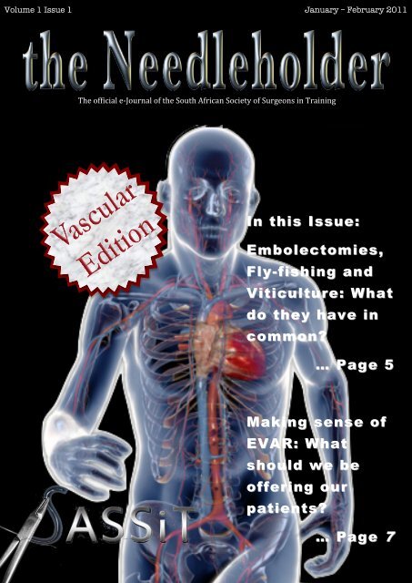
Volume 1 Issue 1 <strong>January</strong> – <strong>February</strong> <strong>2011</strong><br />
The official e-‐Journal of the South African Society of Surgeons in Training<br />
In this Issue:<br />
Embolectomies,<br />
Fly- Fly fishing and<br />
Viticulture: What<br />
do they have in<br />
common?<br />
… Page 5<br />
Making sense of<br />
EVAR: What<br />
should should<br />
we be<br />
offering our<br />
patients?<br />
patients<br />
… Page 7<br />
7
Editor<br />
Bradley A David<br />
Editorial Committee<br />
Ferhana gool<br />
Abri Bezuidenhout<br />
Shreya Rayamaji<br />
Publishing, Design and<br />
Layout<br />
Bradley A David<br />
‘the Needleholder’ is the<br />
official online e-‐Journal of<br />
the South African Society of<br />
Surgeons in Training <strong>SASSiT</strong><br />
Every effort has been made<br />
to ensure that all material is<br />
the original work of its<br />
authors and that where<br />
applicable outside material<br />
is appropriately referenced.<br />
Please feel free to email us<br />
with your concerns,<br />
comments, questions and<br />
ideas on how we can<br />
improve this publication:<br />
theneedleholder@gmail.com<br />
Volume 1 Issue 1 <strong>January</strong> – <strong>February</strong> <strong>2011</strong><br />
Contents<br />
Editorial<br />
Planning a career in vascular surgery. What the future holds?<br />
Dr Phillip Matley, President of VASSA …………………………………………….4<br />
Review<br />
A review of endovascular aortic repair (EVAR) in the elective<br />
management of abdominal aortic aneurysms.<br />
Gool F, Malherbe GF…………………………………………………………………………5<br />
Case Reports<br />
Dysphagia Lusoria: A case of an aberrant right subclavian artery and<br />
a bicarotid trunk<br />
Rogers AD, Nel M, Eloff EP, Naidoo NG……………………………………………13<br />
Case Report: Spontaneous Aortocaval Fistula<br />
Forgan T, Du Toit D…………………………………………………………….…………19<br />
Miscellaneous<br />
A biographical guide to the surgical tray:<br />
Michael E DeBakey…………………………………………………………………………11<br />
Thomas J. Forgarty………………………………………………………………………...18<br />
2
A career in vascular surgery<br />
in Southern Africa presents<br />
numerous interesting<br />
challenges for surgeons in<br />
training. Currently there<br />
are less than 40 registered<br />
specialist vascular surgeons<br />
in active practice in a<br />
country with a population of over 40 million,<br />
making fully trained specialist vascular<br />
surgeons a very precious resource. Out of<br />
sheer necessity, a great deal of vascular care<br />
and many vascular procedures are being<br />
performed by general surgeons but this is not<br />
ideal, given the increasing level of<br />
sophistication of vascular and endovascular<br />
surgery and the increasing demands and<br />
expectations of patients.<br />
Vascular disease is on the increase and this is<br />
contributed to by the fact that in developed<br />
counties, people are simply living to an older age.<br />
The scourge of diabetes has already been<br />
recognized as an international epidemic of<br />
worrying proportions. Tobacco use has not been<br />
brought under control and within our country,<br />
hypertension, hyperlipidaemia and aneurysmal<br />
disease are not only very common but tend to<br />
reflect strong genetic factors. Vascular trauma<br />
and unusual vasculopathies such as Takayasu<br />
Disease and HIV vasculopathy are particularly<br />
prevalent in our sub-‐continent.<br />
Vascular surgery is one of the youngest surgical<br />
specialties. The endovascular revolution,<br />
particularly in the 1990s has almost totally<br />
changed the way that we work. In South Africa,<br />
unlike the United Kingdom and many other parts<br />
of the world, endovascular procedures have<br />
largely remained in the hands of vascular<br />
surgeons rather than radiologists and<br />
cardiologists. This has broadened the scope and<br />
interest level of vascular surgery enormously.<br />
Vascular surgeons provide primary vascular care<br />
and education, diagnosis and assessment as well<br />
as the whole range of medical, surgical and<br />
endovascular treatment. This is in contrast to<br />
several areas of surgical gastro-‐enterology where<br />
PLANNING A<br />
CAREER IN<br />
VASCULAR<br />
SURGERY IN<br />
SOUTH AFRICA<br />
What does the<br />
future hold?<br />
Dr Phillip Matley<br />
President of VASSA<br />
Guest Editorial<br />
primary care and diagnosis is provided by<br />
gastroenterologists, and cardiac surgery where<br />
this is provided almost exclusively by<br />
cardiologists. The modern vascular surgeon has<br />
an intimate knowledge of vascular medicine and<br />
the medical management of the risk factors and<br />
the disease processes themselves. Furthermore,<br />
vascular surgeons have “hands-‐on” involvement<br />
in diagnosis and imaging, particularly with the<br />
increasing use of colour-‐flow duplex ultrasound.<br />
Vascular surgery works well in private practice in<br />
this country. Several private hospitals have<br />
established vascular centres of excellence<br />
including dedicated vascular wards, hybrid<br />
operating theatres (with ceiling-‐mounted<br />
angiographic facilities) and multi-‐disciplinary<br />
medical and para-‐medical teams. Vascular<br />
disease is common in the group of patients who<br />
enjoy private medical cover and there appears to<br />
be an almost limitless number of patients with<br />
varicose veins requesting surgical treatment. The<br />
earning potential of vascular surgeons compares<br />
very well to other specialties in the general<br />
surgery arena.<br />
The endovascular revolution and the increasing<br />
sophistication of vascular care is challenging the<br />
conventional training and career pathways.<br />
3
Increasingly vascular surgeons will be trained in<br />
both radiological and surgical techniques and will<br />
acquire a broad knowledge and experience of<br />
vascular medicine in close association with<br />
cardiology. The historical connections with<br />
general surgery, gastro-‐enterology and oncology<br />
will become ever more tenuous and it is likely<br />
that vascular trainees will have a very limited<br />
general surgery education before commencing a<br />
4-‐5 year training programme of pure vascular<br />
surgery.<br />
Modern vascular practice calls for a wide range of<br />
both cognitive and technical skills including<br />
traditional open surgery as well as percutaneous<br />
catheter-‐based techniques. There is currently<br />
very little role for laparoscopic vascular surgery<br />
but this may change as laparoscopic techniques<br />
develop further,<br />
The speciality is very demanding on the<br />
individual surgeon. The patients tend to be<br />
elderly and high-‐risk with multiple co-‐<br />
morbidities that require attention and control.<br />
Vascular emergencies are a daily phenomenon<br />
that will frequently interrupt weekends and<br />
nights and arranging appropriate cover for<br />
weekends and vacations can be a problem, given<br />
the scarcity of registered vascular surgeons.<br />
Group practices largely solve these issues but are<br />
uncommon among vascular surgeons in this<br />
country. With increasing sophistication may<br />
come increasing cost. The high-‐tech specialties<br />
such as endovascular surgery consume a large<br />
portion of health care expenditure and may be<br />
increasingly unaffordable to patients and funders<br />
in the future.<br />
I practice in a group partnership that includes<br />
three specialist vascular surgeons supported by<br />
three vascular technologists, a full multi-‐<br />
disciplinary team of para-‐medical personnel and<br />
a great network of cardiologists, physicians,<br />
radiologists and anaesthetists. We practice in a<br />
private hospital that has a dedicated 22-‐bed<br />
vascular ward and a hybrid theatre able to handle<br />
everything from a thoraco-‐abdominal aneurysm<br />
to a carotid stent. We cover vascular<br />
emergencies on a one-‐in-‐three rota and allow<br />
ourselves 10 weeks of vacation a year. We do<br />
clinical research and audit and remain very<br />
involved in teaching and in the activities of our<br />
professional societies. It has been a great career<br />
and it keeps getting better.<br />
Surgery<br />
Calender <strong>2011</strong><br />
March UCT Update<br />
22-24<br />
March<br />
12-16<br />
April<br />
15-17<br />
April<br />
Written College exams<br />
European HPB congress/<br />
ASSA-SAGES meeting (Cape<br />
Town)<br />
ASiT congress (Sheffield, UK)<br />
16-18 May Oral College exams (Gauteng)<br />
15 June Closing date for exam entry<br />
21-26<br />
June<br />
28 Aug – 1<br />
Sept<br />
30 Aug-1<br />
Sept<br />
International Surgical Trainees<br />
Congress (Pretoria)<br />
International Surgery Week<br />
(Yokohama, Japan)<br />
Written College exams<br />
October Pretoria Controversies<br />
4
A review of endovascular aneurysm repair (EVAR)<br />
in the elective management of abdominal aortic<br />
aneurysms.<br />
Gool F, Malherbe GF<br />
University of Cape Town<br />
Aneurysms of the abdominal aorta (AAA) have long been recognized as a<br />
fatal disease process and significant advances have been made over the last<br />
50 years in the surgical management thereof. Treatment of an asymptomatic<br />
aortic aneurysm is indicated when the risk of rupture outweighs the risk of<br />
operation.<br />
AAA’s are now more frequently diagnosed, due<br />
mainly to the increase in life expectancy and also<br />
due to the development of better non-invasive<br />
imaging techniques such as Doppler ultrasound,<br />
Computer Tomography (CT) and Magnetic<br />
Resonance Angiography (MRA).<br />
Conventional open aneurysm surgery is<br />
associated with considerable operative mortality<br />
because the operation is extensive and the<br />
patients often have serious co-morbidities.<br />
In published series, the 30-day or in-hospital<br />
mortality has ranged from 3.8% to 8.2% in<br />
patients undergoing elective open repair of AAA’s.<br />
Intensive care and hospital admissions are<br />
extended with open repair. Severe postoperative<br />
complications are common and more importantly,<br />
the time taken for recovery to the preoperative<br />
state of wellbeing is usually prolonged.<br />
There are also subgroups of patients who are at<br />
increased risk during conventional repair because<br />
of their associated medical co-morbidities or<br />
anatomical variations:<br />
1. Patients with myocardial infarction, renal or<br />
liver insufficiency<br />
2. Inflammatory aortic aneurysms<br />
3. Horseshoe kidney, retro-aortic renal vein<br />
4. Hostile abdomen<br />
It was the combination of these factors that<br />
provided the impetus for the development of<br />
minimally invasive procedures as a treatment<br />
option. The advent of minimally invasive<br />
endovascular stent-graft techniques or the<br />
endovascular aneurysm repair (EVAR) as an<br />
alternative treatment has radically extended the<br />
management options for patients with aortic<br />
aneurysms.<br />
However, the advent of new technology to treat<br />
any surgical condition needs to be evaluated<br />
against conventional treatment<br />
before a surgeon can offer this<br />
choice to patients. The questions<br />
that needed to be answered are:<br />
1. What are the advantages of<br />
the novel approach over conventional<br />
treatment?<br />
2. What are the potential drawbacks of the novel<br />
approach?<br />
3. Does the long-term outcome of the novel<br />
approach match the tested outcomes of<br />
conventional management?<br />
4. What are the cost implications of the novel<br />
approach?<br />
The aim of this review will be to outline the<br />
developmental history and the application of<br />
EVAR technology in the elective management of<br />
AAA’s. Finally, this review will aim to assess how<br />
the evidence collected to date answers the<br />
questions posed above in relation to the use of<br />
EVAR.<br />
The history of EVAR<br />
The first animal studies on<br />
EVAR by stent-graft<br />
combinations date from the<br />
mid-1980s. Unpublished<br />
clinical experience in Russia<br />
dates from 1985 but it was<br />
Parodi and colleagues, from<br />
Buenos Aires, Argentina,<br />
who were the first to<br />
publish experience with a<br />
balloon-expandable stent<br />
attached to a polyester<br />
tubular prosthesis to treat<br />
AAAs in patients who were<br />
REVIEW<br />
5
unfit for open surgery. Their home-made stentgraft<br />
evolved from an endograft with one<br />
proximal stent for infrarenal fixation only to<br />
include a second stent for distal fixation to obtain<br />
effective exclusion of the aneurysm from the<br />
blood flow. These early patients had an aortoaortic<br />
tube graft. Early on the graft endograft was<br />
only used in aneurysms not involving the<br />
bifurcation but later an aorto-uni-iliac stent-graft<br />
was also used, resolving the problem of an<br />
inadequate distal aortic cuff for securing “fixation”<br />
and “seal.”<br />
Over the next decade, a succession of industrymanufactured<br />
endoprostheses was introduced into<br />
the market for treatment of a broad spectrum of<br />
patients with a AAA. A variety of endografts from<br />
different manufacturers are currently available.<br />
The ideal endograft should provide lifelong<br />
protection from rupture of the aneurysm without<br />
any risk of migration or displacement from the<br />
attachment sites. Components should be<br />
sufficiently robust and at the same time small<br />
enough to fit into a delivery system that can be<br />
negotiated easily through the access vessels.<br />
Application of EVAR in the elective<br />
management of AAA’s<br />
Although there are significant variations from<br />
brand to brand, each device consists of three key<br />
components:<br />
• A delivery system to allow placement<br />
• An attachment system that forms a blood tight<br />
seal between the graft and the abdominal aorta<br />
• Graft fabric, which acts as a conduit for blood<br />
flow that has been diverted from the diseased<br />
segment of aorta<br />
INITIAL RADIOGRAPHIC EVALUATION<br />
Although the vast majority of abdominal aortic<br />
aneurysms are infrarenal, less than 50 percent<br />
are amenable to endovascular repair due to<br />
anatomic considerations. Angiography and<br />
computed tomography (CT) are commonly<br />
obtained as initial radiographic studies to<br />
determine both the feasibility of an endograft and<br />
the appropriate size and configuration of the<br />
endograft.<br />
1. Angiography<br />
Since the inner lumen is imaged, but not the<br />
wall of the aorta, angiography cannot evaluate<br />
the true lumen diameter, extent of thrombus,<br />
plaque, and calcification.<br />
2. Computed tomography<br />
Measurements of diameters can be<br />
problematic problematic if the aorta is<br />
angulated and the longitudinal axis is not<br />
perpendicular to the imaging plane.<br />
3. CT angiography<br />
Angiography is more sensitive for measuring<br />
small, axially oriented vessels and vascular<br />
stenoses than CTA, combining angiography<br />
with spiral CT and 3D CTA provides the<br />
maximum amount of information<br />
ANATOMIC CONSIDERATIONS<br />
1. Proximal neck length<br />
The minimum length needed varies from<br />
device to device but may be as long as 15<br />
mm. Newer designs allow suprarenal fixation<br />
and require a shorter proximal neck length.<br />
Ideally, it should be a normal appearing<br />
segment of aorta, without abundant thrombus<br />
or heavy circumferential calcification.<br />
2. Distal neck length<br />
A tube endograft may be placed if a distal<br />
neck is of sufficient length and diameter as<br />
specified by the manufacturer. As an example,<br />
the Ancure device requires a distal neck at<br />
least 12 mm in length and no greater than 26<br />
mm in diameter<br />
3. Aneurysm diameter<br />
Undersizing the diameter of the endograft<br />
may lead to an inadequate seal and failure to<br />
exclude the aneurysm, while oversizing may<br />
lead to kinking of the device, causing a nidus<br />
for thrombus formation or a leak. In a report<br />
from a European registry of patients<br />
undergoing endografting (the EUROSTAR<br />
registry), postoperative complications,<br />
postoperative mortality, late rupture, and<br />
6
aneurysm-related death were all more<br />
frequent in patients with a preoperative<br />
aneurysm diameter ≥6.5 cm.<br />
4. Angulation<br />
An angle of 60º or more leads to difficulties in<br />
implantation, kinking, leakage, and the<br />
possibility of downward migration of the<br />
device.<br />
5. Iliac attachment site<br />
The common iliac artery is the preferred<br />
attachment site, although the external iliac<br />
artery may be used. When the attachment site<br />
is in the external iliac artery, the hypogastric<br />
artery will be covered by the endograft. If the<br />
hypogastric artery is patent, the potential<br />
exists to back fill the aneurysm via back flow<br />
through this vessel into the sac, resulting in<br />
ineffective exclusion of the aneurysm. This<br />
problem can be prevented by preprocedural<br />
embolization of the hypogastric artery.<br />
6. Femoral artery diameter<br />
To accommodate the delivery system of most<br />
devices, a minimal femoral artery diameter of<br />
8 mm is usually required.<br />
7. Accessory renal arteries<br />
Accessory renal arteries are present in up to<br />
30 percent of the population and commonly<br />
originate from the lumbar aorta. Exclusion of<br />
an accessory renal vessel by an endograft can<br />
result in partial renal infarction.<br />
8. Inferior mesenteric artery<br />
However, when the inferior mesenteric artery<br />
is patent and there is significant stenosis of<br />
the superior mesenteric artery, the inferior<br />
mesenteric artery may supply important<br />
collateral blood flow to the bowel. In such<br />
patients, covering a patent inferior mesenteric<br />
artery with an endograft may compromise<br />
blood flow to the bowel. Thus, endograft<br />
placement is contraindicated.<br />
PATIENT FOLLOW-UP<br />
The size of the abdominal aortic aneurysm<br />
following endovascular repair and endograft<br />
placement is followed with plain film of the<br />
abdomen, CT scan or Arterial Duplex sonography.<br />
• Follow-up protocols vary, but a typical<br />
schedule after uncomplicated endograft<br />
placement consists of abdominal plain films<br />
before discharge and at one, six, and 12<br />
months, and then every year thereafter.<br />
Abdominal plain films are an economical and<br />
quick way to evaluate the integrity of the graft<br />
and the stability of graft appearance,<br />
alignment, and position.<br />
• CT scans, which are obtained on a similar<br />
schedule, are used to evaluate the diameter<br />
and volume of the aneurysm and to look for<br />
signs of endoleak or of endograft migration<br />
• A typical follow-up schedule in an<br />
uncomplicated patient post EVAR whose BMI is<br />
within normal limits, at our institution would<br />
be:<br />
• CT Abdomen at 4-6 weeks<br />
• Plain AXR at 4-6 weeks, 6 months and 12<br />
months; then annually<br />
• Arterial Duplex Sonography at 6 months, 12<br />
months then annually<br />
COMPLICATIONS<br />
Illustration of<br />
Endoleaks<br />
Type 1 – 4<br />
Complications that have been reported with<br />
endograft use include:<br />
• Vascular injury during deployment (sometimes<br />
leading to aneurysm rupture)<br />
• inadequate fixation or sealing of the graft to<br />
the vessel wall<br />
• stent frame fractures and separations<br />
• breakdown of the graft material<br />
7
Even after an aneurysm is successfully treated<br />
with an endograft, it remains a dynamic entity.<br />
The aneurysm will eventually thrombose and, by<br />
12 months, approximately 50 percent of<br />
aneurysm sacs have shrunk in diameter.<br />
1. Endoleaks<br />
2. Postimplantation syndrome<br />
Patients undergoing endovascular stent graft<br />
placement often experience an acute<br />
inflammatory syndrome characterized by<br />
fever, leukocytosis, elevation of serum Creactive<br />
protein (CRP) concentration, and<br />
perigraft air during the first week to 10 days<br />
after implantation<br />
3. Device migration<br />
Device migration is one of the major causes of<br />
secondary intervention after endovascular<br />
aneurysm repair. If untreated, potential<br />
complications include endoleak, aneurysm<br />
expansion, and rupture.<br />
CLINICAL OUTCOMES<br />
Short-term<br />
The short-term technical success rate for<br />
endovascular aneurysm repair ranges from 83 to<br />
over 95 percent. A 2007 systematic review<br />
identified four randomized trials of 1532 patients<br />
who were considered suitable candidates for<br />
either endovascular or open repair of nonruptured<br />
abdominal aortic aneurysms larger than 5.0 cm in<br />
diameter. 1 The 30 day all-cause mortality was<br />
significantly lower with endovascular repair (1.6<br />
versus 4.8 percent, relative risk 0.33, 95% CI<br />
0.17-0.64).<br />
The two principle contributing trials to this<br />
systematic review were:<br />
• The EVAR 1 trial included 1082 patients who<br />
were at least 60 years of age, with aneurysms<br />
at least 5.5 cm in diameter. At 30 days,<br />
mortality was significantly lower with<br />
endovascular than with open repair (1.6 versus<br />
4.6 percent, adjusted odds ratio 0.34, 95% CI<br />
0.15-0.74). Endovascular repair was also<br />
associated with a significantly shorter hospital<br />
stay (7 versus 12 days), although more<br />
secondary interventions (additional surgical<br />
procedures) were required with endovascular<br />
repair (9.8 versus 5.8 percent).<br />
• The DREAM trial evaluated 345 patients with<br />
aneurysms of at least 5 cm in diameter. There<br />
was an almost significant trend toward lower<br />
operative mortality with endografting than with<br />
surgery (1.2 versus 4.6 percent, risk ratio<br />
0.26, 95% CI 0.03-1.10). Moderate and severe<br />
systemic complications (cardiac, pulmonary,<br />
renal) were more frequent with open repair (26<br />
versus 12 percent), while moderate and severe<br />
local vascular or implant-related complications<br />
were more frequent with endovascular repair<br />
(16 versus 9 percent).<br />
The short-term survival advantage of<br />
endovascular repair appears to be much greater<br />
when endovascular repair is limited to patients at<br />
highest risk from open surgery. This was<br />
illustrated in a report of 454 consecutive patients<br />
who underwent elective repair (206 endovascular<br />
and 248 open surgery) of an abdominal aortic<br />
aneurysm. The overall 30-day mortality rates not<br />
significantly different for endografting and surgery<br />
(2.4 and 4.8 percent, respectively). However,<br />
among patients at highest surgical risk (American<br />
Society of Anesthesiologists class IV), the 30-day<br />
mortality rates were much lower with<br />
endovascular repair (4.7 versus 19.2 percent with<br />
open surgery).<br />
Long term<br />
The early survival benefit seen with endovascular<br />
repair compared to open repair described above is<br />
lost between one and four years, after which<br />
survival appears equivalent. This observation was<br />
seen in the 2007 systematic review discussed<br />
above, which included follow-up of 1473 patients<br />
from the DREAM (two years) and EVAR 1<br />
randomized trials (four years)<br />
Long term outcomes of patients who have<br />
undergone endovascular repair of abdominal<br />
aneurysms have been evaluated with and without<br />
comparison to patients who have undergone open<br />
repair.<br />
EVAR-1:<br />
In this large, randomized trial, endovascular<br />
repair of abdominal aortic aneurysm was<br />
associated with a significantly lower operative<br />
mortality than open surgical repair. However, no<br />
differences were seen in total mortality or<br />
aneurysm-related mortality in the long term.<br />
Endovascular repair was associated with<br />
increased rates of graft-related complications and<br />
re-interventions and was more costly.<br />
EVAR-2:<br />
In this randomized trial involving patients who<br />
were physically ineligible for open repair,<br />
endovascular repair of abdominal aortic aneurysm<br />
was associated with a significantly lower rate of<br />
aneurysm-related mortality than no repair.<br />
8
However, endovascular repair was not associated<br />
with a reduction in the rate of death from any<br />
cause. The rates of graft-related complications<br />
and reinterventions were higher with<br />
endovascular repair, and it was more costly.<br />
Conclusion<br />
1. What are the advantages of EVAR over<br />
conventional open repair?<br />
The results from EVAR1 and DREAM<br />
demonstrate that the short-term morbidity and<br />
mortality related to open repair is clearly<br />
improved in patients undergoing EVAR.<br />
However this benefit does not follow through in<br />
the long-term.<br />
In the select sub-group of patients with<br />
anatomical variants and/or a hostile abdomen<br />
EVAR is a very attractive option to avoid the<br />
unusually excessive risks of open surgery.<br />
2. What are the potential drawbacks of the<br />
novel approach?<br />
Higher long-term aneurysm specific morbidity<br />
and mortality is in large part due to the<br />
Endoleak phenomenon, as discussed earlier.<br />
Stent-technology depends on anatomic<br />
suitability and while EVAR maybe desirable in a<br />
patient it may not be applicable, because of<br />
unsuitable anatomy or problems with access.<br />
In view of uncertain long-term durability it<br />
should be used with caution in younger<br />
relatively low risk patients.<br />
The anticipated use of EVAR to treat patients<br />
who were not fit for open surgery has not been<br />
supported by the EVAR2 trial.<br />
3. Does the long-term outcome of the novel<br />
approach match the tested outcomes of<br />
conventional management?<br />
Long-term follow up in both the EVAR1 and<br />
DREAM trial demonstrate that morbidity and<br />
mortality are equivalent in both groups.<br />
However, the aneurysm specific morbidity and<br />
mortality was significantly higher in the EVAR<br />
arms of the studies. Initial speculation that this<br />
was related to the ‘developmental turnaround<br />
time’ of new technology, may not hold true as<br />
the American-based OVER trial has had very<br />
similar results compared to the EVAR1 and<br />
DREAM trials despite using the latest stent<br />
technology.<br />
4. What are the cost implications of the<br />
novel approach?<br />
EVAR is more costly than open repair not only<br />
because of the initial device costs but also<br />
because of the rigorous CT based follow-up<br />
used in most of the trials. However, in the<br />
EVAR and DREAM trials the long-term costs of<br />
all-cause morbidity associated with open repair<br />
such as wound complications, were not<br />
included in the cost analysis. Moreover, followup<br />
surveillance of the stent is moving away<br />
from CT towards a duplex based regime which<br />
is far more cost effective.<br />
In conclusion, the endovascular management of<br />
abdominal aortic aneurysms should not be viewed<br />
as a replacement for conventional open surgery.<br />
EVAR is a weapon in the armamentarium of the<br />
vascular surgeon, which if used with caution in<br />
the appropriate setting will form part of an<br />
effective elective management strategy for the<br />
treatment of AAA’s.<br />
REFERENCES<br />
1. Sajid MS, Desai M, Baker DM, Hamilton G. Endovascular Aortic Aneurysm<br />
Repair (EVAR) Has Significantly Lower Peri-operative Mortality in Comparison<br />
to Open Repair: A Systemic Review. Asian J Surg. 2008 Jul;31(3):119-23<br />
2. Malas MB, Freischlag JA. Interpretation of Results of OVER in the context of<br />
the EVAR Trial, DREAM, and the EUROSTAR Registry. Semin Vasc Surg. 2010<br />
Sep;23(3):165-9<br />
3. Albuquerque FC Jr, Tonnessen BH, Noll RE Jr, Cires G, Kim JK, Sternbergh WC<br />
3 rd . Paradigm shifts in the treatment of Abdominal Aortic Aneurysms: trends<br />
in 721 patients between 1996 and 2008. J Vasc Surg.2010 Jun;51(6):1348-<br />
52; discussion 1352-3.<br />
4. Deaton DH. Future Technologies to address the failed endoprosthesis. Semin<br />
Vasc Surg. 2009 Jun;22(2):111-8.<br />
5. Lederle, FA, Johnson, GR, Wilson, SE, et al. The aneurysm detection and<br />
management study screening program: validation cohort and final results.<br />
Aneurysm Detection and Management Veterans Affairs Cooperative Study<br />
Investigators. Arch Intern Med 2000; 160:1425.<br />
6. Szilagyi, DE, Smith, RF, DeRusso, FJ, et al. Contribution of abdominal aortic<br />
aneurysmectomy to prolongation of life. Ann Surg 1966; 164:678.<br />
7. Peppelenbosch, N, Buth, J, Harris, PL, et al. Diameter of abdominal aortic<br />
aneurysm and outcome of endovascular aneurysm repair: does size matter? A<br />
report from EUROSTAR. J Vasc Surg 2004; 39:288.<br />
8. Greenhalgh, RM, Brown, LC, Kwong, GP, et al. Comparison of endovascular<br />
aneurysm repair with open repair in patients with abdominal aortic aneurysm<br />
(EVAR trial 1), 30-day operative mortality results: randomised controlled trial.<br />
Lancet 2004; 364:843.<br />
9
Michael Ellis DeBakey was born as Michel Dabaghi (later Anglicized to DeBakey) in Louisiana to<br />
Lebanese immigrants. DeBakey graduated from Tulane University School of Medicine in New Orleans in<br />
1932. He completed his surgical fellowships at the University of Strasbourg, France, under<br />
Professor René Leriche, and at the University of Heidelberg, Germany, under Professor Martin<br />
Kirschner.<br />
‘DeBakey had a machine<br />
shop right outside the<br />
operating room. He<br />
regularly consulted with<br />
an engineer during and<br />
after procedures about<br />
the design of<br />
instruments’<br />
Michael Ellis DeBakey<br />
September 7, 1908 – July 11, 2008<br />
World-renowned Lebanese-American cardiac surgeon, innovator,<br />
scientist, medical educator, and international medical statesman.<br />
During a decorated five-decade medical career, it is without<br />
hyperbole that Dr. Michael E. DeBakey earned the right to be<br />
named as one of the greatest surgeons of the 20th century.<br />
While he was in medical school DeBakey conceived of the<br />
roller pump, the central idea of the heart-lung machine. The<br />
very notion that the heart can be temporarily replaced by a<br />
machine set the stage for a host of medical innovations—from<br />
bypass surgeries and heart transplants to artificial hearts—<br />
and a fundamental change in approach to heart treatment,<br />
heart health and the prevention of heart disease.<br />
DeBakey ushered in, and was often at the forefront of, medical innovations that have saved<br />
millions of lives. He performed over 60,000 operations himself. Chief among these techniques<br />
was arterial bypass and arterial reconstruction. He mastered ways of patching and grafting,<br />
bypassing and re-organizing arteries. He finally put down his scalpel in 1998 at age 90.<br />
Kenneth L. Mattox, chief of surgery at Baylor-affiliated Ben Taub Hospital in the Texas Medical<br />
Center, Houston, has no doubts that his mentor and long-time colleague did that many<br />
operations. “We were doing operations in four different rooms and he would move from room to<br />
room,” Mattox recalls. In the evening, “he sent out for food and we ate between cases.” At 11<br />
p.m., the entire team of residents, nurses, anesthesiologists, everyone, was exhausted, Mattox<br />
says. “I remember Dr. DeBakey sticking his head out a door and saying, ‘Doesn’t anybody else<br />
out there want an operation? We’re just getting warmed up.’”<br />
Mattox also remembers that DeBakey had a machine shop right outside the operating room. He<br />
regularly consulted with an engineer during and after procedures about the design of<br />
instruments. “He would look at the way an instrument fit in his hand, the way a clamp would<br />
spring or close,” Mattox says. “He would talk to the engineer, and a week later he would have a<br />
new instrument.”<br />
The Next time you ask your scrub<br />
sister for a DeBakey forceps<br />
remember the innovative spirit of<br />
its creator – Michael E. DeBakey.<br />
The DeBakey<br />
Forceps designed<br />
with atraumatic<br />
tips so as to<br />
minimize trauma<br />
to delicate tissues.<br />
11
Dysphagia Lusoria: A case of an aberrant right<br />
subclavian artery and a bicarotid trunk<br />
Rogers AD, Nel M, Eloff EP, Naidoo NG<br />
Vascular Surgery Unit, Division of General Surgery, Groote Schuur Hospital, Cape Town<br />
Abstract<br />
Dysphagia Lusoria is dysphagia secondary to an aberrant right subclavian artery<br />
that has a retro-oesophageal course. Adachi and Williams categorized aortic<br />
arch anomalies, showing that the right subclavian artery arising in this fashion<br />
(as the last branch of the descending aorta) is one of the more common.<br />
However, this very rarely co-exists with a bi-carotid trunk.<br />
We present such a case as is it manifested in a 36 year-old lady complaining of<br />
marked weight loss and dysphagia. The diagnosis remained elusive until a CT<br />
scan of the chest was performed; angiography further delineated the pathology.<br />
It is believed that the combination of the common carotid origins with the retrooesophageal<br />
course of the aberrant vessel more frequently accounts for<br />
symptoms in the absence of an aneurysm of the origin of the aberrant vessel.<br />
Several techniques to manage the aberrant vessel have been described in the literature, but we<br />
favoured open ligation and transposition to the right carotid artery.<br />
Case Report<br />
A 36-year-old women presented to the Gastro-<br />
Intestinal Unit of Groote Schuur Hospital with a<br />
three year history of dysphagia. The dysphagia<br />
was initially only for solid food and intermittent,<br />
but more recently she describes a constant ‘stuck<br />
in the throat’ foreign body sensation. She also<br />
admitted to significant (almost 30kg) weight loss.<br />
She was very anxious, and eager to determine<br />
the source of the ailment and was concerned<br />
about the possibility of malignancy.<br />
Physical examination revealed only evidence of<br />
recent weight loss and routine laboratory data<br />
were within normal limits.<br />
There were no abnormalities on chest Xray or<br />
ECG.<br />
Barium Swallow demonstrated an indentation at<br />
the level of the third thoracic vertebra. A small<br />
diverticulum was also reported to be present, just<br />
above the aortic arch.<br />
No pathology was demonstrated during<br />
oesophagoscopy, oesophageal manometry,<br />
larygoscopy and pharyngoscopy.<br />
A course of Antacids and H2 receptor blockers<br />
was initiated for two months, with little benefit.<br />
Psychotherapy was considered and<br />
antidepressants prescribed.<br />
At this time a CT scan of the chest and abdomen<br />
demonstrated an aberrant right subclavian artery,<br />
originating from the descending aorta.<br />
The patient was referred to the Vascular Surgery<br />
Unit for further investigation and possible surgical<br />
intervention. Digital Subtraction Angiogram<br />
confirmed the diagnosis. The ARSA originated<br />
distal to the origin of the left subclavian artery<br />
and coursed through the posterior mediastinum<br />
behind the oesophagus. In addition, the<br />
angiogram also demonstrated a common origin of<br />
the common carotid arteries as a second branch<br />
of the arch.<br />
The patient underwent a right supraclavicular<br />
incision and surgical ligation of the ARSA with<br />
right subclavian–carotid transposition using an<br />
end-to-side anastomosis.<br />
The patient had an uneventful postoperative<br />
course and remains symptom free after follow-up<br />
of twelve months.<br />
Literature Review<br />
Figure 1 – Barium Swallow<br />
The characteristic diagonal<br />
impression at the level of<br />
the third and fourth<br />
vertebrae<br />
The first case of a symptomatic aberrant right<br />
subclavian artery (ARSA) was described in the<br />
medical literature by Hanuld in 1735. 1<br />
The term ‘Dysphagia Lusoria’, however, was<br />
coined by Bayford in 1794 to describe dysphagia<br />
secondary to a retro-oesophageal (aberrant) right<br />
subclavian artery (ARSA). He described a lady<br />
who died of oesophageal obstruction and<br />
resultant emaciation. The condition, in his words<br />
‘may be called lusoria, from Lusus Naturae (Latin<br />
for ‘freak of nature’) that gives rise to it’. 2<br />
13
Figures 2 and 3 – Angiogram – The aberrant right subclavian<br />
artery and its course demonstrated on AP and Right anterior<br />
oblique views. Note also the common origin of the carotid arteries.<br />
Burckhard Komerell is credited with the first<br />
radiological description, in 1936, as the condition<br />
had only been diagnosed at postmortem prior to<br />
that time. Komerell’s name survives as the<br />
eponym for the diverticulum sometimes present<br />
at the origin of this attenuated vessel. He stated ‘<br />
. . . the pulsating mass behind the oesophagus<br />
does not consist of the right subclavian itself,<br />
because the calibre of this vessel is much smaller.<br />
Much more likely this mass consists of an aortic<br />
diverticulum, from which the right subclavian<br />
artery originates.’ 3<br />
The incidence of ARSA varies between 0.4 to<br />
1.8% of the population, and is probably the<br />
commonest significant aortic arch anomaly. 4<br />
The Adachi Williams Classification<br />
In about 80% of individuals, three branches arise<br />
from the aortic arch: the brachiocephalic trunk,<br />
the left subclavian artery and the left common<br />
carotid artery. This Adachi described as Type A. 5<br />
11% of individuals have an Adachi type B pattern,<br />
which consists of a common trunk for the left<br />
common carotid and the brachiocephalic artery<br />
and therefore has only two aortic arch branches. 5<br />
The next most common type, Adachi C, has a<br />
vertebral artery originating proximal to the left<br />
subclavian artery as a 4 th branch of the arch. 5<br />
The origin of the retroesophageal right subclavian<br />
artery as the last branch occurs in between 0.4<br />
and 2% of individuals. 4,5,6<br />
The Adachi and Williams Classification recognizes<br />
four basic morphologies within this group: Types<br />
G, CG, H and N (see figure 7.) 4<br />
This case report demonstrates features of Adachi<br />
H, where the right subclavian artery is anomalous<br />
(as in type G), but where the right and left<br />
common carotids arise from a common stem or<br />
bicarotid trunk.<br />
Embryology<br />
Figure 4 – 3 D Reconstruction –<br />
Right posterior oblique view.<br />
The right subclavian artery develops during the<br />
6 th to 8 th week of gestation. The proximal part<br />
originates from the right 4 th aortic arch artery,<br />
and the distal part from the right dorsal and right<br />
seventh intersegmental arteries. 3,7<br />
In these cases, the right 4 th aortic arch artery<br />
and/or the right dorsal aorta involute cranial to<br />
the seventh intersegmental artery – the<br />
connection between the aortic sac and the right<br />
subclavian artery disappears. The right<br />
subclavian artery develops from the right 7 th<br />
intersegmental artery and the distal segment of<br />
the right dorsal aorta. Differential growth shifts<br />
Figure 7 – Retro-oesophageal Subclavian Anomalies. Note<br />
the rare Type H, with the bicarotid trunk.<br />
the origin cranially and lies close to the origin of<br />
the left subclavian artery. It originates dorsally<br />
and therefore has a retroesophageal course. 3,7<br />
The aberrant right subclavian artery stems from<br />
the dorsal margin of the aortic arch, between the<br />
top of the arch and where it lies against the<br />
vertebral column. The proximal part of the artery<br />
14
has a wider diameter than the distal part and the<br />
artery passes through the mediastium in a retrooesophageal<br />
position. The artery may arise from<br />
a diverticulum at the proximal descending aorta,<br />
referred to as Kommerell’s diverticulum. 3,7<br />
Presentation<br />
There are several descriptions of childhood<br />
dyspnoea resulting from an ARSA, and cases of<br />
pneumonia have been ascribed to the condition<br />
on the basis of aspiration and dysphagia. Relative<br />
tracheal laxity may account for this presentation<br />
8, 9<br />
in childhood.<br />
Two thirds of individuals are believed (on the<br />
basis of autopsy studies and retrospective<br />
studies) to remain completely asymptomatic<br />
despite the anomaly. There are a few proposed<br />
mechanisms as to why certain individuals become<br />
symptomatic:<br />
1. Increased oesophageal rigidity with ageing<br />
2. Aneurysm formation<br />
3. Aortic elongation with ageing<br />
4. The presence of a bicarotid trunk. 7,8<br />
In a study by Klinkhamer, published in 1966 and<br />
reviewing all articles from 1763, it is stated that<br />
the aberrant right subclavian artery was found to<br />
be associated with a bicarotid truncus (common<br />
origin of the right and left carotid arteries) in 85<br />
of 295 cases (29%) In 60% of cases there was a<br />
normal origin of the two carotids, and in 10%<br />
they were observed to be closer to one another<br />
than normal. 8<br />
It is believed that the combination of the common<br />
carotid origins with the retro-oesophageal course<br />
of the ARSA more frequently accounts for<br />
symptoms in the absence of an aneurysm of the<br />
origin of the aberrant vessel. In fact, several<br />
authors have questioned the relationship between<br />
the aberrant vessel (in isolation) and the<br />
dysphagia. 8<br />
Klinkhamer, for instance, felt that the aberrant<br />
artery itself was not an adequate explanation for<br />
the dysphagia because some patients have had<br />
very large ARSA’s, without dysphagia or<br />
respiratory symptoms. He maintained that<br />
symptoms are usually only present when the left<br />
and right carotids arise together or close to one<br />
another and therefore prevent the trachea and<br />
oesophagus from being bent forward where the<br />
ARSA crosses. 8<br />
In 80% of individuals, the Brachiocephalic (BCA)<br />
and Left Common Carotid Arteries (LCCA)<br />
originate from the arch 4cm apart. Because the<br />
arch lies obliquely and curves from front and the<br />
right backward and to the left, the BCA is more<br />
ventral than the LCCA origin. This allows the<br />
ARSA from being bent forward (note diagram). If<br />
they arise commonly, the two carotids form a ‘V’<br />
that prevents forward or flexion movements of<br />
the trachea and oesophagus. These structures<br />
are therefore compressed by the SCA posteriorly<br />
and by the CC origins antero-laterally. 6-10<br />
Investigation 7-11<br />
Barium swallow may demonstrate the<br />
characteristic diagonal impression at the level of<br />
the third and fourth thoracic vertebrae.<br />
A pulsating mass may be visualized at endoscopy.<br />
Digital Subtraction Angiogram, CT with contrast,<br />
or MRI may confirm the diagnosis and enable one<br />
to visualise the arch anatomy.<br />
Motility studies are frequently performed during<br />
the diagnostic phase of investigation. A high<br />
pressure zone in the region of the vessel has been<br />
described. Manometry cannot be used to<br />
diagnose the condition nor has it been of any<br />
assistance in distinguishing which patients may<br />
benefit from surgery.<br />
Therapy<br />
Several reports have described improvement with<br />
conservative therapy. These patients usually had<br />
inconclusive findings on manometry. In light of<br />
the relative infrequency of symptoms in patients<br />
with isolated ARSA, some authors have therefore<br />
advocated trials of therapies like prokinetics and<br />
antireflux medications.<br />
The majority of symptomatic patients have<br />
benefited from surgical intervention. Janssen<br />
concluded that in the absence of another cause of<br />
the symptoms and after a trial of medical<br />
management, surgery should be considered. 7 In<br />
1994, Kieffer reported on 19 patients who<br />
underwent surgery, of whom 16 had complete<br />
resolution of their symptoms. 12<br />
Gross first reported surgical management of this<br />
condition in 1946. He described dividing and<br />
ligating the ARSA via a left thoracotomy, in a 4<br />
month old infant. 13 Lichter was the first to<br />
describe surgery on an adult with this condition in<br />
1963. It is not until the last thirty years that<br />
surgery has become the standard therapy for this<br />
condition, and several authors have advocated<br />
various approaches. 14 Pome, in a review of the<br />
literature published in 1987, found only twenty<br />
reported surgically treated patients. 15<br />
The optimal exposure of the aberrant artery origin<br />
is undoubtedly achieved via a left thoracotomy.<br />
This is particularly important when the origin of<br />
the ARSA is dilated. 7,14<br />
Simple ligation and division has been noted to be<br />
inadequate therapy in a significant number of<br />
patients due to the development of subclavian<br />
15
steal syndromes. The onset of this syndrome<br />
may be immediately postoperatively or late<br />
(described up to seven years later). In addition,<br />
cases of gangrene of the right arm have been<br />
described following simple ligation of the aberrant<br />
vessel. 14<br />
Smith and Pifarre have described reimplanting the<br />
right SCA with a graft onto the ascending arch via<br />
a left thoracotomy. This is a technically<br />
challenging exercise and involves passing the<br />
Right SCA and the graft from the posterior<br />
mediastinum to the anterior, as well as deep<br />
anastomoses. 16,17<br />
The anastomosis of the RSCA to the ascending<br />
arch is much easier to perform via a right<br />
posterolateral thoracotomy, as described by<br />
authors such as Bailey. 18<br />
Pifarre identified a problem with the right<br />
thoracotomy approach in one case report. If the<br />
artery is not divided close to its origin, thrombosis<br />
of the stump may lead to the persistence of<br />
dysphagia. 17<br />
Schumacker described performing an end to side<br />
anastomosis of the R SCA with the right carotid<br />
artery via a median sternotomy. This has been<br />
the method described by at least three further<br />
surgeons. However, this anterior approach also<br />
provides suboptimal control during the dissection<br />
and division of the vessel. The possibility of<br />
transient cerebral ischaemia and the<br />
consequences of damage to an atherosclerotic<br />
carotid artery exist with this approach. Mok<br />
therefore recommended that the divided aberrant<br />
right SCA should be anastomosed to the aortic<br />
arch, with or without an interposition graft. 10<br />
Orvald and Kunlin advocated a cervical approach,<br />
but Kunlin described significant haemorrhage in a<br />
patient when attempting to perform the<br />
procedure via a cervical incision and had to resort<br />
to a median sternotomy. We have already<br />
highlighted the potential complication of residual<br />
dysphagia in cases where a long stump is left. 15<br />
Syders reported a combined approach using both<br />
cervical and left carotid approaches to reimplant<br />
the right SCA onto the right carotid. This, while<br />
considerably safer, involves having to reposition<br />
the patient for the second incision. 15<br />
Lemire described a transternal approach for the<br />
division and reimplanting of the Right SCA to the<br />
ascending aorta. The incidence of pain and<br />
pulmonary complications are probably higher, but<br />
this approach has cosmetic advantages. 19<br />
Pome recommended that a right thoractomy may<br />
be employed for patients without significant<br />
ectasia of the origin of the Right SCA; patients<br />
with ectasia should undergo a left thoracotomy<br />
and cervical incision. 15<br />
The importance of dividing the stump proximally<br />
is again highlighted in the paper by Pome. He<br />
advocated using the aorta rather than the carotid<br />
to avoid a possible subclavian steal syndrome. 15<br />
Janssen reports six cases of dysphagia lusoria<br />
diagnosed and managed between 1992 and 1997.<br />
Three patients responded to either medical<br />
management (antacids etc), or dietary<br />
modification. One patient underwent a right<br />
carotid-subclavian end to side bypass via a right<br />
supraclavicular approach. A persistent RSCA<br />
stump may account for his occasional residual<br />
dysphagia for solid food. Two patients underwent<br />
a two incision approach (ie thoracotomy and<br />
cervical incision). 7<br />
Kieffer et al have the largest single-centre series<br />
of patients who have received therapy for<br />
symptomatic or aneurismal aberrant subclavian<br />
arteries. They divided their patients into four<br />
distinct subgroups:<br />
1. dysphagia lusoria without aneurysm<br />
2. symptomatic occlusive disease of the artery<br />
3. aneurysmal disease of the artery itself<br />
4. aneurysmal disease of the thoracic aorta or<br />
origin of the aberrant artery<br />
It would appear sensible to manage all patients<br />
with aneurismal disease with stentgrafting in light<br />
of the high rupture rate (22.6%) and consequent<br />
mortality rate (100%) independent of the<br />
diameter of the aneurysm. The perioperative rate<br />
of patients undergoing surgical repair of these<br />
aneurysms was 26.9%. 12<br />
There are few reports in the literature of<br />
endovascular or hybrid approaches to this<br />
pathology. Although long term results are still<br />
pending, initial results are promising. Shennib et<br />
al have described minimally invasive, hybrid<br />
endovascular approaches . A right supraclavicular<br />
approach was used to perform a right carotidsubclavian<br />
bypass prior to division of the right<br />
SCA. They then deployed an occluder in an<br />
antegrade fashion into the proximal end of the<br />
right SCA in order to maintain good control of the<br />
deployment and to avoid embolization of the<br />
occluder into the aortic arch from a retrograde<br />
approach. A right femoral artery access was used<br />
via a 9F sheath. 20<br />
Endoluminal grafts have also been used with<br />
some success in the presence of aneurysm of the<br />
ARSA origin. In these cases it may be necessary<br />
to consider performing a further anastomosis<br />
between the left carotid and subclavian artery if<br />
overstenting was intended.<br />
Kopp et al have reported a series of six patients<br />
managed by a variety of techniques during a<br />
seventeen year period. One of the patients had<br />
16
a covered wall stent with an occluded proximal<br />
stent graft lumen inserted via a transbrachial<br />
approach. The right SCA was then anastomosed to<br />
the right common carotid artery. 21<br />
References<br />
1. Williams GD, Aff HM, Schmeckebier M, Edmonds HW, Grand EG. Variations in the<br />
arrangement of the branches arising from the aortic arch in the American whites and<br />
negroes. Anat Rec 1932; 54: 247-251<br />
2. Bayford D. An account of a singular case of obstructed degluitition. Memoirs Med<br />
Soc London 1794; 2: 275-86<br />
3. Kommerell B. Verlagerung des Osophagus durch eine abnorm verlaufende Arteria<br />
subclavia dextra (Arteria lusoria). Fortschr Geb Roentgenstrahlen 1936; 54: 590-595<br />
4. Rahman HA, Sakurai A, Dong K, Setsu T, Umetani T, Yamadori T. The<br />
Retroesophageal Subclavian Artery – A case report and Review. Acta Anat Nippon<br />
1993; 68: 281-287<br />
5. Adachi B. Das Arteriensystem der Japener. Bd 1 1928: 22-42 Kenkyu-sha<br />
Publishing Co, Tokyo<br />
6. Saito T, Tamatsukuri Y et al. Three Cases of Retroeophageal Right Subclavian<br />
Artery. J Nippon Med Sch 2005; 72 (6): 375 – 382<br />
7. Janssen M, Baggen MGA, Veen HF, Smout AJPM, Bekkers JA, Jonkman JGJ,<br />
Ouwendijk RJTh. Dysphagia Lusoria: clinical aspects, manaometric findings, diagnosis,<br />
and therapy. Am J Gastroent 2000; 95: 1411-1416<br />
8. Klinkhamer AC. Aberrant right subclavian artery. Clinical and roentgenologic<br />
aspects. Am J Roentgenol Radium Ther Nucl Med 1966; 97: 438-446.<br />
9. Gross RE, Neuhauser EBD. Compression of trachea by anomalous innominate<br />
artery: operation for its relief. Am J Dis Child 1948; 75: 570-574<br />
10. Mok CK, Cheung KL, Kong SM, Ong GB. Translocating the right subclavian artery<br />
in dysphagia lusoria. Br J Surg 1979; 66: 113-116<br />
11. Harada H, Ito T, Yamamoto N, Abe T. Surgical Treatment of an Aneurysm of the<br />
Aberrant Right Subclavian Artery Involving an Aortic Arch Aneurysm and Coronary<br />
Artery Disease. Am Thorac Cardiovasc Surg 2001; 7 (12): 109-112<br />
12. Kieffer E, Bahnini A, Koskas F. Aberrent subclavian artery: Surgical treatment in<br />
thirty-three adult patients. J Vas Surg 1994; 19: 100-111<br />
13. Gross RE. Surgical treatment of dysphagia lusoria. Ann Surg 1946; 124: 532-534<br />
14. Lichter I. The treatment of dysphagia lusoria in the adult. Br J Surg 1963; 50:<br />
793-796<br />
15. Pome G, Vitali E, Mantovani A, Panzeri E. Surgical treatment of the aberrant<br />
retroesophageal right subclavian artery in adults (dysphagia lusoria). J Cardiovasc<br />
Surg 1987; 28: 403 – 411<br />
16. Smith JM, Reul GJ, Wurash DC, Cooley DA. Retro-oesophageal subclavian arteries:<br />
surgical management of symptomatic children. Card Vasc Disease Texas Heart Inst<br />
1979; 6: 333-334<br />
17. Pifarre R, Dieter, RA, Niedballa RG. Definitive surgical treatment of the aberrant<br />
retro-oesophageal right subclavian artery in the adult. J Thorac Cardiovasc Surg 1971;<br />
61: 154-159.<br />
18. Bailey CP, Hirose T, Alba J. Re-establishment of the continuity of the anomalous<br />
right subclavian artery after operation for dysphagia lusoria. Angiology 1965; 16: 509-<br />
513<br />
19. Lemire GG, Rabbat AG, Trudel J. Dysphagia lusoria: current surgical approach. J<br />
CArdiovasc Surg 1978; 19: 311-313<br />
20. Shennib H, Diethrich EB. Novel approaches for the treatment of the aberrant right<br />
subclavian artery and its aneurysms. J Vasc Surg 2008; 47: 1066-1070.<br />
21. Kopp R, Wizgall I, Kreuzer E, Maimarakis G, Weidenhagen R, Kuhni A, Conrad C,<br />
Jauch KW, Lauterjung L. Surgical and Endovascular Treatment of Symptomatic<br />
Aberrant Right Subclavian Artery (Arteria Lusoria). Vasc 2007;15 (2): 84-91<br />
17
Embolectomies, Fly-fishing &<br />
Viticulture:<br />
What do they have in common?<br />
The answer to the question above is simple when one<br />
explores the biographical history of Thomas J. Fogarty,<br />
who as a physician and professor, inventor and<br />
entrepreneur, has saved tens of millions of lives most<br />
notably, by pioneering the tools and methods of less<br />
invasive vascular surgery.<br />
Fogarty was born and raised in Cincinnati, Ohio. His precocious mechanical ability and business<br />
instincts began as a child, designing and building soapbox derby racers and model airplanes, the latter<br />
of which he sold to children from his neighborhood. Soon, he upgraded to a motor scooter; and when<br />
he became frustrated with its gears, he built (and sold) a centrifugal clutch that is still used today in<br />
some simple motors.<br />
Fogarty's interest in medicine began in early high school when he took a job as an equipment cleaner<br />
and later scrub technician at Cincinnati's Good Samaritan Hospital. In the latter position, Fogarty was<br />
frequently able to observe operations and the problems surgeons encountered. By the time he<br />
graduated from high school, Fogarty knew that his calling was to make surgery simpler, cheaper, faster<br />
and safer through technology.<br />
Before he graduated in 1960 from the University of Cincinnati Medical School, Fogarty had designed his<br />
most significant invention. The Fogarty Balloon Embolectomy Catheter, like many revolutionary<br />
inventions is simple in concept. Fogarty built the prototype in his attic, attaching the fingertip of a latex<br />
surgical glove to a catheter using fly-tying techniques familiar to him from boyhood fishing<br />
expeditions.<br />
In 1969 Fogarty patented his device, which is now being used in over 300,000 procedures every year,<br />
all over the world. The catheter was a great improvement on previous embolectomy methods. First, it<br />
does not cut off blood flow, increasing the risk of the patient's losing a limb; and second, the entire<br />
procedure can be performed in one hour through a single small incision, instead of using many larger<br />
incisions and forceps, with the patient under general anesthesia for hours.<br />
Fogarty's balloon catheter procedure was the first successful example of minimally invasive vascular<br />
surgery. Since its introduction, Fogarty and others have developed numerous spin-off applications: for<br />
example, the first balloon angioplasty, performed with a Fogarty catheter in 1965, has led to over<br />
650,000 such operations per year.<br />
He also provides venture capital to other medical device inventors devoted to solving "real-life clinical<br />
problems." In the 1990s, Fogarty became a Professor of Surgery at Stanford University Medical Center.<br />
As if this were not enough, Fogarty has also founded an award-winning vineyard: Thomas Fogarty<br />
Winery, outside of Palo Alto, California.<br />
To date, Thomas Fogarty has personally earned 63 patents, with many others pending. He has<br />
authored or co-authored over 150 professional articles, and is a member of 29 professional societies.<br />
He has won a series of prestigious awards, including the San Francisco Patent and Trademark<br />
Association's Inventor of the Year (1980).<br />
18
Case Report: Spontaneous<br />
Aortocaval Fistula<br />
Forgan T, Du Toit D<br />
Stellenbosch University; Tygerberg Hospital, Dept<br />
of Vascular Surgery<br />
Abstract<br />
A 58 year old man presented with malena stools<br />
and a pulsatile abdominal mass (which turned out<br />
to be an aortocaval fistula (ACF.) At surgery, a<br />
Introduction<br />
A rare complication of infrarenal abdominal aortic<br />
aneurysms is ACF, which occurs in up to 4% of<br />
ruptured or symptomatic aneurysms. 1,2 Due to<br />
the rarity of ACF and the elusive clinical<br />
presentation, they are easily overlooked.<br />
Presentation ranges from the typical signs of<br />
lower back pain, palpable abdominal aortic<br />
aneurysm, machinery abdominal murmur and<br />
high-output cardiac failure to cardiac and renal<br />
failure with few other obvious signs or symptoms.<br />
16,17 Preoperative diagnosis assists in patient<br />
preparation and the selection of the most<br />
appropriate therapeutic modality.<br />
Case Report<br />
A 58 year old male was referred to our institution<br />
from a district hospital with a history of having<br />
passed malena stools, shortness of breath and<br />
hypotension. He was known to have an abdominal<br />
aortic aneurysm (AAA) of 5 cm. He had been<br />
treated for congestive cardiac failure for the<br />
previous year and was also known to have renal<br />
dysfunction. He was also an ex-smoker.<br />
On arrival he was hypotensive with a mean<br />
arterial pressure of 55mmHg and had a<br />
tachycardia of 120. He had a Glasgow Coma<br />
Score of 15 and was tachypnoeic with a<br />
respiratory rate of 30. He was complaining of<br />
central abdominal pain.<br />
On further examination he was an overweight<br />
man with a mildly tender abdomen and a pulsatile<br />
central abdominal mass. It was difficult to<br />
delineate if the mass extended above the costal<br />
margin. No thrill was palpable and no murmur<br />
was heard on auscultation of his abdomen. On<br />
rectal examination he had malena stool. The<br />
patient had a raised JVP, bibasal crepitations and<br />
bipedal oedema.<br />
A blood gas was done which showed a pH of 7,3,<br />
lactate of 5, base excess of -4 and an hb of 8.<br />
Further blood tests revealed that he had mild<br />
Figure 1, Contrasted CT Abdomen showing early<br />
filling of the IVC with contrast.<br />
Figure 2, Contrasted CT Abdomen revealing an<br />
Aortocaval fistula arising in the distal aorta<br />
renal dysfunction.<br />
At this stage the working diagnosis was that of an<br />
aorta-enteric fistula.<br />
In light of the above information a central venous<br />
catheter was inserted, careful fluid resuscitation<br />
was started and the patient was taken for an<br />
emergency CT scan of his abdomen. On returning<br />
to the resuscitation area after his scan he acutely<br />
dropped his blood pressure, decompensated and<br />
was intubated.<br />
The results of the CT scan revealed a 7.7 cm<br />
infrarenal AAA. The IVC and common iliac veins<br />
were distended and early reflux of contrast was<br />
seen (Fig. 1). A communication between the distal<br />
aorta and the IVC was also demonstrated. (Fig. 2)<br />
After further resuscitation the patient stabilized<br />
and he was taken to theatre, where a midline<br />
laparotomy was done. On opening the abdomen<br />
about a liter of ascitic fluid was noted, in<br />
19
conjunction with a congested liver. In light of his<br />
initial complaint of malena a gastrotomy was<br />
performed, revealing no blood in the stomach and<br />
a small pre-pyloric ulcer. The ulcer had a black<br />
spot in its base, which was over sewn.<br />
At this stage the patient had been fully<br />
resuscitated and was stable. It was thus decided<br />
to continue with the surgery and address his<br />
aortocaval fistula.<br />
A standard transabdominal retroperitoneal<br />
exposure was performed, with medial visceral<br />
rotation and mobilization of the duodenum away<br />
from the aorta. Once the aorta was suitably<br />
exposed, proximal and distal control was achieved<br />
(proximally to the infrarenal aorta and to bilateral<br />
common iliac arteries distally), IVC control was<br />
obtained with digital pressure and the aneurysm<br />
sac was opened. After haematoma evacuation<br />
from the aneurysm sac the fistula was identified<br />
and closed with transaortic gore-tex suture. Once<br />
the fistula was closed lumbar arterial bleeders<br />
were suture ligated. A standard Dacron graft was<br />
used to repair the aneurysm.<br />
Post operatively the patient did well. After initial<br />
worsening in renal function (possibly due to<br />
hypotension and contrast nephropathy) there was<br />
normalization within two weeks. His CCF also<br />
recovered dramatically, and on discharge from<br />
hospital all cardiac failure medication had been<br />
stopped.<br />
At three month follow up the patient was doing<br />
well with normal renal functions and no signs of<br />
cardiac dysfunction.<br />
Discussion<br />
The most common cause of aortocaval fistula is a<br />
degenerative aneurysm that has eroded into the<br />
adjacent IVC. It may also result from penetrating<br />
abdominal trauma and iatrogenic trauma at<br />
lumbar disc surgery. Rare causes include mycotic<br />
aneurysm, syphilis and connective tissue<br />
disorders such as Ehlers-Danlos syndrome and<br />
Marfan’s syndrome. The overwhelming majority of<br />
patients affected are males in their seventh and<br />
eighth decades of life.<br />
The original description of an aortocaval fistula<br />
(ACF) is attributed to James Syme in 1831. 3 In<br />
later years, Javid, 4 Eisman and Hughes 5 and<br />
DeBakey et al 6 all reported successful repairs.<br />
Prior to the production of prosthetic grafts for<br />
vascular replacement, only desperate measures<br />
such as quadruple ligation and packing were<br />
available. 7 Matas, who described the technique of<br />
endoaneurysmorraphy for traumatic fistulas,<br />
significantly influenced the treatment of ACF. 8<br />
Because the aortocaval fistula is rare and has an<br />
elusive clinical presentation, it can easily be<br />
overlooked. Clinical presentation is commonly<br />
acute but long-standing complaints such as<br />
cardiac and renal failure are also reported. Half<br />
the patients present with high-output,<br />
hyperdynamic circulation with a widened pulse<br />
pressure and a relatively low diastolic pressure. 13<br />
The triad of low back pain, a palpable abdominal<br />
aortic aneurysm and a machinery abdominal<br />
murmur/ continuous bruit is diagnostic 16 and may<br />
be associated with high output cardiac failure and<br />
regional venous hypertension. 14,15<br />
In ACF, fluctuations in haemodynamic status are<br />
as a result of the arterio-venous (AV) fistula<br />
diverting blood flow from the high resistance<br />
arterial circuit to the low resistance and high<br />
capacitance venous circuit. There is a resultant<br />
decrease in total peripheral resistance and a<br />
subsequent increase in venous pressure,<br />
resistance and volume leading to acute pulmonary<br />
oedema and CCF (decompensated cardiac failure<br />
due to increased venous return occurs in 35% of<br />
patients). 16 There is therefore a resultant increase<br />
in heart rate, stroke volume and cardiac output. If<br />
the fistula persists, the myocardium<br />
hypertrophies, and can dilate and lead to<br />
irreversible, hyperdynamic cardiac failure. It is<br />
important to note that cardiac failure secondary<br />
to ACF is refractory to medical treatment. 13 In our<br />
case the patient had been managed for<br />
progressively worsening cardiac failure for a year<br />
before his surgery. Post fistula repair his failure<br />
resolved, indicating that he had had a small<br />
fistula that had been slowly enlarging during that<br />
time.<br />
The pathophysiology of the renal dysfunction in<br />
ACF is unclear. It may not simply be the result of<br />
decreased renal blood flow caused by the heart<br />
failure, but may also arise from increased central<br />
venous pressure decreasing the renal perfusion<br />
pressure, or redistribution of renal blood flow. 17<br />
Haematuria and acute renal failure may occur as<br />
a result of a renal infarction due to renal arterial<br />
problems, or due to renal congestion from a<br />
perforation of an AAA into the renal vein. 18<br />
A contrast-enhanced CT scan has become the<br />
standard method for making a definitive preoperative<br />
diagnosis in haemodynamically stable<br />
patients. The findings on CTA include early<br />
detection of contrast medium in a dilated IVC,<br />
which is isodense with the adjacent aorta, an<br />
associated AAA; the loss of the normal anatomic<br />
space between the IVC and the aorta; and rarely,<br />
the fistula itself may be visualized. 18 The<br />
diagnosis may also be made with magnetic<br />
resonance imaging (MRI), duplex Doppler<br />
ultrasound or digital subtraction angiography. 10<br />
20
Perioperative mortality is high, ranging from 20 to<br />
40%. This is due to the typical patient having<br />
multiple comorbidities. 9 Successful treatment<br />
depends on management of perioperative<br />
haemodynamics, control of bleeding from the<br />
fistula and prevention of deep vein thrombosis<br />
and pulmonary embolism.<br />
The treatment options available have evolved as<br />
technology has advanced. Therapeutic options<br />
include standard operative tube graft repair,<br />
endovascular repair or a hybrid approach.<br />
Operative repair entails a transabdominal<br />
approach with transaortic suture of the fistula and<br />
tube graft placement. Endovascular repair<br />
involves the correct patient selection (this<br />
includes overall patient condition and anatomical<br />
considerations), followed by the endovascular<br />
placement of a bifurcated endovascular graft. 19<br />
Endovascular repair is complicated by theoretical<br />
concerns about the persistent communication<br />
between the aortic sac and the IVC. The presence<br />
of this communication could facilitate the<br />
development of a high-flow type II endoleak<br />
which, if allowed to mature, may lead to<br />
persistent increased cardiac output and sac<br />
pressurization with a resultant increase in the<br />
diameter of the aneurysm sac. Case reports have<br />
revealed that, even if present, the endoleak is<br />
often not significant and usually self limiting. 19 If<br />
the patient’s anatomy is unsuitable for<br />
endovascular repair, a hybrid approach may be<br />
considered. In hybrid procedures, the IVC is<br />
stented, thereby closing the fistula; followed by<br />
open aneurysm repair. By stenting the fistula<br />
prior to open repair, the patient’s haemodynamics<br />
are stabilized, and pulmonary embolism and the<br />
high risk of massive haemorrhage from the fistula<br />
during aneurysm repair are prevented. 20<br />
Conclusion<br />
Aortocaval fistula is a rare complication of aortic<br />
aneurysm. The presentation varies widely;<br />
ranging from acute decompensation to insidious<br />
worsening of apparently unrelated medical<br />
problems, such as cardiac and renal failure.<br />
Preoperative identification of the fistula and<br />
selection of the most appropriate therapeutic<br />
modality for the patient are vital to a successful<br />
outcome.<br />
References<br />
1. Baker WH, Sharzer LA, Ehrenhaft JL. Aortocaval fistula as a complication of abdominal aortic aneurysms. Surgery<br />
1972; 72 :933 –8<br />
2. Syme J. Case of spontaneous varicose aneurysm. Med Surg J 1831;36 :pg104-5.<br />
3. Javid H, Dye WS, Grove JW, et al. Resection of ruptured aneurysms of abdominal aorta. Ann Surg 1955;142:621 -<br />
3.<br />
4. Eisman B, Hughes RH. Repair of an abdominal aortic vena cava fistula caused by rupture of an arteriosclerotic<br />
aneurysm. Surgery 1956;39:498-04.<br />
5. Debakey ME, Cooley DA, Morris GC, et al. Arterio-venous fistula involving the abdominal aorta. Report of four cases<br />
with successful repair. Ann Surg 1958;147:646-58.<br />
6. Lehman EP. Spontaneous arteriovenous fistula between the abdominal aorta and the inferior vena cava. Ann Surg<br />
1938;108: 694-700.<br />
7. Matas R. Surgery of the Vascular system. In: Keen WW, editor. Surgery: its principles and practice. Philadelphia: WB<br />
Saunders; 1909.p. 2.<br />
8. Gourdin FW, Salam AA, Smith RB III, Perdue GD. Aortovenous fistulas due to ruptured infrarenal aortic aneurysms.<br />
South Med J 1982; 75 :913 –6<br />
9. R. Schmidt, C. Bruns, M. Walter and H. Erasmi, Aorto-caval fistula—an<br />
uncommon complication of infrarenal aortic aneurysms, Thorac Cardiovasc<br />
Surg 42 (1994) (4), pp. 208–211.<br />
10. Gaa J, Bohm C, Richter A, et al. Aortocaval fistula complicating abdominal aortic aneurysm: diagnosis with<br />
gadolinium-enhanced three-dimensional MR angiography. Eur Radiol 1999; 9: 1438-1440<br />
11. Aorto-caval fistula from acute rupture of an abdominal aortic aneurysm treated with a hybrid approach Matthias<br />
Siepe, Sabrina Koeppe, Wulf Euringer and Christian Schlensak Journal of Vascular Surgery 49(6) June 2009 pg<br />
1574-1576<br />
12. Farid A, Sulivan TM. Aortocaval fistula in ruptured inflammatory abdominal aortic aneurysm. A report of two cases<br />
and literature review. J Cardiovasc Surg. 1996; 37:561-565<br />
13. Rajmohan B. Spontaneous aortocaval fistula. J Post Grad Med. 2002; 48:203-205<br />
14. Abbadi AC, Deldime P, Van Espen D. The spontaneous aorto caval fistula: a complication of the abdominal aortic<br />
aneurysm. Case report and review of the literature. J Cardiovasc Surg. 1998; 39:433-436<br />
15. Sier JC, Meijer S. Aorto-caval fistula as a complication of ruptured abdominal aortic aneurysm. Neth J Surg 1980;<br />
32:154-156<br />
16. Cortis BS, Jablokow VR, Shah AN, Cortis PF. Spontaneous rupture of an abdominal aortic aneurysm into the inferior<br />
vena cava: a case report and review of literature. Mt Sinai J Med (NY) 1972;39:566-72.<br />
17. Albalate M, Octavio JG, Llobregat R, et al. Acute renal failure due to aortocaval<br />
fistula. Nephrol Dial Transplant 1998; 13: 1268-1270.<br />
18. Abrams Angiography- Vascular and interventional radiology (IV ed) vol 1. Pg<br />
962<br />
19. M. Vetrhus, R. McWilliams, C.K. Tan, J. Brennan, G. Gilling-Smith and P.L.<br />
Harris Endovascular Repair of Abdominal Aortic Aneurysms with Aortocaval<br />
Fistula, European Journal of Vascular and Endovascular Surgery Volume 30,<br />
Issue 6, December 2005, Pages 640-643<br />
20. Siepe M, Koeppe S, Euringer W, Schlensak C. Aorto-caval fistula from acute<br />
rupture of an abdominal aortic aneurysm treated with a hybrid approach J<br />
Vasc Surg. 2009 Jun;49(6):1574-6.<br />
"Everyday Challenges in<br />
HPB Surgery"<br />
Surgery"<br />
12 April <strong>2011</strong><br />
Cape Town Convention Centre<br />
9 th EHPBA Congress<br />
Providing a once in a lifetime opportunity<br />
to attend talks by the Premier surgeons in<br />
the Hepatic Pancreatic and Billiary fields.<br />
The full day programme includes talks by the following<br />
surgeons:<br />
You will recognize these names you as authors of<br />
groundbreaking research, textbook chapters, current<br />
journal articles in leading journals<br />
EHPBA and SA Society<br />
Member Trainee / Student<br />
Non Member Trainee /<br />
Student<br />
EHPBA and SA Society<br />
Member Delegate<br />
Non Member Delegate<br />
Registration Fee<br />
R650<br />
R750<br />
R1000<br />
R1200<br />
For further details logon<br />
to<br />
http://www.ehpba<strong>2011</strong>.org.za/Post<br />
21