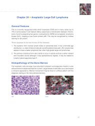
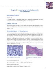
192 | Connection 2010 Figure 9. Hematoxyl<strong>in</strong> and Eos<strong>in</strong> sta<strong>in</strong><strong>in</strong>g of Histoplasma. Figure 10. AFB sta<strong>in</strong><strong>in</strong>g of Histoplasma.
“ Gomori Methenam<strong>in</strong>e Silver (GMS) and Periodic Acid-Schiff (PAS) are the two most common sta<strong>in</strong>s used to look for fungi <strong>in</strong> tissues and <strong>in</strong> cytology specimens <strong>in</strong> the daily practice of pathology. ” sta<strong>in</strong>s. GMS is preferred for screen<strong>in</strong>g, because it gives better contrast, and sta<strong>in</strong>s even degenerated and nonviable fungi that are sometimes refractory to the other two sta<strong>in</strong>s (Fig. 1, 2). GMS also sta<strong>in</strong>s algae (Prototheca and Chlorella spp.), cyst walls of Pneumocystis jiroveci (Fig. 3, 4), pathogenic free liv<strong>in</strong>g soil amebas, the spore coat of most microsporidian parasites, <strong>in</strong>tracytoplasmic granular <strong>in</strong>clusions of Cytomyeolovirus, Act<strong>in</strong>omyces Israeli and related species, Nocardia spp., most Mycobacterium spp., and nonfilamentous bacteria with polysaccharide capsules such as Klebsiella pneumoniae and Streptococcus pneumoniae. Prolonged sta<strong>in</strong><strong>in</strong>g <strong>in</strong> the silver nitrate solution may be required to adequately demonstrate degenerated fungal elements such as the yeast-like cells of Histoplasma capsulatum var. capsulatum <strong>in</strong> granulomas. The disadvantage of GMS and GF fungal sta<strong>in</strong>s is that they mask the natural color of pigmented fungi, mak<strong>in</strong>g it impossible to determ<strong>in</strong>e whether a fungus is colorless hyal<strong>in</strong>e or dematiaceous (pigmented). Such a determ<strong>in</strong>ation is crucial <strong>in</strong> the histologic diagnosis of mycosis caused by dematiaceous fungi such as phaeohyphomycosis (6). Except for the PAS reaction, fungal sta<strong>in</strong>s GMS and GF do not adequately demonstrate the <strong>in</strong>flammatory response to fungal <strong>in</strong>vasion. To counteract this, a GMS-sta<strong>in</strong>ed section can be countersta<strong>in</strong>ed with H&E for a simultaneous study of the fungus and the host response. The PAS sta<strong>in</strong> performs almost as well as GMS, <strong>in</strong> screen<strong>in</strong>g for fungi. It actually demonstrates fungal morphology better than the silver sta<strong>in</strong>s. PAS can sta<strong>in</strong> degenerated fungi that may not be visible on H&E sta<strong>in</strong>. Calcific bodies that are sometimes found <strong>in</strong> caseat<strong>in</strong>g granulomas are also sta<strong>in</strong>ed with PAS, and can be mistaken for yeast-like fungi. This is especially true when calcific bodies are apposed to give the false impression of budd<strong>in</strong>g yeasts, or when the bodies are lam<strong>in</strong>ated to give the appearance of a capsule or thick cell wall. Best sta<strong>in</strong>s to avoid this mis<strong>in</strong>terpretation are GMS and GF sta<strong>in</strong>s, because the chromic acid used as an oxidizer <strong>in</strong> these sta<strong>in</strong>s dissolves the calcium, leav<strong>in</strong>g the calcific bodies unsta<strong>in</strong>ed. Conversely, there are artifacts that mimic fungi on GMS and GF sta<strong>in</strong>s that are not seen on PAS sta<strong>in</strong>, therefore the use of both silver and PAS can reduce the <strong>in</strong>cidence of false positive results. Narrow-Spectrum <strong>Fungal</strong> <strong>Sta<strong>in</strong>s</strong> The differential diagnosis of fungi may require the use of additional special sta<strong>in</strong>s that sta<strong>in</strong> some fungal organism and not others. These are sometimes referred to as “narrow-spectrum” fungal sta<strong>in</strong>s (7, 8). Some of the sta<strong>in</strong>s <strong>in</strong> this category are muc<strong>in</strong> sta<strong>in</strong>s such as alcian blue and Mayer’s, or Southgate’s mucicarm<strong>in</strong>e, that readily demonstrate the mucoid capsule of Cryptococcus neoformans (Fig. 5, 6). This sta<strong>in</strong><strong>in</strong>g reaction differentiates Cryptococcus from other fungi of similar morphology, such as Coccidiodes, Candida, and Histoplasma. These muc<strong>in</strong> sta<strong>in</strong>s are not specific for C. neoformans; the cell walls of B. dermatitidis and Rh<strong>in</strong>osporidium seeberi are often sta<strong>in</strong>ed to vary<strong>in</strong>g degrees with muc<strong>in</strong> sta<strong>in</strong>s. However, these two fungi are nonencapsulated and morphologically dist<strong>in</strong>ct, and not ord<strong>in</strong>arily mistaken for Cryptococcus. In some cases, poorly encapsulated cryptococci <strong>in</strong> tissue sections may not sta<strong>in</strong> positive with mucicarm<strong>in</strong>e sta<strong>in</strong>. In these cases, s<strong>in</strong>ce the cell wall of C. neoformans conta<strong>in</strong>s silver reduc<strong>in</strong>g substances, possibly melan<strong>in</strong> precursors, it can be sta<strong>in</strong>ed with Fontana-Masson’s silver procedure for melan<strong>in</strong> (9, 10). This sta<strong>in</strong> is especially useful <strong>in</strong> those cases of Cryptococcosis with <strong>in</strong>vasive yeast forms that do not have readily detectable capsules, the so-called dry variants. Such forms could possibly be confused with non-encapsulated yeasts of similar morphology. Fontana-Masson and Lillie’s ferrous iron sta<strong>in</strong>s for melan<strong>in</strong> can also be used to confirm and accentuate the presence of melan<strong>in</strong> or melan<strong>in</strong>-like pigments <strong>in</strong> the cell walls of poorly pigmented agents of phaeohyphomycosis <strong>in</strong> tissue sections (11). PAS may be used as a narrow-spectrum fungus sta<strong>in</strong>. For example, <strong>in</strong> the differential diagnosis of small budd<strong>in</strong>g yeast forms, a weak PAS and a strong GMS sta<strong>in</strong><strong>in</strong>g favors a diagnosis of Histoplasma, s<strong>in</strong>ce Candida, microforms of Blastomyces, and yeast forms of Malassezia show a strong cell wall sta<strong>in</strong><strong>in</strong>g with PAS (Fig. 7, 8). Another narrow-spectrum fungus sta<strong>in</strong> is Ziehl-Neelson (ZN). In one study, 60% of blastomyces and 47% of histoplasma organism showed positive cytoplasmic sta<strong>in</strong><strong>in</strong>g of the yeast-like cells with ZN sta<strong>in</strong> (Fig. 9, 10). � Connection 2010 | 193