Chapter 1 | Introduction to Special Stains - Dako
Chapter 1 | Introduction to Special Stains - Dako
Chapter 1 | Introduction to Special Stains - Dako

Create successful ePaper yourself
Turn your PDF publications into a flip-book with our unique Google optimized e-Paper software.
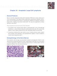
<strong>Chapter</strong> 1 | <strong>Introduction</strong> <strong>to</strong> <strong>Special</strong> <strong>Stains</strong><br />
George L. Kumar, PhD, MBA and Gary W. Gill, CT(ASCP)<br />
special stains are “special” because they are not routine. they are<br />
applied <strong>to</strong> tissue sections in addition <strong>to</strong> hema<strong>to</strong>xylin and eosin<br />
(H&e)-stained sections <strong>to</strong> answer questions that arise above and<br />
beyond those that can be answered by interpreting H&e-stained<br />
tissue morphology (Fig. 1). the term “special stains” is of uncertain<br />
provenance, but one can be certain that it began <strong>to</strong> be used after<br />
1876 when H&e was introduced.<br />
special stains (Fig. 2) can answer these questions:<br />
is a certain class of molecules present or absent?<br />
Where are the molecules located in the preparation?<br />
How many of the molecules are present?<br />
answering the last question requires sophisticated instrumentation<br />
and computation methods and, <strong>to</strong> our knowledge, this aspect of<br />
special stains is neither well-documented nor unders<strong>to</strong>od.<br />
in this article, we will describe some commonly used nonimmunohis<strong>to</strong>chemical<br />
stains. in the first part of the article, we will<br />
compare some key aspects between H&e and special stains, and<br />
certification of special stains by the “Biological stain commission”.<br />
in the second part of the article, we will delve in<strong>to</strong> the technical<br />
details of special stains.<br />
H&E and <strong>Special</strong> <strong>Stains</strong><br />
comparing key aspects of H&e and special stains is instructive.<br />
Classification of <strong>Special</strong> <strong>Stains</strong> by the<br />
Biological Stain Commission<br />
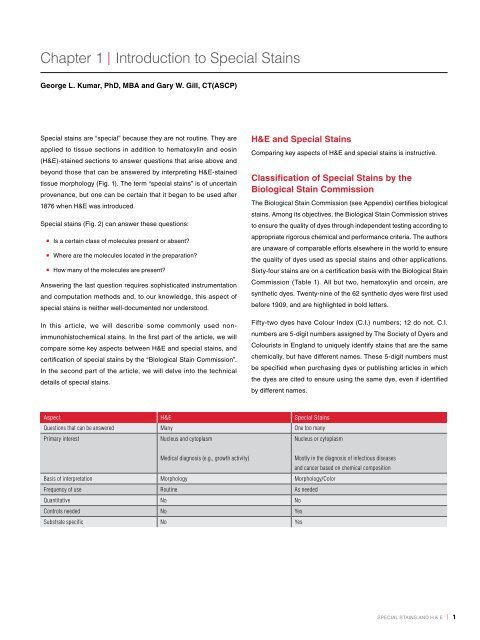
Aspect H&E <strong>Special</strong> <strong>Stains</strong><br />
Questions that can be answered Many One <strong>to</strong>o many<br />
Primary interest Nucleus and cy<strong>to</strong>plasm<br />
Nucleus or cy<strong>to</strong>plasm<br />
Medical diagnosis (e.g., growth activity)<br />
Basis of interpretation Morphology Morphology/Color<br />
Frequency of use Routine As needed<br />
Quantitative No No<br />
Controls needed No Yes<br />
Substrate specific No Yes<br />
the Biological stain commission (see appendix) certifies biological<br />
stains. among its objectives, the Biological stain commission strives<br />
<strong>to</strong> ensure the quality of dyes through independent testing according <strong>to</strong><br />
appropriate rigorous chemical and performance criteria. the authors<br />
are unaware of comparable efforts elsewhere in the world <strong>to</strong> ensure<br />
the quality of dyes used as special stains and other applications.<br />
sixty-four stains are on a certification basis with the Biological stain<br />
commission (table 1). all but two, hema<strong>to</strong>xylin and orcein, are<br />
synthetic dyes. twenty-nine of the 62 synthetic dyes were first used<br />
before 1909, and are highlighted in bold letters.<br />
Fifty-two dyes have colour index (c.i.) numbers; 12 do not. c.i.<br />
numbers are 5-digit numbers assigned by the society of dyers and<br />
colourists in england <strong>to</strong> uniquely identify stains that are the same<br />
chemically, but have different names. these 5-digit numbers must<br />
be specified when purchasing dyes or publishing articles in which<br />
the dyes are cited <strong>to</strong> ensure using the same dye, even if identified<br />
by different names.<br />
Mostly in the diagnosis of infectious diseases<br />
and cancer based on chemical composition<br />
special stains and H & e | 1
<strong>Introduction</strong> <strong>to</strong> <strong>Special</strong> <strong>Stains</strong><br />
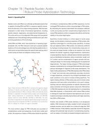
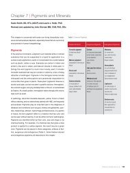
Figure 1. H&E-stained section of skin<br />
with cutaneous blas<strong>to</strong>mycosis. High<br />
magnification (40x) view showing budding<br />
yeast with the inflamma<strong>to</strong>ry infiltrate.<br />
Hema<strong>to</strong>xylin stains the nuclei of cells blue<br />
<strong>to</strong> bluish-purple, and eosin stains other<br />
cellular elements in the tissues from pink<br />
<strong>to</strong> red (Figure courtesy: Sunil Badve, MD,<br />
FRCPath. Indiana University School of<br />
Medicine, Indianapolis, IN, USA).<br />
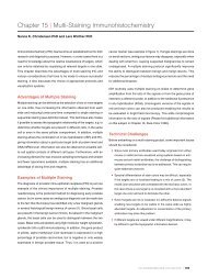
Figure 2. <strong>Special</strong> stained section of small<br />
intestine. The special stain mucicarmine is<br />
used for visualization of neutral epithelial<br />
mucins in small intestine. The mucins are<br />
stained rose <strong>to</strong> red, nuclei are blue/black<br />
(Weigert’s iron hema<strong>to</strong>xylin), and other<br />
tissue elements are yellow (metanil yellow<br />
or tartrazine).<br />
Table 1. 2009 biological stains certified by the Biological Stain Commission.<br />
Acid fuchsin, C.I. 42685<br />
Alcian blue 8 GX, C.I. 74240<br />
Alizarin red S, C.I. 58005<br />
Aniline blue WS, C.I. 42755<br />
Auramine O, C.I. 41000<br />
<strong>Introduction</strong> <strong>to</strong> <strong>Special</strong> <strong>Stains</strong><br />
2 | special stains and H & e special stains and H & e | 3<br />
Azocarmine B<br />
Azocarmine G, C.I. 50085<br />
Azure A, C.I. 52005<br />
Azure B, C.I. 52010<br />
Azure C, C.I. 52002<br />
Basic fuchsine, C.I. 42510<br />
Bismarck brown Y, C.I. 21000<br />
Brilliant cresyl blue, C.I. 51010<br />
Brilliant green, C.I. 42040<br />
Carmine, C.I. 75470<br />
Chlorazol black E, C.I. 30235<br />
Congo red, C.I. 22120<br />
Cresyl violet<br />
Crystal violet, C.I. 42555<br />
Darrow red<br />
Eosin B, C.I. 45400<br />
Eosin Y, C.I. 45380<br />
Erythrosin, C.I. 45430<br />
Ethyl eosin, C.I. 45386<br />
Ethyl green, C.I. 42590<br />
Fast green F C F, C.I. 42053<br />
Fluorescein Isothiocyanate<br />
Giemsa Stain 1902,<br />
modified in 1904.<br />
Hema<strong>to</strong>xylin, C.I. 75290<br />
(Bohmer 1865)<br />
Indigo carmine, C.I. 73015<br />
Janus green B, C.I. 11050<br />
Jenner stain 1899<br />
Light green SF, C.I. 42095<br />
Malachite green, C.I. 42000<br />
Martius yellow, C.I. 10315<br />
Methyl orange, C.I. 13025<br />
Methyl violet 2B, C.I. 42535<br />
Methylene blue<br />
Methylene blue, C.I. 52015<br />
Methylene violet<br />
(Bernthsen), C.I. 52041<br />
Neutral red, C.I. 50040<br />
Nigrosin, C.I. 50420<br />
Nile blue A, C.I. 51180<br />
Nuclear fast red, C.I. 60760<br />
Oil Red O, C.I. 26125<br />
Orange G, C.I. 16230<br />
Orange II, C.I. 15510<br />
Orcein<br />
Pararosaniline, C.I. 42500<br />
Phloxin B, C.I. 45410<br />
Protargol S<br />
Pyronine B, C.I. 45010<br />
Pyronine Y, C.I. 45005<br />
Resazurin<br />
Rose Bengal, C.I. 45435<br />
Safranine O, C.I. 50240<br />
Sudan black B, C.I. 26150<br />
Sudan III, C.I. 26100<br />
Sudan IV, C.I. 26105<br />
Tetrachrome stain (MacNeal)<br />
Thionine, C.I. 52000<br />
Toluidine blue, C.I. 52040<br />
Weigert 1878<br />
Wright stain (1908)
<strong>Introduction</strong> <strong>to</strong> <strong>Special</strong> <strong>Stains</strong> <strong>Introduction</strong> <strong>to</strong> <strong>Special</strong> <strong>Stains</strong><br />
<strong>Special</strong> <strong>Stains</strong><br />
special stain is a term that is mostly used in a labora<strong>to</strong>ry setting.<br />
special stains have two broad areas of application: research and<br />
diagnostic. in research, special stains are used as probes <strong>to</strong> identify<br />
certain chemical constituents in normal and abnormal cells. the<br />
information so obtained is used as a basis for further study and<br />
also as a baseline against which the results of special staining can<br />
be compared in diagnostic applications. On the basis of such a<br />
comparison, the significance of the findings can be interpreted.<br />
special stains can be applied <strong>to</strong> cell biology and his<strong>to</strong>logy. some<br />
useful applications are: (1) the determination of dna and Rna<br />
content, (2) the mode of action of drugs, hormones or of potentially<br />
<strong>to</strong>xic food additives, (3) metabolic biochemistry, (4) biochemistry of<br />
disease processes, (5) primary sites of many metastatic tumors,<br />
(6) identification of non-pigmented metastatic melanomas,<br />
(7) detection of early invading tumors, (8) definition of the margins<br />
of surgically resected tumors, (9) identification of Barr bodies,<br />
(10) staining cells in ways that can be used as a basis for cell<br />
separation by appropriate instrumentation (e.g., fluorescence), and<br />
(11) identification of micro-organisms (e.g., Cryp<strong>to</strong>coccus neoformans,<br />
Helicobacter pylori). see table 2.<br />
the material, methods and interpretation of these special stains can<br />
be found in references 5-7. When working with special stains, keep<br />
in mind the following considerations:<br />
special staining often requires the use of unusual stains and<br />
reagents that are available from only a few sources. Knowledge<br />
of such sources is essential <strong>to</strong> overcome technical bottlenecks.<br />
Be aware of special stains that contain colored and colorless<br />
impurities (e.g., salts) as these substances may interfere with<br />
the staining.<br />
special staining requires broad knowledge of the tissue or<br />
cells targeted.<br />
When working with special stains, care should be taken <strong>to</strong> collect,<br />
fix and prepare the specimen in a manner that will maintain the<br />
molecule of interest within cells or tissues. For example, it is<br />
important <strong>to</strong> work with frozen sections when attempting <strong>to</strong> identify<br />
enzymes and <strong>to</strong> avoid fat solvents such as alcohol and xylene<br />
when attempting <strong>to</strong> identify lipids.<br />
With cell suspensions, it is essential <strong>to</strong> determine by microscopy<br />
whether cells are present, and how many cells are <strong>to</strong> be used when<br />
making the slides. Using this quality control step will improve the<br />
cellular preparations.<br />
control preparations must be run in parallel with experimental<br />
preparations for one or more of the following reasons: (1) <strong>to</strong><br />
determine if the special stain is working, (2) <strong>to</strong> assess the degree<br />
of non-specific staining, (3) <strong>to</strong> determine whether a reagent is still<br />
active, and (4) <strong>to</strong> serve as a standard in fractional reduction of<br />
staining procedures. if a positive reaction is noted when a control<br />
is not used, it can still be determined that the reaction is at least<br />
working (how well or how specifically is open <strong>to</strong> speculation).<br />
However, a negative reaction in the absence of a control may<br />
indicate either that the sought constituent is not present or the<br />
reaction is not working.<br />
control slides should be: (1) sections of tissue/cell high in a<br />
particular molecule/constituent, (2) purified samples of a particular<br />
molecule in smears, (3) samples of the same specimen pretreated<br />
with solvents or enzymes <strong>to</strong> remove the sought constituent,<br />
(4) samples of the same specimen with the omittance of essential<br />
reagents or steps in the staining procedure, or (5) run as a<br />
duplicate cell spread in the same manner as the experiment minus<br />
one essential step.<br />
the amount of special stains within a cell or tissue represents<br />
the difference between the amount taken up during staining and<br />
the amount removed by the rinses following staining. <strong>to</strong> ensure<br />
the optimal amount, the user must employ those materials and<br />
methods that promote stain uptake during and following staining<br />
(e.g., dye concentration, suitable solvent, control of favorable pH,<br />
addition of salts, if necessary, control of ionic concentration, if<br />
necessary, time and temperature).<br />
<strong>to</strong> maintain the right amount and hue of the special stain, mount<br />
the stained specimen in a medium that does not promote bleaching<br />
or leaching.<br />
<strong>to</strong> ensure optimal image quality of the stained specimen, use<br />
the right amount of mounting medium, cover with a no. 1 cover<br />
glass, and use a clean microscope with the illumination adjusted<br />
according <strong>to</strong> the method of Köhler (see appendix, page 283).<br />
Manual vs. Au<strong>to</strong>mated <strong>Special</strong> Staining<br />
Pro<strong>to</strong>cols<br />
depending on the financial situation of the labora<strong>to</strong>ry, specimen<br />
sample size, and the number of personnel available, special stain<br />
pro<strong>to</strong>cols are performed either manually or by using au<strong>to</strong>mated<br />
systems. Manual staining of slides work well in a research setting,<br />
especially, when the number of processed slides are few per day.<br />
However, with increasing numbers of slides <strong>to</strong> be stained, the manual<br />
method becomes prone <strong>to</strong> error resulting in decreased flexibility<br />
Table 2. Commonly used special stains.<br />
and productivity. With the medical community demanding faster<br />
turnaround times, increased flexibility and productivity as well as<br />
greater standardization, au<strong>to</strong>mated instruments have replaced<br />
some manual methods of staining thus becoming an integral part<br />
of the labora<strong>to</strong>ry. au<strong>to</strong>mation combined with specialized software<br />
applications and connectivity have made many instruments capable<br />
of multiprogramming runs resulting in standardized pro<strong>to</strong>cols,<br />
manageable work schedules, enhanced workflow, cost-effectiveness<br />
and the ability <strong>to</strong> adapt <strong>to</strong> regula<strong>to</strong>ry requirements.<br />
<strong>Special</strong> Stain Clinical Application Staining Specificity<br />
For detecting micro-organisms and Helicobacter pylori<br />
Acid-Fast Bacteria (AFB)<br />
(Ziehl-Neelsen Stain)<br />
Detects nocardioform-actinomycete groups of bacteria,<br />
including Mycobacterium Spp (acid fast), Rhodococcus equi<br />
and Nocardia Spp (weakly acid fast) Fig. 3<br />
Alcian Yellow / Toluidine Blue (Leung) Stain Used for the detection of H. pylori<br />
See Fig. 4 for an electron micrograph and illustration<br />
of H. pylori<br />
Dieterle’s Stain Identifies Borrelia burgdorferi, Legionella pneumophila,<br />
Treponema pallidum<br />
Diff-Quik Stain (Diff-Quik is the formerly trademarked<br />
name for a proprietary rapid Giemsa-like stain)<br />
Detects H. pylori and some fungi<br />
(e.g., Pneumocystis jiroveci)<br />
Giemsa Stain Used for staining H. pylori, Plasmodium vivax, Rickettsia<br />
prowazekii, Rickettsia rickettsii, Rickettsia tsutsugamushi,<br />
Trypanosoma cruzi, Giardia lamblia; Fig. 5a, b and c<br />
Gram Stain (Named after its inven<strong>to</strong>r, the Danish scientist<br />
Hans Christian Gram, who developed the technique in<br />
1884 <strong>to</strong> discriminate between two types of bacteria with<br />
similar clinical symp<strong>to</strong>ms)<br />
Used for the detection of Gram-positive (Clostridium<br />
botulinum, Clostridium tetani, Staphylococcus aureus and<br />
Corynebacterium diphtheriae) or Gram-negative bacteria<br />
(Salmonella, Shigella dysenteriae, Escherichia coli and<br />
Pseudomonas aeruginosa). Also used for the detection<br />
of Actinomyces Israeli, Legionella pneumophila, Neisseria<br />
gonorrhea, Neisseria meningitidis, Nocardia asteroides<br />
Acid-fast bacilli retain a cationic dye that is extracted<br />
from all other types of bacteria and animal cells by<br />
acidified alcohol. The waxy wall (with mycolic acid)<br />
of mycobacteria retains the dye<br />
The yellow dye stains oxidized and sulphonated gastric<br />
mucus providing contrast for suspended Helicobacter<br />
organisms that are stained with <strong>to</strong>luidine blue<br />
<strong>Stains</strong> whole organisms<br />
H. pylori and Pneumocystis jiroveci<br />
<strong>Stains</strong> polyanions blue and polycations pink<br />
Bacteria show up blue on account of their nucleic acids.<br />
Acidic capsules (e.g., Anthrax Bacilli, Cryp<strong>to</strong>coccus)<br />
would be expected <strong>to</strong> be blue or purple<br />
<strong>Stains</strong> whole organisms<br />
4 | special stains and H & e special stains and H & e | 5
<strong>Introduction</strong> <strong>to</strong> <strong>Special</strong> <strong>Stains</strong> <strong>Introduction</strong> <strong>to</strong> <strong>Special</strong> <strong>Stains</strong><br />
Table 2. Commonly used special stains.<br />
<strong>Special</strong> Stain Clinical Application Staining Specificity<br />
Grocott’s Methenamine Silver (GMS) Stain Useful in identifying a variety of pathogenic fungi, including<br />
Aspergillus fumigatus, Blas<strong>to</strong>myces dermatitidis, Candida<br />
albicans, Coccidioides immitis, Cryp<strong>to</strong>coccus neoformans,<br />
His<strong>to</strong>plasma capsulatum, Nocardia asteroids, Pneumocystis<br />
carinii, Pneumocystis Jiroveci (human) and Sporothrix<br />
schenckii; Fig. 6-8<br />
Mayer’s Mucicarmine Stain Detects encapsulated yeast-like fungus Cryp<strong>to</strong>coccus<br />
neofarmans<br />
Periodic Acid-Schiff (PAS) Stain Used for the identification of Aspergillus fumigatus,<br />
Blas<strong>to</strong>myces dermatitidis, Candida albicans, Coccidioides<br />
immitis, Cryp<strong>to</strong>coccus neofarmans, Sporothrix schenckii<br />
Sayeed’s Stain (Schiff’s reagent, 0.5% periodic acid,<br />
Mayer’s hemalum)<br />
Detects H. pylori H. pylori<br />
Steiner & Steiner Staining Method Detects spirochetes and legionella, and pneumophila<br />
bacteria, e.g., Borrelia burgdorferi, H. pylori, Legionella<br />
pneumophila, Treponema pallidum; Fig. 9<br />
Warthin-Starry Stain (these are reduced silver<br />
methods)<br />
For demonstrating connective tissue, muscle, collagen, lipid and fibrin<br />
Identifies Alipia feles, Bar<strong>to</strong>nella henselae, Borrelia burgdorferi,<br />
H. pylori, Legionella pneumophila, Treponema pallidum; Fig. 10<br />
Gomori’s One-Step Trichrome Stain Used for distinguishing collagen and smooth muscle fibers;<br />
Fig.11<br />
Jones’ Basement Membrane<br />
Periodic Schiff-Methenamine Silver (PASM) Stain<br />
Used for the identification of basement membranes (of the<br />
glomerulus in the kidney or in tissue samples); Fig. 12<br />
Masson’s Trichrome Stain (TRI) Used for distinguishing cells from surrounding connective<br />
tissue which has several variants and is probably the<br />
trichrome most used in his<strong>to</strong>pathology. Black nuclei, red<br />
cy<strong>to</strong>plasm (including muscle), blue or green collagen<br />
(including fine fibers), cartilage and mucus; Fig. 13<br />
Polysaccharide components of the fungal cell wall<br />
Polysaccharides on the capsule<br />
Polysaccharide components of the fungal cell wall<br />
<strong>Stains</strong> whole organisms<br />
<strong>Stains</strong> whole organisms<br />
Collagen and smooth muscle fibers<br />
Basement membranes<br />
Muscle, collagen fibers, fibrin and erythrocytes<br />
<strong>Special</strong> Stain Clinical Application Staining Specificity<br />
Russel-Movat Pentachrome Stain Used for simultaneous demonstration of muscle, elastic<br />
fibers, collagen/reticular fibers, ground substance and<br />
fibrinoid in tissues<br />
Oil Red O and Sudan Black B <strong>Stains</strong> Used for staining lipids in frozen sections and some<br />
lipoproteins on paraffin sections<br />
Orcein Stain Used for staining elastic fibers Elastic fibers<br />
Lendrum’s Method (Picro-Mallory Stain) Fibrin Fibrin<br />
Phosphotungstic Acid-Hema<strong>to</strong>xylin (PTAH) Stain Used for demonstrating striated muscle fibers<br />
Also used <strong>to</strong> stain abnormal neuroglia (reactive astrocy<strong>to</strong>sis)<br />
Silver methods for reticulum and<br />
basement membranes<br />
(e.g., Reticulin/ Nuclear Fast Red Stain)<br />
Used for the identification of reticulin fibers in tissue<br />
samples; Fig.14<br />
Muscle, elastic fibers, collagen/reticular fibers<br />
Lipids, including triglycerides (which necessarily are<br />
neutral). Oil Red O stains only the most hydrophobic<br />
lipids (triglycerides and cholesterol esters). Sudan<br />
Black B stains these and also phospholipids and<br />
sphingomyelins, which are less hydrophobic<br />
Muscle fibers, collagen<br />
Reticulin (collagen with high level of hexosylation,<br />
including Type IV)<br />
6 | special stains and H & e special stains and H & e | 7<br />
Verhoeff Stain<br />
Van Gieson Stain<br />
For detecting nucleic acids<br />
Used for the identification of elastic laminae and fibers<br />
in tissues; Fig.15<br />
The Verhoeven Stain is specific for elastic fibers.<br />
The Van Gieson Stain is specific for collagen.<br />
Verhoeff’s iron-hema<strong>to</strong>xylin stains elastin and<br />
nuclei black. Van Gieson’s picro-fuchsine gives<br />
yellow cy<strong>to</strong>plasm and red collagen fibers<br />
Ethyl Green-Pyronine Stain Used for differential demonstration of DNA and RNA A buffered mixture of the two dyes gives blue-green<br />
DNA and red RNA (rRNA in cy<strong>to</strong>plasm, nucleoli)<br />
Feulgen Stain Used for the identification of chromosomal material or<br />
deoxyribonucleic acid (DNA in paraffin-embedded tissue<br />
or cell specimens); Fig.16<br />
Deoxyribonucleic acid (DNA)
<strong>Introduction</strong> <strong>to</strong> <strong>Special</strong> <strong>Stains</strong> <strong>Introduction</strong> <strong>to</strong> <strong>Special</strong> <strong>Stains</strong><br />
Table 2. Commonly used special stains.<br />
<strong>Special</strong> Stain Clinical Application Staining Specificity<br />
Neuropathology<br />
Bielschowsky Silver Stain Used for diagnosing Alzheimer’s Disease <strong>to</strong> show neuritic<br />
components of plaques and tangles<br />
Neurofilament protein. Normal axons are also stained<br />
Congo Red Used for the detection of amyloidal plaques in brain; Fig. 17 Extracellular amyloidal deposits<br />
Cresyl Violet Stain Useful in identifying cell bodies of neurons in tissue<br />
sections; Fig. 18<br />
Phosphotungstic Acid-Hema<strong>to</strong>xylin<br />
(PTAH) Stain<br />
For demonstrating myelin<br />
Nissl substance in neurons. The Cresyl Violet Stain<br />
shows cell bodies of neurons by virtue of their abundant<br />
rough ER and ribosomes (rRNA)<br />
Used <strong>to</strong> stain abnormal neuroglia (reactive astrocy<strong>to</strong>sis) Abnormal neuroglia (reactive astrocy<strong>to</strong>sis)<br />
Luxol Fast Blue (MBS) Stain Used for demonstrating myelin; Fig. 18 and 19 Myelin<br />
Page’s Eriochrome Cyanine R Used for demonstrating myelin Myelin<br />
Derma<strong>to</strong>pathology, hema<strong>to</strong>logy, pigment detection, minerals and bone<br />
Alizarin Red S Stain Calcium detection in tissues Complexes with calcium<br />
Chloroacetate Esterase (Leder) Stain Useful as a marker of neutrophils His<strong>to</strong>chemical detection of an enzyme of neutrophil<br />
leukocytes<br />
Hall’s Stain Used for the detection of bile pigment Bilirubin<br />
Masson-Fontana Stain Used for the detection of melanin and some<br />
neuroendocrine cells<br />
Perls’ Prussian Blue Stain Demonstrates hemosiderin in bone marrow macrophages<br />
and within erythroblasts<br />
Sero<strong>to</strong>nin, melanin and other silver-reducing<br />
(argentaffin) substances<br />
Hemosiderin (iron s<strong>to</strong>rage complex)<br />
p-dimethylaminobenzylidenerhodanine Stain Used for the detection of copper in tissues Copper or copper-associated protein<br />
Villanueva Osteochrome Bone Stain Gives uniform and reproducible results for mineralized<br />
or undecalcified bone<br />
Mineralized or undecalcified bone<br />
<strong>Special</strong> Stain Clinical Application Staining Specificity<br />
Miscellaneous and multipurpose stains<br />
Alcian Blue Used in identifying mucins and glycosaminoglycans.<br />
At pH 2.5, Alcian Blue stains sulphated and nonsulphated<br />
acidic carbohydrates. At pH 1.0, only sulphated<br />
carbohydrates are stained; Fig. 20 and 21<br />
Giemsa Stain Used in hema<strong>to</strong>logy, e.g., for the detection of<br />
erythroidcolonies, binucleate normoblast, megaloblasts,<br />
mast cells, etc. Giemsa is also used for chromosome<br />
staining; Fig. 22a, 22b and 23<br />
8 | special stains and H & e special stains and H & e | 9<br />
Mucins<br />
Gomori’s Silver Stain Used for the detection of reticulin in bone marrow Reticulin<br />
Mucicarmine Stain Detects mucins; Fig.1 Mucins<br />
Periodic Acid-Schiff (PAS) Stain Used for staining structures containing a high proportion of<br />
carbohydrate macromolecules (glycogen and glycoprotein),<br />
basement membranes, collagen and primary cell types;<br />
Fig. 24 and 25<br />
Specific for phosphate groups of DNA<br />
Carbohydrate macromolecules by virtue of their content<br />
of galac<strong>to</strong>se, glucose, fucose and mannose<br />
Periodic Acid-Silver Methenamine (PEM) Stain Used for the delineation of basement membranes Carbohydrate macromolecules by virtue of their content<br />
of galac<strong>to</strong>se, glucose, fucose and mannose
<strong>Introduction</strong> <strong>to</strong> <strong>Special</strong> <strong>Stains</strong> <strong>Introduction</strong> <strong>to</strong> <strong>Special</strong> <strong>Stains</strong><br />
Figure 3. Lung stained with Acid-Fast<br />
Bacteria (AFB) Stain, <strong>Dako</strong> Code AR162.<br />
This AFB stain is suitable for the visualization<br />
of acid-fast bacteria belonging<br />
<strong>to</strong> the Mycobacterium genus on the<br />
Artisan Staining System. Application of<br />
carbol-fuchsin stains acid-fast bacteria<br />
fuchsia, followed by decolorization of<br />
all tissue elements except the acid-fast<br />
bacteria. A methylene blue counterstain is<br />
then applied <strong>to</strong> impart a blue color <strong>to</strong> all<br />
background tissue elements.<br />
Figure 4a. Electron micrograph (EM)<br />
(negative staining) of H. pylori possessing<br />
multiple flagella. Courtesy of Wikimedia.<br />
Prof. Yutaka Tsutsumi, MD, Department of<br />
Pathology, Fujita Health University School<br />
of Medicine, Japan.<br />
Sheathed polar flagella<br />
Body of the Bacteria<br />
Figure 4b. Illustration of S-shaped H. pylori with four sheathed polar flagella. The majority of helicobacters possess this basic morphology of an S-shape with polar,<br />
sheathed flagella, though variations in size and the number of spirals are seen in a number of other species. These bacteria are usually around 0.5 × 5 μm, and the<br />
S-shaped morphology has been correlated with maximum in vitro motility. Thin sections of H. pylori revealed through an electron microscope show an outer and inner<br />
membrane separated by the periplasm of approximately 30 nm thickness (see EM picture above). The dense cy<strong>to</strong>plasm contains nucleoid material and ribosomes<br />
(Source: Jani O’Rourke and Günter Bode. Morphology and Ultrastructure of Helicobacter pylori. Physiology and Genetics. Eds. Harry L. T. Mobley, George L. Mendz, and<br />
Stuart L. Hazell. ASM Press. 2001). Illustration by Rashmil Saxena, BFA, HT(ASCP) CM .<br />
10 | special stains and H & e special stains and H & e | 11
<strong>Introduction</strong> <strong>to</strong> <strong>Special</strong> <strong>Stains</strong> <strong>Introduction</strong> <strong>to</strong> <strong>Special</strong> <strong>Stains</strong><br />
Lactael<br />
Lympoid<br />
nodule<br />
Vein<br />
Muscularis<br />
mucosa<br />
Vein<br />
Circular muscule<br />
of muscularis<br />
externa<br />
Longitudinal<br />
muscule of<br />
muscularis<br />
externa<br />
Villus<br />
Surface<br />
epithelium<br />
Crypt of<br />
Lieberkühn<br />
Lamina<br />
propria<br />
Artery<br />
Muscularis<br />
mucosa<br />
Figure 5a. Schematic diagram of intestinal<br />
wall. Illustration by Rashmil Saxena, BFA,<br />
HT(ASCP) CM .<br />
Figure 5b. Giemsa-stained section of<br />
small intestinal mucosa showing clusters<br />
of Giardia that stain purple (arrows) in<br />
the crypts. The background is stained<br />
faint pink by eosin. (Courtesy of Rashmil<br />
Saxena, BFA, HT(ASCP) CM , FRCPath,<br />
Indiana University School of Medicine,<br />
Indianapolis, IN, USA).<br />
Figure 5c. Giardia intestinalis trophozoite:<br />
After ingestion of contaminated food<br />
or water within the small intestine, the<br />
trophozoites reproduce asexually and<br />
either float free or are attached <strong>to</strong> the<br />
mucosa of the lumen. Some trophozoites<br />
then encyst in the small intestine.<br />
Encystation occurs most likely as a result<br />
of exposure <strong>to</strong> bile salts and fatty acids,<br />
and a more alkaline environment. Both<br />
cysts and trophozoites are then passed<br />
in the feces, and are infectious immediately<br />
or shortly afterward (Legend<br />
courtesy: Centers for Disease Control<br />
(CDC, Atlanta, GA, USA). Illustration by<br />
Rashmil Saxena, BFA, HT(ASCP) CM .<br />
12 | special stains and H & e special stains and H & e | 13<br />
Axostyle<br />
Nucleus<br />
Median Body<br />
Flagella<br />
Figure 6. Biopsy stained with GMS, <strong>Dako</strong><br />
Code AR176. The Grocott’s Methenamine<br />
Silver method is utilized for the<br />
visualization of fungi and Pneumocystis<br />
jiroveci in tissue sections using the<br />
Artisan Staining System. Fungi and<br />
P. jiroveci are stained black while other<br />
tissue elements are stained green. This<br />
stain can be used on both tissue and<br />
aspirates or smears.
<strong>Introduction</strong> <strong>to</strong> <strong>Special</strong> <strong>Stains</strong> <strong>Introduction</strong> <strong>to</strong> <strong>Special</strong> <strong>Stains</strong><br />
Figure 7. Methenamine Silver Stain.<br />
His<strong>to</strong>pathologic changes seen in his<strong>to</strong>plasmosis<br />
due <strong>to</strong> His<strong>to</strong>plasma capsulatum<br />
var. duboisii. Note the presence of typical<br />
yeast cells, some of which are undergoing<br />
replication by “budding”. Courtesy of Libero<br />
Ajello, PhD, The Centers for Disease Control<br />
and Prevention, Atlanta, GA, USA/Wikimedia.<br />
Figure 8. Grocott’s Methenamine Silver<br />
(GMS) staining of fungi.<br />
Figure 9. His<strong>to</strong>pathology of Treponema<br />
pallidum spirochetes using a modified<br />
Steiner Silver Stain. Image credit: Dr.<br />
Edwin P. Ewing, Jr., The Centers for<br />
Disease Control and Prevention, Atlanta,<br />
GA, USA/Wikimedia.<br />
Figure 10. Helicobacter stained with<br />
Warthin-Starry, <strong>Dako</strong> Code AR181. The<br />
arrow points <strong>to</strong> some black H. pylori<br />
organisms in yellow mucus.<br />
14 | special stains and H & e special stains and H & e | 15
<strong>Introduction</strong> <strong>to</strong> <strong>Special</strong> <strong>Stains</strong> <strong>Introduction</strong> <strong>to</strong> <strong>Special</strong> <strong>Stains</strong><br />
Figure 11. Liver section stained with<br />
a modification of Gomori’s One-Step<br />
Trichrome method that colors collagen<br />
green rather than blue, <strong>Dako</strong> Code AR166.<br />
Figure 12. Kidney stained with Jones’<br />
Basement Membrane, <strong>Dako</strong> Code AR180.<br />
The Jones’ Basement Membrane stain<br />
is used for visualization of basement<br />
membranes, specifically glomerular and<br />
tubular basement membranes in renal<br />
tissue. The Bowman’s capsule is stained<br />
black, inner basement membrane - black<br />
<strong>to</strong> gray, nuclei - red, collagen - rose, and<br />
cy<strong>to</strong>plasm and other tissue are stained<br />
pink. This stain has been optimized for use<br />
on 2 µm thick tissue sections.<br />
Figure 13. Biopsy stained with Masson’s<br />
Trichrome, <strong>Dako</strong> Code AR173. This stain<br />
is used <strong>to</strong> distinguish collagen from<br />
muscle in tissue specimens using the<br />
Artisan Staining System. The Trichrome<br />
stain is often used <strong>to</strong> differentiate<br />
between collagen and smooth muscle<br />
and <strong>to</strong> identify an increase in collagenous<br />
tissue. With the Masson’s Trichrome stain,<br />
muscle is stained red, collagen - blue,<br />
fibrin - pink, erythrocyte - red and nuclei<br />
- blue/black.<br />
Figure 14. Liver stained with Reticulin/<br />
No Counterstain, <strong>Dako</strong> Code AR182. The<br />
Reticulin/No Counterstain stain is used<br />
for the visualization of reticulin fibers<br />
in tissue sections using the Artisan<br />
Staining System.<br />
16 | special stains and H & e special stains and H & e | 17
<strong>Introduction</strong> <strong>to</strong> <strong>Special</strong> <strong>Stains</strong> <strong>Introduction</strong> <strong>to</strong> <strong>Special</strong> <strong>Stains</strong><br />
Meissner’s<br />
corpuscle<br />
Oil gland<br />
Sweat gland<br />
Epidermis<br />
Dermis<br />
Hypodermis<br />
Hair shaft<br />
Arrec<strong>to</strong>r pili<br />
Hair follicle<br />
Hair root<br />
Pacinian<br />
corpuscle<br />
Vein<br />
Artery<br />
Figure 15a. Skin stained with Elastic<br />
stain, <strong>Dako</strong> Code AR163. In this section<br />
Verhoeff’s hema<strong>to</strong>xylin method has been<br />
counterstained with Van Gieson’s picrofuchsine.<br />
The Elastic stain is based on<br />
Verhoeff’s technique optimized for the<br />
Artisan Staining System. Elastin fibers<br />
and elastic lamina in his<strong>to</strong>logical specimens<br />
are stained black, while remaining<br />
tissue elements are stained as follows:<br />
nuclei - blue/black, collagen - red, other<br />
tissue elements - yellow.<br />
Figure 15b. Schematic diagram of skin:<br />
Cross-section. Dermis contains collagen<br />
and elastin which give the skin its form,<br />
shape and elasticity. Illustration by<br />
Rashmil Saxena, BFA, HT(ASCP) CM .<br />
Figure 16. Breast tissue stained with<br />
Feulgen, <strong>Dako</strong> Code AR174. The Feulgen<br />
stain is used <strong>to</strong> demonstrate DNA in<br />
tissue sections. RNA is not stained<br />
by this procedure. The DNA is stained<br />
magenta with Schiff’s reagent. The stained<br />
DNA is contrasted against a light green<br />
counterstain <strong>to</strong> allow better visualization<br />
by light microscopy or image analysis.<br />
Figure 17. Amyloid stained with Congo<br />
Red, <strong>Dako</strong> Code AR161. The Congo<br />
Red stain is used <strong>to</strong> detect amyloid, an<br />
abnormal protein product that can be<br />
found in various pathologic conditions.<br />
This stain is based on Benhold’s and<br />
demonstrates amyloid in pink <strong>to</strong> dark<br />
salmon with light microscopy or the<br />
characteristic “apple-green birefringence’’<br />
with polarized light. Mayer’s hema<strong>to</strong>xylin<br />
is used as a counterstain. The preferred<br />
method for visualization of amyloid is<br />
under polarized light.<br />
18 | special stains and H & e special stains and H & e | 19
<strong>Introduction</strong> <strong>to</strong> <strong>Special</strong> <strong>Stains</strong> <strong>Introduction</strong> <strong>to</strong> <strong>Special</strong> <strong>Stains</strong><br />
Nucleolus<br />
Cell Body<br />
Nissl body<br />
(or Nissl granule)<br />
Nucleus<br />
Axon Hillock<br />
Dendrites<br />
Internode<br />
Synaptic<br />
terminal<br />
Preterminal<br />
branch<br />
Myelin Sheath<br />
Node of Ranvier<br />
Figure 18. Schematic diagram of a generalized neuron with a myelinated axon. The arrow indicates the direction in which signals are conveyed. Axons conduct signals<br />
away from the cell body, while dendrites receive signals from the axons of other neurons. Around the cell body are dendrites that receive signals from other neurons.<br />
The end of the axon has branching synaptic terminals that release neurotransmitters in<strong>to</strong> a gap called the synaptic cleft (not shown) between the terminals and the<br />
dendrites of the next neuron.<br />
The axons of vertebrate neurons are insulated by a myelin sheath which greatly increases the rate at which axons can conduct a nerve impulse.<br />
The myelin sheath is interrupted at regularly spaced “Nodes of Ranvier” where Na + channels in an axon are concentrated. A myelin sheath is a manylayered<br />
coating, largely composed of a fatty substance called myelin that wraps around the axon and very efficiently insulates it. Nissl bodies or granules<br />
are clumps of free ribosomes attached <strong>to</strong> portions of rough endoplasmic reticulum. These are sites for protein synthesis. Illustration by Rashmil Saxena,<br />
BFA, HT(ASCP) CM .<br />
Axon<br />
20 | special stains and H & e special stains and H & e | 21<br />
500 nm<br />
Schwann cell<br />
nucleus<br />
Mi<strong>to</strong>chondria<br />
inside axon<br />
Myelin sheath<br />
Axon surrounded<br />
by myelin sheath<br />
Figure 19. Transmission electron<br />
micrograph of a myelinated axon. Each<br />
Schwann cell wraps its plasma membrane<br />
concentrically around the axon <strong>to</strong> form<br />
a segment of myelin sheath. Generated<br />
at the Electron Microscopy Facility at<br />
Trinity College, Hartford, CT. (Courtesy of<br />
Wikipedia).<br />
Figure 20. Small intestine stained with<br />
Alcian Blue pH 2.5, <strong>Dako</strong> Code AR160.<br />
Alcian Blue pH 2.5 stains weakly sulphated<br />
mucins, acidic mucopolysaccharides,<br />
sulphomucins, hyaluronic acid and<br />
sialomucins at pH 2.5, blue in color. All<br />
nuclei are stained red, and all other tissue<br />
elements are stained pink <strong>to</strong> red.
<strong>Introduction</strong> <strong>to</strong> <strong>Special</strong> <strong>Stains</strong> <strong>Introduction</strong> <strong>to</strong> <strong>Special</strong> <strong>Stains</strong><br />
Figure 21. Small intestine stained with Alcian Blue/PAS, <strong>Dako</strong> Code AR169. This stain is used for the demonstration of neutral and acidic mucosubstances on the<br />
Artisan Staining System. Alcian Blue pH 2.5 imparts a blue color <strong>to</strong> the acidic mucins and other carboxylated or weakly sulphated acid mucosubstances. The<br />
periodic acid-Schiff (PAS) reaction is then used <strong>to</strong> stain basement membranes, glycogen and neutral mucosubstances pink <strong>to</strong> red. Mixtures of neutral and acidic<br />
mucosubstances will appear purple due <strong>to</strong> positive reactions with both Alcian Blue and PAS.<br />
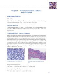
Figure 22a. Cell types seen in normal bone<br />
marrow. Giemsa staining. (Figure from<br />
<strong>Dako</strong> Education Guide, “The Illustrated<br />
Guide <strong>to</strong> Bone Marrow Diagnosis,” 2nd<br />
Edition (2009). Edi<strong>to</strong>rs: Carlos Martin, MD,<br />
and George L. Kumar, PhD).<br />
Figure 22b. Giemsa staining. Atypical<br />
mononuclear megakaryocyte in chronic<br />
myeloid leukemia. (Figure from <strong>Dako</strong><br />
Education Guide, “The Illustrated Guide<br />
<strong>to</strong> Bone Marrow Diagnosis,” 2nd Edition<br />
(2009). Edi<strong>to</strong>rs: Carlos Martin, MD, and<br />
George L. Kumar, PhD).<br />
22 | special stains and H & e special stains and H & e | 23<br />
Erythroblast<br />
Eosinophil<br />
Myelocyte<br />
Neutrophil<br />
Normoblast<br />
Plasma cell<br />
Megakaryocyte
<strong>Introduction</strong> <strong>to</strong> <strong>Special</strong> <strong>Stains</strong> <strong>Introduction</strong> <strong>to</strong> <strong>Special</strong> <strong>Stains</strong><br />
Figure 23. Spleen stained with Giemsa, <strong>Dako</strong> Code AR164. Cell types are stained as follows: mast-cell granules and basophils - purple, eosinophils - bright pink,<br />
lymphocytes - blue.<br />
Figure 24. Trachea stained with Alcian<br />
Blue/PAS/Hema<strong>to</strong>xylin, <strong>Dako</strong> Code AR178.<br />
This stain is used for the demonstration<br />
of neutral and acidic mucosubstances<br />
on the Artisan Staining System. Alcian<br />
Blue pH 2.5 imparts a blue color <strong>to</strong> the<br />
acidic mucins and other carboxylated or<br />
weakly sulfated acid mucosubstances.<br />
The periodic acid-Schiff (PAS) reaction is<br />
then used <strong>to</strong> stain basement membranes,<br />
glycogen and neutral mucosubstances<br />
pink <strong>to</strong> red. Mixtures of neutral and acidic<br />
mucosubstances will appear purple due <strong>to</strong><br />
positive reactions with both Alcian Blue<br />
and PAS. A hema<strong>to</strong>xylin counterstain is<br />
then applied <strong>to</strong> impart a blue/black color<br />
<strong>to</strong> the nuclei.<br />
Figure 25. Kidney stained with PAS, <strong>Dako</strong><br />
Code AR165.<br />
24 | special stains and H & e special stains and H & e | 25
<strong>Introduction</strong> <strong>to</strong> <strong>Special</strong> <strong>Stains</strong> <strong>Introduction</strong> <strong>to</strong> <strong>Special</strong> <strong>Stains</strong><br />
Conclusion<br />
special stains belong <strong>to</strong> an assorted family of stains for microscopic<br />
visualization and general identification of cells, tissues and micro-<br />
organisms. special stains remain an important <strong>to</strong>ol for many<br />
pathologists and technologists providing a powerful complement<br />
<strong>to</strong> immunohis<strong>to</strong>chemistry, flow cy<strong>to</strong>metry, in situ hybridization and<br />
other diagnostic technologies that define a patient’s medical profile.<br />
With the medical community demanding greater standardization and<br />
quality control, special stain pro<strong>to</strong>cols have become increasingly<br />
au<strong>to</strong>mated resulting in higher levels of productivity and flexibility.<br />
au<strong>to</strong>mation is no substitute for a solid understanding of the principles<br />
and practices of a quality staining. We anticipate that this technology<br />
will continue <strong>to</strong> evolve in the foreseeable future and expect it <strong>to</strong> form<br />
an integral part of pathologic diagnosis. in a nutshell, this introduction<br />
was intended <strong>to</strong> provide guidance <strong>to</strong> help interested readers acquire<br />
proficiency in selecting and performing special stains faster than they<br />
might have otherwise done.<br />
Appendix<br />
Biological Stain Commission<br />
the Us-based Biological stain commission was an indirect<br />
consequence of World War i. during the Great War there was a<br />
blockade of German products, including dyes. By 1920, the supply of<br />
pre-war dyes was almost exhausted, foreign supplies were erratic, and<br />
the domestic dyes were still often unsatisfac<strong>to</strong>ry. as a consequence,<br />
several concerned groups and individuals came <strong>to</strong>gether, which<br />
resulted in two key conferences in 1921 on the standardization of<br />
stains. From this activity, the commission on the standardization of<br />
Biological stains originated. By 1923, the commission already had<br />
a constitution that is recognizably the forerunner of the aims of the<br />
present commission. in parallel with this, co-founder dr. Harold J.<br />
conn, while chairman of the commission, published the first edition<br />
of Biological stains in 1925. this book has become a standard<br />
source of reference in technical and research his<strong>to</strong>pathological<br />
and biological labora<strong>to</strong>ries using dyes. the book has been revised<br />
regularly with a 10th edition (2002) as the most recent version. in<br />
1944, the commission on the standardization of Biological stains<br />
became the Biological stain commission.<br />
the objectives of the Biological stain commission are: 1) <strong>to</strong> ensure<br />
an uninterrupted supply of dyes used in biological and medical<br />
applications, 2) <strong>to</strong> promote cooperation and dialogue among<br />
manufacturers, vendors and users of dyes for his<strong>to</strong>chemical<br />
applications, 3) <strong>to</strong> ensure the quality of dyes through independent<br />
testing according <strong>to</strong> appropriately rigorous chemical and performance<br />
criteria, 4) <strong>to</strong> educate users of biological stains about sources of<br />
reliable dyes and how they might best be used, and 5) <strong>to</strong> publish<br />
information concerning new or improved uses for biological dyes and<br />
related his<strong>to</strong>chemical techniques.<br />
these objectives are met by way of: 1) analyzing dye content and<br />
composition of samples supplied voluntarily by dye manufacturers<br />
or vendors, 2) testing the performance of dye samples in rigorous,<br />
standardized procedures known <strong>to</strong> be discerning tests of the staining<br />
quality of the dye, 3) issuing certification labels <strong>to</strong> be attached <strong>to</strong> the<br />
containers used by companies marketing accepted dyes <strong>to</strong> assure<br />
consumers that these dyes have met the performance criteria of the<br />
Biological stain commission, 4) conducting and supporting research<br />
on biological dyes and his<strong>to</strong>chemical techniques dependent on dyes,<br />
5) publishing books concerning biological dyes and his<strong>to</strong>chemical<br />
techniques, and publishing Biotechnic & His<strong>to</strong>chemistry, a bimonthly<br />
journal of microtechnique and his<strong>to</strong>chemistry, and 6) maintaining<br />
an active dialogue among scientists, manufacturers and vendors<br />
concerned with biological stains.<br />
interested readers can learn much more about the Biological stain<br />
commission at its Web site: http://www.biologicalstaincommission.org/.<br />
Ana<strong>to</strong>mic Pathology Checklist by College of<br />
American Pathologists<br />
several thousand Us ana<strong>to</strong>mic pathology labora<strong>to</strong>ries are inspected<br />
by the college of american pathologists for accreditation purposes<br />
required by the clinical labora<strong>to</strong>ry improvement amendments of<br />
1988 (clia ’88). they can expect <strong>to</strong> be asked these two questions<br />
about special stains (see text in bold, page 27):<br />
26 | special stains and H & e special stains and H & e | 27
<strong>Introduction</strong> <strong>to</strong> <strong>Special</strong> <strong>Stains</strong><br />
Bibliography<br />
1. Wissowzky a (1876). Ueber das eosin als reagenz auf Hämoglobin und<br />
die Bildung von Blutgefässen und Blutkörperchen bei säugetier und<br />
Hühnerembryonen. Archiv für mikroskopische Ana<strong>to</strong>mie;13:479-496.<br />
2. Horobin RW, Kiernan Ja, eds (2002). conn’s Biological stains:<br />
3.<br />
a Handbook of dyes, stains and Fluorochromes for Use in Biology and<br />
Medicine. 10th ed. Oxford, UK: BiOs scientific publishers.<br />
the society of dyers and colourists Home page. accessed august 27,<br />
2009 at: http://www.sdc.org.uk/.<br />
4. Rotimi O, cairns a, Gray s, Moayyedi p, dixon MF (2000). His<strong>to</strong>logical<br />
identification of Helicobacter pylori: comparison of staining methods.<br />
J Clin Pathol;53(10):756-759.<br />
5. churukian cJ (2009). Method of the His<strong>to</strong>chemical stains & diagnostic<br />
application, department of pathology and labora<strong>to</strong>ry Medicine,<br />
6.<br />
University of Rochester, Rochester nY, second web edition (2009).<br />
accessed august 27, 2009 at: http://www.urmc.rochester.edu/path/zqu/<br />
stainsManual/index.html.<br />
carson Fl, Hladik c (2009). His<strong>to</strong>technology: a self-instructional text.<br />
3rd ed. chicago, il: ascp press; 2009.<br />
7. Wulff s, (ed.) (2004). education Guide: special stains. carpinteria, ca:<br />
daKO.<br />
8. commission on labora<strong>to</strong>ry accreditation: labora<strong>to</strong>ry accreditation<br />
program. ana<strong>to</strong>mic pathology checklist – Revised 06/15/2009.<br />
9.<br />
college of american pathologists, northfield il.<br />
Baker JR (1958). principles of Biological Microtechnique: a study of<br />
Fixation and dyeing. Bungay, suffolk: Methuen & co., ltd., 1958.<br />
10. Garrett RH, Grisham cM (2010). Biochemistry. 4th ed. Bos<strong>to</strong>n, Ma:<br />
cengage learning.<br />
11. Horobin RW, Bancroft Jd (1998). troubleshooting His<strong>to</strong>logy stains.<br />
new York: churchill livings<strong>to</strong>ne.<br />
12. Horobin RW (1982). His<strong>to</strong>chemistry: an explana<strong>to</strong>ry Outline of<br />
13.<br />
His<strong>to</strong>chemistry and Biophysical staining. london: Butterworths; 1982.<br />
Kiernan Ja (2009). staining, His<strong>to</strong>chemistry, and His<strong>to</strong>technology FaQ.<br />
accessed august 21, 2009 at: http://publish.uwo.ca/~jkiernan/faqlist.htm.<br />
14. Jones dB (1951). inflammation and repair of the glomerulus. Am J Path<br />
27: 991-1009.<br />
28 | special stains and H & e<br />
Acknowledgments<br />
The authors acknowledge with gratitude John A. Kiernan, PhD,<br />
Al<strong>to</strong>n D. Floyd, PhD, and Jamie Nowacek, BS, for their critical<br />
reviews and helpful suggestions. We would also like <strong>to</strong> thank<br />
Sunil Badve, MD and Rashmil Saxena, BFA, HT(ASCP) CM for<br />
providing us H&E stained section of the skin and Giemsa stained<br />
intestinal sample, respectively.